Embed Size (px)
Citation preview
Im. J. Radiatron Oncology Biol. Phys., Vol. I I, pp. 679-686 Printed in the U.S.A. All rights reserved.
0360-3016/85 $03.00 + .oO Copyright 0 1985 Pergamon Press Ltd.
??Original Contribution
FAST NEUTRONS AND MISONIDAZOLE FOR MALIGNANT ASTROCYTOMAS
PARVATHY D. KURUP, M.D.,’ THOMAS F. PAJAK, PH.D.,~ FRANK R. HENDRICKSON, M.D.,’ JAMES S. NELSON, M.D.,3 JOANNE MANSELL, R.N., P.A.,’ LIONEL COHEN, M.D.,’
MIGUEL AWSCHALOM, PH.D.,’ IVAN ROSENBERG, PH.D.~
AND RANDALL K. TEN HAKEN, PH.D.’
‘Fermilab Neutron Therapy Facility, P.O. Box 500, Batavia, IL 60510; ‘Radiation Therapy Oncology Group, 925 Chestnut St., Philadelphia, PA 19107; 3Dept. of Pathology, Washington University School of Medicine, 660 S. Euclid Ave.,
St. Louis, MO 63 110; 4Radiation Therapy Dept., St. Joseph Hospital, Elgin, IL 60120
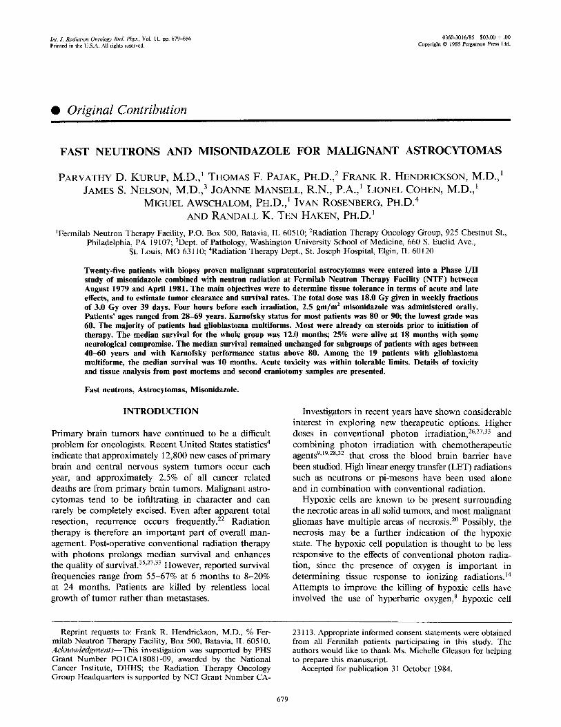
Twenty-five patients with biopsy proven malignant supratentorial astrocytomas were entered into a Phase I/II study of misonidazole combined with neutron radiation at Fermilab Neutron Therapy Facility (NTF) between August 1979 and April 1981. The main objectives were to determine tissue tolerance in terms of acute and late effects, and to estimate tumor clearance and survival rates. The total dose was 18.0 Gy given in weekly fractions of 3.0 Gy over 39 days. Four hours before each irradiation, 2.5 gm/m* misonidazole was administered orally. Patients’ ages ranged from 28-69 years. Karnofsky status for most patients was 80 or 90; the lowest grade was 60. The majority of patients had glioblastoma multiforms. Most were already on steroids prior to initiation of therapy. The median survival for the whole group was 12.0 months; 25% were alive at 18 months with some neurological compromise. The median survival remained unchanged for subgroups of patients with ages between 40-60 years and with Karnofsky performance status above 80. Among the 19 patients with glioblastoma multiforme, the median survival was 10 months. Acute toxicity was within tolerable limits. Details of toxicity and tissue analysis from post mortems and second craniotomy samples are presented.
Fast neutrons, Astrocytomas, Misonidazole.
INTRODUCTION
Primary brain tumors have continued to be a difficult problem for oncologists. Recent United States statistics4 indicate that approximately 12,800 new cases of primary brain and central nervous system tumors occur each year, and approximately 2.5% of all cancer related deaths are from primary brain tumors. Malignant astro- cytomas tend to be infiltrating in character and can rarely be completely excised. Even after apparent total resection, recurrence occurs frequently.22 Radiation therapy is therefore an important part of overall man- agement. Post-operative conventional radiation therapy with photons prolongs median survival and enhances the quality of survival. 25,27,33 However, reported survival frequencies range from 55-67% at 6 months to 8-20% at 24 months. Patients are killed by relentless local growth of tumor rather than metastases.
Investigators in recent years have shown considerable interest in exploring new therapeutic options. Higher doses in conventional photon irradiation,26a27,33 and combining photon irradiation with chemotherapeutic agents 9,19,28,32 that cross the blood brain barrier have been studied. High linear energy transfer (LET) radiations such as neutrons or pi-mesons have been used alone and in combination with conventional radiation.
Hypoxic cells are known to be present surrounding the necrotic areas in all solid tumors, and most malignant gliomas have multiple areas of necrosis.” Possibly, the necrosis may be a further indication of the hypoxic state. The hypoxic cell population is thought to be less responsive to the effects of conventional photon radia- tion, since the presence of oxygen is important in determining tissue response to ionizing radiations. l4 Attempts to improve the killing of hypoxic cells have involved the use of hyperbaric oxygen,8 hypoxic cell
Reprint requests to: Frank R. Hendrickson, M.D., % Fer- milab Neutron Therapy Facility, Box 500, Batavia, IL 605 10. Acknowledgments-This investigation was supported by PHS Grant Number PO 1CA 1808 l-09, awarded by the National Cancer Institute, DHHS; the Radiation Therapy Oncology Group Headquarters is supported by NC1 Grant Number CA-
619
23 113. Appropriate informed consent statements were obtained from all Fermilab patients participating in this study. The authors would like to thank Ms. Michelle Gleason for helping to prepare this manuscript.
Accepted for publication 3 1 October 1984.

680 Radiation Oncology 0 Biology ??Physics April 1985, Volume II, Number 4
sensitize&~’ 1s17,19X31 and high-LET radiation.6,7,‘6,‘8,2’
Neutron irradiation potentially has the radiobiological advantage of a lower OER (oxygen enhancement ratio) which is expected to be beneficial when large hypoxic cell populations are present.12J4J5 Of the sensitizers, the misonidazoles have been used the most in clinical studies. Animal experiments have suggested a reduction of OER from about 1.6 for neutrons alone to nearly 1 .O in combination with hypoxic cell sensitizers.14 A Phase II study was conducted at the Fermilab NTF to evaluate the feasibility and toxicity of combining neutron diations with misonidazole at weekly intervals. results are described in detail in this report.
irra- The
METHODS AND MATERIALS
From August 1979 to April 198 1, 25 patients with malignant astrocytomas were entered into a Phase II study (RTOG 79-03) at Fermilab NTF. Informed consent was obtained from all patients after explaining the nature of the treatment program. Fast neutrons are produced by bombarding a 22 mm thick beryllium target with 66 MeV protons from a linear accelerator.‘,” The skin sparing and depth dose characteristics are similar to those of 6 MeV X rays from a linear accel- erator.2,3,24,29 The relative biological effectiveness (RBE) of the beam is approximately 3 for most tissues,‘5,23 but is considered to be higher for the central nervous system. Patients were treated in a sitting position in an adjustable chair. Immobilization of the head was achieved with individually made plastic masks.* The neutron beam is fixed horizontally. However, all the relative movements provided by conventional isocentric therapy can be achieved with the chair, since it moves in three dimen- sions and rotates about a vertical axis. The source axis distance is 190 cm. Four intersecting laser beams help in the proper alignment of the patient. Planning X rays are done and individual treatment plans are generated in the same chair. The neutron doses reported are actually total absorbed doses which include the gamma ray component.
Treatment technique Patients were treated once a week for 6 weeks with
3.0 neutron Gy per fraction. The whole brain received 12.0 Gy in 4 weeks and boosts to the tumor bed brought the total dose to 18.0 Gy. The whole brain treatments were done with parallel opposing lateral fields with dose calculated at the midplane. The boost was given with single or multiple fields using wedges as needed. Target volume for the boost was determined with the help of CT scans. Figure 1 is a representative isodose distribution.
ISODOSES FOR GLIOELASTOMA MULTIFORME OCCIPITAL LOBE
~(66) Be (49) NEUTRONS
Fig. 1. Typical neutron isodose distribution for a patient treated for malignant astrocytoma.
Misonidazole, 2.5 gm/m2, was given orally 4 hours prior to each neutron irradiation. Blood levels of misonidazole were determined each week.
Follow-up Patients were seen in first follow-up 2-3 weeks after
completion of planned treatment and every 2-3 months thereafter. A physical examination, including a detailed neurological examination, was done to assess the toxicity as well as the effectiveness of therapy. CT scans were done at referring institutions and received at our follow- up clinic to help in these evaluations. In addition, many of the second craniotomy or autopsy specimens were studied in detail by the same pathologist who reviewed the original biopsy material.
The objectives of our study were (1) to lay the basis for a Phase III study with photons versus neutrons in combination with misonidazole; (2) to determine the tolerance of the brain to the unusual radiation doses and fractionation in terms of acute and late normal tissue reaction; and (3) to establish the tumor clearance rate, disease free interval, and the prospect for long- term tumor control.
* Light Cast, Merck, Sharp & Dohme, Orthopedics Co., Inc., Costa Mesa, CA.

Neutrons and misonidazole 0 P. D. KURUP et al. 681
RESULTS
Of the 25 patients treated with fast neutrons in combination with misonidazole, one patient is not eli- gible for evaluation because she was a 14 year-old girl with a brain stem lesion. Two other patients who did not complete the whole course of irradiations are in- cluded in the analysis.
Age, sex and performance status Ages ranged from 28-69 years (median: 52 years).
Eight patients were under 40 years at the time of diagnosis and 5 were 60 years or more. The male to female ratio was 2: 1. Karnofsky performance scale was used to evaluate the general condition of the patient. Karnofsky status for 21 out of 24 patients was 80 or 90.
Location and size of tumor and extent of surgery Many of the lesions involved the frontal lobe. Parietal,
temporal and occipital lobes were involved with equal frequency in this series. The sizes of the tumors were estimated from pre-operative CT scans; 21 of the 24 patients had lesions measuring 5- 10 cm. In one patient the lesion measured less than 5 cm and two had tumors larger than 10 cm. Except for one patient who underwent biopsy only, all others had subtotal resection of their tumor at the time of craniotomy and initial diagnosis.
Pathology Diagnoses of glioblastoma multiforme or Grade IV
astrocytoma were made on 16 patients at their referring institutions. The tumor from one patient was diagnosed as a Grade III-IV lesion. Seven patients had a diagnosis of anaplastic malignant astrocytoma or Grade III astro- cytoma. The pre-treatment slides of 24 of the 25 patients were submitted by the referring institutions and reviewed by one of the authors (J.S.N.). The review diagnosis in 19 patients (including the ineligible patient) was glio- blastoma multiforme. The tumors from the 5 other patients were each diagnosed as astrocytoma with atypical or anaplastic foci. 2o The patient whose pre-treatment slides were not available for pathology review died after receiving only one radiation treatment. The pathology review examination of histologic sections taken at au- topsy from the tumor site disclosed a mixed glioma (oligodendroglioma and astrocytoma). Some atypical appearing tumor cells were present. Radiation necrosis was not evident.
Neurological status In 16 patients, the major symptoms at presentation
were due to increased intracranial pressure, e.g., head- ache, nausea, and seizures. Six patients had weakness of muscles or partial paralysis, and four had sensory symp- toms. Visual problems were reported by seven patients while six patients were noted to have had personality changes. Major neurological deficits were present in only two patients. Six patients had moderate impairment
of motor and/or sensory functions. No neurological deficit was seen in 16 patients prior to neutron irradia- tion.
Treatment All patients aeceived steroids concomitantly. Neutrons
were delivered in full doses to 22 patients. One patient received only 3.0 Gy and another received only 9.0 Gy. Nineteen of the 24 patients received a full course of misonidazole, as required in the protocol. The two patients who did not complete the irradiation also did not receive the full misonidazole dose. In the remaining three patients, five out of the six doses of misonidazole were given. The omission of the last dose was because of acute reactions in one of these patients. Two patients each missed one neutron irradiation because of accel- erator problems after taking the misonidazole as sched- uled. When the neutron dose was given, the misonidazole could not be repeated because of limitations on total dose.
Survival Actuarial analysis of survival for the whole group is
shown graphically in Figure 2. Overall median survival is 12.0 months. The influence of important prognostic factors such as age, Karnofsky performance status, and histology was studied. Median survival for the eight patients under 40 years is 20.0 months and for the group 40-59 years, it is 12.0 months. In the older group of patients (five of them over 60 years) median survival is only six months. Twenty-one of the 24 patients had a Karnofsky performance score of 80 or over, with a median survival of 12.0 months. In the five patients with diagnoses of atypical or anaplastic astrocytoma, the duration of survival after therapy ranges from 15.0 to 36.0 months, with a median survival of 20.0 months. Median survival in the group with glioblastoma multi-
OIL 0 6 12 18 24 30 36 42
TIME FROM START OF TREATMENT (MO.)
Fig. 2. Survival curve of 25 patients treated with neutrons and misonidazole for malignant astrocytomas.

682 Radiation Oncology 0 Biology 0 Physics April 1985, Volume 1 I, Number 4
forme (18 patients) is 10.0 months. Among the 24 evaluable patients, 25% were alive at 18 months and 12.5% at two years. Only 2 patients lived 3 years post- treatment.
DISCUSSION
Tumor clearance and late efects
Quality of survival Five patients did not show any improvement following
treatments. The rest had improvement in their general and neurological status for periods ranging from 3-16 months. In six patients, deterioration started within 6 months of treatment while 10 patients had good neu- rological function for up to 12 months. Only three patients had good quality neurological function for more than 12 months.
Acute reactions Nine patients reported no acute reactions with treat-
ment schedule. Ten patients experienced nausea, vom- iting and headaches of short duration. These symptoms were well controlled with medication. Such symptoms are often produced by intracranial pressure. Transient skin rashes were noticed by two patients. These patients were also taking steroids, phenytoin and phenobarbitol. Four patients developed the sensory symptoms of numb- ness and hyperesthesia of the extremities. Recovery was spontaneous in all patients within one to four months. Only one patient developed a fever of 40°C with chills, and another patient had transient episodes of left ear deafness which resolved spontaneously in two to three months.
Radiographic. CT scans were used to estimate tumor clearance radiographically. The first follow-up scan was done two to three months after the initiation of therapy. Of the 22 patients where scans were available, 10 showed definite improvement in the radiographic appearance. Figure 3a is the CT scan of a patient taken one month prior to initiation of neutron therapy. A large left frontoparietal tumor is demonstrated. An excisional biopsy was performed prior to neutron irradiation. Figure 3b is a CT scan of the same patient taken 7 months later. The lesion is no longer obvious. Appearance was stable in three patients and there was a suggestion of progression of tumor in 6 patients. In another three patients, although tumor itself was not progressing, there was a low density area suggesting edema, which was thought to be a radiation reaction.
Histopathological. Brain tissue including the tumor site was available for histopathological examination from 13 patients following treatment. The histologic specimens were obtained from second craniotomies and debulking surgery and/or autopsies (Table 1). The following account is a brief summary of the principal pathologic findings in these tissues. A detailed study of this material by one of the authors (J.S.N.) is in progress and will be reported in a separate paper. Residual tumor was identified in 12 of the 13 tissues studied. In 6, the histologic diagnoses on the pre- and post-treatment specimens coincided. In
Fig. 3a. Pre-therapy CT scan. A frontoparietal malignant astrocytoma is demonstrated.

Neutrons and misonidazole 0 P. D. KURUP et al. 683
Fig. 3b. CT scan of the same patient taken 7 months after neutrons and misonidazole.
each of the other 6 patients the pre-treatment histologic diagnosis had been glioblastoma multiforme. The post- treatment tumor in these patients, however, lacked the cell density, pleomorphism, and other characteristic features of glioblastoma multiforme. The cell density in these tumors was moderate, and the degree of cellular pleomorphism was variable. Vascular hyperplasia and pseudopalisades with central necrosis were absent. Evi- dence of radiation necrosis with typical vascular changes, chiefly affecting the white matter, was evident in each of the 13 cases. The patient with the mixed oligodenro- glioma-astrocytoma is not included in the above statistics. This patient had only one dose of misonidazole and one fraction of neutron radiation. The pre-treatment speci- men from this patient was not available for pathology review.
Comparison with other RTOG glioma studies Another group of 31 consecutive patients was subse-
quently treated with the same neutron schedule, but
Table 1. Histopathological analysis
Source Number of
patients Residual
tumor Radiation necrosis
Autopsy Post RT
craniotomy Random
biopsy
10 10 10
5 5 5
1 ? 1
without misonidazole. Their median survival was 12.0 months (unpublished data). Since 88% of the evaluable patients in the neutron with misonidazole study had initially favorable Karnofsky performance scores of 80 or higher, the comparison between the two neutron treated patient groups was restricted to patients with such favorable scores. The patient characteristics for both groups are shown in Table 2. Figure 4 shows the historical survival comparison between the patients treated with neutrons and misonidazole versus neutrons only. The two survival curves are very similar, suggesting no augmentation by the misonidazole. Subsequently, patients treated with neutrons with or without the misonidazole were grouped together for comparison with two treatment schedules from other ghoma stud- ies.‘,13 One schedule employed conventional photon radiation to a total dose of 60.0 Gy and was a part of a previous Radiation Therapy Oncology Group (RTOG) study.’
The other schedule employed a conventional photon radiation with a total dose of 70.0 Gy which included a boost of 10.0 Gy. This schedule was the control arm of the RTOG randomized study, comparing a photon boost against a neutron boost. ’ 3
The subsequent survival comparison was restricted to glioblastoma multiforme patients with initially favorable Karnofsky performance scores of 80 or over, in order to reduce the amount of non-homogeneity among the treatment groups with respect to significant prognostic factors. The distribution of patients according to various

684 Radiation Oncology 0 Biology 0 Physics
Table 2. Comparison between neutrons + misonidazole and Table 3. Comparison of neutrons + misonidazole, photon neutrons only (Restricted to Patients with only, photon + boost (restricted to patients with initial
Initial KPS 80-100) KPS 80-100 and with glioblastoma multiforme)
Patients Neutron Neutron characteristic + misonidazole onlv
Photon Patient Neutron Photon + photon
characteristics + misonidazole only boost
Number of evaluable patients 33 44 33
Glioblastoma % centrally
reviewed 58 100 100 Age
Mean 50.5 47.2 48.6 % 40 years 18 27 30 % 40-59 years 55 59 46 % 60+ years 27 14 24
Extent of surgery % none/biopsy 21 17 16* % partial biopsy 67 57 68 % complete 12 26 16 % dead 91 96 97
* Extent of prior surgery unknown in 1 patient.
Number of evaluable cases
Histology % astrocytoma
with atypical or anaplastic foci
% glioblastoma Multiforme
% unknown/ missing
Age Mean % ~40 years % 40-59 years % 260 years
Extent of surgery % none/biopsy % partial resection % complete % dead
21 23
29 17*
71 79
0 4
48.4 48.0 33 22 48 52 19 26
0 39 100 44
0 17 100 83
* Only five of originally eight patients classified as astrocytoma were centrally reviewed.
prognostic criteria is shown in Table 3. No survival advantage is shown for patients treated with neutrons only or in combination with misonidazole when com- pared with conventional radiation (60.0 Gy) (Fig. 5). The group that was treated with photons (60.0 Gy) with a boost (10.0 Gy) showed slight but statistically insignif- icant differences in the latter part of the survival curves (p = .21).
looh-l 1 1 I 1 I
25-
0 I 0 6 12 18 24 30 36 42
TIME FROM START OF TREATMENT (MO.)’
Fig. 4. Survival curves of 44 patients treated with neutrons only (- - -) and with neutrons plus misonidazole (-- ), mean survival in either case was 12.5 months. Patients had a Karnofsky performance scale 2 80.
April 1985, Volume 11, Number 4
This report deals with a unique study in which an attempt was made to attack the ‘hypoxic’ cell population in a situation where relentless regrowth of tumor in the local area has been the major problem. Fast neutron irradiation was used in the past, and pilot studies demonstrated the potential for tumor contro1.7,i8*21 However, many of the patients so treated died of radia- tion necrosis and other related complications. Combining neutrons with conventional photon irradiation, l3 or reducing the dose of neutronsI resulted in failure with persistent tumor. Misonidazole combined with conven- tional radiation yielded survival rates similar to conven-
100x 1 I I I I I I
0 6 12 18 24 30 36 42
--I
TIME FROM START OF TREATMENT(MO.1
Fig. 5. Survival curves of patients treated with neutrons only (+-- ) mean survival 12 months, photons only ( - - - ), mean survival 10 months, and photons + photon boost or neutron boost (- - -) mean survival 12 months.

Neutrons and misonidazole 0 P. D. KURUP et al. 685
tional radiation therapy alone.5,’ 1,17,30,31 No augmentation for misonidazole (plus radiation and BCNU) was shown in a randomized study where the control patients received radiation and BCNU without misonidazole. l9
The present study has combined high-LET radiation with the hypoxic cell sensitizer misonidazole. The major difference in prognostic factors compared to other studies is in the Karnofsky performance status. Twenty-one out of 24 patients from the present series had a Karnofsky performance status over 80. Also, the majority of patients did not have any major neurological deficit since tumors were located in non-critical areas. All underwent subtotal resection, except for one patient who had only a biopsy prior to neutron irradiation. This particular patient died after one fraction of neutron treatment.
One of the authors (J.S.N.) has demonstrated that the presence of coagulation necrosis involving astrocytic tumor cells is a major prognostic criterion with regard to malignant supratentorial astrocytomas.” The useful- ness of this criterion with regard to prognosis, however, is limited in the case of malignant astrocytomas following radiation treatment. This type of therapy frequently induces necrosis within the tumor that is indistinguish- able from the spontaneously occurring necrosis charac- teristic of glioblastoma multiforme. In the present study the diagnosis of glioblastoma multiforme was made on post-treatment specimens only when the following his- tologic features were present: marked cell density and pleomorphism; pseudopalisades with central necrosis; and vascular hyperplasia with formation of glomeruloid structures. As noted previously, in 6 patients the histo- logic characteristics of the post-treatment tumor had changed from those characteristic of glioblastoma to those suggestive of a somewhat less aggressive type of malignant astrocytoma. Little is known, however, about the correlation between the histologic appearance of malignant astrocytomas following neutron treatment and their subsequent biological behavior. It is not clear whether the residual less aggressive tumor would even- tually develop into frank glioblastoma multiforme or
continue to grow in a less aggressive manner. This area requires further investigation.
No augmentation for misonidazole could be shown when the survival curves of the two groups of patients treated with similar doses of neutron radiation, with or without misonidazole, were compared (Fig. 4). No sur- vival advantage was shown for neutron treated patients compared to conventional radiation (Fig. 5). The survival curve of patients treated with 18.0 Gy (Fermi) was almost identical to the photon group receiving 60.0 Gy. Also, persistent or recurrent tumor was identified in post-irradiation specimens whenever tissue was available for histopathological examinations.
Animal experiments and pilot studies with fast neutrons’,” had suggested the possibility of a higher RBE for the central nervous system. Results from the current study seem to suggest that the RBE may be higher for normal tissues in the central nervous system but not necessarily for central nervous system tumors. The absence of survival advantage over conventional radiation suggests that re-oxygenation may take place much more effectively than we believed. Hence, the presumed advantage of low OER becomes merely theo- retical. Attempts to improve hypoxic cell killing by hyperbaric oxygen’ and hypoxic cell sensitizers with conventional radiation5,’ ‘,17*‘9,30,31 were also shown to be ineffective in clinical situations.
(1)
(2)
(3)
CONCLUSIONS Acute tolerance of brain tissue to the unusual com- bination of dose and fractionation is acceptable. Comparison of survival rates for the current study do not show any advantage over conventional irra- diation. Since residual tumor and radiation necrosis are both documented with the treatment schedule used, the histopathological analysis appears to indicate the absence of a therapeutic window, that is, a treatment level at which tumor ablation can be achieved without radionecrosis.
REFERENCES
1. Awschalom, M., Grumboski, L., Hrejsa, A.F., Lee, G.M., Rosenberg, I.: The Fermilab Cancer Therapy Facility: Status report after 2.5 years of operation. Trans. Nucl. Sci. NS-26: 3068-3070, 1979.
6.
Awschalom, M., Rosenberg, I.: Characterization of a p(66)Be(49) neutron therapy beam. II. Skin-sparing and dose transition effects. Med. Phys. 8: 105-107, 198 1. Awschalom, M., Rosenberg, I., Ten Haken, R., Cohen, L., Hendrickson, F.: The Fermilab Neutron Therapy Fa- cility treatment planning for neutron and mixed beams, Stvuhlentherapie 77: 144-149, 198 1. Cancer Statistics, 1984. Ca-A Cancer Journal for Clini- cians (Published by the American Cancer Society). Vol. 34, pp. 7-23, Jan/Feb 1984. Carabell, S.C., Bruno, L.A., Weinstein, A.S., Richter, M.P., Chang, C.H., Weiler, C.B., Goodman, R.L.: Mison- idazole and Radiotherapy to treat malignant glioma: A
phase II trial of the RTOG. Znt. J. Radiat. Oncol. Biol. Phys. 7: 71-77, 1981. Catterall, M., Bewley, D.K.: Fast Neutrons in the Treatment of Cancer. London, Academic Press, 1979. Catterall, M., Bloom, H.J.G., Ash, D.V., Walsh, L., Rich- ardson, A., Uttley, D., Gowing, N.F.C., Lewis, P., Chanter, B.: Fast neutrons compared with megavoltage X-rays in the treatment of patients with supratentorial glioblastoma: A controlled study. Znt. J. Radiat. Oncol. Biol. Phys. 6: 261-266, 1980. Chang, C.H.: Hyperbaric oxygen and radiation therapy in the management of glioblastoma. National Cancer Inst. Monogr. 46: 163-169, 1977. Chang, C.H., Horton, J., Schoenfeld, D., Salazar, O., Perez-Tamayo, R., Kramer, S., Weinstein, A., Nelson, J.S., Tsukada, Y.: Comparison of post-operative radio- therapy and combined post-operative radiotherapy and

686 Radiation Oncology 0 Biology 0 Physics April 198.5, Volume 1 I, Number 4
chemotherapy in the multidisciplinary management of malignant gliomas. A joint RTOG-ECOG study. Cancer 52: 997-1007, 1983.
10. Cohen, L., Awschalom, M.: The Cancer Therapy Facility at the Fermi National Accelerator Laboratory: A prelimi- nary report. Appl. Radiol. 5: 55, 1976.
11. EORTC Brain Tumor Group: Misonidazole in radiotherapy of supmtentorial malignant brain gliomas in adult patients: A randomized double-blind study. Eur. J. Cancer Clin. Oncol. 19: 39-42, 1983.
12. Gragg, R.L., Humphrey, R.M., Meyer, R.E.: The response of Chinese hamster ovary cells in fast neutron radiotherapy beams. II. Sublethal and potentially lethal damage recovery capabilities. Radiat. Rex 71: 461-470, 1977.
13. Griffin, T.W., Davis, R.B., Laramore, G.E., Nelson, J.S., Hendrickson, F.R., Rodriguez-Antunez, A., Hussey, D.H.: Fast neutron irradiation for glioblastoma multiforme: Re- sults of an RTOG Phase III study. Am. J. Clin. Oncol. 6: 661-667, 1983.
14. Hall, E.J.: Radiobiology for the Radiologist. Hagerstown, MD, Harper and Rowe, 1978.
15. Hall, E.J., Kraljeire, U.: Repair of potentially lethal radia- tion damage: Comparison of neutron and X-ray RBE and implications of radiation therapy. Radiology 120: 731- 735, 1976.
16. Herskovic, A., Omitz, R.D., Shell, M., Rogers, C.C.: Treatment experience glioblastoma multiforme treated with 15 MeV fast neutrons. Cancer 49: 2463-2465, 1982.
17. Kapp, D.S., Wagner, F.C., Lawrence, R.: Glioblastoma multiforme: Treatment by large dose fraction irradiation and metronidazole. Int. J. Radiat. 0~01. Biol. Phys. 8: 35 l-355, 1982.
18. Laramore, G.E., Griffin, T.W., Gerdes, A.J., Pancer, R.G.: Fast neutron and mixed beam teletherapy for Grades III and IV astrocytomas. Cancer 42: 96-103, 1978.
19. Nelson, D.F., Schoenfeld, D., Weinstein, A.S., Nelson, J.S., Wasserman, T., Goodman, R.L., Carabell, S.: A Randomized comparison of misonidazole sensitized radio- therapy plus BCNU and radiotherapy plus BCNU for treatment of malignant glioma after surgery: Preliminary results of an RTOG study. Znt. J. Radiat. Oncol. Biol. Phys. 9: 1143-l 151, 1983.
20. Nelson, J.S., Tsukada, Y., Schoenfeld, D., Fulling, K., Lamarche, J., Peress, N.: Necrosis as a prognostic criterion in malignant supratentorial, astrocytic gliomas. Cancer 52: 550-554, 1983.
2 1. Parker, R.G., Berry, H.C., Gerdes, A.J., Soronen, M.D.,
Shaw, CM.: Fast neutron beam radiotherapy of glioblas- toma multiforme. Am. J. Roentgenol. 127: 331-335, 1976.
22. Ransohoff, J.: Surgical management of malignant brain tumors. Natl. Cancer Inst. Monog. 46: 145-150, 1977.
23. Redpath, J.L., David, R.M., Cohen, L.: Dose fractionation studies on mouse gut and marrow: An intercomparison of 6 MeV photons and fast neutrons (E = 25 MeV). Radiat. Res. 75: 642-648, 1978.
24. Rosenberg, I., Awschalom, M.: Characterization of a p(66)Be(49) neutron therapy beam: I. Central axis depth dose and off-axis ratios. Med. Phys. 8: 99-104, 198 1.
25. Rutten, E-H.J.M., Kazam, I., Gloff, J.L., Walder, A.H.D.: Post-operative radiation therapy in the management of brain astrocytoma-A retrospective study of 142 patients. Znt. J. Radiat. Oncol. Biol. Phys. 7: 191-195, 1981.
26. Salazar, O., Rubin, P., Feldstein, M., Pizzutiello, R.: High dose radiation therapy in the treatment of malignant gliomas: Final report. Znt. J. Radiat. Oncol. Biol. Phys. 5: 1733-1740, 1979.
27. Salazar, O.M., Rubin, P., McDonald, J.V., Feldstein, M.L.: Patterns of failure in intracranial astrocytomas after irradiation: Analysis of dose and field factors. Am. J. Roentgenol. 126: 279-292, 1976.
28. Salazar, O.M., Vanhoulte, P.J., Bennett, J.M., Rubin, P., Wheeler, K.T.: High dose radiation therapy with low-dose (pulsed) BCNU in malignant gliomas: An ECOG report. Int. J. Radiat. Oncol. Biol. Phys. 8: 915-9 19, 1982.
29. Ten Haken, R.K., Awschalom, M., Hendrickson, F., Ro- senberg, I.: Comparison of the physical characteristics of a p(66)Be(49) neutron therapy beam to those of correctional radiotherapy beams. Fermilub TM-1021R, 1980.
30. Urtasun, R.C., Band, P.R., Chapman, J.D., Feldstein, M.L.: Radiation plus metronidazole for glioblastoma. NEJM 296: 757, 1977.
31. Urtasun, R.C., Chapman, J.D.: Radiation and high dose metronidazole in supratentorial glioblastomas. NEJM 294: 1364-1367, 1976.
32. Walker, M.D., Hunt, A.E., MacCarty, W.E., Mahaley, C.S., Mealey, J., Norell, H.A., Owens, G., Ranschoff, J., Wilson, C.B., Gehan, E.A., Strike, T.A.: Evaluation of BCNU and/or radiotherapy in the treatment of anaplastic gliomas. A cooperative clinical trial. J. Neurosurg. 49: 333-343, 1978.
33. Walker, M.D., Strike, T.A., Sheline, G.E. (for brain tumor study group): An analysis of dose-effect relationship in the radiotherapy of malignant gliomas. Znt. J. Radiat. Oncol. Biol. Phys. 5: 1725-1731, 1979.