Embed Size (px)
DESCRIPTION
O. Gambhir Singh, Hemalatha N. Fatal craniocerebral injuries in victims who survived for some period. IAIM, 2014; 1(1): 1-6.
Citation preview
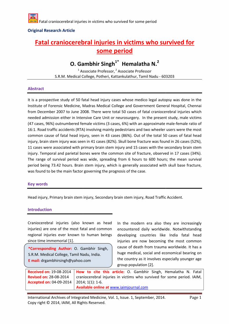
Fatal craniocerebral injuries in victims who survived for some period
International Archives of Integrated Medicine, Vol.
Copy right © 2014, IAIM, All Rights Reserved.
Original Research Article
Fatal craniocerebral injuries in victims who survived for
O. Gambhir Singh1 Associate Professor,
S.R.M. Medical College, Potheri, Kattankulathur, Tamil Nadu
Abstract
It is a prospective study of 50 fatal head injury cases whose medico legal autopsy was done in the
Institute of Forensic Medicine, Madras Medical College and Government General Hospital, Chennai
from December 2007 to June 2008. There were total 50 cases of
needed admission either in Intensive Care Unit or neurosurgery. In the present study, male victims
(47 cases, 96%) outnumbered female victims (3 cases, 6%) with an approximate male
16:1. Road traffic accidents (RTA) involving mainly pedestrians and two wheeler users were the most
common cause of fatal head injury, seen in
injury, brain stem injury was seen in 41 cases (82%). Skull bone fracture was
11 cases were associated with primary brain stem injury and 15 cases with the secondary brain stem
injury. Temporal and parietal bones were the common site of fracture, observed in 17 cases (34%).
The range of survival period was w
period being 73.42 hours. Brain stem injury, which is generally associated with skull base fracture,
was found to be the main factor governing the prognosis of the case.
Key words
Head injury, Primary brain stem injury,
Introduction
Craniocerebral injuries (also known as head
injuries) are one of the most fatal and common
regional injuries ever known to human beings
since time immemorial [1].
Received on: 19-08-2014
Revised on: 28-08-2014
Accepted on: 04-09-2014
How to cite this article:
craniocerebral injuries in victims who survived for some period.
2014; 1(1):
Available
*Corresponding Author: O. Gambhir Singh
S.R.M. Medical College, Tamil Nadu
E mail: [email protected]
Fatal craniocerebral injuries in victims who survived for some period
International Archives of Integrated Medicine, Vol. 1, Issue. 1, September, 2014.
Copy right © 2014, IAIM, All Rights Reserved.
Fatal craniocerebral injuries in victims who survived for
some period
O. Gambhir Singh1*
Hemalatha N.2
Associate Professor, 2
Associate Professor
S.R.M. Medical College, Potheri, Kattankulathur, Tamil Nadu - 603203
It is a prospective study of 50 fatal head injury cases whose medico legal autopsy was done in the
Institute of Forensic Medicine, Madras Medical College and Government General Hospital, Chennai
from December 2007 to June 2008. There were total 50 cases of fatal craniocerebral injuries which
needed admission either in Intensive Care Unit or neurosurgery. In the present study, male victims
(47 cases, 96%) outnumbered female victims (3 cases, 6%) with an approximate male
cidents (RTA) involving mainly pedestrians and two wheeler users were the most
common cause of fatal head injury, seen in 43 cases (86%). Out of the total 50 cases of fatal head
injury, brain stem injury was seen in 41 cases (82%). Skull bone fracture was found in 26 cases (52%),
11 cases were associated with primary brain stem injury and 15 cases with the secondary brain stem
injury. Temporal and parietal bones were the common site of fracture, observed in 17 cases (34%).
The range of survival period was wide, spreading from 6 hours to 600 hours; the mean survival
period being 73.42 hours. Brain stem injury, which is generally associated with skull base fracture,
was found to be the main factor governing the prognosis of the case.
Head injury, Primary brain stem injury, Secondary brain stem injury, Road Traffic Accident
Craniocerebral injuries (also known as head
injuries) are one of the most fatal and common
regional injuries ever known to human beings
In the modern era also they are increasingly
encountered daily worldwide. Notwithstandi
developing countries like India fatal head
injuries are now becoming the most common
cause of death from trauma worldwide. It has a
huge medical, social and economical bearing on
the country as it involves especially younger age
group population [2].
How to cite this article: O. Gambhir Singh, Hemalatha N.
craniocerebral injuries in victims who survived for some period.
2014; 1(1): 1-6.
Available online at www.iaimjournal.com
O. Gambhir Singh,
S.R.M. Medical College, Tamil Nadu, India.
Page 1
Fatal craniocerebral injuries in victims who survived for
603203
It is a prospective study of 50 fatal head injury cases whose medico legal autopsy was done in the
Institute of Forensic Medicine, Madras Medical College and Government General Hospital, Chennai
fatal craniocerebral injuries which
needed admission either in Intensive Care Unit or neurosurgery. In the present study, male victims
(47 cases, 96%) outnumbered female victims (3 cases, 6%) with an approximate male-female ratio of
cidents (RTA) involving mainly pedestrians and two wheeler users were the most
Out of the total 50 cases of fatal head
found in 26 cases (52%),
11 cases were associated with primary brain stem injury and 15 cases with the secondary brain stem
injury. Temporal and parietal bones were the common site of fracture, observed in 17 cases (34%).
ide, spreading from 6 hours to 600 hours; the mean survival
period being 73.42 hours. Brain stem injury, which is generally associated with skull base fracture,
Secondary brain stem injury, Road Traffic Accident.
In the modern era also they are increasingly
encountered daily worldwide. Notwithstanding
developing countries like India fatal head
injuries are now becoming the most common
cause of death from trauma worldwide. It has a
huge medical, social and economical bearing on
the country as it involves especially younger age
O. Gambhir Singh, Hemalatha N. Fatal
craniocerebral injuries in victims who survived for some period. IAIM,
Fatal craniocerebral injuries in victims who survived for some period
International Archives of Integrated Medicine, Vol.
Copy right © 2014, IAIM, All Rights Reserved.
The present study was conducted in the
Institute of Forensic Medicine, Madras Medical
College, Chennai, Tamil Nadu (India). It is one of
the oldest and largest medical colleges and
tertiary multidisciplinary health care centre in
the country catering to a vast Chennai
metropolitan city and different parts of the
state. Because of heavy traffic congestion, the
speed is limited and in majority of the incidents
the anatomy of head is comparatively preserved
though it is fatally damaged. In our study
included only those cases having fatal head
injuries as defined by The Royal College of
Surgeons [3], U.K. Our main aim of the present
study is to analyze the autopsy
important regional injury in details and compare
our findings with the previous studies.
Material and method
We selected 50 cases of fatal head injury that
were admitted and died in our hospital. All these
cases were either admitted in Intensive Care
Unit or performed neurosurgery. Other brought
in dead cases of fatal head injuries or cases of
crushed head injuries were excluded from the
present study because in such cases brain
structure would be grossly damaged and
moreover we wanted to include the likely future
prognosis. After reflecting the scalp tissues, the
vault of the skull and meninges were dissected
to expose the brain by following the routine
autopsy techniques. Detail information such as
post mortem number, name, age, sex, date and
time of injury, mode of injury, site of impact and
other relevant data were noted. Other relevant
information was also collected from the hospital
records, police papers and relatives.
Involvement of brain stem was confirmed with
histopathological examination and identified as
primary and secondary brain stem injuries.
These data were tabulated for easy study and
comparison with the previous available studies.
Fatal craniocerebral injuries in victims who survived for some period
International Archives of Integrated Medicine, Vol. 1, Issue. 1, September, 2014.
Copy right © 2014, IAIM, All Rights Reserved.
The present study was conducted in the
Institute of Forensic Medicine, Madras Medical
College, Chennai, Tamil Nadu (India). It is one of
medical colleges and
tertiary multidisciplinary health care centre in
the country catering to a vast Chennai
metropolitan city and different parts of the
state. Because of heavy traffic congestion, the
speed is limited and in majority of the incidents
anatomy of head is comparatively preserved
though it is fatally damaged. In our study, we
included only those cases having fatal head
injuries as defined by The Royal College of
, U.K. Our main aim of the present
study is to analyze the autopsy findings of this
important regional injury in details and compare
our findings with the previous studies.
We selected 50 cases of fatal head injury that
were admitted and died in our hospital. All these
cases were either admitted in Intensive Care
Unit or performed neurosurgery. Other brought
in dead cases of fatal head injuries or cases of
es were excluded from the
present study because in such cases brain
structure would be grossly damaged and
moreover we wanted to include the likely future
prognosis. After reflecting the scalp tissues, the
vault of the skull and meninges were dissected
expose the brain by following the routine
Detail information such as
post mortem number, name, age, sex, date and
time of injury, mode of injury, site of impact and
other relevant data were noted. Other relevant
lected from the hospital
records, police papers and relatives.
n stem was confirmed with
pathological examination and identified as
primary and secondary brain stem injuries.
These data were tabulated for easy study and
ith the previous available studies.
Observation
These 50 cases of fatal craniocerebral injuries
comprised about 2.82% of all medico legal
autopsies conducted during the study period.
The incidence of brain stem involvement in fatal
craniocerebral injury cases was very high,
observed in 41 cases (82%). Amongst these 41
cases of brain stem injury, 16 cases (39.02%)
were primary brainstem injury and 25 cases
(60.98%) were secondary brainstem injury.
Majority of the victims were middle aged male
though the age range was wide spread from 4
87 years as per Table - 1.
Road Traffic Accidents (RTA) was the single most
common cause of fatal head injury which was
seen in 43 cases (86%) as shown in
Homicidal head injury was observed only in one
case. The most common site for brain stem
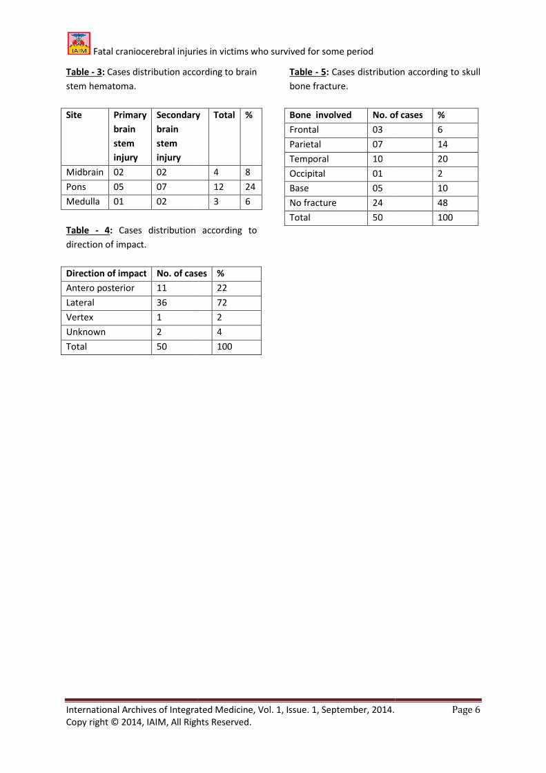
hematoma was the Pons, seen in 12 case
(63.16%) as shown in Table -
The incidence was high with lateral i.e. side to
side force, 36 cases (72%) as shown in
Skull bone fracture was encountered in 26 cases
(52%). The occipital bone was the least
commonly involved; seen in only one
as shown in Table - 5. In primary brainstem
lesions gross hemorrhagic lesions were seen in
dorsal, dorsolateral aspect of midbrain and
dorsal aspect of upper Pons. In se
brainstem lesions gross hemorrhagic lesions
were seen in the midline and paramedian aspect
of tegmentum of midbrain and Pons.
In majority of the cases, death occurred within
24 hours of hospitalization. The mean survival
period was about 73.42 hours (range being 6
hours to 600 hours). The mean survival period of
the primary brainstem injury was 41.55 hours
and that of the secondary brainstem injury was
103.2 hours. Out of 16 cases of primary
brainstem injury, 5 cases (31.25%) died within
Page 2
These 50 cases of fatal craniocerebral injuries
comprised about 2.82% of all medico legal
autopsies conducted during the study period.
The incidence of brain stem involvement in fatal
cases was very high,
observed in 41 cases (82%). Amongst these 41
cases of brain stem injury, 16 cases (39.02%)
were primary brainstem injury and 25 cases
(60.98%) were secondary brainstem injury.
Majority of the victims were middle aged male
e range was wide spread from 4 –
Road Traffic Accidents (RTA) was the single most
common cause of fatal head injury which was
ases (86%) as shown in Table - 2.
Homicidal head injury was observed only in one
The most common site for brain stem
hematoma was the Pons, seen in 12 cases
- 3.
The incidence was high with lateral i.e. side to
ases (72%) as shown in Table - 4.
Skull bone fracture was encountered in 26 cases
(52%). The occipital bone was the least
commonly involved; seen in only one case (2%)
In primary brainstem
lesions gross hemorrhagic lesions were seen in
dorsal, dorsolateral aspect of midbrain and
dorsal aspect of upper Pons. In secondary
brainstem lesions gross hemorrhagic lesions
were seen in the midline and paramedian aspect
of tegmentum of midbrain and Pons.
In majority of the cases, death occurred within
24 hours of hospitalization. The mean survival
period was about 73.42 hours (range being 6
hours to 600 hours). The mean survival period of
the primary brainstem injury was 41.55 hours
ry brainstem injury was
103.2 hours. Out of 16 cases of primary
brainstem injury, 5 cases (31.25%) died within
Fatal craniocerebral injuries in victims who survived for some period
International Archives of Integrated Medicine, Vol.
Copy right © 2014, IAIM, All Rights Reserved.
24 hours and out of 25 cases of secondary
brainstem injury, 4 cases (16%) died within 24
hours.
Discussion
In the present study male victims,
(94%) outnumbered female victims, 3 cases (6%)
with an approximate male-female ratio of 16:1.
Male dominance was also reported by various
authors [4, 5, 6, 7] and is attributed to the fact
that males are more mobile and frequently
involved in outdoor activities than females. Male
preponderance was observed in all age groups,
most commonly affected age range being 21 to
50 years. Similar findings pertaining to age group
were also reported by Amit MP et al
AK et al. [6] and Akang EEU et a
emerged as the single most common cause of
fatal head injury which was seen in 43 cases
(86%). Most of the victims were two wheeler
users or pedestrians in the age group of 20 plus
to 50 years. In this respect our findings were
consistent with the works of Kumar A et al
Amit MP et al. [2], Tyagi Ak et al. [6]
MR et al. [8]. However, in the western countries
the majority of people injured in road traffic
accidents are car occupants [2, 9]
due to differences in comm
transportation, two wheelers being more
popular conveyance in Chennai city and in fact in
India. In most of the circumstances the manner
of head injury was accidental in nature, 49 cases
(98%) and there was 1 case, 2% of assault. Most
of fall from height cases, 5 cases (10%), were
reported from construction site. Two young
patients were injured due to fall from the first
floor (20 feet height) while playing.
Gross hemorrhagic lesions were seen in 19
cases, out of which 6 cases (31.58%) were
associated with primary brainstem injury and 13
cases (68.42%) were associated with secondary
brainstem injury. Hemorrhagic contusions were
Fatal craniocerebral injuries in victims who survived for some period
International Archives of Integrated Medicine, Vol. 1, Issue. 1, September, 2014.
Copy right © 2014, IAIM, All Rights Reserved.
24 hours and out of 25 cases of secondary
brainstem injury, 4 cases (16%) died within 24
In the present study male victims, 47 cases
(94%) outnumbered female victims, 3 cases (6%)
female ratio of 16:1.
Male dominance was also reported by various
and is attributed to the fact
that males are more mobile and frequently
oor activities than females. Male
preponderance was observed in all age groups,
most commonly affected age range being 21 to
50 years. Similar findings pertaining to age group
were also reported by Amit MP et al. [2], Tyagi
and Akang EEU et al. [7]. RTA
emerged as the single most common cause of
fatal head injury which was seen in 43 cases
(86%). Most of the victims were two wheeler
users or pedestrians in the age group of 20 plus
to 50 years. In this respect our findings were
the works of Kumar A et al. [4],
. [6] and Johnson
in the western countries
the majority of people injured in road traffic
[2, 9]. It could be
due to differences in common mode of
transportation, two wheelers being more
popular conveyance in Chennai city and in fact in
India. In most of the circumstances the manner
of head injury was accidental in nature, 49 cases
(98%) and there was 1 case, 2% of assault. Most
om height cases, 5 cases (10%), were
reported from construction site. Two young
patients were injured due to fall from the first
floor (20 feet height) while playing.
Gross hemorrhagic lesions were seen in 19
cases, out of which 6 cases (31.58%) were
ciated with primary brainstem injury and 13
cases (68.42%) were associated with secondary
brainstem injury. Hemorrhagic contusions were
seen in midbrain in 6 cases (31.58%), Pons in 12
cases (63.16%) and medulla in one case (5.26%).
In cases of primary bra
hemorrhagic lesions were seen in the dorsal and
dorsolateral aspect of the midbrain and the
dorsal aspect of upper Pons. In cases of
secondary brainstem injuries, hemorrhagic
lesions were found in the midline and
paramedian aspect of tegme
midbrain and the Pons. Present findings agreed
more or less with the works of Chattopadhyay S,
Tripathi C [10] and Ella FT
cases the direction of force was “Lateral”, i.e.
from side to side, seen in 36 cases (72%). The
unique observation during our present study is
that those cases showing lateral impact also
sustained secondary brainstem injury due to
associated supratentorial traumatic mass with
the midline shift. The second most common
direction of force was from front to
was seen in 11 cases (22%).
Skull bone fracture was seen in 26 cases (52%);
11 cases associated with primary brain stem
injury and 15 cases with secondary brain stem
injury. Temporal and parietal bones were the
common sites of fracture which
cases (34%). More or less similar observation
was also reported by Chattopadhyay S, Tripathi
C [10], Ghosh PK [12],
Fimate L et al
Salgado MSL, Colombage SM
et al. [15]. In case of skull base fracture,
involvement of the middle cranial fossa was the
maximum and similar observation was also
reported by Menon A et al
[16]. Moreover, in majority of the fatal
cranicerebral injury cases, skull vault fractures
were found to be extended up to skull base. One
unique observation in the present study was the
higher incidence of “skull base fractures” with
the primary brainstem injury cases and the
“temporo-parietal skull fracture
secondary brainstem injury cases. We did not
find any literature regarding this brain stem
Page 3
seen in midbrain in 6 cases (31.58%), Pons in 12
cases (63.16%) and medulla in one case (5.26%).
In cases of primary brainstem injuries,
hemorrhagic lesions were seen in the dorsal and
dorsolateral aspect of the midbrain and the
dorsal aspect of upper Pons. In cases of
secondary brainstem injuries, hemorrhagic
lesions were found in the midline and
paramedian aspect of tegmentum of the
midbrain and the Pons. Present findings agreed
more or less with the works of Chattopadhyay S,
[11]. In majority of
cases the direction of force was “Lateral”, i.e.
from side to side, seen in 36 cases (72%). The
ue observation during our present study is
that those cases showing lateral impact also
sustained secondary brainstem injury due to
associated supratentorial traumatic mass with
the midline shift. The second most common
direction of force was from front to back, which
Skull bone fracture was seen in 26 cases (52%);
11 cases associated with primary brain stem
injury and 15 cases with secondary brain stem
injury. Temporal and parietal bones were the
common sites of fracture which was seen in 17
cases (34%). More or less similar observation
was also reported by Chattopadhyay S, Tripathi
Fimate L et al [13],
Salgado MSL, Colombage SM [14] and Yavuz M
In case of skull base fracture,
he middle cranial fossa was the
maximum and similar observation was also
reported by Menon A et al [9] and Tirpude BH
. Moreover, in majority of the fatal
injury cases, skull vault fractures
were found to be extended up to skull base. One
unique observation in the present study was the
higher incidence of “skull base fractures” with
the primary brainstem injury cases and the
parietal skull fractures” with the
secondary brainstem injury cases. We did not
find any literature regarding this brain stem
Fatal craniocerebral injuries in victims who survived for some period
International Archives of Integrated Medicine, Vol.
Copy right © 2014, IAIM, All Rights Reserved.
injury relationship with skull bone fracture
except this present study.
Marks PV and Lavy CBD [17] observed that age
to be a major determinant of the d
recovery following fatal head injury, stating:
younger the age group better will be the
prognosis. Though it is partly true with the
present study also, we observed a strong
relationship between the brain stem injury and
the case fatality. Amongst the brain stem injury
cases also, those showing primary brainstem
injury were found to be more fatal. This finding
is also in agreement with the findings of Shukla
D et al [5], Pilz P et al [18] and Simpson DA et al
[19].
Conclusion
Fatal craniocerebral injury cases constitute
considerable size of morbidity and mortality in
our study centre. Incidence of brain stem injury
in such a case is also very high. RTA involving the
pedestrians and two wheelers users was the
single most important cause of fatal
craniocerebral injuries. In majority of the cases
the primary brainstem lesions have been
associated with basal skull fractures whereas
majority of the secondary brainstem lesions
were associated with vault fractures.
Involvement of brain stem, primarily pr
brain stem injury, was found to be an important
factor determining the survival and prognosis of
the victim.
Acknowledgement
Authors acknowledge the immense help
received from the scholars whose articles are
cited and included in references of this
manuscript. The authors are also grateful to
authors / editors /publishers of all those articles,
journals and books from where the literature for
this article has been reviewed and discussed.
Fatal craniocerebral injuries in victims who survived for some period
International Archives of Integrated Medicine, Vol. 1, Issue. 1, September, 2014.
Copy right © 2014, IAIM, All Rights Reserved.
injury relationship with skull bone fracture
observed that age
to be a major determinant of the degree of
recovery following fatal head injury, stating:
younger the age group better will be the
prognosis. Though it is partly true with the
present study also, we observed a strong
relationship between the brain stem injury and
the brain stem injury
rimary brainstem
injury were found to be more fatal. This finding
is also in agreement with the findings of Shukla
Simpson DA et al.
l injury cases constitute
considerable size of morbidity and mortality in
our study centre. Incidence of brain stem injury
in such a case is also very high. RTA involving the
pedestrians and two wheelers users was the
single most important cause of fatal
raniocerebral injuries. In majority of the cases
the primary brainstem lesions have been
associated with basal skull fractures whereas
majority of the secondary brainstem lesions
were associated with vault fractures.
Involvement of brain stem, primarily primary
brain stem injury, was found to be an important
factor determining the survival and prognosis of
Authors acknowledge the immense help
received from the scholars whose articles are
cited and included in references of this
manuscript. The authors are also grateful to
authors / editors /publishers of all those articles,
journals and books from where the literature for
this article has been reviewed and discussed.
References
1. Tedeschi CG, Eckert WG, Tedeschi LG.
“Forensic Medicine: A study in trauma
and environmental hazards, Vol. I,
Mechanical Trauma “section 1,
Mechanical Injury: Chapter 3: Head
injury, WB Saunders Co. Philadelphia,
1977, p 30.
2. Amit M P, Walter F Vaz.
Blunt Head Injury: A Two Year
Retrospective / Prospective Medico
Legal Autopsy Study.
Forensic Med, 2010; 32(2):144
3. Available at:
http://www.rcseng.ac.uk/publications/d
ocs/report_head_injuries
Accessed on 08.05.2013.
4. Kumar A, Lalwani S, Agrawal D, Rautji R,
Dogra TD. Fatal road traffic accidents
and their relationship with head injuries:
An epidemiological survey of five years.
IJNT, 2008; 5(2): 63-67.
5. Shukla D, Mahadevan A, Sastry
Shankar SK. Pathology of post traumatic
brainstem and hypothalamic injuries,
Clinical Neuropathology, 2007;
197-209.
6. Tyagi AK, Sharma G
cerebral damage by blunt force impact: J
Indian Acad Forensic Med, 1986; 1:
39.
7. Akang EEU, Okati MAO, Osunkaya AO,
Komolate EO, Malomo AO, Shokunbi
MT, Amutta SB. Pattern of fatal head
injuries in Ibadan –
Medicine Science and Law, 2002; 42(2):
160-166.
8. Johnson MR, McCarthy MC, Miller SF,
People JB. Craniofacial trau
motorcyclists: The impact of helmet
usage. J Trauma, 1995;
9. Menon A, Pai VK, Rajeev A. Pattern of
fatal head injuries due to vehicular
Page 4
Tedeschi CG, Eckert WG, Tedeschi LG. In
Medicine: A study in trauma
and environmental hazards, Vol. I,
Mechanical Trauma “section 1,
Mechanical Injury: Chapter 3: Head
WB Saunders Co. Philadelphia,
Amit M P, Walter F Vaz. Pattern of Fatal
Blunt Head Injury: A Two Year
ective / Prospective Medico
Legal Autopsy Study. J Indian Acad
Forensic Med, 2010; 32(2):144-149.
Available at:
http://www.rcseng.ac.uk/publications/d
ocs/report_head_injuries.html.
ccessed on 08.05.2013.
Kumar A, Lalwani S, Agrawal D, Rautji R,
Dogra TD. Fatal road traffic accidents
and their relationship with head injuries:
An epidemiological survey of five years.
67.
Shukla D, Mahadevan A, Sastry KVR.,
Pathology of post traumatic
brainstem and hypothalamic injuries,
cal Neuropathology, 2007; 26(5):
Tyagi AK, Sharma GK, Bishnu K. Cranio
cerebral damage by blunt force impact: J
Indian Acad Forensic Med, 1986; 1: 24-
Akang EEU, Okati MAO, Osunkaya AO,
Komolate EO, Malomo AO, Shokunbi
B. Pattern of fatal head
– A 10 year review,
Medicine Science and Law, 2002; 42(2):
Johnson MR, McCarthy MC, Miller SF,
People JB. Craniofacial trauma in injured
motorcyclists: The impact of helmet
1995; 38(6): 876-8.
Menon A, Pai VK, Rajeev A. Pattern of
fatal head injuries due to vehicular
Fatal craniocerebral injuries in victims who survived for some period
International Archives of Integrated Medicine, Vol.
Copy right © 2014, IAIM, All Rights Reserved.
accidents in Mangalore. J Forensic Leg
Med, 2008; 15(2): 75-7.
10. Chattopadhyay S, Tripathi C. Sk
fracture and haemorrhage pattern
among fatal and nonfatal
assault victims – A critical analysis. J Inj
Violence Res., 2010; 2(2):
11. Ella FT. Autopsy finding in head injuries.
Lancet, 1963; 1: 265.
12. Ghosh PK. Post-mortem Study of patter
of injury involving pedestrian victims.
J Forens Med Toxicol, 1991
8.
13. Fimate L, Chandra J, Dikshit PC.
time in relation to consciousness and
severity of head injury. Journal of
Forensic Medicine & Toxicology,
8(3&4): 15-19.
14. Salgado MSL, Colombage SM. Analysis of
fatalities in Road Accidents.
Science International, 1988; 36: 91
15. Yavuz M, Asirdizer M, Cetin G, Balci GY,
Kok MA. The correlation between skull
fractures and intracranial lesions due to
Table - 1: Cases distribution according to age
and sex.
Age in Years Male Female
0-10 - 1
11-20 5 -
21-30 6 -
31-40 9 -
41-50 11 2
51-60 9 -
61-70 4 -
71-80 1 -
Above 80 2 -
Total 47 3
Fatal craniocerebral injuries in victims who survived for some period
International Archives of Integrated Medicine, Vol. 1, Issue. 1, September, 2014.
Copy right © 2014, IAIM, All Rights Reserved.
accidents in Mangalore. J Forensic Leg
Chattopadhyay S, Tripathi C. Skull
fracture and haemorrhage pattern
among fatal and nonfatal head injury
A critical analysis. J Inj
2010; 2(2): 99-103.
Ella FT. Autopsy finding in head injuries.
mortem Study of pattern
ry involving pedestrian victims.
1991; VIII(3-4): 1-
Fimate L, Chandra J, Dikshit PC. Survival
time in relation to consciousness and
severity of head injury. Journal of
Forensic Medicine & Toxicology, 1992;
Salgado MSL, Colombage SM. Analysis of
fatalities in Road Accidents. Forensic
1988; 36: 91-96.
Yavuz M, Asirdizer M, Cetin G, Balci GY,
Kok MA. The correlation between skull
fractures and intracranial lesions due to
traffic accidents: American Journal of
Forensic Medicine and Pathology,
24(4): 339-345.
16. Tirpude BH, Naik RS, Anjankar AJ,
Khajuria BK. A study of the pattern of
cranio-cerebral injuries in road traffic
accidents. JIAFM, 1998; 20(1): 9
17. Marks PV, Lavy CBD.
head injury management. W.B.
Saunders, London, 1992.
18. Pliz P, Strohecker. Survival after
traumatic pontomedullary tear
Neurol Neuro Surg Psychiatry, 1982; 45:
422–427.
19. Simpson D.A., Blumbergs P
R.D., Kilminster M., McLe
Pontomedullary tears and other gross
brainstem injuries after vehicular
accidents. Journal of Trauma,
29(11): 1519-1525.
Source of support: Nil
Conflict of interest: None declared.
Cases distribution according to age
Total %
1 2
5 10
6 12
9 18
13 26
9 18
4 8
1 2
2 4
50 100
Table - 2: Cases distribution according to
mode of injury.
Mode of injury
Road traffic accident
Fall from height
Assault
Total
Page 5
accidents: American Journal of
ic Medicine and Pathology, 2003;
Tirpude BH, Naik RS, Anjankar AJ,
BK. A study of the pattern of
uries in road traffic
1998; 20(1): 9-12.
Marks PV, Lavy CBD. A practical guide to
head injury management. W.B.
1992.
Pliz P, Strohecker. Survival after
traumatic pontomedullary tears. J
Neurol Neuro Surg Psychiatry, 1982; 45:
Simpson D.A., Blumbergs P.C., Cooter
D., Kilminster M., McLean A.J., Scott G.
Pontomedullary tears and other gross
brainstem injuries after vehicular
Journal of Trauma, 1989;
None declared.
Cases distribution according to
Total %
43 86
06 12
01 2
50 100
Fatal craniocerebral injuries in victims who survived for some period
International Archives of Integrated Medicine, Vol.
Copy right © 2014, IAIM, All Rights Reserved.
Table - 3: Cases distribution according to brain
stem hematoma.
Site Primary
brain
stem
injury
Secondary
brain
stem
injury
Midbrain 02 02
Pons 05 07
Medulla 01 02
Table - 4: Cases distribution according to
direction of impact.
Direction of impact No. of cases
Antero posterior 11
Lateral 36
Vertex 1
Unknown 2
Total 50
Fatal craniocerebral injuries in victims who survived for some period
International Archives of Integrated Medicine, Vol. 1, Issue. 1, September, 2014.
Copy right © 2014, IAIM, All Rights Reserved.
Cases distribution according to brain
Secondary Total %
4 8
12 24
3 6
Cases distribution according to
No. of cases %
22
72
2
4
100
Table - 5: Cases distribution according to skull
bone fracture.
Bone involved No. of cases
Frontal 03
Parietal 07
Temporal 10
Occipital 01
Base 05
No fracture 24
Total 50
Page 6
Cases distribution according to skull
No. of cases %
6
14
20
2
10
48
100