Embed Size (px)
Citation preview
FDA Regulation of Digital Pathology Devices
Yun-Fu Hu, Ph.D.Deputy Director
Division of Molecular Genetics and PathologyOffice of In Vitro Diagnostics and Radiological Health
Center for Devices and Radiological Health

Presentation Topics
• Introduction to FDA regulation of medical devices
• FDA review of digital pathology devices• FDA proposals for validation of whole slide
image devices• Take aways
2

3
• …an instrument, apparatus, implement, machine, contrivance, implant, or in vitro reagent …– Intended for use in the diagnosis of disease or other
conditions, or in the cure, mitigation, treatment, or prevention of disease… or
– Intended to affect the structure or any function of the body– Does not achieve its primary intended purposes through
chemical action within or on the body… and – Is not dependent upon being metabolized for the
achievement of its primary intended purposes
21 U.S.C. §321(h)
Medical Device

4
• Subject to General Controls– Prohibition against adulteration and misbranding– Registration and Listing– Quality System Regulation (GMPs)– MDR Reporting / Reports of Corrections and Removals
• General Controls known to be sufficient to provide reasonable assurance of the safety and effectiveness
• Or not intended to support or sustain human life or prevent impairment of human health, and without a potential unreasonable risk of illness or injury
• Most exempt from premarket submission
Class I: Common, Low Risk Devices

5
• Cannot be Class I• Sufficient information to establish Special Controls
– Promulgation of performance standards– Development and dissemination of guidelines– Postmarket surveillance / Patient registries– Recommendations, and other appropriate actions – For a device intended “for a use in supporting or
sustaining human life, the Secretary shall examine and identify the special controls, if any, that are necessary to provide adequate assurance of safety and effectiveness and describe how such controls provide such assurance”
• Premarket Notification [510(k)]
Class II: Moderate Risk Device
6
• Cannot be Class I– Insufficient information exists to determine that the
application of general controls are sufficient to provide reasonable assurance of the safety and effectiveness, and
• Cannot be Class II– Insufficient information exists to determine that the
special controls would provide reasonable assurance of its safety and effectiveness, and
• Intended to support or sustain human life or prevent impairment of human health, or presents a potential unreasonable risk of illness or injury
• Premarket Application [PMA]
Class III: High Risk (or by default)

7
• 21 CFR §864.5260 (Class II; Procode: JOY)
– CellaVision® DM 1200 with the body fluid application (k102778): …intended for differential count of white blood cells. The system automatically locates and presents images of cells on cytocentrifuged body fluid preparations. The operator identifies and verifies the suggested classification of each cell according to type…
– EasyCell Cell Locator (k092116): …is intended to locate and display images of white cells, red cells, and platelets acquired from fixed and stained peripheral blood smears and assists a qualified technologist in conducting a WBC differential, RBC morphology evaluation, and platelet estimate using those images…
Automated Cell-Locating Device

8
• 21 CFR §864.1860 (Class II; Procode: NQN, NOT, OEO)
– Imaging Devices for Digital Read/Imaging Analysis• GenASIs HiPath IHC Family (140957…)• Aperio ePathology eIHC IVD System/ScanScope® XT (k141109…)• Ventana Virtuoso™ System (k130515, k121516, k122143…)• BioImagene PATHIAM System with iScan (k080910)• ChromaVision Automated Cellular Imaging System (k032113)• Applied Imaging Ariol™ (k031715)
– Assay kits• anti-HER2/neu (4B5, HercepTest™), anti-ER (SP1, 1D5), anti-PR
(1E2, PgR 636), anti-Ki67 (30-9, MIB1), anti-p53 (DO-7)
– Not all imaging devices cleared for use with all assay kits
Automated IHC Imaging System

9
• 21 CFR §866.4700 (Class II; Procode: NTH)
– Imaging Devices• Duet™ System (k130775)• GenASIs ScanView System (k122554)• Ikoniscope® oncoFISH™ HER2 Test System (k080909)
– Probe kits• CEP XY probes• Vysis UroVysion™ Bladder Cancer Kit aneuploidy for
chromosomes 3, 7, 17, and loss of the 9p21 locus• Vysis® PathVysion™ HER-2 DNA Probe kit HER2/CEP 17
• Vysis® ALK (Anaplastic Lymphoma Kinase) Break Apart FISH Probe kit Rearrangements of ALK gene (2p23)
– Not all imaging devices cleared for use with all probe kits
Automated FISH enumeration System

10
• Hologic ThinPrep® Imaging System (P020002): …assist in primary cervical cancer screening of ThinPrep Pap Test slides for the presence of atypical cells, cervical neoplasia, including its precursor lesions (LSIL, HSIL), and carcinoma as well as all other cytologic criteria as defined by 2001 Bethesda System: Terminology for Reporting Results of Cervical Cytology…
• BD FocalPoint™ Slide Profiler (P950009): …intended for use in initial screening of cervical cytology slides… identifies up to 25% of successfully processed slides as requiring no further review… also identifies at least 15% of all successfully processed slides for a second manual review…to detect slides with evidence of squamous carcinoma and adenocarcinoma and their usual precursor conditions.…
• Class III (Procode: MNM)
Cervical Cytology Screening Device

11
• Whole Slide Image (WSI)– A digitized histopathology glass slide created on a slide
scanner– The digitized glass slide represents a high-resolution
replica of the original glass that can then be manipulated through software to mimic microscope review and diagnosis
– Also referred to as a virtual slide
• Whole Slide Imaging– The acquisition process of creating a virtual slide or whole
slide image on a slide scanner
Whole Slide Image (WSI)

12
CDRH Mission• To protect and promote the public health
Ensure that medical devices/systems on the market are safe and effective
Assure that patients and providers have timely access to medical devices/systems

13
• Safety– There is reasonable assurance …that the probable benefits
…outweigh any probable risks [21CFR860.7(d)(1)].
• Effectiveness– There is reasonable assurance that …the use of the device
…will provide clinically significant results [21CFR860.7(e)(1)].
In Vitro Diagnostic Device

14
• Intended Use Intended for primary surgical pathology diagnosis in lieu of optical microscopy– Not an adjunct
• Indications for Use Broad applications– Different organ systems– Different diseases/conditions/cases (e.g., simple vs
complicated, common vs rare)– Different specimen types (e.g., cytology preps vs biopsies)– Different stains (e.g., H&E, special stains)– Different users (e.g., generalists vs specialists)– Different clinical settings (e.g., intranet vs internet access)
Intended Use of WSI Systems

15
• Components vs System– Technical assessment of individual components vs
Characterization of integrated systems or subsystems
• Non-Clinical vs Clinical– Technical vs non-technical (e.g., human elements)
• Clinical Representation vs Statistical Power– Number of organs vs numbers of cases per organ– Number of readers vs number of cases– Consecutive/representative cases vs enrichment cases
• Different organ systems or diseases/conditions
Claims vs limitations
• Premarket vs Postmarket
FDA Considerations for WSI Validations

Technical Assessment of WSI System
Mechanical scanner movement
Imaging Processing Software
Imaging Composition
Digital Imaging Sensor
Imaging Optics
Light source Image Data File
Image review manipulation software
Computer Environment
Display
Human Reader
Glass Slide
Slide Feeder
Imag
e Acq
uisit
ion
Wor
k Sta
tion
17
• Does the system function accurately and reliably in image acquisition and processing processes?
• Levels of Testing– Components– Integrated subsystems – Complete system
• Methodology of Testing – Test materials– Testing methods
• Product specifications and limitations
Technical Assessment of WSI System

18
• Does the system output digital images accurately and reliably for interpretation in the hands of the intended users with various sources of variability?– Precision– Instrument-to-Instrument Reproducibility – Reader-to-Reader Reproducibility – Feature Studies
• Accuracy and reproducibility in identification of histological features critical to diagnosis or differential diagnosis of diseases
Analytical Validation of WSI System
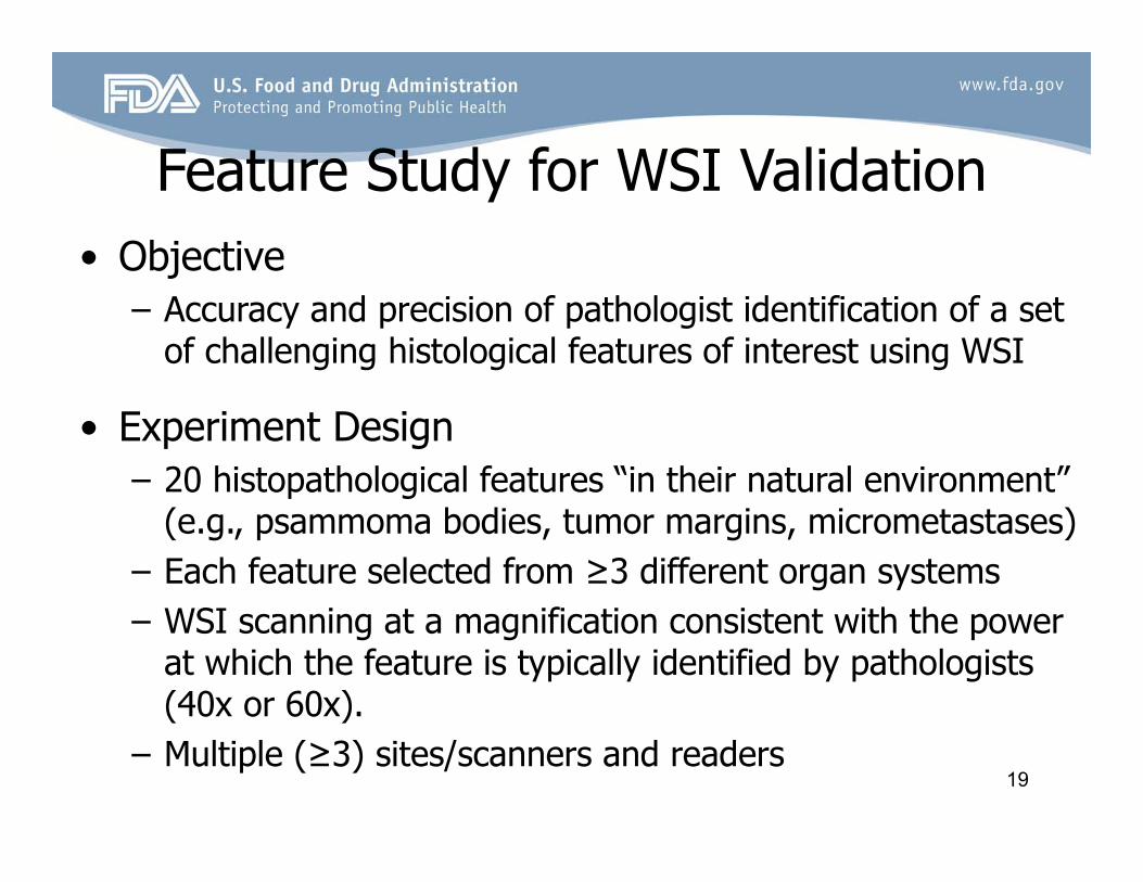
19
• Objective– Accuracy and precision of pathologist identification of a set
of challenging histological features of interest using WSI
• Experiment Design– 20 histopathological features “in their natural environment”
(e.g., psammoma bodies, tumor margins, micrometastases)– Each feature selected from ≥3 different organ systems– WSI scanning at a magnification consistent with the power
at which the feature is typically identified by pathologists (40x or 60x).
– Multiple (≥3) sites/scanners and readers
Feature Study for WSI Validation
20
• Does WSI system allow intended users to make diagnosis of surgical pathology specimens as accurately and reliably as optical microscopy? – Serious consequences to public health if misdiagnosis
caused by suboptimal images
Clinical Validation of WSI System
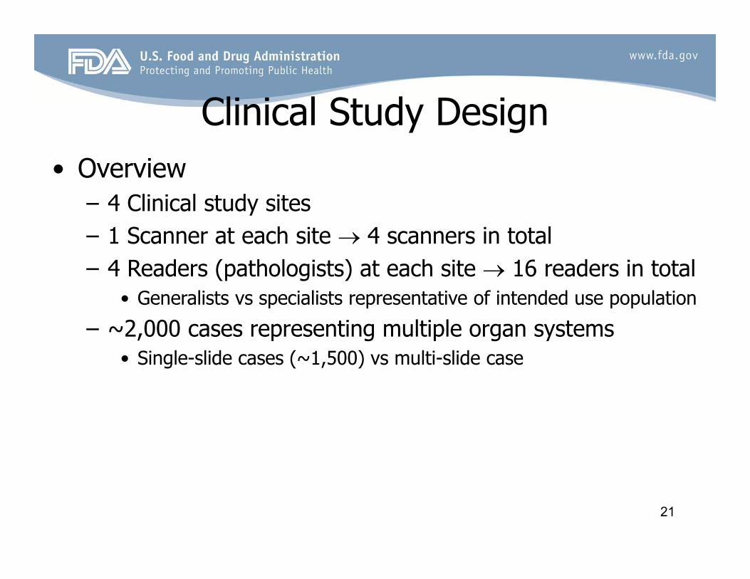
21
• Overview– 4 Clinical study sites– 1 Scanner at each site 4 scanners in total– 4 Readers (pathologists) at each site 16 readers in total
• Generalists vs specialists representative of intended use population
– ~2,000 cases representing multiple organ systems• Single-slide cases (~1,500) vs multi-slide case
Clinical Study Design
22
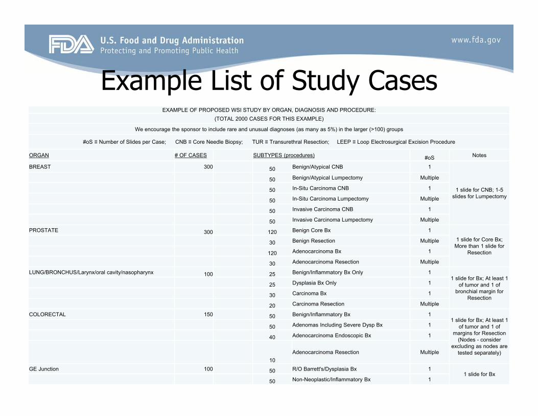
Example List of Study CasesEXAMPLE OF PROPOSED WSI STUDY BY ORGAN, DIAGNOSIS AND PROCEDURE:
(TOTAL 2000 CASES FOR THIS EXAMPLE)
We encourage the sponsor to include rare and unusual diagnoses (as many as 5%) in the larger (>100) groups
#oS ≡ Number of Slides per Case; CNB ≡ Core Needle Biopsy; TUR ≡ Transurethral Resection; LEEP ≡ Loop Electrosurgical Excision Procedure
ORGAN # OF CASES SUBTYPES (procedures) #oS Notes
BREAST 300 50 Benign/Atypical CNB 1
1 slide for CNB; 1-5 slides for Lumpectomy
50 Benign/Atypical Lumpectomy Multiple
50 In-Situ Carcinoma CNB 1
50 In-Situ Carcinoma Lumpectomy Multiple
50 Invasive Carcinoma CNB 1
50 Invasive Carcinoma Lumpectomy Multiple
PROSTATE 300 120 Benign Core Bx 1
1 slide for Core Bx; More than 1 slide for
Resection
30 Benign Resection Multiple
120 Adenocarcinoma Bx 1
30 Adenocarcinoma Resection Multiple
LUNG/BRONCHUS/Larynx/oral cavity/nasopharynx 100 25 Benign/Inflammatory Bx Only 11 slide for Bx; At least 1
of tumor and 1 of bronchial margin for
Resection
25 Dysplasia Bx Only 1
30 Carcinoma Bx 1
20 Carcinoma Resection Multiple
COLORECTAL 150 50 Benign/Inflammatory Bx 11 slide for Bx; At least 1
of tumor and 1 of margins for Resection
(Nodes - consider excluding as nodes are
tested separately)
50 Adenomas Including Severe Dysp Bx 1
40 Adenocarcinoma Endoscopic Bx 1
10Adenocarcinoma Resection Multiple
GE Junction 100 50 R/O Barrett's/Dysplasia Bx 11 slide for Bx
50 Non-Neoplastic/Inflammatory Bx 1
23
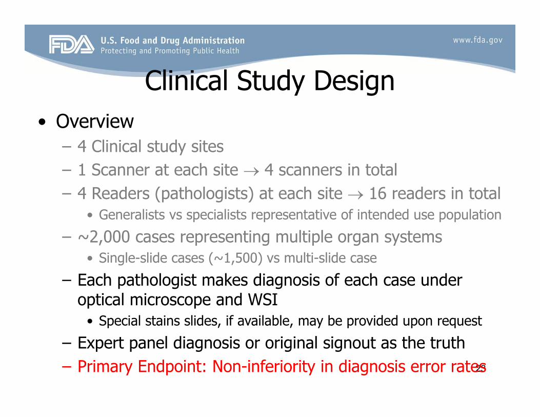
• Overview– 4 Clinical study sites– 1 Scanner at each site 4 scanners in total– 4 Readers (pathologists) at each site 16 readers in total
• Generalists vs specialists representative of intended use population
– ~2,000 cases representing multiple organ systems• Single-slide cases (~1,500) vs multi-slide case
– Each pathologist makes diagnosis of each case under optical microscope and WSI
• Special stains slides, if available, may be provided upon request
– Expert panel diagnosis or original signout as the truth – Primary Endpoint: Non-inferiority in diagnosis error rates
Clinical Study Design

24
• Subgroup analysis– Limitations of WSI vs requirement of postmarket studies
• Intranet, internet, mobile apps– Access vs fidelity vs confidentiality (cybersecurity)
• Human factors– Training, clueing – Scanning magnification vs digital magnification
• Component replacement– Swaps, upgrades
• Availability of glass slides
Future Considerations

25
• Digital pathology devices including WSI regulated by FDA based on intended use
• FDA has published draft guidance for technical assessment of WSI system for primary diagnosis
• FDA has outline WSI validation studies for sponsors– A clinical study to validate WSI for a broad intended use
(i.e., primary surgical pathology diagnosis in lieu of optical microscopy)
– A feature study to supplement the non-clinical and clinical validation studies
• Please use the pre-submission process to seek FDA guidance on digital pathology devices
Take Home Messages
26
• Technical assessment of WSI system draft guidance– http://www.fda.gov/ucm/groups/fdagov-public/@fdagov-meddev-
gen/documents/document/ucm435355.pdf
• Cybersecurity draft guidance– http://www.fda.gov/downloads/medicaldevices/deviceregulationandgu
idance/guidancedocuments/ucm356190.pdf
• CDRH device advice– http://www.fda.gov/MedicalDevices/DeviceRegulationandGuidance/
• Pre-submission guidance– http://www.fda.gov/downloads/medicaldevices/deviceregulationandgu
idance/guidancedocuments/ucm311176.pdf
• Division of Industry and Consumer Education (DICE)– 800-638-2041/301-796-7100
Resources