Embed Size (px)
Citation preview

FEATURE ARTICLE
SylvanaA. Brickley,MSN, FNP-BC,DCNP,University of RochesterMedical Center, Rochester, NY.Abigail Franco, MD, University of Rochester Medical Center,Rochester, NY.Kathryn Somers, MD, University of Rochester Medical Center,Rochester, NY.
The authors declare no conflict of interest.
Correspondence concerning this article should be addressed to SylvanaA. Brickley, MSN, FNP-BC, DCNP, University of Rochester MedicalCenter, Department of Dermatology, 601 Elmwood Ave, Box 697Rochester, New York 14642. E-mail: [email protected] © 2021 by the Dermatology Nurses’ Association.DOI: 10.1097/JDN.0000000000000606
102 Journal of the Dermatology Nurses’ A
Copyright © 2021 Dermatology Nurses' Association. Unauthorized reproduction of this article is prohibited.
2.0ContactHours
Skin Cancer: Back toBasics—Merkel Cell Carcinoma
Sylvana A. Brickley, Abigail Franco, and Kathryn Somers
ABSTRACT:Merkel cell carcinoma (MCC) is a rare andag-gressive skin cancer. Advanced disease portends a poorprognosis in most patients. We review the pathogenesis,clinical features, dermoscopic findings, differential diag-nosis, workup, treatment modalities, and follow-up ofMCC for dermatology nurses. It is important for nurses tobe familiar with MCC to prevent delayed diagnosis.Keywords:Dermoscopy,Malignancy,Merkel,Merkel CellCarcinoma, Skin Cancer
BACKGROUNDMerkel cell carcinoma (MCC) is a rare neuroendocrine tu-mor that is associated with a high rate of recurrence anddistant metastases. It most commonly affects elderly whitemen over the age of 70 years with an extensive history ofpast sun exposure, although MCC has been reported inBlack, Asian, American Indian, and Pacific Islander popula-tions. The risk of MCC is up to 25 times higher in the whitepopulation, compared with other ethnicities (Coggshallet al., 2018; Goldstein&DeCaprio, 2019).MCC typicallyoccurs on the sun-exposed areas of the head and neck(Coggshall et al., 2018). The incidence ofMCC has increasedsteadily over the past 30 years and continues to increase;however, it remains low, with approximately 1,600 casesdiagnosed annually in the United States (Coggshall et al.,2018; Tello et al., 2018). Annual incidence is much higherin patients with lymphoproliferative malignancies, historyof solid organ transplant, and HIV infection (Goldstein &DeCaprio, 2019; Ma & Brewer, 2014). MCC is highly
aggressive, and more than one third of patients die ofthe disease; thus, MCC has a case fatality rate that is cur-rently higher thanmelanoma (Becker et al., 2017). Despiteits rarity, it is important for dermatology nurses to be fa-miliar with MCC because of its high case fatality rate.
PATHOGENESISThe current understanding of MCC is that it is a primarycutaneous tumor derived from theMerkel cell (MC).MCsare considered neuroendocrine cells that play a role inlight touch/sensory responses and nerve guidance, as wellas endocrine/paracrine effects and somatostatin synthesis(Coggshall et al., 2018).Oncogenesis of theMC is consideredsecondary to genetic and environmental factors. Evidencesupports a role of ultraviolet (UV) radiation, with increasedincidence on sun-exposed sites. In fact, patients with psoriasistreated with psoralen and UVA light were found to have a100-fold increase in MCC development (Lunder & Stern,1998). Immunosuppression and immunosenescence also playa role, with higher rates seen in patients on immunosuppres-sive therapies (10-fold increased risk in transplant patients),in those withHIV/AIDS (13-fold increased risk), and amongthe elderly population (with 76% of cases in people olderthan 65 years; Agelli & Clegg, 2003; Arora et al., 2012).
In 2008, Feng et al. discovered the MC polyomavirus(MCV) and found MCV's DNA to be integrated into thehost cell's genome in a clonal pattern. MCV was foundin eight of 10 (80%) MCC tumors, versus only five of59 (8%) control tissues from various body sites and fourof 24 (16%) control skin tissues (Feng et al., 2008). Sincethen, this has been further corroborated with 1,743 of2,354 (74.2%) MCC tumors testing positive for MCV inmultiple studies worldwide (Arora et al., 2012). Interestingly,although MCV is ubiquitous in humans (~60%–80% posi-tive serologic tests in the general population), integration intothe host genome is not part of the normal life cycle of MCVand integration inhibits the virus's ability to replicate. It hasbeen postulated that UV radiation may induce mutations inMCV that drive integration into the genome and subsequentoncogenesis (Coggshall et al., 2018).
ssociation
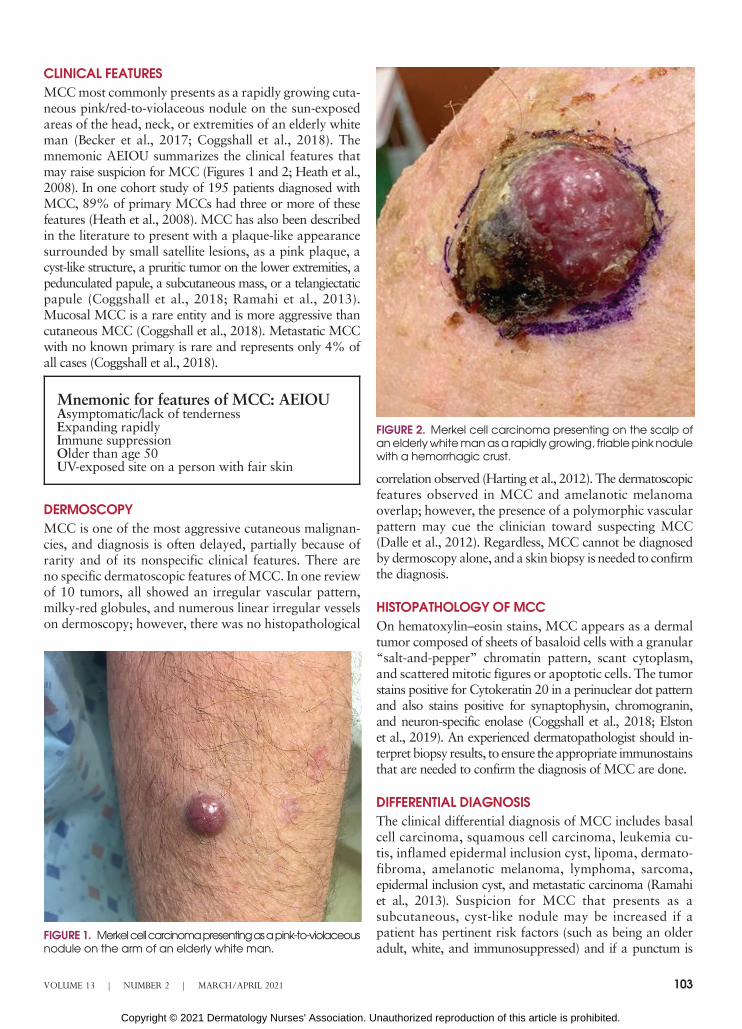
CLINICAL FEATURESMCCmost commonly presents as a rapidly growing cuta-neous pink/red-to-violaceous nodule on the sun-exposedareas of the head, neck, or extremities of an elderly whiteman (Becker et al., 2017; Coggshall et al., 2018). Themnemonic AEIOU summarizes the clinical features thatmay raise suspicion for MCC (Figures 1 and 2; Heath et al.,2008). In one cohort study of 195 patients diagnosed withMCC, 89% of primary MCCs had three or more of thesefeatures (Heath et al., 2008). MCC has also been describedin the literature to present with a plaque-like appearancesurrounded by small satellite lesions, as a pink plaque, acyst-like structure, a pruritic tumor on the lower extremities, apedunculated papule, a subcutaneous mass, or a telangiectaticpapule (Coggshall et al., 2018; Ramahi et al., 2013).Mucosal MCC is a rare entity and is more aggressive thancutaneous MCC (Coggshall et al., 2018). Metastatic MCCwith no known primary is rare and represents only 4% ofall cases (Coggshall et al., 2018).
FIGURE 2. Merkel cell carcinoma presenting on the scalp ofanelderlywhitemanasa rapidly growing, friablepink nodulewith a hemorrhagic crust.
Mnemonic for features of MCC: AEIOUAsymptomatic/lack of tendernessExpanding rapidlyImmune suppressionOlder than age 50UV-exposed site on a person with fair skin
DERMOSCOPYMCC is one of the most aggressive cutaneous malignan-cies, and diagnosis is often delayed, partially because ofrarity and of its nonspecific clinical features. There areno specific dermatoscopic features ofMCC. In one reviewof 10 tumors, all showed an irregular vascular pattern,milky-red globules, and numerous linear irregular vesselson dermoscopy; however, there was no histopathological
FIGURE 1. Merkelcellcarcinomapresentingasapink-to-violaceousnodule on the arm of an elderly white man.
VOLUME 13 | NUMBER 2 | MARCH/APRIL 2021
Copyright © 2021 Dermatology Nurses' Association. U
correlation observed (Harting et al., 2012). The dermatoscopicfeatures observed in MCC and amelanotic melanomaoverlap; however, the presence of a polymorphic vascularpattern may cue the clinician toward suspecting MCC(Dalle et al., 2012). Regardless, MCC cannot be diagnosedby dermoscopy alone, and a skin biopsy is needed to confirmthe diagnosis.
HISTOPATHOLOGY OF MCCOn hematoxylin–eosin stains, MCC appears as a dermaltumor composed of sheets of basaloid cells with a granular“salt-and-pepper” chromatin pattern, scant cytoplasm,and scattered mitotic figures or apoptotic cells. The tumorstains positive for Cytokeratin 20 in a perinuclear dot patternand also stains positive for synaptophysin, chromogranin,and neuron-specific enolase (Coggshall et al., 2018; Elstonet al., 2019). An experienced dermatopathologist should in-terpret biopsy results, to ensure the appropriate immunostainsthat are needed to confirm the diagnosis of MCC are done.
DIFFERENTIAL DIAGNOSISThe clinical differential diagnosis of MCC includes basalcell carcinoma, squamous cell carcinoma, leukemia cu-tis, inflamed epidermal inclusion cyst, lipoma, dermato-fibroma, amelanotic melanoma, lymphoma, sarcoma,epidermal inclusion cyst, and metastatic carcinoma (Ramahiet al., 2013). Suspicion for MCC that presents as asubcutaneous, cyst-like nodule may be increased if apatient has pertinent risk factors (such as being an olderadult, white, and immunosuppressed) and if a punctum is
103
nauthorized reproduction of this article is prohibited.

not visible clinically. Suspicion for MCC may be heightenedby a history of a lesion that is expanding rapidly (doublingin less than 3 months), chronic immunosuppression, ageolder than 50 years, and lesion location on a UV-exposedarea (Sarnaik et al., 2010).
WORKUP AND TREATMENT MODALITIESThe National Comprehensive Cancer Network (NCCN)in the United States proposed specific, updated guidelinesfor diagnostic evaluation ofMCC; these guidelines reviewthe clinical presentation, preliminary workup, diagnosis,additional workup, and then treatment depending on stag-ing (NCCN, 2019). A table outlining a simplified versionof the 2019 NCCN guideline is shown below (Table 1).
If a lesion suspicious for MCC is identified on exam-ination, a skin biopsy should be performed and sent forhematoxylin-and-eosin and immunopanel studies, andthe patient should have a complete skin and lymph nodeexamination. The provider should note suspicion for MCCon the pathology requisition for the biopsy to aid thedermatopathologist in making an accurate diagnosis. Upondiagnosis of MCC, the patient should be referred to anoncologist for appropriate staging and further workupaccording to NCCN (2019) guidelines.
For patients with clinically negative lymph nodes, themost reliable tool for evaluating regional nodes for metas-tases is a sentinel lymph node biopsy (SLNB; Coggshallet al., 2018). SLNB is recommended in patients with clin-ically negative lymph nodes because approximately 26%of these patients will have pathologically positive lymphnodes (Allen et al., 2005; Lemos & Ngheim, 2007; Simset al., 2018). Every effort should be made for SLNB to beperformed preceding or concurrently with excision to pre-vent a false-negative SLNB result (NCCN, 2019). Imagingat the time of diagnosis using magnetic resonance imaging,
TABLE 1. Summary of National CancerCoalition Network: Merkel Cell Carcinoma2019 Guidelines
1. Complete skin and lymph node examination2. Biopsy specimen, sent to an experienced
dermatopathologist for hematoxylin–eosin stains andimmunostaining
3. For patients with negative clinical nodes, refer tooncology to obtain a sentinel lymph node biopsy
4. For patients with positive clinical nodes, refer tooncology for fine needle aspiration or core biopsy firsta. If negative, oncology may consider open lymph
node biopsyb. If positive, oncology may proceed to 5
5. Imaging as clinically indicated with magneticresonance imaging, computed tomography (CT), orpositron emission tomography/CT
6. Consider consultation with a multidisciplinary tumorboard
104
Copyright © 2021 Dermatology Nurses' Association. Un
computed tomography, or positron emission tomography/computed tomographymay be ordered as clinically indicated(Coggshall et al., 2018). Excision of the tumor by a board-certified dermatologist or surgical oncologist is the first-linetherapy; however, if not feasible, radiation monotherapymay be considered (NCCN, 2019). For advanced-stage orrefractory MCC, chemotherapy or immune-checkpoint in-hibitors may be utilized by oncology (Becker et al., 2017;NCCN, 2019).
ROLE OF THE DERMATOLOGY NURSEBecause of the rarity and aggressiveness of MCC, it is im-portant for nurses (such as licensed practical nurses, regis-tered nurses, and nurse practitioners) to be familiar withits clinical features andmanagement to ensure timely diag-nosis and appropriate course of care. Patients face muchuncertainty amid a diagnosis ofMCC.Nurse practitionersmay conduct skin cancer screenings and perform skin biop-sies, so familiarity with MCC is critical to prevent delayeddiagnosis. Nurse practitioners who diagnose MCC needto also be aware of appropriate treatment and follow-upso that timely and appropriate referral can be made to on-cology. Nurses at all levels of practice also may play an im-portant role in educating patients about risk factors andclinical features of MCC. Any nurse caring for a patientwith MCC may be expected to provide patient educationand help guide the patient through treatment and follow-up. A team-based approach to treatment with coordinationof care between dermatology, surgery, and oncology is nec-essary, and nurses may help to facilitate care coordination.Knowledge ofMCC enables nurses to provide the best pos-sible care to patients withMCC,within the nurses' scope ofpractice and specialty.
FOLLOW-UPMCC is an aggressive tumor with a poor prognosis, es-pecially if there is metastatic disease at presentation. At5 years, overall survival is approximately 51% for localdisease, 35% for nodal disease, and 14% for distant met-astatic disease (Harms et al., 2016). Patients with a historyof MCC should undergo a complete skin and lymph nodeexamination every 3–6 months for 3 years and then every6–12 months indefinitely (NCCN, 2019). For high-riskpatients, routine imaging may be considered to monitorfor metastasis. ▪
REFERENCESAgelli, M., & Clegg, L. X. (2003). Epidemiology of primaryMerkel cell carci-noma in the United States. Journal of the American Academy of Derma-tology, 49(5), 832–841. 10.1016/s0190-9622(03)02108-x
Allen, P. J., Bowne, W. B., Jaques, D. P., Brennan, M. F., Busam, K., &Coit, D. G. (2005). Merkel cell carcinoma: Prognosis and treatment ofpatients from a single institution. Journal of Clinical Oncology, 23(10),2300–2309.
Arora, R., Chang, Y., &Moore, P. S. (2012). MCVandMerkel cell carcinoma:A molecular success story. Current Opinion in Virology, 2(4), 489–498. 10.1016/j.coviro.2012.05.007
Journal of the Dermatology Nurses’ Association
authorized reproduction of this article is prohibited.
Becker, J. C., Stang, A., DeCaprio, J. A., Cerroni, L., Lebbé, C., Veness,M., &Nghiem, P. (2017). Merkel cell carcinoma. Nature Reviews DiseasePrimers, 3, 17077. 10.1038/nrdp.2017.77
Coggshall, K., Tello, T. L., North, J. P., & Yu, S. S. (2018). Merkel cell carci-noma: An update and review: Pathogenesis, diagnosis, and staging. Jour-nal of the American Academy of Dermatology, 78(3), 433–442. 10.1016/j.jaad.2017.12.001
Dalle, S., Parmentier, L.,Moscarella, E., Phan, A., Argenziano, G.,&Thomas,L. (2012). Dermoscopy of Merkel cell carcinoma. Dermatology (Basel,Switzerland), 224(2), 140–144. 10.1159/000337411
Elston, D. M., Ferringer, T., & Ko, C. J. (2019). Dermatopathology (pp.421–422). Elsevier.
Feng, H., Shuda, M., Chang, Y., &Moore, P. S. (2008). Clonal integration ofa polyomavirus in humanMerkel cell carcinoma. Science (NewYork, N.Y.),319(5866), 1096–1100. 10.1126/science.1152586
Goldstein, R. H., & DeCaprio, J. A. (2019). Merkel cell carcinoma in theHIV-1/AIDS patient. Cancer Treatment and Research, 177, 211–229.10.1007/978-3-030-03502-0_8
Harms, K. L., Healy, M. A., Nghiem, P., Sober, A. J., Johnson, T. M.,Bichakjian, C. K., & Wong, S. L. (2016). Analysis of prognostic factorsfrom 9387 Merkel cell carcinoma cases forms the basis for the new 8thedition AJCC staging system. Annals of Surgical Oncology, 23(11),3564–3571. 10.1245/s10434-016-5266-4
Harting, M. S., Ludgate, M.W., Fullen, D. R., Johnson, T. M., & Bichakjian,C. K. (2012). Dermatoscopic vascular patterns in cutaneous Merkel cellcarcinoma. Journal of the American Academy of Dermatology, 66(6),923–927. 10.1016/j.jaad.2011.06.020
VOLUME 13 | NUMBER 2 | MARCH/APRIL 2021
Copyright © 2021 Dermatology Nurses' Association. U
Heath, M., Jaimes, N., Lemos, B., Mostaghimi, A., Wang, L. C., Peñas, P. F.,& Nghiem, P. (2008). Clinical characteristics of Merkel cell carcinomaat diagnosis in 195 patients: The AEIOU features. Journal of the AmericanAcademy of Dermatology, 58(3), 375–381. 10.1016/j.jaad.2007.11.020
Lemos, B., &Nghiem, P. (2007). Merkel cell carcinoma:More deaths but stillno pathway to blame. Journal of Investigative Dermatology, 127(9), 2100–2103.10.1038/sj.jid.5700925
Lunder, E. J., & Stern, R. S. (1998).Merkel-cell carcinomas in patients treatedwith methoxsalen and ultraviolet A radiation. New England Journal ofMedicine, 339, 1247–1248.
Ma, J. E., & Brewer, J. D. (2014). Merkel cell carcinoma in immunosuppressedpatients. Cancers (Basel), 6(3), 1328–1350. 10.3390/cancers6031328
National Comprehensive Cancer Network. (2019). NCCN clinical practiceguidelines in oncologyMerkel cell carcinoma. NCCNGuidelines, Version1.2020. https://www.nccn.org/professionals/physician_gls/pdf/mcc.pdf
Ramahi, E., Choi, J., Fuller, C.D.,&Eng, T. Y. (2013).Merkel cell carcinoma.American Journal of Clinical Oncology, 36(3), 299–309. 10.1097/COC.0b013e318210f83c
Sims, J. R., Grotz, T. E., Pockaj, B. A., Joseph, R.W., Foote, R. L., Otley, C. C.,Weaver, A. L., Jakub, J. W., & Price, D. L. (2018). Sentinel lymph nodebiopsy in Merkel cell carcinoma: The Mayo Clinic experience of 150patients. Surgical Oncology, 27(1), 11–17. 10.1016/j.suronc.2017.10.005
Tello, T. L., Coggshall, K., Yom, S. S.,&Yu, S. S. (2018).Merkel cell carcinoma:An update and review: Current and future therapy. Journal of the AmericanAcademy of Dermatology, 78(3), 445–454. 10.1016/j.jaad.2017.12.004
For more than 64 additional continuing professional development articles related to dermatologic conditions, go toNursingCenter.com/ce.
Nursing Continuing Professional Development
TEST INSTRUCTIONS• Read the article. The test for this nursing continuing professionaldevelopment (NCPD) activity is to be taken online atwww.NursingCenter.com/CE/JDNA. Tests can no longer be mailed or faxed.• You'll need to create an account (it's free!) and log in to accessMy Planner before taking online tests. Your planner will keep trackof all your Lippincott Professional Development online NCPD activitiesfor you.• There's only one correct answer for each question. A passing score forthis test is 7 correct answers. If you pass, you can print your certificateof earned contact hours and access the answer key. If you fail, you have theoption of taking the test again at no additional cost.• For questions, contact Lippincott Professional Development:1-800-787-8985.• Registration deadline is March 3, 2023.
PROVIDER ACCREDITATIONLippincott Professional Development will award 2.0 contact hours for thisnursing continuing professional development activity.
Lippincott Professional Development is accredited as a provider ofnursing continuing professional development by the American NursesCredentialing Center's Commission on Accreditation.
This activity is also provider approved by the California Board ofRegistered Nursing, Provider Number CEP 11749 for 2.0 contact hours.Lippincott Professional Development is also an approved provider ofcontinuing nursing education by the District of Columbia, Georgia,and Florida, CE Broker #50-1223. Your certificate is valid in all states.
Payment: The registration fee for this test is $10 for members; $20 fornonmembers.
105
nauthorized reproduction of this article is prohibited.