Embed Size (px)
Citation preview
Dental Implants
: Dental Implants Patient Selection Factors
- Evidence Based Review
Dental Implants
, Mark Ayson MBChB DPH
ACC
-Peer reviewers and co authers
,Jonathan Leichter and Karl Lyons , University of Otago School of Dentistry
Table of Contents
Summary.....................................................................................................................................5
Introduction.................................................................................................................................7
Objectives....................................................................................................................................7
Criteria for selecting studies for this review...............................................................................7
Search strategy............................................................................................................................8
Methodological quality...............................................................................................................8
Evidence Based Healthcare Team Page 2
Dental Implants
Results and discussion................................................................................................................9
Patient selection factors............................................................................................................10
Effect of smoking..................................................................................................................10
Effect of periodontitis...........................................................................................................19
Effect of oral hygiene/habits.................................................................................................23
Effect of location...................................................................................................................24
Effect of bone quality............................................................................................................33
Effect of bone quantity..........................................................................................................37
Effect of bone grafts/augmentation.......................................................................................39
Effect of osteoporosis...........................................................................................................43
Effect of hormone replacement therapy (HRT), radical hysterectomy &/or menopausal status.....................................................................................................................................46
Effect of age..........................................................................................................................48
Effect of radiotherapy...........................................................................................................51
Effect of chemotherapy.........................................................................................................53
Effect of coagulation problems.............................................................................................54
Effect of edentulousness/proximity to natural teeth.............................................................56
Effect of gastric disease........................................................................................................58
Effect of Crohn’s disease......................................................................................................59
Effect of cardiac disease.......................................................................................................61
Effect of hypercholesterolaemia...........................................................................................63
Effect of hypertension...........................................................................................................64
Effect of prosthesis type........................................................................................................65
Effect of diabetes..................................................................................................................67
Effect of asthma or pulmonary disease.................................................................................70
Effect of rheumatoid arthritis................................................................................................71
Effect of thyroid disease.......................................................................................................73
Effect of medication and other factors..................................................................................74
Additional Information.............................................................................................................78
Conclusions...............................................................................................................................82
References.................................................................................................................................84
Appendix A...........................................................................................................................87
Evidence Based Healthcare Team Page 3

Dental Implants
Appendix B...........................................................................................................................90
Appendix C.........................................................................................................................152
Evidence Based Healthcare Team Page 4
Dental Implants
: Dental Implant Patient Selection Factors
Reviewer Dr Mark Ayson, MBChB;
ACC
Contributing authors Jonathan Leichter, D.M.D.,
Karl Lyons. BDS, MS;
University of Otago, School of Dentistry
Literature Search Amanda Bowens
Date of Draft 16 September 2009
Important Notes:
This evidence based review summarises information on dental implants published since 2004. It is not intended to replace clinical judgement, or be used as a clinical protocol.
If a particular factor (e.g. bruxism or any other parafunctional habit) is not discussed in this report, it is because no publihed literature regarding that factor was located. In these circumstances, the conclusions of the previous report1 still apply.
A reasonable attempt has been made to find and review papers relevant to the focus of this EBH review. It does not claim to be exhaustive.
The content of this document does not necessarily represent the official view of ACC or represent ACC policy.
Summary
An evidence-based review of patient selection factors that affect dental implant survival and/or success has been conducted to supply evidence to update the existing ACC Dental Implant Guidelines (2006)2. Fifty-five papers were reviewed. The overall quality of the published research was relatively poor and so, the guideline development will involve considering the existing evidence, expert opinion and a consensus process.
The main findings of the review are:
• Smoking less than ten cigarettes per day, as a single risk factor, may not increase the risk of failure sufficiently to deny treatment but dental implants may be contra-indicted in smokers who have other relevant risk factors.
• There is insufficient evidence that people with a history of treated periodontitis have a significantly elevated risk of implant failure but the consensus view is that people with periodontitis should be treated for this condition before being considered for implant therapy.
Evidence Based Healthcare Team Page 5
Dental Implants
• There is a consensus of opinion that people with poor oral hygiene, infection or uncontrolled caries should not be offered dental implants.
• There is weak evidence that dental implants placed in the maxilla may have a greater risk of failure.
• There is conflicting evidence that bone quality or quantity have a significnat effect on the risk of implant failure.
• There is some weak evidence that implants placed after maxillary sinus augmentation may have an increased risk of failure but there is no evidence that other bone graft/augmentation techniques alter significantly the risk of implant failure.
• Older age, as a single risk factor, is not a contraindication for implant placement.
• There is a consensus of opinion that implant placement should be delayed in young people until growth is complete.
• There is some evidence that implant failure rates in the maxilla are usually significantly increased in people who have previously had irradiation of the jaw, regardless of the total radiation dose.
• There is a consensus opinion that dental implants should not be placed in people undergoing active chemotherapy.
• Medical advice should be sought for people with coagulation disorders or taking anticoagulant medication before proceeding with implant placement.
• Bisphosphonate medication is a contraindication to implant placement due to the risk of osteonecrosis of the jaw.
• There is insufficient evidence that well controlled diabetes type II is a significant risl factor for implant failure
• There is no evidence whether diabetes type I, asthma, rheumatoid arthritis is or is not a significant risk factor for implant failure
• There is insufficient evidence that the following factors are significant risk factors for implant failure:
o type of edentulousness or proximity to natural dentition
o placement in the posterior region of a jaw
o osteoporosis
o type of prosthesis
o a history of chemotherapy treatment
o cardiovascular disease, including hypertension and hypercholesterolaemia
o thyroid disease, gastric problems or Crohn’s disease
o hormone replacement therapy, menopausal status or history of a radical hysterectomy
Evidence Based Healthcare Team Page 6
Dental Implants
o steroid, antidepressant, antihypertensive, statin, antimicrobial or non-steroidal anti-inflammatory medications.
Dental Implants
Introduction
Missing teeth have traditionally been replaced with dentures or bridges permitting restoration of chewing function, speech, and aesthetics. Dental implants offer an alternative.
These implants are inserted into the jawbones to support a dental prosthesis and are retained because of the intimacy of bone growth on to their surface. This direct structural and functional connection between living bone and implant surface, termed osseointegration, was first described by Brånemark 1977.3
Studies have reported a high level of predictability and success using this treatment modality for a variety of clinical situations in both people who are edentulous and partially dentate.4
ACC pays for around 80% of all dental implants in New Zealand and the total costs are increasing. In 1999 the surgical costs were approximately $1m and increased to $5.1m in the July 2008-June 2009 period. [personal communication with Rosemary Kennedy, Dental Advisor, Health Purchasing Provider Relationships, ACC]
In 2004 guidelines were developed by ACC to supply guidance on patient selection factors, patient satisfaction and cost factors. A request was made in 2007 to update the evidence review for these guidelines so as to keep entitlements in line with necessary and appropriate treatment.
Objectives
The objective is to update the evidence base of the ACC Dental Implant Guidelines focussing on the patient selection factors.
Criteria for selecting studies for this review
Types of studies:
Randomised controlled studies, systematic reviews, clinical practice guidelines, and cohort, case-control or case series studies that reported on patient selection factors.
Types of participants:
People of any age who had dental implants placed for whatever reason.
Evidence Based Healthcare Team Page 7
Dental Implants
Types of exposures:
Risk factors for implant failure i.e. smoking, diabetes, parafunction including bruxism, bone grafting, bone quality/volume, osteoporosis, menopause and hormone replacement therapy, hypothyroidism, cardiovascular disease, irradiation of jaw, oral health (infection, periodontitis, poor oral hygiene, uncontrolled caries), general health status, site of implant placement (mandible vs. maxilla & posterior vs. anterior location), degree of edentulousness, force on implant, age and medication.
Types of outcome measures:
Risk of implant survival, success or failure expressed as relative risk, odds ratio or statistical significance (p-value), and pooled survival estimates.
Search strategy
The following databases were searched: Medline, EMBASE, Premedline, CINAHL, All EBM reviews, and PsychInfo. The search strategies were run in January 2008 and repeated in November 2008.
Search strategies are shown in Appendix A
Inclusion criteria:
• only papers published in English that were readily available
• papers published from 2004 onwards or before 2004 if not in previous review.
Exclusion criteria:
Papers that dealt exclusively with:
• gene polymorphism
• implant overdentures/fixed complete prosthesis
• timing of loading of implant
• placement protocols (1 vs. 2-stage)
• type/surface of implant
• orthodontic anchorage.
Methodological quality
Reviewed papers were assigned a level of evidence according to the SIGN5 revised grading system (Table 1).
Table 1: Scottish Intercollegiate Guidelines Network (SIGN) Revised Grading System
LEVELS OF EVIDENCE
Evidence Based Healthcare Team Page 8
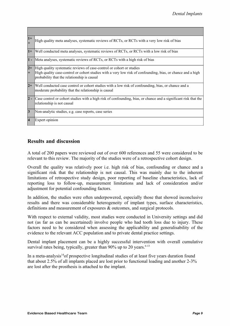
Dental Implants
1++
High quality meta analyses, systematic reviews of RCTs, or RCTs with a very low risk of bias
1+ Well conducted meta analyses, systematic reviews of RCTs, or RCTs with a low risk of bias
1 - Meta analyses, systematic reviews of RCTs, or RCTs with a high risk of bias
2++
High quality systematic reviews of case-control or cohort or studiesHigh quality case-control or cohort studies with a very low risk of confounding, bias, or chance and a high probability that the relationship is causal
2+ Well conducted case control or cohort studies with a low risk of confounding, bias, or chance and a moderate probability that the relationship is causal
2 - Case control or cohort studies with a high risk of confounding, bias, or chance and a significant risk that the relationship is not causal
3 Non-analytic studies, e.g. case reports, case series
4 Expert opinion
Results and discussion
A total of 200 papers were reviewed out of over 600 references and 55 were considered to be relevant to this review. The majority of the studies were of a retrospective cohort design.
Overall the quality was relatively poor i.e. high risk of bias, confounding or chance and a significant risk that the relationship is not causal. This was mainly due to the inherent limitations of retrospective study design, poor reporting of baseline characteristics, lack of reporting loss to follow-up, measurement limitations and lack of consideration and/or adjustment for potential confounding factors.
In addition, the studies were often underpowered, especially those that showed inconclusive results and there was considerable heterogeneity of implant types, surface characteristics, definitions and measurement of exposures & outcomes, and surgical protocols.
With respect to external validity, most studies were conducted in University settings and did not (as far as can be ascertained) involve people who had tooth loss due to injury. These factors need to be considered when assessing the applicability and generalisability of the evidence to the relevant ACC population and to private dental practice settings.
Dental implant placement can be a highly successful intervention with overall cumulative survival rates being, typically, greater than 90% up to 20 years.6-13
In a meta-analysis14of prospective longitudinal studies of at least five years duration found that about 2.5% of all implants placed are lost prior to functional loading and another 2-3% are lost after the prosthesis is attached to the implant.
Evidence Based Healthcare Team Page 9
Dental Implants
Patient selection factors
Effect of smoking
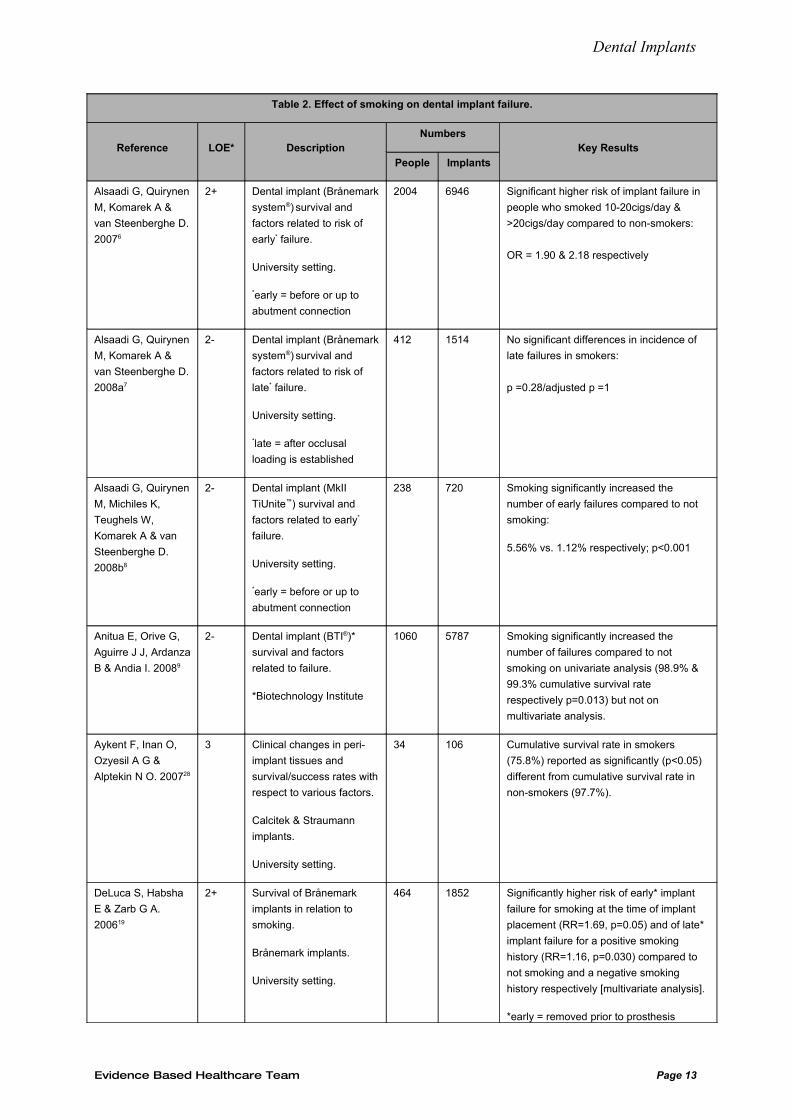
Thirty-seven papers were reviewed that evaluated the effect of smoking on implant survival/success, including 3 systematic reviews, 18 cohort studies, 6 case series, 9 narrative reviews and one consensus guideline (Table 2).
Of the 3 systematic reviews, all found that smoking significantly increased the risk of implant failure:
• Two of the reviews15, 16 estimated the pooled odds ratio (OR) to be 2.17 & 2.38 respectively
• The other12 estimated the pooled difference in survival rates between smokers and non-smokers to be 2.98%
• The systematic review15 also found that there was a significant increase in the risk of failure of implants placed in the maxilla of smokers compared to non-smokers: OR [maxilla] = 2.06 (95%CI: 1.61-2.65) but no significant increase in the risk of failure of implants placed in the mandible of smokers compared to non-smokers: OR [mandible] = 1.32 (95%CI: 0.72-2.4), suggesting that smoking has more influence on implants placed in the maxilla.
All of the four cohort studies considered to have a low risk of confounding, bias or chance found that smoking had a detrimental effect on implant survival:
• People who smoked between 10 & 20 cigarettes per day had a statistically significant greater risk of early implant failure compared to people who don’t smoke (OR=1.90) and that those who smoked >20 cigarettes per day had an even greater risk (OR=2.18)6
• Smokers had an significantly increased risk of overall complications (HR=2.31 (95%CI: 1.29-4.16); p=0.0051) and ‘inflammatory’ complications (including mobility, pain, infection, & peri-implantitis) (HR=3.26 (1.74-6.10) p=0.0002)17
• Smokers had an increased risk of implant failure (defined simply as “removal”) (HR=3.5 (95%CI: 1.7-7.2) p<0.001) on multivariate analysis17
• An increased risk of implant loss (HR=2.6 (95%CI: 1.8-3.9)) was found on multivariate analysis18.
Eight of the 14 cohort studies considered to have a high risk of confounding, bias or chance also found that smoking had a detrimental effect on implant survival:
• Smoking increased the risk of early failure from 1.12% to 5.56% (p<0.001)8
• Smoking at the time of implant placement surgery increased the risk of early implant failure (RR=1.69; p=0.05) and that a positive smoking history was associated with late (after the prosthesis was placed) implant failure (RR=1.91; p=0.05)19
• A study11 that looked at the effect of osteoporosis on implant survival in women found that smoking increased the risk of implant failure (HR=2.6 (95%CI: 1.20-5.63) p=0.016)
Evidence Based Healthcare Team Page 10
Dental Implants
• Moheng20 found that smoking increased the risk of implant failure (RR=14.4; p<0.0001) in a small prospective study of a highly selective population
• A larger study21 found that the risk of failure was increased by 1.39 times in smokers (p=0.03)
• A study13 assessing the long-term survival and risk factors for implant failure of screw-type implants in a private practice found that smoking increased the risk of implant failure by 1.04 times
• Smoking more than 20 cigarettes per day increased the risk of implant failure (OR = 2.50) compared to smoking less than 20 per day in a retrospective cohort study22
• A study23 investigating the survival rates of dental implants in people who had had a successful dentoalveolar reconstructive procedure found that the risk of failure in smokers was increased by 4.4 times.
Six of the 14 cohort studies considered to have a high risk of confounding, bias or chance found that smoking did not adversely affect the risk of failure:
• Smoking7 did not significantly alter the risk of late implant failure i.e. up to abutment connection
• A lower survival rate was found in smokers (p<0.013) in another study9 but this was not significant when adjusted for potential confounders
• There was no significant difference in implant failure rate between those who had never smoked compared to a group consisting of current & ex-smokers (p=0.16)24
• Another25 found that non-smokers had a non-significant reduction in the odds of failure (OR=0.42 (95%CI: 0.12-1.24) on multivariate analysis)
• Sverzut (2008)26 found no significant difference in risk of early failure in smokers compared to non-smokers (HR=1.2390; p=0.5994)
• Another study27 found that the risk of failure was not significantly different for smokers compared to non-smokers (5.6% vs. 3.7%; p=0.342).
Of the 6 case series, 3 found that smoking adversely affected implant survival and the other 3 found no significant difference in survival in smokers compared to non-smokers:
• A small study28 reported a significant difference in survival rates in smokers and non-smokers (75.8% vs. 97.7% respectively) but no statistics were given
• More implants failed in people who smoked compared to those who didn’t (p<0.001) in another case series29
• A significant (p<0.001) difference in success rates between smokers & non-smokers (84.2% vs. 98.6%) was found in one study30. The authors also found a dose-response curve of relative risks: 6.5 light smoker; 8.5 moderate smoker; 21.8 heavy smoker
• Another small study31 reported that smoking at least 20 cigarettes per day did not affect survival of dental implants placed in fully edentulous arches using a single-stage surgical protocol
Evidence Based Healthcare Team Page 11
Dental Implants
• Lemmerman (2005)32 found that there was no statistical difference between implant failure in smokers compared to non-smokers (p=0.945). In this study smokers were encouraged to quit and not sure whether they were still included in the smoking group
• Another smaller study33 evaluating implants replacing a single molar between two natural teeth, reported that there no significant difference in failure/complication rates in smokers compared to non-smokers.
A consensus statement from the Academy of Osseointegration34 and the nine non-systematic reviews35-43 identified, have all concluded that smoking has an adverse effect on implant survival and success and two papers went on to state that this effect seems to be more pronounced in loose trabecular bone34 and the maxilla42.
While smoking is reported not to be an absolute contraindication to implant therapy40, these people should be informed of the elevated risk35, 36, 40 of implant complications and counselled to stop smoking35, 36.
Discussion
Smoking is an established risk factor for periodontitis and delayed wound healing after tooth extraction42. The previous ACC Dental Implant Evidence-based Review (2004)1 concluded that smoking as a single risk factor may not increase the risk of implant failure sufficiently to deny treatment but that dental implants may be contraindicated in smokers who have other relevant risk factors.
The evidence since 2004, reviewed above, supports these recommendations and strengthens the evidence base for smoking as a risk factor for dental implant failure. It seems reasonable to conclude that smoking does decrease the risk of implant survival by approximately 3%12
with an RR of about 215, 16.
Moreover, there is some evidence of a dose-response relationship between smoking and risk of implant failure6, 30 and a biologically plausible mechanism12, 15, 16 for greater implant failure in smokers, which supports the conclusions above.
Therefore it is concluded that there is moderate evidence that smoking reduces implant survival but that the effect size is so small as not to be clinically relevant. However, when associated with other risk factors, it may become relevant.
Suggested Recommendations:
Smoking less than ten cigarettes per day as a single risk factor may not increase the risk of failure sufficiently to deny treatment.
Dental implants may be contra-indicted in smokers who have other relevant risk factors.
Evidence Based Healthcare Team Page 12
Dental Implants
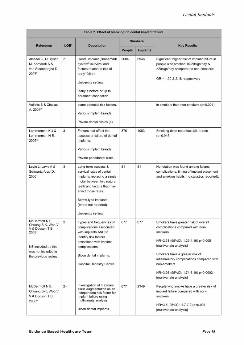
Table 2. Effect of smoking on dental implant failure.
Reference LOE* DescriptionNumbers
People Implants
Key Results
Alsaadi G, Quirynen
M, Komarek A &
van Steenberghe D.
20076
2+ Dental implant (Brånemark
system®) survival and
factors related to risk of
early* failure.
University setting.
*early = before or up to
abutment connection
2004 6946 Significant higher risk of implant failure in
people who smoked 10-20cigs/day &
>20cigs/day compared to non-smokers:
OR = 1.90 & 2.18 respectively
Alsaadi G, Quirynen
M, Komarek A &
van Steenberghe D.
2008a7
2- Dental implant (Brånemark
system®) survival and
factors related to risk of
late* failure.
University setting.
*late = after occlusal
loading is established
412 1514 No significant differences in incidence of
late failures in smokers:
p =0.28/adjusted p =1
Alsaadi G, Quirynen
M, Michiles K,
Teughels W,
Komarek A & van
Steenberghe D.
2008b8
2- Dental implant (MkII
TiUnite™) survival and
factors related to early*
failure.
University setting.
*early = before or up to
abutment connection
238 720 Smoking significantly increased the
number of early failures compared to not
smoking:
5.56% vs. 1.12% respectively; p<0.001
Anitua E, Orive G,
Aguirre J J, Ardanza
B & Andia I. 20089
2- Dental implant (BTI®)*
survival and factors
related to failure.
*Biotechnology Institute
1060 5787 Smoking significantly increased the
number of failures compared to not
smoking on univariate analysis (98.9% &
99.3% cumulative survival rate
respectively p=0.013) but not on
multivariate analysis.
Aykent F, Inan O,
Ozyesil A G &
Alptekin N O. 200728
3 Clinical changes in peri-
implant tissues and
survival/success rates with
respect to various factors.
Calcitek & Straumann
implants.
University setting.
34 106 Cumulative survival rate in smokers
(75.8%) reported as significantly (p<0.05)
different from cumulative survival rate in
non-smokers (97.7%).
DeLuca S, Habsha
E & Zarb G A.
200619
2+ Survival of Brånemark
implants in relation to
smoking.
Brånemark implants.
University setting.
464 1852 Significantly higher risk of early* implant
failure for smoking at the time of implant
placement (RR=1.69, p=0.05) and of late*
implant failure for a positive smoking
history (RR=1.16, p=0.030) compared to
not smoking and a negative smoking
history respectively [multivariate analysis].
*early = removed prior to prosthesis
Evidence Based Healthcare Team Page 13
Dental Implants
Table 2. Effect of smoking on dental implant failure.
Reference LOE* DescriptionNumbers
People Implants
Key Results
Alsaadi G, Quirynen
M, Komarek A &
van Steenberghe D.
20076
2+ Dental implant (Brånemark
system®) survival and
factors related to risk of
early* failure.
University setting.
*early = before or up to
abutment connection
2004 6946 Significant higher risk of implant failure in
people who smoked 10-20cigs/day &
>20cigs/day compared to non-smokers:
OR = 1.90 & 2.18 respectively
installation
*late = removed after prosthesis
installation
Hinode D, Tanabe
S, Yokoyama M,
Fujisawa K,
Yamauchi E &
Miyamoto Y. 200615
2- Systematic review of the
effect of smoking on
dental implant failure and
influence of implant
location on failure in
smokers.
(N=19 studies)
NR 17278 Odds of implant failure in smokers
compared to non-smokers significantly
increased:
OR=2.17 (95%CI: 1.61-2.65)
Range of ORs: 0.64-23.1
Holahan C M, Koka
S, Kennel K A,
Weaver A L, Assad
D A, Regennitter F J
& Kademani D.
200811
2- Effect of a diagnosis of
osteoporosis or
osteopenia in women
50yrs or over on the
survival rate of
osseointegrated dental
implants.
University setting.
192 646 Significantly higher risk of failure in
smokers compared to non-smokers:
HR = 2.6 (95%CI: 1.20-5.63), p=0.016
Kinsel R P & Liss M.
200731
3 Effect of various factors on
the survival of Straumann
dental implants placed in
edentulous arches.
Private practice.
43 344 No significant differences in failure rates
between smokers (defined as at least
20cigs/day) compared to non-smokers.
p=0.111
Klokkevold P R &
Han T J. 200712
2- Systematic review of the
effect of smoking, diabetes
& periodontitis on the
survival or success of
dental implants.
(N=14 studies)
1150 10904 Implant survival in smokers 89.7% (95%CI:
87-92.4%) compared to non-smokers
93.3% (95%CI: 91-95.6%).
Significant difference in survival = 2.68%
(95%CI: 1.1-4.26%) [p=0.0009].
Implant success in smokers 77% (95%CI:
66.1-87.9%) compared to non-smokers
91% (95%CI: 86.6-95.4%).
Significant difference in success = 11.28%
(95%CI: 3.41-19.15%) [p=0.005].
Kourtis S G,
Sotiriadou S,
3 Associate the causes of
dental implant failure with
405 1692 Implant failures were significantly greater
Evidence Based Healthcare Team Page 14
Dental Implants
Table 2. Effect of smoking on dental implant failure.
Reference LOE* DescriptionNumbers
People Implants
Key Results
Alsaadi G, Quirynen
M, Komarek A &
van Steenberghe D.
20076
2+ Dental implant (Brånemark
system®) survival and
factors related to risk of
early* failure.
University setting.
*early = before or up to
abutment connection
2004 6946 Significant higher risk of implant failure in
people who smoked 10-20cigs/day &
>20cigs/day compared to non-smokers:
OR = 1.90 & 2.18 respectively
Voliotis S & Challas
A. 200429
some potential risk factors.
Various implant brands.
Private dental clinics (4).
in smokers than non-smokers (p<0.001).
Lemmerman K J &
Lemmerman N E.
200532
3 Factors that affect the
success or failure of dental
implants.
Various implant brands.
Private periodontal clinic.
376 1003 Smoking does not affect failure rate
(p=0.945)
Levin L, Laviv A &
Schwartz-Arad D.
200633
3 Long-term success &
survival rates of dental
implants replacing a single
molar between two natural
teeth and factors that may
affect those rates.
Screw-type implants
(brand not reported)
University setting.
81 81 No relation was found among failure,
complications, timing of implant placement
and smoking habits (no statistics reported).
McDermott N E, Chuang S-K, Woo V V & Dodson T B. 200317
NB included as this
was not included in
the previous review.
2+ Types and frequencies of
complications associated
with implants AND to
identify risk factors
associated with implant
complications.
Bicon dental implants.
Hospital Dentistry Centre.
677 677 Smokers have greater risk of overall
complications compared with non-
smokers.
HR=2.31 (95%CI: 1.29-4.16) p=0.0051
[multivariate analysis]
Smokers have a greater risk of
inflammatory complications compared with
non-smokers
HR=3.26 (95%CI: 1.74-6.10) p=0.0002
[multivariate analysis]
McDermott N E,
Chuang S-K, Woo V
V & Dodson T B.
200644
2+ Investigation of maxillary sinus augmentation as an independent risk factor for implant failure using multivariate analysis.
Bicon dental implants.
677 2349 People who smoke have a greater risk of
implant failure compared with non-
smokers.
HR=3.5 (95%CI: 1.7-7.2) p<0.001
[multivariate analysis]
Evidence Based Healthcare Team Page 15
Dental Implants
Table 2. Effect of smoking on dental implant failure.
Reference LOE* DescriptionNumbers
People Implants
Key Results
Alsaadi G, Quirynen
M, Komarek A &
van Steenberghe D.
20076
2+ Dental implant (Brånemark
system®) survival and
factors related to risk of
early* failure.
University setting.
*early = before or up to
abutment connection
2004 6946 Significant higher risk of implant failure in
people who smoked 10-20cigs/day &
>20cigs/day compared to non-smokers:
OR = 1.90 & 2.18 respectively
Hospital Dentistry Centre.
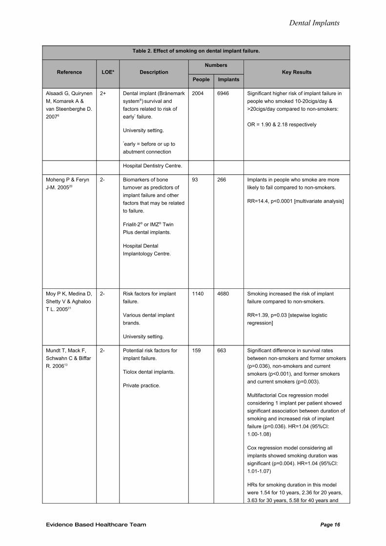
Moheng P & Feryn
J-M. 200520
2- Biomarkers of bone
turnover as predictors of
implant failure and other
factors that may be related
to failure.
Frialit-2® or IMZ® Twin
Plus dental implants.
Hospital Dental
Implantology Centre.
93 266 Implants in people who smoke are more
likely to fail compared to non-smokers.
RR=14.4, p<0.0001 [multivariate analysis]
Moy P K, Medina D,
Shetty V & Aghaloo
T L. 200521
2- Risk factors for implant
failure.
Various dental implant
brands.
University setting.
1140 4680 Smoking increased the risk of implant
failure compared to non-smokers.
RR=1.39, p=0.03 [stepwise logistic
regression]
Mundt T, Mack F,
Schwahn C & Biffar
R. 200613
2- Potential risk factors for
implant failure.
Tiolox dental implants.
Private practice.
159 663 Significant difference in survival rates
between non-smokers and former smokers
(p=0.036), non-smokers and current
smokers (p<0.001), and former smokers
and current smokers (p=0.003).
Multifactorial Cox regression model
considering 1 implant per patient showed
significant association between duration of
smoking and increased risk of implant
failure (p=0.036). HR=1.04 (95%CI:
1.00-1.08)
Cox regression model considering all
implants showed smoking duration was
significant (p=0.004). HR=1.04 (95%CI:
1.01-1.07)
HRs for smoking duration in this model
were 1.54 for 10 years, 2.36 for 20 years,
3.63 for 30 years, 5.58 for 40 years and
Evidence Based Healthcare Team Page 16
Dental Implants
Table 2. Effect of smoking on dental implant failure.
Reference LOE* DescriptionNumbers
People Implants
Key Results
Alsaadi G, Quirynen
M, Komarek A &
van Steenberghe D.
20076
2+ Dental implant (Brånemark
system®) survival and
factors related to risk of
early* failure.
University setting.
*early = before or up to
abutment connection
2004 6946 Significant higher risk of implant failure in
people who smoked 10-20cigs/day &
>20cigs/day compared to non-smokers:
OR = 1.90 & 2.18 respectively
6.92 for 45 years.
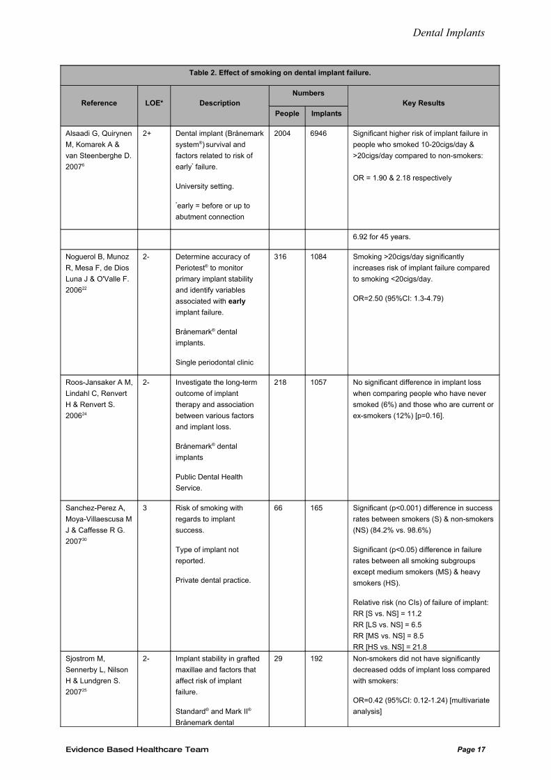
Noguerol B, Munoz
R, Mesa F, de Dios
Luna J & O'Valle F.
200622
2- Determine accuracy of
Periotest® to monitor
primary implant stability
and identify variables
associated with early
implant failure.
Brånemark® dental
implants.
Single periodontal clinic
316 1084 Smoking >20cigs/day significantly
increases risk of implant failure compared
to smoking <20cigs/day.
OR=2.50 (95%CI: 1.3-4.79)
Roos-Jansaker A M,
Lindahl C, Renvert
H & Renvert S.
200624
2- Investigate the long-term
outcome of implant
therapy and association
between various factors
and implant loss.
Brånemark® dental
implants
Public Dental Health
Service.
218 1057 No significant difference in implant loss
when comparing people who have never
smoked (6%) and those who are current or
ex-smokers (12%) [p=0.16].
Sanchez-Perez A,
Moya-Villaescusa M
J & Caffesse R G.
200730
3 Risk of smoking with
regards to implant
success.
Type of implant not
reported.
Private dental practice.
66 165 Significant (p<0.001) difference in success
rates between smokers (S) & non-smokers
(NS) (84.2% vs. 98.6%)
Significant (p<0.05) difference in failure
rates between all smoking subgroups
except medium smokers (MS) & heavy
smokers (HS).
Relative risk (no CIs) of failure of implant:
RR [S vs. NS] = 11.2
RR [LS vs. NS] = 6.5
RR [MS vs. NS] = 8.5
RR [HS vs. NS] = 21.8
Sjostrom M,
Sennerby L, Nilson
H & Lundgren S.
200725
2- Implant stability in grafted
maxillae and factors that
affect risk of implant
failure.
Standard® and Mark II®
Brånemark dental
29 192 Non-smokers did not have significantly
decreased odds of implant loss compared
with smokers:
OR=0.42 (95%CI: 0.12-1.24) [multivariate
analysis]
Evidence Based Healthcare Team Page 17
Dental Implants
Table 2. Effect of smoking on dental implant failure.
Reference LOE* DescriptionNumbers
People Implants
Key Results
Alsaadi G, Quirynen
M, Komarek A &
van Steenberghe D.
20076
2+ Dental implant (Brånemark
system®) survival and
factors related to risk of
early* failure.
University setting.
*early = before or up to
abutment connection
2004 6946 Significant higher risk of implant failure in
people who smoked 10-20cigs/day &
>20cigs/day compared to non-smokers:
OR = 1.90 & 2.18 respectively
implants.
University setting.
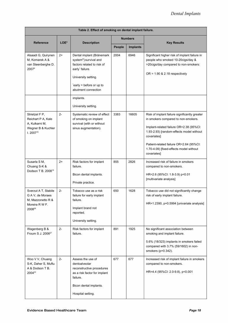
Strietzel F P,
Reichart P A, Kale
A, Kulkarni M,
Wegner B & Kuchler
I. 200716
2- Systematic review of effect
of smoking on implant
survival (with or without
sinus augmentation).
3383 16605 Risk of implant failure significantly greater
in smokers compared to non-smokers.
Implant-related failure OR=2.38 (95%CI:
1.93-2.93) [random-effects model without
covariates]
Patient-related failure OR=2.64 (95%CI:
1.70-4.09) [fixed-effects model without
covariates]
Susarla S M,
Chuang S-K &
Dodson T B. 200818
2+ Risk factors for implant
failure.
Bicon dental implants.
Private practice.
855 2826 Increased risk of failure in smokers
compared to non-smokers.
HR=2.6 (95%CI: 1.8-3.9) p<0.01
[multivariate analysis]
Sverzut A T, Stabile
G A V, de Moraes
M, Mazzonetto R &
Moreira R W F.
200826
2- Tobacco use as a risk
failure for early implant
failure.
Implant brand not
reported.
University setting.
650 1628 Tobacco use did not significantly change
risk of early implant failure.
HR=1.2390, p=0.5994 [univariate analysis]
Wagenberg B &
Froum S J. 200627
2- Risk factors for implant
failure.
891 1925 No significant association between
smoking and implant failure.
5.6% (18/323) implants in smokers failed
compared with 3.7% (59/1602) in non-
smokers (p=0.342).
Woo V V, Chuang
S-K, Daher S, Muftu
A & Dodson T B.
200423
2- Assess the use of
dentoalveolar
reconstructive procedures
as a risk factor for implant
failure.
Bicon dental implants.
Hospital setting.
677 677 Increased risk of implant failure in smokers
compared to non-smokers.
HR=4.4 (95%CI: 2.0-9.8), p<0.001
Evidence Based Healthcare Team Page 18
Dental Implants
*LOE = level of evidence
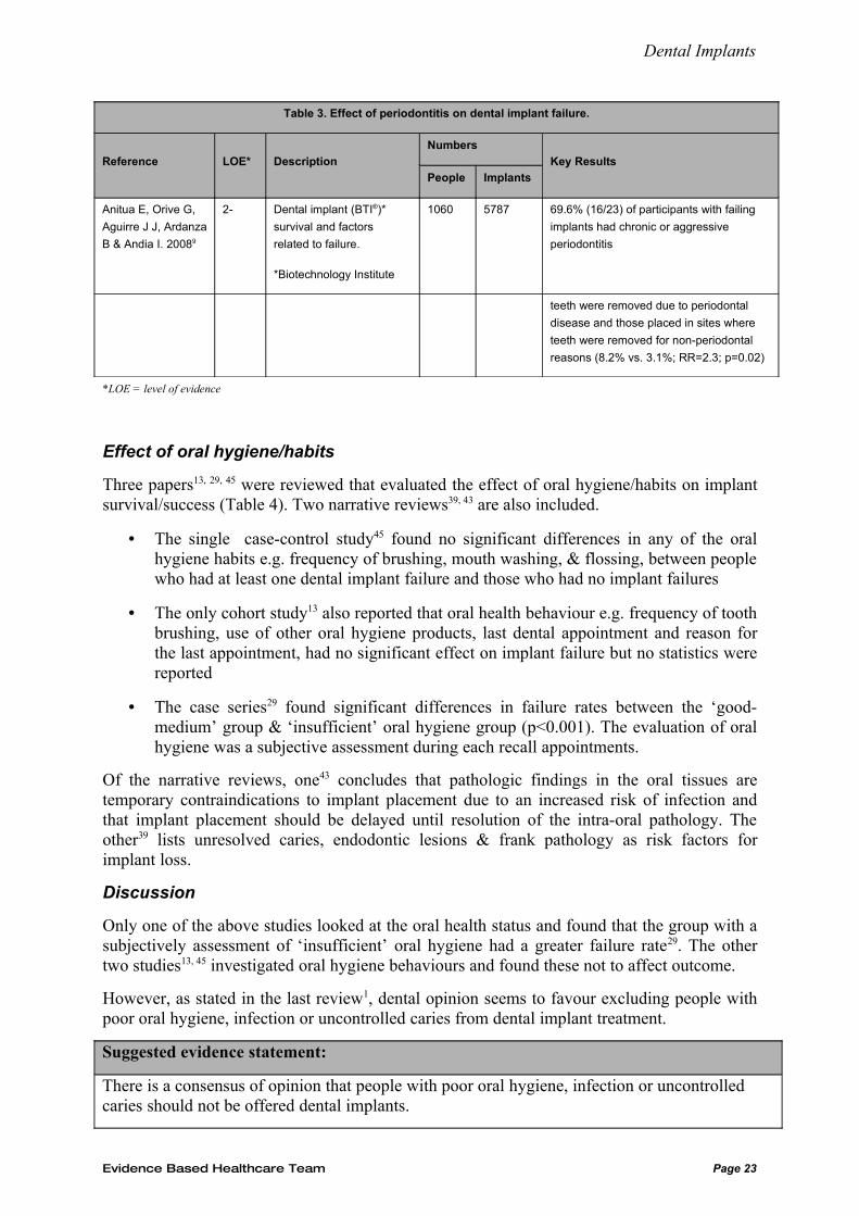
Effect of periodontitis
Nine papers9, 12, 22, 27, 45-50 were found that referred to the effect of a history of periodontitis on implant failure, including 5 systematic reviews and 4 cohort studies (Table 3).
One consensus guideline34 and 3 narrative reviews39, 42, 43 also discussed periodontal health with respect to dental implants.
Of the 5 systematic reviews:
• Karoussis (2007)47 found no statistically significant differences in both short-term and long-term implant survival in partially edentulous people with a history of chronic periodontitis compared to people without such a history. However, significantly greater long-term probing pocket depth, peri-implant marginal bone loss and incidence of peri-implantitis were found in this group of people with periodontitis
• Another review12 found that there was no significant difference in survival of implants placed in people who had a history of treated periodontitis but there was a significant difference in implant success rate (pooled difference in success rates = -11.05% (95%CI: -20.06 to -2.03%); p=0.0163) even though the pooled success rates were similar i.e. 89% vs. 89.2%
• Ong (2008)48, in a narrative analysis, reported that all studies (N=5) except one, found that implant survival was better for the non-periodontitis group but that only two studies reported a statistically significant difference. Four of five studies reporting on implant success found that success was better in the non-periodontitis groups, but only one found a statistically significant difference
• The other two systematic reviews49, 50 found no significant increase in implant failure in people with periodontitis-associated tooth loss at 5- and 10-yr follow-up. Nor was there a significant increase in suprastructure loss. However, the authors did find a significant increase in peri-implantitis & marginal bone loss (based on only one study each).
Of the 4 cohort studies:
• One study9 found that 69.6% (16/23) of participants with failing implants had chronic or aggressive periodontitis but no statistics were reported
• Another46 found that people with current manifestations or history of periodontitis had a significantly increased the risk of implant failure (RR=2.49 (95%CI: 1.03-6.02) over a mean follow-up period of almost 2yrs
• In a study22 to identify variables associated with early implant failure, a non-significant increase in risk of failure was found in people with treated periodontitis compared to those who did not have periodontitis or who were edentulous. (OR=2.36 (95%CI: 0.9-6.21))
• Another study27 found a significant difference in failure rates between implants placed in sites where teeth were removed due to periodontal disease and those placed in sites where teeth were removed for non-periodontal reasons (8.2% vs. 3.1%; RR=2.3; p=0.02).
Evidence Based Healthcare Team Page 19
Dental Implants
The consensus based guideline34 concluded that a history of treated periodontitis does not appear to adversely affect implant survival rates, but periodontitis may have a negative effect on implant success rates, particularly over longer periods. They recommended that a periodontal evaluation and appropriate treatment be provided to ensure people have the most optimal periodontal health possible42 prior to implant placement. This is supported by Wood (2004)42, who adds that preventative periodontal therapy should be maintained after implant placement.
One of the two other narrative reviews39, 43 reported that chronic periodontitis was a risk factor for implant failure39 and the other that people with a history of aggressive periodontitis had an increased risk of developing peri-implantitis43 (which can result in implant failure).
Discussion
Periodontitis is one of the common causes of tooth loss and dental implants are being increasingly used to replace missing teeth in these people48. The same pathogens responsible for periodontitis have been implicated in peri-implant infections and implant loss47. Hence, it has been suggested that a history of past periodontitis may be an adverse prognostic factor for the survival of dental implants48.
In the previous ACC dental implant review1, the recommendation, based mainly on consensus opinion, was that people with periodontitis should be excluded as candidates for dental implants.
The evidence since 2004, reviewed above, suggests that although a history of treated periodontitis appears not to be a significant risk factor for implant failure, it may significantly affect implant success, especially over longer periods of time. There is also some suggestion that periodontitis present at the time of implant placement may increase the risk of failure to a greater extent, although this is not certain.
The consensus of opinion is that periodontitis at the time of implant placement reduces implant survival and any periodontitis present should be treated first (and consequently reduce the risk of further tooth loss) before consideration of implant therapy and also to continue preventative periodontitis therapy if the decision is made to use dental implants in that person.
Suggested evidence statement:
There is insufficient evidence that people with a history of treated periodontitis have a significantly elevated risk of implant failure.
There is a consensus view that people with periodontitis must be treated for this condition before being considered for implant therapy.
There is a consensus view that preventative therapy for periodontitis should continue after the placement of implants in people with a history of periodontitis.
Evidence Based Healthcare Team Page 20
Dental Implants
Table 3. Effect of periodontitis on dental implant failure.
Reference LOE* Description
Numbers
People Implants
Key Results
Anitua E, Orive G,
Aguirre J J, Ardanza
B & Andia I. 20089
2- Dental implant (BTI®)*
survival and factors
related to failure.
*Biotechnology Institute
1060 5787 69.6% (16/23) of participants with failing
implants had chronic or aggressive
periodontitis
Evian C I, Emling R,
Rosenberg E S,
Waasdorp J A,
Halpern W, Shah S
& Garcia M. 200446
2- Dental implant survival
and effect of periodontal
disease & immediate
implant placement after
tooth extraction on long-
term survival
Private practice setting.
149 149 Global survival rate = 85.2%
Periodontitis significantly increased the risk
of implant failure (RR=2.49 (95%CI:
1.03-6.02))
Karoussis I K,
Kotsovilis S &
Fourmousis I.
200747
2- Systematic review of
prospective studies
regarding the short- &
long-term prognosis of
dental implants placed in
periodontally
compromised partially
edentulous patients.
(N=15 studies)
Short-term (7 studies) and long-term (6
studies) survival rate in people with a
history of chronic periodontitis similar to
general population.
Short-term survival in people with a history
of aggressive periodontitis was
approximately 95-100% (2 studies) & long-
term survival was 88.8% (1 study).
Klokkevold P R &
Han T J. 200712
2- Systematic review of the
effect of smoking,
periodontitis & diabetes on
implant survival &
success.
(N=13 studies)
1150 10904 Implant survival in people with a history of
treated periodontitis [95% (95%CI:
91.8-98.2%)] compared to people without
such a history [97.1% (95%CI:
94.8-99.4%)].
No significant difference in survival =
-3.14% (95%CI: -6.97 to 0.68%)
[p=0.1075].
Implant success in people with a history of
treated periodontitis [89% (95%CI:
82.3-95.7%)] compared to people without
such a history [89.2% (95%CI:
81.2-97.2%)].
Significant difference in success =
-11.05% (95%CI: -20.06 to -2.03%)
[p=0.0163].
Noguerol B, Munoz
R, Mesa F, de Dios
Luna J & O'Valle F.
200622
2- Determine accuracy of
Periotest® to monitor
primary implant stability
and identify variables
associated with early
implant failure.
Brånemark® dental
implants.
316 1084 Non-significant increase in risk of failure in
people with treated periodontitis compared
to those who were did not have
periodontitis or were edentulous.
OR=2.36 (95%CI: 0.9-6.21)
Evidence Based Healthcare Team Page 21
Dental Implants
Table 3. Effect of periodontitis on dental implant failure.
Reference LOE* Description
Numbers
People Implants
Key Results
Anitua E, Orive G,
Aguirre J J, Ardanza
B & Andia I. 20089
2- Dental implant (BTI®)*
survival and factors
related to failure.
*Biotechnology Institute
1060 5787 69.6% (16/23) of participants with failing
implants had chronic or aggressive
periodontitis
Single periodontal clinic
Ong C T T,
Ivanovski S,
Needleman I G,
Retzepi M, Moles D
R, Tonetti M S &
Donos N. 200848
2- Systematic review to
determine the effect of a
history of periodontitis on
survival & success of
dental implants in partially
dentate people.
(N=9 studies)
Four of 5 studies reported better implant
survival for the non-periodontitis group,
however only 2 studies reported a
statistically significant difference.
Four of 5 studies reported better implant
success for the non-periodontitis group,
however only one found a statistically
significant difference.
Schou S. 200849 2- Systematic review of the
outcomes of dental
implant treatment in
periodontally-susceptible
patients.
(N=23 studies)
No significant increase of risk of implant or
suprastructure failure in people with
periodontitis-associated tooth loss.
Significant increased risk of peri-implantitis
& peri-implant bone loss in people with
periodontitis-associated tooth loss.
For RRs & WMD – see Shou 2006 below
Schou S, Holmstrup
P, Worthington H V
& Esposito M.
200650
2- Systematic review of the
outcomes of dental
implant treatment in
periodontally-susceptible
patients.
(N=2 studies)
No significant increase in risk of implant
loss in people with periodontitis-associated
tooth loss at 5- & 10-yr follow-up:
RR=2.24 (0.71-7.04) at 5yr follow-up (both
studies)
RR=3.75 (0.74-19.02) at 10yr follow-up
(Hardt 2002)
No significant increase in risk of
suprastructure failure in people with
periodontitis-associated tooth loss:
RR=5.00 (95% CI: 0.25-99.16) at 5yr
follow-up (Hardt 2002)
Significant increase risk of peri-implantitis
& marginal bone loss in people with
periodontitis-associated tooth loss:
RR=9.00 (3.94-20.57) at 10yr follow-up
(Karoussis 2003)
WMD=0.50 (0.06-0.94) at 5yr follow-up
(Hardt 2002)
Wagenberg B &
Froum S J. 200627
2- Risk factors for implant
failure.
891 1925 A significant difference in failure rates
between implants placed in sites where
Evidence Based Healthcare Team Page 22
Dental Implants
Table 3. Effect of periodontitis on dental implant failure.
Reference LOE* Description
Numbers
People Implants
Key Results
Anitua E, Orive G,
Aguirre J J, Ardanza
B & Andia I. 20089
2- Dental implant (BTI®)*
survival and factors
related to failure.
*Biotechnology Institute
1060 5787 69.6% (16/23) of participants with failing
implants had chronic or aggressive
periodontitis
teeth were removed due to periodontal
disease and those placed in sites where
teeth were removed for non-periodontal
reasons (8.2% vs. 3.1%; RR=2.3; p=0.02)
*LOE = level of evidence
Effect of oral hygiene/habits
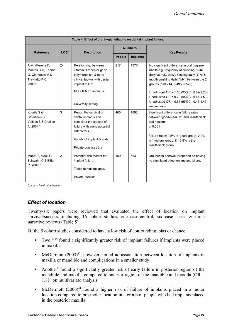
Three papers13, 29, 45 were reviewed that evaluated the effect of oral hygiene/habits on implant survival/success (Table 4). Two narrative reviews39, 43 are also included.
• The single case-control study45 found no significant differences in any of the oral hygiene habits e.g. frequency of brushing, mouth washing, & flossing, between people who had at least one dental implant failure and those who had no implant failures
• The only cohort study13 also reported that oral health behaviour e.g. frequency of tooth brushing, use of other oral hygiene products, last dental appointment and reason for the last appointment, had no significant effect on implant failure but no statistics were reported
• The case series29 found significant differences in failure rates between the ‘good-medium’ group & ‘insufficient’ oral hygiene group (p<0.001). The evaluation of oral hygiene was a subjective assessment during each recall appointments.
Of the narrative reviews, one43 concludes that pathologic findings in the oral tissues are temporary contraindications to implant placement due to an increased risk of infection and that implant placement should be delayed until resolution of the intra-oral pathology. The other39 lists unresolved caries, endodontic lesions & frank pathology as risk factors for implant loss.
Discussion
Only one of the above studies looked at the oral health status and found that the group with a subjectively assessment of ‘insufficient’ oral hygiene had a greater failure rate29. The other two studies13, 45 investigated oral hygiene behaviours and found these not to affect outcome.
However, as stated in the last review1, dental opinion seems to favour excluding people with poor oral hygiene, infection or uncontrolled caries from dental implant treatment.
Suggested evidence statement:
There is a consensus of opinion that people with poor oral hygiene, infection or uncontrolled caries should not be offered dental implants.
Evidence Based Healthcare Team Page 23
Dental Implants
Table 4. Effect of oral hygiene/habits on dental implant failure.
Reference LOE* Description
Numbers
People Implants
Key Results
Alvim-Pereira F,
Montes C C, Thome
G, Olandoski M &
Trevilatto P C.
200845
2- Relationship between
vitamin D receptor gene
polymorphism & other
clinical factors with dental
implant failure.
NEODENT™ implants
University setting.
217 1376 No significant difference in oral hygiene
habits e.g. frequency of brushing [1-3X
daily vs. >3X daily], flossing daily [Y/N] &
mouth washing daily [Y/N], between the 2
groups (p=0.744, 0.495, 0.575).
Unadjusted OR = 1.18 (95%CI: 0.62-2.26)
Unadjusted OR = 0.79 (95%CI: 0.41-1.53)
Unadjusted OR = 0.84 (95%CI: 0.48-1.46)
respectively
Kourtis S G,
Sotiriadou S,
Voliotis S & Challas
A. 200429
3 Report the survival of
dental implants and
associate the causes of
failure with some potential
risk factors.
Variety of implant brands.
Private practices (4).
405 1692 Significant difference in failure rates
between ‘good-medium’, and ‘insufficient’
oral hygiene
p<0.001
Failure rates: 2.5% in ‘good’ group, 2.9%
in ‘medium’ group, & 13.8% in the
‘insufficient’ group
Mundt T, Mack F,
Schwahn C & Biffar
R. 200613
2- Potential risk factors for
implant failure.
Tiolox dental implants.
Private practice.
159 663 Oral health behaviour reported as having
no significant effect on implant failure.
*LOE = level of evidence
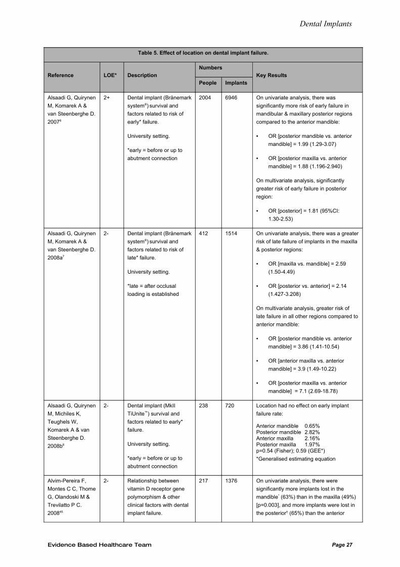
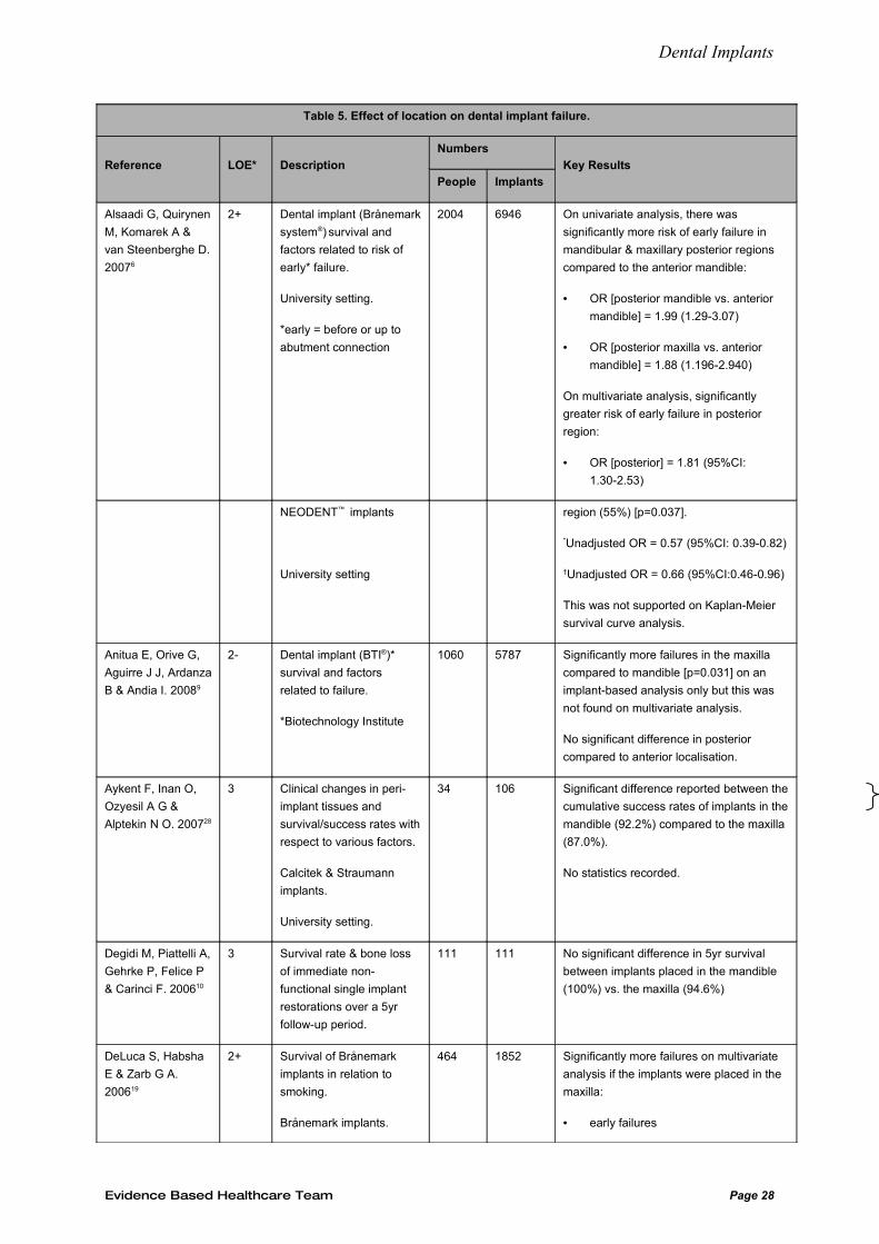
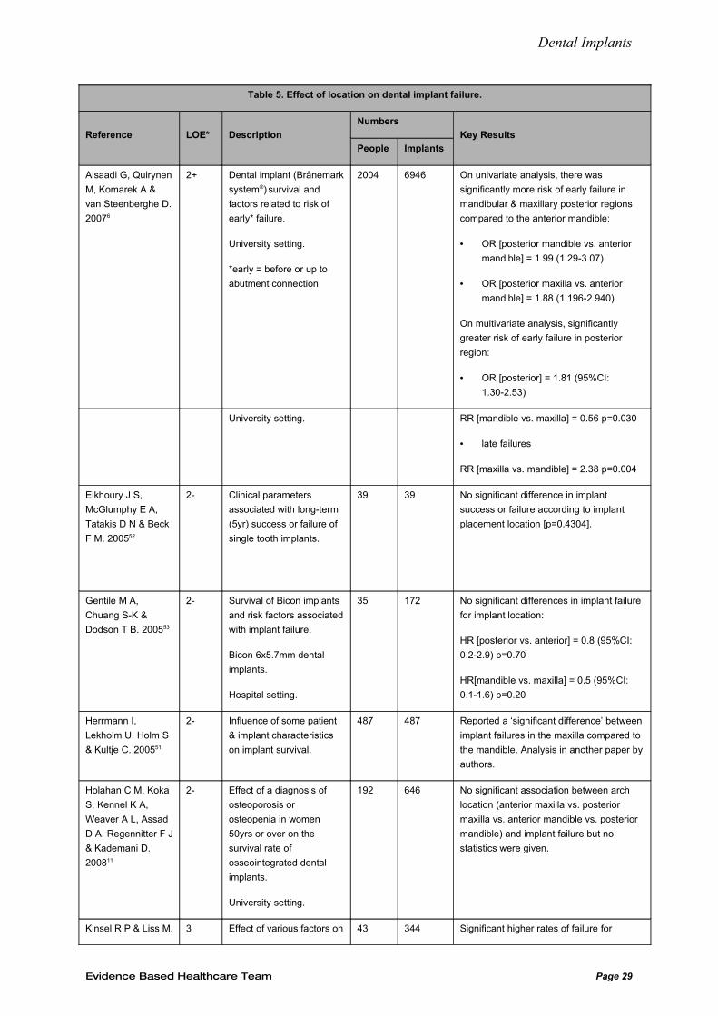
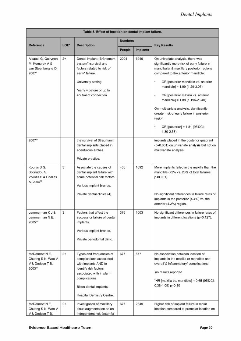
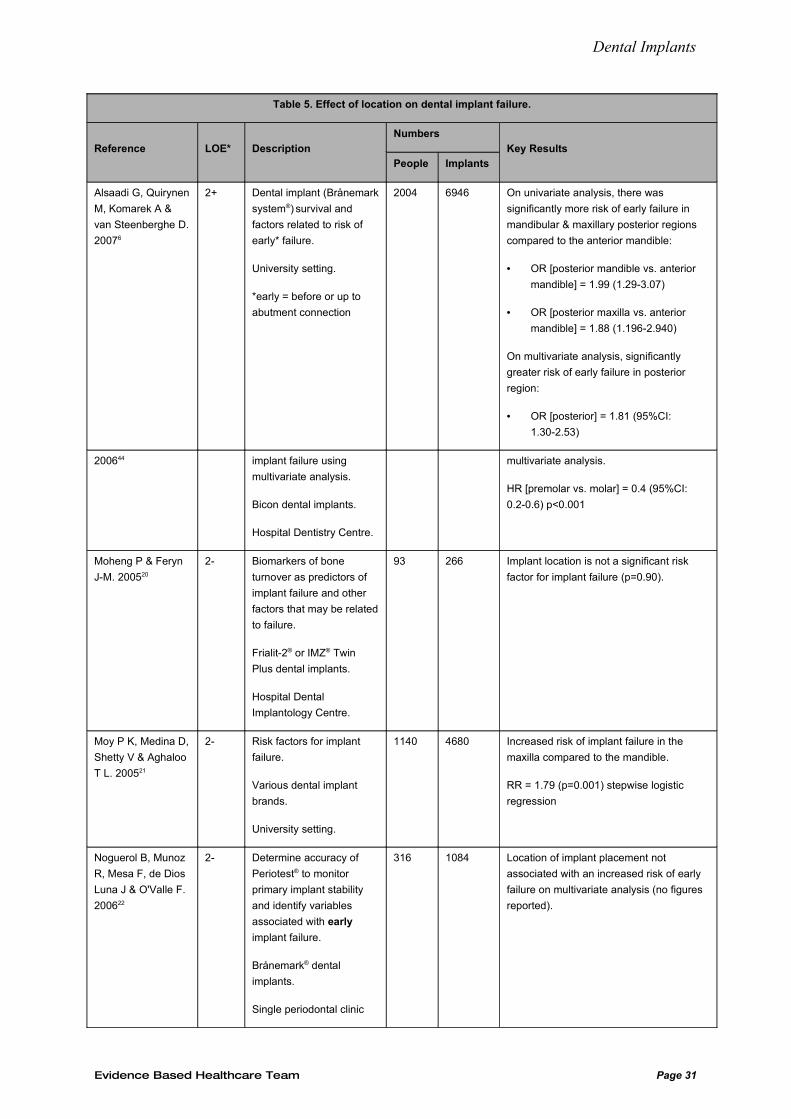
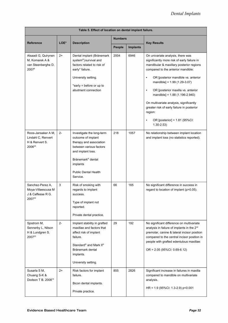
Effect of location
Twenty-six papers were reviewed that evaluated the effect of location on implant survival/success, including 16 cohort studies, one case-control, six case series & three narrative reviews (Table 5).
Of the 5 cohort studies considered to have a low risk of confounding, bias or chance,
• Two18, 19 found a significantly greater risk of implant failures if implants were placed in maxilla
• McDermott (2003)17, however, found no association between location of implants in maxilla or mandible and complications in a smaller study
• Another6 found a significantly greater risk of early failure in posterior region of the mandible and maxilla compared to anterior region of the mandible and maxilla (OR = 1.81) on multivariate analysis
• McDermott (2006)44 found a higher risk of failure of implants placed in a molar location compared to pre-molar location in a group of people who had implants placed in the posterior maxilla.
Evidence Based Healthcare Team Page 24
Dental Implants
Of the 11 cohort studies considered to have a high risk of confounding, bias or chance, 4 found a significant effect of location:
• Two21, 51 found an increased risk of implant failure in the maxilla compared to the mandible
• Alsaadi (2008a)7 found a greater risk of late failure in all regions of the maxilla and the posterior mandible compared to the anterior mandible on multivariate analysis
• Sverzet (2008)26 found that ‘Location 3’ (defined as: “maxillary anterior, maxillary posterior, mandibular anterior, and mandibular posterior) had a hazard ratio of 0.3900 (p=0.0023) on multivariate analysis but which actual location had the greater (or lesser) risk was not reported.
The remaining 7 studies8, 9, 11, 20, 22, 24, 25, 52, 53 found no significant association between the risk of failure and location.
• Three studies25, 52, 53 were of a small sample i.e. 35, 39 & 29 participants respectively
• No statistics were reported in two other studies11, 24
• One study compared a limited number of locations (anterior only) in people with a grafted edentulous maxilla.
The only case-control study45 also found that location had no effect on implant failure on multivariate analysis.
Of the case series,
• Kourtis (2004)29 found that more implants failed in the maxilla compared to the mandible but no significant differences between posterior and anterior regions was found
• A smaller study28 found a better success rate of implants placed in the mandible compared to the maxilla but no statistics were reported
• The four other case series10, 30-32 found no association between implant location and risk of failure.
Of the 3 narrative reviews,
Paquette (2006)38 reported that according to one study, the location of the implant (either maxilla vs. mandible or anterior vs. posterior) did not alter implant survival significantly. Another study in the review was reported as showing that placement in the maxilla increased the risk of implant failure on multivariate analysis.
Another54, states that implant location plays an important role in implant success and that the cumulative survival rate of implants in the mandible seem to be slightly higher than in the maxilla – about a 4% difference. The success rate of implants in the anterior region seems to be higher than in the posterior regions of the jaws, mostly due to the quality of bone: about 12% difference between the anterior maxilla and posterior maxilla, and about 4% difference between anterior mandible and posterior mandible. NB: three of the six papers used in this narrative review have been included in this or the previous dental implant review.
Evidence Based Healthcare Team Page 25
Dental Implants
The other narrative review39 states that most authors agree that mandibular implants have a greater chance for success than those placed in the maxilla. The posterior maxilla is cited in a table of predictors of implant success or failure to be particularly prone to implant failure.
Discussion
Implant location has been on of the factors identified that may influence the success or failure of the dental implant54. In the last review1, the conclusions reached were that the failure rate of implants in the edentulous maxilla may be higher than the mandible and this may be of clinical significance and a relevant patient selection factor. There was, however, inadequate evidence to conclude that implant failure was greater at posterior locations compared to anterior.
Taking this into consideration and adding the more recent evidence, there is some evidence that dental implants are at greater risk of failure when placed in the maxilla and weaker, conflicting evidence suggesting that in a posterior position implants may also have a greater risk.
The relevance of these findings is unclear. It may be that position/location per se is not a risk factor but that it may be confounded by other factors such as occlusal forces, blood supply, bone quality and quantity and/or the presence of a bone graft. A consensus opinion is needed as to any recommendation stemming from this evidence.
Suggested evidence statement:
There is weak evidence that dental implants placed in the maxilla may have a greater risk of failure
There is insufficient evidence that dental implants placed in the posterior region of a jaw have a greater risk of failure than those placed in the anterior region.
Evidence Based Healthcare Team Page 26
Dental Implants
Table 5. Effect of location on dental implant failure.
Reference LOE* Description
Numbers
People Implants
Key Results
Alsaadi G, Quirynen
M, Komarek A &
van Steenberghe D.
20076
2+ Dental implant (Brånemark
system®) survival and
factors related to risk of
early* failure.
University setting.
*early = before or up to
abutment connection
2004 6946 On univariate analysis, there was
significantly more risk of early failure in
mandibular & maxillary posterior regions
compared to the anterior mandible:
• OR [posterior mandible vs. anterior
mandible] = 1.99 (1.29-3.07)
• OR [posterior maxilla vs. anterior
mandible] = 1.88 (1.196-2.940)
On multivariate analysis, significantly
greater risk of early failure in posterior
region:
• OR [posterior] = 1.81 (95%CI:
1.30-2.53)
Alsaadi G, Quirynen
M, Komarek A &
van Steenberghe D.
2008a7
2- Dental implant (Brånemark
system®) survival and
factors related to risk of
late* failure.
University setting.
*late = after occlusal
loading is established
412 1514 On univariate analysis, there was a greater
risk of late failure of implants in the maxilla
& posterior regions:
• OR [maxilla vs. mandible] = 2.59
(1.50-4.49)
• OR [posterior vs. anterior] = 2.14
(1.427-3.208)
On multivariate analysis, greater risk of
late failure in all other regions compared to
anterior mandible:
• OR [posterior mandible vs. anterior
mandible] = 3.86 (1.41-10.54)
• OR [anterior maxilla vs. anterior
mandible] = 3.9 (1.49-10.22)
• OR [posterior maxilla vs. anterior
mandible] = 7.1 (2.69-18.78)
Alsaadi G, Quirynen
M, Michiles K,
Teughels W,
Komarek A & van
Steenberghe D.
2008b8
2- Dental implant (MkII
TiUnite™) survival and
factors related to early*
failure.
University setting.
*early = before or up to
abutment connection
238 720 Location had no effect on early implant
failure rate:
Anterior mandible 0.65% Posterior mandible 2.82%Anterior maxilla 2.16%Posterior maxilla 1.97%p=0.54 (Fisher); 0.59 (GEE*)
*Generalised estimating equation
Alvim-Pereira F,
Montes C C, Thome
G, Olandoski M &
Trevilatto P C.
200845
2- Relationship between
vitamin D receptor gene
polymorphism & other
clinical factors with dental
implant failure.
217 1376 On univariate analysis, there were
significantly more implants lost in the
mandible* (63%) than in the maxilla (49%)
[p=0.003], and more implants were lost in
the posterior† (65%) than the anterior
Evidence Based Healthcare Team Page 27
Dental Implants
Table 5. Effect of location on dental implant failure.
Reference LOE* Description
Numbers
People Implants
Key Results
Alsaadi G, Quirynen
M, Komarek A &
van Steenberghe D.
20076
2+ Dental implant (Brånemark
system®) survival and
factors related to risk of
early* failure.
University setting.
*early = before or up to
abutment connection
2004 6946 On univariate analysis, there was
significantly more risk of early failure in
mandibular & maxillary posterior regions
compared to the anterior mandible:
• OR [posterior mandible vs. anterior
mandible] = 1.99 (1.29-3.07)
• OR [posterior maxilla vs. anterior
mandible] = 1.88 (1.196-2.940)
On multivariate analysis, significantly
greater risk of early failure in posterior
region:
• OR [posterior] = 1.81 (95%CI:
1.30-2.53)
NEODENT™ implants
University setting
region (55%) [p=0.037].
*Unadjusted OR = 0.57 (95%CI: 0.39-0.82)
†Unadjusted OR = 0.66 (95%CI:0.46-0.96)
This was not supported on Kaplan-Meier
survival curve analysis.
Anitua E, Orive G,
Aguirre J J, Ardanza
B & Andia I. 20089
2- Dental implant (BTI®)*
survival and factors
related to failure.
*Biotechnology Institute
1060 5787 Significantly more failures in the maxilla
compared to mandible [p=0.031] on an
implant-based analysis only but this was
not found on multivariate analysis.
No significant difference in posterior
compared to anterior localisation.
Aykent F, Inan O,
Ozyesil A G &
Alptekin N O. 200728
3 Clinical changes in peri-
implant tissues and
survival/success rates with
respect to various factors.
Calcitek & Straumann
implants.
University setting.
34 106 Significant difference reported between the
cumulative success rates of implants in the
mandible (92.2%) compared to the maxilla
(87.0%).
No statistics recorded.
Degidi M, Piattelli A,
Gehrke P, Felice P
& Carinci F. 200610
3 Survival rate & bone loss
of immediate non-
functional single implant
restorations over a 5yr
follow-up period.
111 111 No significant difference in 5yr survival
between implants placed in the mandible
(100%) vs. the maxilla (94.6%)
DeLuca S, Habsha
E & Zarb G A.
200619
2+ Survival of Brånemark
implants in relation to
smoking.
Brånemark implants.
464 1852 Significantly more failures on multivariate
analysis if the implants were placed in the
maxilla:
• early failures
Evidence Based Healthcare Team Page 28
Dental Implants
Table 5. Effect of location on dental implant failure.
Reference LOE* Description
Numbers
People Implants
Key Results
Alsaadi G, Quirynen
M, Komarek A &
van Steenberghe D.
20076
2+ Dental implant (Brånemark
system®) survival and
factors related to risk of
early* failure.
University setting.
*early = before or up to
abutment connection
2004 6946 On univariate analysis, there was
significantly more risk of early failure in
mandibular & maxillary posterior regions
compared to the anterior mandible:
• OR [posterior mandible vs. anterior
mandible] = 1.99 (1.29-3.07)
• OR [posterior maxilla vs. anterior
mandible] = 1.88 (1.196-2.940)
On multivariate analysis, significantly
greater risk of early failure in posterior
region:
• OR [posterior] = 1.81 (95%CI:
1.30-2.53)
University setting. RR [mandible vs. maxilla] = 0.56 p=0.030
• late failures
RR [maxilla vs. mandible] = 2.38 p=0.004
Elkhoury J S,
McGlumphy E A,
Tatakis D N & Beck
F M. 200552
2- Clinical parameters
associated with long-term
(5yr) success or failure of
single tooth implants.
39 39 No significant difference in implant
success or failure according to implant
placement location [p=0.4304].
Gentile M A,
Chuang S-K &
Dodson T B. 200553
2- Survival of Bicon implants
and risk factors associated
with implant failure.
Bicon 6x5.7mm dental
implants.
Hospital setting.
35 172 No significant differences in implant failure
for implant location:
HR [posterior vs. anterior] = 0.8 (95%CI:
0.2-2.9) p=0.70
HR[mandible vs. maxilla] = 0.5 (95%CI:
0.1-1.6) p=0.20
Herrmann I,
Lekholm U, Holm S
& Kultje C. 200551
2- Influence of some patient
& implant characteristics
on implant survival.
487 487 Reported a ‘significant difference’ between
implant failures in the maxilla compared to
the mandible. Analysis in another paper by
authors.
Holahan C M, Koka
S, Kennel K A,
Weaver A L, Assad
D A, Regennitter F J
& Kademani D.
200811
2- Effect of a diagnosis of
osteoporosis or
osteopenia in women
50yrs or over on the
survival rate of
osseointegrated dental
implants.
University setting.
192 646 No significant association between arch
location (anterior maxilla vs. posterior
maxilla vs. anterior mandible vs. posterior
mandible) and implant failure but no
statistics were given.
Kinsel R P & Liss M. 3 Effect of various factors on 43 344 Significant higher rates of failure for
Evidence Based Healthcare Team Page 29
Dental Implants
Table 5. Effect of location on dental implant failure.
Reference LOE* Description
Numbers
People Implants
Key Results
Alsaadi G, Quirynen
M, Komarek A &
van Steenberghe D.
20076
2+ Dental implant (Brånemark
system®) survival and
factors related to risk of
early* failure.
University setting.
*early = before or up to
abutment connection
2004 6946 On univariate analysis, there was
significantly more risk of early failure in
mandibular & maxillary posterior regions
compared to the anterior mandible:
• OR [posterior mandible vs. anterior
mandible] = 1.99 (1.29-3.07)
• OR [posterior maxilla vs. anterior
mandible] = 1.88 (1.196-2.940)
On multivariate analysis, significantly
greater risk of early failure in posterior
region:
• OR [posterior] = 1.81 (95%CI:
1.30-2.53)
200731 the survival of Straumann
dental implants placed in
edentulous arches.
Private practice.
implants placed in the posterior quadrant
(p=0.001) on univariate analysis but not on
multivariate analysis.
Kourtis S G,
Sotiriadou S,
Voliotis S & Challas
A. 200429
3 Associate the causes of
dental implant failure with
some potential risk factors.
Various implant brands.
Private dental clinics (4).
405 1692 More implants failed in the maxilla than the
mandible (72% vs. 28% of total failures;
p<0.001).
No significant differences in failure rates of
implants in the posterior (4.4%) vs. the
anterior (4.2%) region.
Lemmerman K J &
Lemmerman N E.
200532
3 Factors that affect the
success or failure of dental
implants.
Various implant brands.
Private periodontal clinic.
376 1003 No significant differences in failure rates of
implants in different locations (p=0.127).
McDermott N E,
Chuang S-K, Woo V
V & Dodson T B.
200317
2+ Types and frequencies of
complications associated
with implants AND to
identify risk factors
associated with implant
complications.
Bicon dental implants.
Hospital Dentistry Centre.
677 677 No association between location of
implants in the maxilla or mandible and
overall* & inflammatory† complications.
*no results reported
†HR [maxilla vs. mandible] = 0.65 (95%CI:
0.38-1.09) p=0.10
McDermott N E,
Chuang S-K, Woo V
V & Dodson T B.
2+ Investigation of maxillary
sinus augmentation as an
independent risk factor for
677 2349 Higher risk of implant failure in molar
location compared to premolar location on
Evidence Based Healthcare Team Page 30
Dental Implants
Table 5. Effect of location on dental implant failure.
Reference LOE* Description
Numbers
People Implants
Key Results
Alsaadi G, Quirynen
M, Komarek A &
van Steenberghe D.
20076
2+ Dental implant (Brånemark
system®) survival and
factors related to risk of
early* failure.
University setting.
*early = before or up to
abutment connection
2004 6946 On univariate analysis, there was
significantly more risk of early failure in
mandibular & maxillary posterior regions
compared to the anterior mandible:
• OR [posterior mandible vs. anterior
mandible] = 1.99 (1.29-3.07)
• OR [posterior maxilla vs. anterior
mandible] = 1.88 (1.196-2.940)
On multivariate analysis, significantly
greater risk of early failure in posterior
region:
• OR [posterior] = 1.81 (95%CI:
1.30-2.53)
200644 implant failure using
multivariate analysis.
Bicon dental implants.
Hospital Dentistry Centre.
multivariate analysis.
HR [premolar vs. molar] = 0.4 (95%CI:
0.2-0.6) p<0.001
Moheng P & Feryn
J-M. 200520
2- Biomarkers of bone
turnover as predictors of
implant failure and other
factors that may be related
to failure.
Frialit-2® or IMZ® Twin
Plus dental implants.
Hospital Dental
Implantology Centre.
93 266 Implant location is not a significant risk
factor for implant failure (p=0.90).
Moy P K, Medina D,
Shetty V & Aghaloo
T L. 200521
2- Risk factors for implant
failure.
Various dental implant
brands.
University setting.
1140 4680 Increased risk of implant failure in the
maxilla compared to the mandible.
RR = 1.79 (p=0.001) stepwise logistic
regression
Noguerol B, Munoz
R, Mesa F, de Dios
Luna J & O'Valle F.
200622
2- Determine accuracy of
Periotest® to monitor
primary implant stability
and identify variables
associated with early
implant failure.
Brånemark® dental
implants.
Single periodontal clinic
316 1084 Location of implant placement not
associated with an increased risk of early
failure on multivariate analysis (no figures
reported).
Evidence Based Healthcare Team Page 31
Dental Implants
Table 5. Effect of location on dental implant failure.
Reference LOE* Description
Numbers
People Implants
Key Results
Alsaadi G, Quirynen
M, Komarek A &
van Steenberghe D.
20076
2+ Dental implant (Brånemark
system®) survival and
factors related to risk of
early* failure.
University setting.
*early = before or up to
abutment connection
2004 6946 On univariate analysis, there was
significantly more risk of early failure in
mandibular & maxillary posterior regions
compared to the anterior mandible:
• OR [posterior mandible vs. anterior
mandible] = 1.99 (1.29-3.07)
• OR [posterior maxilla vs. anterior
mandible] = 1.88 (1.196-2.940)
On multivariate analysis, significantly
greater risk of early failure in posterior
region:
• OR [posterior] = 1.81 (95%CI:
1.30-2.53)
Roos-Jansaker A M,
Lindahl C, Renvert
H & Renvert S.
200624
2- Investigate the long-term
outcome of implant
therapy and association
between various factors
and implant loss.
Brånemark® dental
implants
Public Dental Health
Service.
218 1057 No relationship between implant location
and implant loss (no statistics reported).
Sanchez-Perez A,
Moya-Villaescusa M
J & Caffesse R G.
200730
3 Risk of smoking with
regards to implant
success.
Type of implant not
reported.
Private dental practice.
66 165 No significant difference in success in
regard to location of implant (p>0.05).
Sjostrom M,
Sennerby L, Nilson
H & Lundgren S.
200725
2- Implant stability in grafted
maxillae and factors that
affect risk of implant
failure.
Standard® and Mark II®
Brånemark dental
implants.
University setting.
29 192 No significant difference on multivariate
analysis in failure of implants in the 2nd
premolar, canine & lateral incisor position
compared to the central incisor position in
people with grafted edentulous maxillae:
OR = 2.05 (95%CI: 0.69-6.12)
Susarla S M,
Chuang S-K &
Dodson T B. 200818
2+ Risk factors for implant
failure.
Bicon dental implants.
Private practice.
855 2826 Significant increase in failures in maxilla
compared to mandible on multivariate
analysis.
HR = 1.9 (95%CI: 1.3-2.9) p<0.001
Evidence Based Healthcare Team Page 32
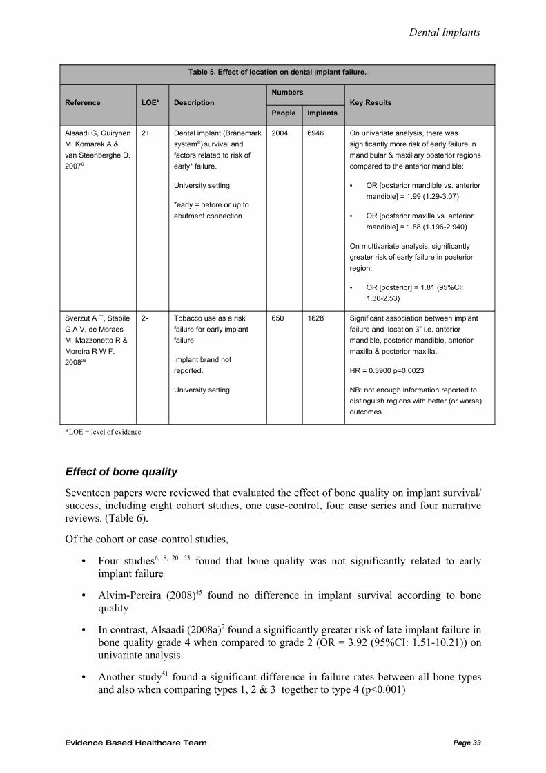
Dental Implants
Table 5. Effect of location on dental implant failure.
Reference LOE* Description
Numbers
People Implants
Key Results
Alsaadi G, Quirynen
M, Komarek A &
van Steenberghe D.
20076
2+ Dental implant (Brånemark
system®) survival and
factors related to risk of
early* failure.
University setting.
*early = before or up to
abutment connection
2004 6946 On univariate analysis, there was
significantly more risk of early failure in
mandibular & maxillary posterior regions
compared to the anterior mandible:
• OR [posterior mandible vs. anterior
mandible] = 1.99 (1.29-3.07)
• OR [posterior maxilla vs. anterior
mandible] = 1.88 (1.196-2.940)
On multivariate analysis, significantly
greater risk of early failure in posterior
region:
• OR [posterior] = 1.81 (95%CI:
1.30-2.53)
Sverzut A T, Stabile
G A V, de Moraes
M, Mazzonetto R &
Moreira R W F.
200826
2- Tobacco use as a risk
failure for early implant
failure.
Implant brand not
reported.
University setting.
650 1628 Significant association between implant
failure and ‘location 3” i.e. anterior
mandible, posterior mandible, anterior
maxilla & posterior maxilla.
HR = 0.3900 p=0.0023
NB: not enough information reported to
distinguish regions with better (or worse)
outcomes.
*LOE = level of evidence
Effect of bone quality
Seventeen papers were reviewed that evaluated the effect of bone quality on implant survival/success, including eight cohort studies, one case-control, four case series and four narrative reviews. (Table 6).
Of the cohort or case-control studies,
• Four studies6, 8, 20, 53 found that bone quality was not significantly related to early implant failure
• Alvim-Pereira (2008)45 found no difference in implant survival according to bone quality
• In contrast, Alsaadi (2008a)7 found a significantly greater risk of late implant failure in bone quality grade 4 when compared to grade 2 (OR = 3.92 (95%CI: 1.51-10.21)) on univariate analysis
• Another study51 found a significant difference in failure rates between all bone types and also when comparing types 1, 2 & 3 together to type 4 (p<0.001)
Evidence Based Healthcare Team Page 33
Dental Implants
• Whereas Noguerol (2006)22 found a greater risk of early implant failure in bone types 1, 3 & 4 together compared to type 2 (OR=1.93)
• And Elkhoury (2005)52 found that the mean bone quality in people with a failed implant significantly different from mean bone quality in those with a successful implant (2.90 vs. 1.79 respectively; p<0.001).
Of the 4 case series,
• One10 found a significant difference in 5yr survival between type 1 (100%) and type 4 (95.5%) bone (p<0.05)
• Another29 found a significantly higher failure rate in type 4 bone compared to the other types (p<0.001)
• However, Lemmerman (2005)32 and Sanchez-Perez (2007)30 found no significant difference in failure rates and success rates respectively according to bone type.
Of the narrative reviews:
There appears to be a consensus that good bone quality (and adequate bone quantity) are essential for implant success39, 42 and that poor bone quality (type 4) is a significant determinant for implant failure38, 39, 41.
Discussion
As stated in the last review1, the evidence for bone quality affecting implant success is not strong. This is perhaps not surprising when you consider that often people with poor bone quality are not included in a study30, the effect of bone quality on implant failure is usually a secondary goal, and there appears to be a complex relationship between factors; for example, bone quantity, bone quality, type of edentulism, & smoking. Often studies do not have sufficient power to detect differences in success/failure according to bone quality7 and can use different criteria or combinations of criteria for quality, making it difficult to compare studies.
The more recent evidence is conflicting, with seven of the thirteen observational studies finding no significant association between bone quality and failure. Even amongst the other studies that did find a significant association the results are conflicting i.e. more failures in type 4 compared to type 27; more failures in type 4 compared to types 1, 2 & 3 combined51; more failures in types 1, 3 & 4 combined compared to type 222.
However, there is a strong consensus from the narrative reviews that bone quality is an important determinant for implant success.
Suggested evidence statement:
There is conflicting evidence that bone quality has a significant effect on the risk of implant failure.
Evidence Based Healthcare Team Page 34
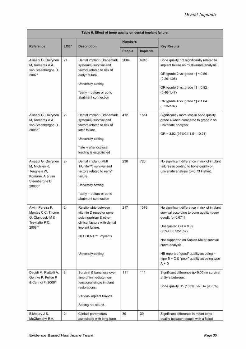
Dental Implants
Table 6. Effect of bone quality on dental implant failure.
Reference LOE* Description
Numbers
People Implants
Key Results
Alsaadi G, Quirynen
M, Komarek A &
van Steenberghe D.
20076
2+ Dental implant (Brånemark
system®) survival and
factors related to risk of
early* failure.
University setting.
*early = before or up to
abutment connection
2004 6946 Bone quality not significantly related to
implant failure on multivariate analysis:
OR [grade 2 vs. grade 1] = 0.56
(0.29-1.05)
OR [grade 3 vs. grade 1] = 0.82
(0.46-1.47)
OR [grade 4 vs. grade 1] = 1.04
(0.53-2.07)
Alsaadi G, Quirynen
M, Komarek A &
van Steenberghe D.
2008a7
2- Dental implant (Brånemark
system®) survival and
factors related to risk of
late* failure.
University setting.
*late = after occlusal
loading is established
412 1514 Significantly more loss in bone quality
grade 4 when compared to grade 2 on
univariate analysis:
OR = 3.92 (95%CI: 1.51-10.21)
Alsaadi G, Quirynen
M, Michiles K,
Teughels W,
Komarek A & van
Steenberghe D.
2008b8
2- Dental implant (MkII
TiUnite™) survival and
factors related to early*
failure.
University setting.
*early = before or up to
abutment connection
238 720 No significant difference in risk of implant
failures according to bone quality on
univariate analysis (p=0.73 Fisher).
Alvim-Pereira F,
Montes C C, Thome
G, Olandoski M &
Trevilatto P C.
200845
2- Relationship between
vitamin D receptor gene
polymorphism & other
clinical factors with dental
implant failure.
NEODENT™ implants
University setting
217 1376 No significant difference in risk of implant
survival according to bone quality (poor/
good). [p=0.671]
Unadjusted OR = 0.89
(95%CI:0.52-1.52)
Not supported on Kaplan-Meier survival
curve analysis.
NB reported “good” quality as being =
type B + C & “poor” quality as being type
A + D
Degidi M, Piattelli A,
Gehrke P, Felice P
& Carinci F. 200610
3 Survival & bone loss over
time of immediate non-
functional single implant
restorations.
Various implant brands
Setting not stated.
111 111 Significant difference (p<0.05) in survival
at 5yrs between:
Bone quality D1 (100%) vs. D4 (95.5%)
Elkhoury J S,
McGlumphy E A,
2- Clinical parameters
associated with long-term
39 39 Significant difference in mean bone
quality between people with a failed
Evidence Based Healthcare Team Page 35
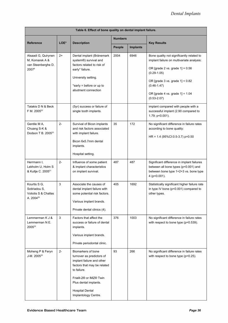
Dental Implants
Table 6. Effect of bone quality on dental implant failure.
Reference LOE* Description
Numbers
People Implants
Key Results
Alsaadi G, Quirynen
M, Komarek A &
van Steenberghe D.
20076
2+ Dental implant (Brånemark
system®) survival and
factors related to risk of
early* failure.
University setting.
*early = before or up to
abutment connection
2004 6946 Bone quality not significantly related to
implant failure on multivariate analysis:
OR [grade 2 vs. grade 1] = 0.56
(0.29-1.05)
OR [grade 3 vs. grade 1] = 0.82
(0.46-1.47)
OR [grade 4 vs. grade 1] = 1.04
(0.53-2.07)
Tatakis D N & Beck
F M. 200552
(5yr) success or failure of
single tooth implants.
implant compared with people with a
successful implant (2.90 compared to
1.79; p<0.001).
Gentile M A,
Chuang S-K &
Dodson T B. 200553
2- Survival of Bicon implants
and risk factors associated
with implant failure.
Bicon 6x5.7mm dental
implants.
Hospital setting.
35 172 No significant difference in failure rates
according to bone quality:
HR = 1.4 (95%CI:0.5-3.7) p=0.50
Herrmann I,
Lekholm U, Holm S
& Kultje C. 200551
2- Influence of some patient
& implant characteristics
on implant survival.
487 487 Significant difference in implant failures
between all bone types (p<0.001) and
between bone type 1+2+3 vs. bone type
4 (p<0.001).
Kourtis S G,
Sotiriadou S,
Voliotis S & Challas
A. 200429
3 Associate the causes of
dental implant failure with
some potential risk factors.
Various implant brands.
Private dental clinics (4).
405 1692 Statistically significant higher failure rate
in type IV bone (p<0.001) compared to
other types.
Lemmerman K J &
Lemmerman N E.
200532
3 Factors that affect the
success or failure of dental
implants.
Various implant brands.
Private periodontal clinic.
376 1003 No significant difference in failure rates
with respect to bone type (p=0.539).
Moheng P & Feryn
J-M. 200520
2- Biomarkers of bone
turnover as predictors of
implant failure and other
factors that may be related
to failure.
Frialit-2® or IMZ® Twin
Plus dental implants.
Hospital Dental
Implantology Centre.
93 266 No significant difference in failure rates
with respect to bone type (p=0.25).
Evidence Based Healthcare Team Page 36
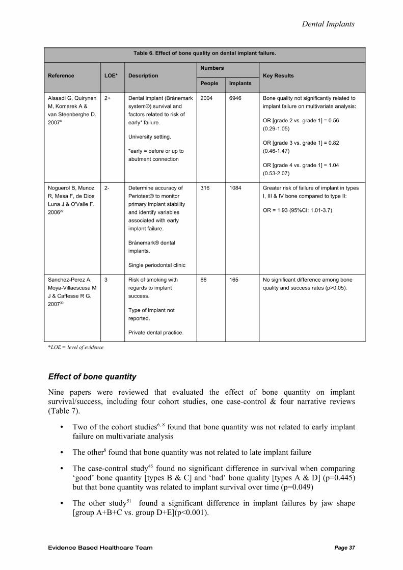
Dental Implants
Table 6. Effect of bone quality on dental implant failure.
Reference LOE* Description
Numbers
People Implants
Key Results
Alsaadi G, Quirynen
M, Komarek A &
van Steenberghe D.
20076
2+ Dental implant (Brånemark
system®) survival and
factors related to risk of
early* failure.
University setting.
*early = before or up to
abutment connection
2004 6946 Bone quality not significantly related to
implant failure on multivariate analysis:
OR [grade 2 vs. grade 1] = 0.56
(0.29-1.05)
OR [grade 3 vs. grade 1] = 0.82
(0.46-1.47)
OR [grade 4 vs. grade 1] = 1.04
(0.53-2.07)
Noguerol B, Munoz
R, Mesa F, de Dios
Luna J & O'Valle F.
200622
2- Determine accuracy of
Periotest® to monitor
primary implant stability
and identify variables
associated with early
implant failure.
Brånemark® dental
implants.
Single periodontal clinic
316 1084 Greater risk of failure of implant in types
I, III & IV bone compared to type II:
OR = 1.93 (95%CI: 1.01-3.7)
Sanchez-Perez A,
Moya-Villaescusa M
J & Caffesse R G.
200730
3 Risk of smoking with
regards to implant
success.
Type of implant not
reported.
Private dental practice.
66 165 No significant difference among bone
quality and success rates (p>0.05).
*LOE = level of evidence
Effect of bone quantity
Nine papers were reviewed that evaluated the effect of bone quantity on implant survival/success, including four cohort studies, one case-control & four narrative reviews (Table 7).
• Two of the cohort studies6, 8 found that bone quantity was not related to early implant failure on multivariate analysis
• The other8 found that bone quantity was not related to late implant failure
• The case-control study45 found no significant difference in survival when comparing ‘good’ bone quantity [types B & C] and ‘bad’ bone quality [types A & D] (p=0.445) but that bone quantity was related to implant survival over time (p=0.049)
• The other study51 found a significant difference in implant failures by jaw shape [group A+B+C vs. group D+E](p<0.001).
Evidence Based Healthcare Team Page 37
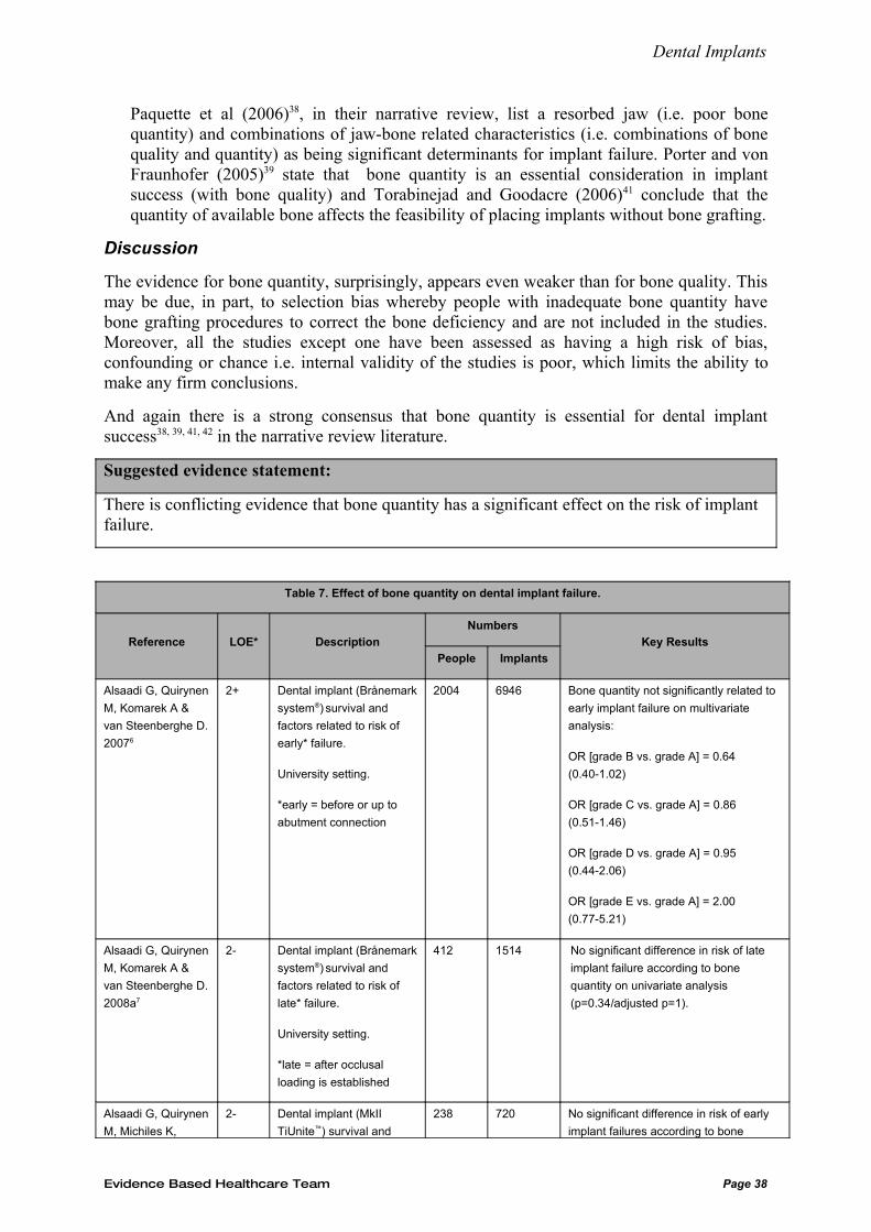
Dental Implants
Paquette et al (2006)38, in their narrative review, list a resorbed jaw (i.e. poor bone quantity) and combinations of jaw-bone related characteristics (i.e. combinations of bone quality and quantity) as being significant determinants for implant failure. Porter and von Fraunhofer (2005)39 state that bone quantity is an essential consideration in implant success (with bone quality) and Torabinejad and Goodacre (2006)41 conclude that the quantity of available bone affects the feasibility of placing implants without bone grafting.
Discussion
The evidence for bone quantity, surprisingly, appears even weaker than for bone quality. This may be due, in part, to selection bias whereby people with inadequate bone quantity have bone grafting procedures to correct the bone deficiency and are not included in the studies. Moreover, all the studies except one have been assessed as having a high risk of bias, confounding or chance i.e. internal validity of the studies is poor, which limits the ability to make any firm conclusions.
And again there is a strong consensus that bone quantity is essential for dental implant success38, 39, 41, 42 in the narrative review literature.
Suggested evidence statement:
There is conflicting evidence that bone quantity has a significant effect on the risk of implant failure.
Table 7. Effect of bone quantity on dental implant failure.
Reference LOE* Description
Numbers
People Implants
Key Results
Alsaadi G, Quirynen
M, Komarek A &
van Steenberghe D.
20076
2+ Dental implant (Brånemark
system®) survival and
factors related to risk of
early* failure.
University setting.
*early = before or up to
abutment connection
2004 6946 Bone quantity not significantly related to
early implant failure on multivariate
analysis:
OR [grade B vs. grade A] = 0.64
(0.40-1.02)
OR [grade C vs. grade A] = 0.86
(0.51-1.46)
OR [grade D vs. grade A] = 0.95
(0.44-2.06)
OR [grade E vs. grade A] = 2.00
(0.77-5.21)
Alsaadi G, Quirynen
M, Komarek A &
van Steenberghe D.
2008a7
2- Dental implant (Brånemark
system®) survival and
factors related to risk of
late* failure.
University setting.
*late = after occlusal
loading is established
412 1514 No significant difference in risk of late
implant failure according to bone
quantity on univariate analysis
(p=0.34/adjusted p=1).
Alsaadi G, Quirynen
M, Michiles K,
2- Dental implant (MkII
TiUnite™) survival and
238 720 No significant difference in risk of early
implant failures according to bone
Evidence Based Healthcare Team Page 38
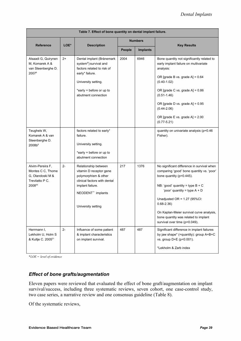
Dental Implants
Table 7. Effect of bone quantity on dental implant failure.
Reference LOE* Description
Numbers
People Implants
Key Results
Alsaadi G, Quirynen
M, Komarek A &
van Steenberghe D.
20076
2+ Dental implant (Brånemark
system®) survival and
factors related to risk of
early* failure.
University setting.
*early = before or up to
abutment connection
2004 6946 Bone quantity not significantly related to
early implant failure on multivariate
analysis:
OR [grade B vs. grade A] = 0.64
(0.40-1.02)
OR [grade C vs. grade A] = 0.86
(0.51-1.46)
OR [grade D vs. grade A] = 0.95
(0.44-2.06)
OR [grade E vs. grade A] = 2.00
(0.77-5.21)
Teughels W,
Komarek A & van
Steenberghe D.
2008b8
factors related to early*
failure.
University setting.
*early = before or up to
abutment connection
quantity on univariate analysis (p=0.46
Fisher).
Alvim-Pereira F,
Montes C C, Thome
G, Olandoski M &
Trevilatto P C.
200845
2- Relationship between
vitamin D receptor gene
polymorphism & other
clinical factors with dental
implant failure.
NEODENT™ implants
University setting
217 1376 No significant difference in survival when
comparing ‘good’ bone quantity vs. ‘poor’
bone quantity (p=0.445).
NB: ‘good’ quantity = type B + C
‘poor’ quantity = type A + D
Unadjusted OR = 1.27 (95%CI:
0.68-2.36)
On Kaplan-Meier survival curve analysis,
bone quantity was related to implant
survival over time (p=0.049).
Herrmann I,
Lekholm U, Holm S
& Kultje C. 200551
2- Influence of some patient
& implant characteristics
on implant survival.
487 487 Significant difference in implant failures
by jaw shape* (=quantity): group A+B+C
vs. group D+E (p<0.001).
*Lekholm & Zarb index
*LOE = level of evidence
Effect of bone grafts/augmentation
Eleven papers were reviewed that evaluated the effect of bone graft/augmentation on implant survival/success, including three systematic reviews, seven cohort, one case-control study, two case series, a narrative review and one consensus guideline (Table 8).
Of the systematic reviews,
Evidence Based Healthcare Team Page 39

Dental Implants
• One3 found no significant difference in implant or prosthesis failure rates or major complication rates after bone augmentation compared to no augmentation [based on 2 studies]
• Whereas Strietzel (2007)16 reported that the odds ratio was 2.15 for smokers without sinus floor augmentation procedures and 3.16 for smokers with augmentation which were significantly different (p=0.039) suggesting that augmentation procedure is a risk factor for implant loss
• Graziani (2004)55 stated that the results suggest that the range of survival rates is greater in those who had sinus floor augmentation (36-100%) than those who did not (73-100%) on a per patient basis [2 studies] and that survival varied between 75 and 100% for both augmented and non-augmented sites on a per implant basis [6 studies].
Of the observational studies,
• Four studies23, 44, 45, 53 found no significant difference in failure between those who had a bone graft and those who didn’t
• One17 found that the use of any reconstructive procedure significantly increased the risk of overall and inflammatory complication (HR = 1.18 & 1.17 respectively)
• Another26 found a greater risk of failure when an alveolar distraction technique was used (HR = 7.1).
The 2 case series31, 32 found that a bone graft procedure had no effect on outcome.
In their narrative review, Porter (2005)39 states that the literature suggests that the osseointegration of dental implants can be affected by bone grafting and that if the reason for inadequate bone is due to periodontitis, infection or osteoporosis, it is possible that these conditions will affect the successful integration of the graft. They report that the success rate for implants placed in grafted bone has ranged from 77-85% compared to a success rate of 95% or more for implants placed in mature ungrafted bone.
The Academy of Osseointegration guideline (2008)34 reported that maxillary sinus augmentation had been well documented and implants survival/success compares favourably to conventionally placed implants. Other bone augmentation techniques may not have as much evidence but the studies that met their inclusion criteria “were comparable and yielded favourable results supporting dental implants.”
Discussion
Various bone grafting procedures and materials are used to ensure adequate bone quantity and quality for successful dental implant placement34.
The previous review1 concluded that it was likely that implants placed after maxillary sinus augmentation have a clinically significantly greater failure rate.
The newer evidence, however, is equivocal and when combined with the previous review’s evidence weakens the conclusion that implants placed after maxillary sinus augmentation have a clinically significant risk of failure. The evidence for other augmentation techniques is too sparse to make any firm conclusion.
In addition, the success of implants placed in grafted bone will likely to be influenced by the magnitude of the graft, the type of implant material and the location of the implant. Other potential factors include the experience of the surgeon and the surgical technique chosen1.
Evidence Based Healthcare Team Page 40
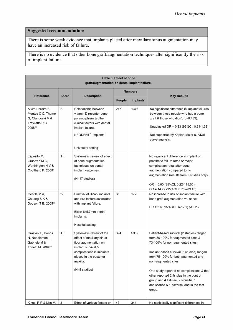
Dental Implants
Suggested recommendation:
There is some weak evidence that implants placed after maxillary sinus augmentation may have an increased risk of failure.
There is no evidence that other bone graft/augmentation techniques alter significantly the risk of implant failure.
Table 8. Effect of bone
graft/augmentation on dental implant failure.
Reference LOE* Description
Numbers
People Implants
Key Results
Alvim-Pereira F,
Montes C C, Thome
G, Olandoski M &
Trevilatto P C.
200845
2- Relationship between
vitamin D receptor gene
polymorphism & other
clinical factors with dental
implant failure.
NEODENT™ implants
University setting
217 1376 No significant difference in implant failures
between those people who had a bone
graft & those who didn’t (p=0.433).
Unadjusted OR = 0.83 (95%CI: 0.51-1.33)
Not supported by Kaplan-Meier survival
curve analysis.
Esposito M,
Grusovin M G,
Worthington H V &
Coulthard P. 20083
1+ Systematic review of effect
of bone augmentation
techniques on dental
implant outcomes.
(N=17 studies)
No significant difference in implant or
prosthetic failure rates or major
complication rates after bone
augmentation compared to no
augmentation (results from 2 studies only).
OR = 5.00 (95%CI: 0.22-115.05)
OR = 14.79 (95%CI: 0.76-289.43)
Gentile M A,
Chuang S-K &
Dodson T B. 200553
2- Survival of Bicon implants
and risk factors associated
with implant failure.
Bicon 6x5.7mm dental
implants.
Hospital setting.
35 172 No increase in risk of implant failure with
bone graft augmentation vs. none:
HR = 2.6 995%CI: 0.6-12.1) p=0.23
Graziani F, Donos
N, Needleman I,
Gabriele M &
Tonetti M. 200455
1+ Systematic review of the
effect of maxillary sinus
floor augmentation on
implant survival &
complications in implants
placed in the posterior
maxilla.
(N=5 studies)
394 >989 Patient-based survival (2 studies) ranged
from 36-100% for augmented sites &
73-100% for non-augmented sites
Implant-based survival (6 studies) ranged
from 75-100% for both augmented and
non-augmented sites
One study reported no complications & the
other reported 2 fistulae in the control
group and 4 fistulae, 2 sinusitis, 1
dehiscence & 1 adverse load in the test
group.
Kinsel R P & Liss M. 3 Effect of various factors on 43 344 No statistically significant differences in
Evidence Based Healthcare Team Page 41
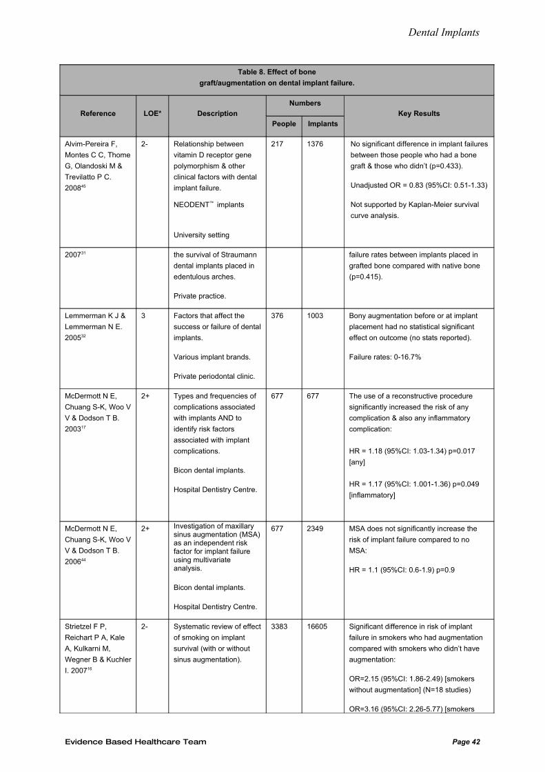
Dental Implants
Table 8. Effect of bone
graft/augmentation on dental implant failure.
Reference LOE* Description
Numbers
People Implants
Key Results
Alvim-Pereira F,
Montes C C, Thome
G, Olandoski M &
Trevilatto P C.
200845
2- Relationship between
vitamin D receptor gene
polymorphism & other
clinical factors with dental
implant failure.
NEODENT™ implants
University setting
217 1376 No significant difference in implant failures
between those people who had a bone
graft & those who didn’t (p=0.433).
Unadjusted OR = 0.83 (95%CI: 0.51-1.33)
Not supported by Kaplan-Meier survival
curve analysis.
200731 the survival of Straumann
dental implants placed in
edentulous arches.
Private practice.
failure rates between implants placed in
grafted bone compared with native bone
(p=0.415).
Lemmerman K J &
Lemmerman N E.
200532
3 Factors that affect the
success or failure of dental
implants.
Various implant brands.
Private periodontal clinic.
376 1003 Bony augmentation before or at implant
placement had no statistical significant
effect on outcome (no stats reported).
Failure rates: 0-16.7%
McDermott N E,
Chuang S-K, Woo V
V & Dodson T B.
200317
2+ Types and frequencies of
complications associated
with implants AND to
identify risk factors
associated with implant
complications.
Bicon dental implants.
Hospital Dentistry Centre.
677 677 The use of a reconstructive procedure
significantly increased the risk of any
complication & also any inflammatory
complication:
HR = 1.18 (95%CI: 1.03-1.34) p=0.017
[any]
HR = 1.17 (95%CI: 1.001-1.36) p=0.049
[inflammatory]
McDermott N E,
Chuang S-K, Woo V
V & Dodson T B.
200644
2+ Investigation of maxillary sinus augmentation (MSA) as an independent risk factor for implant failure using multivariate analysis.
Bicon dental implants.
Hospital Dentistry Centre.
677 2349 MSA does not significantly increase the
risk of implant failure compared to no
MSA:
HR = 1.1 (95%CI: 0.6-1.9) p=0.9
Strietzel F P,
Reichart P A, Kale
A, Kulkarni M,
Wegner B & Kuchler
I. 200716
2- Systematic review of effect
of smoking on implant
survival (with or without
sinus augmentation).
3383 16605 Significant difference in risk of implant
failure in smokers who had augmentation
compared with smokers who didn’t have
augmentation:
OR=2.15 (95%CI: 1.86-2.49) [smokers
without augmentation] (N=18 studies)
OR=3.16 (95%CI: 2.26-5.77) [smokers
Evidence Based Healthcare Team Page 42
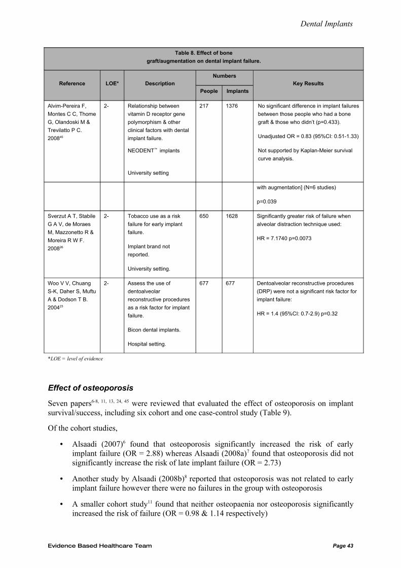
Dental Implants
Table 8. Effect of bone
graft/augmentation on dental implant failure.
Reference LOE* Description
Numbers
People Implants
Key Results
Alvim-Pereira F,
Montes C C, Thome
G, Olandoski M &
Trevilatto P C.
200845
2- Relationship between
vitamin D receptor gene
polymorphism & other
clinical factors with dental
implant failure.
NEODENT™ implants
University setting
217 1376 No significant difference in implant failures
between those people who had a bone
graft & those who didn’t (p=0.433).
Unadjusted OR = 0.83 (95%CI: 0.51-1.33)
Not supported by Kaplan-Meier survival
curve analysis.
with augmentation] (N=6 studies)
p=0.039
Sverzut A T, Stabile
G A V, de Moraes
M, Mazzonetto R &
Moreira R W F.
200826
2- Tobacco use as a risk
failure for early implant
failure.
Implant brand not
reported.
University setting.
650 1628 Significantly greater risk of failure when
alveolar distraction technique used:
HR = 7.1740 p=0.0073
Woo V V, Chuang
S-K, Daher S, Muftu
A & Dodson T B.
200423
2- Assess the use of
dentoalveolar
reconstructive procedures
as a risk factor for implant
failure.
Bicon dental implants.
Hospital setting.
677 677 Dentoalveolar reconstructive procedures
(DRP) were not a significant risk factor for
implant failure:
HR = 1.4 (95%CI: 0.7-2.9) p=0.32
*LOE = level of evidence
Effect of osteoporosis
Seven papers6-8, 11, 13, 24, 45 were reviewed that evaluated the effect of osteoporosis on implant survival/success, including six cohort and one case-control study (Table 9).
Of the cohort studies,
• Alsaadi (2007)6 found that osteoporosis significantly increased the risk of early implant failure (OR = 2.88) whereas Alsaadi (2008a)7 found that osteoporosis did not significantly increase the risk of late implant failure (OR = 2.73)
• Another study by Alsaadi (2008b)8 reported that osteoporosis was not related to early implant failure however there were no failures in the group with osteoporosis
• A smaller cohort study11 found that neither osteopaenia nor osteoporosis significantly increased the risk of failure (OR = 0.98 & 1.14 respectively)
Evidence Based Healthcare Team Page 43
Dental Implants
• In the only case-control study45, osteoporosis did not significantly change the odds of failure (unadjusted OR = 0.57 (95%CI: 0.05-5.53))
• In the last two cohort studies13, 24 the presence of osteoporosis was recorded but no results specific to this disorder was reported.
Of the narrative reviews,
Wood (2004)42 concluded that there is no evidence for systemic osteoporosis being a risk factor for implant failure but that “for patients with extreme osteoporosis, it may be wise to be cautious with maxillary implant treatment”.
Hwang (2007)56 concluded that osteoporosis alone does not affect implant success
Zitzmann (2008)43 lists osteoporosis as a condition that increases the risk of implant failure and suggests consideration of calcium substitution, a longer healing period and avoidance of high torque for abutment screw fixation.
Porter (2005)39 found few published clinical studies on the topic with conflicting opinions and concluded that visual assessment of bone density at placement42 may be more pertinent to implant success.
Scully (2007)57 in their review found similar success rates in people with osteoporosis compared to those without but adds that sinus lifts may be contraindicated.
Discussion
Osteoporosis may theoretically affect osseointegration42 because bone metabolism is impaired. However, the conclusion in the previous ACC review1 was that the evidence did not support the idea that osteoporosis was strongly associated with implant loss.
Of the primary studies reviewed above, only one found an elevated risk of implant failure in people with osteoporosis. The rest of the studies had problems with their methodology and were considered to be at high risk of bias, confounding or chance findings. Moreover, the narrative reviews’ conclusions were often based on very few studies, studies that were included in the previous ACC dental implant review1 and they often cited the same studies. This introduces bias by making the body of evidence appear stronger than it actually is.
Suggested recommendation:
There is insufficient evidence that osteoporosis is a relevant risk factor for implant failure.
Evidence Based Healthcare Team Page 44
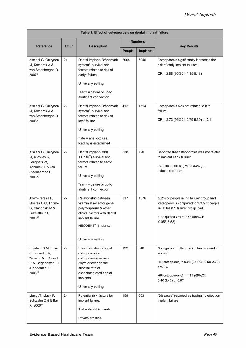
Dental Implants
Table 9. Effect of osteoporosis on dental implant failure.
Reference LOE* Description
Numbers
People Implants
Key Results
Alsaadi G, Quirynen
M, Komarek A &
van Steenberghe D.
20076
2+ Dental implant (Brånemark
system®) survival and
factors related to risk of
early* failure.
University setting.
*early = before or up to
abutment connection
2004 6946 Osteoporosis significantly increased the
risk of early implant failure:
OR = 2.88 (95%CI: 1.15-5.48)
Alsaadi G, Quirynen
M, Komarek A &
van Steenberghe D.
2008a7
2- Dental implant (Brånemark
system®) survival and
factors related to risk of
late* failure.
University setting.
*late = after occlusal
loading is established
412 1514 Osteoporosis was not related to late
failure:
OR = 2.73 (95%CI: 0.79-9.39) p=0.11
Alsaadi G, Quirynen
M, Michiles K,
Teughels W,
Komarek A & van
Steenberghe D.
2008b8
2- Dental implant (MkII
TiUnite™) survival and
factors related to early*
failure.
University setting.
*early = before or up to
abutment connection
238 720 Reported that osteoporosis was not related
to implant early failure:
0% (osteoporosis) vs. 2.03% (no
osteoporosis) p=1
Alvim-Pereira F,
Montes C C, Thome
G, Olandoski M &
Trevilatto P C.
200845
2- Relationship between
vitamin D receptor gene
polymorphism & other
clinical factors with dental
implant failure.
NEODENT™ implants
University setting.
217 1376 2.2% of people in ‘no failure’ group had
osteoporosis compared to 1.3% of people
in ‘at least 1 failure’ group [p=1]
Unadjusted OR = 0.57 (95%CI:
0.058-5.53)
Holahan C M, Koka
S, Kennel K A,
Weaver A L, Assad
D A, Regennitter F J
& Kademani D.
200811
2- Effect of a diagnosis of
osteoporosis or
osteopenia in women
50yrs or over on the
survival rate of
osseointegrated dental
implants.
University setting.
192 646 No significant effect on implant survival in
women:
HR[osteopenia] = 0.98 (95%CI: 0.50-2.60)
p=0.76
HR[osteoporosis] = 1.14 (95%CI:
0.40-2.42) p=0.97
Mundt T, Mack F,
Schwahn C & Biffar
R. 200613
2- Potential risk factors for
implant failure.
Tiolox dental implants.
Private practice.
159 663 “Diseases” reported as having no effect on
implant failure
Evidence Based Healthcare Team Page 45
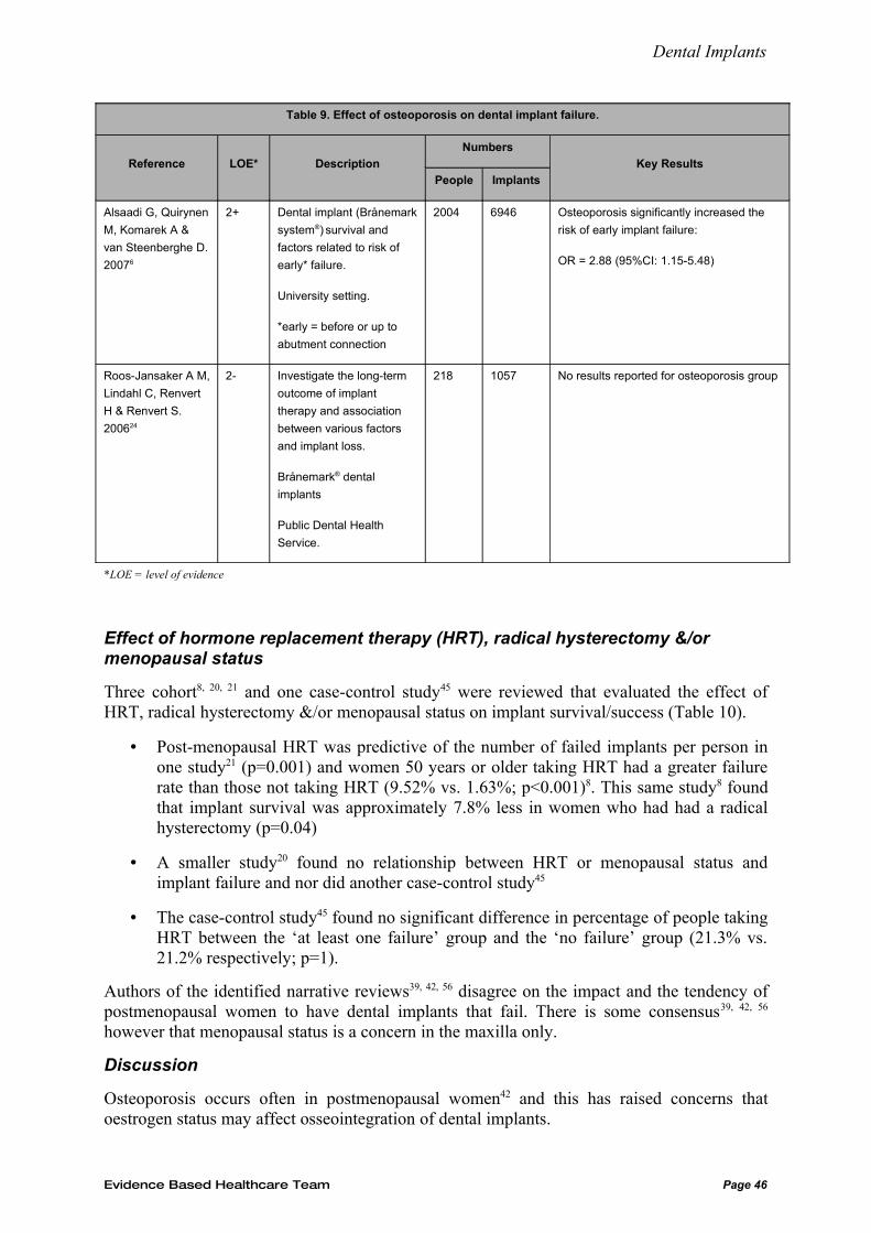
Dental Implants
Table 9. Effect of osteoporosis on dental implant failure.
Reference LOE* Description
Numbers
People Implants
Key Results
Alsaadi G, Quirynen
M, Komarek A &
van Steenberghe D.
20076
2+ Dental implant (Brånemark
system®) survival and
factors related to risk of
early* failure.
University setting.
*early = before or up to
abutment connection
2004 6946 Osteoporosis significantly increased the
risk of early implant failure:
OR = 2.88 (95%CI: 1.15-5.48)
Roos-Jansaker A M,
Lindahl C, Renvert
H & Renvert S.
200624
2- Investigate the long-term
outcome of implant
therapy and association
between various factors
and implant loss.
Brånemark® dental
implants
Public Dental Health
Service.
218 1057 No results reported for osteoporosis group
*LOE = level of evidence
Effect of hormone replacement therapy (HRT), radical hysterectomy &/or menopausal status
Three cohort8, 20, 21 and one case-control study45 were reviewed that evaluated the effect of HRT, radical hysterectomy &/or menopausal status on implant survival/success (Table 10).
• Post-menopausal HRT was predictive of the number of failed implants per person in one study21 (p=0.001) and women 50 years or older taking HRT had a greater failure rate than those not taking HRT (9.52% vs. 1.63%; p<0.001)8. This same study8 found that implant survival was approximately 7.8% less in women who had had a radical hysterectomy (p=0.04)
• A smaller study20 found no relationship between HRT or menopausal status and implant failure and nor did another case-control study45
• The case-control study45 found no significant difference in percentage of people taking HRT between the ‘at least one failure’ group and the ‘no failure’ group (21.3% vs. 21.2% respectively; p=1).
Authors of the identified narrative reviews39, 42, 56 disagree on the impact and the tendency of postmenopausal women to have dental implants that fail. There is some consensus39, 42, 56
however that menopausal status is a concern in the maxilla only.
Discussion
Osteoporosis occurs often in postmenopausal women42 and this has raised concerns that oestrogen status may affect osseointegration of dental implants.
Evidence Based Healthcare Team Page 46
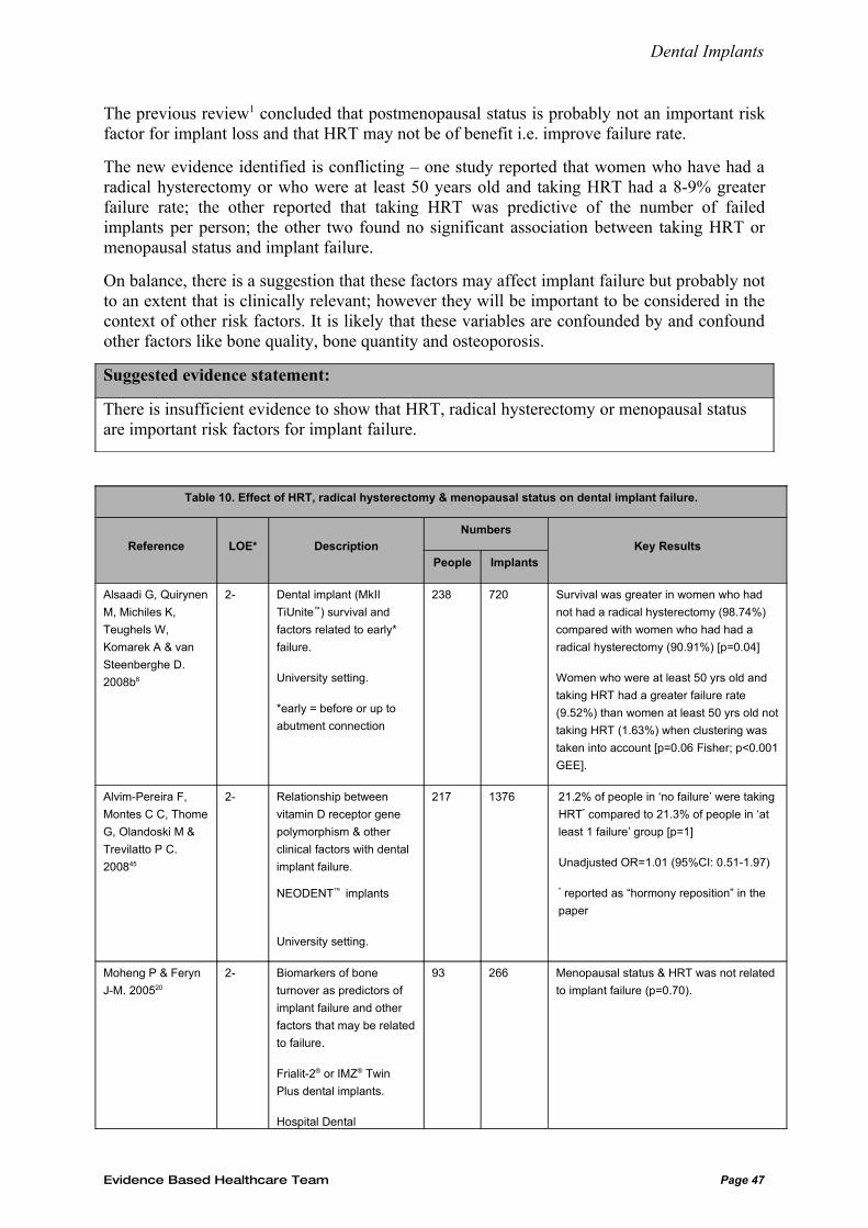
Dental Implants
The previous review1 concluded that postmenopausal status is probably not an important risk factor for implant loss and that HRT may not be of benefit i.e. improve failure rate.
The new evidence identified is conflicting – one study reported that women who have had a radical hysterectomy or who were at least 50 years old and taking HRT had a 8-9% greater failure rate; the other reported that taking HRT was predictive of the number of failed implants per person; the other two found no significant association between taking HRT or menopausal status and implant failure.
On balance, there is a suggestion that these factors may affect implant failure but probably not to an extent that is clinically relevant; however they will be important to be considered in the context of other risk factors. It is likely that these variables are confounded by and confound other factors like bone quality, bone quantity and osteoporosis.
Suggested evidence statement:
There is insufficient evidence to show that HRT, radical hysterectomy or menopausal status are important risk factors for implant failure.
Table 10. Effect of HRT, radical hysterectomy & menopausal status on dental implant failure.
Reference LOE* Description
Numbers
People Implants
Key Results
Alsaadi G, Quirynen
M, Michiles K,
Teughels W,
Komarek A & van
Steenberghe D.
2008b8
2- Dental implant (MkII
TiUnite™) survival and
factors related to early*
failure.
University setting.
*early = before or up to
abutment connection
238 720 Survival was greater in women who had
not had a radical hysterectomy (98.74%)
compared with women who had had a
radical hysterectomy (90.91%) [p=0.04]
Women who were at least 50 yrs old and
taking HRT had a greater failure rate
(9.52%) than women at least 50 yrs old not
taking HRT (1.63%) when clustering was
taken into account [p=0.06 Fisher; p<0.001
GEE].
Alvim-Pereira F,
Montes C C, Thome
G, Olandoski M &
Trevilatto P C.
200845
2- Relationship between
vitamin D receptor gene
polymorphism & other
clinical factors with dental
implant failure.
NEODENT™ implants
University setting.
217 1376 21.2% of people in ‘no failure’ were taking
HRT* compared to 21.3% of people in ‘at
least 1 failure’ group [p=1]
Unadjusted OR=1.01 (95%CI: 0.51-1.97)
* reported as “hormony reposition” in the
paper
Moheng P & Feryn
J-M. 200520
2- Biomarkers of bone
turnover as predictors of
implant failure and other
factors that may be related
to failure.
Frialit-2® or IMZ® Twin
Plus dental implants.
Hospital Dental
93 266 Menopausal status & HRT was not related
to implant failure (p=0.70).
Evidence Based Healthcare Team Page 47
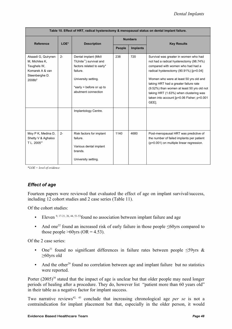
Dental Implants
Table 10. Effect of HRT, radical hysterectomy & menopausal status on dental implant failure.
Reference LOE* Description
Numbers
People Implants
Key Results
Alsaadi G, Quirynen
M, Michiles K,
Teughels W,
Komarek A & van
Steenberghe D.
2008b8
2- Dental implant (MkII
TiUnite™) survival and
factors related to early*
failure.
University setting.
*early = before or up to
abutment connection
238 720 Survival was greater in women who had
not had a radical hysterectomy (98.74%)
compared with women who had had a
radical hysterectomy (90.91%) [p=0.04]
Women who were at least 50 yrs old and
taking HRT had a greater failure rate
(9.52%) than women at least 50 yrs old not
taking HRT (1.63%) when clustering was
taken into account [p=0.06 Fisher; p<0.001
GEE].
Implantology Centre.
Moy P K, Medina D,
Shetty V & Aghaloo
T L. 200521
2- Risk factors for implant
failure.
Various dental implant
brands.
University setting.
1140 4680 Post-menopausal HRT was predictive of
the number of failed implants per patient
(p=0.001) on multiple linear regression.
*LOE = level of evidence
Effect of age
Fourteen papers were reviewed that evaluated the effect of age on implant survival/success, including 12 cohort studies and 2 case series (Table 11).
Of the cohort studies:
• Eleven 9, 17-21, 26, 44, 51-53found no association between implant failure and age
• And one22 found an increased risk of early failure in those people ≤60yrs compared to those people >60yrs (OR = 4.53).
Of the 2 case series:
• One31 found no significant differences in failure rates between people ≤59yrs & ≥60yrs old
• And the other29 found no correlation between age and implant failure but no statistics were reported.
Porter (2005)39 stated that the impact of age is unclear but that older people may need longer periods of healing after a procedure. They do, however list “patient more than 60 years old” in their table as a negative factor for implant success.
Two narrative reviews42, 43 conclude that increasing chronological age per se is not a contraindication for implant placement but that, especially in the older person, it would
Evidence Based Healthcare Team Page 48
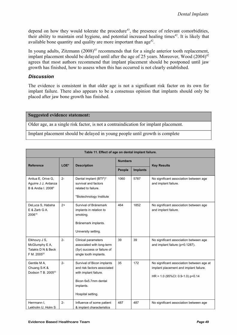
Dental Implants
depend on how they would tolerate the procedure43, the presence of relevant comorbidities, their ability to maintain oral hygiene, and potential increased healing times42. It is likely that available bone quantity and quality are more important than age42.
In young adults, Zitzmann (2008)43 recommends that for a single anterior tooth replacement, implant placement should be delayed until after the age of 25 years. Moreover, Wood (2004)42
agrees that most authors recommend that implant placement should be postponed until jaw growth has finished, how to assess when this has occurred is not clearly established.
Discussion
The evidence is consistent in that older age is not a significant risk factor on its own for implant failure. There also appears to be a consensus opinion that implants should only be placed after jaw bone growth has finished.
Suggested evidence statement:
Older age, as a single risk factor, is not a contraindication for implant placement.
Implant placement should be delayed in young people until growth is complete
Table 11. Effect of age on dental implant failure.
Reference LOE* Description
Numbers
People Implants
Key Results
Anitua E, Orive G,
Aguirre J J, Ardanza
B & Andia I. 20089
2- Dental implant (BTI®)*
survival and factors
related to failure.
*Biotechnology Institute
1060 5787 No significant association between age
and implant failure.
DeLuca S, Habsha
E & Zarb G A.
200619
2+ Survival of Brånemark
implants in relation to
smoking.
Brånemark implants.
University setting.
464 1852 No significant association between age
and implant failure.
Elkhoury J S,
McGlumphy E A,
Tatakis D N & Beck
F M. 200552
2- Clinical parameters
associated with long-term
(5yr) success or failure of
single tooth implants.
39 39 No significant association between age
and implant failure (p=0.1287).
Gentile M A,
Chuang S-K &
Dodson T B. 200553
2- Survival of Bicon implants
and risk factors associated
with implant failure.
Bicon 6x5.7mm dental
implants.
Hospital setting.
35 172 No significant association between age at
implant placement and implant failure.
HR = 1.0 (95%CI: 0.9-1.0) p=0.14
Herrmann I,
Lekholm U, Holm S
2- Influence of some patient
& implant characteristics
487 487 No significant association between age
Evidence Based Healthcare Team Page 49
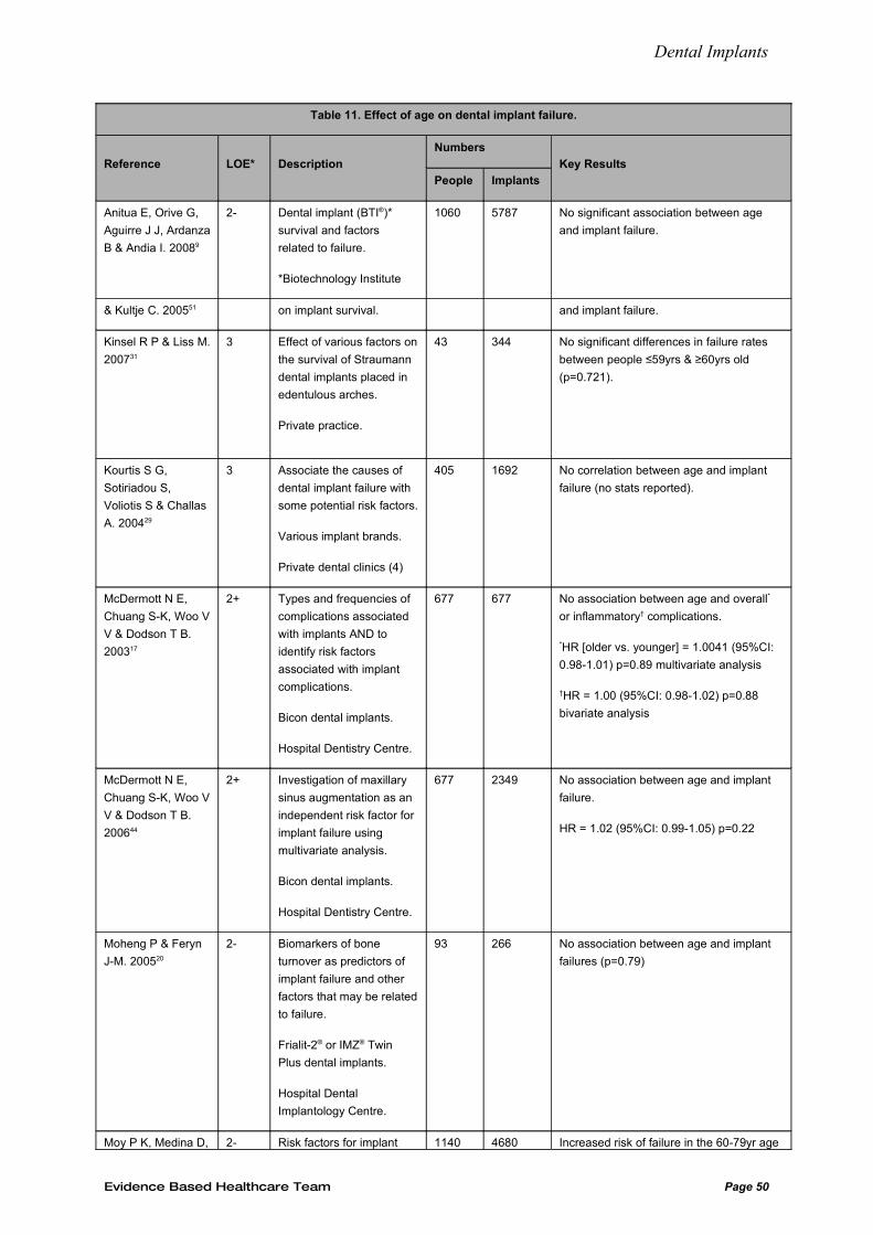
Dental Implants
Table 11. Effect of age on dental implant failure.
Reference LOE* Description
Numbers
People Implants
Key Results
Anitua E, Orive G,
Aguirre J J, Ardanza
B & Andia I. 20089
2- Dental implant (BTI®)*
survival and factors
related to failure.
*Biotechnology Institute
1060 5787 No significant association between age
and implant failure.
& Kultje C. 200551 on implant survival. and implant failure.
Kinsel R P & Liss M.
200731
3 Effect of various factors on
the survival of Straumann
dental implants placed in
edentulous arches.
Private practice.
43 344 No significant differences in failure rates
between people ≤59yrs & ≥60yrs old
(p=0.721).
Kourtis S G,
Sotiriadou S,
Voliotis S & Challas
A. 200429
3 Associate the causes of
dental implant failure with
some potential risk factors.
Various implant brands.
Private dental clinics (4)
405 1692 No correlation between age and implant
failure (no stats reported).
McDermott N E,
Chuang S-K, Woo V
V & Dodson T B.
200317
2+ Types and frequencies of
complications associated
with implants AND to
identify risk factors
associated with implant
complications.
Bicon dental implants.
Hospital Dentistry Centre.
677 677 No association between age and overall*
or inflammatory† complications.
*HR [older vs. younger] = 1.0041 (95%CI:
0.98-1.01) p=0.89 multivariate analysis
†HR = 1.00 (95%CI: 0.98-1.02) p=0.88
bivariate analysis
McDermott N E,
Chuang S-K, Woo V
V & Dodson T B.
200644
2+ Investigation of maxillary
sinus augmentation as an
independent risk factor for
implant failure using
multivariate analysis.
Bicon dental implants.
Hospital Dentistry Centre.
677 2349 No association between age and implant
failure.
HR = 1.02 (95%CI: 0.99-1.05) p=0.22
Moheng P & Feryn
J-M. 200520
2- Biomarkers of bone
turnover as predictors of
implant failure and other
factors that may be related
to failure.
Frialit-2® or IMZ® Twin
Plus dental implants.
Hospital Dental
Implantology Centre.
93 266 No association between age and implant
failures (p=0.79)
Moy P K, Medina D, 2- Risk factors for implant 1140 4680 Increased risk of failure in the 60-79yr age
Evidence Based Healthcare Team Page 50
Dental Implants
Table 11. Effect of age on dental implant failure.
Reference LOE* Description
Numbers
People Implants
Key Results
Anitua E, Orive G,
Aguirre J J, Ardanza
B & Andia I. 20089
2- Dental implant (BTI®)*
survival and factors
related to failure.
*Biotechnology Institute
1060 5787 No significant association between age
and implant failure.
Shetty V & Aghaloo
T L. 200521
failure.
Various dental implant
brands.
University setting.
group compared to those <40yrs on
univariate analysis* but not on multivariate
analysis.
*RR =2.24 (95%CI: 1.28-3.93)
Noguerol B, Munoz
R, Mesa F, de Dios
Luna J & O'Valle F.
200622
2- Determine accuracy of
Periotest® to monitor
primary implant stability
and identify variables
associated with early
implant failure.
Brånemark® dental
implants.
Single periodontal clinic
316 1084 Increased risk of early failure in those
people ≤60yrs compared to those people
>60yrs:
OR = 4.53 (95%CI: 1.34-15.27)
Susarla S M,
Chuang S-K &
Dodson T B. 200818
2+ Risk factors for implant
failure.
Bicon dental implants.
Private practice.
855 2826 Age was not a significant risk factor for
implant failure:
HR = 1.00 (95%CI: 0.98-1.0) p=0.57
Sverzut A T, Stabile
G A V, de Moraes
M, Mazzonetto R &
Moreira R W F.
200826
2- Tobacco use as a risk
failure for early implant
failure.
Implant brand not
reported.
University setting.
650 1628 No significant difference in risk of implant
failure with respect to age:
HR = 1.0750 p=0.7358
*LOE = level of evidence
Effect of radiotherapy
Four cohort studies6-8, 21 and 4 narrative reviews were reviewed that evaluated the effect of radiotherapy on implant survival/success (Table 12).
• Alsaadi (2007)6 and Alsaadi (2008b)8 found that radiotherapy of the concerned area did not increase the risk of early implant failure
• Alsaadi (2008a)7, on the other hand, found that radiotherapy increased the risk of late implant failure (OR = 3.32 (95%CI: 1.49-7.35)) although this was only based on 2 people with 6 implants
Evidence Based Healthcare Team Page 51
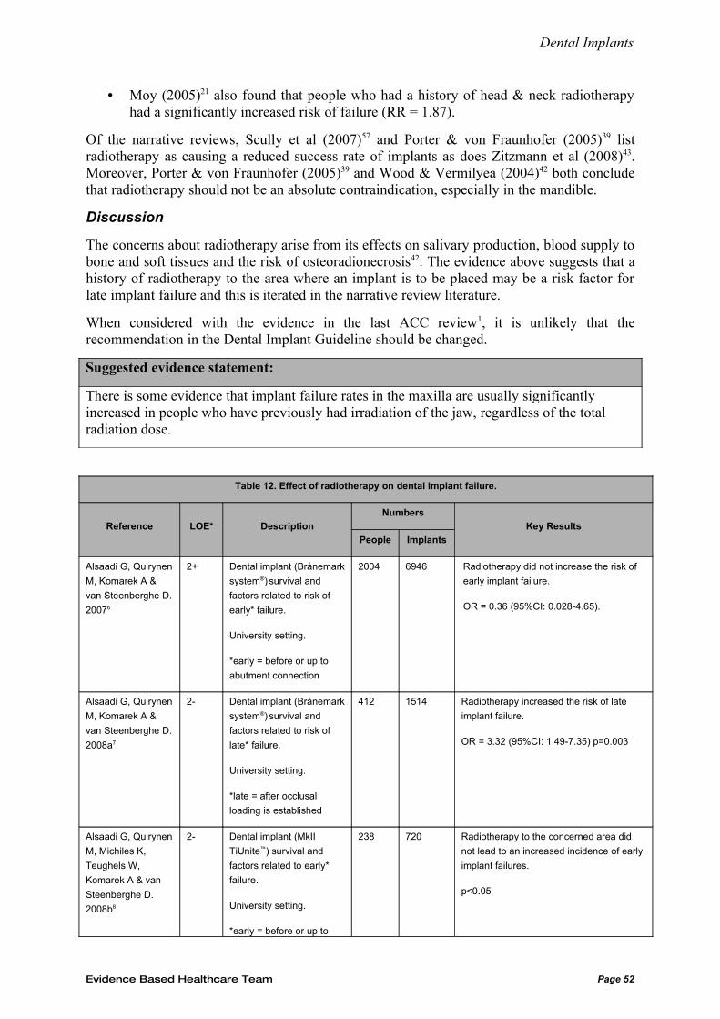
Dental Implants
• Moy (2005)21 also found that people who had a history of head & neck radiotherapy had a significantly increased risk of failure (RR = 1.87).
Of the narrative reviews, Scully et al (2007)57 and Porter & von Fraunhofer (2005)39 list radiotherapy as causing a reduced success rate of implants as does Zitzmann et al (2008)43. Moreover, Porter & von Fraunhofer (2005)39 and Wood & Vermilyea (2004)42 both conclude that radiotherapy should not be an absolute contraindication, especially in the mandible.
Discussion
The concerns about radiotherapy arise from its effects on salivary production, blood supply to bone and soft tissues and the risk of osteoradionecrosis42. The evidence above suggests that a history of radiotherapy to the area where an implant is to be placed may be a risk factor for late implant failure and this is iterated in the narrative review literature.
When considered with the evidence in the last ACC review1, it is unlikely that the recommendation in the Dental Implant Guideline should be changed.
Suggested evidence statement:
There is some evidence that implant failure rates in the maxilla are usually significantly increased in people who have previously had irradiation of the jaw, regardless of the total radiation dose.
Table 12. Effect of radiotherapy on dental implant failure.
Reference LOE* Description
Numbers
People Implants
Key Results
Alsaadi G, Quirynen
M, Komarek A &
van Steenberghe D.
20076
2+ Dental implant (Brånemark
system®) survival and
factors related to risk of
early* failure.
University setting.
*early = before or up to
abutment connection
2004 6946 Radiotherapy did not increase the risk of
early implant failure.
OR = 0.36 (95%CI: 0.028-4.65).
Alsaadi G, Quirynen
M, Komarek A &
van Steenberghe D.
2008a7
2- Dental implant (Brånemark
system®) survival and
factors related to risk of
late* failure.
University setting.
*late = after occlusal
loading is established
412 1514 Radiotherapy increased the risk of late
implant failure.
OR = 3.32 (95%CI: 1.49-7.35) p=0.003
Alsaadi G, Quirynen
M, Michiles K,
Teughels W,
Komarek A & van
Steenberghe D.
2008b8
2- Dental implant (MkII
TiUnite™) survival and
factors related to early*
failure.
University setting.
*early = before or up to
238 720 Radiotherapy to the concerned area did
not lead to an increased incidence of early
implant failures.
p<0.05
Evidence Based Healthcare Team Page 52
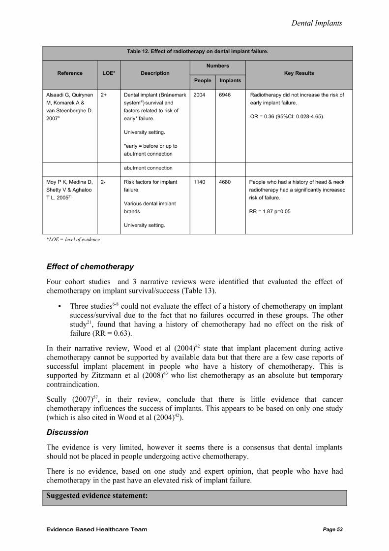
Dental Implants
Table 12. Effect of radiotherapy on dental implant failure.
Reference LOE* Description
Numbers
People Implants
Key Results
Alsaadi G, Quirynen
M, Komarek A &
van Steenberghe D.
20076
2+ Dental implant (Brånemark
system®) survival and
factors related to risk of
early* failure.
University setting.
*early = before or up to
abutment connection
2004 6946 Radiotherapy did not increase the risk of
early implant failure.
OR = 0.36 (95%CI: 0.028-4.65).
abutment connection
Moy P K, Medina D,
Shetty V & Aghaloo
T L. 200521
2- Risk factors for implant
failure.
Various dental implant
brands.
University setting.
1140 4680 People who had a history of head & neck
radiotherapy had a significantly increased
risk of failure.
RR = 1.87 p=0.05
*LOE = level of evidence
Effect of chemotherapy
Four cohort studies and 3 narrative reviews were identified that evaluated the effect of chemotherapy on implant survival/success (Table 13).
• Three studies6-8 could not evaluate the effect of a history of chemotherapy on implant success/survival due to the fact that no failures occurred in these groups. The other study21, found that having a history of chemotherapy had no effect on the risk of failure (RR = 0.63).
In their narrative review, Wood et al (2004)42 state that implant placement during active chemotherapy cannot be supported by available data but that there are a few case reports of successful implant placement in people who have a history of chemotherapy. This is supported by Zitzmann et al (2008)43 who list chemotherapy as an absolute but temporary contraindication.
Scully (2007)57, in their review, conclude that there is little evidence that cancer chemotherapy influences the success of implants. This appears to be based on only one study (which is also cited in Wood et al (2004)42).
Discussion
The evidence is very limited, however it seems there is a consensus that dental implants should not be placed in people undergoing active chemotherapy.
There is no evidence, based on one study and expert opinion, that people who have had chemotherapy in the past have an elevated risk of implant failure.
Suggested evidence statement:
Evidence Based Healthcare Team Page 53
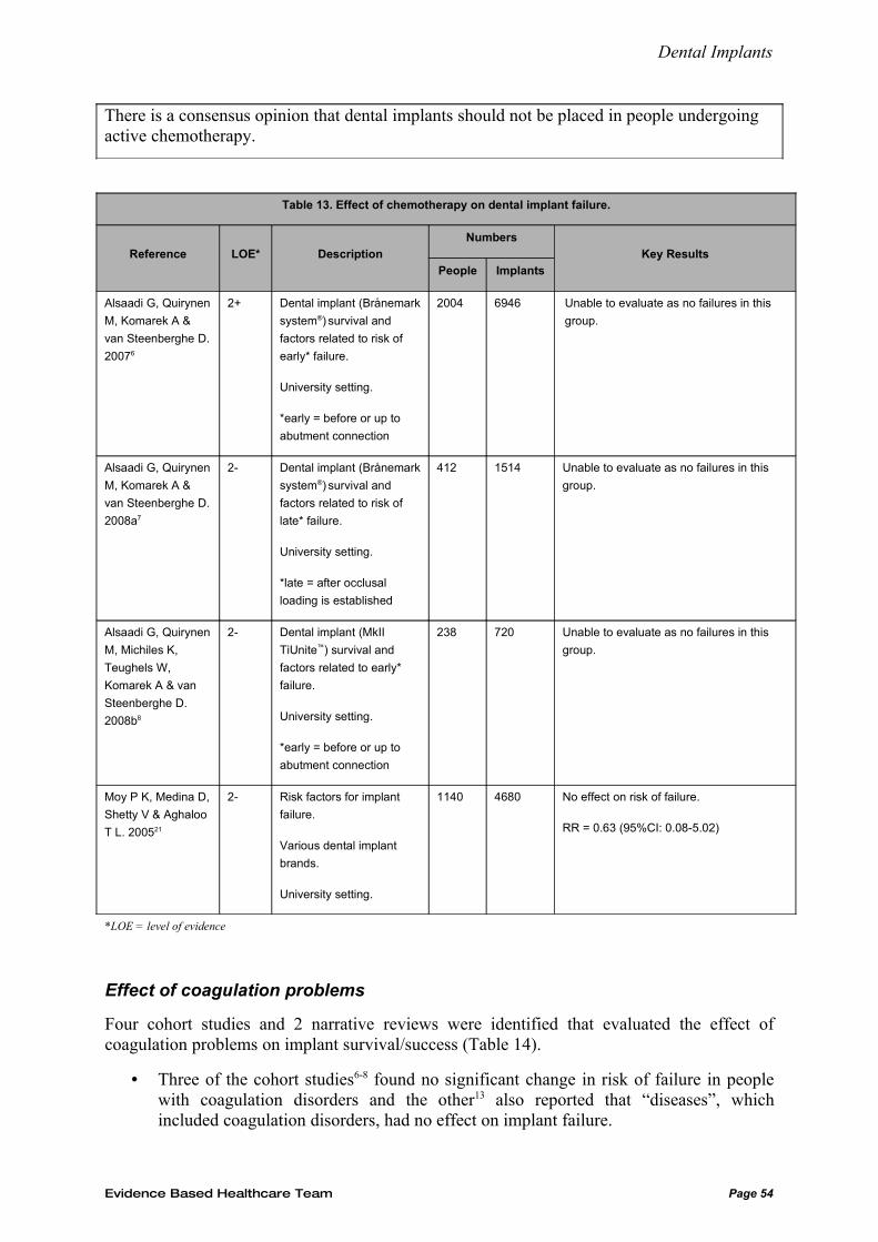
Dental Implants
There is a consensus opinion that dental implants should not be placed in people undergoing active chemotherapy.
Table 13. Effect of chemotherapy on dental implant failure.
Reference LOE* Description
Numbers
People Implants
Key Results
Alsaadi G, Quirynen
M, Komarek A &
van Steenberghe D.
20076
2+ Dental implant (Brånemark
system®) survival and
factors related to risk of
early* failure.
University setting.
*early = before or up to
abutment connection
2004 6946 Unable to evaluate as no failures in this
group.
Alsaadi G, Quirynen
M, Komarek A &
van Steenberghe D.
2008a7
2- Dental implant (Brånemark
system®) survival and
factors related to risk of
late* failure.
University setting.
*late = after occlusal
loading is established
412 1514 Unable to evaluate as no failures in this
group.
Alsaadi G, Quirynen
M, Michiles K,
Teughels W,
Komarek A & van
Steenberghe D.
2008b8
2- Dental implant (MkII
TiUnite™) survival and
factors related to early*
failure.
University setting.
*early = before or up to
abutment connection
238 720 Unable to evaluate as no failures in this
group.
Moy P K, Medina D,
Shetty V & Aghaloo
T L. 200521
2- Risk factors for implant
failure.
Various dental implant
brands.
University setting.
1140 4680 No effect on risk of failure.
RR = 0.63 (95%CI: 0.08-5.02)
*LOE = level of evidence
Effect of coagulation problems
Four cohort studies and 2 narrative reviews were identified that evaluated the effect of coagulation problems on implant survival/success (Table 14).
• Three of the cohort studies6-8 found no significant change in risk of failure in people with coagulation disorders and the other13 also reported that “diseases”, which included coagulation disorders, had no effect on implant failure.
Evidence Based Healthcare Team Page 54
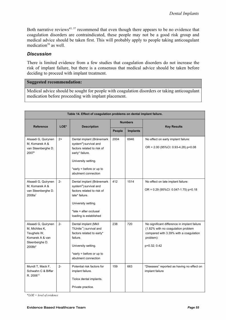
Dental Implants
Both narrative reviews43, 57 recommend that even though there appears to be no evidence that coagulation disorders are contraindicated, these people may not be a good risk group and medical advice should be taken first. This will probably apply to people taking anticoagulant medication58 as well.
Discussion
There is limited evidence from a few studies that coagulation disorders do not increase the risk of implant failure, but there is a consensus that medical advice should be taken before deciding to proceed with implant treatment.
Suggested recommendation:
Medical advice should be sought for people with coagulation disorders or taking anticoagulant medication before proceeding with implant placement.
Table 14. Effect of coagulation problems on dental implant failure.
Reference LOE* Description
Numbers
People Implants
Key Results
Alsaadi G, Quirynen
M, Komarek A &
van Steenberghe D.
20076
2+ Dental implant (Brånemark
system®) survival and
factors related to risk of
early* failure.
University setting.
*early = before or up to
abutment connection
2004 6946 No effect on early implant failure:
OR = 2.00 (95%CI: 0.93-4.28) p=0.08
Alsaadi G, Quirynen
M, Komarek A &
van Steenberghe D.
2008a7
2- Dental implant (Brånemark
system®) survival and
factors related to risk of
late* failure.
University setting.
*late = after occlusal
loading is established
412 1514 No effect on late implant failure:
OR = 0.29 (95%CI: 0.047-1.75) p=0.18
Alsaadi G, Quirynen
M, Michiles K,
Teughels W,
Komarek A & van
Steenberghe D.
2008b8
2- Dental implant (MkII
TiUnite™) survival and
factors related to early*
failure.
University setting.
*early = before or up to
abutment connection
238 720 No significant difference in implant failure
(1.82% with no coagulation problem
compared with 3.39% with a coagulation
problem):
p=0.32; 0.42
Mundt T, Mack F,
Schwahn C & Biffar
R. 200613
2- Potential risk factors for
implant failure.
Tiolox dental implants.
Private practice.
159 663 “Diseases” reported as having no effect on
implant failure
*LOE = level of evidence
Evidence Based Healthcare Team Page 55
Dental Implants
Effect of edentulousness/proximity to natural teeth
Six papers were reviewed that evaluated the effect of edentulousness or proximity to natural teeth on implant survival/success, including four cohort, one case-control and one case series (Table 15).
• Of the cohort studies, one6 found that the risk of early failure was 2.77 times higher in implants neighbouring teeth than in implants placed in fully edentulous mouths. This increased to 4.8 times when compared to implants in the presence of teeth in the antagonistic jaw only. Even though these results reach statistical significance, the data was available for only one third of the study population & the summary estimates were not adjusted for potential confounders i.e. high risk of bias and confounding
• Another study8 which found higher early failure rates in implants neighbouring natural teeth compared to those in fully edentulous mouths (p=0.004). Of note, there were no failures of implants placed in a jaw having teeth (but not neighbouring the implant) or of implants placed in an edentulous jaw opposing a jaw with teeth
• McDermott (2006)44 found that the dental implant’s proximity to natural teeth was not significantly associated with implant failure on multivariate analysis
• Degree of edentulousness was not related to late implant failure in another study7
• The case-control study45 found that there were more totally edentulous participants in the group with no implant failures compared to the group with at least one failure (19% vs. 6.3%; p=0.009)
• The single case series29 found that there was a significant difference in the failure rates of implants replacing a single tooth, implants placed in partially edentulous people or implants placed in fully edentulous people (7%, 56%, 37% respectively).
Discussion
The evidence for the effect of the type of edentulousness and proximity to natural dentition is inconclusive and conflicting. In addition, there are considerable concerns regarding the internal validity of the studies, and it is unclear whether these variables are primary risk factors or proxies for other risk factors e.g. reason for tooth loss, occlusal forces. Moreover, the utility of these variables as patient selection factors for the ACC population is debatable.
Suggested evidence statement:
There is insufficient evidence that the types of edentulousness or proximity to natural dentition are significant risk factors for implant failure.
Evidence Based Healthcare Team Page 56
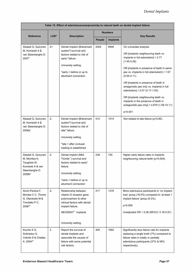
Dental Implants
Table 15. Effect of edentulousness/proximity to natural teeth on dental implant failure.
Reference LOE* Description
Numbers
People Implants
Key Results
Alsaadi G, Quirynen
M, Komarek A &
van Steenberghe D.
20076
2+ Dental implant (Brånemark
system®) survival and
factors related to risk of
early* failure.
University setting.
*early = before or up to
abutment connection
2004 6946 On univariate analysis:
OR [implants neighbouring teeth vs.
implants in full edentulism] = 2.77
(1.45-5.28)
OR [implants in presence of teeth in same
jaw vs. implants in full edentulism] = 1.97
(0.94-4.11)
OR [implants in presence of teeth in
antagonistic jaw only vs. implants in full
edentulism] = 0.57 (0.17-1.93)
OR [implants neighbouring teeth vs.
implants in the presence of teeth in
antagonistic jaw only] = 4.879 (1.59-15.11)
p<0.001
Alsaadi G, Quirynen
M, Komarek A &
van Steenberghe D.
2008a7
2- Dental implant (Brånemark
system®) survival and
factors related to risk of
late* failure.
University setting.
*late = after occlusal
loading is established
412 1514 Not related to late failure (p=0.85).
Alsaadi G, Quirynen
M, Michiles K,
Teughels W,
Komarek A & van
Steenberghe D.
2008b8
2- Dental implant (MkII
TiUnite™) survival and
factors related to early*
failure.
University setting.
*early = before or up to
abutment connection
238 720 Higher early failure rates in implants
neighbouring natural teeth (p=0.004).
Alvim-Pereira F,
Montes C C, Thome
G, Olandoski M &
Trevilatto P C.
200845
2- Relationship between
vitamin D receptor gene
polymorphism & other
clinical factors with dental
implant failure.
NEODENT™ implants
University setting.
217 1376 More edentulous participants in ‘no implant
loss’ group (19.0%) compared to ‘at least 1
implant failure’ group (6.3%).
p=0.009
Unadjusted OR = 0.28 (95%CI: 0.18-0.91)
Kourtis S G,
Sotiriadou S,
Voliotis S & Challas
A. 200429
3 Report the survival of
dental implants and
associate the causes of
failure with some potential
risk factors.
405 1692 Significantly less failure rate for implants
replacing a single tooth (7%) compared to
failure rates in totally or partially
edentulous participants (37% & 56%
respectively).
Evidence Based Healthcare Team Page 57
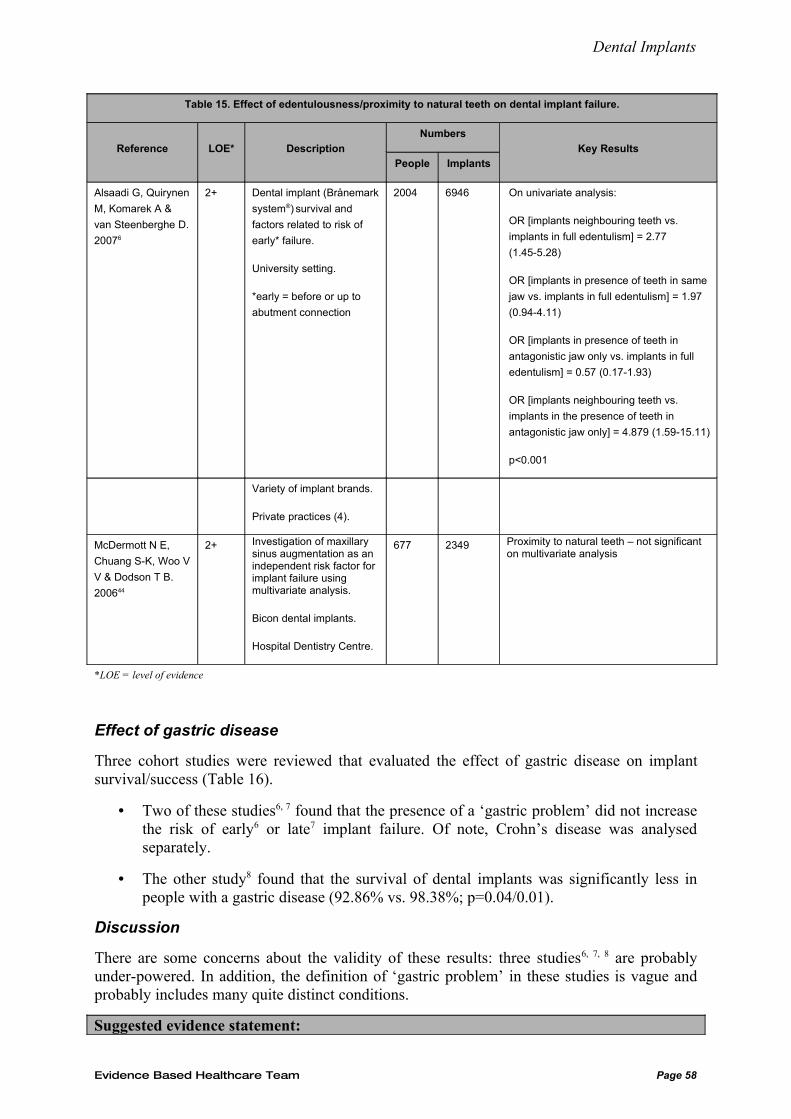
Dental Implants
Table 15. Effect of edentulousness/proximity to natural teeth on dental implant failure.
Reference LOE* Description
Numbers
People Implants
Key Results
Alsaadi G, Quirynen
M, Komarek A &
van Steenberghe D.
20076
2+ Dental implant (Brånemark
system®) survival and
factors related to risk of
early* failure.
University setting.
*early = before or up to
abutment connection
2004 6946 On univariate analysis:
OR [implants neighbouring teeth vs.
implants in full edentulism] = 2.77
(1.45-5.28)
OR [implants in presence of teeth in same
jaw vs. implants in full edentulism] = 1.97
(0.94-4.11)
OR [implants in presence of teeth in
antagonistic jaw only vs. implants in full
edentulism] = 0.57 (0.17-1.93)
OR [implants neighbouring teeth vs.
implants in the presence of teeth in
antagonistic jaw only] = 4.879 (1.59-15.11)
p<0.001
Variety of implant brands.
Private practices (4).
McDermott N E,
Chuang S-K, Woo V
V & Dodson T B.
200644
2+ Investigation of maxillary sinus augmentation as an independent risk factor for implant failure using multivariate analysis.
Bicon dental implants.
Hospital Dentistry Centre.
677 2349 Proximity to natural teeth – not significant on multivariate analysis
*LOE = level of evidence
Effect of gastric disease
Three cohort studies were reviewed that evaluated the effect of gastric disease on implant survival/success (Table 16).
• Two of these studies6, 7 found that the presence of a ‘gastric problem’ did not increase the risk of early6 or late7 implant failure. Of note, Crohn’s disease was analysed separately.
• The other study8 found that the survival of dental implants was significantly less in people with a gastric disease (92.86% vs. 98.38%; p=0.04/0.01).
Discussion
There are some concerns about the validity of these results: three studies6, 7, 8 are probably under-powered. In addition, the definition of ‘gastric problem’ in these studies is vague and probably includes many quite distinct conditions.
Suggested evidence statement:
Evidence Based Healthcare Team Page 58
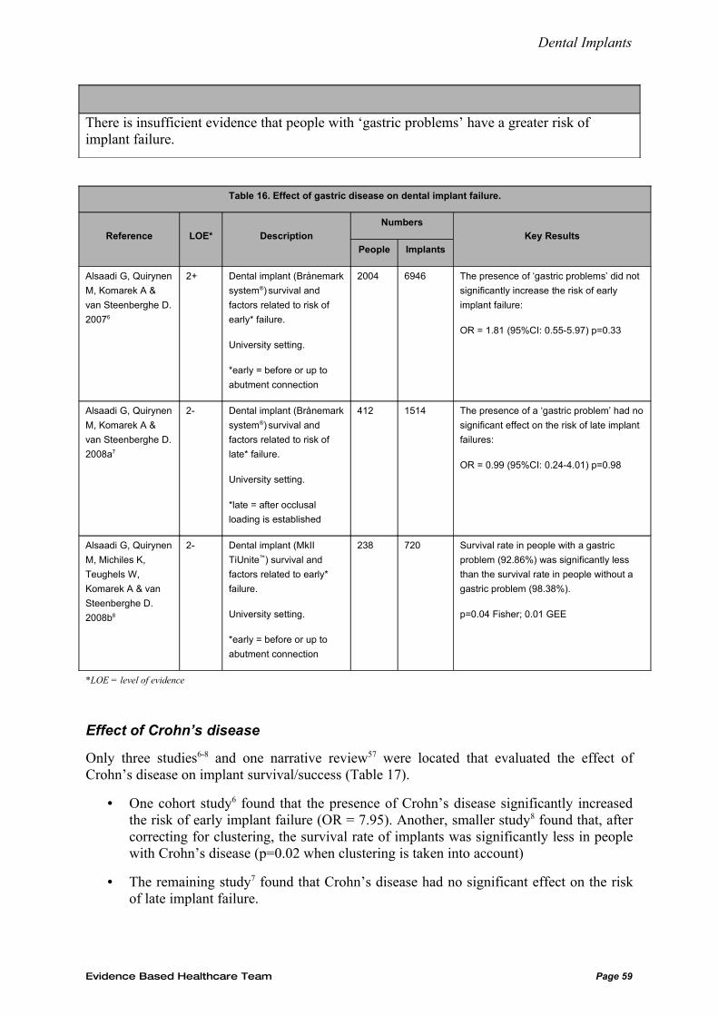
Dental Implants
There is insufficient evidence that people with ‘gastric problems’ have a greater risk of implant failure.
Table 16. Effect of gastric disease on dental implant failure.
Reference LOE* Description
Numbers
People Implants
Key Results
Alsaadi G, Quirynen
M, Komarek A &
van Steenberghe D.
20076
2+ Dental implant (Brånemark
system®) survival and
factors related to risk of
early* failure.
University setting.
*early = before or up to
abutment connection
2004 6946 The presence of ‘gastric problems’ did not
significantly increase the risk of early
implant failure:
OR = 1.81 (95%CI: 0.55-5.97) p=0.33
Alsaadi G, Quirynen
M, Komarek A &
van Steenberghe D.
2008a7
2- Dental implant (Brånemark
system®) survival and
factors related to risk of
late* failure.
University setting.
*late = after occlusal
loading is established
412 1514 The presence of a ‘gastric problem’ had no
significant effect on the risk of late implant
failures:
OR = 0.99 (95%CI: 0.24-4.01) p=0.98
Alsaadi G, Quirynen
M, Michiles K,
Teughels W,
Komarek A & van
Steenberghe D.
2008b8
2- Dental implant (MkII
TiUnite™) survival and
factors related to early*
failure.
University setting.
*early = before or up to
abutment connection
238 720 Survival rate in people with a gastric
problem (92.86%) was significantly less
than the survival rate in people without a
gastric problem (98.38%).
p=0.04 Fisher; 0.01 GEE
*LOE = level of evidence
Effect of Crohn’s disease
Only three studies6-8 and one narrative review57 were located that evaluated the effect of Crohn’s disease on implant survival/success (Table 17).
• One cohort study6 found that the presence of Crohn’s disease significantly increased the risk of early implant failure (OR = 7.95). Another, smaller study8 found that, after correcting for clustering, the survival rate of implants was significantly less in people with Crohn’s disease (p=0.02 when clustering is taken into account)
• The remaining study7 found that Crohn’s disease had no significant effect on the risk of late implant failure.
Evidence Based Healthcare Team Page 59
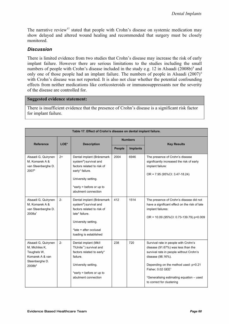
Dental Implants
The narrative review57 stated that people with Crohn’s disease on systemic medication may show delayed and altered wound healing and recommended that surgery must be closely monitored.
Discussion
There is limited evidence from two studies that Crohn’s disease may increase the risk of early implant failure. However there are serious limitations to the studies including the small numbers of people with Crohn’s disease included in the study e.g. 12 in Alsaadi (2008b)8 and only one of those people had an implant failure. The numbers of people in Alsaadi (2007)6
with Crohn’s disease was not reported. It is also not clear whether the potential confounding effects from neither medications like corticosteroids or immunosuppressants nor the severity of the disease are controlled for.
Suggested evidence statement:
There is insufficient evidence that the presence of Crohn’s disease is a significant risk factor for implant failure.
Table 17. Effect of Crohn’s disease on dental implant failure.
Reference LOE* Description
Numbers
People Implants
Key Results
Alsaadi G, Quirynen
M, Komarek A &
van Steenberghe D.
20076
2+ Dental implant (Brånemark
system®) survival and
factors related to risk of
early* failure.
University setting.
*early = before or up to
abutment connection
2004 6946 The presence of Crohn’s disease
significantly increased the risk of early
implant failure:
OR = 7.95 (95%CI: 3.47-18.24)
Alsaadi G, Quirynen
M, Komarek A &
van Steenberghe D.
2008a7
2- Dental implant (Brånemark
system®) survival and
factors related to risk of
late* failure.
University setting.
*late = after occlusal
loading is established
412 1514 The presence of Crohn’s disease did not
have a significant effect on the risk of late
implant failures:
OR = 10.09 (95%CI: 0.73-139.79) p=0.009
Alsaadi G, Quirynen
M, Michiles K,
Teughels W,
Komarek A & van
Steenberghe D.
2008b8
2- Dental implant (MkII
TiUnite™) survival and
factors related to early*
failure.
University setting.
*early = before or up to
abutment connection
238 720 Survival rate in people with Crohn’s
disease (91.67%) was less than the
survival rate in people without Crohn’s
disease (98.16%).
Depending on the method used: p=0.21
Fisher; 0.02 GEE†
†Generalising estimating equation – used
to correct for clustering
Evidence Based Healthcare Team Page 60
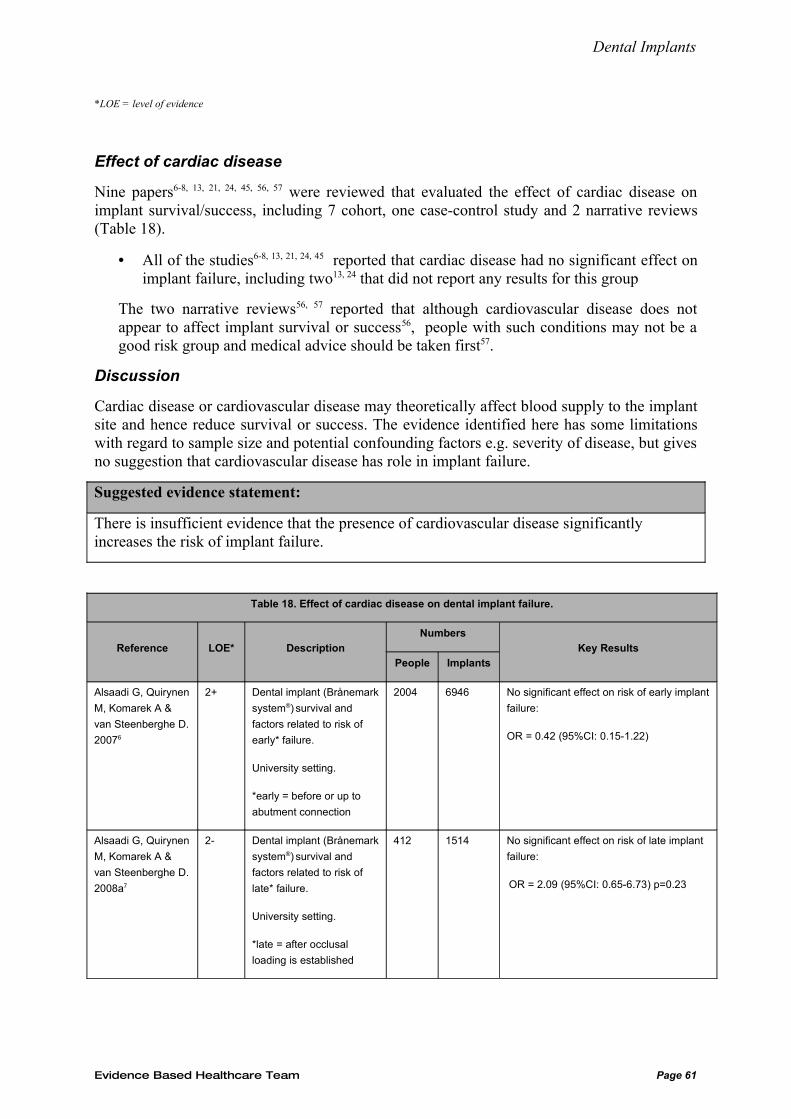
Dental Implants
*LOE = level of evidence
Effect of cardiac disease
Nine papers6-8, 13, 21, 24, 45, 56, 57 were reviewed that evaluated the effect of cardiac disease on implant survival/success, including 7 cohort, one case-control study and 2 narrative reviews (Table 18).
• All of the studies6-8, 13, 21, 24, 45 reported that cardiac disease had no significant effect on implant failure, including two13, 24 that did not report any results for this group
The two narrative reviews56, 57 reported that although cardiovascular disease does not appear to affect implant survival or success56, people with such conditions may not be a good risk group and medical advice should be taken first57.
Discussion
Cardiac disease or cardiovascular disease may theoretically affect blood supply to the implant site and hence reduce survival or success. The evidence identified here has some limitations with regard to sample size and potential confounding factors e.g. severity of disease, but gives no suggestion that cardiovascular disease has role in implant failure.
Suggested evidence statement:
There is insufficient evidence that the presence of cardiovascular disease significantly increases the risk of implant failure.
Table 18. Effect of cardiac disease on dental implant failure.
Reference LOE* Description
Numbers
People Implants
Key Results
Alsaadi G, Quirynen
M, Komarek A &
van Steenberghe D.
20076
2+ Dental implant (Brånemark
system®) survival and
factors related to risk of
early* failure.
University setting.
*early = before or up to
abutment connection
2004 6946 No significant effect on risk of early implant
failure:
OR = 0.42 (95%CI: 0.15-1.22)
Alsaadi G, Quirynen
M, Komarek A &
van Steenberghe D.
2008a7
2- Dental implant (Brånemark
system®) survival and
factors related to risk of
late* failure.
University setting.
*late = after occlusal
loading is established
412 1514 No significant effect on risk of late implant
failure:
OR = 2.09 (95%CI: 0.65-6.73) p=0.23
Evidence Based Healthcare Team Page 61
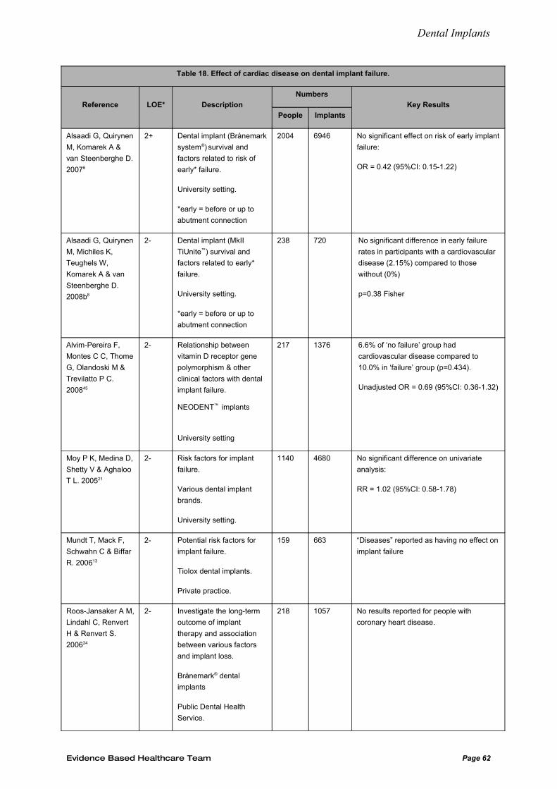
Dental Implants
Table 18. Effect of cardiac disease on dental implant failure.
Reference LOE* Description
Numbers
People Implants
Key Results
Alsaadi G, Quirynen
M, Komarek A &
van Steenberghe D.
20076
2+ Dental implant (Brånemark
system®) survival and
factors related to risk of
early* failure.
University setting.
*early = before or up to
abutment connection
2004 6946 No significant effect on risk of early implant
failure:
OR = 0.42 (95%CI: 0.15-1.22)
Alsaadi G, Quirynen
M, Michiles K,
Teughels W,
Komarek A & van
Steenberghe D.
2008b8
2- Dental implant (MkII
TiUnite™) survival and
factors related to early*
failure.
University setting.
*early = before or up to
abutment connection
238 720 No significant difference in early failure
rates in participants with a cardiovascular
disease (2.15%) compared to those
without (0%)
p=0.38 Fisher
Alvim-Pereira F,
Montes C C, Thome
G, Olandoski M &
Trevilatto P C.
200845
2- Relationship between
vitamin D receptor gene
polymorphism & other
clinical factors with dental
implant failure.
NEODENT™ implants
University setting
217 1376 6.6% of ‘no failure’ group had
cardiovascular disease compared to
10.0% in ‘failure’ group (p=0.434).
Unadjusted OR = 0.69 (95%CI: 0.36-1.32)
Moy P K, Medina D,
Shetty V & Aghaloo
T L. 200521
2- Risk factors for implant
failure.
Various dental implant
brands.
University setting.
1140 4680 No significant difference on univariate
analysis:
RR = 1.02 (95%CI: 0.58-1.78)
Mundt T, Mack F,
Schwahn C & Biffar
R. 200613
2- Potential risk factors for
implant failure.
Tiolox dental implants.
Private practice.
159 663 “Diseases” reported as having no effect on
implant failure
Roos-Jansaker A M,
Lindahl C, Renvert
H & Renvert S.
200624
2- Investigate the long-term
outcome of implant
therapy and association
between various factors
and implant loss.
Brånemark® dental
implants
Public Dental Health
Service.
218 1057 No results reported for people with
coronary heart disease.
Evidence Based Healthcare Team Page 62
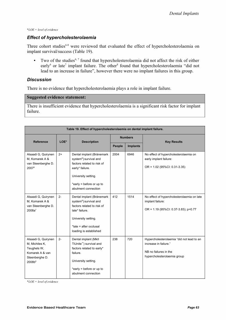
Dental Implants
*LOE = level of evidence
Effect of hypercholesterolaemia
Three cohort studies6-8 were reviewed that evaluated the effect of hypercholesterolaemia on implant survival/success (Table 19).
• Two of the studies6, 7 found that hypercholesterolaemia did not affect the risk of either early6 or late7 implant failure. The other8 found that hypercholesterolaemia “did not lead to an increase in failure”, however there were no implant failures in this group.
Discussion
There is no evidence that hypercholesterolaemia plays a role in implant failure.
Suggested evidence statement:
There is insufficient evidence that hypercholesterolaemia is a significant risk factor for implant failure.
Table 19. Effect of hypercholesterolaemia on dental implant failure.
Reference LOE* Description
Numbers
People Implants
Key Results
Alsaadi G, Quirynen
M, Komarek A &
van Steenberghe D.
20076
2+ Dental implant (Brånemark
system®) survival and
factors related to risk of
early* failure.
University setting.
*early = before or up to
abutment connection
2004 6946 No effect of hypercholesterolaemia on
early implant failure:
OR = 1.02 (95%CI: 0.31-3.35)
Alsaadi G, Quirynen
M, Komarek A &
van Steenberghe D.
2008a7
2- Dental implant (Brånemark
system®) survival and
factors related to risk of
late* failure.
University setting.
*late = after occlusal
loading is established
412 1514 No effect of hypercholesterolaemia on late
implant failure:
OR = 1.19 (95%CI: 0.37-3.83); p=0.77
Alsaadi G, Quirynen
M, Michiles K,
Teughels W,
Komarek A & van
Steenberghe D.
2008b8
2- Dental implant (MkII
TiUnite™) survival and
factors related to early*
failure.
University setting.
*early = before or up to
abutment connection
238 720 Hypercholesterolaemia “did not lead to an
increase in failure.”
NB no failures in the
hypercholesterolaemia group
*LOE = level of evidence
Evidence Based Healthcare Team Page 63
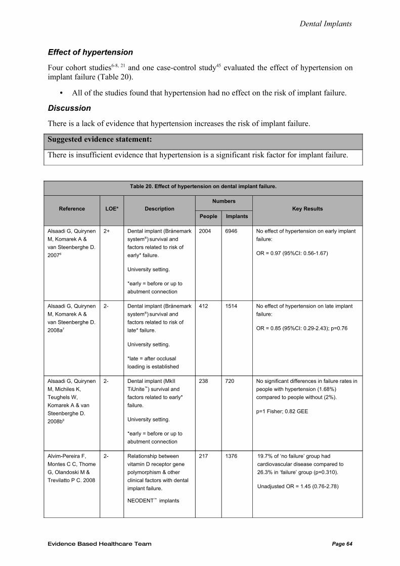
Dental Implants
Effect of hypertension
Four cohort studies6-8, 21 and one case-control study45 evaluated the effect of hypertension on implant failure (Table 20).
• All of the studies found that hypertension had no effect on the risk of implant failure.
Discussion
There is a lack of evidence that hypertension increases the risk of implant failure.
Suggested evidence statement:
There is insufficient evidence that hypertension is a significant risk factor for implant failure.
Table 20. Effect of hypertension on dental implant failure.
Reference LOE* Description
Numbers
People Implants
Key Results
Alsaadi G, Quirynen
M, Komarek A &
van Steenberghe D.
20076
2+ Dental implant (Brånemark
system®) survival and
factors related to risk of
early* failure.
University setting.
*early = before or up to
abutment connection
2004 6946 No effect of hypertension on early implant
failure:
OR = 0.97 (95%CI: 0.56-1.67)
Alsaadi G, Quirynen
M, Komarek A &
van Steenberghe D.
2008a7
2- Dental implant (Brånemark
system®) survival and
factors related to risk of
late* failure.
University setting.
*late = after occlusal
loading is established
412 1514 No effect of hypertension on late implant
failure:
OR = 0.85 (95%CI: 0.29-2.43); p=0.76
Alsaadi G, Quirynen
M, Michiles K,
Teughels W,
Komarek A & van
Steenberghe D.
2008b8
2- Dental implant (MkII
TiUnite™) survival and
factors related to early*
failure.
University setting.
*early = before or up to
abutment connection
238 720 No significant differences in failure rates in
people with hypertension (1.68%)
compared to people without (2%).
p=1 Fisher; 0.82 GEE
Alvim-Pereira F,
Montes C C, Thome
G, Olandoski M &
Trevilatto P C. 2008
2- Relationship between
vitamin D receptor gene
polymorphism & other
clinical factors with dental
implant failure.
NEODENT™ implants
217 1376 19.7% of ‘no failure’ group had
cardiovascular disease compared to
26.3% in ‘failure’ group (p=0.310).
Unadjusted OR = 1.45 (0.76-2.78)
Evidence Based Healthcare Team Page 64
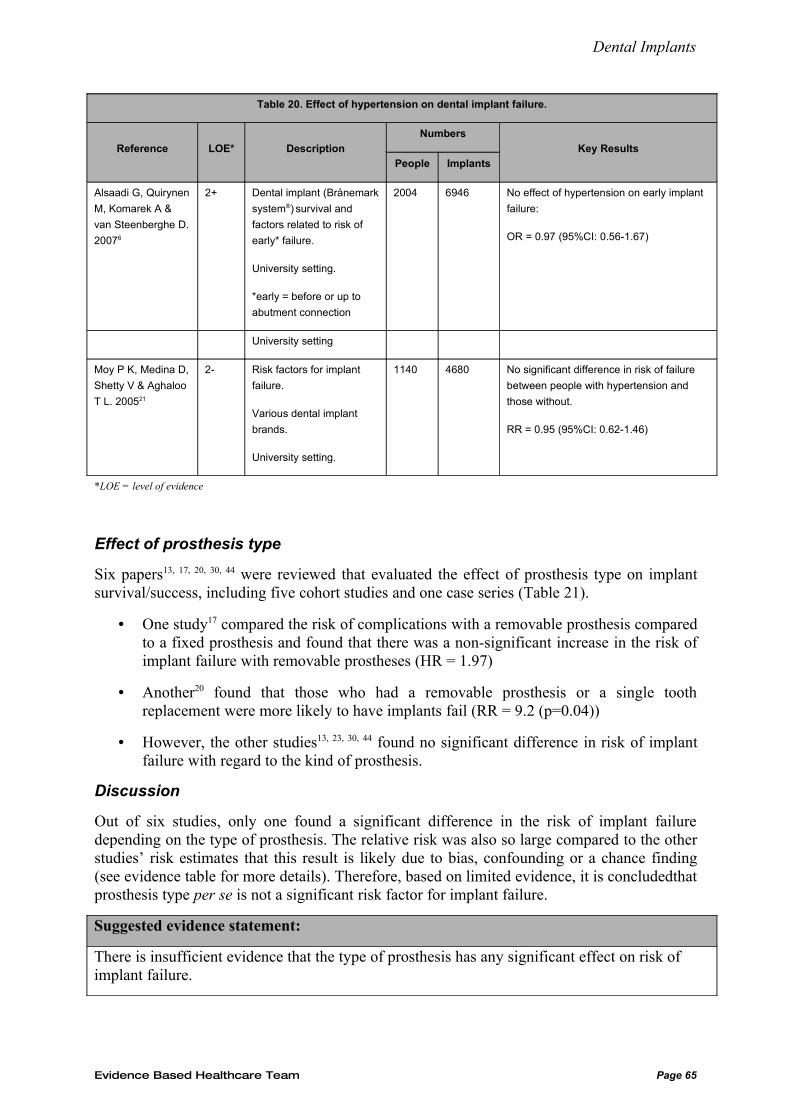
Dental Implants
Table 20. Effect of hypertension on dental implant failure.
Reference LOE* Description
Numbers
People Implants
Key Results
Alsaadi G, Quirynen
M, Komarek A &
van Steenberghe D.
20076
2+ Dental implant (Brånemark
system®) survival and
factors related to risk of
early* failure.
University setting.
*early = before or up to
abutment connection
2004 6946 No effect of hypertension on early implant
failure:
OR = 0.97 (95%CI: 0.56-1.67)
University setting
Moy P K, Medina D,
Shetty V & Aghaloo
T L. 200521
2- Risk factors for implant
failure.
Various dental implant
brands.
University setting.
1140 4680 No significant difference in risk of failure
between people with hypertension and
those without.
RR = 0.95 (95%CI: 0.62-1.46)
*LOE = level of evidence
Effect of prosthesis type
Six papers13, 17, 20, 30, 44 were reviewed that evaluated the effect of prosthesis type on implant survival/success, including five cohort studies and one case series (Table 21).
• One study17 compared the risk of complications with a removable prosthesis compared to a fixed prosthesis and found that there was a non-significant increase in the risk of implant failure with removable prostheses (HR = 1.97)
• Another20 found that those who had a removable prosthesis or a single tooth replacement were more likely to have implants fail (RR = 9.2 (p=0.04))
• However, the other studies13, 23, 30, 44 found no significant difference in risk of implant failure with regard to the kind of prosthesis.
Discussion
Out of six studies, only one found a significant difference in the risk of implant failure depending on the type of prosthesis. The relative risk was also so large compared to the other studies’ risk estimates that this result is likely due to bias, confounding or a chance finding (see evidence table for more details). Therefore, based on limited evidence, it is concludedthat prosthesis type per se is not a significant risk factor for implant failure.
Suggested evidence statement:
There is insufficient evidence that the type of prosthesis has any significant effect on risk of implant failure.
Evidence Based Healthcare Team Page 65
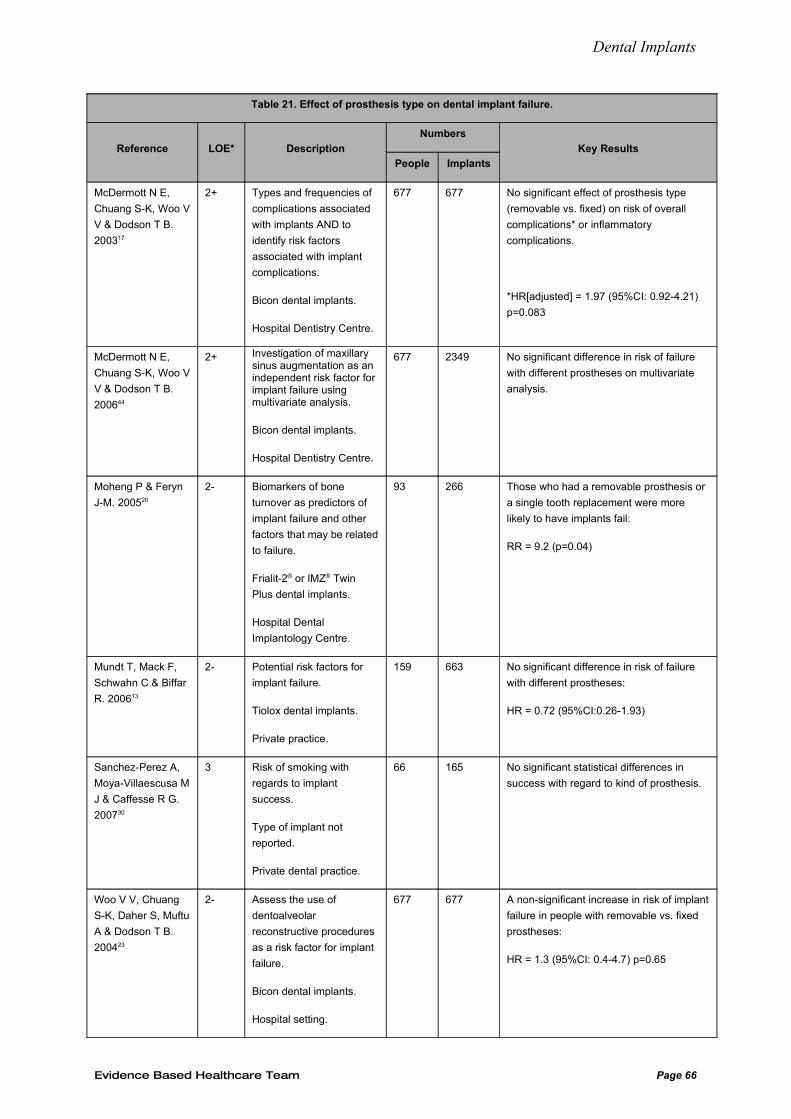
Dental Implants
Table 21. Effect of prosthesis type on dental implant failure.
Reference LOE* Description
Numbers
People Implants
Key Results
McDermott N E,
Chuang S-K, Woo V
V & Dodson T B.
200317
2+ Types and frequencies of
complications associated
with implants AND to
identify risk factors
associated with implant
complications.
Bicon dental implants.
Hospital Dentistry Centre.
677 677 No significant effect of prosthesis type
(removable vs. fixed) on risk of overall
complications* or inflammatory
complications.
*HR[adjusted] = 1.97 (95%CI: 0.92-4.21)
p=0.083
McDermott N E,
Chuang S-K, Woo V
V & Dodson T B.
200644
2+ Investigation of maxillary sinus augmentation as an independent risk factor for implant failure using multivariate analysis.
Bicon dental implants.
Hospital Dentistry Centre.
677 2349 No significant difference in risk of failure
with different prostheses on multivariate
analysis.
Moheng P & Feryn
J-M. 200520
2- Biomarkers of bone
turnover as predictors of
implant failure and other
factors that may be related
to failure.
Frialit-2® or IMZ® Twin
Plus dental implants.
Hospital Dental
Implantology Centre.
93 266 Those who had a removable prosthesis or
a single tooth replacement were more
likely to have implants fail:
RR = 9.2 (p=0.04)
Mundt T, Mack F,
Schwahn C & Biffar
R. 200613
2- Potential risk factors for
implant failure.
Tiolox dental implants.
Private practice.
159 663 No significant difference in risk of failure
with different prostheses:
HR = 0.72 (95%CI:0.26-1.93)
Sanchez-Perez A,
Moya-Villaescusa M
J & Caffesse R G.
200730
3 Risk of smoking with
regards to implant
success.
Type of implant not
reported.
Private dental practice.
66 165 No significant statistical differences in
success with regard to kind of prosthesis.
Woo V V, Chuang
S-K, Daher S, Muftu
A & Dodson T B.
200423
2- Assess the use of
dentoalveolar
reconstructive procedures
as a risk factor for implant
failure.
Bicon dental implants.
Hospital setting.
677 677 A non-significant increase in risk of implant
failure in people with removable vs. fixed
prostheses:
HR = 1.3 (95%CI: 0.4-4.7) p=0.65
Evidence Based Healthcare Team Page 66
Dental Implants
*LOE = level of evidence
Effect of diabetes
Six papers6, 8, 12, 13, 21, 29 were reviewed that evaluated the effect of diabetes on implant survival/success, including one systematic review, 4 cohort studies and one case series (Table 22). Eight narrative reviews and one consensus guideline are also included.
• In the systematic review12, the survival rate was 91.7% (95%CI: 89.1-94.3%) in people with type II diabetes and 93.2% (95%CI: 92.2-94.1%) in people without diabetes. The authors could not calculate a pooled estimate for the difference in survival rates as there was only one study that included both diabetics and non-diabetics.
Of the 4 cohort studies,
• One6 found no significant difference in risk of early implant failure in people with type II diabetes (OR = 0.25). There were no failures in the group of people with type I diabetes therefore any effect of this condition on implant failure could not be calculated
• Another,21 found that people with diabetes had a greater risk of implant failure (RR = 1.94 p=0.003). Whether the participants had type I or II diabetes is not stated.
• Alsaadi (2008b)8 reported a significantly greater risk of late failure in people with type I diabetes (p=0.02), however there was only one implant placed in a person with diabetes type I and it failed i.e. failure rate = 100%, an extremely unlikely true estimate of risk. When considering type II diabetes, there was no significant difference in the risk of implant failure (p=0.39 & 0.36)
• The last study13 reported that ‘diseases’ (which included diabetes) were not significantly correlated with implant failure. Like the previous study, this result is unlikely to be of any validity.
The last two studies29, 45 reported no significance difference in risk in people with diabetes; in the case-control study45 the p-value was 0.096 and no statistics were reported in the case series29.
Of the eight narrative reviews and one consensus guideline,
• Four of these reviews35, 39, 41, 43 state that ‘uncontrolled’ diabetes is among the factors that contribute to an elevated risk of implant failure. The evidence base used to reach this conclusion is limited i.e. is based on only one or two studies and, at times, only on other narrative reviews
• Another57 lists diabetes as increasing implant failure (citing one study) and slightly reducing implant success to between 86-96% (citing five studies)
• Paquette (2006)38 reports from one study and concludes that “endocrine disease, particularly diabetes, may also pose a significant risk for implant failure”
• Hwang (2007)56 conclude that if glycaemic control is adequate (according to the HbA1c level) diabetes does not compromise implant success
• In contrast, another review42 and the guideline34, could not come to a definitive conclusion about whether diabetes is a significant risk factor for implant failure.
Evidence Based Healthcare Team Page 67
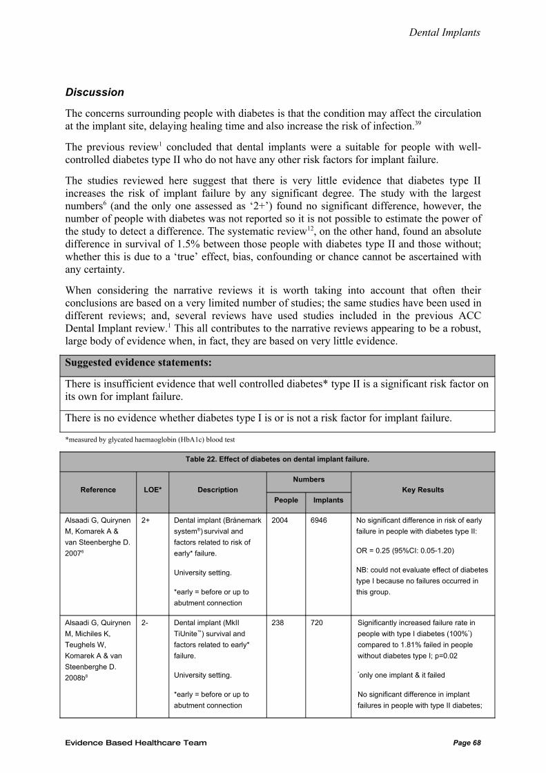
Dental Implants
Discussion
The concerns surrounding people with diabetes is that the condition may affect the circulation at the implant site, delaying healing time and also increase the risk of infection.39
The previous review1 concluded that dental implants were a suitable for people with well-controlled diabetes type II who do not have any other risk factors for implant failure.
The studies reviewed here suggest that there is very little evidence that diabetes type II increases the risk of implant failure by any significant degree. The study with the largest numbers6 (and the only one assessed as ‘2+’) found no significant difference, however, the number of people with diabetes was not reported so it is not possible to estimate the power of the study to detect a difference. The systematic review12, on the other hand, found an absolute difference in survival of 1.5% between those people with diabetes type II and those without; whether this is due to a ‘true’ effect, bias, confounding or chance cannot be ascertained with any certainty.
When considering the narrative reviews it is worth taking into account that often their conclusions are based on a very limited number of studies; the same studies have been used in different reviews; and, several reviews have used studies included in the previous ACC Dental Implant review.1 This all contributes to the narrative reviews appearing to be a robust, large body of evidence when, in fact, they are based on very little evidence.
Suggested evidence statements:
There is insufficient evidence that well controlled diabetes* type II is a significant risk factor on its own for implant failure.
There is no evidence whether diabetes type I is or is not a risk factor for implant failure.
*measured by glycated haemaoglobin (HbA1c) blood test
Table 22. Effect of diabetes on dental implant failure.
Reference LOE* Description
Numbers
People Implants
Key Results
Alsaadi G, Quirynen
M, Komarek A &
van Steenberghe D.
20076
2+ Dental implant (Brånemark
system®) survival and
factors related to risk of
early* failure.
University setting.
*early = before or up to
abutment connection
2004 6946 No significant difference in risk of early
failure in people with diabetes type II:
OR = 0.25 (95%CI: 0.05-1.20)
NB: could not evaluate effect of diabetes
type I because no failures occurred in
this group.
Alsaadi G, Quirynen
M, Michiles K,
Teughels W,
Komarek A & van
Steenberghe D.
2008b8
2- Dental implant (MkII
TiUnite™) survival and
factors related to early*
failure.
University setting.
*early = before or up to
abutment connection
238 720 Significantly increased failure rate in
people with type I diabetes (100%*)
compared to 1.81% failed in people
without diabetes type I; p=0.02
*only one implant & it failed
No significant difference in implant
failures in people with type II diabetes;
Evidence Based Healthcare Team Page 68
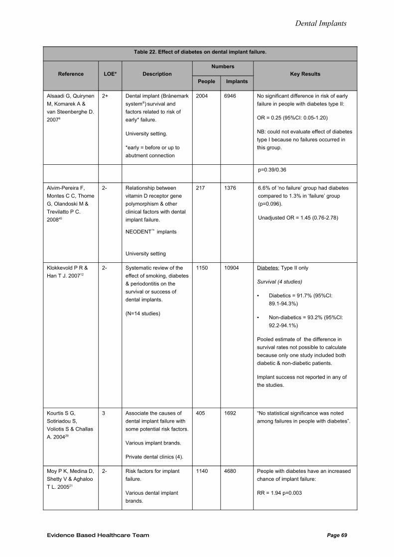
Dental Implants
Table 22. Effect of diabetes on dental implant failure.
Reference LOE* Description
Numbers
People Implants
Key Results
Alsaadi G, Quirynen
M, Komarek A &
van Steenberghe D.
20076
2+ Dental implant (Brånemark
system®) survival and
factors related to risk of
early* failure.
University setting.
*early = before or up to
abutment connection
2004 6946 No significant difference in risk of early
failure in people with diabetes type II:
OR = 0.25 (95%CI: 0.05-1.20)
NB: could not evaluate effect of diabetes
type I because no failures occurred in
this group.
p=0.39/0.36
Alvim-Pereira F,
Montes C C, Thome
G, Olandoski M &
Trevilatto P C.
200845
2- Relationship between
vitamin D receptor gene
polymorphism & other
clinical factors with dental
implant failure.
NEODENT™ implants
University setting
217 1376 6.6% of ‘no failure’ group had diabetes
compared to 1.3% in ‘failure’ group
(p=0.096).
Unadjusted OR = 1.45 (0.76-2.78)
Klokkevold P R &
Han T J. 200712
2- Systematic review of the
effect of smoking, diabetes
& periodontitis on the
survival or success of
dental implants.
(N=14 studies)
1150 10904 Diabetes: Type II only
Survival (4 studies)
• Diabetics = 91.7% (95%CI:
89.1-94.3%)
• Non-diabetics = 93.2% (95%CI:
92.2-94.1%)
Pooled estimate of the difference in
survival rates not possible to calculate
because only one study included both
diabetic & non-diabetic patients.
Implant success not reported in any of
the studies.
Kourtis S G,
Sotiriadou S,
Voliotis S & Challas
A. 200429
3 Associate the causes of
dental implant failure with
some potential risk factors.
Various implant brands.
Private dental clinics (4).
405 1692 “No statistical significance was noted
among failures in people with diabetes”.
Moy P K, Medina D,
Shetty V & Aghaloo
T L. 200521
2- Risk factors for implant
failure.
Various dental implant
brands.
1140 4680 People with diabetes have an increased
chance of implant failure:
RR = 1.94 p=0.003
Evidence Based Healthcare Team Page 69
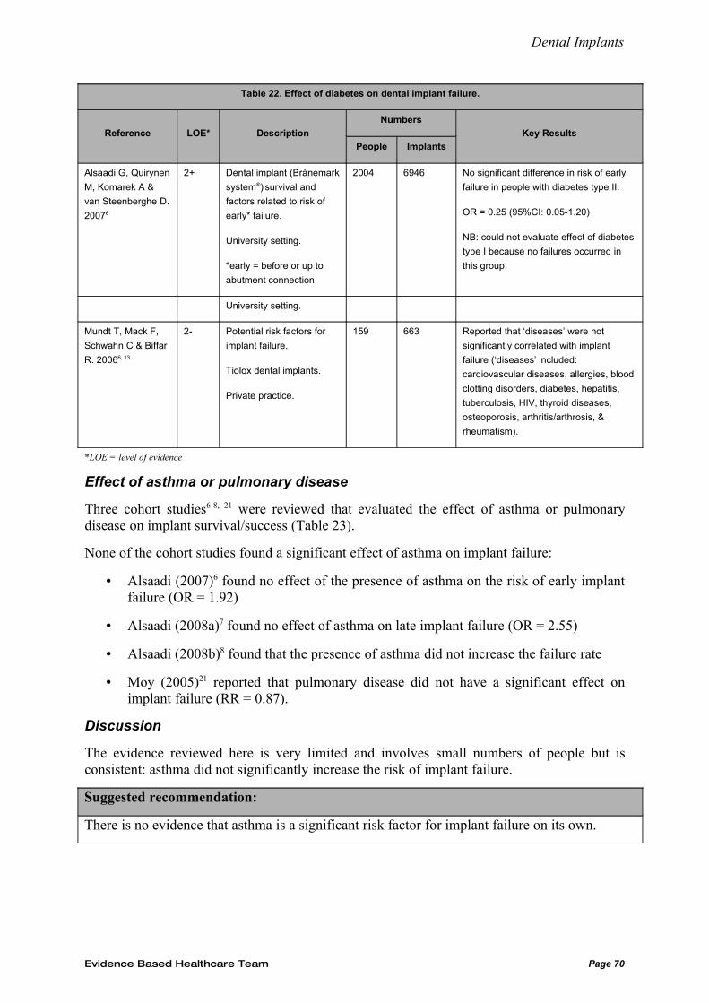
Dental Implants
Table 22. Effect of diabetes on dental implant failure.
Reference LOE* Description
Numbers
People Implants
Key Results
Alsaadi G, Quirynen
M, Komarek A &
van Steenberghe D.
20076
2+ Dental implant (Brånemark
system®) survival and
factors related to risk of
early* failure.
University setting.
*early = before or up to
abutment connection
2004 6946 No significant difference in risk of early
failure in people with diabetes type II:
OR = 0.25 (95%CI: 0.05-1.20)
NB: could not evaluate effect of diabetes
type I because no failures occurred in
this group.
University setting.
Mundt T, Mack F,
Schwahn C & Biffar
R. 20066, 13
2- Potential risk factors for
implant failure.
Tiolox dental implants.
Private practice.
159 663 Reported that ‘diseases’ were not
significantly correlated with implant
failure (‘diseases’ included:
cardiovascular diseases, allergies, blood
clotting disorders, diabetes, hepatitis,
tuberculosis, HIV, thyroid diseases,
osteoporosis, arthritis/arthrosis, &
rheumatism).
*LOE = level of evidence
Effect of asthma or pulmonary disease
Three cohort studies6-8, 21 were reviewed that evaluated the effect of asthma or pulmonary disease on implant survival/success (Table 23).
None of the cohort studies found a significant effect of asthma on implant failure:
• Alsaadi (2007)6 found no effect of the presence of asthma on the risk of early implant failure (OR = 1.92)
• Alsaadi (2008a)7 found no effect of asthma on late implant failure (OR = 2.55)
• Alsaadi (2008b)8 found that the presence of asthma did not increase the failure rate
• Moy (2005)21 reported that pulmonary disease did not have a significant effect on implant failure (RR = 0.87).
Discussion
The evidence reviewed here is very limited and involves small numbers of people but is consistent: asthma did not significantly increase the risk of implant failure.
Suggested recommendation:
There is no evidence that asthma is a significant risk factor for implant failure on its own.
Evidence Based Healthcare Team Page 70
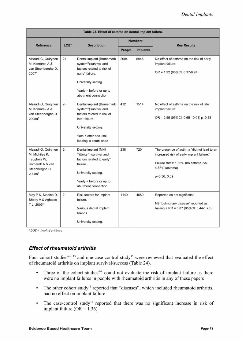
Dental Implants
Table 23. Effect of asthma on dental implant failure.
Reference LOE* Description
Numbers
People Implants
Key Results
Alsaadi G, Quirynen
M, Komarek A &
van Steenberghe D.
20076
2+ Dental implant (Brånemark
system®) survival and
factors related to risk of
early* failure.
University setting.
*early = before or up to
abutment connection
2004 6946 No effect of asthma on the risk of early
implant failure:
OR = 1.92 (95%CI: 0.37-9.97)
Alsaadi G, Quirynen
M, Komarek A &
van Steenberghe D.
2008a7
2- Dental implant (Brånemark
system®) survival and
factors related to risk of
late* failure.
University setting.
*late = after occlusal
loading is established
412 1514 No effect of asthma on the risk of late
implant failure:
OR = 2.55 (95%CI: 0.65-10.01) p=0.18
Alsaadi G, Quirynen
M, Michiles K,
Teughels W,
Komarek A & van
Steenberghe D.
2008b8
2- Dental implant (MkII
TiUnite™) survival and
factors related to early*
failure.
University setting.
*early = before or up to
abutment connection
238 720 The presence of asthma “did not lead to an
increased risk of early implant failure.”
Failure rates: 1.86% (no asthma) vs.
4.55% (asthma)
p=0.36; 0.39
Moy P K, Medina D,
Shetty V & Aghaloo
T L. 200521
2- Risk factors for implant
failure.
Various dental implant
brands.
University setting.
1140 4680 Reported as not significant.
NB “pulmonary disease” reported as
having a RR = 0.87 (95%CI: 0.44-1.73)
*LOE = level of evidence
Effect of rheumatoid arthritis
Four cohort studies6-8, 13 and one case-control study45 were reviewed that evaluated the effect of rheumatoid arthritis on implant survival/success (Table 24).
• Three of the cohort studies6-8 could not evaluate the risk of implant failure as there were no implant failures in people with rheumatoid arthritis in any of these papers
• The other cohort study13 reported that “diseases”, which included rheumatoid arthritis, had no effect on implant failure
• The case-control study45 reported that there was no significant increase in risk of implant failure (OR = 1.36).
Evidence Based Healthcare Team Page 71
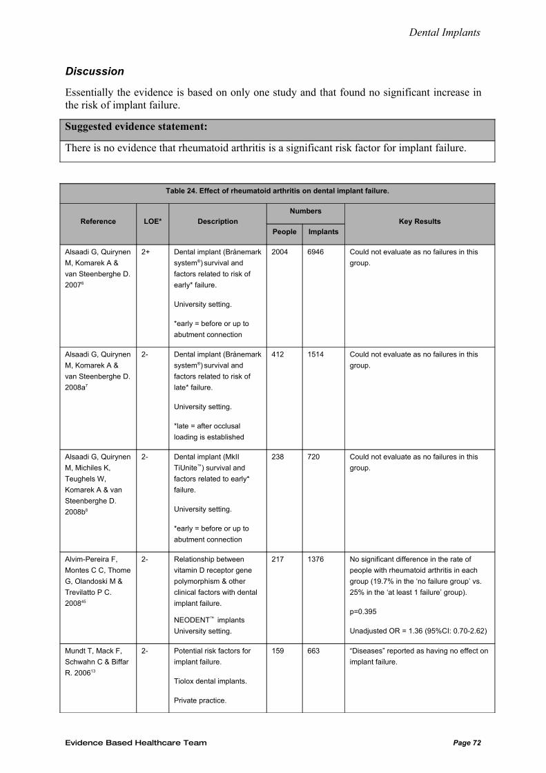
Dental Implants
Discussion
Essentially the evidence is based on only one study and that found no significant increase in the risk of implant failure.
Suggested evidence statement:
There is no evidence that rheumatoid arthritis is a significant risk factor for implant failure.
Table 24. Effect of rheumatoid arthritis on dental implant failure.
Reference LOE* Description
Numbers
People Implants
Key Results
Alsaadi G, Quirynen
M, Komarek A &
van Steenberghe D.
20076
2+ Dental implant (Brånemark
system®) survival and
factors related to risk of
early* failure.
University setting.
*early = before or up to
abutment connection
2004 6946 Could not evaluate as no failures in this
group.
Alsaadi G, Quirynen
M, Komarek A &
van Steenberghe D.
2008a7
2- Dental implant (Brånemark
system®) survival and
factors related to risk of
late* failure.
University setting.
*late = after occlusal
loading is established
412 1514 Could not evaluate as no failures in this
group.
Alsaadi G, Quirynen
M, Michiles K,
Teughels W,
Komarek A & van
Steenberghe D.
2008b8
2- Dental implant (MkII
TiUnite™) survival and
factors related to early*
failure.
University setting.
*early = before or up to
abutment connection
238 720 Could not evaluate as no failures in this
group.
Alvim-Pereira F,
Montes C C, Thome
G, Olandoski M &
Trevilatto P C.
200845
2- Relationship between
vitamin D receptor gene
polymorphism & other
clinical factors with dental
implant failure.
NEODENT™ implants
University setting.
217 1376 No significant difference in the rate of
people with rheumatoid arthritis in each
group (19.7% in the ‘no failure group’ vs.
25% in the ‘at least 1 failure’ group).
p=0.395
Unadjusted OR = 1.36 (95%CI: 0.70-2.62)
Mundt T, Mack F,
Schwahn C & Biffar
R. 200613
2- Potential risk factors for
implant failure.
Tiolox dental implants.
Private practice.
159 663 “Diseases” reported as having no effect on
implant failure.
Evidence Based Healthcare Team Page 72
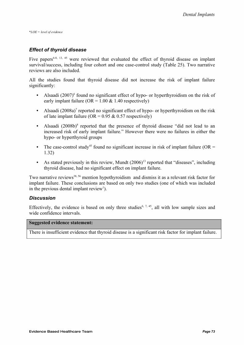
Dental Implants
*LOE = level of evidence
Effect of thyroid disease
Five papers6-8, 13, 45 were reviewed that evaluated the effect of thyroid disease on implant survival/success, including four cohort and one case-control study (Table 25). Two narrative reviews are also included.
All the studies found that thyroid disease did not increase the risk of implant failure significantly:
• Alsaadi (2007)6 found no significant effect of hypo- or hyperthyroidism on the risk of early implant failure (OR = 1.00 & 1.40 respectively)
• Alsaadi (2008a)7 reported no significant effect of hypo- or hyperthyroidism on the risk of late implant failure (OR = 0.95 & 0.57 respectively)
• Alsaadi (2008b)8 reported that the presence of thyroid disease “did not lead to an increased risk of early implant failure.” However there were no failures in either the hypo- or hyperthyroid groups
• The case-control study45 found no significant increase in risk of implant failure (OR = 1.32)
• As stated previously in this review, Mundt (2006)13 reported that “diseases”, including thyroid disease, had no significant effect on implant failure.
Two narrative reviews38, 56 mention hypothyroidism and dismiss it as a relevant risk factor for implant failure. These conclusions are based on only two studies (one of which was included in the previous dental implant review1).
Discussion
Effectively, the evidence is based on only three studies6, 7, 45, all with low sample sizes and wide confidence intervals.
Suggested evidence statement:
There is insufficient evidence that thyroid disease is a significant risk factor for implant failure.
Evidence Based Healthcare Team Page 73
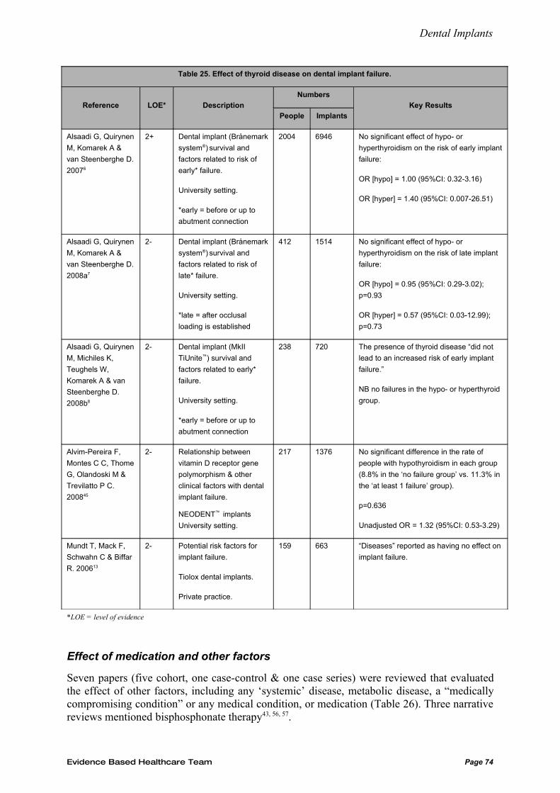
Dental Implants
Table 25. Effect of thyroid disease on dental implant failure.
Reference LOE* Description
Numbers
People Implants
Key Results
Alsaadi G, Quirynen
M, Komarek A &
van Steenberghe D.
20076
2+ Dental implant (Brånemark
system®) survival and
factors related to risk of
early* failure.
University setting.
*early = before or up to
abutment connection
2004 6946 No significant effect of hypo- or
hyperthyroidism on the risk of early implant
failure:
OR [hypo] = 1.00 (95%CI: 0.32-3.16)
OR [hyper] = 1.40 (95%CI: 0.007-26.51)
Alsaadi G, Quirynen
M, Komarek A &
van Steenberghe D.
2008a7
2- Dental implant (Brånemark
system®) survival and
factors related to risk of
late* failure.
University setting.
*late = after occlusal
loading is established
412 1514 No significant effect of hypo- or
hyperthyroidism on the risk of late implant
failure:
OR [hypo] = 0.95 (95%CI: 0.29-3.02);
p=0.93
OR [hyper] = 0.57 (95%CI: 0.03-12.99);
p=0.73
Alsaadi G, Quirynen
M, Michiles K,
Teughels W,
Komarek A & van
Steenberghe D.
2008b8
2- Dental implant (MkII
TiUnite™) survival and
factors related to early*
failure.
University setting.
*early = before or up to
abutment connection
238 720 The presence of thyroid disease “did not
lead to an increased risk of early implant
failure.”
NB no failures in the hypo- or hyperthyroid
group.
Alvim-Pereira F,
Montes C C, Thome
G, Olandoski M &
Trevilatto P C.
200845
2- Relationship between
vitamin D receptor gene
polymorphism & other
clinical factors with dental
implant failure.
NEODENT™ implants
University setting.
217 1376 No significant difference in the rate of
people with hypothyroidism in each group
(8.8% in the ‘no failure group’ vs. 11.3% in
the ‘at least 1 failure’ group).
p=0.636
Unadjusted OR = 1.32 (95%CI: 0.53-3.29)
Mundt T, Mack F,
Schwahn C & Biffar
R. 200613
2- Potential risk factors for
implant failure.
Tiolox dental implants.
Private practice.
159 663 “Diseases” reported as having no effect on
implant failure.
*LOE = level of evidence
Effect of medication and other factors
Seven papers (five cohort, one case-control & one case series) were reviewed that evaluated the effect of other factors, including any ‘systemic’ disease, metabolic disease, a “medically compromising condition” or any medical condition, or medication (Table 26). Three narrative reviews mentioned bisphosphonate therapy43, 56, 57.
Evidence Based Healthcare Team Page 74
Dental Implants
Medical conditions:
• The case-control study45 found no differences in the proportions of people with a systemic disease in either group
• The three cohort studies13, 27, 53 found no association between having a ‘medically compromising condition’53, ‘diseases’13 (including cardiovascular disease, allergies, blood clotting disorders, diabetes, hepatitis, TB, HIV, thyroid disease, osteoporosis, rheumatism, and arthritis/arthrosis) or a past history of any medical condition and dental implant failure27
• In contrast, the case series29 found that more implants failed in people who had a metabolic disease (p<0.001).
Medications:
• Three studies8, 21, 45 concluded that taking steroid medication was not associated with implant failure
• Alvim-Pereira (2008)45 also found no association between taking no medication, or taking anti-hypertensive, statin, antimicrobial, non-steroidal anti-inflammatory or hormone replacement medication and risk of implant failure
• Alsaadi (2008b)8 also found no association between taking antidepressant medication and implant failure
• In regard to antibiotics, one study8 found no relationship between taking antibiotics at the time of implant surgery and implant failure, another27 found that people who were allergic to penicillin had a greater failure rate than those people who were not, but the statistical significance is unclear
• Wagenberg and Froum (2006)27 also found no correlation between implant failure and taking any single medication or combination of medications.
• All three narrative reviews43, 56, 57 stated that sytemic bisphosphonate medication was a contraindication to implant placement due to the risk of osteonecrosis of the jaw.
Discussion
All of the studies, except one case series29, found no association between various ‘diseases’ and implant failure. In addition, no association was found with steroid, antidepressant, antihypertensive, statin, antimicrobial or non-steroidal anti-inflammatory medications. However, there is a strong consensus that bisphosphonate medications are a contraindication to implant placement due to the risk of osteonecrosis of the jaw.
Most of the variables investigated in these studies were not the primary focus of the respective studies, consisted of small numbers of people and were rather crude measures – all of which jeopardises the validity of the findings for these ‘secondary’ exposures of interest.
Suggested evidence statement:
There is insufficient evidence that any medical condition (other than those already evaluated in this review) is a significant risk factor for implant failure.
There is insufficient evidence that steroid, antidepressant, antihypertensive, statin, antimicrobial
Evidence Based Healthcare Team Page 75
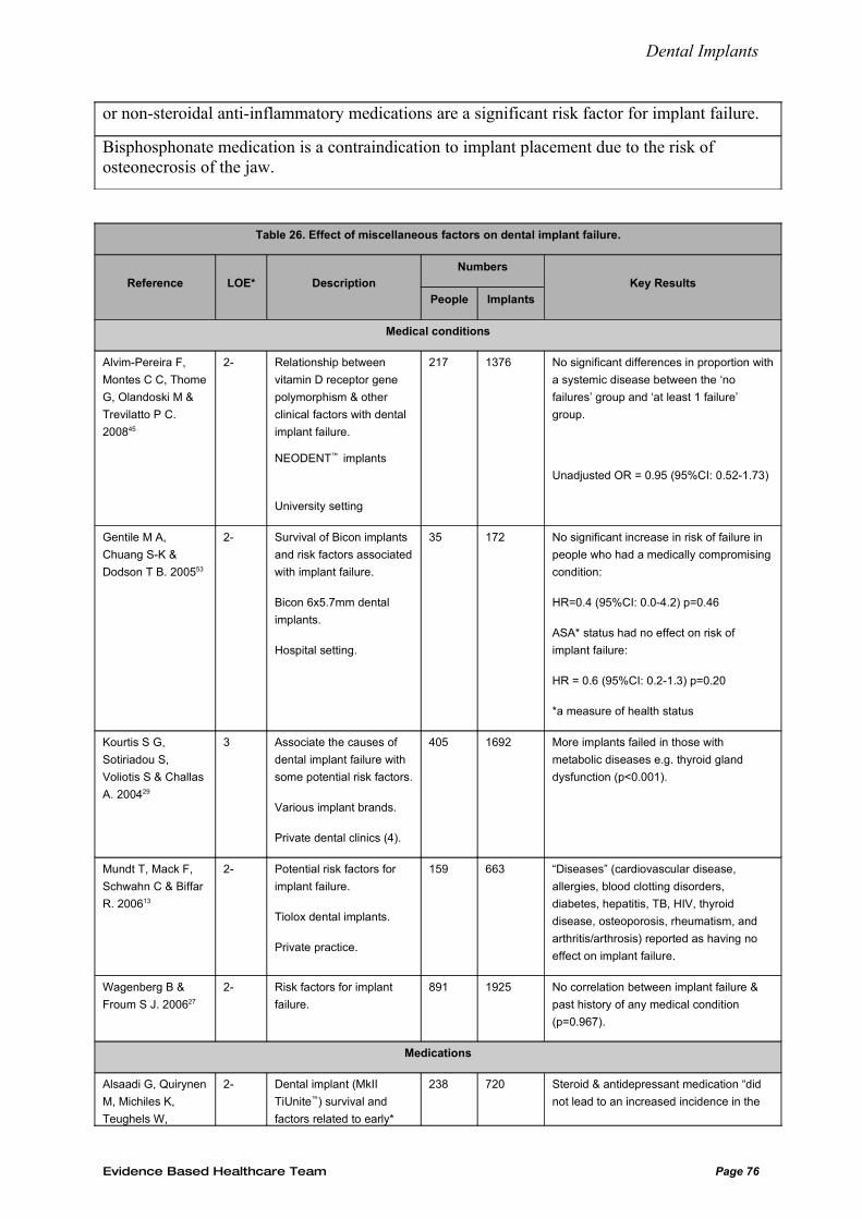
Dental Implants
or non-steroidal anti-inflammatory medications are a significant risk factor for implant failure.
Bisphosphonate medication is a contraindication to implant placement due to the risk of osteonecrosis of the jaw.
Table 26. Effect of miscellaneous factors on dental implant failure.
Reference LOE* Description
Numbers
People Implants
Key Results
Medical conditions
Alvim-Pereira F,
Montes C C, Thome
G, Olandoski M &
Trevilatto P C.
200845
2- Relationship between
vitamin D receptor gene
polymorphism & other
clinical factors with dental
implant failure.
NEODENT™ implants
University setting
217 1376 No significant differences in proportion with
a systemic disease between the ‘no
failures’ group and ‘at least 1 failure’
group.
Unadjusted OR = 0.95 (95%CI: 0.52-1.73)
Gentile M A,
Chuang S-K &
Dodson T B. 200553
2- Survival of Bicon implants
and risk factors associated
with implant failure.
Bicon 6x5.7mm dental
implants.
Hospital setting.
35 172 No significant increase in risk of failure in
people who had a medically compromising
condition:
HR=0.4 (95%CI: 0.0-4.2) p=0.46
ASA* status had no effect on risk of
implant failure:
HR = 0.6 (95%CI: 0.2-1.3) p=0.20
*a measure of health status
Kourtis S G,
Sotiriadou S,
Voliotis S & Challas
A. 200429
3 Associate the causes of
dental implant failure with
some potential risk factors.
Various implant brands.
Private dental clinics (4).
405 1692 More implants failed in those with
metabolic diseases e.g. thyroid gland
dysfunction (p<0.001).
Mundt T, Mack F,
Schwahn C & Biffar
R. 200613
2- Potential risk factors for
implant failure.
Tiolox dental implants.
Private practice.
159 663 “Diseases” (cardiovascular disease,
allergies, blood clotting disorders,
diabetes, hepatitis, TB, HIV, thyroid
disease, osteoporosis, rheumatism, and
arthritis/arthrosis) reported as having no
effect on implant failure.
Wagenberg B &
Froum S J. 200627
2- Risk factors for implant
failure.
891 1925 No correlation between implant failure &
past history of any medical condition
(p=0.967).
Medications
Alsaadi G, Quirynen
M, Michiles K,
Teughels W,
2- Dental implant (MkII
TiUnite™) survival and
factors related to early*
238 720 Steroid & antidepressant medication “did
not lead to an increased incidence in the
Evidence Based Healthcare Team Page 76
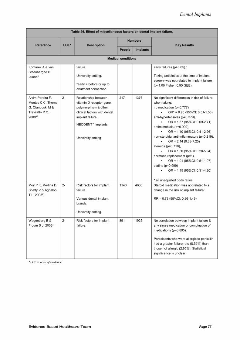
Dental Implants
Table 26. Effect of miscellaneous factors on dental implant failure.
Reference LOE* Description
Numbers
People Implants
Key Results
Medical conditions
Komarek A & van
Steenberghe D.
2008b8
failure.
University setting.
*early = before or up to
abutment connection
early failures (p>0.05).”
Taking antibiotics at the time of implant
surgery was not related to implant failure
(p=1.00 Fisher; 0.95 GEE).
Alvim-Pereira F,
Montes C C, Thome
G, Olandoski M &
Trevilatto P C.
200845
2- Relationship between
vitamin D receptor gene
polymorphism & other
clinical factors with dental
implant failure.
NEODENT™ implants
University setting
217 1376 No significant differences in risk of failure
when taking:
no medication (p=0.777),
• OR* = 0.90 (95%CI: 0.51-1.56)
anti-hypertensives (p=0.379),
• OR = 1.37 (95%CI: 0.69-2.71)
antimicrobials (p=0.999),
• OR = 1.10 (95%CI: 0.41-2.96)
non-steroidal anti-inflammatory (p=0.219),
• OR = 2.14 (0.63-7.25)
steroids (p=0.710),
• OR = 1.30 (95%CI: 0.28-5.94)
hormone replacement (p=1),
• OR = 1.01 (95%CI: 0.51-1.97)
statins (p=0.999)
• OR = 1.15 (95%CI: 0.31-4.20)
* all unadjusted odds ratios
Moy P K, Medina D,
Shetty V & Aghaloo
T L. 200521
2- Risk factors for implant
failure.
Various dental implant
brands.
University setting.
1140 4680 Steroid medication was not related to a
change in the risk of implant failure:
RR = 0.73 (95%CI: 0.36-1.49)
Wagenberg B &
Froum S J. 200627
2- Risk factors for implant
failure.
891 1925 No correlation between implant failure &
any single medication or combination of
medications (p=0.895).
Participants who were allergic to penicillin
had a greater failure rate (8.52%) than
those not allergic (2.95%). Statistical
significance is unclear.
*LOE = level of evidence
Evidence Based Healthcare Team Page 77
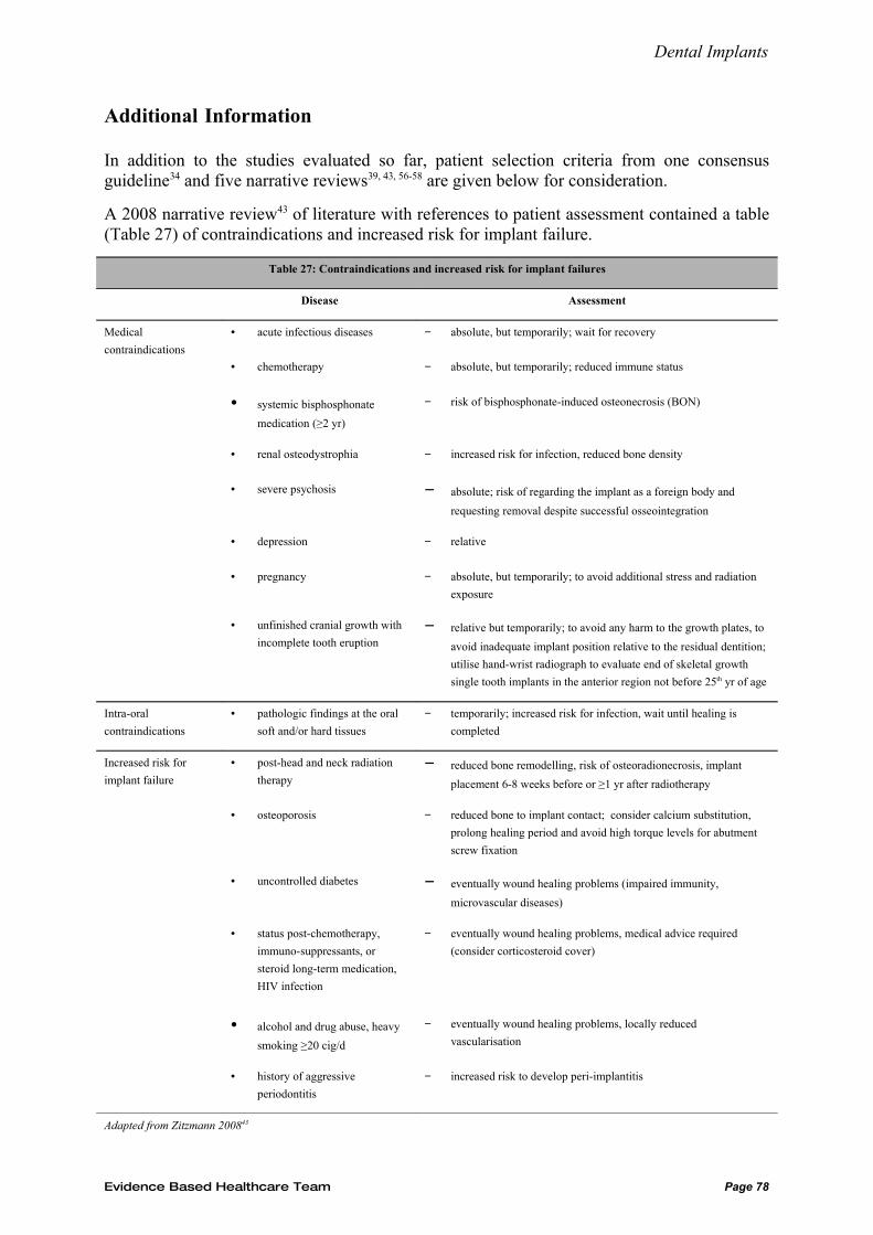
Dental Implants
Additional Information
In addition to the studies evaluated so far, patient selection criteria from one consensus guideline34 and five narrative reviews39, 43, 56-58 are given below for consideration.
A 2008 narrative review43 of literature with references to patient assessment contained a table (Table 27) of contraindications and increased risk for implant failure.
Table 27: Contraindications and increased risk for implant failures
Disease Assessment
Medical
contraindications
• acute infectious diseases − absolute, but temporarily; wait for recovery
• chemotherapy − absolute, but temporarily; reduced immune status
• systemic bisphosphonate
medication (≥2 yr)
− risk of bisphosphonate-induced osteonecrosis (BON)
• renal osteodystrophia − increased risk for infection, reduced bone density
• severe psychosis − absolute; risk of regarding the implant as a foreign body and
requesting removal despite successful osseointegration
• depression − relative
• pregnancy − absolute, but temporarily; to avoid additional stress and radiation
exposure
• unfinished cranial growth with
incomplete tooth eruption− relative but temporarily; to avoid any harm to the growth plates, to
avoid inadequate implant position relative to the residual dentition;
utilise hand-wrist radiograph to evaluate end of skeletal growth
single tooth implants in the anterior region not before 25th yr of age
Intra-oral
contraindications
• pathologic findings at the oral
soft and/or hard tissues
− temporarily; increased risk for infection, wait until healing is
completed
Increased risk for
implant failure
• post-head and neck radiation
therapy− reduced bone remodelling, risk of osteoradionecrosis, implant
placement 6-8 weeks before or ≥1 yr after radiotherapy
• osteoporosis − reduced bone to implant contact; consider calcium substitution,
prolong healing period and avoid high torque levels for abutment
screw fixation
• uncontrolled diabetes − eventually wound healing problems (impaired immunity,
microvascular diseases)
• status post-chemotherapy,
immuno-suppressants, or
steroid long-term medication,
HIV infection
− eventually wound healing problems, medical advice required
(consider corticosteroid cover)
• alcohol and drug abuse, heavy
smoking ≥20 cig/d
− eventually wound healing problems, locally reduced
vascularisation
• history of aggressive
periodontitis
− increased risk to develop peri-implantitis
Adapted from Zitzmann 200843
Evidence Based Healthcare Team Page 78
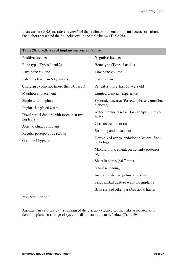
Dental Implants
In an earlier (2005) narrative review39 of the predictors of dental implant success or failure, the authors presented their conclusions in the table below (Table 28).
Table 28: Predictors of implant success or failure.
Positive factors
Bone type (Types 1 and 2)
High bone volume
Patient is less than 60 years old
Clinician experience (more than 50 cases)
Mandibular placement
Single tooth implant
Implant length >0.8 mm
Fixed partial denture with more than two implants
Axial loading of implant
Regular postoperative recalls
Good oral hygiene
Negative factors
Bone type (Types 3 and 4)
Low bone volume
Osteonecrosis
Patient is more than 60 years old
Limited clinician experience
Systemic diseases (for example, uncontrolled diabetes)
Auto-immune disease (for example, lupus or HIV)
Chronic periodontitis
Smoking and tobacco use
Unresolved caries, endodontic lesions, frank pathology
Maxillary placement, particularly posterior region
Short implants (<0.7 mm)
Acentric loading
Inappropriate early clinical loading
Fixed partial denture with two implants
Bruxism and other parafunctional habits
Adapted from Porter 200539
Another narrative review57 summarised the current evidence for the risks associated with dental implants in a range of systemic disorders in the table below (Table 29).
Evidence Based Healthcare Team Page 79
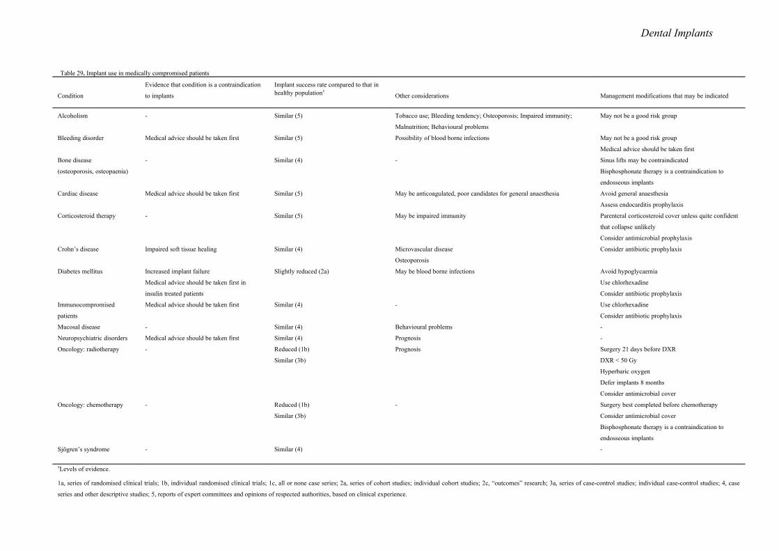
Dental Implants
Table 29. Implant use in medically compromised patients
Condition
Evidence that condition is a contraindication
to implants
Implant success rate compared to that in healthy population†
Other considerations Management modifications that may be indicated
Alcoholism - Similar (5) Tobacco use; Bleeding tendency; Osteoporosis; Impaired immunity;
Malnutrition; Behavioural problems
May not be a good risk group
Bleeding disorder Medical advice should be taken first Similar (5) Possibility of blood borne infections May not be a good risk group
Medical advice should be taken first
Bone disease
(osteoporosis, osteopaenia)
- Similar (4) - Sinus lifts may be contraindicated
Bisphosphonate therapy is a contraindication to
endosseous implants
Cardiac disease Medical advice should be taken first Similar (5) May be anticoagulated, poor candidates for general anaesthesia Avoid general anaesthesia
Assess endocarditis prophylaxis
Corticosteroid therapy - Similar (5) May be impaired immunity Parenteral corticosteroid cover unless quite confident
that collapse unlikely
Consider antimicrobial prophylaxis
Crohn’s disease Impaired soft tissue healing Similar (4) Microvascular disease
Osteoporosis
Consider antibiotic prophylaxis
Diabetes mellitus Increased implant failure
Medical advice should be taken first in
insulin treated patients
Slightly reduced (2a) May be blood borne infections Avoid hypoglycaemia
Use chlorhexadine
Consider antibiotic prophylaxis
Immunocompromised
patients
Medical advice should be taken first Similar (4) - Use chlorhexadine
Consider antibiotic prophylaxis
Mucosal disease - Similar (4) Behavioural problems -
Neuropsychiatric disorders Medical advice should be taken first Similar (4) Prognosis -
Oncology: radiotherapy - Reduced (1b)
Similar (3b)
Prognosis Surgery 21 days before DXR
DXR < 50 Gy
Hyperbaric oxygen
Defer implants 8 months
Consider antimicrobial cover
Oncology: chemotherapy - Reduced (1b)
Similar (3b)
- Surgery best completed before chemotherapy
Consider antimicrobial cover
Bisphosphonate therapy is a contraindication to
endosseous implants
Sjögren’s syndrome - Similar (4) -
†Levels of evidence.
1a, series of randomised clinical trials; 1b, individual randomised clinical trials; 1c, all or none case series; 2a, series of cohort studies; individual cohort studies; 2c, “outcomes” research; 3a, series of case-control studies; individual case-control studies; 4, case
series and other descriptive studies; 5, reports of expert committees and opinions of respected authorities, based on clinical experience.
Dental Implants
Two narrative reviews of the literature, Hwang (200658 & 200756) list both the absolute and relative contraindications to implant therapy.
The absolute contraindications include:
• recent myocardial infarction and cerebrovascular accident
• valvular prosthesis surgery
• immunosuppression
• bleeding issues
• active treatment of malignancy
• drug abuse
• psychiatric illness
• intravenous bisphosphonate use.
The relative contraindications include:
• adolescence
• aging
• osteoporosis
• smoking
• diabetes
• positive interleukin-1 genotype
• being HIV positive
• cardiovascular disease
• hypothyroidism.
In a consensus guideline developed by the Academy of Osseointegration59 based on the results their 2006 Consensus Conference on the State of the Science on Implant Dentistry, the potential risk factors for implant failure were discussed. They concluded that:
• Smoking had an adverse effect on implant survival and success, especially in areas of trabecular bone.
• Type II diabetes may have an adverse effect on survival rates, although the limited number of studies on diabetes precluded a definitive conclusion.
• A history of treated periodontitis did not appear to affect implant survival rates adversely but periodontitis may have a negative effect on implant success rates, particularly over longer periods. The authors recommended that a periodontal evaluation and appropriate treatment be provided prior to implant placement.
Evidence Based Healthcare Team page 81
Dental Implants
• Several grafting procedures have proven successful in providing adequate bone quantity and quality in patients who have bone loss. The long-term (>5yrs) clinical success/survival of implants placed after maxillary sinus augmentation, regardless of grafting materials, compared favourably to implant placed with no grafting procedure.
Conclusions
It is concluded that dental implants are a successful intervention to replace teeth in University settings. Due to the relatively poor quality of the papers reviewed, guideline development will rely on consideration of the existing evidence, expert opinion and consensus.
The evidence for significant risk factors for implant failure is summarised, as follows:
• Smoking as a single risk factor may not increase the risk of failure sufficiently to deny treatment but dental implants may be contra-indicted in smokers who have other relevant risk factors. In addition, smokers seeking dental implant treatment should have their smoking history taken, be advised of the risk of implant failure and advised to stop smoking.
• Even though there is insufficient evidence that people with a history of treated periodontitis have a significantly elevated risk of implant failure, the consensus view is that people with periodontitis should be treated for this condition before being considered for implant therapy.
• In addition, even though there is insufficient evidence that oral health status or oral hygiene behaviour has a significant effect on the risk of implant failure, the consensus view is that people with poor oral hygiene, infection or uncontrolled caries should not be offered dental implants.
• There is weak evidence that dental implants placed in the maxilla may have a greater risk of failure.
• There is some weak evidence that implants placed after maxillary sinus augmentation may have an increased risk of failure but there is no evidence that other bone graft/augmentation techniques alter significantly the risk of implant failure.
• Older age, as a single risk factor, is not a contraindication for implant placement.
• There is a consensus opinion that implant placement should be delayed in young people until growth is complete
• There is some evidence that implant failure rates in the maxilla are usually significantly increased in people who have previously had irradiation of the jaw, regardless of the total radiation dose.
• There is a consensus opinion that dental implants should not be placed in people undergoing active chemotherapy.
• Medical advice should be sought for people with coagulation disorders or taking anticoagulant medication before proceeding with implant placement.
Evidence Based Healthcare Team Page 82
Dental Implants
• Bisphosphonate medication is a contraindication to implant placement due to the risk of osteonecrosis of the jaw.
• There is insufficient evidence that well controlled diabetes type II is a significant risl factor for implant failure
• There is no evidence whether diabetes type I, asthma, rheumatoid arthritis is or is not a significant risk factor for implant failure
• There is insufficient evidence that the following factors are significant risk factors for implant failure:
o type of edentulousness or proximity to natural dentition
o placement in the posterior region of a jaw
o osteoporosis
o type of prosthesis
o a history of chemotherapy treatment
o cardiovascular disease, including hypertension and hypercholesterolaemia
o thyroid disease, gastric problems or Crohn’s disease
o hormone replacement therapy, menopausal status or history of a radical hysterectomy
o steroid, antidepressant, antihypertensive, statin, antimicrobial or non-steroidal anti-inflammatory medications.
Evidence Based Healthcare Team Page 83
Dental Implants
References
1. Larking, P., Dental Implants: Patient Selection, Patient Satisfaction and Cost Factors, in Referees - Leichter J & Lyons K. 2004, Accident Compensation Commission.
2. Accident Compensation Corporation, Dental Implant Guideline. 2006.3. Esposito, M., et al., Interventions for replacing missing teeth:bone augmentation
techniques for dental implant treatment (Review). Cochrane Database of Systematic Reviews, 2008(1).
4. Vartoukian, S.R., et al., Does the referral and selection for NHS-funded dental implant treatment in the UK follow National Guidelines? Annals of the Royal College of Surgeons of England, 2007. 89(3): p. 247-51.
5. Scottish Intercollegiate Guidelines Network (2008 ).6. Alsaadi, G., et al., Impact of local and systemic factors on the incidence of oral
implant failures, up to abutment connection. Journal of Clinical Periodontology, 2007. 34(7): p. 610-7.
7. Alsaadi, G., et al., Impact of local and systemic factors on the incidence of late oral implant loss. Clinical Oral Implants Research, 2008a. 19(7): p. 670-6.
8. Alsaadi, G., et al., Impact of local and systemic factors on the incidence of failures up to abutment connection with modified surface oral implants. Journal of Clinical Periodontology, 2008b. 35(1): p. 51-7.
9. Anitua, E., et al., 5-year clinical experience with BTI dental implants: risk factors for implant failure. Journal of Clinical Periodontology, 2008. 35(8): p. 724-32.
10. Degidi, M., et al., Five-year outcome of 111 immediate nonfunctional single restorations. Journal of Oral Implantology, 2006. 32(6): p. 277-85.
11. Holahan, C.M., et al., Effect of osteoporotic status on the survival of titanium dental implants. International Journal of Oral & Maxillofacial Implants, 2008. 23(5): p. 905-10.
12. Klokkevold, P.R. and T.J. Han, How do smoking, diabetes, and periodontitis affect outcomes of implant treatment?[erratum appears in Int J Oral Maxillofac Implants. 2008 Jan-Feb;23(1):56]. International Journal of Oral & Maxillofacial Implants, 2007. 22 Suppl: p. 173-202.
13. Mundt, T., et al., Private practice results of screw-type tapered implants: survival and evaluation of risk factors. International Journal of Oral & Maxillofacial Implants, 2006. 21(4): p. 607-14.
14. Berglundh, T., L. Persson, and B. Klinge, A systematic review of the incidence of biological and technical complications in implant dentistry reported in prospective longitudinal studies of at least 5 years. Journal of Clinical Periodontology, 2002. 29 Suppl 3: p. 197-212; discussion 232-3.
15. Hinode, D., et al., Influence of smoking on osseointegrated implant failure: a meta-analysis. Clinical Oral Implants Research, 2006. 17(4): p. 473-8.
16. Strietzel, F.P., et al., Smoking interferes with the prognosis of dental implant treatment: a systematic review and meta-analysis. Journal of Clinical Periodontology, 2007. 34(6): p. 523-44.
17. McDermott, N.E., et al., Complications of dental implants: identification, frequency, and associated risk factors. International Journal of Oral & Maxillofacial Implants, 2003. 18(6): p. 848-55.
Evidence Based Healthcare Team Page 84
Dental Implants
18. Susarla, S.M., S.-K. Chuang, and T.B. Dodson, Delayed versus immediate loading of implants: survival analysis and risk factors for dental implant failure. Journal of Oral & Maxillofacial Surgery, 2008. 66(2): p. 251-5.
19. DeLuca, S., E. Habsha, and G.A. Zarb, The effect of smoking on osseointegrated dental implants. Part I: implant survival. International Journal of Prosthodontics, 2006. 19(5): p. 491-8.
20. Moheng, P. and J.-M. Feryn, Clinical and biologic factors related to oral implant failure: a 2-year follow-up study. Implant Dentistry, 2005. 14(3): p. 281-8.
21. Moy, P.K., et al., Dental implant failure rates and associated risk factors. International Journal of Oral and Maxillofacial Implants, 2005. 20(4): p. 569-77.
22. Noguerol, B., et al., Early implant failure. Prognostic capacity of Periotest: retrospective study of a large sample.[see comment]. Clinical Oral Implants Research, 2006. 17(4): p. 459-64.
23. Woo, V.V., et al., Dentoalveolar reconstructive procedures as a risk factor for implant failure. Journal of Oral & Maxillofacial Surgery, 2004. 62(7): p. 773-80.
24. Roos-Jansaker, A.M., et al., Nine- to fourteen-year follow-up of implant treatment. Part I: implant loss and associations to various factors. Journal of Clinical Periodontology, 2006. 33(4): p. 283-9.
25. Sjostrom, M., et al., Reconstruction of the atrophic edentulous maxilla with free iliac crest grafts and implants: a 3-year report of a prospective clinical study. Clinical Implant Dentistry & Related Research, 2007. 9(1): p. 46-59.
26. Sverzut, A.T., et al., The influence of tobacco on early dental implant failure. Journal of Oral & Maxillofacial Surgery, 2008. 66(5): p. 1004-9.
27. Wagenberg, B. and S.J. Froum, A retrospective study of 1925 consecutively placed immediate implants from 1988 to 2004. International Journal of Oral & Maxillofacial Implants, 2006. 21(1): p. 71-80.
28. Aykent, F., et al., A 1- to 12-year clinical evaluation of 106 endosseous implants supporting fixed and removable prostheses. International Journal of Periodontics & Restorative Dentistry, 2007. 27(4): p. 358-67.
29. Kourtis, S.G., et al., Private practice results of dental implants. Part I: survival and evaluation of risk factors--Part II: surgical and prosthetic complications. Implant Dentistry, 2004. 13(4): p. 373-85.
30. Sanchez-Perez, A., M.J. Moya-Villaescusa, and R.G. Caffesse, Tobacco as a risk factor for survival of dental implants. Journal of Periodontology, 2007. 78(2): p. 351-9.
31. Kinsel, R.P. and M. Liss, Retrospective analysis of 56 edentulous dental arches restored with 344 single-stage implants using an immediate loading fixed provisional protocol: statistical predictors of implant failure. International Journal of Oral & Maxillofacial Implants, 2007. 22(5): p. 823-30.
32. Lemmerman, K.J. and N.E. Lemmerman, Osseointegrated dental implants in private practice: a long-term case series study. Journal of Periodontology, 2005. 76(2): p. 310-9.
33. Levin, L., A. Laviv, and D. Schwartz-Arad, Long-term success of implants replacing a single molar. Journal of Periodontology, 2006. 77(9): p. 1528-1532.
34. Academy of Osseointegration, et al., Guidelines for the provision of dental implants.[see comment]. International Journal of Oral & Maxillofacial Implants, 2008. 23(3): p. 471-3.
35. A.D.A. Council on Scientific Affairs, Dental endosseous implants: an update. Journal of the American Dental Association, 2004. 135(1): p. 92-7.
Evidence Based Healthcare Team Page 85
Dental Implants
36. Baig, M.R. and M. Rajan, Effects of smoking on the outcome of implant treatment: a literature review. Indian Journal of Dental Research, 2007. 18(4): p. 190-5.
37. Johnson, G.K. and J.M. Guthmiller, The impact of cigarette smoking on periodontal disease and treatment. Periodontology 2000, 2007. 44: p. 178-94.
38. Paquette, D.W., N. Brodala, and R.C. Williams, Risk factors for endosseous dental implant failure. Dental Clinics of North America, 2006. 50(3): p. 361-74.
39. Porter, J.A. and J.A. von Fraunhofer, Implants. Success or failure of dental implants? A literature review with treatment considerations. General Dentistry, 2005. 53(6): p. 423-33.
40. Shah, K.C., et al., Treatment planning for the single-tooth implant restoration--general considerations and the pretreatment evaluation. Journal of the California Dental Association, 2008. 36(11): p. 827-34.
41. Torabinejad, M. and C.J. Goodacre, Endodontic or dental implant therapy: the factors affecting treatment planning. Journal of the American Dental Association, 2006. 137(7): p. 973-7; quiz 1027-8.
42. Wood, M.R. and S.G. Vermilyea, A review of selected dental literature on evidence-based treatment planning for dental implants: Report of the Committee on Research in Fixed Prosthodontics of the Academy of Fixed Prosthodontics. The Journal of Prosthetic Dentistry, 2004. 92(5): p. 447-462.
43. Zitzmann, N.U., et al., Patient assessment and diagnosis in implant treatment. Australian Dental Journal, 2008. 53 Suppl 1: p. S3-10.
44. McDermott, N.E., et al., Maxillary sinus augmentation as a risk factor for implant failure. International Journal of Oral & Maxillofacial Implants, 2006. 21(3): p. 366-74.
45. Alvim-Pereira, F., et al., Analysis of association of clinical aspects and vitamin D receptor gene polymorphism with dental implant loss. Clinical Oral Implants Research, 2008. 19(8): p. 786-95.
46. Evian, C.I., et al., Retrospective analysis of implant survival and the influence of periodontal disease and immediate placement on long-term results.[see comment]. International Journal of Oral & Maxillofacial Implants, 2004. 19(3): p. 393-8.
47. Karoussis, I.K., S. Kotsovilis, and I. Fourmousis, A comprehensive and critical review of dental implant prognosis in periodontally compromised partially edentulous patients. Clinical Oral Implants Research, 2007. 18(6): p. 669-79.
48. Ong, C.T.T., et al., Systematic review of implant outcomes in treated periodontitis subjects. Journal of Clinical Periodontology, 2008. 35(5): p. 438-62.
49. Schou, S., Implant treatment in periodontitis-susceptible patients: a systematic review. Journal of Oral Rehabilitation, 2008. 35 Suppl 1: p. 9-22.
50. Schou, S., et al., Outcome of implant therapy in patients with previous tooth loss due to periodontitis. Clinical Oral Implants Research, 2006. 17 Suppl 2: p. 104-23.
51. Herrmann, I., et al., Evaluation of patient and implant characteristics as potential prognostic factors for oral implant failures.[see comment]. International Journal of Oral & Maxillofacial Implants, 2005. 20(2): p. 220-30.
52. Elkhoury, J.S., et al., Clinical parameters associated with success and failure of single-tooth titanium plasma-sprayed cylindric implants under stricter criteria: a 5-year retrospective study. International Journal of Oral & Maxillofacial Implants, 2005. 20(5): p. 687-94.
53. Gentile, M.A., S.-K. Chuang, and T.B. Dodson, Survival estimates and risk factors for failure with 6 x 5.7-mm implants. International Journal of Oral & Maxillofacial Implants, 2005. 20(6): p. 930-7.
54. Tolstunov, L. and L. Tolstunov, Implant zones of the jaws: implant location and related success rate. Journal of Oral Implantology, 2007. 33(4): p. 211-20.
Evidence Based Healthcare Team Page 86
Dental Implants
55. Graziani, F., et al., Comparison of implant survival following sinus floor augmentation procedures with implants placed in pristine posterior maxillary bone: a systematic review.[see comment]. Clinical Oral Implants Research, 2004. 15(6): p. 677-82.
56. Hwang, D., et al., Medical contraindications to implant therapy: Part II: Relative contraindications. Implant Dentistry, 2007. 16(1): p. 13-23.
57. Scully, C., J. Hobkirk, and P.D. Dios, Dental endosseous implants in the medically compromised patient. Journal of Oral Rehabilitation, 2007. 34(8): p. 590-9.
58. Hwang, D., et al., Medical contraindications to implant therapy: part I: absolute contraindications. Implant Dentistry, 2006. 15(4): p. 353-60.
59. Academy of Osseointegration.60. Amorim, M.A.L., et al., Comparative study of axial and femoral bone mineral density
and parameters of mandibular bone quality in patients receiving dental implants.[see comment][republished in Osteoporos Int. 2007 May;18(5):703-9; PMID: 17506127]. Osteoporosis International, 2006. 17(10): p. 1494-500.
Appendix A:
Patient Factors:
MEDLINE
1. exp Dental Implants/px, ct [Psychology, Contraindications]2. exp Dental Implants/3. ((dental or tooth) adj implant$).mp.4. exp Decision Making/5. exp Patient Selection/6. exp Dentist-Patient Relations/7. Judgment/8. (patient selection or patient characteristics or psychological factor$).mp.9. or/4-810. (2 or 3) and 911. 10 or 112. limit 11 to human
EMBASE
1. exp Tooth Implantation/2. ((dental or tooth) adj implant$).af.3. exp Decision Making/4. exp Patient Selection/5. exp Psychological Aspect/6. exp TREATMENT CONTRAINDICATION/7. exp TREATMENT INDICATION/8. (patient selection or patient characteristics or psychological factor$).af.9. or/3-810. (1 or 2) and 9
PREMEDLINE
Evidence Based Healthcare Team Page 87
Dental Implants
1. ((dental or tooth) adj implant$).af.2. (patient selection or patient characteristics or psychological factor$).af.3. (contraindication$ or indication$ or risk factor$).af.4. 1 and (2 or 3)
CINAHL
1. exp Dental Implants/2. ((dental or tooth) adj implant$).mp.3. exp Decision Making/4. exp Patient Selection/5. exp Dentist-Patient Relations/6. Judgment/7. exp Risk Factors/8. Psychosocial Aspects of Illness/9. (patient selection or patient characteristic$ or contraindication$ or psychological factor$).mp.10. exp Patients/11. or/3-1012. (1 or 2) and 11
All EBM Reviews (Cochrane DSR, ACP Journal Club, DARE, CCTR)
exp dental implants/ (((dental or tooth) adj implant$) or (branemark or stereos)).mp. or/1-2 exp decision making/ exp patient selection/ exp dentist-patient relations/ exp judgment/ exp risk factors/ (patient selection or patient characteristic$ or contraindication$ or risk factor$ or psychological factor$).mp. or/4-9 3 and 10
PsycINFO
((dental or tooth or teeth) and implant$).af.prosthodontic$.mp. not 2 [mp=title, abstract, heading word, table of contents, key concepts]1 or 2
Risk Factors:
MEDLINE
1. exp Risk Factors/2. exp Dental Implants/3. ((dental or tooth) adj implant$).af.
Evidence Based Healthcare Team Page 88

Dental Implants
4. risk factor$.af.5. (1 or 4) and (2 or 3)
EMBASE
1. exp Risk Factor/2. risk factor$.af.3. exp Tooth Implantation/4. ((dental or tooth) adj implant$).af.5. (1 or 2) and (3 or 4)
Evidence Based Healthcare Team Page 89
Dental Implants Appendix B:
Evidence Based Healthcare Table Reference: Alsaadi 2007 Bibliographic Number: 5
Design Participants Exposures(total patients/implants)
Outcomes
Evidence Based Healthcare Team page 90
Dental ImplantsRetrospective cohort
Biases/weaknesses:
Age not reported or
adjusted for
~17% implants not
included in multivariate
analysis
Type of edentulism and
PTV value still possible
confounders
Oral status e.g.
presence of
periodontitis, & oral
hygiene habits not
accounted for as
potential confounders
Description:
N=2004 consecutive patients “from the total patient population” who had had
6946 dental implants placed between 1982-2003 at a single university
department in Belgium.
Group:
Mean Age: not reported
Gender: 1212 women, 792 men
Implants: Brånemark system® (6316 machined & 630 Ti-Unite® surface)
Drop-outs: Unable to get records for 12% (232/2004) patients with 17%
implants
Follow-up = 21yrs
Setting: University
Reliability of surgical records tested by comparing with medical records (700
people)
Inclusions:
Those patients with full records available.
Exclusions:
Patient files could not be retrieved.
Selection Notes:
• Failure = peri-implant radiolucency on intra-oral X-rays; implants
showed slightest sign of mobility measured by PTV ≥ 5 or patient
showed subjective signs of infection or pain that required removal.
• Logistic regression analysis was used: univariate effect then
multivariate effect for those that were univariately significant at 5% i.e.
smoking, implant length/diameter/location, bone quantity/quality. NB
type of edentulism and PTV not used as only available for a limited
number of patients.
• Generalising estimating equation (GEE) used to account for that
repeated observations (several implants ) were available for a single
participant
• For the multivariate analysis, implants with missing data on bone quality
and quantity were removed (1100 implants). Also, it was not possible to
statistically evaluate effect of chemotherapy, diabetes type I and
rheumatoid arthritis as there were no failures in these groups.
• No statistical difference between failure rate of both type of implant
surface (p>0.05) therefore all implants grouped together.
• Smoking, hypertension,
ischaemic cardiac
problems, coagulation
anomalies, gastric
problems, osteoporosis,
hypo- or
hyperthyroidism,
hypercholesterolaemia,
asthma, diabetes type I
or II, Crohn’s disease,
rheumatoid arthritis,
chemotherapy and
medications
(2004/6946).
• Implant length
(2004/6946)
• Implant diameter
(2004/6936)
• Implant location
(2000/6931)
• Bone quantity
(1759/5800)
• Bone quality
(1759/5782)
• Type of edentulism
(676/2448)
• Antibiotics pre- or
immediately after
surgery (not reported)
• Dehiscence during
surgery (430/1380)
• Perforation of jaw during
surgery (418/1345)
• Periotest® value (71/189)
• Placement torque
measurement (138/320)
Outcome Measures:
Overall success rate (%)
Odds of early implant failure* (OR; 95%CI; p-value)
*Early failure = before or up to abutment connection
Results:
Overall success rate: 96.4%
252/6946 implants failed 1-6 months after placement
(3.6%) in 178 patients.
Odds of early implant failure:
Univariate analysis:
• Implant location
OR [posterior mandible vs. anterior mandible]
= 1.99 (1.29-3.07)
OR [posterior maxilla vs. anterior mandible] =
1.88 (1.196-2.940)
p=0.008
• Smoking
OR [<10 cigs/day vs. none] = 1.42
(0.48-4.23)
OR [10-20 cigs vs. no cigs] = 1.87 (1.07-3.26)
OR [>20 vs. 0] = 2.72 (1.63-4.54)
p<0.001
• Type of edentulism
OR [implants neighbouring teeth vs. implants
in full edentulism] = 2.77 (1.45-5.28)
OR [implants neighbouring teeth vs. implants
in the presence of teeth in antagonistic jaw
only] = 4.879 (1.59-15.11)
p<0.001
• Bone quantity
OR [grade E vs. grade A] = 3.43 (1.49-7.89)
OR [grade E vs. grade B] = 5.21 (2.34-11.61)
OR [grade E vs. grade C] = 3.90 (1.73-8.79)
p=0.002
• Bone quality
OR [grade 4 vs. grade 2] = 3.05 (1.73-5.38)
OR [grade 2 vs. grade 1] = 0.42 (0.23-0.77)
p<0.001
Multivariate analysis: (controlled for smoking, bone
quality/quantity, implant site, length & diameter)Evidence Based Healthcare Team Page 91
Dental Implants
Evidence Based Healthcare Table Reference: Alsaadi 2008a Bibliographic Number: 6
Design Participants Exposures Outcomes
Evidence Based Healthcare Team Page 92
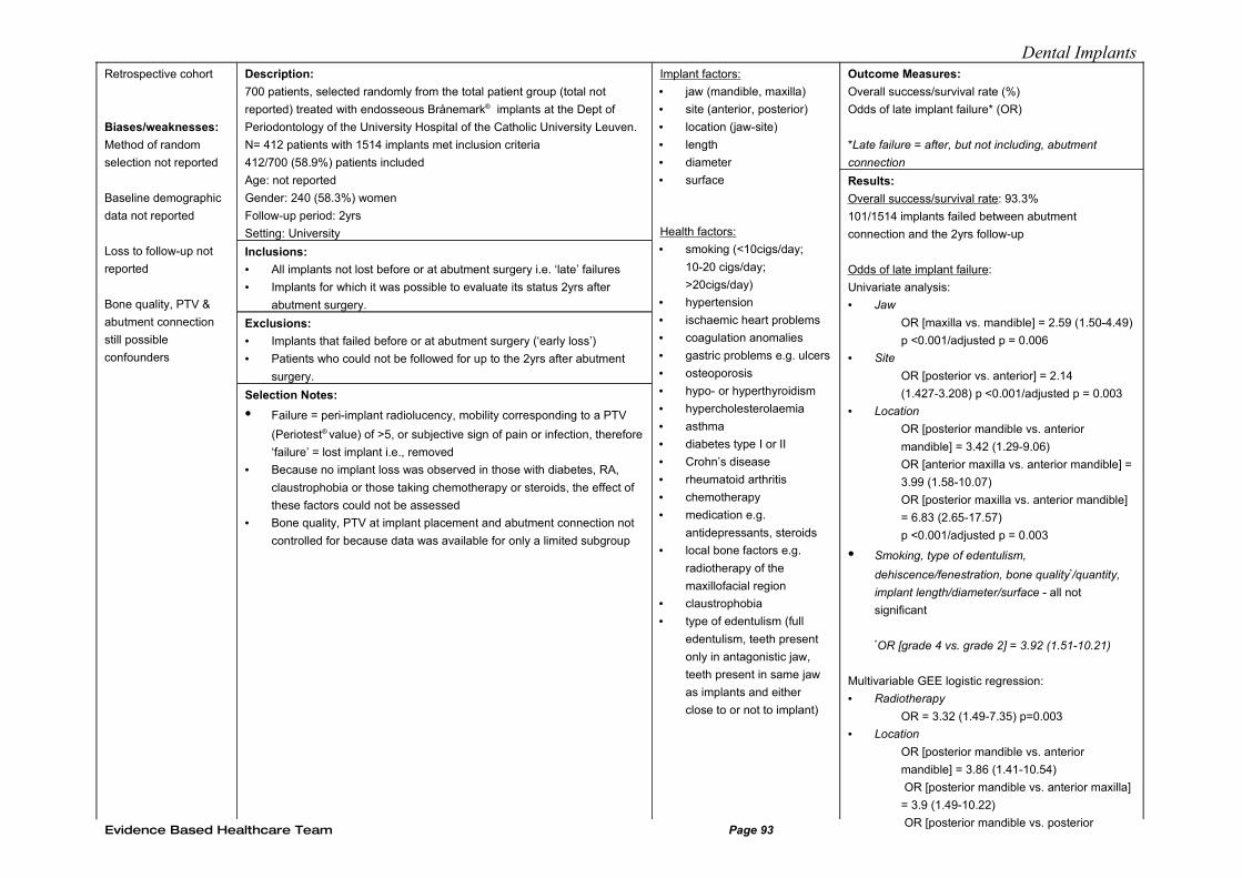
Dental ImplantsRetrospective cohort
Biases/weaknesses:
Method of random
selection not reported
Baseline demographic
data not reported
Loss to follow-up not
reported
Bone quality, PTV &
abutment connection
still possible
confounders
Description:
700 patients, selected randomly from the total patient group (total not
reported) treated with endosseous Brånemark® implants at the Dept of
Periodontology of the University Hospital of the Catholic University Leuven.
N= 412 patients with 1514 implants met inclusion criteria
412/700 (58.9%) patients included
Age: not reported
Gender: 240 (58.3%) women
Follow-up period: 2yrs
Setting: University
Inclusions:
• All implants not lost before or at abutment surgery i.e. ‘late’ failures
• Implants for which it was possible to evaluate its status 2yrs after
abutment surgery.
Exclusions:
• Implants that failed before or at abutment surgery (‘early loss’)
• Patients who could not be followed for up to the 2yrs after abutment
surgery.
Selection Notes:
• Failure = peri-implant radiolucency, mobility corresponding to a PTV
(Periotest® value) of >5, or subjective sign of pain or infection, therefore
‘failure’ = lost implant i.e., removed
• Because no implant loss was observed in those with diabetes, RA,
claustrophobia or those taking chemotherapy or steroids, the effect of
these factors could not be assessed
• Bone quality, PTV at implant placement and abutment connection not
controlled for because data was available for only a limited subgroup
Implant factors:
• jaw (mandible, maxilla)
• site (anterior, posterior)
• location (jaw-site)
• length
• diameter
• surface
Health factors:
• smoking (<10cigs/day;
10-20 cigs/day;
>20cigs/day)
• hypertension
• ischaemic heart problems
• coagulation anomalies
• gastric problems e.g. ulcers
• osteoporosis
• hypo- or hyperthyroidism
• hypercholesterolaemia
• asthma
• diabetes type I or II
• Crohn’s disease
• rheumatoid arthritis
• chemotherapy
• medication e.g.
antidepressants, steroids
• local bone factors e.g.
radiotherapy of the
maxillofacial region
• claustrophobia
• type of edentulism (full
edentulism, teeth present
only in antagonistic jaw,
teeth present in same jaw
as implants and either
close to or not to implant)
Outcome Measures:
Overall success/survival rate (%)
Odds of late implant failure* (OR)
*Late failure = after, but not including, abutment
connection
Results:
Overall success/survival rate: 93.3%
101/1514 implants failed between abutment
connection and the 2yrs follow-up
Odds of late implant failure:
Univariate analysis:
• Jaw
OR [maxilla vs. mandible] = 2.59 (1.50-4.49)
p <0.001/adjusted p = 0.006
• Site
OR [posterior vs. anterior] = 2.14
(1.427-3.208) p <0.001/adjusted p = 0.003
• Location
OR [posterior mandible vs. anterior
mandible] = 3.42 (1.29-9.06)
OR [anterior maxilla vs. anterior mandible] =
3.99 (1.58-10.07)
OR [posterior maxilla vs. anterior mandible]
= 6.83 (2.65-17.57)
p <0.001/adjusted p = 0.003
• Smoking, type of edentulism,
dehiscence/fenestration, bone quality*/quantity,
implant length/diameter/surface - all not
significant
*OR [grade 4 vs. grade 2] = 3.92 (1.51-10.21)
Multivariable GEE logistic regression:
• Radiotherapy
OR = 3.32 (1.49-7.35) p=0.003
• Location
OR [posterior mandible vs. anterior
mandible] = 3.86 (1.41-10.54)
OR [posterior mandible vs. anterior maxilla]
= 3.9 (1.49-10.22)
OR [posterior mandible vs. posterior Evidence Based Healthcare Team Page 93
Dental Implants
Evidence Based Healthcare Table Reference: Alsaadi 2008b Bibliographic Number: 7
Design Participants Exposures Outcomes
Evidence Based Healthcare Team Page 94
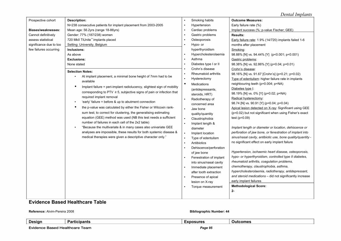
Dental ImplantsProspective cohort
Biases/weaknesses:
Cannot definitively
assess statistical
significance due to too
few failures occurring
Description:
N=238 consecutive patients for implant placement from 2003-2005
Mean age: 56.2yrs (range 18-86yrs)
Gender: 77% (187/238) women
720 MkII TiUnite™ implants placed
Setting: University, Belgium
Inclusions:
As above
Exclusions:
None stated
Selection Notes:
• At implant placement, a minimal bone height of 7mm had to be
available
• Implant failure = peri-implant radiolucency, slightest sign of mobility
corresponding to PTV ≥ 5, subjective signs of pain or infection that
required implant removal
• ‘early’ failure = before & up to abutment connection
• the p-value was calculated by either the Fisher or Wilcoxin rank-
sum test; to correct for clustering, the generalising estimating
equation (GEE) method was used (NB this test needs a sufficient
number of failures in each cell of the 2x2 table)
• “Because the multivariate & in many cases also univariate GEE
analyses are impossible, these results for both systemic disease &
medical therapies were given a descriptive character only.”
• Smoking habits
• Hypertension
• Cardiac problems
• Gastric problems
• Osteoporosis
• Hypo- or
hyperthyroidism
• Hypercholesterolaemia
• Asthma
• Diabetes type I or II
• Crohn’s disease
• Rheumatoid arthritis
• Hysterectomy
• Medications
(antidepressants,
steroids, HRT)
• Radiotherapy of
concerned area
• Jaw bone
quality/quantity
• Claustrophobia
• Implant length &
diameter
• Implant location
• Type of edentulism
• Antibiotics
• Dehiscence/perforation
of jaw bone
• Fenestration of implant
into sinus/nasal cavity
• Immediate placement
after tooth extraction
• Presence of apical
lesion on X-ray
• Torque measurement
Outcome Measures:
Early failure rate (%)
Implant success (%; p-value Fischer; GEE)
Results:
Early failure rate: 1.9% (14/720) implants failed 1-6
months after placement
Smoking:
98.88% [N] vs. 94.44% [Y] (p<0.001; p<0.001)
Gastric problems:
98.38% [N] vs. 92.86% [Y] (p=0.04; p=0.01)
Crohn’s disease:
98.16% [N] vs. 91.67 [Crohn’s] (p=0.21; p=0.02)
Type of edentulism: higher failure rate in implants
neighbouring teeth (p=0.004; p=NA)
Diabetes type I:
98.19% [N] vs. 0% [Y] (p=0.02; p=NA)
Radical hysterectomy:
98.74 [N] vs. 90.91 [Y] (p=0.04; p=0.04)
Apical lesion detected on X-ray: Significant using GEE
(p=0.02) but not significant when using Fisher’s exact
test (p=0.09)
Implant length or diameter or location, dehiscence or
perforation of jaw bone, or fenestration of implant into
sinus/nasal cavity, antibiotic use, bone quality/quantity -
no significant effect on early implant failure
Hypertension, ischaemic heart disease, osteoporosis,
hypo- or hyperthyroidism, controlled type II diabetes,
rheumatoid arthritis, coagulation problems,
chemotherapy, claustrophobia, asthma,
hypercholesterolaemia, radiotherapy, antidepressant,
and steroid medications – did not significantly increase
early implant failures
Methodological Score:
2-
Evidence Based Healthcare Table
Reference: Alvim-Pereira 2008 Bibliographic Number: 44
Design Participants Exposures Outcomes
Evidence Based Healthcare Team Page 95

Dental Implants
Evidence Based Healthcare Team Page 96
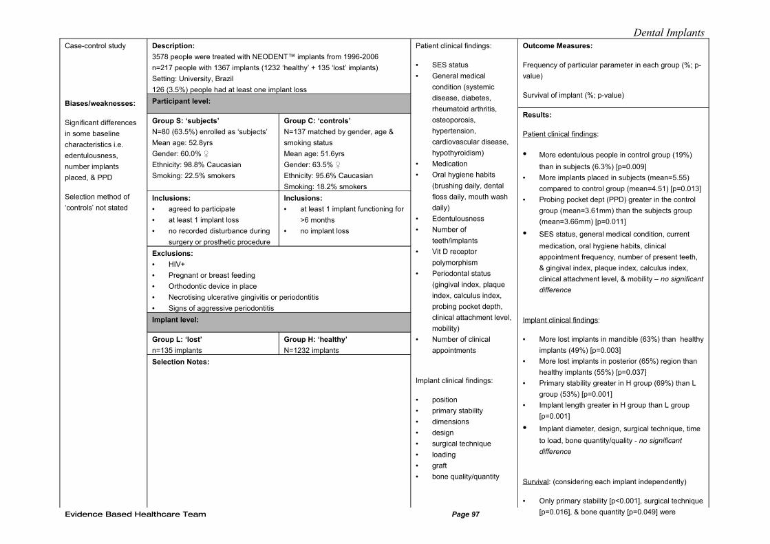
Dental ImplantsCase-control study
Biases/weaknesses:
Significant differences
in some baseline
characteristics i.e.
edentulousness,
number implants
placed, & PPD
Selection method of
‘controls’ not stated
Description:
3578 people were treated with NEODENT™ implants from 1996-2006
n=217 people with 1367 implants (1232 ‘healthy’ + 135 ‘lost’ implants)
Setting: University, Brazil
126 (3.5%) people had at least one implant loss
Participant level:
Group S: ‘subjects’
N=80 (63.5%) enrolled as ‘subjects’
Mean age: 52.8yrs
Gender: 60.0% ♀
Ethnicity: 98.8% Caucasian
Smoking: 22.5% smokers
Group C: ‘controls’
N=137 matched by gender, age &
smoking status
Mean age: 51.6yrs
Gender: 63.5% ♀
Ethnicity: 95.6% Caucasian
Smoking: 18.2% smokers
Inclusions:
• agreed to participate
• at least 1 implant loss
• no recorded disturbance during
surgery or prosthetic procedure
Inclusions:
• at least 1 implant functioning for
>6 months
• no implant loss
Exclusions:
• HIV+
• Pregnant or breast feeding
• Orthodontic device in place
• Necrotising ulcerative gingivitis or periodontitis
• Signs of aggressive periodontitis
Implant level:
Group L: ‘lost’
n=135 implants
Group H: ‘healthy’
N=1232 implants
Selection Notes:
Patient clinical findings:
• SES status
• General medical
condition (systemic
disease, diabetes,
rheumatoid arthritis,
osteoporosis,
hypertension,
cardiovascular disease,
hypothyroidism)
• Medication
• Oral hygiene habits
(brushing daily, dental
floss daily, mouth wash
daily)
• Edentulousness
• Number of
teeth/implants
• Vit D receptor
polymorphism
• Periodontal status
(gingival index, plaque
index, calculus index,
probing pocket depth,
clinical attachment level,
mobility)
• Number of clinical
appointments
Implant clinical findings:
• position
• primary stability
• dimensions
• design
• surgical technique
• loading
• graft
• bone quality/quantity
Outcome Measures:
Frequency of particular parameter in each group (%; p-
value)
Survival of implant (%; p-value)
Results:
Patient clinical findings:
• More edentulous people in control group (19%)
than in subjects (6.3%) [p=0.009]
• More implants placed in subjects (mean=5.55)
compared to control group (mean=4.51) [p=0.013]
• Probing pocket dept (PPD) greater in the control
group (mean=3.61mm) than the subjects group
(mean=3.66mm) [p=0.011]
• SES status, general medical condition, current
medication, oral hygiene habits, clinical
appointment frequency, number of present teeth,
& gingival index, plaque index, calculus index,
clinical attachment level, & mobility – no significant
difference
Implant clinical findings:
• More lost implants in mandible (63%) than healthy
implants (49%) [p=0.003]
• More lost implants in posterior (65%) region than
healthy implants (55%) [p=0.037]
• Primary stability greater in H group (69%) than L
group (53%) [p=0.001]
• Implant length greater in H group than L group
[p=0.001]
• Implant diameter, design, surgical technique, time
to load, bone quantity/quality - no significant
difference
Survival: (considering each implant independently)
• Only primary stability [p<0.001], surgical technique
[p=0.016], & bone quantity [p=0.049] were Evidence Based Healthcare Team Page 97

Dental Implants
Evidence Based Healthcare Team Page 98
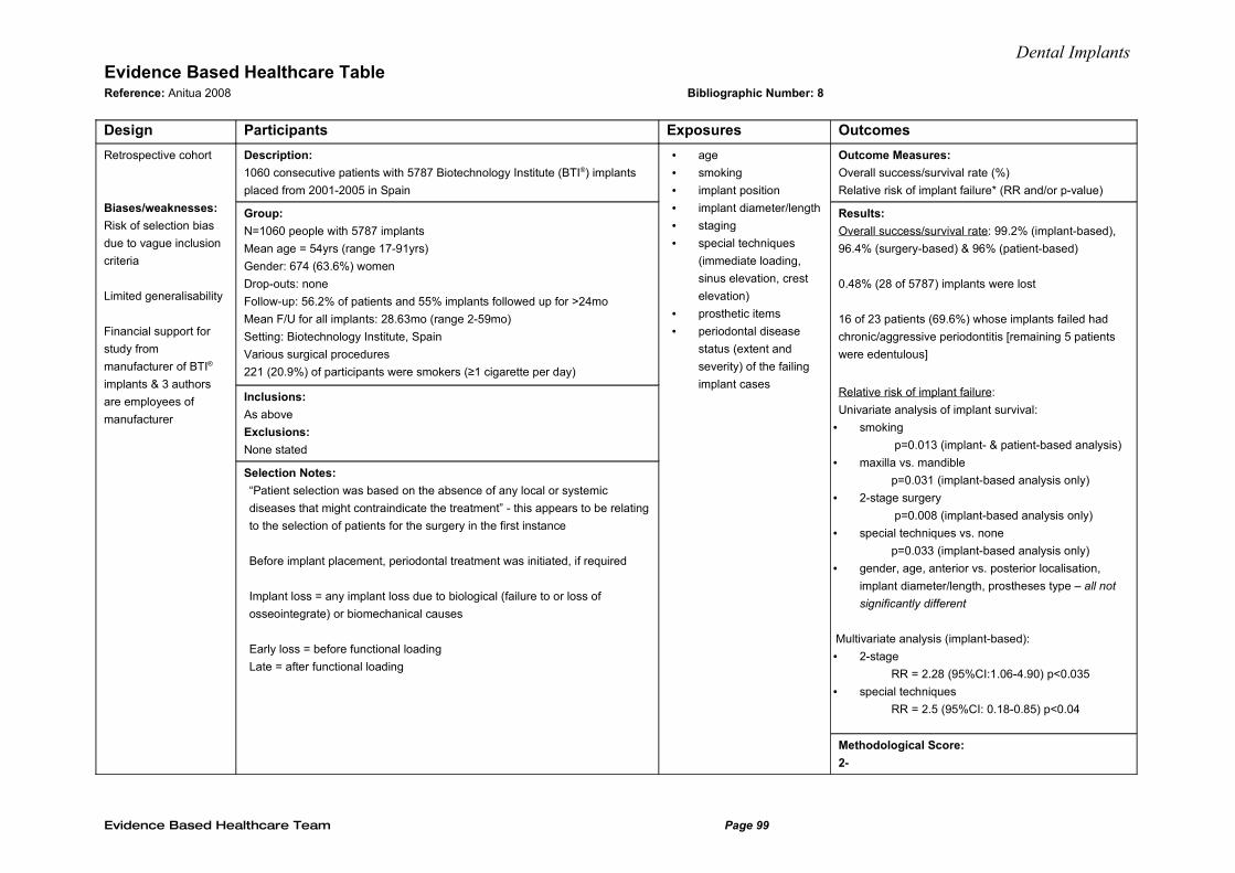
Dental ImplantsEvidence Based Healthcare Table Reference: Anitua 2008 Bibliographic Number: 8
Design Participants Exposures Outcomes
Retrospective cohort
Biases/weaknesses:
Risk of selection bias
due to vague inclusion
criteria
Limited generalisability
Financial support for
study from
manufacturer of BTI®
implants & 3 authors
are employees of
manufacturer
Description:
1060 consecutive patients with 5787 Biotechnology Institute (BTI®) implants
placed from 2001-2005 in Spain
Group:
N=1060 people with 5787 implants
Mean age = 54yrs (range 17-91yrs)
Gender: 674 (63.6%) women
Drop-outs: none
Follow-up: 56.2% of patients and 55% implants followed up for >24mo
Mean F/U for all implants: 28.63mo (range 2-59mo)
Setting: Biotechnology Institute, Spain
Various surgical procedures
221 (20.9%) of participants were smokers (≥1 cigarette per day)
Inclusions:
As above
Exclusions:
None stated
Selection Notes:
“Patient selection was based on the absence of any local or systemic
diseases that might contraindicate the treatment” - this appears to be relating
to the selection of patients for the surgery in the first instance
Before implant placement, periodontal treatment was initiated, if required
Implant loss = any implant loss due to biological (failure to or loss of
osseointegrate) or biomechanical causes
Early loss = before functional loading
Late = after functional loading
• age
• smoking
• implant position
• implant diameter/length
• staging
• special techniques
(immediate loading,
sinus elevation, crest
elevation)
• prosthetic items
• periodontal disease
status (extent and
severity) of the failing
implant cases
Outcome Measures:
Overall success/survival rate (%)
Relative risk of implant failure* (RR and/or p-value)
Results:
Overall success/survival rate: 99.2% (implant-based),
96.4% (surgery-based) & 96% (patient-based)
0.48% (28 of 5787) implants were lost
16 of 23 patients (69.6%) whose implants failed had
chronic/aggressive periodontitis [remaining 5 patients
were edentulous]
Relative risk of implant failure:
Univariate analysis of implant survival:
• smoking
p=0.013 (implant- & patient-based analysis)
• maxilla vs. mandible
p=0.031 (implant-based analysis only)
• 2-stage surgery
p=0.008 (implant-based analysis only)
• special techniques vs. none
p=0.033 (implant-based analysis only)
• gender, age, anterior vs. posterior localisation,
implant diameter/length, prostheses type – all not
significantly different
Multivariate analysis (implant-based):
• 2-stage
RR = 2.28 (95%CI:1.06-4.90) p<0.035
• special techniques
RR = 2.5 (95%CI: 0.18-0.85) p<0.04
Methodological Score:
2-
Evidence Based Healthcare Team Page 99
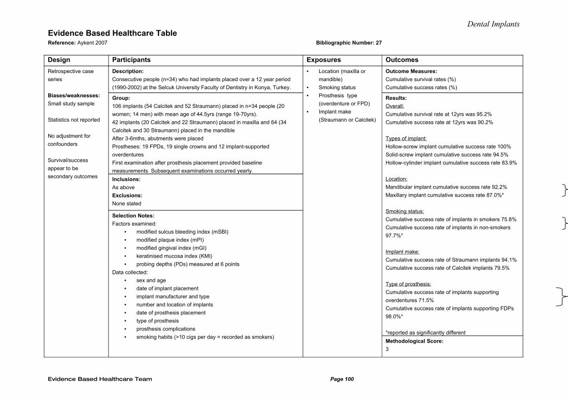
Dental ImplantsEvidence Based Healthcare Table Reference: Aykent 2007 Bibliographic Number: 27
Design Participants Exposures Outcomes
Retrospective case
series
Biases/weaknesses:
Small study sample
Statistics not reported
No adjustment for
confounders
Survival/success
appear to be
secondary outcomes
Description:
Consecutive people (n=34) who had implants placed over a 12 year period
(1990-2002) at the Selcuk University Faculty of Dentistry in Konya, Turkey.
Group:
106 implants (54 Calcitek and 52 Straumann) placed in n=34 people (20
women; 14 men) with mean age of 44.5yrs (range 19-70yrs).
42 implants (20 Calcitek and 22 Straumann) placed in maxilla and 64 (34
Calcitek and 30 Straumann) placed in the mandible
After 3-6mths, abutments were placed
Prostheses: 19 FPDs, 19 single crowns and 12 implant-supported
overdentures
First examination after prosthesis placement provided baseline
measurements. Subsequent examinations occurred yearly.
Inclusions:
As above
Exclusions:
None stated
Selection Notes:
Factors examined:
• modified sulcus bleeding index (mSBI)
• modified plaque index (mPI)
• modified gingival index (mGI)
• keratinised mucosa index (KMI)
• probing depths (PDs) measured at 6 points
Data collected:
• sex and age
• date of implant placement
• implant manufacturer and type
• number and location of implants
• date of prosthesis placement
• type of prosthesis
• prosthesis complications
• smoking habits (>10 cigs per day = recorded as smokers)
• Location (maxilla or
mandible)
• Smoking status
• Prosthesis type
(overdenture or FPD)
• Implant make
(Straumann or Calcitek)
Outcome Measures:
Cumulative survival rates (%)
Cumulative success rates (%)
Results:
Overall:
Cumulative survival rate at 12yrs was 95.2%
Cumulative success rate at 12yrs was 90.2%
Types of implant:
Hollow-screw implant cumulative success rate 100%
Solid-screw implant cumulative success rate 94.5%
Hollow-cylinder implant cumulative success rate 83.9%
Location:
Mandibular implant cumulative success rate 92.2%
Maxillary implant cumulative success rate 87.0%*
Smoking status:
Cumulative success rate of implants in smokers 75.8%
Cumulative success rate of implants in non-smokers
97.7%*
Implant make:
Cumulative success rate of Straumann implants 94.1%
Cumulative success rate of Calcitek implants 79.5%
Type of prosthesis:
Cumulative success rate of implants supporting
overdentures 71.5%
Cumulative success rate of implants supporting FDPs
98.0%*
*reported as significantly different
Methodological Score:
3
Evidence Based Healthcare Team Page 100
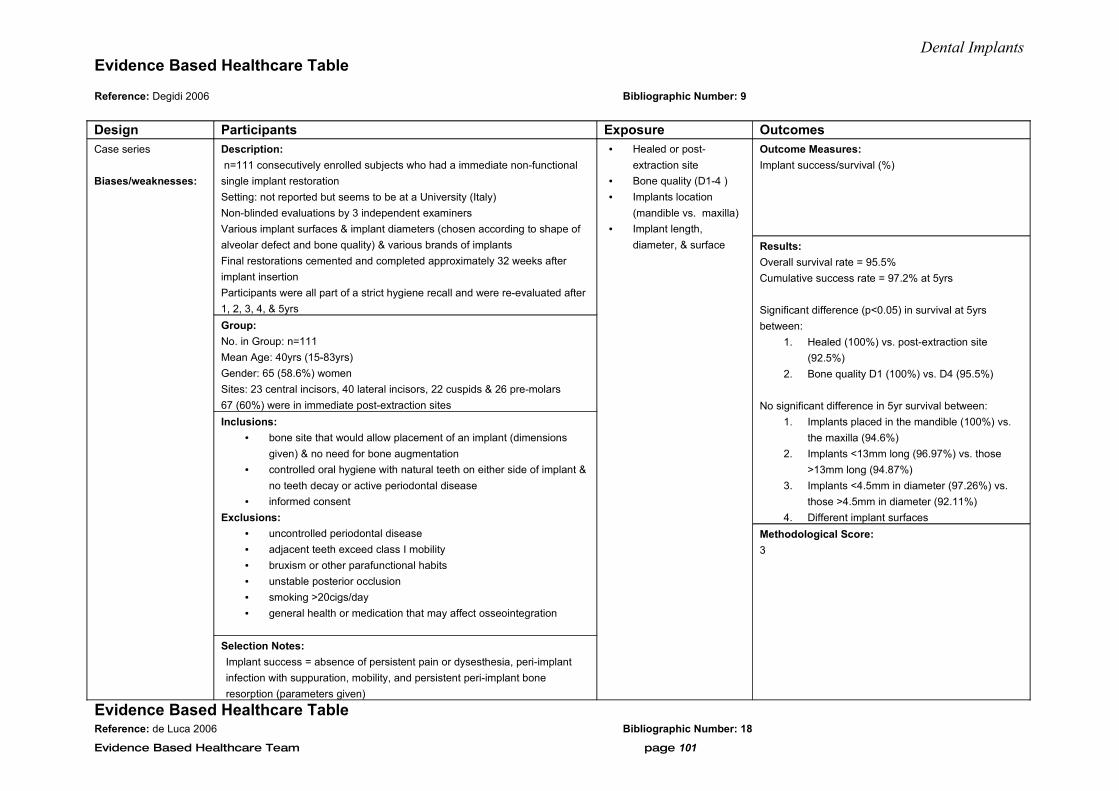
Dental ImplantsEvidence Based Healthcare Table
Reference: Degidi 2006 Bibliographic Number: 9
Design Participants Exposure OutcomesCase series
Biases/weaknesses:
Description:
n=111 consecutively enrolled subjects who had a immediate non-functional
single implant restoration
Setting: not reported but seems to be at a University (Italy)
Non-blinded evaluations by 3 independent examiners
Various implant surfaces & implant diameters (chosen according to shape of
alveolar defect and bone quality) & various brands of implants
Final restorations cemented and completed approximately 32 weeks after
implant insertion
Participants were all part of a strict hygiene recall and were re-evaluated after
1, 2, 3, 4, & 5yrs
Group:
No. in Group: n=111
Mean Age: 40yrs (15-83yrs)
Gender: 65 (58.6%) women
Sites: 23 central incisors, 40 lateral incisors, 22 cuspids & 26 pre-molars
67 (60%) were in immediate post-extraction sites
Inclusions:
• bone site that would allow placement of an implant (dimensions
given) & no need for bone augmentation
• controlled oral hygiene with natural teeth on either side of implant &
no teeth decay or active periodontal disease
• informed consent
Exclusions:
• uncontrolled periodontal disease
• adjacent teeth exceed class I mobility
• bruxism or other parafunctional habits
• unstable posterior occlusion
• smoking >20cigs/day
• general health or medication that may affect osseointegration
Selection Notes:
Implant success = absence of persistent pain or dysesthesia, peri-implant
infection with suppuration, mobility, and persistent peri-implant bone
resorption (parameters given)
• Healed or post-
extraction site
• Bone quality (D1-4 )
• Implants location
(mandible vs. maxilla)
• Implant length,
diameter, & surface
Outcome Measures:
Implant success/survival (%)
Results:
Overall survival rate = 95.5%
Cumulative success rate = 97.2% at 5yrs
Significant difference (p<0.05) in survival at 5yrs
between:
1. Healed (100%) vs. post-extraction site
(92.5%)
2. Bone quality D1 (100%) vs. D4 (95.5%)
No significant difference in 5yr survival between:
1. Implants placed in the mandible (100%) vs.
the maxilla (94.6%)
2. Implants <13mm long (96.97%) vs. those
>13mm long (94.87%)
3. Implants <4.5mm in diameter (97.26%) vs.
those >4.5mm in diameter (92.11%)
4. Different implant surfaces
Methodological Score:
3
Evidence Based Healthcare Table Reference: de Luca 2006 Bibliographic Number: 18
Evidence Based Healthcare Team page 101

Dental Implants
Design Description
Participants Exposure Outcomes
Evidence Based Healthcare Team Page 102
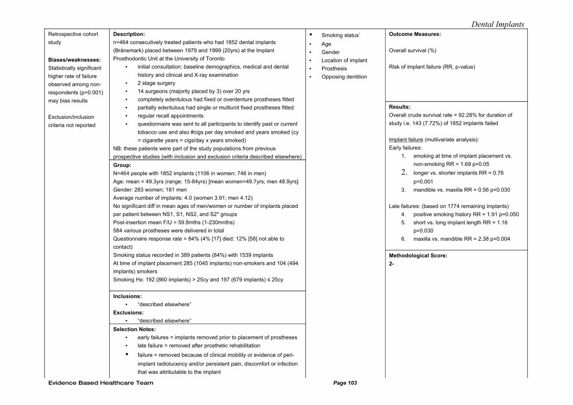
Dental ImplantsRetrospective cohort
study
Biases/weaknesses:
Statistically significant
higher rate of failure
observed among non-
respondents (p=0.001)
may bias results
Exclusion/inclusion
criteria not reported
Description:
n=464 consecutively treated patients who had 1852 dental implants
(Brånemark) placed between 1979 and 1999 (20yrs) at the Implant
Prosthodontic Unit at the University of Toronto
• initial consultation: baseline demographics, medical and dental
history and clinical and X-ray examination
• 2 stage surgery
• 14 surgeons (majority placed by 3) over 20 yrs
• completely edentulous had fixed or overdenture prostheses fitted
• partially edentulous had single or multiunit fixed prostheses fitted
• regular recall appointments
• questionnaire was sent to all participants to identify past or current
tobacco use and also #cigs per day smoked and years smoked (cy
= cigarette years = cigs/day x years smoked)
NB: these patients were part of the study populations from previous
prospective studies (with inclusion and exclusion criteria described elsewhere)
Group:
N=464 people with 1852 implants (1106 in women; 746 in men)
Age: mean = 49.3yrs (range: 15-84yrs) [mean women=49.7yrs; men 48.9yrs]
Gender: 283 women; 181 men
Average number of implants: 4.0 (women 3.91; men 4.12)
No significant diff in mean ages of men/women or number of implants placed
per patient between NS1, S1, NS2, and S2* groups
Post-insertion mean F/U = 59.8mths (1-230mnths)
584 various prostheses were delivered in total
Questionnaire response rate = 84% (4% [17] died; 12% [58] not able to
contact)
Smoking status recorded in 389 patients (84%) with 1539 implants
At time of implant placement 285 (1045 implants) non-smokers and 104 (494
implants) smokers
Smoking Hx: 192 (860 implants) > 25cy and 197 (679 implants) ≤ 25cy
Inclusions:
• “described elsewhere”
Exclusions:
• “described elsewhere”
Selection Notes:
• early failures = implants removed prior to placement of prostheses
• late failure = removed after prosthetic rehabilitation
• failure = removed because of clinical mobility or evidence of peri-
implant radiolucency and/or persistent pain, discomfort or infection
that was attributable to the implant
• Smoking status*
• Age
• Gender
• Location of implant
• Prosthesis
• Opposing dentition
Outcome Measures:
Overall survival (%)
Risk of implant failure (RR, p-value)
Results:
Overall crude survival rate = 92.28% for duration of
study i.e. 143 (7.72%) of 1852 implants failed
Implant failure (multivariate analysis):
Early failures:
1. smoking at time of implant placement vs.
non-smoking RR = 1.69 p=0.05
2. longer vs. shorter implants RR = 0.76
p=0.001
3. mandible vs. maxilla RR = 0.56 p=0.030
Late failures: (based on 1774 remaining implants)
4. positive smoking history RR = 1.91 p=0.050
5. short vs. long implant length RR = 1.16
p=0.030
6. maxilla vs. mandible RR = 2.38 p=0.004
Methodological Score:
2-
Evidence Based Healthcare Team Page 103

Dental Implants
Evidence Based Healthcare Team Page 104
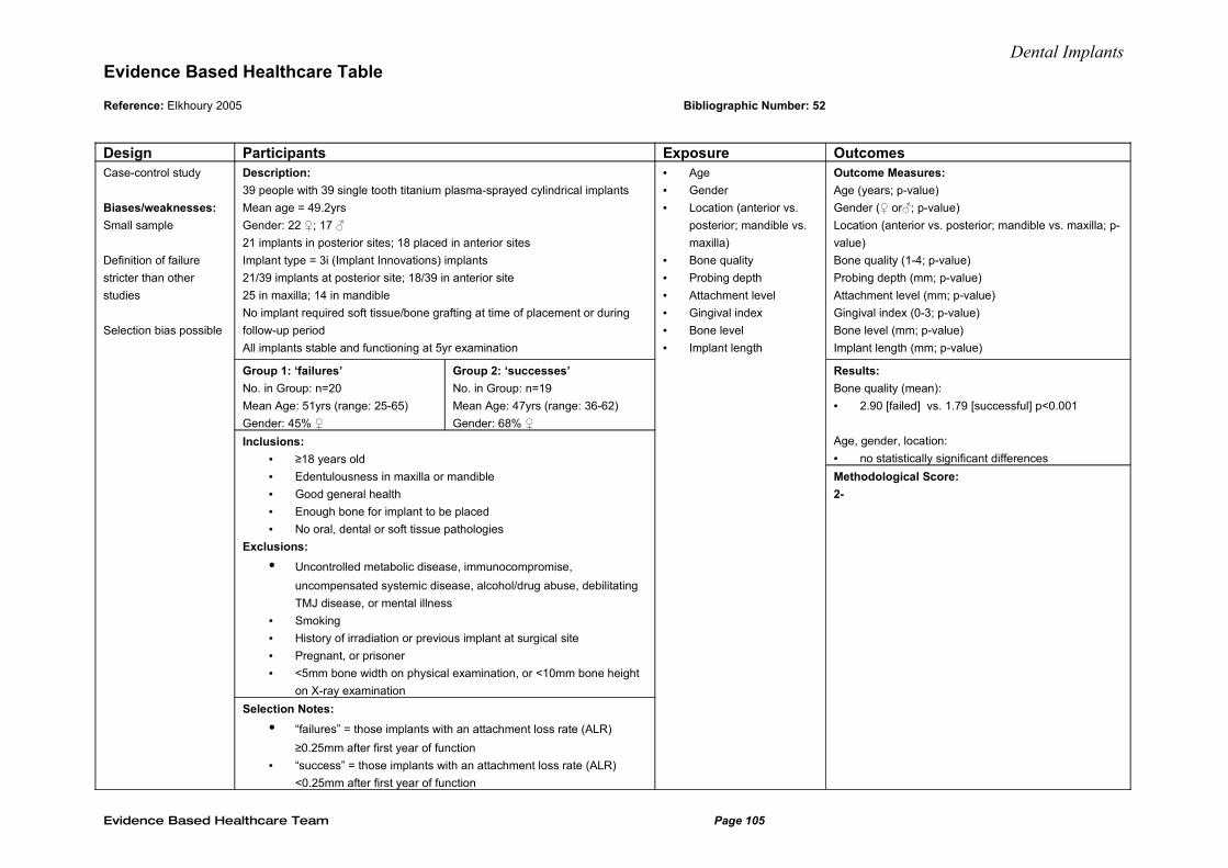
Dental ImplantsEvidence Based Healthcare Table
Reference: Elkhoury 2005 Bibliographic Number: 52
Design Participants Exposure OutcomesCase-control study
Biases/weaknesses:
Small sample
Definition of failure
stricter than other
studies
Selection bias possible
Description:
39 people with 39 single tooth titanium plasma-sprayed cylindrical implants
Mean age = 49.2yrs
Gender: 22 ♀; 17 ♂
21 implants in posterior sites; 18 placed in anterior sites
Implant type = 3i (Implant Innovations) implants
21/39 implants at posterior site; 18/39 in anterior site
25 in maxilla; 14 in mandible
No implant required soft tissue/bone grafting at time of placement or during
follow-up period
All implants stable and functioning at 5yr examination
Group 1: ‘failures’
No. in Group: n=20
Mean Age: 51yrs (range: 25-65)
Gender: 45% ♀
Group 2: ‘successes’
No. in Group: n=19
Mean Age: 47yrs (range: 36-62)
Gender: 68% ♀
Inclusions:
• ≥18 years old
• Edentulousness in maxilla or mandible
• Good general health
• Enough bone for implant to be placed
• No oral, dental or soft tissue pathologies
Exclusions:
• Uncontrolled metabolic disease, immunocompromise,
uncompensated systemic disease, alcohol/drug abuse, debilitating
TMJ disease, or mental illness
• Smoking
• History of irradiation or previous implant at surgical site
• Pregnant, or prisoner
• <5mm bone width on physical examination, or <10mm bone height
on X-ray examination
Selection Notes:
• “failures” = those implants with an attachment loss rate (ALR)
≥0.25mm after first year of function
• “success” = those implants with an attachment loss rate (ALR)
<0.25mm after first year of function
• Age
• Gender
• Location (anterior vs.
posterior; mandible vs.
maxilla)
• Bone quality
• Probing depth
• Attachment level
• Gingival index
• Bone level
• Implant length
Outcome Measures:
Age (years; p-value)
Gender (♀ or♂; p-value)
Location (anterior vs. posterior; mandible vs. maxilla; p-
value)
Bone quality (1-4; p-value)
Probing depth (mm; p-value)
Attachment level (mm; p-value)
Gingival index (0-3; p-value)
Bone level (mm; p-value)
Implant length (mm; p-value)
Results:
Bone quality (mean):
• 2.90 [failed] vs. 1.79 [successful] p<0.001
Age, gender, location:
• no statistically significant differences
Methodological Score:
2-
Evidence Based Healthcare Team Page 105
Dental ImplantsEvidence Based Healthcare Table Reference: Esposito 2008 Bibliographic Number: 2
Design Description
Participants Intervention Outcomes
Evidence Based Healthcare Team Page 106
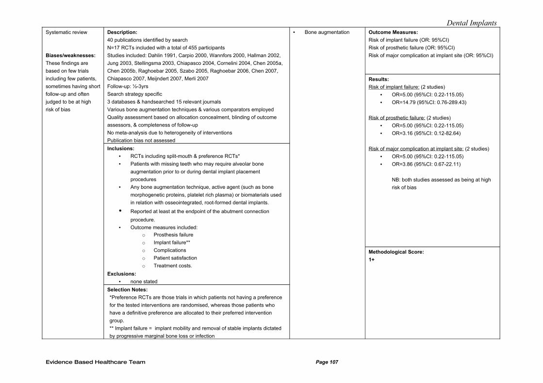
Dental ImplantsSystematic review
Biases/weaknesses:
These findings are
based on few trials
including few patients,
sometimes having short
follow-up and often
judged to be at high
risk of bias
Description:
40 publications identified by search
N=17 RCTs included with a total of 455 participants
Studies included: Dahlin 1991, Carpio 2000, Wannfors 2000, Hallman 2002,
Jung 2003, Stellingsma 2003, Chiapasco 2004, Cornelini 2004, Chen 2005a,
Chen 2005b, Raghoebar 2005, Szabo 2005, Raghoebar 2006, Chen 2007,
Chiapasco 2007, Meijndert 2007, Merli 2007
Follow-up: ½-3yrs
Search strategy specific
3 databases & handsearched 15 relevant journals
Various bone augmentation techniques & various comparators employed
Quality assessment based on allocation concealment, blinding of outcome
assessors, & completeness of follow-up
No meta-analysis due to heterogeneity of interventions
Publication bias not assessed
Inclusions:
• RCTs including split-mouth & preference RCTs*
• Patients with missing teeth who may require alveolar bone
augmentation prior to or during dental implant placement
procedures
• Any bone augmentation technique, active agent (such as bone
morphogenetic proteins, platelet rich plasma) or biomaterials used
in relation with osseointegrated, root-formed dental implants.
• Reported at least at the endpoint of the abutment connection
procedure.
• Outcome measures included:
o Prosthesis failure
o Implant failure**
o Complications
o Patient satisfaction
o Treatment costs.
Exclusions:
• none stated
Selection Notes:
*Preference RCTs are those trials in which patients not having a preference
for the tested interventions are randomised, whereas those patients who
have a definitive preference are allocated to their preferred intervention
group.
** Implant failure = implant mobility and removal of stable implants dictated
by progressive marginal bone loss or infection
• Bone augmentation Outcome Measures:
Risk of implant failure (OR: 95%CI)
Risk of prosthetic failure (OR: 95%CI)
Risk of major complication at implant site (OR: 95%CI)
Results:
Risk of implant failure: (2 studies)
• OR=5.00 (95%CI: 0.22-115.05)
• OR=14.79 (95%CI: 0.76-289.43)
Risk of prosthetic failure: (2 studies)
• OR=5.00 (95%CI: 0.22-115.05)
• OR=3.16 (95%CI: 0.12-82.64)
Risk of major complication at implant site: (2 studies)
• OR=5.00 (95%CI: 0.22-115.05)
• OR=3.86 (95%CI: 0.67-22.11)
NB: both studies assessed as being at high
risk of bias
Methodological Score:
1+
Evidence Based Healthcare Team Page 107

Dental Implants
Evidence Based Healthcare Team Page 108
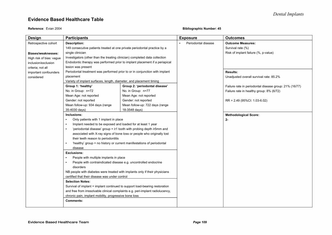
Dental ImplantsEvidence Based Healthcare Table
Reference: Evian 2004 Bibliographic Number: 45
Design Participants Exposure OutcomesRetrospective cohort
Biases/weaknesses:
High risk of bias: vague
inclusion/exclusion
criteria; not all
important confounders
considered
Description:
149 consecutive patients treated at one private periodontal practice by a
single clinician
Investigators (other than the treating clinician) completed data collection
Endodontic therapy was performed prior to implant placement if a periapical
lesion was present
Periodontal treatment was performed prior to or in conjunction with implant
placement
Variety of implant surfaces, length, diameter, and placement timing
Group 1: ‘healthy’
No. in Group: n=72
Mean Age: not reported
Gender: not reported
Mean follow-up: 934 days (range
35-4030 days)
Group 2: ‘periodontal disease’
No. in Group: n=77
Mean Age: not reported
Gender: not reported
Mean follow-up: 722 days (range
18-3548 days)
Inclusions:
• Only patients with 1 implant in place
• Implant needed to be exposed and loaded for at least 1 year
• ‘periodontal disease’ group = ≥1 tooth with probing depth ≥5mm and
associated with X-ray signs of bone loss or people who originally lost
their teeth reason to periodontitis
• ‘healthy’ group = no history or current manifestations of periodontal
disease
Exclusions:
• People with multiple implants in place
• People with contraindicated disease e.g. uncontrolled endocrine
disorders
NB people with diabetes were treated with implants only if their physicians
certified that their disease was under control
Selection Notes:
Survival of implant = implant continued to support load-bearing restoration
and free from irresolvable clinical complaints e.g. peri-implant radiolucency,
chronic pain, implant mobility, progressive bone loss
Comments:
• Periodontal disease Outcome Measures:
Survival rate (%)
Risk of implant failure (%, p-value)
Results:
Unadjusted overall survival rate: 85.2%
Failure rate in periodontal disease group: 21% (16/77)
Failure rate in healthy group: 8% (6/72)
RR = 2.49 (95%CI: 1.03-6.02)
Methodological Score:
2-
Evidence Based Healthcare Team Page 109
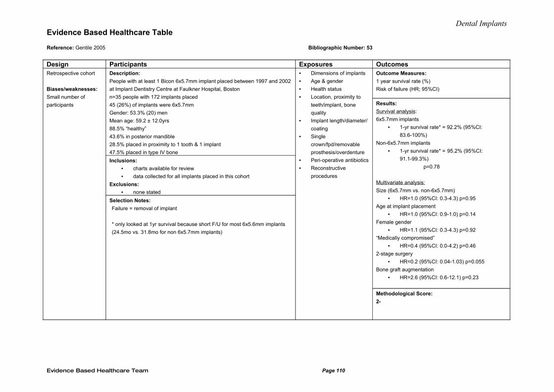
Dental ImplantsEvidence Based Healthcare Table
Reference: Gentile 2005 Bibliographic Number: 53
Design Participants Exposures OutcomesRetrospective cohort
Biases/weaknesses:
Small number of
participants
Description:
People with at least 1 Bicon 6x5.7mm implant placed between 1997 and 2002
at Implant Dentistry Centre at Faulkner Hospital, Boston
n=35 people with 172 implants placed
45 (26%) of implants were 6x5.7mm
Gender: 53.3% (20) men
Mean age: 59.2 ± 12.0yrs
88.5% “healthy”
43.6% in posterior mandible
28.5% placed in proximity to 1 tooth & 1 implant
47.5% placed in type IV bone
Inclusions:
• charts available for review
• data collected for all implants placed in this cohort
Exclusions:
• none stated
Selection Notes:
Failure = removal of implant
* only looked at 1yr survival because short F/U for most 6x5.6mm implants
(24.5mo vs. 31.8mo for non 6x5.7mm implants)
• Dimensions of implants
• Age & gender
• Health status
• Location, proximity to
teeth/implant, bone
quality
• Implant length/diameter/
coating
• Single
crown/fpd/removable
prosthesis/overdenture
• Peri-operative antibiotics
• Reconstructive
procedures
Outcome Measures:
1 year survival rate (%)
Risk of failure (HR; 95%CI)
Results:
Survival analysis:
6x5.7mm implants
• 1-yr survival rate* = 92.2% (95%CI:
83.6-100%)
Non-6x5.7mm implants
• 1-yr survival rate* = 95.2% (95%CI:
91.1-99.3%)
p=0.78
Multivariate analysis:
Size (6x5.7mm vs. non-6x5.7mm)
• HR=1.0 (95%CI: 0.3-4.3) p=0.95
Age at implant placement
• HR=1.0 (95%CI: 0.9-1.0) p=0.14
Female gender
• HR=1.1 (95%CI: 0.3-4.3) p=0.92
“Medically compromised”
• HR=0.4 (95%CI: 0.0-4.2) p=0.46
2-stage surgery
• HR=0.2 (95%CI: 0.04-1.03) p=0.055
Bone graft augmentation
• HR=2.6 (95%CI: 0.6-12.1) p=0.23
Methodological Score:
2-
Evidence Based Healthcare Team Page 110
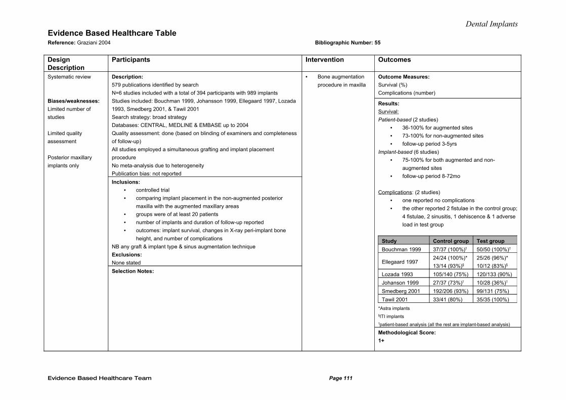
Dental ImplantsEvidence Based Healthcare Table Reference: Graziani 2004 Bibliographic Number: 55
Design Description
Participants Intervention Outcomes
Systematic review
Biases/weaknesses:
Limited number of
studies
Limited quality
assessment
Posterior maxillary
implants only
Description:
579 publications identified by search
N=6 studies included with a total of 394 participants with 989 implants
Studies included: Bouchman 1999, Johansson 1999, Ellegaard 1997, Lozada
1993, Smedberg 2001, & Tawil 2001
Search strategy: broad strategy
Databases: CENTRAL, MEDLINE & EMBASE up to 2004
Quality assessment: done (based on blinding of examiners and completeness
of follow-up)
All studies employed a simultaneous grafting and implant placement
procedure
No meta-analysis due to heterogeneity
Publication bias: not reported
Inclusions:
• controlled trial
• comparing implant placement in the non-augmented posterior
maxilla with the augmented maxillary areas
• groups were of at least 20 patients
• number of implants and duration of follow-up reported
• outcomes: implant survival, changes in X-ray peri-implant bone
height, and number of complications
NB any graft & implant type & sinus augmentation technique
Exclusions:
None stated
Selection Notes:
• Bone augmentation
procedure in maxilla
Outcome Measures:
Survival (%)
Complications (number)
Results:
Survival:
Patient-based (2 studies)
• 36-100% for augmented sites
• 73-100% for non-augmented sites
• follow-up period 3-5yrs
Implant-based (6 studies)
• 75-100% for both augmented and non-
augmented sites
• follow-up period 8-72mo
Complications: (2 studies)
• one reported no complications
• the other reported 2 fistulae in the control group;
4 fistulae, 2 sinusitis, 1 dehiscence & 1 adverse
load in test group
Study Control group Test group
Bouchman 1999 37/37 (100%)† 50/50 (100%)†
Ellegaard 199724/24 (100%)*
13/14 (93%)§
25/26 (96%)*
10/12 (83%)§
Lozada 1993 105/140 (75%) 120/133 (90%)
Johanson 1999 27/37 (73%)† 10/28 (36%)†
Smedberg 2001 192/206 (93%) 99/131 (75%)
Tawil 2001 33/41 (80%) 35/35 (100%)
*Astra implants§ITI implants†patient-based analysis (all the rest are implant-based analysis)
Methodological Score:
1+
Evidence Based Healthcare Team Page 111
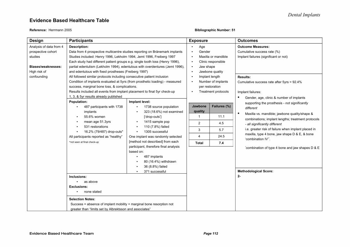
Dental ImplantsEvidence Based Healthcare Table
Reference: Herrmann 2005 Bibliographic Number: 51
Design Participants Exposure Outcomes
Analysis of data from 4
prospective cohort
studies
Biases/weaknesses:
High risk of
confounding
Description:
Data from 4 prospective multicentre studies reporting on Brånemark implants
Studies included: Henry 1996, Lekholm 1994, Jemt 1996, Freiberg 1997
Each study had different patient groups e.g. single tooth loss (Henry 1996),
partial edentulism (Lekholm 1994), edentulous with overdentures (Jemt 1996),
and edentulous with fixed prostheses (Freiberg 1997)
All followed similar protocols including consecutive patient inclusion
Condition of implants evaluated at 5yrs (from prosthetic loading) - measured
success, marginal bone loss, & complications.
Results included all events from implant placement to final 5yr check-up
1, 3, & 5yr results already published
Population:
• 487 participants with 1738
implants
• 55.6% women
• mean age 51.3yrs
• 531 restorations
• 16.2% (79/487) drop-outs*
All participants reported as “healthy”
*not seen at final check-up
Implant level:
• 1738 source population
• 323 (18.6%) not examined
[‘drop-outs’]
• 1415 sample pop
• 110 (7.8%) failed
• 1305 successful
One implant was randomly selected
[method not described] from each
participant, therefore final analysis
based on:
• 487 implants
• 80 (16.4%) withdrawn
• 36 (8.8%) failed
• 371 successful
Inclusions:
• as above
Exclusions:
• none stated
Selection Notes:
Success = absence of implant mobility + marginal bone resorption not
greater than “limits set by Albrektsson and associates”
• Age
• Gender
• Maxilla or mandible
• Clinic responsible
• Jaw shape
• Jawbone quality
• Implant length
• Number of implants
per restoration
• Treatment protocols
Jawbone
quality
Failures (%)
1 11.1
2 4.5
3 5.7
4 24.5
Total 7.4
Outcome Measures:
Cumulative success rate (%)
Implant failures (significant or not)
Results:
Cumulative success rate after 5yrs = 92.4%
Implant failures:
• Gender, age, clinic & number of implants
supporting the prosthesis - not significantly
different
• Maxilla vs. mandible; jawbone quality/shape &
combinations; implant lengths; treatment protocols
- all significantly different
i.e. greater risk of failure when implant placed in
maxilla, type 4 bone, jaw shape D & E, & bone
‘combination IV’*.
*combination of type 4 bone and jaw shapes D & E
Methodological Score:
2-
Evidence Based Healthcare Team Page 112
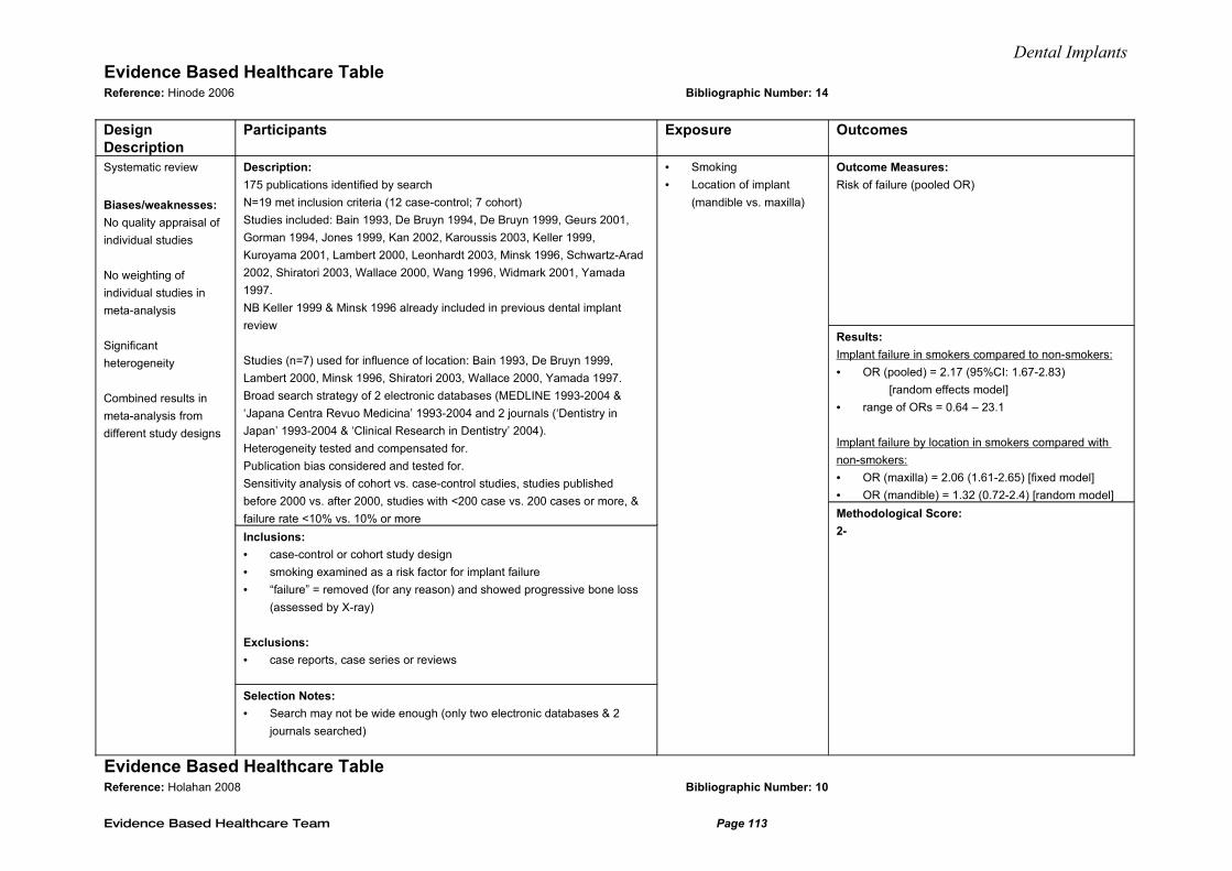
Dental ImplantsEvidence Based Healthcare Table Reference: Hinode 2006 Bibliographic Number: 14
Design Description
Participants Exposure Outcomes
Systematic review
Biases/weaknesses:
No quality appraisal of
individual studies
No weighting of
individual studies in
meta-analysis
Significant
heterogeneity
Combined results in
meta-analysis from
different study designs
Description:
175 publications identified by search
N=19 met inclusion criteria (12 case-control; 7 cohort)
Studies included: Bain 1993, De Bruyn 1994, De Bruyn 1999, Geurs 2001,
Gorman 1994, Jones 1999, Kan 2002, Karoussis 2003, Keller 1999,
Kuroyama 2001, Lambert 2000, Leonhardt 2003, Minsk 1996, Schwartz-Arad
2002, Shiratori 2003, Wallace 2000, Wang 1996, Widmark 2001, Yamada
1997.
NB Keller 1999 & Minsk 1996 already included in previous dental implant
review
Studies (n=7) used for influence of location: Bain 1993, De Bruyn 1999,
Lambert 2000, Minsk 1996, Shiratori 2003, Wallace 2000, Yamada 1997.
Broad search strategy of 2 electronic databases (MEDLINE 1993-2004 &
‘Japana Centra Revuo Medicina’ 1993-2004 and 2 journals (‘Dentistry in
Japan’ 1993-2004 & ‘Clinical Research in Dentistry’ 2004).
Heterogeneity tested and compensated for.
Publication bias considered and tested for.
Sensitivity analysis of cohort vs. case-control studies, studies published
before 2000 vs. after 2000, studies with <200 case vs. 200 cases or more, &
failure rate <10% vs. 10% or more
Inclusions:
• case-control or cohort study design
• smoking examined as a risk factor for implant failure
• “failure” = removed (for any reason) and showed progressive bone loss
(assessed by X-ray)
Exclusions:
• case reports, case series or reviews
Selection Notes:
• Search may not be wide enough (only two electronic databases & 2
journals searched)
• Smoking
• Location of implant
(mandible vs. maxilla)
Outcome Measures:
Risk of failure (pooled OR)
Results:
Implant failure in smokers compared to non-smokers:
• OR (pooled) = 2.17 (95%CI: 1.67-2.83)
[random effects model]
• range of ORs = 0.64 – 23.1
Implant failure by location in smokers compared with
non-smokers:
• OR (maxilla) = 2.06 (1.61-2.65) [fixed model]
• OR (mandible) = 1.32 (0.72-2.4) [random model]
Methodological Score:
2-
Evidence Based Healthcare Table Reference: Holahan 2008 Bibliographic Number: 10
Evidence Based Healthcare Team Page 113
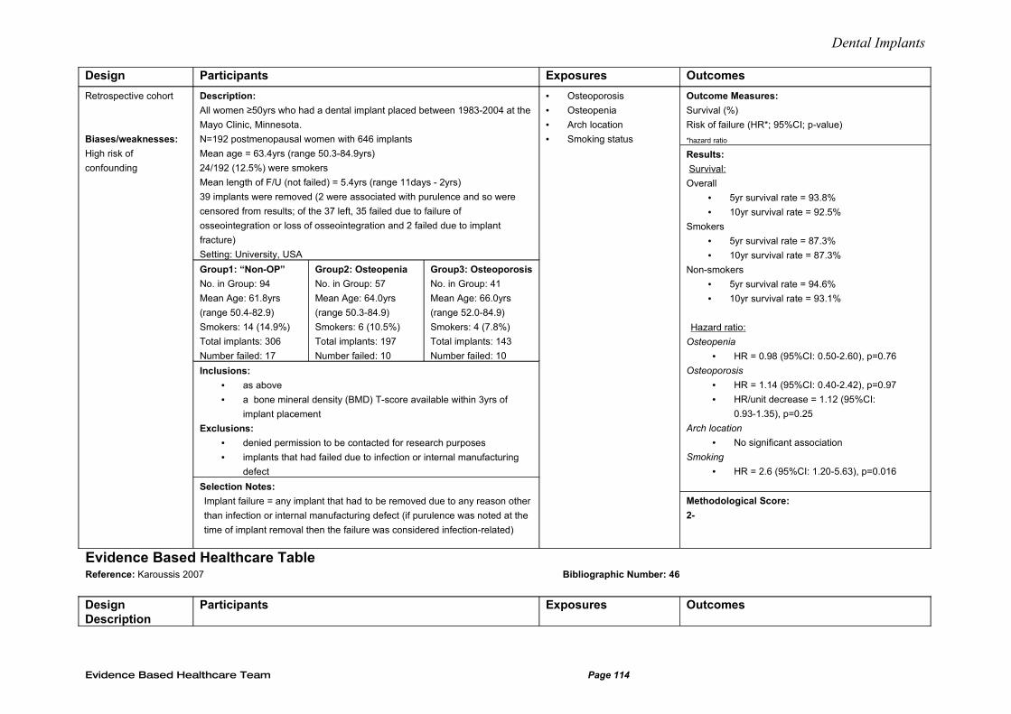
Dental Implants
Design Participants Exposures Outcomes
Retrospective cohort
Biases/weaknesses:
High risk of
confounding
Description:
All women ≥50yrs who had a dental implant placed between 1983-2004 at the
Mayo Clinic, Minnesota.
N=192 postmenopausal women with 646 implants
Mean age = 63.4yrs (range 50.3-84.9yrs)
24/192 (12.5%) were smokers
Mean length of F/U (not failed) = 5.4yrs (range 11days - 2yrs)
39 implants were removed (2 were associated with purulence and so were
censored from results; of the 37 left, 35 failed due to failure of
osseointegration or loss of osseointegration and 2 failed due to implant
fracture)
Setting: University, USA
Group1: “Non-OP”
No. in Group: 94
Mean Age: 61.8yrs
(range 50.4-82.9)
Smokers: 14 (14.9%)
Total implants: 306
Number failed: 17
Group2: Osteopenia
No. in Group: 57
Mean Age: 64.0yrs
(range 50.3-84.9)
Smokers: 6 (10.5%)
Total implants: 197
Number failed: 10
Group3: Osteoporosis
No. in Group: 41
Mean Age: 66.0yrs
(range 52.0-84.9)
Smokers: 4 (7.8%)
Total implants: 143
Number failed: 10
Inclusions:
• as above
• a bone mineral density (BMD) T-score available within 3yrs of
implant placement
Exclusions:
• denied permission to be contacted for research purposes
• implants that had failed due to infection or internal manufacturing
defect
Selection Notes:
Implant failure = any implant that had to be removed due to any reason other
than infection or internal manufacturing defect (if purulence was noted at the
time of implant removal then the failure was considered infection-related)
• Osteoporosis
• Osteopenia
• Arch location
• Smoking status
Outcome Measures:
Survival (%)
Risk of failure (HR*; 95%CI; p-value)
*hazard ratio
Results:
Survival:
Overall
• 5yr survival rate = 93.8%
• 10yr survival rate = 92.5%
Smokers
• 5yr survival rate = 87.3%
• 10yr survival rate = 87.3%
Non-smokers
• 5yr survival rate = 94.6%
• 10yr survival rate = 93.1%
Hazard ratio:
Osteopenia
• HR = 0.98 (95%CI: 0.50-2.60), p=0.76
Osteoporosis
• HR = 1.14 (95%CI: 0.40-2.42), p=0.97
• HR/unit decrease = 1.12 (95%CI:
0.93-1.35), p=0.25
Arch location
• No significant association
Smoking
• HR = 2.6 (95%CI: 1.20-5.63), p=0.016
Methodological Score:
2-
Evidence Based Healthcare Table Reference: Karoussis 2007 Bibliographic Number: 46
Design Description
Participants Exposures Outcomes
Evidence Based Healthcare Team Page 114
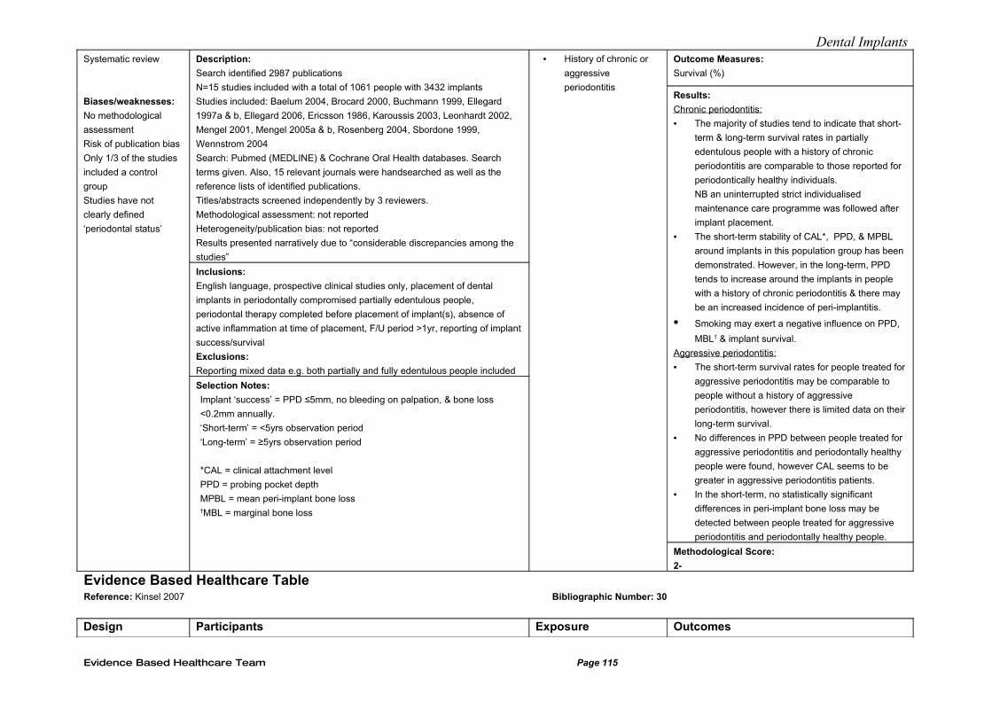
Dental ImplantsSystematic review
Biases/weaknesses:
No methodological
assessment
Risk of publication bias
Only 1/3 of the studies
included a control
group
Studies have not
clearly defined
‘periodontal status’
Description:
Search identified 2987 publications
N=15 studies included with a total of 1061 people with 3432 implants
Studies included: Baelum 2004, Brocard 2000, Buchmann 1999, Ellegard
1997a & b, Ellegard 2006, Ericsson 1986, Karoussis 2003, Leonhardt 2002,
Mengel 2001, Mengel 2005a & b, Rosenberg 2004, Sbordone 1999,
Wennstrom 2004
Search: Pubmed (MEDLINE) & Cochrane Oral Health databases. Search
terms given. Also, 15 relevant journals were handsearched as well as the
reference lists of identified publications.
Titles/abstracts screened independently by 3 reviewers.
Methodological assessment: not reported
Heterogeneity/publication bias: not reported
Results presented narratively due to “considerable discrepancies among the
studies”
Inclusions:
English language, prospective clinical studies only, placement of dental
implants in periodontally compromised partially edentulous people,
periodontal therapy completed before placement of implant(s), absence of
active inflammation at time of placement, F/U period >1yr, reporting of implant
success/survival
Exclusions:
Reporting mixed data e.g. both partially and fully edentulous people included
Selection Notes:
Implant ‘success’ = PPD ≤5mm, no bleeding on palpation, & bone loss
<0.2mm annually.
‘Short-term’ = <5yrs observation period
‘Long-term’ = ≥5yrs observation period
*CAL = clinical attachment level
PPD = probing pocket depth
MPBL = mean peri-implant bone loss†MBL = marginal bone loss
• History of chronic or
aggressive
periodontitis
Outcome Measures:
Survival (%)
Results:
Chronic periodontitis:
• The majority of studies tend to indicate that short-
term & long-term survival rates in partially
edentulous people with a history of chronic
periodontitis are comparable to those reported for
periodontically healthy individuals.
NB an uninterrupted strict individualised
maintenance care programme was followed after
implant placement.
• The short-term stability of CAL*, PPD, & MPBL
around implants in this population group has been
demonstrated. However, in the long-term, PPD
tends to increase around the implants in people
with a history of chronic periodontitis & there may
be an increased incidence of peri-implantitis.
• Smoking may exert a negative influence on PPD,
MBL† & implant survival.
Aggressive periodontitis:
• The short-term survival rates for people treated for
aggressive periodontitis may be comparable to
people without a history of aggressive
periodontitis, however there is limited data on their
long-term survival.
• No differences in PPD between people treated for
aggressive periodontitis and periodontally healthy
people were found, however CAL seems to be
greater in aggressive periodontitis patients.
• In the short-term, no statistically significant
differences in peri-implant bone loss may be
detected between people treated for aggressive
periodontitis and periodontally healthy people.
Methodological Score:
2-
Evidence Based Healthcare Table Reference: Kinsel 2007 Bibliographic Number: 30
Design Participants Exposure Outcomes
Evidence Based Healthcare Team Page 115
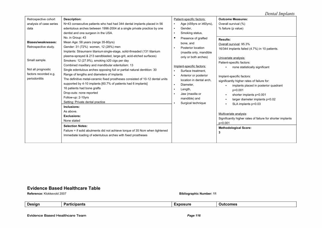
Dental ImplantsRetrospective cohort
analysis of case series
data
Biases/weaknesses:
Retrospective study.
Small sample.
Not all prognostic
factors recorded e.g.
periodontitis
Description:
N=43 consecutive patients who had had 344 dental implants placed in 56
edentulous arches between 1996-2004 at a single private practice by one
dentist and one surgeon in the USA.
No. in Group: 43
Mean Age: 58 years (range 35-80yrs)
Gender: 31 (72%) women, 12 (28%) men
Implants: Straumann titanium single-stage, solid-threaded (131 titanium
plasma-sprayed & 213 sandblasted, large-grit, acid-etched surfaces)
Smokers: 12 (27.9%), smoking ≥20 cigs per day
Combined maxillary and mandibular edentulism: 13
Single edentulous arches opposing full or partial natural dentition: 30
Range of lengths and diameters of implants
The definitive metal-ceramic fixed prostheses consisted of 10-12 dental units
supported by 4-10 implants [60.7% of patients had 6 implants]
16 patients had bone grafts
Drop-outs: none reported
Follow-up: 2-10yrs
Setting: Private dental practice
Inclusions:
As above.
Exclusions:
None stated
Selection Notes:
Failure = if solid abutments did not achieve torque of 35 Ncm when tightened
Immediate loading of edentulous arches with fixed prostheses
Patient-specific factors:
• Age (≤59yrs or ≥60yrs),
• Gender,
• Smoking status,
• Presence of grafted
bone, and
• Posterior location
(maxilla only, mandible
only or both arches)
Implant-specific factors:
• Surface treatment,
• Anterior or posterior
location in dental arch,
• Diameter,
• Length,
• Jaw (maxilla or
mandible) and
• Surgical technique
Outcome Measures:
Overall survival (%)
% failure (p value)
Results:
Overall survival: 95.3%
16/344 implants failed (4.7%) in 10 patients.
Univariate analysis:
Patient-specific factors:
• none statistically significant
Implant-specific factors: significantly higher rates of failure for:
• implants placed in posterior quadrant
p=0.001
• shorter implants p<0.001
• larger diameter implants p<0.02
• SLA implants p<0.03
Multivariate analysis:
Significantly higher rates of failure for shorter implants
p<0.001
Methodological Score:
3
Evidence Based Healthcare Table Reference: Klokkevold 2007 Bibliographic Number: 11
Design Participants Exposure Outcomes
Evidence Based Healthcare Team Page 116
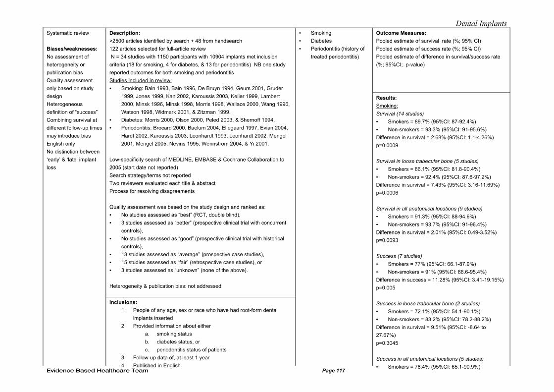
Dental ImplantsSystematic review
Biases/weaknesses:
No assessment of
heterogeneity or
publication bias
Quality assessment
only based on study
design
Heterogeneous
definition of “success”
Combining survival at
different follow-up times
may introduce bias
English only
No distinction between
‘early’ & ‘late’ implant
loss
Description:
>2500 articles identified by search + 48 from handsearch
122 articles selected for full-article review
N = 34 studies with 1150 participants with 10904 implants met inclusion
criteria (18 for smoking, 4 for diabetes, & 13 for periodontitis) NB one study
reported outcomes for both smoking and periodontitis
Studies included in review:
• Smoking: Bain 1993, Bain 1996, De Bruyn 1994, Geurs 2001, Gruder
1999, Jones 1999, Kan 2002, Karoussis 2003, Keller 1999, Lambert
2000, Minsk 1996, Minsk 1998, Morris 1998, Wallace 2000, Wang 1996,
Watson 1998, Widmark 2001, & Zitzman 1999.
• Diabetes: Morris 2000, Olson 2000, Peled 2003, & Shernoff 1994.
• Periodontitis: Brocard 2000, Baelum 2004, Ellegaard 1997, Evian 2004,
Hardt 2002, Karoussis 2003, Leonhardt 1993, Leonhardt 2002, Mengel
2001, Mengel 2005, Nevins 1995, Wennstrom 2004, & Yi 2001.
Low-specificity search of MEDLINE, EMBASE & Cochrane Collaboration to
2005 (start date not reported)
Search strategy/terms not reported
Two reviewers evaluated each title & abstract
Process for resolving disagreements
Quality assessment was based on the study design and ranked as:
• No studies assessed as “best” (RCT, double blind),
• 3 studies assessed as “better” (prospective clinical trial with concurrent
controls),
• No studies assessed as “good” (prospective clinical trial with historical
controls),
• 13 studies assessed as “average” (prospective case studies),
• 15 studies assessed as “fair” (retrospective case studies), or
• 3 studies assessed as “unknown” (none of the above).
Heterogeneity & publication bias: not addressed
Inclusions:
1. People of any age, sex or race who have had root-form dental
implants inserted
2. Provided information about either
a. smoking status
b. diabetes status, or
c. periodontitis status of patients
3. Follow-up data of, at least 1 year
4. Published in English
• Smoking
• Diabetes
• Periodontitis (history of
treated periodontitis)
Outcome Measures:
Pooled estimate of survival rate (%; 95% CI)
Pooled estimate of success rate (%; 95% CI)
Pooled estimate of difference in survival/success rate
(%; 95%CI; p-value)
Results:
Smoking:
Survival (14 studies)
• Smokers = 89.7% (95%CI: 87-92.4%)
• Non-smokers = 93.3% (95%CI: 91-95.6%)
Difference in survival = 2.68% (95%CI: 1.1-4.26%)
p=0.0009
Survival in loose trabecular bone (5 studies)
• Smokers = 86.1% (95%CI: 81.8-90.4%)
• Non-smokers = 92.4% (95%CI: 87.6-97.2%)
Difference in survival = 7.43% (95%CI: 3.16-11.69%)
p=0.0006
Survival in all anatomical locations (9 studies)
• Smokers = 91.3% (95%CI: 88-94.6%)
• Non-smokers = 93.7% (95%CI: 91-96.4%)
Difference in survival = 2.01% (95%CI: 0.49-3.52%)
p=0.0093
Success (7 studies)
• Smokers = 77% (95%CI: 66.1-87.9%)
• Non-smokers = 91% (95%CI: 86.6-95.4%)
Difference in success = 11.28% (95%CI: 3.41-19.15%)
p=0.005
Success in loose trabecular bone (2 studies)
• Smokers = 72.1% (95%CI: 54.1-90.1%)
• Non-smokers = 83.2% (95%CI: 78.2-88.2%)
Difference in survival = 9.51% (95%CI: -8.64 to
27.67%)
p=0.3045
Success in all anatomical locations (5 studies)
• Smokers = 78.4% (95%CI: 65.1-90.9%)Evidence Based Healthcare Team Page 117
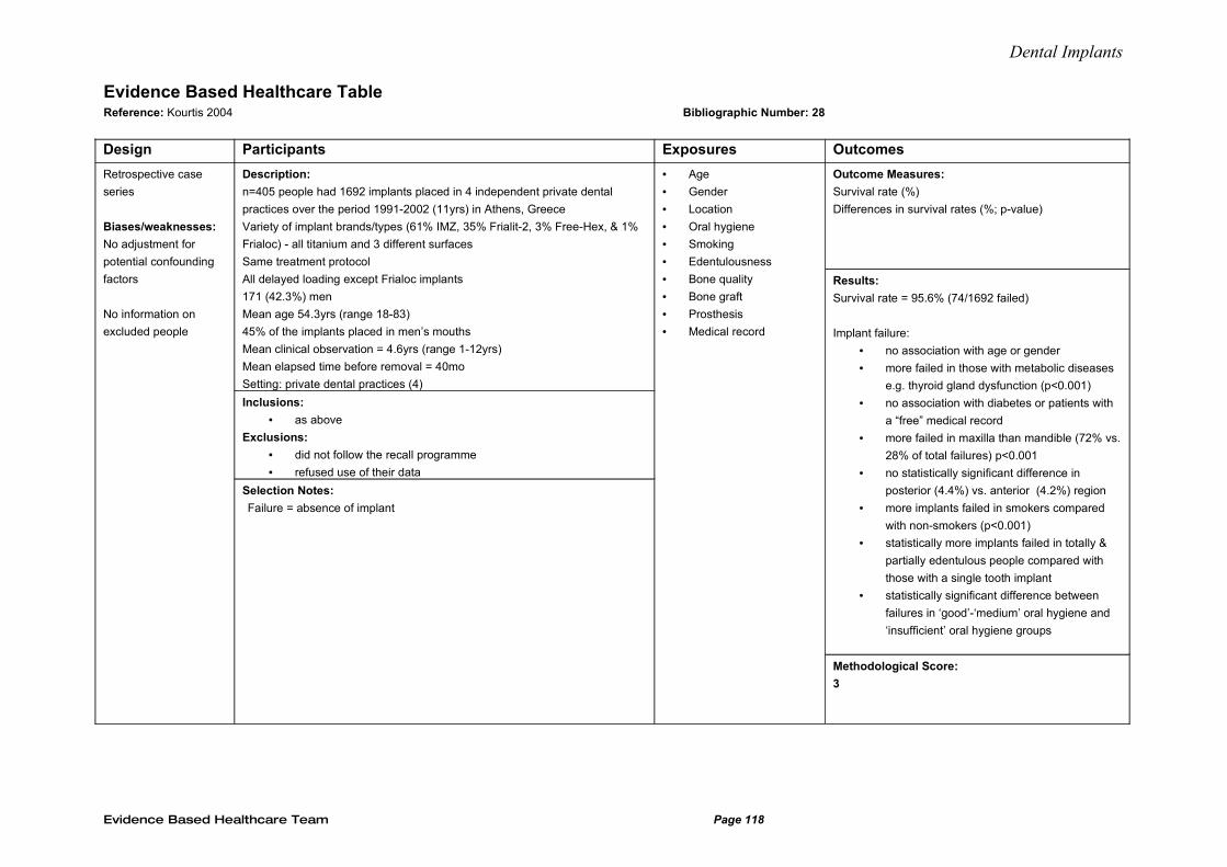
Dental Implants
Evidence Based Healthcare Table Reference: Kourtis 2004 Bibliographic Number: 28
Design Participants Exposures Outcomes
Retrospective case
series
Biases/weaknesses:
No adjustment for
potential confounding
factors
No information on
excluded people
Description:
n=405 people had 1692 implants placed in 4 independent private dental
practices over the period 1991-2002 (11yrs) in Athens, Greece
Variety of implant brands/types (61% IMZ, 35% Frialit-2, 3% Free-Hex, & 1%
Frialoc) - all titanium and 3 different surfaces
Same treatment protocol
All delayed loading except Frialoc implants
171 (42.3%) men
Mean age 54.3yrs (range 18-83)
45% of the implants placed in men’s mouths
Mean clinical observation = 4.6yrs (range 1-12yrs)
Mean elapsed time before removal = 40mo
Setting: private dental practices (4)
Inclusions:
• as above
Exclusions:
• did not follow the recall programme
• refused use of their data
Selection Notes:
Failure = absence of implant
• Age
• Gender
• Location
• Oral hygiene
• Smoking
• Edentulousness
• Bone quality
• Bone graft
• Prosthesis
• Medical record
Outcome Measures:
Survival rate (%)
Differences in survival rates (%; p-value)
Results:
Survival rate = 95.6% (74/1692 failed)
Implant failure:
• no association with age or gender
• more failed in those with metabolic diseases
e.g. thyroid gland dysfunction (p<0.001)
• no association with diabetes or patients with
a “free” medical record
• more failed in maxilla than mandible (72% vs.
28% of total failures) p<0.001
• no statistically significant difference in
posterior (4.4%) vs. anterior (4.2%) region
• more implants failed in smokers compared
with non-smokers (p<0.001)
• statistically more implants failed in totally &
partially edentulous people compared with
those with a single tooth implant
• statistically significant difference between
failures in ‘good’-‘medium’ oral hygiene and
‘insufficient’ oral hygiene groups
Methodological Score:
3
Evidence Based Healthcare Team Page 118
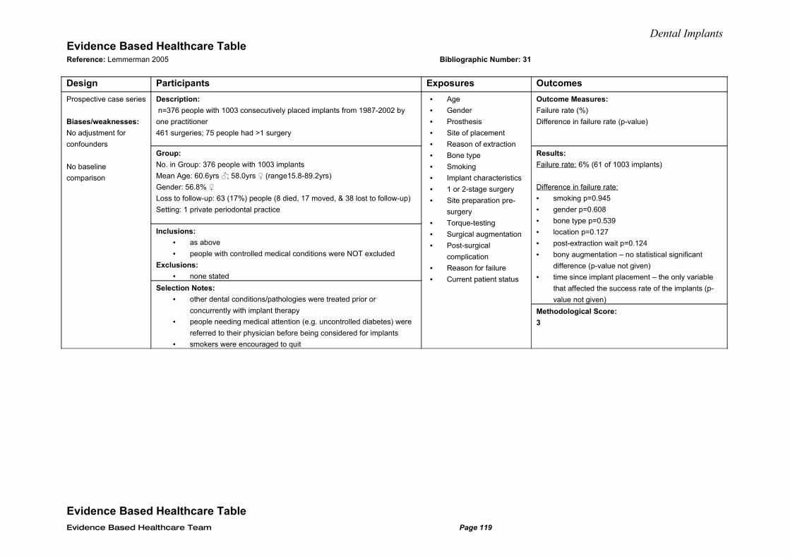
Dental ImplantsEvidence Based Healthcare Table Reference: Lemmerman 2005 Bibliographic Number: 31
Design Participants Exposures Outcomes
Prospective case series
Biases/weaknesses:
No adjustment for
confounders
No baseline
comparison
Description:
n=376 people with 1003 consecutively placed implants from 1987-2002 by
one practitioner
461 surgeries; 75 people had >1 surgery
Group:
No. in Group: 376 people with 1003 implants
Mean Age: 60.6yrs ♂; 58.0yrs ♀ (range15.8-89.2yrs)
Gender: 56.8% ♀
Loss to follow-up: 63 (17%) people (8 died, 17 moved, & 38 lost to follow-up)
Setting: 1 private periodontal practice
Inclusions:
• as above
• people with controlled medical conditions were NOT excluded
Exclusions:
• none stated
Selection Notes:
• other dental conditions/pathologies were treated prior or
concurrently with implant therapy
• people needing medical attention (e.g. uncontrolled diabetes) were
referred to their physician before being considered for implants
• smokers were encouraged to quit
• Age
• Gender
• Prosthesis
• Site of placement
• Reason of extraction
• Bone type
• Smoking
• Implant characteristics
• 1 or 2-stage surgery
• Site preparation pre-
surgery
• Torque-testing
• Surgical augmentation
• Post-surgical
complication
• Reason for failure
• Current patient status
Outcome Measures:
Failure rate (%)
Difference in failure rate (p-value)
Results:
Failure rate: 6% (61 of 1003 implants)
Difference in failure rate:
• smoking p=0.945
• gender p=0.608
• bone type p=0.539
• location p=0.127
• post-extraction wait p=0.124
• bony augmentation – no statistical significant
difference (p-value not given)
• time since implant placement – the only variable
that affected the success rate of the implants (p-
value not given)
Methodological Score:
3
Evidence Based Healthcare Table Evidence Based Healthcare Team Page 119
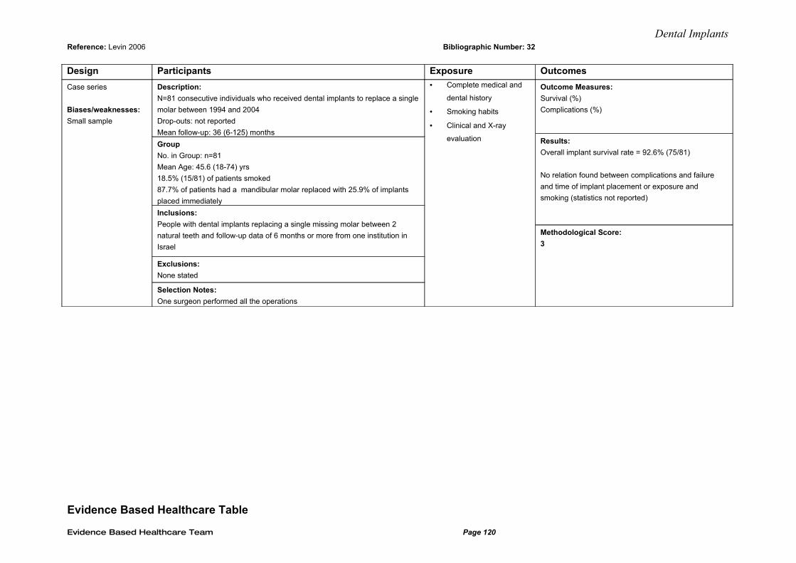
Dental ImplantsReference: Levin 2006 Bibliographic Number: 32
Design Participants Exposure Outcomes
Case series
Biases/weaknesses:
Small sample
Description:
N=81 consecutive individuals who received dental implants to replace a single
molar between 1994 and 2004
Drop-outs: not reported
Mean follow-up: 36 (6-125) months
Group
No. in Group: n=81
Mean Age: 45.6 (18-74) yrs
18.5% (15/81) of patients smoked
87.7% of patients had a mandibular molar replaced with 25.9% of implants
placed immediately
Inclusions:
People with dental implants replacing a single missing molar between 2
natural teeth and follow-up data of 6 months or more from one institution in
Israel
Exclusions:
None stated
Selection Notes:
One surgeon performed all the operations
• Complete medical and
dental history
• Smoking habits
• Clinical and X-ray
evaluation
Outcome Measures:
Survival (%)
Complications (%)
Results:
Overall implant survival rate = 92.6% (75/81)
No relation found between complications and failure
and time of implant placement or exposure and
smoking (statistics not reported)
Methodological Score:
3
Evidence Based Healthcare Table
Evidence Based Healthcare Team Page 120
Dental ImplantsReference: McDermott 2003 Bibliographic Number: 16
Design Participants Exposures Outcomes
Evidence Based Healthcare Team Page 121
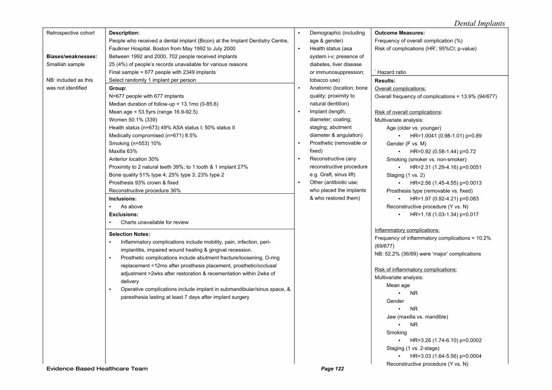
Dental ImplantsRetrospective cohort
Biases/weaknesses:
Smallish sample
NB: included as this
was not identified
Description:
People who received a dental implant (Bicon) at the Implant Dentistry Centre,
Faulkner Hospital, Boston from May 1992 to July 2000
Between 1992 and 2000, 702 people received implants
25 (4%) of people’s records unavailable for various reasons
Final sample = 677 people with 2349 implants
Select randomly 1 implant per person
Group:
N=677 people with 677 implants
Median duration of follow-up = 13.1mo (0-85.6)
Mean age = 53.5yrs (range 16.9-92.5)
Women 50.1% (339)
Health status (n=673) 49% ASA status I; 50% status II
Medically compromised (n=671) 8.5%
Smoking (n=553) 10%
Maxilla 63%
Anterior location 30%
Proximity to 2 natural teeth 39%; to 1 tooth & 1 implant 27%
Bone quality 51% type 4; 25% type 3; 23% type 2
Prosthesis 93% crown & fixed
Reconstructive procedure 36%
Inclusions:
• As above
Exclusions:
• Charts unavailable for review
Selection Notes:
• Inflammatory complications include mobility, pain, infection, peri-
implantitis, impaired wound healing & gingival recession.
• Prosthetic complications include abutment fracture/loosening, O-ring
replacement <12mo after prosthesis placement, prosthetic/occlusal
adjustment >2wks after restoration & recementation within 2wks of
delivery
• Operative complications include implant in submandibular/sinus space, &
paresthesia lasting at least 7 days after implant surgery
• Demographic (including
age & gender)
• Health status (asa
system i-v; presence of
diabetes, liver disease
or immunosuppression;
tobacco use)
• Anatomic (location; bone
quality; proximity to
natural dentition)
• Implant (length;
diameter; coating;
staging; abutment
diameter & angulation)
• Prosthetic (removable or
fixed)
• Reconstructive (any
reconstructive procedure
e.g. Graft, sinus lift)
• Other (antibiotic use;
who placed the implants
& who restored them)
Outcome Measures:
Frequency of overall complication (%)
Risk of complications (HR*; 95%CI; p-value)
* Hazard ratio
Results:
Overall complications:
Overall frequency of complications = 13.9% (94/677)
Risk of overall complications:
Multivariate analysis:
Age (older vs. younger)
• HR=1.0041 (0.98-1.01) p=0.89
Gender (F vs. M)
• HR=0.92 (0.58-1.44) p=0.72
Smoking (smoker vs. non-smoker)
• HR=2.31 (1.29-4.16) p=0.0051
Staging (1 vs. 2)
• HR=2.56 (1.45-4.55) p=0.0013
Prosthesis type (removable vs. fixed)
• HR=1.97 (0.92-4.21) p=0.083
Reconstructive procedure (Y vs. N)
• HR=1.18 (1.03-1.34) p=0.017
Inflammatory complications:
Frequency of inflammatory complications = 10.2%
(69/677)
NB: 52.2% (36/69) were ‘major’ complications
Risk of inflammatory complications:
Multivariate analysis:
Mean age
• NR
Gender
• NR
Jaw (maxilla vs. mandible)
• NR
Smoking
• HR=3.26 (1.74-6.10) p=0.0002
Staging (1 vs. 2-stage)
• HR=3.03 (1.64-5.56) p=0.0004
Reconstructive procedure (Y vs. N) Evidence Based Healthcare Team Page 122

Dental Implants
Evidence Based Healthcare Team Page 123

Dental ImplantsEvidence Based Healthcare Table Reference: McDermott 2006 Bibliographic Number: 50
Design Participants Exposures Outcomes
Evidence Based Healthcare Team Page 124
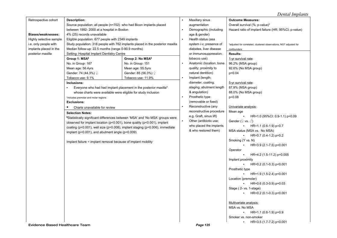
Dental ImplantsRetrospective cohort
Biases/weaknesses:
Highly selective sample
i.e. only people with
implants placed in the
posterior maxilla
Description:
Source population: all people (n=702) who had Bicon implants placed
between 1992- 2000 at a hospital in Boston
4% (25) records unavailable
Eligible population: 677 people with 2349 implants
Study population: 318 people with 762 implants placed in the posterior maxilla
Median follow-up: 22.5 months (range 0-90.9 months)
Setting: Hospital Implant Dentistry Centre
Group 1: MSA§
No. in Group: 167
Mean age: 56.4yrs
Gender: 74 (44.3%) ♀
Tobacco use: 9.1%
Group 2: No MSA§
No. in Group: 151
Mean age: 55.5yrs
Gender: 85 (56.3%) ♀
Tobacco use: 11.9%
Inclusions:
• Everyone who had had implant placement in the posterior maxilla*
whose charts were available were eligible for study inclusion
*includes premolar and molar regions
Exclusions:
• Charts unavailable for review
Selection Notes:§Statistically significant differences between ‘MSA’ and ‘No MSA’ groups were
observed for implant location (p<0.001), bone quality (p<0.001), implant
coating (p<0.001), well size (p=0.008), implant staging (p=0.006), immediate
implant (p=0.001), and abutment angle (p=0.009)
Implant failure = implant removal because of implant mobility
• Maxillary sinus
augmentation
• Demographic (including
age & gender)
• Health status (asa
system i-v; presence of
diabetes, liver disease
or immunosuppression;
tobacco use)
• Anatomic (location; bone
quality; proximity to
natural dentition)
• Implant (length;
diameter; coating;
staging; abutment length
& angulation)
• Prosthetic type
(removable or fixed)
• Reconstructive (any
reconstructive procedure
e.g. Graft, sinus lift)
• Other (antibiotic use;
who placed the implants
& who restored them)
Outcome Measures:
Overall survival (%; p-value)*
Hazard ratio of implant failure (HR; 95%CI; p-value)
*adjusted for correlated, clustered observations, NOT adjusted for
confounders
Results:
1-yr survival rate:
96.2% (MSA group)
92.6% (No MSA group)
p=0.04
5-yr survival rate:
87.9% (MSA group)
88.0% (No MSA group)
p=0.08
Univariate analysis:
Mean age
• HR=1.0 (95%CI: 0.9-1.1) p=0.09
Gender (♀ vs. ♂)
• HR=1.1 (0.6-1.9) p=0.7
MSA status (MSA vs. No MSA)
• HR=0.7 (0.4-1.2) p=0.2
Smoking (Y vs. N)
• HR=3.9 (2.1-7.5) p<0.001
Operator
• HR=4.2 (1.5-11.2) p=0.005
Implant proximity
• HR=0.2 (0.1-0.3) p<0.001
Prosthetic type
• HR=1.9 (1.5-2.4) p<0.001
Location (premolar)
• HR=0.6 (0.3-0.9) p=0.03
Stage ( 2- vs. 1-stage)
• HR=0.2 (0.1-0.3) p<0.001
Multivariate analysis:
MSA vs. No MSA
• HR=1.1 (0.6-1.9) p=0.9
Smoker vs. non-smoker
• HR=3.5 (1.7-7.2) p<0.001Evidence Based Healthcare Team Page 125

Dental Implants
Evidence Based Healthcare Team Page 126
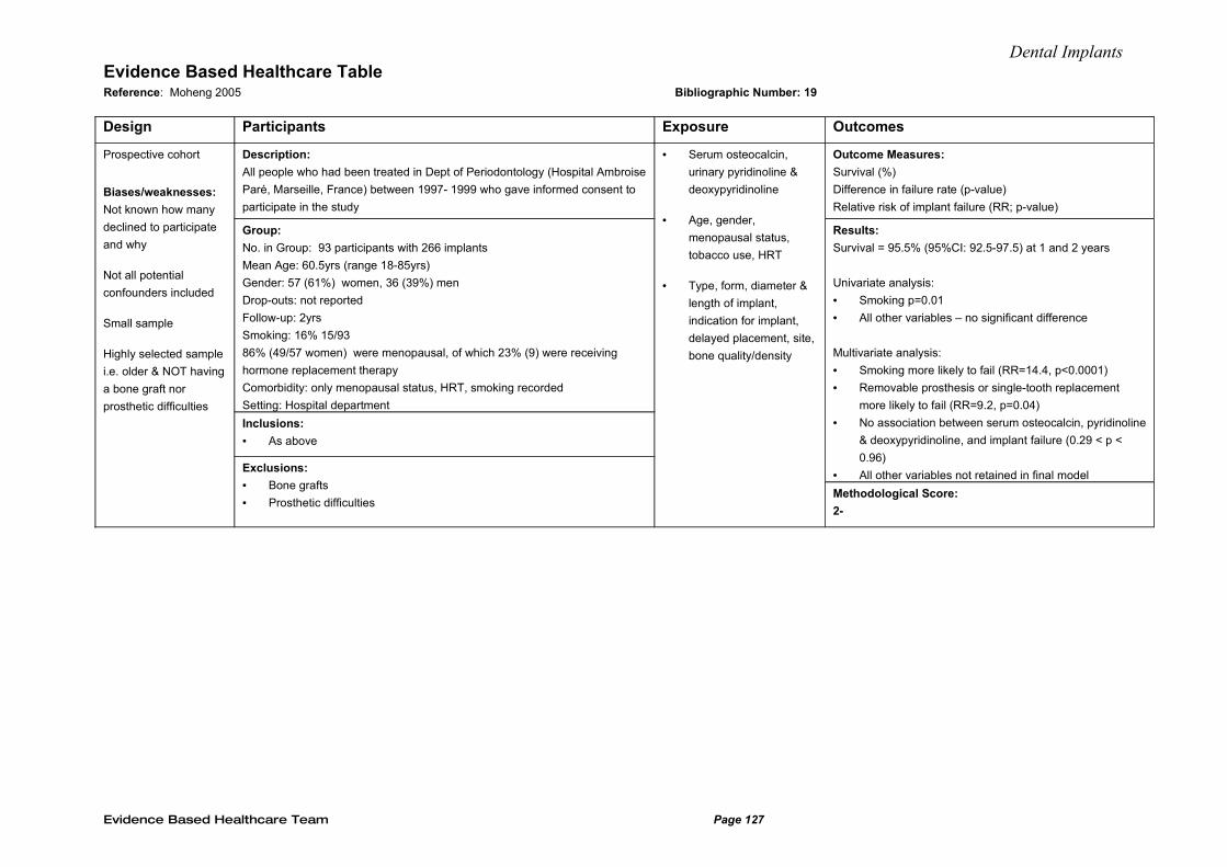
Dental ImplantsEvidence Based Healthcare Table Reference: Moheng 2005 Bibliographic Number: 19
Design Participants Exposure Outcomes
Prospective cohort
Biases/weaknesses:
Not known how many
declined to participate
and why
Not all potential
confounders included
Small sample
Highly selected sample
i.e. older & NOT having
a bone graft nor
prosthetic difficulties
Description:
All people who had been treated in Dept of Periodontology (Hospital Ambroise
Paré, Marseille, France) between 1997- 1999 who gave informed consent to
participate in the study
Group:
No. in Group: 93 participants with 266 implants
Mean Age: 60.5yrs (range 18-85yrs)
Gender: 57 (61%) women, 36 (39%) men
Drop-outs: not reported
Follow-up: 2yrs
Smoking: 16% 15/93
86% (49/57 women) were menopausal, of which 23% (9) were receiving
hormone replacement therapy
Comorbidity: only menopausal status, HRT, smoking recorded
Setting: Hospital department
Inclusions:
• As above
Exclusions:
• Bone grafts
• Prosthetic difficulties
• Serum osteocalcin,
urinary pyridinoline &
deoxypyridinoline
• Age, gender,
menopausal status,
tobacco use, HRT
• Type, form, diameter &
length of implant,
indication for implant,
delayed placement, site,
bone quality/density
Outcome Measures:
Survival (%)
Difference in failure rate (p-value)
Relative risk of implant failure (RR; p-value)
Results:
Survival = 95.5% (95%CI: 92.5-97.5) at 1 and 2 years
Univariate analysis:
• Smoking p=0.01
• All other variables – no significant difference
Multivariate analysis:
• Smoking more likely to fail (RR=14.4, p<0.0001)
• Removable prosthesis or single-tooth replacement
more likely to fail (RR=9.2, p=0.04)
• No association between serum osteocalcin, pyridinoline
& deoxypyridinoline, and implant failure (0.29 < p <
0.96)
• All other variables not retained in final model
Methodological Score:
2-
Evidence Based Healthcare Team Page 127
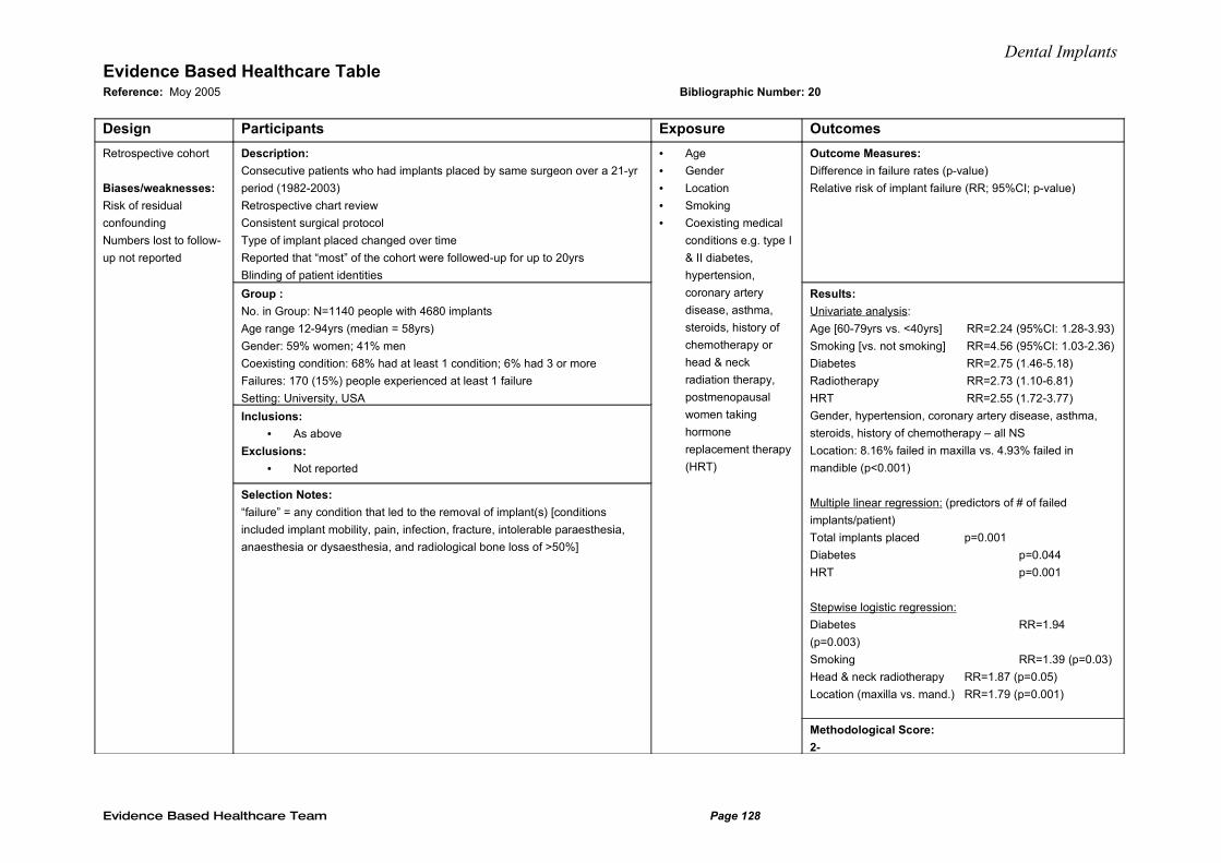
Dental ImplantsEvidence Based Healthcare Table Reference: Moy 2005 Bibliographic Number: 20
Design Participants Exposure Outcomes
Retrospective cohort
Biases/weaknesses:
Risk of residual
confounding
Numbers lost to follow-
up not reported
Description:
Consecutive patients who had implants placed by same surgeon over a 21-yr
period (1982-2003)
Retrospective chart review
Consistent surgical protocol
Type of implant placed changed over time
Reported that “most” of the cohort were followed-up for up to 20yrs
Blinding of patient identities
Group :
No. in Group: N=1140 people with 4680 implants
Age range 12-94yrs (median = 58yrs)
Gender: 59% women; 41% men
Coexisting condition: 68% had at least 1 condition; 6% had 3 or more
Failures: 170 (15%) people experienced at least 1 failure
Setting: University, USA
Inclusions:
• As above
Exclusions:
• Not reported
Selection Notes:
“failure” = any condition that led to the removal of implant(s) [conditions
included implant mobility, pain, infection, fracture, intolerable paraesthesia,
anaesthesia or dysaesthesia, and radiological bone loss of >50%]
• Age
• Gender
• Location
• Smoking
• Coexisting medical
conditions e.g. type I
& II diabetes,
hypertension,
coronary artery
disease, asthma,
steroids, history of
chemotherapy or
head & neck
radiation therapy,
postmenopausal
women taking
hormone
replacement therapy
(HRT)
Outcome Measures:
Difference in failure rates (p-value)
Relative risk of implant failure (RR; 95%CI; p-value)
Results:
Univariate analysis:
Age [60-79yrs vs. <40yrs] RR=2.24 (95%CI: 1.28-3.93)
Smoking [vs. not smoking] RR=4.56 (95%CI: 1.03-2.36)
Diabetes RR=2.75 (1.46-5.18)
Radiotherapy RR=2.73 (1.10-6.81)
HRT RR=2.55 (1.72-3.77)
Gender, hypertension, coronary artery disease, asthma,
steroids, history of chemotherapy – all NS
Location: 8.16% failed in maxilla vs. 4.93% failed in
mandible (p<0.001)
Multiple linear regression: (predictors of # of failed
implants/patient)
Total implants placed p=0.001
Diabetes p=0.044
HRT p=0.001
Stepwise logistic regression:
Diabetes RR=1.94
(p=0.003)
Smoking RR=1.39 (p=0.03)
Head & neck radiotherapy RR=1.87 (p=0.05)
Location (maxilla vs. mand.) RR=1.79 (p=0.001)
Methodological Score:
2-
Evidence Based Healthcare Team Page 128
Dental ImplantsEvidence Based Healthcare Table Reference: Mundt 2006 Bibliographic Number: 12
Design Participants Exposure Outcomes
Evidence Based Healthcare Team Page 129
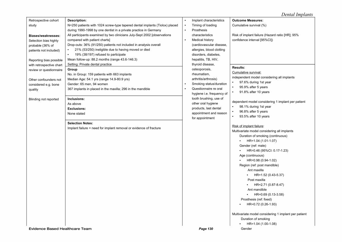
Dental ImplantsRetrospective cohort
study
Biases/weaknesses:
Selection bias highly
probable (36% of
patients not included)
Reporting bias possible
with retrospective chart
review or questionnaire
Other confounders not
considered e.g. bone
quality
Blinding not reported
Description:
N=250 patients with 1024 screw-type tapered dental implants (Tiolox) placed
during 1990-1998 by one dentist in a private practice in Germany
All participants examined by two clinicians July-Sept 2002 [observations
compared with patient charts]
Drop-outs: 36% (91/250) patients not included in analysis overall
• 21% (53/250) ineligible due to having moved or died
• 19% (38/197) refused to participate
Mean follow-up: 88.2 months (range 43.6-146.3)
Setting: Private dental practice
Group
No. in Group: 159 patients with 663 implants
Median Age: 54.1 yrs (range 14.9-80.9 yrs)
Gender: 65 men, 94 women
367 implants in placed in the maxilla; 296 in the mandible
Inclusions:
As above
Exclusions:
None stated
Selection Notes:
Implant failure = need for implant removal or evidence of fracture
• Implant characteristics
• Timing of loading
• Prosthesis
characteristics
• Medical history
(cardiovascular disease,
allergies, blood clotting
disorders, diabetes,
hepatitis, TB, HIV,
thyroid disease,
osteoporosis,
rheumatism,
arthritis/arthrosis)
• Smoking status/duration
• Questionnaire re oral
hygiene i.e. frequency of
tooth brushing, use of
other oral hygiene
products, last dental
appointment and reason
for appointment
Outcome Measures:
Cumulative survival (%)
Risk of implant failure (Hazard ratio [HR]; 95%
confidence interval [95%CI])
Results:
Cumulative survival:
independent model considering all implants
• 97.6% during 1st year
• 95.9% after 5 years
• 91.8% after 10 years
dependent model considering 1 implant per patient
• 98.1% during 1st year
• 96.8% after 5 years
• 93.5% after 10 years
Risk of implant failure:
Multivariate model considering all implants
Duration of smoking (continuous)
• HR=1.04 (1.01-1.07)
Gender (ref: male)
• HR=0.46 (95%CI: 0.17-1.23)
Age (continuous)
• HR=0.98 (0.94-1.02)
Region (ref: post mandible)
Ant maxilla
• HR=1.52 (0.43-5.37)
Post maxilla
• HR=2.71 (0.87-8.47)
Ant mandible
• HR=0.69 (0.13-3.58)
Prosthesis (ref: fixed)
• HR=0.72 (0.26-1.93)
Multivariate model considering 1 implant per patient
Duration of smoking
• HR=1.04 (1.00-1.08)
GenderEvidence Based Healthcare Team Page 130

Dental Implants
Evidence Based Healthcare Team Page 131
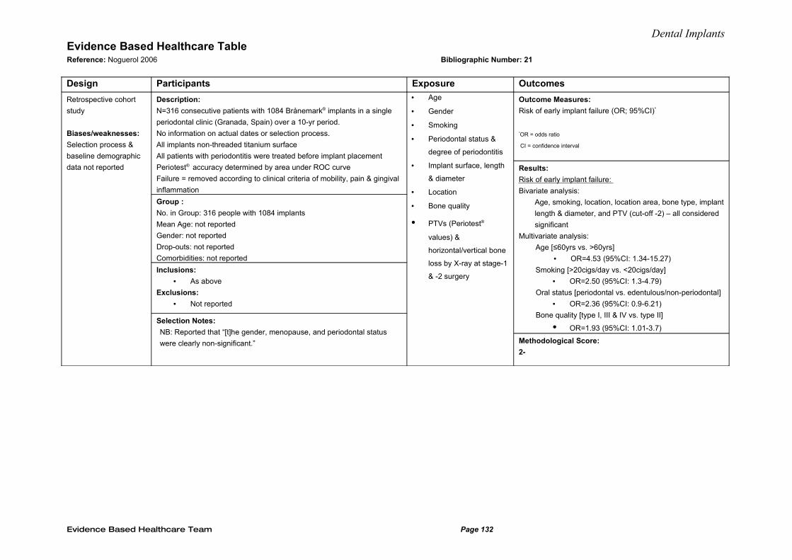
Dental ImplantsEvidence Based Healthcare Table Reference: Noguerol 2006 Bibliographic Number: 21
Design Participants Exposure Outcomes
Retrospective cohort
study
Biases/weaknesses:
Selection process &
baseline demographic
data not reported
Description:
N=316 consecutive patients with 1084 Brånemark® implants in a single
periodontal clinic (Granada, Spain) over a 10-yr period.
No information on actual dates or selection process.
All implants non-threaded titanium surface
All patients with periodontitis were treated before implant placement
Periotest® accuracy determined by area under ROC curve
Failure = removed according to clinical criteria of mobility, pain & gingival
inflammation
Group :
No. in Group: 316 people with 1084 implants
Mean Age: not reported
Gender: not reported
Drop-outs: not reported
Comorbidities: not reported
Inclusions:
• As above
Exclusions:
• Not reported
Selection Notes:
NB: Reported that “[t]he gender, menopause, and periodontal status
were clearly non-significant.”
• Age
• Gender
• Smoking
• Periodontal status &
degree of periodontitis
• Implant surface, length
& diameter
• Location
• Bone quality
• PTVs (Periotest®
values) &
horizontal/vertical bone
loss by X-ray at stage-1
& -2 surgery
Outcome Measures:
Risk of early implant failure (OR; 95%CI)*
*OR = odds ratio
CI = confidence interval
Results:
Risk of early implant failure:
Bivariate analysis:
Age, smoking, location, location area, bone type, implant
length & diameter, and PTV (cut-off -2) – all considered
significant
Multivariate analysis:
Age [≤60yrs vs. >60yrs]
• OR=4.53 (95%CI: 1.34-15.27)
Smoking [>20cigs/day vs. <20cigs/day]
• OR=2.50 (95%CI: 1.3-4.79)
Oral status [periodontal vs. edentulous/non-periodontal]
• OR=2.36 (95%CI: 0.9-6.21)
Bone quality [type I, III & IV vs. type II]
• OR=1.93 (95%CI: 1.01-3.7)
Methodological Score:
2-
Evidence Based Healthcare Team Page 132
Dental ImplantsEvidence Based Healthcare Table Reference: Ong 2008 Bibliographic Number: 47
Design Description
Participants Exposures Outcomes
Evidence Based Healthcare Team Page 133
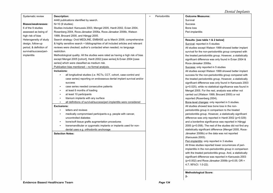
Dental ImplantsSystematic review
Biases/weaknesses:
6 of the 9 studies
assessed as being of
high risk of bias
Heterogeneity of study
design, follow-up
period, & definition of
survival/success/peri-
implantitis
Description:
4448 publications identified by search.
N=10 (9 studies)
Studies included: Karoussis 2003, Mengel 2005, Hardt 2002, Evian 2004,
Rosenberg 2004, Roos-Jånsaker 2006a, Roos-Jånsaker 2006b, Watson
1999, Brocard 2000, and Hänggi 2005.
Search strategy: Ovid MEDLINE, EMBASE up to March 2006; comprehensive
& highly sensitive search + bibliographies of all included articles and relevant
reviews were checked; author’s contacted when needed; no language
restriction.
Methodological quality: All the studies were rated as having a high risk of bias
except Mengel 2005 [cohort], Hardt 2002 [case series] & Evian 2004 [case
series] which were classified as medium risk.
Publication bias mentioned – no formal analysis.
Inclusions:
• all longitudinal studies (i.e. RCTs, CCT, cohort, case-control and
case series) reporting on endosseous dental implant survival and/or
success
• case series needed consecutive patients
• at least 6 months of loading
• at least 10 participants
• titanium implants with any surface
• all definitions of survival/success/peri-implantitis were considered
Exclusions:
• letters and reviews
• medically compromised participants e.g. people with cancer,
uncontrolled diabetes
• bone/soft tissue grafts augmentation procedures
• transmandibular or zygomatic implants or implants used for non-
dental uses e.g. orthodontic anchorage
Selection Notes:
• Periodontitis Outcome Measures:
Survival
Success
Bone loss
Peri-implantitis
Results: [see table 1 & 2 below]
Survival: reported in 5 studies
All studies except Watson 1999 showed better implant
survival for the non-periodontitis group compared with
the treated periodontitis group. However, a statistically
significant difference was only found in Evian 2004 &
Roos-Jånsaker 2006a.
Success: only reported in 5 studies
All studies except Watson 1999 showed better implant
success for the non-periodontitis group compared with
the treated periodontitis group. However, a statistically
significant difference was only found in Karoussis 2003
(p<0.025), while no statistical significance was found in
Mengel 2005. For the rest, analysis was either not
carried out (Watson 1999, Brocard 2000) or not
reported (Rosenberg 2004).
Bone-level changes: only reported in 5 studies.
All studies showed less bone loss in the non-
periodontitis group in comparison to the treated
periodontitis group. However, a statistically significant
difference was only reported in Hardt 2002 (p=0.029)
and a borderline significance was reported in Hänggi
2005 (p=0.058). The rest of the studies did not find any
statistically significant difference (Mengel 2005, Roos-
Jånsaker 2006b) or the data was not reported
(Karoussis 2003).
Peri-implantitis: only reported in 3 studies
All three studies reported lower occurrences of peri-
implantitis in the non-periodontitis group in comparison
with the treated periodontitis group. And, a statistically
significant difference was reported in Karoussis 2003
(p=0.002) and Roos-Jånsaker 2006b (p=0.05; OR =
4.7; 95%CI: 1.0-22).
Methodological Score:
2-
Evidence Based Healthcare Team Page 134
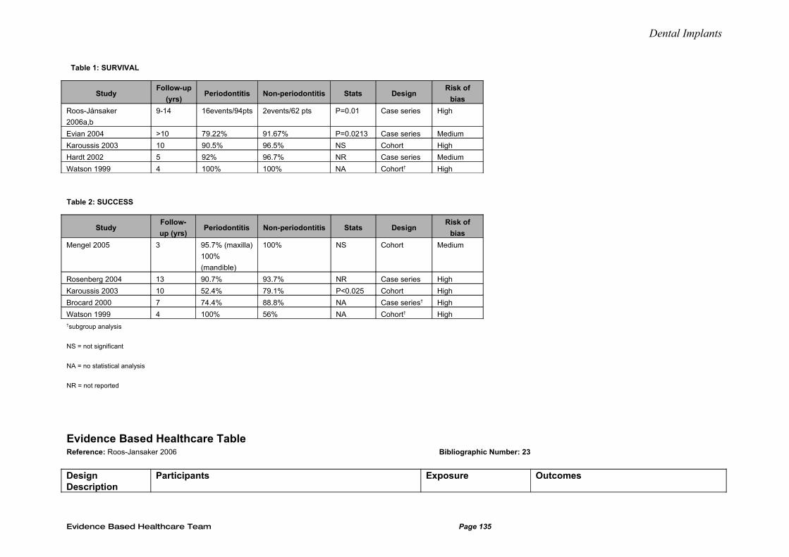
Dental Implants
Table 1: SURVIVAL
StudyFollow-up
(yrs)Periodontitis Non-periodontitis Stats Design
Risk of
bias
Roos-Jånsaker
2006a,b
9-14 16events/94pts 2events/62 pts P=0.01 Case series High
Evian 2004 >10 79.22% 91.67% P=0.0213 Case series Medium
Karoussis 2003 10 90.5% 96.5% NS Cohort High
Hardt 2002 5 92% 96.7% NR Case series Medium
Watson 1999 4 100% 100% NA Cohort† High
Table 2: SUCCESS
StudyFollow-
up (yrs)Periodontitis Non-periodontitis Stats Design
Risk of
bias
Mengel 2005 3 95.7% (maxilla)
100%
(mandible)
100% NS Cohort Medium
Rosenberg 2004 13 90.7% 93.7% NR Case series High
Karoussis 2003 10 52.4% 79.1% P<0.025 Cohort High
Brocard 2000 7 74.4% 88.8% NA Case series† High
Watson 1999 4 100% 56% NA Cohort† High†subgroup analysis
NS = not significant
NA = no statistical analysis
NR = not reported
Evidence Based Healthcare Table Reference: Roos-Jansaker 2006 Bibliographic Number: 23
Design Description
Participants Exposure Outcomes
Evidence Based Healthcare Team Page 135
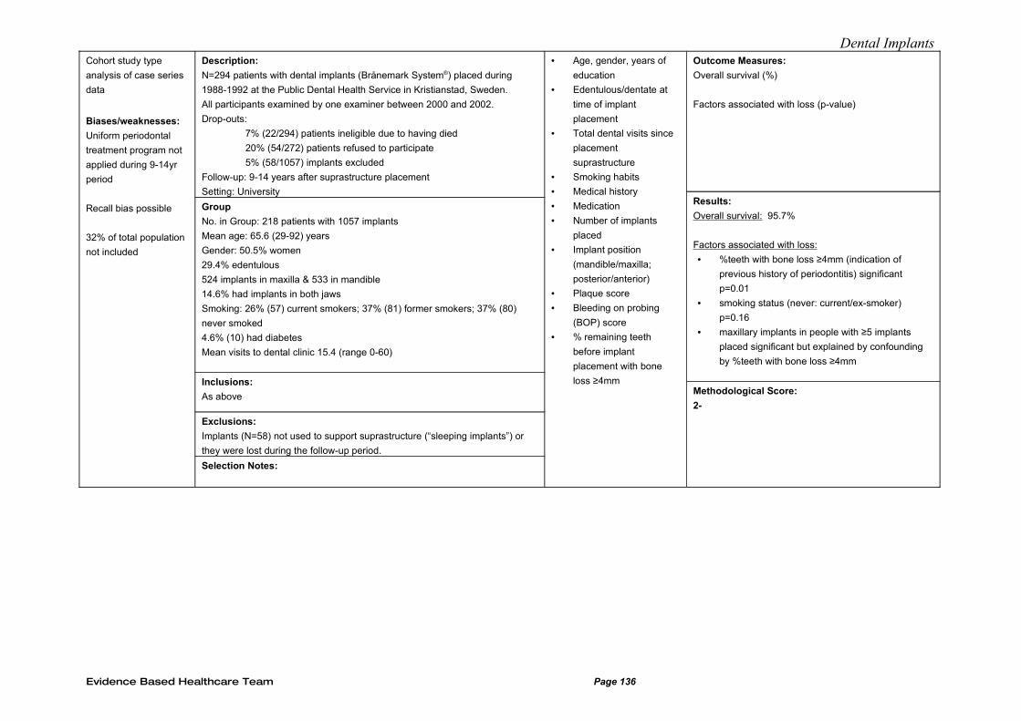
Dental ImplantsCohort study type
analysis of case series
data
Biases/weaknesses:
Uniform periodontal
treatment program not
applied during 9-14yr
period
Recall bias possible
32% of total population
not included
Description:
N=294 patients with dental implants (Brånemark System®) placed during
1988-1992 at the Public Dental Health Service in Kristianstad, Sweden.
All participants examined by one examiner between 2000 and 2002.
Drop-outs:
7% (22/294) patients ineligible due to having died
20% (54/272) patients refused to participate
5% (58/1057) implants excluded
Follow-up: 9-14 years after suprastructure placement
Setting: University
Group
No. in Group: 218 patients with 1057 implants
Mean age: 65.6 (29-92) years
Gender: 50.5% women
29.4% edentulous
524 implants in maxilla & 533 in mandible
14.6% had implants in both jaws
Smoking: 26% (57) current smokers; 37% (81) former smokers; 37% (80)
never smoked
4.6% (10) had diabetes
Mean visits to dental clinic 15.4 (range 0-60)
Inclusions:
As above
Exclusions:
Implants (N=58) not used to support suprastructure (“sleeping implants”) or
they were lost during the follow-up period.
Selection Notes:
• Age, gender, years of
education
• Edentulous/dentate at
time of implant
placement
• Total dental visits since
placement
suprastructure
• Smoking habits
• Medical history
• Medication
• Number of implants
placed
• Implant position
(mandible/maxilla;
posterior/anterior)
• Plaque score
• Bleeding on probing
(BOP) score
• % remaining teeth
before implant
placement with bone
loss ≥4mm
Outcome Measures:
Overall survival (%)
Factors associated with loss (p-value)
Results:
Overall survival: 95.7%
Factors associated with loss:
• %teeth with bone loss ≥4mm (indication of
previous history of periodontitis) significant
p=0.01
• smoking status (never: current/ex-smoker)
p=0.16
• maxillary implants in people with ≥5 implants
placed significant but explained by confounding
by %teeth with bone loss ≥4mm
Methodological Score:
2-
Evidence Based Healthcare Team Page 136
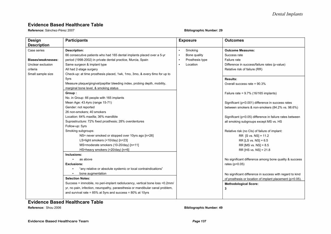
Dental Implants
Evidence Based Healthcare Table Reference: Sánchez-Pérez 2007 Bibliographic Number: 29
Design Description
Participants Exposure Outcomes
Case series
Biases/weaknesses:
Unclear exclusion
criteria
Small sample size
Description:
66 consecutive patients who had 165 dental implants placed over a 5-yr
period (1998-2002) in private dental practice, Murcia, Spain
Same surgeon & implant type
All had 2-stage surgery
Check-up: at time prosthesis placed, 1wk, 1mo, 3mo, & every 6mo for up to
5yrs
Measure plaque/gingival/papillar bleeding index, probing depth, mobility,
marginal bone level, & smoking status
Group :
No. in Group: 66 people with 165 implants
Mean Age: 43.4yrs (range 15-71)
Gender: not reported
26 non-smokers; 40 smokers
Location: 64% maxilla; 36% mandible
Suprastructure: 72% fixed prosthesis; 28% overdentures
Follow-up: 5yrs
Smoking subgroups:
NS= never smoked or stopped over 10yrs ago [n=26]
LS=light smokers (<10/day) [n=23]
MS=moderate smokers (10-20/day) [n=11]
HS=heavy smokers (>20/day) [n=6]
Inclusions:
• as above
Exclusions:
• “any relative or absolute systemic or local contraindications”
• bone augmentation
Selection Notes:
Success = immobile, no peri-implant radiolucency, vertical bone loss <0.2mm/
yr, no pain, infection, neuropathy, paraesthesia or mandibular canal problem,
and survival rate = 85% at 5yrs and success = 80% at 10yrs
• Smoking
• Bone quality
• Prosthesis type
• Location
Outcome Measures:
Success rate
Failure rate
Difference in success/failure rates (p-value)
Relative risk of failure (RR)
Results:
Overall success rate = 90.3%
Failure rate = 9.7% (16/165 implants)
Significant (p<0.001) difference in success rates
between smokers & non-smokers (84.2% vs. 98.6%)
Significant (p<0.05) difference in failure rates between
all smoking subgroups except MS vs. HS
Relative risk (no CIs) of failure of implant:
RR [S vs. NS] = 11.2
RR [LS vs. NS] = 6.5
RR [MS vs. NS] = 8.5
RR [HS vs. NS] = 21.8
No significant difference among bone quality & success
rates (p>0.05)
No significant difference in success with regard to kind
of prosthesis or location of implant placement (p>0.05).
Methodological Score:
3
Evidence Based Healthcare Table Reference: Shou 2006 Bibliographic Number: 49
Evidence Based Healthcare Team Page 137
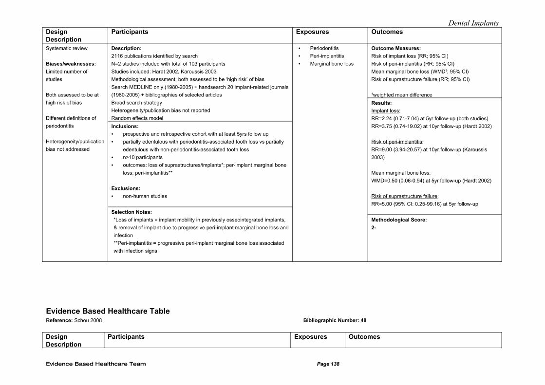
Dental ImplantsDesign Description
Participants Exposures Outcomes
Systematic review
Biases/weaknesses:
Limited number of
studies
Both assessed to be at
high risk of bias
Different definitions of
periodontitis
Heterogeneity/publication
bias not addressed
Description:
2116 publications identified by search
N=2 studies included with total of 103 participants
Studies included: Hardt 2002, Karoussis 2003
Methodological assessment: both assessed to be ‘high risk’ of bias
Search MEDLINE only (1980-2005) + handsearch 20 implant-related journals
(1980-2005) + bibliographies of selected articles
Broad search strategy
Heterogeneity/publication bias not reported
Random effects model
Inclusions:
• prospective and retrospective cohort with at least 5yrs follow up
• partially edentulous with periodontitis-associated tooth loss vs partially
edentulous with non-periodontitis-associated tooth loss
• n>10 participants
• outcomes: loss of suprastructures/implants*; per-implant marginal bone
loss; peri-implantitis**
Exclusions:
• non-human studies
Selection Notes:
*Loss of implants = implant mobility in previously osseointegrated implants,
& removal of implant due to progressive peri-implant marginal bone loss and
infection
**Peri-implantitis = progressive peri-implant marginal bone loss associated
with infection signs
• Periodontitis
• Peri-implantitis
• Marginal bone loss
Outcome Measures:
Risk of implant loss (RR; 95% CI)
Risk of peri-implantitis (RR; 95% CI)
Mean marginal bone loss (WMD†; 95% CI)
Risk of suprastructure failure (RR; 95% CI)
†weighted mean difference
Results:
Implant loss:
RR=2.24 (0.71-7.04) at 5yr follow-up (both studies)
RR=3.75 (0.74-19.02) at 10yr follow-up (Hardt 2002)
Risk of peri-implantitis:
RR=9.00 (3.94-20.57) at 10yr follow-up (Karoussis
2003)
Mean marginal bone loss:
WMD=0.50 (0.06-0.94) at 5yr follow-up (Hardt 2002)
Risk of suprastructure failure:
RR=5.00 (95% CI: 0.25-99.16) at 5yr follow-up
Methodological Score:
2-
Evidence Based Healthcare Table Reference: Schou 2008 Bibliographic Number: 48
Design Description
Participants Exposures Outcomes
Evidence Based Healthcare Team Page 138
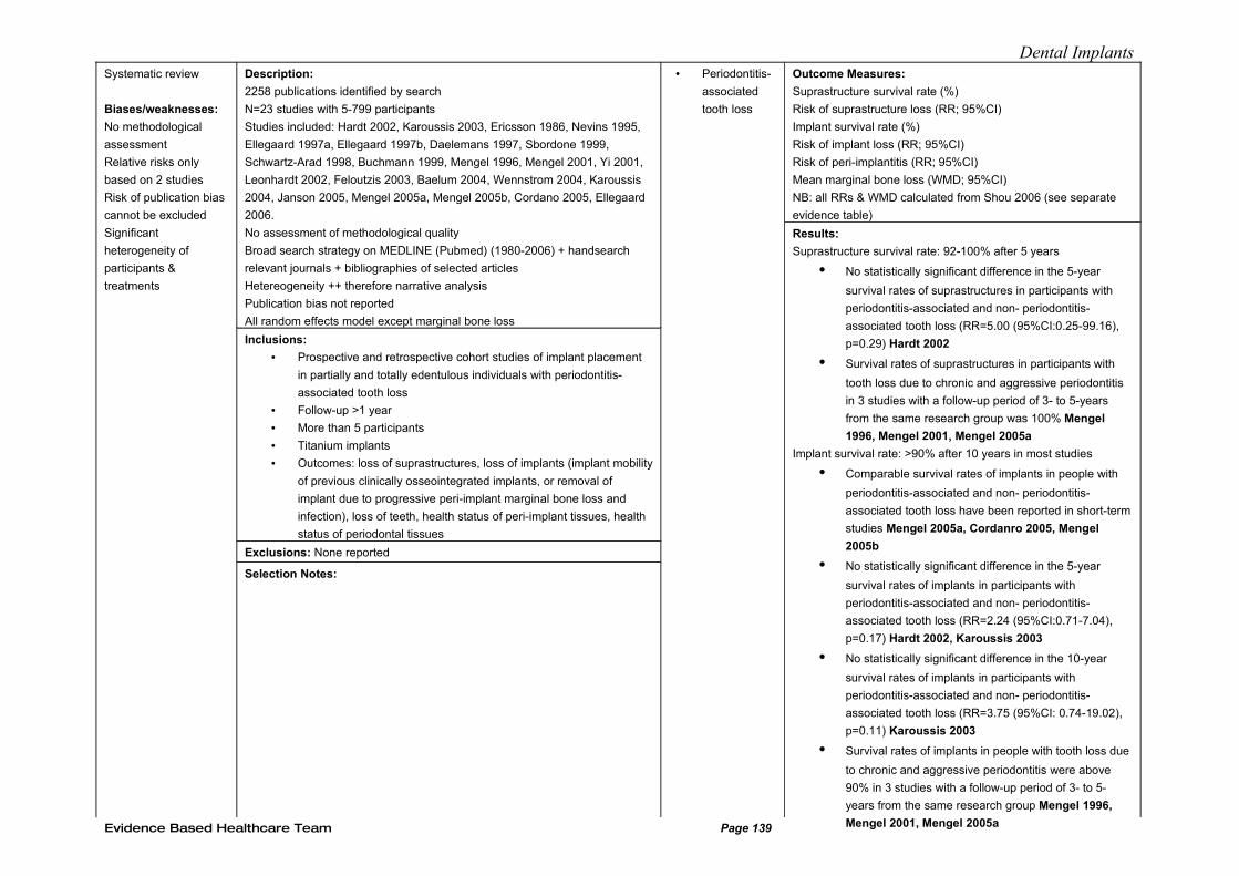
Dental ImplantsSystematic review
Biases/weaknesses:
No methodological
assessment
Relative risks only
based on 2 studies
Risk of publication bias
cannot be excluded
Significant
heterogeneity of
participants &
treatments
Description:
2258 publications identified by search
N=23 studies with 5-799 participants
Studies included: Hardt 2002, Karoussis 2003, Ericsson 1986, Nevins 1995,
Ellegaard 1997a, Ellegaard 1997b, Daelemans 1997, Sbordone 1999,
Schwartz-Arad 1998, Buchmann 1999, Mengel 1996, Mengel 2001, Yi 2001,
Leonhardt 2002, Feloutzis 2003, Baelum 2004, Wennstrom 2004, Karoussis
2004, Janson 2005, Mengel 2005a, Mengel 2005b, Cordano 2005, Ellegaard
2006.
No assessment of methodological quality
Broad search strategy on MEDLINE (Pubmed) (1980-2006) + handsearch
relevant journals + bibliographies of selected articles
Hetereogeneity ++ therefore narrative analysis
Publication bias not reported
All random effects model except marginal bone loss
Inclusions:
• Prospective and retrospective cohort studies of implant placement
in partially and totally edentulous individuals with periodontitis-
associated tooth loss
• Follow-up >1 year
• More than 5 participants
• Titanium implants
• Outcomes: loss of suprastructures, loss of implants (implant mobility
of previous clinically osseointegrated implants, or removal of
implant due to progressive peri-implant marginal bone loss and
infection), loss of teeth, health status of peri-implant tissues, health
status of periodontal tissues
Exclusions: None reported
Selection Notes:
• Periodontitis-
associated
tooth loss
Outcome Measures:
Suprastructure survival rate (%)
Risk of suprastructure loss (RR; 95%CI)
Implant survival rate (%)
Risk of implant loss (RR; 95%CI)
Risk of peri-implantitis (RR; 95%CI)
Mean marginal bone loss (WMD; 95%CI)
NB: all RRs & WMD calculated from Shou 2006 (see separate
evidence table)
Results:
Suprastructure survival rate: 92-100% after 5 years
• No statistically significant difference in the 5-year
survival rates of suprastructures in participants with
periodontitis-associated and non- periodontitis-
associated tooth loss (RR=5.00 (95%CI:0.25-99.16),
p=0.29) Hardt 2002
• Survival rates of suprastructures in participants with
tooth loss due to chronic and aggressive periodontitis
in 3 studies with a follow-up period of 3- to 5-years
from the same research group was 100% Mengel
1996, Mengel 2001, Mengel 2005a
Implant survival rate: >90% after 10 years in most studies
• Comparable survival rates of implants in people with
periodontitis-associated and non- periodontitis-
associated tooth loss have been reported in short-term
studies Mengel 2005a, Cordanro 2005, Mengel
2005b
• No statistically significant difference in the 5-year
survival rates of implants in participants with
periodontitis-associated and non- periodontitis-
associated tooth loss (RR=2.24 (95%CI:0.71-7.04),
p=0.17) Hardt 2002, Karoussis 2003
• No statistically significant difference in the 10-year
survival rates of implants in participants with
periodontitis-associated and non- periodontitis-
associated tooth loss (RR=3.75 (95%CI: 0.74-19.02),
p=0.11) Karoussis 2003
• Survival rates of implants in people with tooth loss due
to chronic and aggressive periodontitis were above
90% in 3 studies with a follow-up period of 3- to 5-
years from the same research group Mengel 1996,
Mengel 2001, Mengel 2005aEvidence Based Healthcare Team Page 139
Dental Implants
Evidence Based Healthcare Table Reference: Sjostrum 2007 Bibliographic Number: 24
Design Description
Participants Exposure Outcomes
Evidence Based Healthcare Team Page 140
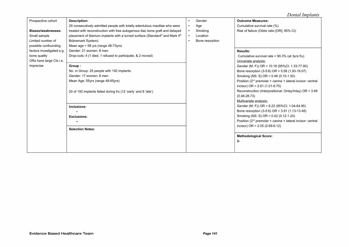
Dental ImplantsProspective cohort
Biases/weaknesses:
Small sample
Limited number of
possible confounding
factors investigated e.g.
bone quality
ORs have large CIs i.e.
imprecise
Description:
29 consecutively admitted people with totally edentulous maxillae who were
treated with reconstruction with free autogenous iliac bone graft and delayed
placement of titanium implants with a turned surface (Standard® and Mark II®
Brånemark System)
Mean age = 58 yrs (range 48-73yrs)
Gender: 21 women; 8 men
Drop-outs: 4 (1 died, 1 refused to participate, & 2 moved)
Group :
No. in Group: 25 people with 192 implants
Gender: 17 women; 8 men
Mean Age: 55yrs (range 48-65yrs)
20 of 192 implants failed during f/u (12 ‘early’ and 8 ‘late’)
Inclusions:
•Exclusions:
•
Selection Notes:
• Gender
• Age
• Smoking
• Location
• Bone resorption
Outcome Measures:
Cumulative survival rate (%)
Risk of failure (Odds ratio [OR]; 95% CI)
Results:
Cumulative survival rate = 90.3% (at 3yrs f/u)
Univariate analysis:
Gender (M: F)) OR = 10.18 (95%CI: 1.33-77.90)
Bone resorption (3-5:6) OR = 5.56 (1.93-16.07)
Smoking (NS: S) OR = 0.48 (0.15-1.50)
Position (2nd premolar + canine + lateral incisor: central
incisor) OR = 2.61 (1.01-6.75)
Reconstruction (Interpositional: Onlay/Inlay) OR = 3.69
(0.48-28.73)
Multivariate analysis:
Gender (M: F)) OR = 8.22 (95%CI: 1.04-64.95)
Bone resorption (3-5:6) OR = 3.91 (1.13-13.48)
Smoking (NS: S) OR = 0.42 (0.12-1.24)
Position (2nd premolar + canine + lateral incisor: central
incisor) OR = 2.05 (0.69-6.12)
Methodological Score:
2-
Evidence Based Healthcare Team Page 141
Dental ImplantsEvidence Based Healthcare Table Reference: Strietzel, 2007 Bibliographic Number: 15
Design Description Studies Exposure Outcomes
Evidence Based Healthcare Team Page 142
Dental ImplantsSystematic review and meta-
analysis of observational studies.
Biases/weaknesses:
No methodological assessment of
individual studies
Description:
Number of studies: N=29 (meta-analysis); N=35 (systematic review)
Total number of patients in the studies: n=16605 implants; n=3383 patients
Databases used: Medline and Embase (1989-2005) in English and manual
search of three German language peer-reviewed dental journals
(1968/1996/1999-2005)
Description of the methodological assessment of studies: none reported
Length of treatment: any period of follow-up
Description of comparison (placebo): not smoking
Co-interventions: none
Inclusions:
SR/meta-analyses, RCTs, prospective and retrospective clinical studies,
cohort studies, and case-control studies that include implant survival rates or
numbers of failed implants on a patient- or implant-related basis and numbers
of smokers and non-smokers. Also studies of implant survival after bone
augmentation procedures in smokers compared with non-smokers.
Other publications that didn’t meet the above criteria were included in a
separate systematic review if ORs, RRs or HRs for implant failures or
biological complications among smokers were reported.
Exclusions:
Case reports, animal/in vitro studies, educational statements, expert opinion
or did not match inclusion criteria or the focussed question.
Selection Notes:
Studies included in meta-analysis: Bain 1996, Bain 1993, Balshi 1999, Berge
2000, Beschnidt 2003, De Bruyn 1994, De Bruyn 1999, Geurs 2001, Gorman
1994, Grunder 1999, Jones 1999, Kan 2002, Karoussis 2003, Keller 1999,
Kronstrom 2001, Kumar 2002, Lambert 2000, Mayfield 2001, Minsk 1998,
Moheng 2005, Olson 2000a, Ortorp 2002, Penarrocha 2002, Schwartz-Arad
2000, Schwartz-Arad 2002, Van Steenberghe 2004, Wallace 2000, Widmark
2001.
Studies included for systematic review: Aalam 2005, Ataoglu 2002, Attard
2002, Baelum 2004, Bain 2002, Carlsson 2000, Chuang 2002b, Eckert 2001,
Feloutzis 2003, Galindo-Moreno 2005, Gruica 2004, Haas 1996, Karoussis
2004, Kourtis 2004, Lemmermen 2005, Leonhardt 2003, Levin 2004, Lindquist
1996, Lindquist 1997, McDermott 2003, Moy 2005, Nitzan 2005, Oates 2004,
Ortorp 2004, Penarroucha 2004, Schwartz-Arad 2005a, Schwartz-Arad
2005b, Strietzel 2001, Strietzel 2004, Wennstrom 2004a, Wennstrom 2004b,
Weyant 1994, Wilson 1999, Woo 2004, Zitzman 1999.
• Smoking status
• Sinus augmentation
Outcome Measures:
Meta-analysis: Odds ratios (OR) of failure in smokers
compared with rate in non-smokers + 95% confidence
intervals (95% CI).
Systematic review: Narrative synthesis
Results:
Meta-analysis
Implant-related failure (18 studies):
OR = 2.38 (95% CI: 1.93-2.93) random effects model
Patient-related failure (10 studies):
OR = 2.64 (95% CI: 1.70-4.09) fixed effects model
Methodological Score: 2-
Evidence Based Healthcare Team Page 143

Dental Implants
Evidence Based Healthcare Team Page 144
Dental ImplantsEvidence Based Healthcare Table Reference: Suslarla 2008 Bibliographic Number: 17
Design Description
Participants Exposures Outcomes
Evidence Based Healthcare Team Page 145
Dental ImplantsRetrospective cohort
Biases/weaknesses:
Unclear whether the
study population is
everyone who had an
implant placed between
2001-3 or a sample of
this population
Loss to follow-up may
introduce bias
Description: People who had at least one Bicon dental implant placed at a
private dental practice, Boston, during period 1992-2003.
• n=855 people with 2826 implants
Delayed loading group had implants placed between 1992-2000, those in the
immediate loading group had implants placed between 2001-2003.
Lost to follow-up: not reported
Setting: private dental practice
Group 1: delayed loading
No in group: 677 (79.2%) people with
2349 (83.1%) implants
Mean age: 53.1yrs (range 16.9-92.5)
Gender: 49.6% men
Smoking: 10.3%
Immediate implant placement: 10.3%
Location: 60.2% in maxilla; 31.1%
anterior
Bone quality: 0.9% type I, 26.6% type
II, 20.2% type III, 52.4% type IV
Dentoalveolar reconstructive
procedure: 36.1%
Group 2: immediate loading
No in group: 178 (20.8%) people
with 477 (16.9%) implants
Mean age: 53.0yrs (range 14.9-91.4)
Gender: 47.8% men
Smoking: 9.0%
Immediate implant placement: 50.1%
Location: 75.7% in maxilla; 53%
anterior
Bone quality: 4.3% type I, 9.8% type
II, 35.4% type III, 50.6% type IV
Dentoalveolar reconstructive
procedure: 9.0%
Statistically significant differences between the two groups in location,
dentoalveolar reconstructive procedure, immediate implant placement &
implant length, diameter, coating & well size (p<0.01)
Inclusions:
• as above
Exclusions:
• missing records
Selection Notes:*immediate loading =implant provisionally restored & stabilised by bonding to
adjacent teeth or implants & placed into functional occlusion the same day as
implant placement*delayed loading =implant restored at some time interval after implant
placement
Implant failure = removal of the implant
• Loading of implant
(immediate vs. delayed)*
• Age
• Gender
• Smoking
• Bone quality (type I-IV)
• Reconstructive
parameters (sinus lift,
barrier membrane, bone
graft, ridge split & graft
materials)
• Implant characteristics
Outcome Measures:
Implant survival at one year (%; 95%CI; p-value)
Relative risk of failure at 1 year (RR; 95%CI)
Results:
1-yr survival:
95.5% (95%CI: 94.6-96.4) delayed loading group
90.3% (95%CI: 87-93.7) immediate loading group
p<0.01
Risk of failure:
Univariate analysis:
• immediate loading
HR=1.9 (95%CI: 1.3-2.8) p<0.01
• age
HR=1.0 (95%CI: 1.0-1.0) p=0.26
• gender (F)
HR=1.1 (95%CI: 0.81-1.5) p=0.51
• smoking
HR=2.5 (95%CI: 1.7-3.6) p<0.01
• maxilla
HR=1.7 (95%CI: 1.2-2.3) p<0.01
• immediate implant placement
HR=1.4 (95%CI: 0.92-2.0) p=0.12
Multivariate analysis:
• immediate loading
HR=2.7 (95%CI: 1.2-5.9) p=0.01
• age
HR=1.00 (95%CI: 0.98-1.0) p=0.57
• gender (F)
HR=1.10 (95%CI: 0.78-1.5) p=0.59
• smoking
HR=2.6 (95%CI: 1.8-3.9) p<0.01
• maxilla
HR=1.9 (95%CI: 1.3-2.9) p<0.01
• immediate implant placement
HR=1.04 (95%CI: 0.61-1.8) p=0.88
Methodological Score:
2+Evidence Based Healthcare Team Page 146

Dental Implants
Evidence Based Healthcare Team Page 147
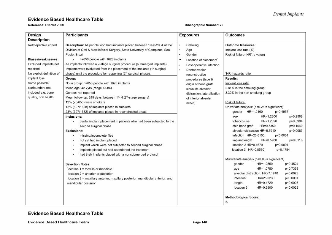
Dental ImplantsEvidence Based Healthcare Table Reference: Sverzut 2008 Bibliographic Number: 25
Design Description
Participants Exposures Outcomes
Retrospective cohort
Biases/weaknesses:
Excluded implants not
reported
No explicit definition of
implant loss
Some possible
confounders not
included e.g. bone
quality, oral health
Description: All people who had implants placed between 1996-2004 at the
Division of Oral & Maxillofacial Surgery, State University of Campinas, Sao
Paulo, Brazil
• n=650 people with 1628 implants
All implants followed a 2-stage surgical procedure (submerged implants).
Implants were evaluated from the placement of the implants (1st surgical
phase) until the procedure for reopening (2nd surgical phase).
Group:
No in group: n=650 people with 1628 implants
Mean age: 42.7yrs (range 13-84)
Gender: not reported
Mean follow-up: 249 days [between 1st- & 2nd-stage surgery]
12% (76/650) were smokers
12% (197/1628) of implants placed in smokers
23% (397/1682) of implants placed in reconstructed areas
Inclusions:
• dental implant placement in patients who had been subjected to the
second surgical phase
Exclusions:
• missing/incomplete files
• not yet had implant placed
• implant which were not subjected to second surgical phase
• implants placed but had abandoned the treatment
• had their implants placed with a nonsubmerged protocol
Selection Notes:*location 1 = maxilla or mandible
location 2 = anterior or posterior
location 3 = maxillary anterior, maxillary posterior, mandibular anterior, and
mandibular posterior
• Smoking
• Age
• Gender
• Location of placement*
• Post-operative infection
• Dentoalveolar
reconstructive
procedures (type &
origin of bone graft,
sinus lift, alveolar
distraction, lateralisation
of inferior alveolar
nerve)
Outcome Measures:
Implant loss rate (%)
Risk of failure (HR*; p-value)
*HR=hazards ratio
Results:
Implant loss rate:
2.81% in the smoking group
3.32% in the non-smoking group
Risk of failure:
Univariate analysis: (p<0.25 = significant)
gender HR=1.2160 p=0.4957
age HR=1.2600 p=0.2588
tobacco use HR=1.2390 p=0.5994
chin bone graft HR=0.5350 p=0.1640
alveolar distraction HR=6.7910 p=0.0083
infection HR=23.6150 p<0.0001
implant length HR=0.5980 p=0.0116
location 2 HR=0.4670 p=0.0091
location 3 HR=0.8530 p=0.1784
Multivariate analysis (p<0.05 = significant)
gender HR=1.2550 p=0.4524
age HR=1.0750 p=0.7358
alveolar distraction HR=7.1740 p=0.0073
infection HR=25.0230 p<0.0001
length HR=0.4720 p=0.0006
location 3 HR=0.3900 p=0.0023
Methodological Score:
2-
Evidence Based Healthcare Table
Evidence Based Healthcare Team Page 148
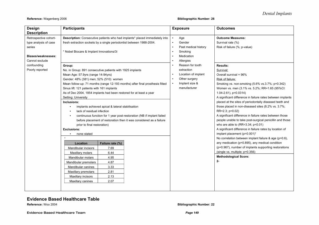
Dental ImplantsReference: Wagenberg 2006 Bibliographic Number: 26
Design Description
Participants Exposure Outcomes
Retrospective cohort-
type analysis of case
series
Biases/weaknesses:
Cannot exclude
confounding
Poorly reported
Description: Consecutive patients who had implants* placed immediately into
fresh extraction sockets by a single periodontist between 1988-2004.
* Nobel Biocare & Implant Innovations/3i
Group:
No. in Group: 891 consecutive patients with 1925 implants
Mean Age: 57.9yrs (range 14-94yrs)
Gender: 48% (381) men; 52% (510) women
Mean follow-up: 71 months (range 12-193 months) after final prosthesis fitted
Sinus lift: 121 patients with 161 implants
As of Dec 2004, 1854 implants had been restored for at least a year
Setting: University
Inclusions:
• implants achieved apical & lateral stabilisation
• lack of residual infection
• continuous function for 1 year post-restoration (NB if implant failed
before placement of restoration then it was considered as a failure
prior to final restoration)
Exclusions:
• none stated†
Location Failure rate (%)
Mandibular incisors 7.69
Maxillary molars 6.44
Mandibular molars 4.95
Mandibular premolars 4.87
Mandibular canines 3.33
Maxillary premolars 2.81
Maxillary incisors 2.13
Maxillary canines 2.07
• Age
• Gender
• Past medical history
• Smoking
• Medication
• Allergies
• Reason for tooth
extraction
• Location of implant
• Other surgery
• Implant size &
manufacturer
Outcome Measures:
Survival rate (%)
Risk of failure (%; p-value)
Results:
Survival:
Overall survival = 96%
Risk of failure:
Smoking vs. non-smoking (5.6% vs.3.7%; p=0.342)
Women vs. men (3.1% vs. 5.2%; RR=1.65 (95%CI:
1.04-2.61), p=0.0314)
A significant difference in failure rates between implants
placed at the sites of periodontally diseased teeth and
those placed in non-diseased sites (8.2% vs. 3.7%;
RR=2.3; p=0.02)
A significant difference in failure rates between those
people unable to take post-surgical penicillin and those
who are able to (RR=3.34; p<0.01)
A significant difference in failure rates by location of
implant placement (p=0.001)†
No correlation between implant failure & age (p>0.6),
any medication (p=0.895), any medical condition
(p=0.967), number of implants supporting restorations
(single vs. multiple; p=0.356)
Methodological Score:
2-
Evidence Based Healthcare Table Reference: Woo 2004 Bibliographic Number: 22
Evidence Based Healthcare Team Page 149
Dental Implants
Design Description
Participants Exposure Outcomes
Evidence Based Healthcare Team Page 150
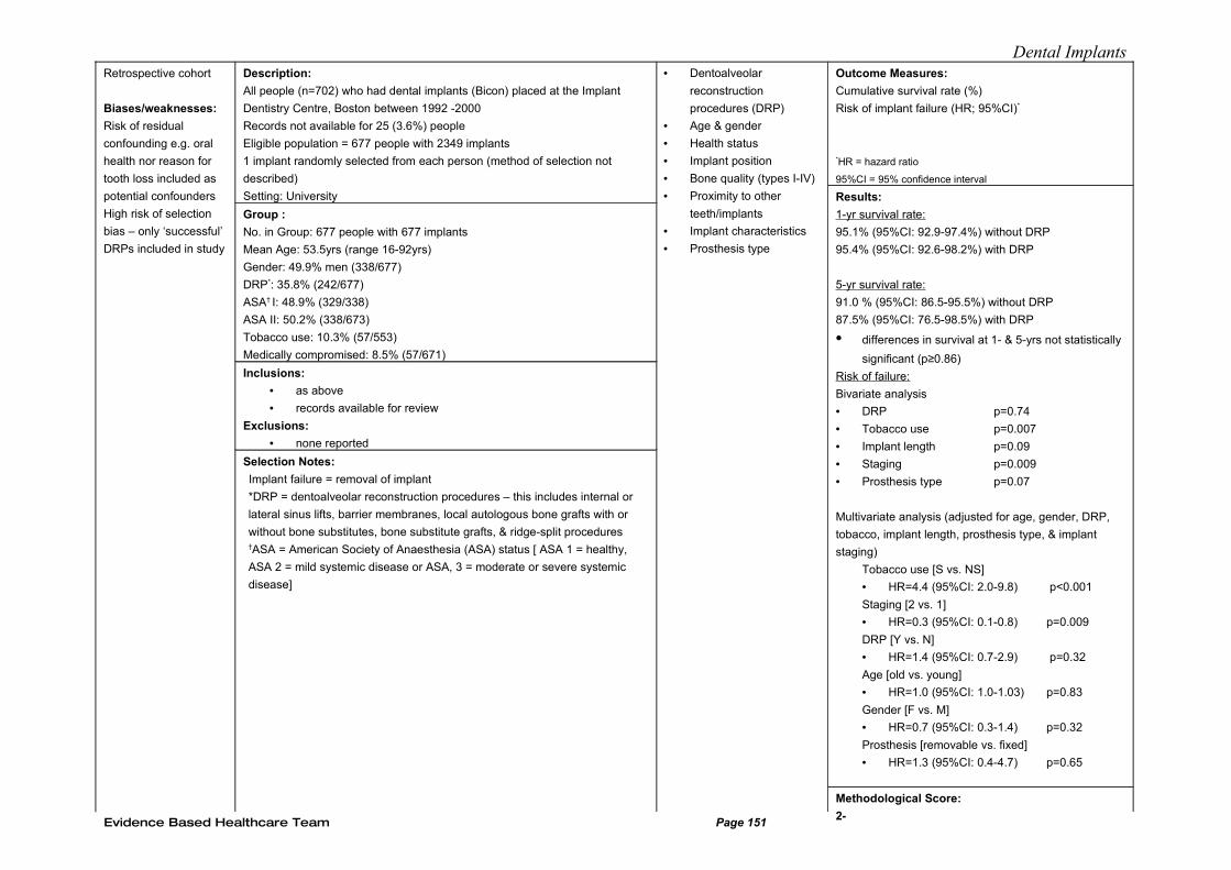
Dental ImplantsRetrospective cohort
Biases/weaknesses:
Risk of residual
confounding e.g. oral
health nor reason for
tooth loss included as
potential confounders
High risk of selection
bias – only ‘successful’
DRPs included in study
Description:
All people (n=702) who had dental implants (Bicon) placed at the Implant
Dentistry Centre, Boston between 1992 -2000
Records not available for 25 (3.6%) people
Eligible population = 677 people with 2349 implants
1 implant randomly selected from each person (method of selection not
described)
Setting: University
Group :
No. in Group: 677 people with 677 implants
Mean Age: 53.5yrs (range 16-92yrs)
Gender: 49.9% men (338/677)
DRP*: 35.8% (242/677)
ASA† I: 48.9% (329/338)
ASA II: 50.2% (338/673)
Tobacco use: 10.3% (57/553)
Medically compromised: 8.5% (57/671)
Inclusions:
• as above
• records available for review
Exclusions:
• none reported
Selection Notes:
Implant failure = removal of implant
*DRP = dentoalveolar reconstruction procedures – this includes internal or
lateral sinus lifts, barrier membranes, local autologous bone grafts with or
without bone substitutes, bone substitute grafts, & ridge-split procedures†ASA = American Society of Anaesthesia (ASA) status [ ASA 1 = healthy,
ASA 2 = mild systemic disease or ASA, 3 = moderate or severe systemic
disease]
• Dentoalveolar
reconstruction
procedures (DRP)
• Age & gender
• Health status
• Implant position
• Bone quality (types I-IV)
• Proximity to other
teeth/implants
• Implant characteristics
• Prosthesis type
Outcome Measures:
Cumulative survival rate (%)
Risk of implant failure (HR; 95%CI)*
*HR = hazard ratio
95%CI = 95% confidence interval
Results:
1-yr survival rate:
95.1% (95%CI: 92.9-97.4%) without DRP
95.4% (95%CI: 92.6-98.2%) with DRP
5-yr survival rate:
91.0 % (95%CI: 86.5-95.5%) without DRP
87.5% (95%CI: 76.5-98.5%) with DRP
• differences in survival at 1- & 5-yrs not statistically
significant (p≥0.86)
Risk of failure:
Bivariate analysis
• DRP p=0.74
• Tobacco use p=0.007
• Implant length p=0.09
• Staging p=0.009
• Prosthesis type p=0.07
Multivariate analysis (adjusted for age, gender, DRP,
tobacco, implant length, prosthesis type, & implant
staging)
Tobacco use [S vs. NS]
• HR=4.4 (95%CI: 2.0-9.8) p<0.001
Staging [2 vs. 1]
• HR=0.3 (95%CI: 0.1-0.8) p=0.009
DRP [Y vs. N]
• HR=1.4 (95%CI: 0.7-2.9) p=0.32
Age [old vs. young]
• HR=1.0 (95%CI: 1.0-1.03) p=0.83
Gender [F vs. M]
• HR=0.7 (95%CI: 0.3-1.4) p=0.32
Prosthesis [removable vs. fixed]
• HR=1.3 (95%CI: 0.4-4.7) p=0.65
Methodological Score:
2-Evidence Based Healthcare Team Page 151
Dental Implants
Appendix C:
Exclusion Table:
Paper Reason for Exclusion
Albrektsson, T., J. Gottlow, et al. (2007). "Survival of NobelDirect implants: an analysis of 550
consecutively placed implants at 18 different clinical centers." Clinical Implant Dentistry &
Related Research 9(2): 65-70.
No risk factor analysis
Amorim, M. A. L., L. Takayama, et al. (2006). "Comparative study of axial and femoral bone
mineral density and parameters of mandibular bone quality in patients receiving dental
implants.[see comment][republished in Osteoporos Int. 2007 May;18(5):703-9; PMID:
17506127]." Osteoporosis International 17(10): 1494-500.
Survival was a secondary
outcome
Arvidson, K., O. Esselin, et al. (2008). "Early loading of mandibular full-arch bridges screw
retained after 1 week to four to five Monotype implants: 3-year results from a prospective
multicentre study." Clinical Oral Implants Research 19(7): 693-703.
No risk factor analysis
Astrand, P., B. Engquist, et al. (2004). "Astra Tech and Branemark system implants: a 5-year
prospective study of marginal bone reactions." Clinical Oral Implants Research 15(4): 413-20.
Primary outcome was bone
loss
Bedrossian, E., B. Rangert, et al. (2006). "Immediate function with the zygomatic implant: a
graftless solution for the patient with mild to advanced atrophy of the maxilla." International
Journal of Oral and Maxillofacial Implants 21(6): 937-42.
No risk factor analysis
Bell, B. M. and R. E. Bell (2008). "Oral bisphosphonates and dental implants: a retrospective
study." Journal of Oral & Maxillofacial Surgery 66(5): 1022-4.
Bisphosphonates
Bianchi, A. E. and F. Sanfilippo (2004). "Single-tooth replacement by immediate implant and
connective tissue graft: a 1-9-year clinical evaluation." Clinical Oral Implants Research 15(3):
269-77.
No risk factor analysis
Blanco, J., A. Alonso, et al. (2005). "Long-term results and survival rate of implants treated
with guided bone regeneration: a 5-year case series prospective study." Clinical Oral Implants
Research 16(3): 294-301.
No risk factor analysis
Blanes, R. J., J. P. Bernard, et al. (2007). "A 10-year prospective study of ITI dental implants
placed in the posterior region. I: Clinical and radiographic results." Clinical Oral Implants
Research 18(6): 699-706.
Survival only
Buser, D., M. M. Bornstein, et al. (2008). "Early implant placement with simultaneous guided
bone regeneration following single-tooth extraction in the esthetic zone: a cross-sectional,
retrospective study in 45 subjects with a 2- to 4-year follow-up." Journal of Periodontology
79(9): 1773-81.
Wrong exposure
Chou, C.-T., H. F. Morris, et al. (2004). "AICRG, Part II: Crestal bone loss associated with the
Ankylos implant: loading to 36 months." Journal of Oral Implantology 30(3): 134-43.
Wrong outcome (crestal
bone loss)
Chuang, S. (2005). "Jawbone, quality, jaw shape, implant length, and overdenture treatment
protocol were associated with implant failures." Journal of Evidence Based Dental Practice
5(4): 215-6.
Appraisal of Herrmann 2005
Chuang, S. K. and T. Cai (2006). "Predicting clustered dental implant survival using frailty
methods." Journal of Dental Research 85(12): 1147-51.
Reanalysis of Chuang 2002
Chuang, S. K., T. Cai, et al. (2005). "Frailty approach for the analysis of clustered failure time
observations in dental research." Journal of Dental Research 84(1): 54-8.
Reanalysis of Chuang 2002
Evidence Based Healthcare Team page 152
Dental Implants
Chung, D. M., T.-J. Oh, et al. (2006). "Significance of keratinized mucosa in maintenance of
dental implants with different surfaces." Journal of Periodontology 77(8): 1410-20.
No outcomes of interest
Chung, D. M., T.-J. Oh, et al. (2007). "Factors affecting late implant bone loss: a retrospective
analysis." International Journal of Oral & Maxillofacial Implants 22(1): 117-26.
Wrong outcome (bone loss)
Cordaro, L., F. Torsello, et al. (2006). "Retrospective evaluation of mandibular incisor
replacement with narrow neck implants." Clinical Oral Implants Research 17(6): 730-5.
No risk factor analysis
Cornelini, R., C. Rubini, et al. (2003). "Transforming growth factor-beta 1 expression in the
peri-implant soft tissues of healthy and failing dental implants." Journal of Periodontology
74(4): 446-50.
No risk factor analysis
Cornelini, R., F. Cangini, et al. (2005). "Immediate restoration of implants placed into fresh
extraction sockets for single-tooth replacement: a prospective clinical study." International
Journal of Periodontics & Restorative Dentistry 25(5): 438-47.
No risk factor analysis
Coulthard, P., M. Esposito, et al. (2007). "Interventions for replacing missing teeth: surgical
techniques for placing dental implants." Cochrane Database of Systematic Reviews 4.
Withdrawn from Cochrane
library
Coulthard, P., M. Esposito, et al. (2007). "Interventions for replacing missing teeth: hyperbaric
oxygen therapy for irradiated patients who require dental implants." Cochrane Database of
Systematic Reviews 4.
No risk factors
Covani, U., A. Barone, et al. (2006). "Clinical outcome of implants placed immediately after
implant removal." Journal of Periodontology 77(4): 722-7.
Wrong exposure (timing of
placement)
Covani, U., R. Crespi, et al. (2004). "Immediate implants supporting single crown restoration: A
4-year prospective study." Journal of Periodontology 75(7): 982-988.
Wrong exposure (timing of
placement)
Cune, M. S., J. W. Verhoeven, et al. (2004). "A prospective evaluation of Frialoc implants with
ball-abutments in the edentulous mandible: 1-year results." Clinical Oral Implants Research
15(2): 167-73.
Overdentures
das Neves, F. D., D. Fones, et al. (2006). "Short implants--an analysis of longitudinal studies."
International Journal of Oral and Maxillofacial Implants 21(1): 86-93.
Risks dealt with narratively
Short implants
Davarpanah, M., M. Caraman, et al. (2007). "Prosthetic success with a maxillary immediate-
loading protocol in the multiple-risk patient." International Journal of Periodontics & Restorative
Dentistry 27(2): 161-9.
Case reports
De Backer, H., G. Van Maele, et al. (2007). "The influence of gender and age on fixed
prosthetic restoration longevity: an up to 18- to 20-year follow-up in an undergraduate clinic."
International Journal of Prosthodontics 20(6): 579-86.
Cannot tell if implants used
or not
De Rouck, T., K. Collys, et al. (2008). "Immediate single-tooth implants in the anterior maxilla:
a 1-year case cohort study on hard and soft tissue response." Journal of Clinical
Periodontology 35(7): 649-57.
No risk factor analysis
Degidi, M. and A. Piattelli (2005). "Comparative analysis study of 702 dental implants
subjected to immediate functional loading and immediate non-functional loading to traditional
healing periods with a follow-up of up to 24 months.[see comment][erratum appears in Int J
Oral Maxillofac Implants. 2005 Mar-Apr;20(2):306]." International Journal of Oral &
Maxillofacial Implants 20(1): 99-107.
Effect of timing of loading
Degidi, M., A. Piattelli, et al. (2007). "Do longer implants improve clinical outcome in immediate
loading?" International Journal of Oral & Maxillofacial Surgery 36(12): 1172-6.
Wrong exposure (implant
length)
Del Fabbro, M., T. Testori, et al. (2006). "Systematic review of survival rates for immediately
loaded dental implants." International Journal of Periodontics & Restorative Dentistry 26(3):
Only risks looked at was
implant surface & timing of
Evidence Based Healthcare Team Page 153
Dental Implants
249-63. loading
DeLuca, S. and G. Zarb (2006). "The effect of smoking on osseointegrated dental implants.
Part II: peri-implant bone loss." International Journal of Prosthodontics 19(6): 560-6.
Wrong outcome (peri-
implant bone loss)
Dijakiewicz, M., A. Wojtowicz, et al. (2007). "Is implanto-prosthodontic treatment available for
haemodialysis patients? [2]." Nephrology Dialysis Transplantation 22(9): 2722-2724.
Letter
Dos Santos, M. C. L. G., M. I. G. Campos, et al. (2004). "Analysis of the transforming growth
factor-beta 1 gene promoter polymorphisms in early osseointegrated implant failure." Implant
Dentistry 13(3): 262-9.
Wrong exposure
(gene polymorphism)
Durham, T. M., T. King, et al. (2006). "Dental implants in edentulous adults with cognitive
disabilities: report of a pilot project." Special Care in Dentistry 26(1): 40-6.
No risk factors
Duskova, M., M. Kotova, et al. (2004). "Reconstruction of maxilla alveolus for application of
dental implant in patients with cleft defect." Acta Chirurgiae Plasticae 46(4): 115-121.
Wrong population
Eliasson, A., T. Eriksson, et al. (2006). "Fixed partial prostheses supported by 2 or 3 implants:
a retrospective study up to 18 years." International Journal of Oral and Maxillofacial Implants
21(4): 567-74.
Focus on prosthesis survival
Ellis, I. E. and D. McFadden (2007). "The Value of a Diagnostic Setup for Full Fixed Maxillary
Implant Prosthetics." Journal of Oral and Maxillofacial Surgery 65(9): 1764-1771.
Not concerning risk factors
for implant loss
Erdogan, O., D. M. Shafer, et al. (2007). "A review of the association between osteoporosis
and alveolar ridge augmentation." Oral Surgery Oral Medicine Oral Pathology Oral Radiology
& Endodontics 104(6): 738.e1-13.
Wrong outcomes
Esposito, M. A. B., A. Koukoulopoulou, et al. (2006). "Interventions for replacing missing teeth:
dental implants in fresh extraction sockets (immediate, immediate-delayed and delayed
implants)." Cochrane Database of Systematic Reviews(4): CD005968.
Timing of implant
Esposito, M., H. V. Worthington, et al. (2005). "Interventions for replacing missing teeth: dental
implants in zygomatic bone for the rehabilitation of the severely deficient edentulous maxilla.
[update of Cochrane Database Syst Rev. 2003;(3):CD004151; PMID: 12918005]." Cochrane
Database of Systematic Reviews(4): CD004151.
Not relevant to guideline
Esposito, M., L. Murray-Curtis, et al. (2007). "Interventions for replacing missing teeth: different
types of dental implants." Cochrane Database of Systematic Reviews(4): CD003815.
Types of implants
Esposito, M., M. G. Grusovin, et al. (2006). "Interventions for replacing missing teeth:
treatment of perimplantitis.[update of Cochrane Database Syst Rev. 2004;(4):CD004970;
PMID: 15495132]." Cochrane Database of Systematic Reviews 3: CD004970.
Treatment
Esposito, M., M. G. Grusovin, et al. (2006). "Interventions for replacing missing teeth: bone
augmentation techniques for dental implant treatment.[update of Cochrane Database Syst
Rev. 2003;(3):CD003607; PMID: 12917975]." Cochrane Database of Systematic Reviews(1):
CD003607.
Updated by Esposito 20083
Esposito, M., M. G. Grusovin, et al. (2007). "Interventions for replacing missing teeth: different
times for loading dental implants.[update of Cochrane Database Syst Rev. 2004;
(3):CD003878; PMID: 15266505]." Cochrane Database of Systematic Reviews(2): CD003878.
Timing of loading
Esposito, M., M. Grusovin, et al. (2008). "Interventions for replacing missing teeth: hyperbaric
oxygen therapy for irradiated patients who require dental implants [Systematic Review]."
Cochrane Database of Systematic Reviews 3.
Treatment
Fardal, O. and G. J. Linden (2008). "Tooth loss and implant outcomes in patients refractory to
treatment in a periodontal practice." Journal of Clinical Periodontology 35(8): 733-8.
No risk analysis
Evidence Based Healthcare Team Page 154
Dental Implants
Feldman, S., N. Boitel, et al. (2004). "Five-year survival distributions of short-length (10 mm or
less) machined-surfaced and Osseotite implants." Clinical Implant Dentistry & Related
Research 6(1): 16-23.
No risk factor analysis
Ferrara, A., C. Galli, et al. (2006). "Immediate provisional restoration of postextraction implants
for maxillary single-tooth replacement." International Journal of Periodontics & Restorative
Dentistry 26(4): 371-7.
Immediate placement
Ferreira, S. D., G. L. M. Silva, et al. (2006). "Prevalence and risk variables for peri-implant
disease in Brazilian subjects." Journal of Clinical Periodontology 33(12): 929-35.
Wrong outcomes
Finnema, K. J., G. M. Raghoebar, et al. (2005). "Oral rehabilitation with dental implants in
oligodontia patients." International Journal of Prosthodontics 18(3): 203-9.
Wrong population
Fischer, K. and T. Stenberg (2006). "Three-year data from a randomized, controlled study of
early loading of single-stage dental implants supporting maxillary full-arch prostheses."
International Journal of Oral & Maxillofacial Implants 21(2): 245-52.
No risk factor
Fugazzotto, P. A., J. R. Beagle, et al. (2004). "Success and failure rates of 9 mm of shorter
implants in the replacement of missing maxillary molars when restored with individual crowns:
Preliminary results 0 to 84 months in function. A retrospective study." Journal of
Periodontology 75(2): 327-332.
No risk factor
Garfinkle, J. S., L. L. Cunningham, Jr., et al. (2008). "Evaluation of orthodontic mini-implant
anchorage in premolar extraction therapy in adolescents." American Journal of Orthodontics &
Dentofacial Orthopedics 133(5): 642-53.
Orthodontic anchorage
Gotfredsen, K. (2004). "A 5-year prospective study of single-tooth replacements supported by
the Astra Tech implant: a pilot study." Clinical Implant Dentistry & Related Research 6(1): 1-8.
No risk factor
Iqbal, M. K. and S. Kim (2008). "A review of factors influencing treatment planning decisions of
single-tooth implants versus preserving natural teeth with nonsurgical endodontic therapy."
Journal of Endodontics 34(5): 519-29.
Treatment planning
Hagi, D., D. A. Deporter, et al. (2004). "A targeted review of study outcomes with short (< or =
7 mm) endosseous dental implants placed in partially edentulous patients." Journal of
Periodontology 75(6): 798-804.
Could not calculate risk of
failure
Henry, P. J., D. van Steenberghe, et al. (2003). "Prospective multicenter study on immediate
rehabilitation of edentulous lower jaws according to the Branemark Novum protocol." Clinical
Implant Dentistry & Related Research 5(3): 137-42.
Fixed complete prosthesis
Herrmann, I., U. Lekholm, et al. (2005). "Evaluation of patient and implant characteristics as
potential prognostic factors for oral implant failures.[see comment]." International Journal of
Oral & Maxillofacial Implants 20(2): 220-30.
Already in previous review
Herzberg, R., E. Dolev, et al. (2006). "Implant marginal bone loss in maxillary sinus grafts."
International Journal of Oral & Maxillofacial Implants 21(1): 103-10.
Wrong outcome
Hirsch, J.-M., L.-O. Ohrnell, et al. (2004). "A clinical evaluation of the Zygoma fixture: one year
of follow-up at 16 clinics." Journal of Oral & Maxillofacial Surgery 62(9 Suppl 2): 22-9.
Zygomatic implants
Horwitz, J., O. Zuabi, et al. (2007). "Immediate and delayed restoration of dental implants in
periodontally susceptible patients: 1-year results." International Journal of Oral & Maxillofacial
Implants 22(3): 423-9.
Timing of restoration
Hug, S., D. Mantokoudis, et al. (2006). "Clinical evaluation of 3 overdenture concepts with
tooth roots and implants: 2-year results." International Journal of Prosthodontics 19(3): 236-43.
Overdentures
Evidence Based Healthcare Team Page 155
Dental Implants
Ioannidou, E. and A. Doufexi (2005). "Does loading time affect implant survival? A meta-
analysis of 1,266 implants." Journal of Periodontology 76(8): 1252-8.
No risk factors
Jemt, T. and P. Hager (2006). "Early complete failures of fixed implant-supported prostheses
in the edentulous maxilla: a 3-year analysis of 17 consecutive cluster failure patients." Clinical
Implant Dentistry & Related Research 8(2): 77-86.
Fixed complete prostheses
Jemt, T., G. Ahlberg, et al. (2007). "Tooth movements adjacent to single-implant restorations
after more than 15 years of follow-up." International Journal of Prosthodontics 20(6): 626-32.
Wrong outcome
Johansson, L. and A. Ekfeldt (2003). "Implant-supported fixed partial prostheses: a
retrospective study." International Journal of Prosthodontics 16(2): 172-6.
No risk factor analysis
Jung, R. E., B. E. Pjetursson, et al. (2008). "A systematic review of the 5-year survival and
complication rates of implant-supported single crowns." Clinical Oral Implants Research 19(2):
119-30.
Survival only
Jungner, M., P. Lundqvist, et al. (2005). "Oxidized titanium implants (Nobel Biocare TiUnite)
compared with turned titanium implants (Nobel Biocare mark III) with respect to implant failure
in a group of consecutive patients treated with early functional loading and two-stage protocol."
Clinical Oral Implants Research 16(3): 308-12.
No risk factor analysis
Kahnberg, K.-E., P. J. Henry, et al. (2007). "Clinical evaluation of the zygoma implant: 3-year
follow-up at 16 clinics." Journal of Oral & Maxillofacial Surgery 65(10): 2033-8.
No risk factor analysis
Kan, J. Y. K., K. Rungcharassaeng, et al. (2003). "Immediate placement and provisionalization
of maxillary anterior single implants: 1-year prospective study." International Journal of Oral
and Maxillofacial Implants 18(1): 31-9.
Timing of placement
Karoussis, I. K., G. E. Salvi, et al. (2003). "Long-term implant prognosis in patients with and
without a history of chronic periodontitis: a 10-year prospective cohort study of the ITI Dental
Implant System." Clinical Oral Implants Research 14(3): 329-39.
Included in Schou (2006)50
systematic review
Karoussis, I. K., S. Muller, et al. (2004). "Association between periodontal and peri-implant
conditions: a 10-year prospective study." Clinical Oral Implants Research 15(1): 1-7.
Wrong outcomes
Karoussis, I. K., U. Bragger, et al. (2004). "Effect of implant design on survival and success
rates of titanium oral implants: a 10-year prospective cohort study of the ITI Dental Implant
System." Clinical Oral Implants Research 15(1): 8-17.
Wrong exposure (implant
design)
Kim, D. M., R. L. Badovinac, et al. (2008). "A 10-year prospective clinical and radiographic
study of one-stage dental implants." Clinical Oral Implants Research 19(3): 254-8.
Wrong outcome
Knauf, M., T. Gerds, et al. (2007). "Survival and success rates of 3i implants in partially
edentulous patients: results of a prospective study with up to 84-months' follow-up."
Quintessence International 38(8): 643-51.
No risk factors
Kramer, F. J., C. Baethge, et al. (2005). "Dental implants in patients with orofacial clefts: a
long-term follow-up study." International Journal of Oral & Maxillofacial Surgery 34(7): 715-21.
Wrong population (orofacial
clefts)
Lang, N. P., T. Berglundh, et al. (2004). "Consensus statements and recommended clinical
procedures regarding implant survival and complications." International Journal of Oral &
Maxillofacial Implants 19 Suppl: 150-4.
Wrong outcomes
Laster, Z. and O. T. Jensen (2008). "Secondarily stabilized dental implants: an alternative to
sinus grafting--early clinical results of 63 consecutively placed implants." Journal of Oral &
Maxillofacial Surgery 66(10): 2136-9.
Surgical technique
Leung, A. C. F. and L. K. Cheung (2003). "Dental implants in reconstructed jaws: patients'
evaluation of functional and quality-of-life outcomes." International Journal of Oral and
No risk factor analysis
Evidence Based Healthcare Team Page 156
Dental Implants
Maxillofacial Implants 18(1): 127-34.
Lin, Y.-H., P. Huang, et al. (2007). "The relationship between IL-1 gene polymorphism and
marginal bone loss around dental implants." Journal of Oral & Maxillofacial Surgery 65(11):
2340-4.
Wrong outcome
Lindh, T. (2008). "Should we extract teeth to avoid tooth-implant combinations?" Journal of
Oral Rehabilitation 35 Suppl 1: 44-54.
No risk factor analysis
Narrative review
Lundgren, D., H. Rylander, et al. (2008). "To save or to extract, that is the question. Natural
teeth or dental implants in periodontitis-susceptible patients: clinical decision-making and
treatment strategies exemplified with patient case presentations." Periodontology 2000 47:
27-50.
Case reports
Martinez Iturriaga, M. T. and C. Colmenero Ruiz (2004). "Maxillary Sinus Reconstruction with
Calvarium Bone Grafts and Endosseous Implants." Journal of Oral and Maxillofacial Surgery
62(3): 344-347.
No risk factors
Meijer, H. J. A., G. M. Raghoebar, et al. (2003). "Comparison of implant-retained mandibular
overdentures and conventional complete dentures: a 10-year prospective study of clinical
aspects and patient satisfaction." International Journal of Oral & Maxillofacial Implants 18(6):
879-85.
Overdenture
Merli, M., F. Bernardelli, et al. (2008). "Immediate versus early nonocclusal loading of dental
implants placed with a flapless procedure in partially edentulous patients: preliminary results
from a randomized controlled clinical trial." International Journal of Periodontics & Restorative
Dentistry 28(5): 453-9.
Wrong exposure (timing of
loading)
Mesa F, Munoz R, Noguerol B, de Dios Luna J, Galindo P, O'Valle F. Multivariate study of
factors influencing primary dental implant stability. Clinical Oral Implants Research
2008;19(2):196-200.
Wrong outcome (implant
stability)
Miyamoto, Y., K. Fujisawa, et al. (2003). "Effect of the additional installation of implants in the
posterior region on the prognosis of treatment in the edentulous mandibular jaw." Clinical Oral
Implants Research 14(6): 727-33.
Wrong exposure
Molly, L. (2006). "Bone density and primary stability in implant therapy." Clinical Oral Implants
Research 17 Suppl 2: 124-35.
No risk analysis
Moradi, D. R., P. K. Moy, et al. (2006). "Evidence-based research in alternative protocols to
dental implantology: a closer look at publication bias." Journal of the California Dental
Association 34(11): 877-86.
Wrong exposure
Morris, H. F., S. Ochi, et al. (2004). "AICRG, Part V: Factors influencing implant stability at
placement and their influence on survival of Ankylos implants." Journal of Oral Implantology
30(3): 162-70.
Wrong outcome
Nakai, H., Y. Okazaki, et al. (2003). "Clinical application of zygomatic implants for rehabilitation
of the severely resorbed maxilla: a clinical report." International Journal of Oral and
Maxillofacial Implants 18(4): 566-70.
Zygomatic implants
Nedir, R., M. Bischof, et al. (2004). "A 7-year life table analysis from a prospective study on ITI
implants with special emphasis on the use of short implants. Results from a private practice."
Clinical Oral Implants Research 15(2): 150-7.
No risk factors
Nedir, R., M. Bischof, et al. (2006). "Prosthetic complications with dental implants: from an up-
to-8-year experience in private practice." International Journal of Oral and Maxillofacial
Implants 21(6): 919-28.
Wrong outcomes
Nentwig, G. H. (2004). "Ankylos implant system: concept and clinical application." Journal of No risk factor analysis
Evidence Based Healthcare Team Page 157
Dental Implants
Oral Implantology 30(3): 171-7.
Nickenig, H.-J., H. Spiekermann, et al. (2008). "Survival and complication rates of combined
tooth-implant-supported fixed and removable partial dentures." International Journal of
Prosthodontics 21(2): 131-7.
Wrong outcomes
Nkenke, E. and M. Fenner (2006). "Indications for immediate loading of implants and implant
success." Clinical Oral Implants Research 17 Suppl 2: 19-34.
Timing of loading
Ormianer, Z. and A. Palti (2006). "Long-term clinical evaluation of tapered multi-threaded
implants: results and influences of potential risk factors." Journal of Oral Implantology 32(6):
300-7.
No outcomes of interest
Ostman, P.-O. (2008). "Immediate/early loading of dental implants. Clinical documentation and
presentation of a treatment concept." Periodontology 2000 47: 90-112.
Timing of loading
Ottoni JMP, Oliveira ZFL, Mansini R, Cabral AM. Correlation between placement torque and
survival of single-tooth implants. International Journal of Oral & Maxillofacial Implants
2005;20(5):769-76.
No outcomes of interest
Park W, Kim HS. Osteoma of maxillary sinus: a case report. Oral Surgery Oral Medicine Oral
Pathology Oral Radiology & Endodontics 2006;102(6):e26-7.
Case report
Paton, G., J. Fuss, et al. (2002). "The transmandibular implant: a 5- and 15-year single-center
study." Journal of Oral and Maxillofacial Surgery 60(8): 851-7.
Transmandibular implant
Payne, A. G. T., A. Tawse-Smith, et al. (2003). "Early functional loading of unsplinted
roughened surface implants with mandibular overdentures 2 weeks after surgery." Clinical
Implant Dentistry & Related Research 5(3): 143-53.
Overdentures
Payne, A. G. T., A. Tawse-Smith, et al. (2004). "One-stage surgery and early loading of three
implants for maxillary overdentures: a 1-year report." Clinical Implant Dentistry & Related
Research 6(2): 61-74.
Overdentures
Peled, M., L. Ardekian, et al. (2003). "Dental implants in patients with type 2 diabetes mellitus:
a clinical study." Implant Dentistry 12(2): 116-22.
No risk factors
Pinholt, E. M. (2003). "Branemark and ITI dental implants in the human bone-grafted maxilla: a
comparative evaluation." Clinical Oral Implants Research 14(5): 584-92.
No risk factor analysis
Polo, W. C. K., N. S. de Araujo, et al. (2007). "Peri-implant bone loss around posterior
mandible dental implants placed after distraction osteogenesis: preliminary findings." Journal
of Periodontology 78(2): 204-8.
Wrong outcome
Quirynen, M., R. Vogels, et al. (2005). "Predisposing conditions for retrograde peri-implantitis,
and treatment suggestions." Clinical Oral Implants Research 16(5): 599-608.
Wrong outcome
Rao, N., Y. Cao, et al. (2006). "Investigation of peri-implant status and risk variables for
implant failure in body of maxilla after oral tumor surgery." Journal of Huazhong University of
Science and Technology Medical Sciences 26(6): 756-8.
Wrong population
Renouard, F. and D. Nisand (2006). "Impact of implant length and diameter on survival rates."
Clinical Oral Implants Research 17 Suppl 2: 35-51.
Wrong exposures
Romeo, E., D. Lops, et al. (2003). "Implant-supported fixed cantilever prostheses in partially
edentulous arches. A seven-year prospective study." Clinical Oral Implants Research 14(3):
303-11.
No risk factor analysis
Sadowsky, S. J. (2007). "Treatment considerations for maxillary implant overdentures: a
systematic review." Journal of Prosthetic Dentistry 97(6): 340-8.
Overdentures
Santos, M. C. L. and M. I. G. Campos (2004). "Analysis of MMP-1 and MMP-9 promoter Polymorphism
Evidence Based Healthcare Team Page 158
Dental Implants
polymorphisms in early osseointegrated implant failure." International Journal of Oral and
Maxillofacial Implants 19(1): 38-43.
Schepers, R. H., A. P. Slagter, et al. (2006). "Effect of postoperative radiotherapy on the
functional result of implants placed during ablative surgery for oral cancer." International
Journal of Oral and Maxillofacial Surgery 35(9): 803-808.
Wrong population
Schleier, P., G. Bierfreund, et al. (2008). "Simultaneous dental implant placement and
endoscope-guided internal sinus floor elevation: 2-year post-loading outcomes." Clinical Oral
Implants Research 19(11): 1163-70.
Surgical technique
Schoen, P. J., G. M. Raghoebar, et al. (2007). "Rehabilitation of oral function in head and neck
cancer patients after radiotherapy with implant-retained dentures: effects of hyperbaric oxygen
therapy." Oral Oncology 43(4): 379-88.
Treatment
Schropp, L. and F. Isidor (2008). "Clinical outcome and patient satisfaction following full-flap
elevation for early and delayed placement of single-tooth implants: a 5-year randomized
study." International Journal of Oral & Maxillofacial Implants 23(4): 733-43.
Timing of implant placement
Schropp, L. and F. Isidor (2008). "Timing of implant placement relative to tooth extraction."
Journal of Oral Rehabilitation 35 Suppl 1: 33-43.
Timing of implant placement
Schropp, L., L. Kostopoulos, et al. (2005). "Clinical and radiographic performance of delayed-
immediate single-tooth implant placement associated with peri-implant bone defects. A 2-year
prospective, controlled, randomized follow-up report." Journal of Clinical Periodontology 32(5):
480-7.
Timing of implant placement
Sennerby, L., A. Rocci, et al. (2008). "Short-term clinical results of Nobel Direct implants: a
retrospective multicentre analysis." Clinical Oral Implants Research 19(3): 219-26.
No risk factors
Shimpuku, H., Y. Nosaka, et al. (2003). "Genetic polymorphisms of the interleukin-1 gene and
early marginal bone loss around endosseous dental implants." Clinical Oral Implants Research
14(4): 423-9.
Wrong exposure and
outcome
Shin, S.-W., S. R. Bryant, et al. (2004). "A retrospective study on the treatment outcome of
wide-bodied implants." International Journal of Prosthodontics 17(1): 52-8.
No patient-related risk
factors
Slagter, K. W., G. M. Raghoebar, et al. (2008). "Osteoporosis and edentulous jaws."
International Journal of Prosthodontics 21(1): 19-26.
Only four studies in this
systematic review relevant –
2 were already included in
the previous 2004 review1;
one was excluded60; the
remaining one was retrieved
(Holahan 200811)
Stricker, A., R. Gutwald, et al. (2004). "Immediate loading of 2 interforaminal dental implants
supporting an overdenture: clinical and radiographic results after 24 months." International
Journal of Oral & Maxillofacial Implants 19(6): 868-72.
Overdenture
Teoh, K. H., J. M. Huryn, et al. (2005). "Implant prosthodontic rehabilitation of fibula free-flap
reconstructed mandibles: a Memorial Sloan-Kettering Cancer Center review of prognostic
factors and implant outcomes." International Journal of Oral & Maxillofacial Implants 20(5):
738-46.
Wrong population
Trakas, T., K. Michalakis, et al. (2006). "Attachment systems for implant retained
overdentures: a literature review." Implant Dentistry 15(1): 24-34.
Overdentures
Turkyilmaz, I., M. Avci, et al. (2007). "A 4-year prospective clinical and radiological study of
maxillary dental implants supporting single-tooth crowns using early and delayed loading
protocols." Clinical Implant Dentistry & Related Research 9(4): 222-7.
Loading protocol
Evidence Based Healthcare Team Page 159
Dental Implants
Van der Weijden, G. A., K. M. van Bemmel, et al. (2005). "Implant therapy in partially
edentulous, periodontally compromised patients: a review." Journal of Clinical Periodontology
32(5): 506-11.
Superseded by Schou
200650
Visser, A., G. M. Raghoebar, et al. (2005). "Mandibular overdentures supported by two or four
endosseous implants. A 5-year prospective study." Clinical Oral Implants Research 16(1):
19-25.
Overdentures
Visser, A., H. J. A. Meijer, et al. (2006). "Implant-retained mandibular overdentures versus
conventional dentures: 10 years of care and aftercare." International Journal of Prosthodontics
19(3): 271-8.
Overdentures
Watzak, G., W. Zechner, et al. (2006). "Radiological and clinical follow-up of machined- and
anodized-surface implants after mean functional loading for 33 months." Clinical Oral Implants
Research 17(6): 651-7.
Wrong outcomes
Weng, D. and E.-J. Richter (2007). "Maxillary removable prostheses retained by telescopic
crowns on two implants or two canines." International Journal of Periodontics & Restorative
Dentistry 27(1): 35-41.
Survival of dentures
Wennstrom, J. L., A. Ekestubbe, et al. (2005). "Implant-supported single-tooth restorations: a
5-year prospective study." Journal of Clinical Periodontology 32(6): 567-74.
No risk factor analysis
Yerit, K. C., M. Posch, et al. (2004). "Long-term implant survival in the grafted maxilla: results
of a 12-year retrospective study." Clinical Oral Implants Research 15(6): 693-9.
Wrong exposure (1 or 2
stage procedure)
Zollner, A., J. Ganeles, et al. (2008). "Immediate and early non-occlusal loading of Straumann
implants with a chemically modified surface (SLActive) in the posterior mandible and maxilla:
interim results from a prospective multicenter randomized-controlled study." Clinical Oral
Implants Research 19(5): 442-50.
No risk factor analysis
Evidence Based Healthcare Team Page 160