Embed Size (px)
Citation preview
Feller, K. D., Jordan, T. M., Wilby, D., & Roberts, N. W. (2017). Selectionof the intrinsic polarization properties of animal optical materials createsenhanced structural reflectivity and camouflage. Philosophical TransactionsB: Biological Sciences, 372(1724), [20160336]. DOI:10.1098/rstb.2016.0336
Publisher's PDF, also known as Version of record
License (if available):CC BY
Link to published version (if available):10.1098/rstb.2016.0336
Link to publication record in Explore Bristol ResearchPDF-document
This is the final published version of the article (version of record). It first appeared online via the Royal Societyat http://rstb.royalsocietypublishing.org/content/372/1724/20160336. Please refer to any applicable terms of useof the publisher.
University of Bristol - Explore Bristol ResearchGeneral rights
This document is made available in accordance with publisher policies. Please cite only the publishedversion using the reference above. Full terms of use are available:http://www.bristol.ac.uk/pure/about/ebr-terms
on June 19, 2017http://rstb.royalsocietypublishing.org/Downloaded from
rstb.royalsocietypublishing.org
ResearchCite this article: Feller KD, Jordan TM, Wilby
D, Roberts NW. 2017 Selection of the intrinsic
polarization properties of animal optical
materials creates enhanced structural
reflectivity and camouflage. Phil. Trans.
R. Soc. B 372: 20160336.
http://dx.doi.org/10.1098/rstb.2016.0336
Accepted: 3 March 2017
One contribution of 19 to a theme issue
‘Animal coloration: production, perception,
function and application’.
Subject Areas:behaviour, biomaterials, ecology
Keywords:structural colour, photonics,
Anderson localization, evolution
Author for correspondence:Nicholas W. Roberts
e-mail: [email protected]
& 2017 The Authors. Published by the Royal Society under the terms of the Creative Commons AttributionLicense http://creativecommons.org/licenses/by/4.0/, which permits unrestricted use, provided the originalauthor and source are credited.
Selection of the intrinsic polarizationproperties of animal optical materialscreates enhanced structural reflectivityand camouflage
Kathryn D. Feller1, Thomas M. Jordan2, David Wilby1 and Nicholas W. Roberts1
1School of Biological Sciences, University of Bristol, Bristol BS8 1TQ, UK2School of Geographical Sciences, University of Bristol, Bristol, BS8 1SS, UK
DW, 0000-0002-6553-8739; NWR, 0000-0002-4540-6683
Many animals use structural coloration to create bright and conspicuous
visual signals. Selection of the size and shape of the optical structures
animals use defines both the colour and intensity of the light reflected.
The material used to create these reflectors is also important; however,
animals are restricted to a limited number of materials: commonly chitin,
guanine and the protein, reflectin. In this work we highlight that a particular
set of material properties can also be under selection in order to increase the
optical functionality of structural reflectors. Specifically, polarization proper-
ties, such as birefringence (the difference between the refractive indices of a
material) and chirality (which relates to molecular asymmetry) are both
under selection to create enhanced structural reflectivity. We demonstrate
that the structural coloration of the gold beetle Chrysina resplendens and silv-
ery reflective sides of the Atlantic herring, Clupea harengus are two examples
of this phenomenon. Importantly, these polarization properties are not
selected to control the polarization of the reflected light as a source of
visual information per se. Instead, by creating higher levels of reflectivity
than are otherwise possible, such internal polarization properties improve
intensity-matching camouflage.
This article is part of the themed issue ‘Animal coloration: production,
perception, function and application’.
1. IntroductionAnimals use structural optics to produce highly reflective coloration [1–4].
Many of these optical structures follow well-understood physics [5]; however,
there are also several examples where no synthetic analogues exist [6]. Struc-
tural optics represents the most efficient solution for creating coloration. The
architectures responsible for structural colour can undergo adaptation with
little inclusive cost or expense. Only small changes in the initial properties of
materials are required for a structural optical mechanism to access any point
within the visual colour space of an intended animal receiver. However, the
natural world has to rely on a limited diversity of materials to produce struc-
tural reflections, typically using chitin [7,8], guanine [1,9] or the regulation of
the protein reflectin [10]. In many cases such structural coloration has been
under strong selective pressure, whether in the context of sexual selection, for
example, the remarkable nape feather displays of the species of birds of para-
dise [11], or under natural selection where structural colours provide forms
of camouflage [9,12] or aposematic signals [2].
This is the standard picture of structural optics, one where the spectral
reflectivity is controlled by a combination of the spatial arrangement and isotro-
pic optical properties of the structure [1,5]. However, the intrinsic polarization
rstb.royalsocietypublishing.orgPhil.Trans.R.Soc.B
372:20160336
2
on June 19, 2017http://rstb.royalsocietypublishing.org/Downloaded from
properties of biological materials [6,13–15] can also strongly
influence the task-related function of structural optics. In this
work we use the term intrinsic polarization properties to refer
to optical properties such as refractive index or birefringence
(the difference between two refractive indices); these are
properties that are inherent to individual materials but
affect the polarization of light. The polarization of light, or
just polarization, is a term used to describe three characteristic
physical properties of electromagnetic waves. (i) The angle ofpolarization describes the average angle at which the electric
fields of the waves of light oscillate, and (ii) the degree or percen-tage polarization defines the ratio of the (averaged) intensity of
the polarized portion of the beam to its total (averaged) inten-
sity [16]. Polarization can also have a circular component and
(iii) the ellipticity, which ranges from 21 (left-handed circularly)
to 0 (linearly) to 1 (right-handed circularly) polarized light,
respectively. It is well understood that many animals, such as
insects [17,18], crustaceans [18] and some vertebrates [18–21],
exhibit different levels of visual sensitivity to the polarization
of light. It is established in many of these cases that visually
guided behaviours depend on polarization information found
in natural light environments [22,23]. Nonetheless, it is not
the polarization of light per se or associated behaviour we are
concerned with in this paper, and this is an important point
to communicate. Our objective is to discuss only the intrinsic
polarization properties of the materials and structures, and to
establish how these properties can be under selection to control
the overall reflectivity in novel ways, something that has never
been addressed before.
The often iridescent, and metal-like reflections from the
insect order Coleoptera are probably the most widely studied
examples of structural colour in nature, dating back to
Michelson in the 1920s [24], who first began to examine the
optical mechanism responsible. In the 1970s Neville estab-
lished the more general link that chitinous structures
display a close similarity to cholesteric liquid crystal materials
in the way they self-assemble to form a helical organization
based on the intrinsic chirality of the constituents [25]. This
underlying chiral design plan has a significant number of
advantages, particularly in terms of ease of spectral manipu-
lation. Relatively simple structural variations can change the
optics considerably. Changes to the pitch (helix repeat dis-
tance) can move the wavelength of maximum reflection,
and a distribution in the pitch can create a broadband reflec-
tor [26]. Moreover, spatial variation in the pitch and
the creation of a pointillist surface manipulates the spectral
signature in the eyes (and visual acuity) of the intended
receiver [27–29].
Terrestrial animals are not alone in their use of structural
coloration. The silvery reflectors in the sides of many species
of fish are one-dimensional, disordered photonic structures
consisting of alternating layers of guanine and cytoplasm,
analogous to optical structures known as distributed Bragg
stacks [9]. These stacks act as mirror-like reflectors and are
used by the fish for camouflage in the open ocean [9].
E.J. Denton, J.A.C. Nicol and M.F. Land carried out the vast
majority of the original physical characterization of reflective
guanine crystal stacks in fish in the 1960s and the early 1970s
[9,30–33]. However, more recent investigations into the
polarization and disordered optics that underlie these broad-
band reflections—in particular the effect of the remarkably
high optical anisotropy (birefringence) of the guanine
crystals—provide a greater understanding of the structure,
the optics and the selective pressure to match the background
radiance [6,34–37].
It is the symmetry properties of the underwater light field
that provide an explanation for how the guanine multilayer
reflectors in fish are able to function as an effective camou-
flage strategy [9,31]. While in the upper layers of the open
ocean the radiance distribution of light is strongly dependent
upon the position of the sun, with increasing depth the
underwater radiance distribution becomes more symmetrical
about the vertical axis [38]. At a certain depth this can be
approximated as being cylindrically symmetric about the ver-
tical axis [9,39,40]. In this ideal open-ocean light
environment, a vertical 100% reflective mirror provides an
ideal form of camouflage, perfectly matching the intensity
and spectral properties of the background light field (see
fig. 6.7 in [41]).
In this paper, we use examples of chiral chitin–based
reflectors of the beetle, C. resplendens, and the birefringent
guanine crystal stacks of fish such as Clupea harengus (Atlantic
herring) to demonstrate how the intrinsic polarization pro-
perties of the optical structures increase the reflectivity over
a range of viewing angles. Specifically, for both structures,
we demonstrate how the intrinsic polarization properties
enable reflectivity values to be increased above a theoretical
threshold of approximately 50% that occurs in similar struc-
tures that are not affected by the symmetry breaking of the
chirality or birefringence. Again, we would like to stress that
these properties are not being selected to affect the polarization
of the reflected light, but rather to control and improve the over-
all reflectivity. This is a new idea for considering the evolution of
structural coloration.
2. The polarization properties of biologicalreflectors
Many of the optical materials and arrangements of optical
materials found in the structural reflectors of animals are ani-
sotropic; that is, exhibit a structural dependence on direction
and the polarization state of the light. Optical anisotropy can
arise through a variety of mechanisms that are characterized
by the length scale relative to the optical wavelength:
(i) Intrinsic anisotropy is due to atomic structure, for example
intrinsic birefringence is defined as the difference between
two refractive indices of a material, or chirality which creates
an optical handedness. (ii) Form birefringence is an induced
difference in the effective refractive indices of a material due
to sub-wavelength periodic structure in that material [42,43].
The effective refractive indices that the incident light experi-
ence are defined by the boundary conditions of Maxwell’s
equations [42]. (iii) Finally, structural anisotropy occurs from
anisotropic arrangements of scattering elements at the order
of the wavelength and results in different optical responses
due to different periodicities (and associated Bragg reson-
ances) in different directions [44]. Table one illustrates the
scale hierarchy of anisotropy present in biological reflectors
and gives several examples where material anisotropy and
chirality, and structural anisotropy affect the optical response.
Intrinsic and form anisotropy, as described above, can
readily be included in theoretical calculations of the reflectiv-
ity from one-dimensional biological reflectors using the 4 3 4
transfer matrix technique [45,46]. While we do not set out the
theory again here, it is important to note that this technique
rstb.royalsocietypublishing.orgPhil.T
3
on June 19, 2017http://rstb.royalsocietypublishing.org/Downloaded from
represents an exact numerical solution to Maxwell’s field
equations [47]. Transfer matrix models of biological reflectors
typically then apply a statistical averaging procedure when
calculating the reflectivity, which accounts for local variation
in the reflective structure that averages out when illuminated
by a macroscopic light source [35,36]. For off-axis illumination,
isotropic one-dimensional models of biological reflectors, and
the majority of anisotropic models, are predicted to have a
Brewster angle, approximately 50–55 degrees for most isotro-
pic biological materials. At the Brewster angle the polarization
component of light that is polarized tangentially to the plane
of incidence is completely transmitted, and is an effect that
arises purely due to the geometry of the stack system (table 1).
rans.R.Soc.B372:201603363. Chitin in golden beetles: increasing thereflectivity via polarization mode conversion
The chiral structures found in beetles are documented in
many publications and the optics of the cholesteric or chiral
nematic-like chitin is well understood [2,7,28]. A fact that is
not often discussed is that this mechanism imposes a limit
on the maximum reflectivity of material. For incident unpo-
larized light, 50% reflectivity is a fundamental maximum.
Resolving the incident light into equal amounts of left-
handed and right-handed circularly polarized light, it can
be seen that the component that matches the handedness of
the structure will be transmitted and the component of the
light that has the opposite handedness will be reflected. In
reality, a degree of disorder and defects in the structures
make this a theoretical maximum and the reflectivity is
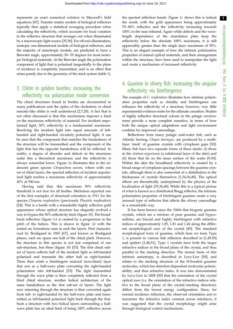
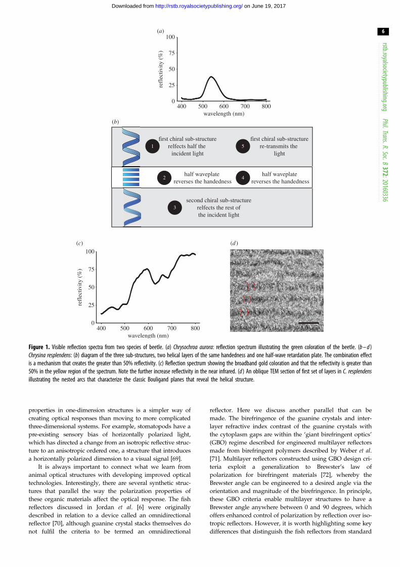
always somewhat lower. Figure 1a illustrates this in the iri-
descent green species Chrysochroa aurora, where with one
set of chiral layers, the spectral reflection of incident unpolar-
ized light reaches a maximum reflectivity of approximately
40% at 540 nm.
Having said that, this maximum 50% reflectivity
threshold is not true for all beetles. Michelson reported one
of the first examples of structural coloration in 1911 for the
species Chrysina resplendens (previously Plusiotis resplendens)
[24]. This is a beetle with a remarkable highly reflective gold
appearance whose optical structure has elegantly evolved a
way to bypass the 50% reflectivity limit (figure 1b). The broad-
band reflection (figure 1c) is created by a progression in the
pitch of the helices. This is shown in figure 1d with the
nested arc formations seen in each the layers. First character-
ized by Bouligand in 1965 [67], and known as Bouligand
planes, each arc spans one half of the chiral pitch. However,
the structure in this species is not just comprised of one
sub-structure, but three (figure 1b) [53]. The first chiral sub-
set of layers reflects half of the incident light as left-handed
polarized and transmits the other half as right-handed.
There then exists a birefringent uniaxial (non-chiral) layer
that acts as a half-wave plate converting the right-handed
polarization into left-handed [53]. The light transmitted
through the wave plate is then completely reflected from a
third chiral structure, another helical architecture of the
same handedness as the first sub-set of layers. The light
now returning through the structure is then converted again
from left- to right-handed by the half-wave plate and trans-
mitted as left-handed polarized light back through the first.
Such a structure with two helical layers surrounding a half-
wave plate has an ideal limit of being 100% reflective across
the spectral reflection bands. Figure 1c shows this is indeed
the result, with the gold appearance being approximately
70–80% reflective and the reflectivity increasing towards
100% in the near infrared. Again while defects and the wave-
length dependence of the retardation plate keep the
reflectivity below the absolute 100% maximum, it is still
appreciably greater than the single layer maximum of 50%.
This is an elegant example of how the intrinsic polarization
properties of animal optical materials, and their arrangement
within the structure, have been used to manipulate the light
and create a mechanism of increased reflectivity.
4. Guanine in silvery fish: increasing the angularreflectivity via birefringence
The example of C. resplendens illustrates how intrinsic polariz-
ation properties such as chirality and birefringence can
influence the reflectivity of a structure; however, very little
experimental evidence exists for the ultimate causation. Studies
of highly reflective structural colours in the pelagic environ-
ment provide a more complete narrative, in terms of how
both the unique optical adaptations and light environment
combine for improved camouflage.
Reflections from many pelagic mid-water fish, such as
Atlantic herring, Clupea harengus, are produced by a multi-
layer ‘stack’ of guanine crystals with cytoplasm gaps [30].
Many fish have two separate forms of these stacks: (i) those
in the stratum argenteum (a subdermal layer of the skin) and
(ii) those that lie on the inner surface of the scales [9,30].
Within the skin the broadband reflectivity is created by a
broad range of cytoplasm spacings between the guanine crys-
tals, although there is also somewhat of a distribution in the
thicknesses of crystals themselves [1,34,36,48]. The optical
effects are theoretically underpinned by the physics of the
localization of light [35,36,68]. While this is a typical picture
of what is known as a distributed Bragg reflector, the intrinsic
polarization properties of birefringent guanine create a very
unusual type of reflector that affects the silvery camouflage
in a remarkable way.
It has been known since the 1960s that biogenic guanine
crystals, which are a mixture of pure guanine and hypox-
anthine, are biaxial and highly birefringent with refractive
indices of approximately 1.85, 1.81 and 1.46 along the differ-
ent morphological axes of the crystal [49]. The standard
morphological form of guanine, which here we term Type
1, is present in various fish reflectors described in [1,49,50]
and spiders [1,48,51]. Type 1 crystals have both the larger
refractive indices in the broad plane of the crystal, and thus
parallel to the stacking direction. The atomic basis of this
intrinsic anisotropy, is described in Levy-Lior [50], and
relates to the stacking structure of the H-bonded guanine
molecules, which has direction-dependent molecular polariz-
ability, and thus refractive index. It was also demonstrated
by Levy-Lior in 2008 [50] that the orientation of the crystal
optical axes (i.e. the orientation of the refractive indices rela-
tive to the broad plane of the crystal/stacking direction),
differs from the lowest energy configuration. Since, for
normal incidence reflection, the observed orientation acts to
maximize the refractive index contrast across interfaces, it
was suggested that the crystal morphology might arise
through biological control mechanisms.
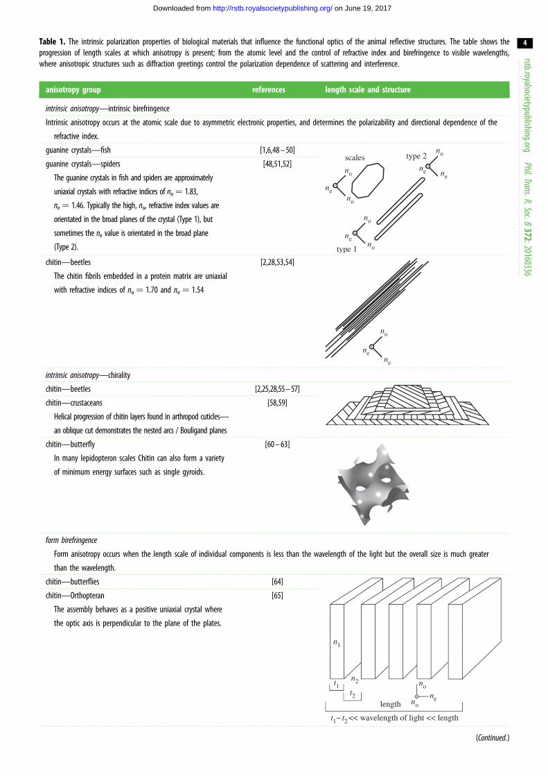
Table 1. The intrinsic polarization properties of biological materials that influence the functional optics of the animal reflective structures. The table shows theprogression of length scales at which anisotropy is present; from the atomic level and the control of refractive index and birefringence to visible wavelengths,where anisotropic structures such as diffraction greetings control the polarization dependence of scattering and interference.
anisotropy group references length scale and structure
intrinsic anisotropy—intrinsic birefringence
Intrinsic anisotropy occurs at the atomic scale due to asymmetric electronic properties, and determines the polarizability and directional dependence of the
refractive index.
guanine crystals—fish [1,6,48 – 50]scales type 2
type 1
no
no
no
ne
ne
ne
no
no
ne
guanine crystals—spiders
The guanine crystals in fish and spiders are approximately
uniaxial crystals with refractive indices of no ¼ 1.83,
ne¼ 1.46. Typically the high, no, refractive index values are
orientated in the broad planes of the crystal (Type 1), but
sometimes the ne value is orientated in the broad plane
(Type 2).
[48,51,52]
chitin—beetles
The chitin fibrils embedded in a protein matrix are uniaxial
with refractive indices of no ¼ 1.70 and ne ¼ 1.54
[2,28,53,54]
no
nene
intrinsic anisotropy—chirality
chitin—beetles [2,25,28,55 – 57]
chitin—crustaceans
Helical progression of chitin layers found in arthropod cuticles—
an oblique cut demonstrates the nested arcs / Bouligand planes
[58,59]
chitin—butterfly
In many lepidopteron scales Chitin can also form a variety
of minimum energy surfaces such as single gyroids.
[60 – 63]
form birefringence
Form anisotropy occurs when the length scale of individual components is less than the wavelength of the light but the overall size is much greater
than the wavelength.
chitin—butterflies [64]
n1
n2 no
no
ne
t1t2
length
t1~ t2 << wavelength of light << length
chitin—Orthopteran
The assembly behaves as a positive uniaxial crystal where
the optic axis is perpendicular to the plane of the plates.
[65]
(Continued.)
rstb.royalsocietypublishing.orgPhil.Trans.R.Soc.B
372:20160336
4
on June 19, 2017http://rstb.royalsocietypublishing.org/Downloaded from
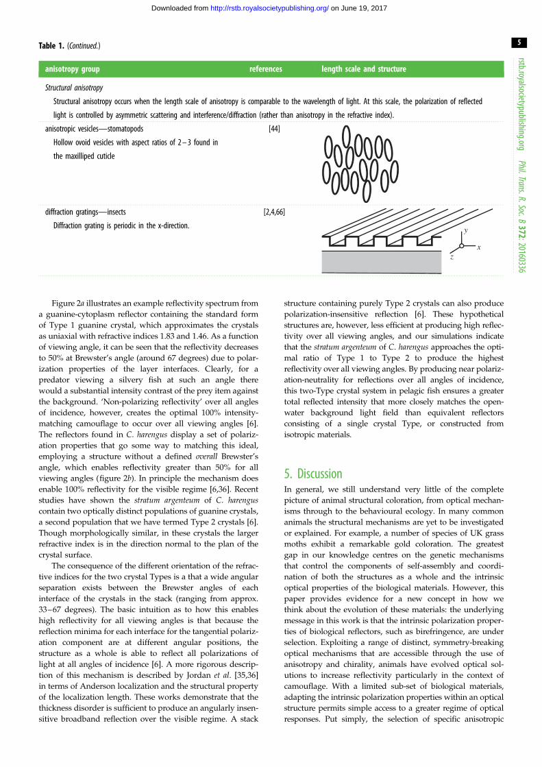
Table 1. (Continued.)
anisotropy group references length scale and structure
Structural anisotropy
Structural anisotropy occurs when the length scale of anisotropy is comparable to the wavelength of light. At this scale, the polarization of reflected
light is controlled by asymmetric scattering and interference/diffraction (rather than anisotropy in the refractive index).
anisotropic vesicles—stomatopods
Hollow ovoid vesicles with aspect ratios of 2 – 3 found in
the maxilliped cuticle
[44]
diffraction gratings—insects
Diffraction grating is periodic in the x-direction.
[2,4,66]
y
xz
rstb.royalsocietypublishing.orgPhil.Trans.R.Soc.B
372:20160336
5
on June 19, 2017http://rstb.royalsocietypublishing.org/Downloaded from
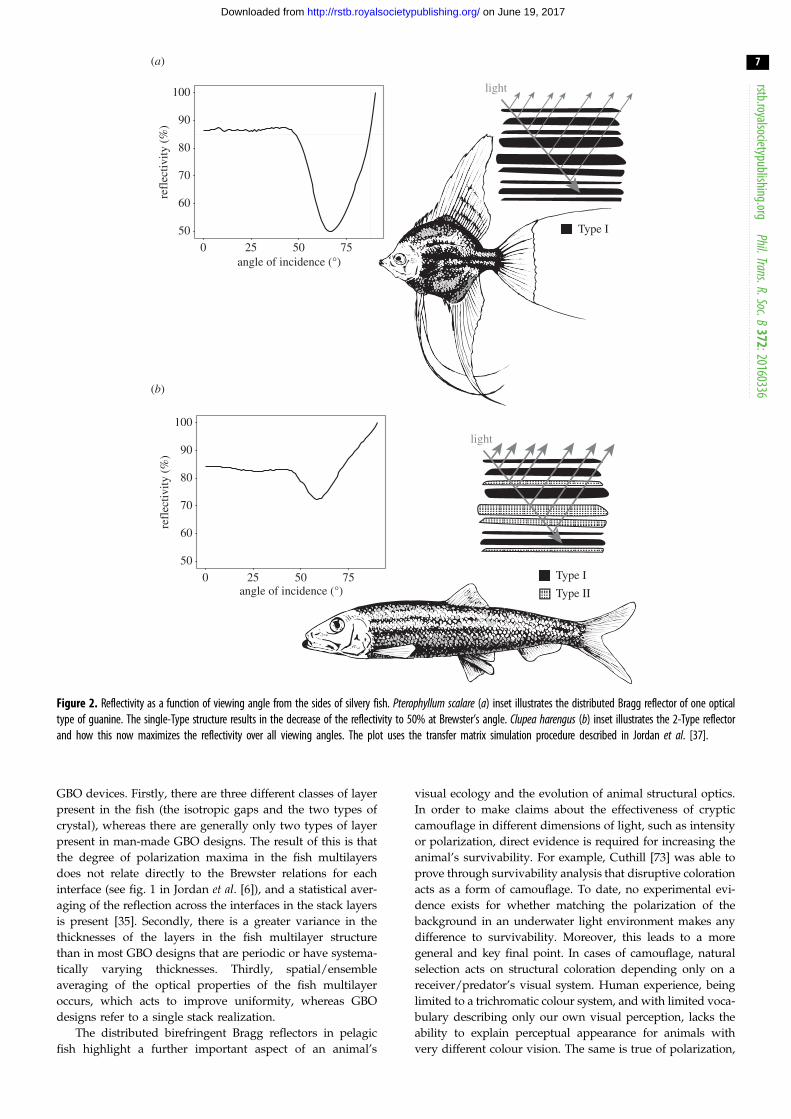
Figure 2a illustrates an example reflectivity spectrum from
a guanine-cytoplasm reflector containing the standard form
of Type 1 guanine crystal, which approximates the crystals
as uniaxial with refractive indices 1.83 and 1.46. As a function
of viewing angle, it can be seen that the reflectivity decreases
to 50% at Brewster’s angle (around 67 degrees) due to polar-
ization properties of the layer interfaces. Clearly, for a
predator viewing a silvery fish at such an angle there
would a substantial intensity contrast of the prey item against
the background. ‘Non-polarizing reflectivity’ over all angles
of incidence, however, creates the optimal 100% intensity-
matching camouflage to occur over all viewing angles [6].
The reflectors found in C. harengus display a set of polariz-
ation properties that go some way to matching this ideal,
employing a structure without a defined overall Brewster’s
angle, which enables reflectivity greater than 50% for all
viewing angles (figure 2b). In principle the mechanism does
enable 100% reflectivity for the visible regime [6,36]. Recent
studies have shown the stratum argenteum of C. harenguscontain two optically distinct populations of guanine crystals,
a second population that we have termed Type 2 crystals [6].
Though morphologically similar, in these crystals the larger
refractive index is in the direction normal to the plan of the
crystal surface.
The consequence of the different orientation of the refrac-
tive indices for the two crystal Types is a that a wide angular
separation exists between the Brewster angles of each
interface of the crystals in the stack (ranging from approx.
33–67 degrees). The basic intuition as to how this enables
high reflectivity for all viewing angles is that because the
reflection minima for each interface for the tangential polariz-
ation component are at different angular positions, the
structure as a whole is able to reflect all polarizations of
light at all angles of incidence [6]. A more rigorous descrip-
tion of this mechanism is described by Jordan et al. [35,36]
in terms of Anderson localization and the structural property
of the localization length. These works demonstrate that the
thickness disorder is sufficient to produce an angularly insen-
sitive broadband reflection over the visible regime. A stack
structure containing purely Type 2 crystals can also produce
polarization-insensitive reflection [6]. These hypothetical
structures are, however, less efficient at producing high reflec-
tivity over all viewing angles, and our simulations indicate
that the stratum argenteum of C. harengus approaches the opti-
mal ratio of Type 1 to Type 2 to produce the highest
reflectivity over all viewing angles. By producing near polariz-
ation-neutrality for reflections over all angles of incidence,
this two-Type crystal system in pelagic fish ensures a greater
total reflected intensity that more closely matches the open-
water background light field than equivalent reflectors
consisting of a single crystal Type, or constructed from
isotropic materials.
5. DiscussionIn general, we still understand very little of the complete
picture of animal structural coloration, from optical mechan-
isms through to the behavioural ecology. In many common
animals the structural mechanisms are yet to be investigated
or explained. For example, a number of species of UK grass
moths exhibit a remarkable gold coloration. The greatest
gap in our knowledge centres on the genetic mechanisms
that control the components of self-assembly and coordi-
nation of both the structures as a whole and the intrinsic
optical properties of the biological materials. However, this
paper provides evidence for a new concept in how we
think about the evolution of these materials: the underlying
message in this work is that the intrinsic polarization proper-
ties of biological reflectors, such as birefringence, are under
selection. Exploiting a range of distinct, symmetry-breaking
optical mechanisms that are accessible through the use of
anisotropy and chirality, animals have evolved optical sol-
utions to increase reflectivity particularly in the context of
camouflage. With a limited sub-set of biological materials,
adapting the intrinsic polarization properties within an optical
structure permits simple access to a greater regime of optical
responses. Put simply, the selection of specific anisotropic
0
25
50
75
100
400 500 600 700 800
refl
ectiv
ity (
%)
wavelength (nm)
0
25
50
75
100
400 500 600 700 800
refl
ectiv
ity (
%)
wavelength (nm)1.0 µm
first chiral sub-structurerelfects half the
incident light
half waveplate reverses the handedness
second chiral sub-structurerelfects the rest ofthe incident light
half waveplate reverses the handedness
1
2
3
4
first chiral sub-structurere-transmits the
light5
(b)
(a)
(c) (d )
Figure 1. Visible reflection spectra from two species of beetle. (a) Chrysochroa aurora: reflection spectrum illustrating the green coloration of the beetle. (b – d )Chrysina resplendens: (b) diagram of the three sub-structures, two helical layers of the same handedness and one half-wave retardation plate. The combination effectis a mechanism that creates the greater than 50% reflectivity. (c) Reflection spectrum showing the broadband gold coloration and that the reflectivity is greater than50% in the yellow region of the spectrum. Note the further increase reflectivity in the near infrared. (d ) An oblique TEM section of first set of layers in C. resplendensillustrating the nested arcs that characterize the classic Bouligand planes that reveal the helical structure.
rstb.royalsocietypublishing.orgPhil.Trans.R.Soc.B
372:20160336
6
on June 19, 2017http://rstb.royalsocietypublishing.org/Downloaded from
properties in one-dimension structures is a simpler way of
creating optical responses than moving to more complicated
three-dimensional systems. For example, stomatopods have a
pre-existing sensory bias of horizontally polarized light,
which has directed a change from an isotropic reflective struc-
ture to an anisotropic ordered one, a structure that introduces
a horizontally polarized dimension to a visual signal [69].
It is always important to connect what we learn from
animal optical structures with developing improved optical
technologies. Interestingly, there are several synthetic struc-
tures that parallel the way the polarization properties of
these organic materials affect the optical response. The fish
reflectors discussed in Jordan et al. [6] were originally
described in relation to a device called an omnidirectional
reflector [70], although guanine crystal stacks themselves do
not fulfil the criteria to be termed an omnidirectional
reflector. Here we discuss another parallel that can be
made. The birefringence of the guanine crystals and inter-
layer refractive index contrast of the guanine crystals with
the cytoplasm gaps are within the ‘giant birefringent optics’
(GBO) regime described for engineered multilayer reflectors
made from birefringent polymers described by Weber et al.[71]. Multilayer reflectors constructed using GBO design cri-
teria exploit a generalization to Brewster’s law of
polarization for birefringent materials [72], whereby the
Brewster angle can be engineered to a desired angle via the
orientation and magnitude of the birefringence. In principle,
these GBO criteria enable multilayer structures to have a
Brewster angle anywhere between 0 and 90 degrees, which
offers enhanced control of polarization by reflection over iso-
tropic reflectors. However, it is worth highlighting some key
differences that distinguish the fish reflectors from standard
0 25 50 75angle of incidence (°)
refl
ectiv
ity (
%)
Type I
Type II
light
Type I
light
50
60
70
80
90
100
0 25 50 75angle of incidence (°)
refl
ectiv
ity (
%)
(a)
(b)
50
60
70
80
90
100
Figure 2. Reflectivity as a function of viewing angle from the sides of silvery fish. Pterophyllum scalare (a) inset illustrates the distributed Bragg reflector of one opticaltype of guanine. The single-Type structure results in the decrease of the reflectivity to 50% at Brewster’s angle. Clupea harengus (b) inset illustrates the 2-Type reflectorand how this now maximizes the reflectivity over all viewing angles. The plot uses the transfer matrix simulation procedure described in Jordan et al. [37].
rstb.royalsocietypublishing.orgPhil.Trans.R.Soc.B
372:20160336
7
on June 19, 2017http://rstb.royalsocietypublishing.org/Downloaded from
GBO devices. Firstly, there are three different classes of layer
present in the fish (the isotropic gaps and the two types of
crystal), whereas there are generally only two types of layer
present in man-made GBO designs. The result of this is that
the degree of polarization maxima in the fish multilayers
does not relate directly to the Brewster relations for each
interface (see fig. 1 in Jordan et al. [6]), and a statistical aver-
aging of the reflection across the interfaces in the stack layers
is present [35]. Secondly, there is a greater variance in the
thicknesses of the layers in the fish multilayer structure
than in most GBO designs that are periodic or have systema-
tically varying thicknesses. Thirdly, spatial/ensemble
averaging of the optical properties of the fish multilayer
occurs, which acts to improve uniformity, whereas GBO
designs refer to a single stack realization.
The distributed birefringent Bragg reflectors in pelagic
fish highlight a further important aspect of an animal’s
visual ecology and the evolution of animal structural optics.
In order to make claims about the effectiveness of cryptic
camouflage in different dimensions of light, such as intensity
or polarization, direct evidence is required for increasing the
animal’s survivability. For example, Cuthill [73] was able to
prove through survivability analysis that disruptive coloration
acts as a form of camouflage. To date, no experimental evi-
dence exists for whether matching the polarization of the
background in an underwater light environment makes any
difference to survivability. Moreover, this leads to a more
general and key final point. In cases of camouflage, natural
selection acts on structural coloration depending only on a
receiver/predator’s visual system. Human experience, being
limited to a trichromatic colour system, and with limited voca-
bulary describing only our own visual perception, lacks the
ability to explain perceptual appearance for animals with
very different colour vision. The same is true of polarization,
rstb.royalso
8
on June 19, 2017http://rstb.royalsocietypublishing.org/Downloaded from
but seems often forgotten. The inter-relationship between the
reflectivity and the polarization properties must always take
into account the receiver’s visual system; animals know noth-
ing of angle or degree of polarization or the mathematical
constructs we use to describe them.
cietypublishing.orgPhil.Trans.R.Soc.B372:201
6. ConclusionIn this study we focused upon the role the intrinsic
polarization properties of biological materials, such as the
birefringence of guanine and the chirality of chitin, play in
controlling animal structural coloration. The golden reflectors
in the beetle C. resplendens and the silvery reflectors in the fish
C. harengus both illustrate that the intrinsic polarization prop-
erties can act to control and improve the overall percentage
reflectivity of the structure. Thus, these intrinsic polarization
properties directly influence the intensity component of
visual information. The fish reflector in the pelagic environ-
ment provides a model example for such an adaptation of
optical properties, where improved reflectivity as a result of
intrinsic polarization properties acts to improve the selective
advantage of the reflector. Future studies of biological reflec-
tors that include anisotropic materials should always address
whether the polarization properties may be an adaptation to
enhance reflectivity, and should also consider carefully the
context of intended receivers’ visual system.
Authors’ contributions. K.D.F. undertook the TEM study on the beetlesand provided the illustrations; T.M.J. produced the reflectivity datafor the fish reflector and developed the theoretical description ofthe structure; N.W.R. conceived the work and measured the beetlereflection spectra. N.W.R. wrote the initial draft and all authorscontributed further editing of the manuscript.
Competing interests. We have no competing interests.
Funding. This work was funded by the US Airforce Office of ScientificResearch FA9550-09-1-0149 and the Engineering and PhysicalSciences Research Council EP/E501214/1.
Acknowledgements. We thank Thomas Cronin, Andrew Radford andMartin How for helpful discussions and the reviewers who providedmany helpful comments.
60336
References
1. Land MF. 1972 The physics and biology of animalreflectors. Prog. Biophys. Mol. Biol. 24, 75 – 106.(doi:10.1016/0079-6107(72)90004-1)
2. Seago AE, Brady P, Vigneron JP, Schultz TD. 2009Gold bugs and beyond: a review of iridescenceand structural colour mechanisms in beetles(Coleoptera). J. R. Soc. Interface 6(Suppl. 2),S165 – S184. (doi:10.1098/rsif.2008.0354.focus)
3. Prum RO, Torres RH, Williamson S, Dyck J. 1998Coherent light scattering by blue feather barbs.Nature 396, 28 – 29. (doi:10.1038/23838)
4. Feller KD, Cronin TW. 2014 Hiding opaque eyes intransparent organisms: a potential role for larvaleyeshine in stomatopod crustaceans. J. Exp. Biol.217, 3263 – 3273.
5. Kinoshita S, Yoshioka S, Miyazaki J. 2008 Physics ofstructural colors. Rep. Prog. Phys. 71, 076401.(doi:10.1088/0034-4885/71/7/076401)
6. Jordan TM, Partridge JC, Roberts NW. 2012 Non-polarizing broadband multilayer reflectors in fish.Nat. Photonics 6, 759 – 763. (doi:10.1038/nphoton.2012.260)
7. Neville AC. 1967 Chitin orientation in cuticle and itscontrol. Adv. Insect Physiol. 4, 213 – 286. (doi:10.1016/S0065-2806(08)60209-X)
8. Neville AC, Luke BM. 1969 A two-system model forchitin-protein complexes in insect cuticles. TissueCell 1, 689 – 707. (doi:10.1016/S0040-8166(69)80041-8)
9. Denton EJ. 1970 Review lecture: on the organizationof reflecting surfaces in some marine animals. Phil.Trans. R. Soc. Lond. B 258, 285 – 313. (doi:10.1098/rstb.1970.0037)
10. Crookes WJ, Ding LL, Huang QL, Kimbell JR, HorwitzJ, McFall-Ngai MJ. 2004 Reflectins: the unusualproteins of squid reflective tissues. Science 303,235 – 238. (doi:10.1126/science.1091288)
11. Wilts BD, Michielsen K, De Raedt H, Stavenga DG.2014 Sparkling feather reflections of a bird-of-paradise explained by finite-difference time-domainmodeling. Proc. Natl Acad. Sci. USA 111,4363 – 4368. (doi:10.1073/pnas.1323611111)
12. Wilts BD, Michielsen K, Kuipers J, De Raedt H,Stavenga DG. 2012 Brilliant camouflage: photoniccrystals in the diamond weevil, Entimus imperialis.Proc. R. Soc. B 279, 2524 – 2530. (doi:10.1098/rspb.2011.2651)
13. Roberts NW, Gleeson HF. 2004 The absorption ofpolarized light by vertebrate photoreceptors. Vision Res.44, 2643 – 2652. (doi:10.1016/j.visres.2004.06.001)
14. Roberts NW. 2006 The optics of vertebratephotoreceptors: anisotropy and form birefringence.Vision Res. 46, 3259 – 3266. (doi:10.1016/j.visres.2006.03.019)
15. Roberts NW, Chiou TH, Marshall NJ, Cronin TW.2009 A biological quarter-wave retarder withexcellent achromaticity in the visible wavelengthregion. Nat. Photonics 3, 641 – 644. (doi:10.1038/nphoton.2009.189)
16. Al-Qasimi A, Korotkova O, James D, Wolf E. 2007Definitions of the degree of polarization of a lightbeam. Opt. Lett. 32, 1015 – 1016. (doi:10.1364/OL.32.001015)
17. Wehner R. 2001 Polarization vision—a uniformsensory capacity? J. Exp. Biol. 204, 2589 – 2596.
18. Roberts NW, Porter ML, Cronin TW. 2011 Themolecular basis of mechanisms underlyingpolarization vision. Phil. Trans. R. Soc. B. 366,627 – 637. (doi:10.1098/rstb.2010.0206)
19. Parkyn DC, Hawryshyn CW. 1993. Polarized-lightsensitivity in rainbow trout (Oncorhynchus mykiss):characterization from multi-unit responses in theoptic nerve. J. Comp. Physiol. A 172, 493 – 500.(doi:10.1007/BF00213531)
20. Roberts NW, Gleeson HF, Temple SE, Haimberger TJ,Hawryshyn CW. 2004 Differences in the opticalproperties of vertebrate photoreceptor classesleading to axial polarization sensitivity. J. Opt. Soc.Am. A 21, 335 – 345. (doi:10.1364/JOSAA.21.000335)
21. Roberts NW. 2014 Polarisation vision of fishes. InPolarized light and polarization vision in animalsciences, pp. 225 – 247. Berlin: Springer.
22. El Jundi B, Pfeiffer K, Heinze S, Homberg U. 2014Integration of polarization and chromatic cues inthe insect sky compass. J. Comp. Physiol. A 200,575 – 589. (doi:10.1007/s00359-014-0890-6)
23. How MJ, Christy JH, Temple SE, Hemmi JM, MarshallNJ, Roberts NW. 2015 Target detection is enhancedby polarization vision in a fiddler crab. Curr. Biol. 25,3069 – 3073. (doi:10.1016/j.cub.2015.09.073)
24. Michelson AA. 1911 LXI. On metallic colouring inbirds and insects. Philos. Mag. 21, 554 – 567.(doi:10.1080/14786440408637061)
25. Neville AC, Luke BM. 1971 A biological systemproducing a self-assembling cholesteric proteinliquid crystal. J. Cell Sci. 8, 93 – 109.
26. Mckenzie D, Large M. 1998 Multilayer reflectors inanimals using green and gold beetles as contrastingexamples. J. Exp. Biol. 201, 1307 – 1313.
27. Schultz TD, Bernard GD. 1989 Pointillistic mixing ofinterference colours in cryptic tiger beetles. Nature337, 72 – 73. (doi:10.1038/337072a0)
28. Jewell SA, Vukusic P, Roberts NW. 2007 Circularlypolarized colour reflection from helicoidal structuresin the beetle Plusiotis boucardi. New J. Phys. 9, 99.(doi:10.1088/1367-2630/9/4/099)
29. Roberts NW, Marshall NJ, Cronin TW. 2012 Highlevels of reflectivity and pointillist structural colorin fish, cephalopods, and beetles. Proc. Natl Acad.Sci. USA 109, E3387. (doi:10.1073/pnas.1216282109)
rstb.royalsocietypublishing.orgPhil.Trans.R.Soc.B
372:20160336
9
on June 19, 2017http://rstb.royalsocietypublishing.org/Downloaded from
30. Denton EJ, Nicol JA. 1965 Reflexion of light byexternal surfaces of the herring, Clupea harengus.J. Mar. Biol. Assoc. U. K. 45, 711 – 738. (doi:10.1017/S0025315400016544)
31. Denton EJ, Nicol JA. 1966 A survey of reflectivityin silvery teleosts. J. Mar. Biol. Assoc. U. K. 46,685 – 722. (doi:10.1017/S0025315400033439)
32. Denton EJ, Land MF. 1971 Mechanism of reflexionin silvery layers of fish and cephalopods.Proc. R. Soc. Lond. B 178, 43 – 61. (doi:10.1098/rspb.1971.0051)
33. Denton EJ, Gilpin-Brown JB, Wright PG. 1972 Theangular distribution of the light produced by somemesopelagic fish in relation to their camouflage.Proc. R. Soc. Lond. B 182, 145 – 158. (doi:10.1098/rspb.1972.0071)
34. McKenzie DR, Yin Y, McFall WD. 1995 Silvery fishskin as an example of a chaotic reflector.Proc. R. Soc. Lond. A 451, 579 – 584. (doi:10.1098/rspa.1995.0144)
35. Jordan TM, Partridge JC, Roberts NW. 2013Suppression of Brewster delocalization anomalies inan alternating isotropic-birefringent random layeredmedium. Phys. Rev. B 88, 041105. (doi:10.1103/PhysRevB.88.041105)
36. Jordan TM, Partridge JC, Roberts NW. 2014Disordered animal multilayer reflectors and thelocalization of light. J. R. Soc. Interface 11,20140948. (doi:10.1098/rsif.2014.0948)
37. Cronin TW, Gagnon YL, Johnsen S, Marshall NJ,Roberts NW. 2016 Comment on ‘Open-ocean fishreveal an omnidirectional solution to camouflage inpolarized environments’. Science 353, 552. (doi:10.1126/science.aaf4481)
38. Johnsen S, Gassmann E, Reynolds RA, Stramski D,Mobley C. 2014 The asymmetry of the underwaterhorizontal light field and its implications for mirror-based camouflage in silvery pelagic fish. Limnol.Oceanogr. 59, 1839 – 1852. (doi:10.4319/lo.2014.59.6.1839)
39. Jerlov NG, Fukuda M. 1960 Radiance distribution inthe upper layers of the sea. Tellus 12, 348 – 355.(doi:10.3402/tellusa.v12i3.9393)
40. Tyler JE. 1960 Radiance distribution as a function ofdepth in an underwater environment. Berkeley, CA:University of California Press.
41. Johnsen S. 2012. The optics of life: a biologist’sguide to light in nature. Princeton, NJ: PrincetonUniversity Press.
42. Born M, Wolf E. 1999 Principles of optics:electromagnetic theory of propagation, interferenceand diffraction of light, 7th edn, pp. 837 – 840.Cambridge, UK: Cambridge University Press.
43. Beche B, Gaviot E. 2003 Matrix formalism toenhance the concept of effective dielectric constant.Opt. Commun. 219, 15 – 19. (doi:10.1016/S0030-4018(03)01291-4)
44. Jordan TM, Wilby D, Chiou TH, Feller KD, CaldwellRL, Cronin TW, Roberts NW. 2016 A shape-anisotropic
reflective polarizer in a stomatopod crustacean. Sci.Rep. 6, 21744. (doi:10.1038/srep21744)
45. Berreman DW. 1972 Optics in stratified andanisotropic media: 4� 4-matrix formulation. J. Opt.Soc. A 62, 502 – 510. (doi:10.1364/JOSA.62.000502)
46. Azzam RM, Bashara NM. 1987 Ellipsometry andpolarized light. North-Holland: Elsevier SciencePublishing.
47. Orfanidis S. 2002 Electromagnetic Waves andAntennas. New Brunswick, NJ: Rutgers University.See http://www.ece.rutgers.edu/orfanidi/ewa/(accessed September 2016).
48. Levy-Lior A, Shimoni E, Schwartz O, Gavish-Regev E,Oron D, Oxford G, Weiner S, Addadi L. 2010Guanine-based biogenic photonic-crystal arrays infish and spiders. Adv. Funct. Mater. 20, 320 – 329.(doi:10.1002/adfm.200901437)
49. Greenstein L. 1966 Nacreous pigments and their?properties. Proc. Sci. Sec. Toilet Goods Assoc. 26,20 – 26.
50. Levy-Lior A, Pokroy B, Levavi-Sivan B, Leiserowitz L,Weiner S, Addadi L. 2008 Biogenic guanine crystalsfrom the skin of fish may be designed to enhancelight reflectance. Cryst. Growth Des. 8, 507 – 511.(doi:10.1021/cg0704753)
51. Dacke M, Nilsson DE, Warrant EJ, Blest AD, Land MF,O’carroll DC. 1999 Built-in polarizers form part of acompass organ in spiders. Nature 401, 470 – 473.(doi:10.1038/46773)
52. Mueller KP, Labhart T. 2010 Polarizing optics in aspider eye. J. Comp. Physiol. A 196, 335 – 348.(doi:10.1007/s00359-010-0516-6)
53. Caveney S. 1971 Cuticle reflectivity and opticalactivity in scarab beetles: the role of uric acid.Proc. R. Soc. Lond. B 178, 205 – 225. (doi:10.1098/rspb.1971.0062)
54. Brink DJ, Van der Berg NG, Prinsloo LC, Hodgkinson IJ.2007 Unusual coloration in scarabaeid beetles. J. Phys.D 40, 2189. (doi:10.1088/0022-3727/40/7/050)
55. Neville AC, Caveney S. 1969 Scarabaeid beetleexocuticle as an optical analogue of cholestericliquid crystals. Biol. Rev. 44, 531 – 562. (doi:10.1111/j.1469-185X.1969.tb00611.x)
56. De Silva L, Hodgkinson I, Murray P, Wu QH, ArnoldM, Leader J, Mcnaughton A. 2005 Natural andnanoengineered chiral reflectors: structural color ofmanuka beetles and titania coatings.Electromagnetics 25, 391 – 408. (doi:10.1080/02726340590957399)
57. Arwin H, Magnusson R, Landin J, Jarrendahl K. 2012Chirality-induced polarization effects in the cuticleof scarab beetles: 100 years after Michelson. Philos.Mag. 92, 1583 – 1599. (doi:10.1080/14786435.2011.648228)
58. Neville AC, Parry DA, Woodhead-Galloway J. 1976The chitin crystallite in arthropod cuticle. J. Cell Sci.21, 73 – 82.
59. Giraud-Guille MM. 1984 Fine structure of thechitin-protein system in the crab cuticle.
Tissue Cell 16, 75 – 92. (doi:10.1016/0040-8166(84)90020-X)
60. Argyros A, Manos S, Large MCJ, McKenzie DR,Cox GC, Dwarte DM. 2002 Electron tomographyand computer visualization of a three-dimensionalphotonic crystal in a butterfly wing-scale. Micron33, 483 – 487. (doi:10.1016/S0968-4328(01)00044-0)
61. Michielsen K, Stavenga DG. 2008 Gyroid cuticularstructures in butterfly wing scales: biologicalphotonic crystals. J. R. Soc. Interface 5, 85 – 94.(doi:10.1098/rsif.2007.1065)
62. Schroder-Turk GE, Wickham S, Averdunk H, Brink F,Gerald JF, Poladian L, Large MC, Hyde ST. 2011 Thechiral structure of porous chitin within thewing-scales of Callophrys rubi. J. Struct. Biol. 174,290 – 295. (doi:10.1016/j.jsb.2011.01.004)
63. Saba M, Wilts BD, Hielscher J, Schroder-Turk GE.2014 Absence of circular polarisation in reflectionsof butterfly wing scales with chiral gyroid structure.Mater. Today 1, 193 – 208. (doi:10.1016/j.matpr.2014.09.023)
64. de Campos Vidal B. 2011 Butterfly scale formbirefringence related to photonics. Micron 42,801 – 807. (doi:10.1016/j.micron.2011.04.006)
65. Neville AC. 1965 Chitin lamellogenesis in locustcuticle. J. Cell Sci. 3, 269 – 286.
66. Zhang K, Zhou S, Tang Y, Wang G, Zhou H, Fan T,Zhang D. 2014 Polarization-sensitive color iniridescent scales of butterfly Ornithoptera. RSC Adv.4, 51 865 – 51 871. (doi:10.1039/C4RA07988D)
67. Bouligand Y. 1978 Liquid crystalline order inbiological materials. In Liquid crystalline order inpolymers (ed. A Blumstein), pp. 261 – 297. NewYork, NY, USA: Academic Press.
68. Berry MV, Klein S. 1997 Transparent mirrors: rays,waves and localization. Eur. J. Phys. 18, 222 – 228.(doi:10.1088/0143-0807/18/3/017)
69. How MJ, Porter ML, Radford AN, Feller KD, TempleSE, Caldwell RL, Marshall NJ, Cronin TW, RobertsNW. 2014 Out of the blue: the evolution ofhorizontally polarized signals in Haptosquilla(Crustacea, Stomatopoda, Protosquillidae). J. Exp.Biol. 217, 3425 – 3431. (doi:10.1242/jeb.107581)
70. Fink Y, Winn JN, Fan S, Chen C, Michel J,Joannopoulos JD, Thomas EL. 1998 A dielectricomnidirectional reflector. Science 282, 1679 – 1682.(doi:10.1126/science.282.5394.1679)
71. Weber MF, Stover CA, Gilbert LR, Nevitt TJ,Ouderkirk AJ. 2000 Giant birefringent opticsin multilayer polymer mirrors. Science 287,2451 – 2456. (doi:10.1126/science.287.5462.2451)
72. Lekner J. 1991 Reflection and refraction by uniaxialcrystals. J. Phys. 3, 6121 – 6133. (doi:10.1088/0953-8984/3/32/017)
73. Cuthill IC, Stevens M, Sheppard J, Maddocks T,Parraga CA, Troscianko TS. 2005 Disruptivecoloration and background pattern matching. Nature434, 72 – 74. (doi:10.1038/nature03312)