Embed Size (px)
Citation preview

84 Feige et al., Blood glucose, insulin and acid base observations
J. Perinat. Med.5(1977) 84
Fetal and maternal blood glucose, insulin and acid base observationsfollowing maternal glucose infusion
Axel Feige, Wolfgang Künzel, Hans J. Mitzkat
Universitäts-Frauenklinik WürzburgArbeitsgruppe Diabetologie im Dept. Innere Medizin der Medizinischen HochschuleHannover
In the past conflicting results for the fetal insulinincrease following maternal glucose infusion werereported [2, 7, 21]. In conscious rhesus monkeysconstant glucose infusion to the mother wasfollowed by maternal and fetal hyperglycemiaand äs a response to it a 2 to 4 fold increment infetal insulin occured [22]. Fetal glucose andinsulin responses following maternal glucose loadshave been repeatedly investigated in fetal cordblood in humans [16, 19, 23, 28]. However fetaland maternal glucose levels increase duringparturition and are correlated to the base excess[10]. In addition fetal insulin secretion is suppressedin severe acidosis [9], so that cord blood studiesare masked by hypoxic episodes which occureduring the second stage of labor.Maternal and fetal glucose and insulin concentrationwere studied in normal and diabetic pregnanciesfollowing a single intravenous injection of glucoseto the mother [4]. The fetal insulin responsehowever was of a different kind probably due tothe short lasting Stimulation of the fetal pancreaticislet cells.We were therefore interested in the human maternaland fetal glucose and insulin response to a constantglucose infusion to the mother during the firststage of labor sustained for 60 minutes. Elevatedfetal glucose concentrations lead in a mild hypoxicfetus to a deterioration of the fetal pH due toincreasing lactate production [13, 24]. We investi-
Curriculum vitae
AXEL FEIGE was born in1941 and obtained hismedical degree in 1968.He did clinical and researchwork from 1970 to 1974at the Department of Ob-stetrics and Gynecölogy,Hannover, since 1974at theUniversitäts-FrauenklinikWürzburg. Chief areas ofinterest: Carbohydratemetabolism in pregnancy,pregnancy and diabetesmellitus, ultrasonics inobstetrics.
gated therefore in addition the fetal acid baseParameters.The present experiments demonstrate a closerelationship between the maternal and fetal glucoseconcentration. The fetal insulin increased by about86% after 60 min glucose infusion to the motherA deterioration of the fetal pH and base excesscould not be established even at high glucose con-centrations.
l Methods1.1 Patient material
The study comprises 11 primigravid patients (age17-28 years) with a gestational age of 38-40
J. Perinat. Med. 5 (1977)
Feige et al., Blood glucose, insulin and acid base observations 85
weeks. The course of pregnancy had been withoutany complications. The birthweight of the babieswas 3330 g (SD 480). Neither the family historynor the case history revealed any signs of disturb-ances in carbohydrate metabolism.
l .2 Experimental procedure
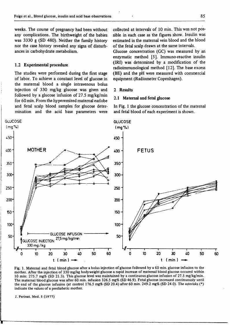
The studies were performed during the first stageof labor. To achieve a constant level of glucose inthe maternal blood a single intravenous bolusinjection of 330 mg/kg glucose was given andfollowed by a glucose infusion of 27.5 mg/kg/minfor 60 min. From the hyperemized maternal earlobeand fetal scalp blood samples for glucose deter-mination and the acid base parameters were
GLUCOSE(mg0/.)
400-
350-
300-
250-
200-
150-
100-
50-
MOTHER
L/-rGLUCOSE INJECTION
330 mg/kg
GLUCOSE INFUSION27,5 mg/kg/min
collected at intervals of 10 min. This was not pos-sible in each case äs the figures show. Insulin wasestimated in the maternal vein blood and the bloodof the fetal scalp drawn at the same intervals.Glucose concentration (GC) was measured by anenzymatic method [5]. Immuno-reactive insulin(IRI) was determined by a modification of theradioimmunological method [12]. The base excess(BE) and the pH were measured with commercialequipment (Radiometer Copenhagen).
2 Results
2. l Maternal and fetal glucose
In Fig. l the glucose concentration of the maternaland fetal blood of each experiment is shown.
GLUCOSE(mge/o)
10 20 30 40 50t ( min ) —**
450-
400-
350
300-
250-
200
150
100-
50-
FETUS
60 10 20 30 40 50t ( min ) —+»
60
Fig. 1. Maternal and fetal blood glucose after a bolus injection of glucose followed by a 60 min. glucose infusion to themother. After the injection of 330 mg/kg body weight glucose a rapid increase of maternal blood glucose occured within10 min: 275.7 mg% (SD 21.3). This glucose level was maintained by a continuous glucose infusion of 27.5 mg/kg/min.The maternal blood glucose was after 60 min. infusion 326.5 mg% (SD 46.9). Fetal glucose increased continuously untilthe end of the glucose infusion (at control 176.5 mg% (SD 20.4) after 60 min. 249.2 mg% (SD 24.0). The asterisks (*)indicate the values of a prediabetic mother.
J. Perinat. Med. 5 (1977)

86 Feige et al., Blood glucose, Insulin and acid base observations
Following the injection of 330 mg/kg glucoseintravenously the maternal bloodglucoseincreasedwithin 10 min from 85.9 mg/100 ml (SD 7.9) to280 mg/100 ml (SD 25.9). This level was main-tained roughly by a continuous glucose Infusion of27.5 mg/kg/min. The maternal glucose concentra-tion after 60 min infusion was 327.5 mg/100 ml(SD 43.6).Before the injection of glucose to the mother thefetal blood glucose was 64.1 mg/100 ml (SD 7.6).As a result of the glucose infusion the glucoseconcentration in the fetal scalp blood increased to191.0 mg/100 ml (SD 24.8) after 10 min and to
247.5 mg/100 ml (SD 25.0) after 60 min infusion,respectively.In Fig. 2 the glucose concentration of the fetalscalp blood is plotted to the blood glucose in thematernal capillary blood. The calculated regressionline deviates from the line of identity with increas-ing maternal blood glucose. The different symbolsin high maternal glucose concentrations indicatethe time of sampling. Compared with the line ofidentity the difference between the maternal andthe fetal blood glucose during the control periodwas about 22 mg/100 ml and rose to 80 mg/100 nüat the end of the infusion.
LJJO
¥ 300 -
250 -
8 200ÜJ(/)8 150D
LU
100 -
50 -
GLUCOSE (F) = 3,4 * 0,74 GLUCOSE ( M )r = 0,96; 2«<0,001
IDENTITY LINE
PRESENT PAPER
s /./ HUMAN [FEIGE et al. 1976]
50 100 150 200 250 300 350 400 450
MATERNAL GLUCOSE CONCENTRATION (mg%)
Fig. 2. The relationship between fetal and maternal blood glucose. The solid line shows the regression line of the fetaland maternal blood glucose at control (·) and after 60 min. (*) glucose infusion to the mother. The maternal-fetalglucose difference rises with increasing maternal glucose concentration.The dotted line shows the fetal-maternal relationship of blood glucose in human under hypoxic conditions.This regression line is identical with the present observations, revealing a fairly rapid glucose transfer across the hemo-chorial placenta.
J. Perinat. Med. S (1977)

Feige et al . iiiood glutusc. insulm und acid base obscrvations 87
The present results and the regression line arc inAgreement with our previous obscrvations |10]which had bccn achicved under quitc differentconditions (sce discussion) and deviate froni theresults ohtained in sheep (17, 18, 26).
2.2 Maternal and fetal insulm
In Fig. 3 the insulin rcsponse to the increasedmaternal and fetal bloodglucose of eachexperimentis dcmonstratcd. The increase of the glucose con-ccntrationinthe mate rna l bloodwasnotparalleled
by the same rise of insulin in each casc. Tliematernal insulin concentration at control was24.0 /iU/ml. In one case (asterisks) the insulinsecretion rose after 10 min up to 232 μυ/mJfollowed by a rapid decrease to about 60 μυ/ml.In the fetal blood there was only a slight responseof insulin secretion to the elevated glucose level.The IRI-concentration at control was 17.0 μυ/ml(SD 5.2). After 40 and 60 min of glucose infusionto the mother the IRI-concentration was 163%and 180% of its control, respectively. In the caseof an abnormal maternal insulin secretion thefetal insulin however showed a continuous increase
IRI(iiU/ml)
250-
200-
150
90-
MOTHER
ι GLUCOSE NJECT NΓ 330 mg/kg
GU COSE INFUSION
IRI(uU7mL)
250-
200
150-
100
SO-
FETUS
ιK) 20 30
t ( min) -*-
l
i SO 60 K) 20 30
t (nw)
l
50 60
Fig. 3. Maternal and fetal insulin (IRI) concentration after a bolus injection of glucose followed by a 60 min. glucoseinfusion to the mother. The increase of maternal blood glucose is accompanied with an increase of maternal insulin(IRI). The IRI-concentration was at control 24.0 μυ/ml (SD 8.0) and rose continuously up to 98.7 μυ/ml (SD 22.8),(10 min.) and 152.3 μυ/ml (SD 51.5) after 60 min. glucose infusion. The values of the prediabetic mother (asterisks) areexcluded by calculating the mean. Fetal insulin concentration did not change significantly during the initial time course(at control 17.0 μΐΐ/ml) (SD 5.2). After 30 min. however, a small increase up to 33.0 μυ/ml (SD 26.4) and 30.1 μυ/ml(SD 14,1) (60 min.), respectiveiy took place. The fetus of the prediabetic mother (*) showed a tremendous rise in in-sulin. These values were not used in calculating the mean.
J. Perinat. Med. 5 (1977)

88 Feige et al., Blood glucose, insulin and acid base observations
(asteriks) from 26.0/zU/ml at control to 216 μυ/mlat the end of infusion.
2.3 Fetal weight and insulin response
In Fig. 4 the insulin secretion s a response to therise of the fetal blood glucose after 60 min isrelated to the fetal birth weight. The increase ofthe insulin secretion following the glucoseStimulation may be assumed to indicate thereactivity of the pancreatic islet cells. The calculatedquotient is low in normal fetal birth weight, i.e.the insulin secretion related to the glucose increaseis less if the fetal weight is in a normal r nge. How-ever there is a steep increment of the quotient in
cases of high birth weight, i.e. the fetal insulinsecretion by approximately the same rise of glu-cose is much more pronounced in babies withhigher birthweights.
2.4 pH and base excess of the matemal and fetalblood
In Tab. I the pH and base excess of the maternaland fetal blood are listed. The pH and base excessin the fetal blood by the first analyses was 7.37(SD 0.05) and - 3.8 meq/1 (SD 2.0), respectively.The maternal pH was 7.45 (SD 0.05) and the baseexcess - 3.5 meq/1 (SD 2.1). Neither in the fetalblood nor in the maternal blood were significant
IRI-INCREASE ( u U / m l )GLUCOSE-INCREASE (mg%)
0.8 -
0,7-
0,6 -
0,5 -
0,4 -
0,3 -
0,2 -
0,1 -
A I R IΔ GLUCOSE
= 7,05 - 0,0046 BW* 7,5-lcftBW)2
r = 0,91
2«< 0,001
3000 3500 4000
BIRTH WEIGHT (g)
Fig. 4. The relationship between fetal insulin secretion and fetal birth weight after 60 min. glucose infusion to themother. A polynomial regression line was adapted to the respective data. Birthweight over 3500 g is associated with anelevated insulin secretion by an equal rise of glucose. This fact demonstrates a hyperinsulinism probably due topancreatic islet cell hypertrophy generated by the chronic fetal glucose load.
J. Perinat. Med. 5 (1977)

Feige et al., Blood glucose, Insulin and acid base observations 89
Tab. I. Acid base observation in the maternal capillary and fetal scalp blood during the first s tage oflabor before andafter 60 min. glucose infusion to the mother. There is no difference in pH and base excess in the maternal and fetalblood before and after glucose-infusion. (x = mean, SD = Standard deviation, N = number of cases)
Fetal scalp blood
Maternal capiUaryblood
pH κSON
Base Excess (meq/1) χSDN
pH χSDN
Base Excess (meq/1) χSDN
Beforeglucose infusioito the mother
7.370,0510
-3,82,010
7.450.059
-3,52,18
After 60 min Utesn glucose infusion
to the mother
7,350,048
-3.81,48
7.460,058
-3.71,77
changes of the above mentioned parameters noted.A relationship between the glucose concentrationand the base excess could not be proved.
3 Discussion
The present investigations were designed to studythe effect of a maternal glucose load on fetal bloodglucose and its stimulatory effect on the fetalinsulin secretion s well s its effect on the acidbase Status of the fetus during the first stage oflabor. This time was chosen because alterations infetal blood glucose and base excess take placeduring the second stage of labor and might alterfetal insulin secretion [9,10].
3.1 The effect of glucose infusion to the motheron maternal and fetal blood glucose andinsulin
Glucose: The bolus injection of 330 mg/kg glucoseto the mother and the continuation of the glucoseload by infusion of 27.5 mg/kg/min to the motherraised the maternal gjucose level from 85.9 mg/100 ml to 280 mg/100 ml after 10 min. Thisconcentration was fairiy well maintained during aperiod of 60 min. The rapid increase in maternalblood glucose was not in parallel with the glucoseelevation in the fetal blood. After 10 min it was
176 mg/100 ml and reached its highest level after60 min infusion: 247.5 mg/100 ml. The slow riseof the fetal blood glucose may be a result of theplacental barrier to glucose.The maternal glucose difference increased signifi-cantly during maternal hyperglycemia (Fig. 2).This is in accordance with earlier observations[3, 6, 7, 10, 17, 20, 21]. There is no differencebetween the present and the previous findingsunder hypoxic conditions [10). This may accountfor the fairiy rapid glucose transfer across theplacent .Factors which might be responsible for the increasedmaternal-fetal glucose difference are a Saturationof the transport mechanism for glucose [8, 14, 31]and an increased glucose consumption by theplacent . Recently published observations in sheepby BATTAGLIA and MESCHIA [3] and SIMMONS etal. [27] however indicate, that for the increment inmaternal-fetal glucose difference the rise in fetalglucose uptake may be the most important factor.This is supported by the increase of fetal insulinand thus glucose utilization.
Insulin: The maternal and fetal glucose load wasparalleled by an increase of maternal and fetalinsulin. The level for maternal insulin prior toinfusion was 24.0 μυ/ml. The maternal insulinincrease was scattered over a wide r nge. The
J. Perinat. Med. 5 (1977)

90 Feige et al., Blood glucose, insulin and acid base observations
maternal insulin response in this series is compatiblewith previous observations by FEIGE and MITZKAT[11] and the studies by OAKLEY [20] and GARD-MARK [13]. The baseline fetal insulin concentrationwas 17.0 μυ/ml and increased only by 86%. Thissmall increase in fetal insulin is not consistent withthe fmdings in rhesus monkeys [22], and in sheep[2], In fetal rhesus monkeys a two to four foldincrement in fetal insulin (50-240 μΙΤ/πύ) isassociated with a fetal hyperglycemia of 225—325 mg%. In sheep an even higher insulin releasewas observed with fetal glucose concentrations of100-160 mg/100 ml. The low fetal pancreaticislet cell response in the present studies may beexplained by in-vitro studies concerning the insulinrelease of fetal pancreas slices during incubation inmedia with different glucose concentration [l ]. The.maximum insulin release was observed in amediumcontaining 80-150 mg/100 ml glucose. In higherglucose concentrations (300 mg/100 ml) therelease was equal with the release at zero con-centrations.However, in one case of an accidentally studiedmother with initial hyperinsulinism we observed atremendous fetal insulin secretion following glucoseload rising from 26.0 μυ/ml to 216 μΐΐ/πύ. This caseimplies that a chronic fetal glucose load whichoccurs in mothers, with a poor insulin response toglucose, results in a hypertrophy of the fetalpancreatic islet cells. A very interesting observationin this context was the correlation of the fetalweight to the rise of insulin per glucose elevationobserved after 60 min glucose infusion (Fig. 4).Therefore maternal hyperglycemia s seen inprediabetic and diabetic patients with poor controlof maternal blood sugar can be considered s theinitial Step for fetal overweight and complications
such s placental hypertrophy and fall in diffusingcapacity [30]. , f
3.2 The effect of maternal glucose load on thefetal acid-base-status
The increase in fetal blood glucose in humans[13], and in sheep [2, 25] is associated with an in-crease in plasma lactate. In sheep there exists agood correlation between plasma lactate andglucose which is still lacking for the human fetus.The rise of plasma lactate should correlate withthe fall of the fetal buffer base, i.e. the base excessof the fetus. With the present investigations nochange in the fetal pH and base excess could bedemonstrated. The base excess was — 3.8 meq/1before and — 3.8 meq/1 60 min. after glucoseinfusion to the mother. This is consistent with thefindings of GARDMARK et al. [11] describinghowever an increase of maternal-fetal pH differenceto0.17units.Two factors may be responsible for the differentfindings in sheep and human. As pointed out bySHELLEY et al. [25] hyperglycemia of the fetusmight be fatal only if it coincides with hypoxicepisodes. Hypoxic episodes however can beneglected in the present studies because no fetalheart rate decelerations could be seen during thetime of observation.In addition it is known that lactic acid does notcross the syndesmo-chorial placenta of the sheep[29] however is transferred in exchange to bi-carbonate in both directions of the human hemo-chorial placenta [15]. Taking this into)considerationit may be assumed that at physiological flow rateson both sides of the placenta an accumulation ofplasma lactate in the human fetus will not occur,while it takes place in the sheep fetus.
Summary
The aim of the present investigation was to examine thefetal and maternal blood glucose and insulin responsefollowing glucose infusion to the mother.The studies were performed on 11 primigravid patientswith a gestational age of 38-40 weeks during the firststage of labor. Glucose was given intravenously by a bolusinjection of 330 mg/kg body weight, followed by aglucose infusion of 27.5 mg/kg/min for 60 min. Glucoseconcentration, immuno-reactive insulin (IRI), pH andbase excess of the maternal and fetal blood were measuredbefore and during maternal glucose load. Maternal blood
glucose rose within 10 min. up to 280.0 mg% (SD 25.9).This level could be fairly maintained throughout the ex-periment. The maternal glucose was after 60 min. infusion326.5 mg% (SD 46.9). Fetal glucose concentration rosecontinuously fiom 65.8 mg% (SD 5.8) at control to249.2 mg% (SD 23.3) after 60 min.The increase of maternal and fetal glucose was associatedwith an elevation of immuno-reactive insulin (IRI). Thematernal insulin was 24.0 μυ/ml (SD 8.0). It was scatteredover a wide r nge (55.4M /ml-217.1 μυ/ml) after 60 min.glucose infusion. The fetal insulin was 17.0 μυ/ml (SD 5.2)
J. Perinat. Med. S (1977)

Feige et al., Blood glucose, Insulin and acid base observations 91
at control and rose by 86.5% (SD 80.5) after 60 min.glucose load.One case of a mother with a subclinical diabetes mellitusdeviated where the fetal insulin rose from 26.0 MU/ml atcontrol to 215.6 /ml after 60 min. infusion.The increase of insulin per glucose rise was correlated tofetal body weight. During glucose infusion to the motherof both, fetal and maternal, acid base parameters remainedunchanged.
From these observations it may be concluded that in thehuman fetus insulin secretion following a single glucoseload is generally low, however, it increases in cases wherethe maternal insulin response to glucose load is abnormal.This might be related to a chronic Stimulation by glucoseof the fetal pancreatic islet cells in poorly controlleddiabetic and possibly prediabetic patients.
Keywords: Acid-base-balance, blood glucose, fetus, glucose infusion, insulin, mother.
Zusammenfassung
Glukosekonzentration, Insulinsekretion und Säure-Basen-Status im fetalen und mütterlichen Blut nach Glukose-Infusion an die MutterDas Ziel der vorliegenden Arbeit war die Glukosekonzen-tration im mütterlichen und fetalen Blut und die Insulin-sekretion nach Glukoseinfusion an die Mutter zu unter-suchen.Die Untersuchungen wurden an 11 Primiparae in der38.-40. Schwangerschaftswoche während der Eröffnungs-periode durchgeführt. Nach einer Bolusinjektion von330 mg Glukose pro Kilogramm Körpergewicht wurdezur Aufrechterhaltung eines konstanten Glukosespiegelsbei der Mutter 27.5 mg/kg Körpergewicht/min Glukoseüber 60 min infundiert. Bei Mutter und Fet wurden vorund während der Infusion die Glukosekonzentration, dasimmuno-reaktive Insulin, pH und Base-Excess bestimmt.Die materne Glukosekonzentration stieg innerhalb von10 min auf 280.0 mg/100 ml (SD 25.9) an und betrugnach 60 min Infusionsdauer 326.5 mg/100 ml (SD 46.9}.Die fetale Glukosekonzentration stieg kontinuierlich von65.8 mg/100 ml (SD 5.8) zu Beginn der Infusion auf249.2 mg/100 ml (SD 24.0) nach 60 min dauernder In-fusion an.Gleichzeitig mit dem Anstieg der fetalen und maternenGlukosekonzentration stieg das immuno-reaktive Insulin
(IRI) an. Zu Beginn der Infusion betrug das materne IRI24.0 /ml (SD 8.0), nach 60 min Infusionszeit betrugdas IRI 152.3 /ml (SD 51.5). Die fetale Insulinkonzen-tration betrug zu Beginn der Infusion 17.0 /ml (SD 5.2)und war nach 60 min dauernder Glukoscinfusion an dieMutter um 86,5% (SD 80,5) erhöht. Deutlich anders ver-hielt sich die Insulinsekretion des Feten einer Mutter mitlatentem Diabetes mellitus. Das immuno-reaktive Insulinbetrug vor der Infusion 26.0 /ml und stieg nach 60Minuten dauernder Infusion auf 215,6 /ml an.Der Anstieg der fetalen Insulinkonzentration pro Anstiegder fetalen Glukosekonzentration war nach 60 Minutendauernder Infusion mit dem Geburtsgewicht korreliert.Während der Glukoseinfusion an die Mutter waren dieParameter des Säure-Basen-Haushaltes sowohl bei derMutter als beim Feten konstant.Die vorliegenden Untersuchungen zeigen, daß Feten vonstoffwechselgesunden Müttern auf eine Glukosebelastungnur mit einer geringfügigen Insulinsekretion reagieren.Eine chronische Hyperglykämie, wie sie bei Feten vonMüttern mit schlecht eingestelltem Diabetes mellitus oderlatentem Diabetes mellitus vorliegt, könnte über eineStimulation des fetalen Pankreas zur vorzeitigen Reifungder Inselzellen führen. Daraus resultiert bei Glukosebe-lastung eine übermäßig gesteigerte Sekretion von Insulin.
Schlüsselwörter: Fet, Glukon, Glukoseinfusionen, Insulin, Mutter, Säure-Basen-Haushalt.
Resume
Glucose du sang maternel et foetal, controle de l'insulineet de l'eqiiilibre acidobasique apres infusion de glucose ala mereLe present article se propose d'etudier la concentration deglucose et la secretion d'insuline maternelles et foetalesapres infusion de glucose a la mere.Les examens ont ete effectues sur 11 prirnipares dans la38-40eme semaine de grossesse pendant la periode dedilatation du col. Apres une injection de bpl de 330 mgde glucose/kg de poids du corps, on a infuse ä la mere27,5 mg/kg poids du corps/min. de glucose pendant 60minutes pour maintenir un taux de glucose constant.Avant et durant l'infusion on a determine chez la mereet chez le foetus la concentration de glucose, l'insulineimmuno-reactive, le pH et l'exces basique. La concen-tration de glucose de la mere a atteint en 10 min. 280,0 mg%
(SD 25,9) et au bout des 60 min. d'infusion 326,5 mg%(SD 46,9). La concentration de glucose du foetus aaugmente de fa£on continue pour passer des 65,8 mg%(SD 5,8) du debut de l'infusion a 249,2 mg% (SD 24,0)ä la fin des 60 min. d'infusion.En meine temps, que la hausse de la concentration deglucose chez la mere et le foetus, on a observe uneaugmentation de l'insuline immuno-reactive (IRI). Audebut de l'infusion, l'IRI maternelle etait de 24,0 /ml(SD 8,0) et, apres 60 min-d'infusion, de 152,3 /ml(SD 51,5). La concentration d'insuline foetale etait de17,0 /ml (SD 5,2) au debut de Pinfusion et augmentade 86,5% (SD 80,5) en fin d'une infusion de 60 minutesde glucose ä la mere. Par contre, la secretion d'insuline dufoetus evolue tout differemment lorsque la mere souffred'un diabetes mellitus latent. L'IRI, de 26,0 /ml avant
J. Perinat. Med. 5 (1977)

92 Feige et al., Blood glucose, insulin and acid base observations
l'infusion, monta a 215,6 /ml apres 60 min. d'infusioncontinuc.On a observe une correlation entre la hausse de la concen-tration d'insuline foetale pro hausse de la concentrationde glucose du foetus avec le poids a la naissance apres uneinfusion continue de 60 min.Pendant Hnfusion de glucose a la mere, les parametres derequilibre acidobasique ont ete constants aussi bien chezla mere que chcz le foetus.
Les analyses presentes montrent que les foetus de meresau metabolisme normal ne reagissent que par une faiblesecretion d'insuline a une Charge de glucose. Une hyper-glycemie chronique, comme c'est le cas pour les foetus demeres ayant un diabetes mellitus mal regle ou latent,pourrait conduire par Stimulation du pancreas foetal aune maturation precoce des ilots de Langerhans avec,pour consequence d'une Charge de glucose, une secretionexcessivement accrue d'insuline.
Mots-cles: Equilibre acidobasique, foetus, glucose du sang, infusion de glucose, insuline, mere
Bibliography
( l ] BASSETT, J. M., D. MADILL, D. H. NICOL, G. D.THORBURN: Further studies on the regulation ofinsulin release in foetal and post-natal lambs: Therole of glucose äs a physiological regulator of insulinrelease in utero. In: Foetal and Neonatal Physiology,Cambridge University Press (1973)
[2] BASSETT, J. M., D. MADILL: The influence ofmaternal nutrition on plasma hormone and metabolicconcentrations of foetal lambs. J. Endocr. 61 (1974)465
13] BATTAGLIA, F.C., G. MESCHIA: Foetalmetabolismand Substrate utilization. In: Foetal and NeonatalPhysiology, Cambridge University Press (1973)
[4)BEARD, R. W., R. C. TURNER, N. W. OAKLEY:Fetal response to glucose loading. Post-Grad. Med. J.,London 47 (1971) 68
[5] BERGMEYER, H. U.: Methoden der enzymatischenAnalyse. Chemie, Weinheim/Bergstraße 1974
[6] BOSSART, H., S. PAPADOPOULOS: La glycemiematernelle et foetale sub partu. Gynaecologia, Basel162 (1966) 360
|7]CORDERO, L., J. A. GRÜNT, G. ANDERSON:Hypertonie glucose infusion during labor. Amer. J.Obstet. Gynec. 15 (1970) 560
[8]ELY, A. P.: The placental transfer of hexoses andpolyols in the guinea-pig, äs shown by umbilicalperfusion of the placenta. J. Physiol., Lond. 184(1966) 255
[9] FEIGE, A., W. KÜNZEL, M. CORNELY, H. J.MITZKAT: Die Beziehung von Glucosestoffwechselund Säure-Basen-Status des Feten während der Ge-burt. In: DUDENHAUSEN, J. W., E. SALING,E. SCHMIDT: Perinatale Medizin Band VI. ThiemeStuttgart
[10] FEIGE, A., W. KÜNZEL, M. CORNELY, H. J.MITZKAT: Die Beziehung zwischen Glukosekon-zentration und Säure-Basen-Status im maternen undfetalen Blut während der Geburt. Z. Geburtsh.Perinat. 180(1976)106
|11J FEIGE, A., H. J. MITZKAT: Correlations betweenglucose disappearance rate (K-value) and serum IRIand HPL levels in pregnancy. In: ALMQUIST andWIKSELL International, Perinatal Medicine Stock-holm (1976) 112
[12]FRIEDEL, R., A. DWENGER, S. KÄSTNER,I. TRAUTSCHOLD: Teilautomatisierung von Radio-immunoassays mit Hilfe eines0 diskontinuierlichenAnalysensystems. Z. Klin. Chem. Klin. Bioch. l(1974)45
[13] GARDMARK, s., G. GENNSER, L. JACOBSON,G. ROOTH, J. THORELL; Influence on fetalcarbohydrate and fat metabolism and on acid-basebalance of glucose administration to the motherduring labour. Biol. Neonate 26 (1975) 129
[14] HOLMBERG, N. G., B. KAPLAN, M. J. KARVONEN,J. LIND, M. MALM: Permeability of human placentato glucose, fructose and xylose. Acta physiol. scand.,Stockholm 36 (1956) 291
[15]KASTENDIECK, E., W. MOLL: The transfer oflactic acid across the guinea-pig placenta. In: PerinatalMedicine, distributed by ALMQUIST and WIKSELLInternational, Stockholm (1976) 50
[16] LIND, T., E. A. GILMORE, M. MCCLARENCE:Cord plasma glucose and insulin concentrations andmaternal-fetal relations. Brit. J. Obstet. Gynaecol.82 (1975)562
[17] MANN, L. L, J. w. PRICHARD, D. SYMMES: Theeffect of glucose loading on the fetal response tohypoxia. Amer. J. Obstet. Gynec. 107 (1970) 610
[18] MESCHIA, G., F. C. BATTAGLIA: Acute changesof oxygen pressure and the regulation of uterineblood flow. In: Foetal and Neonatal Physiology,Cambridge University Press (1973)
[19]MILNER, R. D. G., C. N. HALES: Effect of intra-venous glucose on concentration of insulin in maternaland umbilical cord plasma. Brit. med. J. l (1965)284 ' OK
[20] OAKLEY, N. W., R. W. BEARD, R.o£Ü TURNER:Effect of sustained maternal hyperglycaemia on thefetus in normal and diabetic pregnancies. Brit. med.J. l (1972)466
[21] PATERSON, P., D. PAGE, P. TAFT, L. PHILLIPS,C. WOOD: Study of fetal and maternal insulin levelsduring labour. J. Obstet. Gynec. Brit. Cwlth. 75(1968)917
[22] REYNOLDS, W. A., R. M. PITKIN: Fetal insulinresponse: a reexamination. Gynec. Invest. 7 (1976)48
[23] SABATA, V.,H.FRERICHS, H. WOLF, P. STUB3P:Insulin and glucose levels in umbilical cord bloodafter infusions of glucose and glucose with insulin towomen in labour. J. Obstetr. Gynaec. Brit. Comm.77 (1970) 121
[24] SHELLEY, H. J.: The use of chronically catheterizedfoetal lambs for the study of foetal metabolism. In:Foetal and Neonatal Physiology, Cambridge Univer-sity Press (1973) 360
J. Perinat. Med. 5 (1977)

Feige et al., Blood glucose, insulin and arid base observations ' 93
[25] SHELLEY, H. J., J. M. BASSETT: Control of Carbo- [29] STEELE, S. M., G. B. JACKSON, A. STARKHydrate Metabolism in the fetus and Newborn. B. WOLKOFF: Some aspects of blood lactate levels inMed. Bull. 31 (1975)37 mother and fetus. Amer. J. Obstet. Gynec. 105
[26)SILVER, M., D. H. STEVEN, R. S. COMLINE: (1969)569Placental exchange and morphology in ruminants [30] WERNER, GH., W. SCHNEIDERHAN: Plazenta-and the märe. In: Foetal and Neonatal Physiology, morphologie und Plazentafunktion in AbhängigkeitCambridge University Press (1973) 245 von der diabetischen Stoffwechselführung. Geburtsh.
[27] SIMMONS, M. A., G. MESCHIA, E. L. MAKOWSKI, u. Frauenheilk. 32 (1972) 959F. C. BATTAGLIA: Fetal metabolic response to [31|WIDDAS, W. F.: Inability of diffusion to accountmaternal starvation. Pediat. Res. 8 (1974) 830 for placental glucose transfer in the sheep and
[28] SPELLACY, W. N.: Maternal and fetal metabolic consideration of the kinetics of a possible carrierinterrelationships. In: SUTHERLAND, H. W., J. M. transfer. J. Physiol. 118 (1952) 23STOWERS: Carbohydrate Metabolism in Pregnancyand the Newborn. Churchill Livingstone, Edinburgh-London-New York 1975 . . T f _ 0 1 _, . 4 . XT , 0/l .Qnii' Received July 28, 1976. Accepted November 24, 1976.
Dr. med. A. FeigeProf. Dr. med. W. KünzelUniversitäts-Frauenklinik WürzburgJosef-Schneider-Straße 4D-8700 WürzburgProf. Dr. med. H.-J. MitzkatArbeitsgruppe Diabetologie imDept. Innere Medizin,Med. Hochschule HannoverPasteurallee 5D-3000 Hannover
J. Perinat. Med. 5 (1977)