Embed Size (px)
Citation preview

Fetal EchocardiographyFetal Echocardiography
Dr. Avisa Tabib Dr. Avisa Tabib
Pediatric CardiologistPediatric CardiologistRajaei Cardiovascular Medical and Research CenterRajaei Cardiovascular Medical and Research Center

INDICATIONS
Fetal :
• Abnormal screening obstetric ultrasound.• Extracardiac anomalies (omphalocele, duodenal atresia
VACTERL,spina bifida )• Chromosomal abnormalities (trisomies, microdeletion)• Increased first – trimester nuchal translucency • Nonimmune hydrops • Tachyarrhythmias• Bradyarrhythmias

INDICATIONS
Maternal:
• Diabetes • Phenylketonuria• Teratogen exposure (lithium ,phenytoin, valproic acid• ,carbamazepin,isotretinoin )• Viral infection during first-trimester

INDICATIONS
• Familial :
• Previous child with CHD • Paternal congenital heart defect• Tuberous sclerosis• Noonan syndrome• Velocardiofacial syndrome

Timing of Examination
• Assessment of the central cardiovascular connection is electively performed from 18 weeks gestation onwards. How ever , major CHD can also be detected earlier , at 12_ 14 weeks , in high-risk cases

Ultrasound approach and scanning planes
• Axial views• 4-chamber view• 3-vessel view
• Oblique views• long axis of the LV • long axis of the RV• short axis of the RV
• Sagittal views• cavo- atrial junction • aortic arch• ductal arch

4 chamber view
• This presents anan axial view of the fetal thorax.
• Apical 4 chamber viewApical 4 chamber view : cardiac apex is directed towards the transducer and the interventricular septum is aligned with the insonating beam .
• Transverse 4 chamber viewTransverse 4 chamber view: the interventricular septum is at 90 angle with the insonating beam .

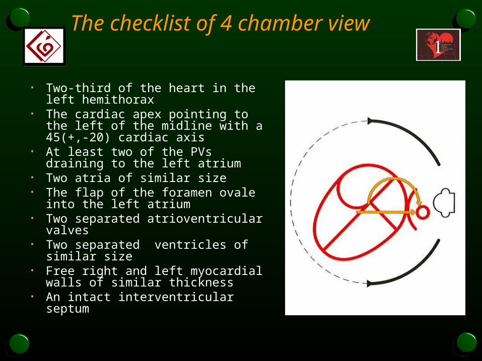
The checklist of 4 chamber view
• Two-third of the heart in the left hemithorax
• The cardiac apex pointing to the left of the midline with a 45(+,-20) cardiac axis
• At least two of the PVs draining to the left atrium
• Two atria of similar size • The flap of the foramen ovale into the
left atrium • Two separated atrioventricular valves • Two separated ventricles of similar
size • Free right and left myocardial walls of
similar thickness• An intact interventricular septum

Standard 4 chamber view


Cardiac Axis

3-vessel view
• This is an axial view of the upper mediastinum .The three vessels are (from right to left) , SVC , the Aorta , and the PA.


Long axis of left ventricle
• This view is obtained by rotating the transducer slightly towards the right fetal shoulder .

Checklist :
• The presence of a vessel that connects with LV.
• Septo-aortic continuity .• Semilunar valve with
normal systo-diastolic excursion .
• The presence of crossover.

Long axis of right ventricle • This view is obtained from the long axis of the LV by curving the
transducer towards the fetal head .


Checklist :
• The presence of a vessel that connects with RV.
• Semilunar valve with normal systo-diastolic excursion .
• The presence of crossover.
• The size of the vessel is similar or slightly larger than Aorta .

Short axis of the Right ventricle
• To obtain this view from the 4-chamber view ,the transducer should perform a rotation mirroring that needed for the long axis of the LV.
Checklist:• Same as for the long axis of the RV.

Cavo-atrial junction
• In this longitudinal view of the fetal thorax , both vanae cavas are seen entering the right atrium (seagull wings view)
• Checklist:• Presence of both venae
cavae .• Both venae cavae should
have the same size.

Longitudinal view of the Aortic Arch
On this view the whole course of the Aorta from the left ventricle to the abdominal Aorta , is displayed. The aortic arch has regular diameter with 3 vessels of the head and neck .

Ductal arch view
Ductal arch takes a trajectory that is practically rectilinear , anteroposterior ,beginning in the retrosternal region with a ductal arch of an obtuse angle.

A FAST GLANCE :
• Before going into the details of the examination we begin by taking a quick preliminary look round the entire area .
• We move up wards perpendicular to the axis of the spine towards the cephalic pole , beginning from the transverse abdominal view ,and the pass to the 4 chamber view , and then beginning of the aorta and pulmonary trunk .
• Continuing the movement , we see the horizontal portion of the aortic arch and 3 vessel view.