Embed Size (px)
Citation preview
Fetal Monitoring and Umbilical Cord Gases:
What’s the Secret?
Patricia A. Heale, DNP, RNC-OB, C-EFM
The learner will be able to
describe the process for umbilical cord blood collection.
define the normal acid-base parameters of umbilical cord blood gases.
discuss the clinical value of determining acid-base parameters.
Objectives
Low levels of oxygen in the tissues
What is Hypoxia?
Acidemia – state of low blood pH
Acidosis – the process of becoming acidemic
Acidemia & Acidosis
Birth asphyxia occurs when a baby doesn't receive enough oxygen before, during or just after birth
Hypoxia, hypercapnia, and respiratory and/or metabolic acidemia
What is Asphyxia?
ACOG reports that fetal asphyxia occurs in 25 of 1000 live births and 15% of those are moderate to severe
Fetal Asphyxia Occurrence
Ultrasound
Biophysical Profile (BPP)
Doppler studies
Electronic Fetal Monitor
Umbilical Cord Blood Analysis
Assessing Fetal Well-Being

“…a valuable but imperfect tool.”(Clark, et al. 2017, p. 163)
Electronic Fetal Monitor
Decrease incidence of cerebral palsy and intrapartum stillbirth
Expected Outcomes
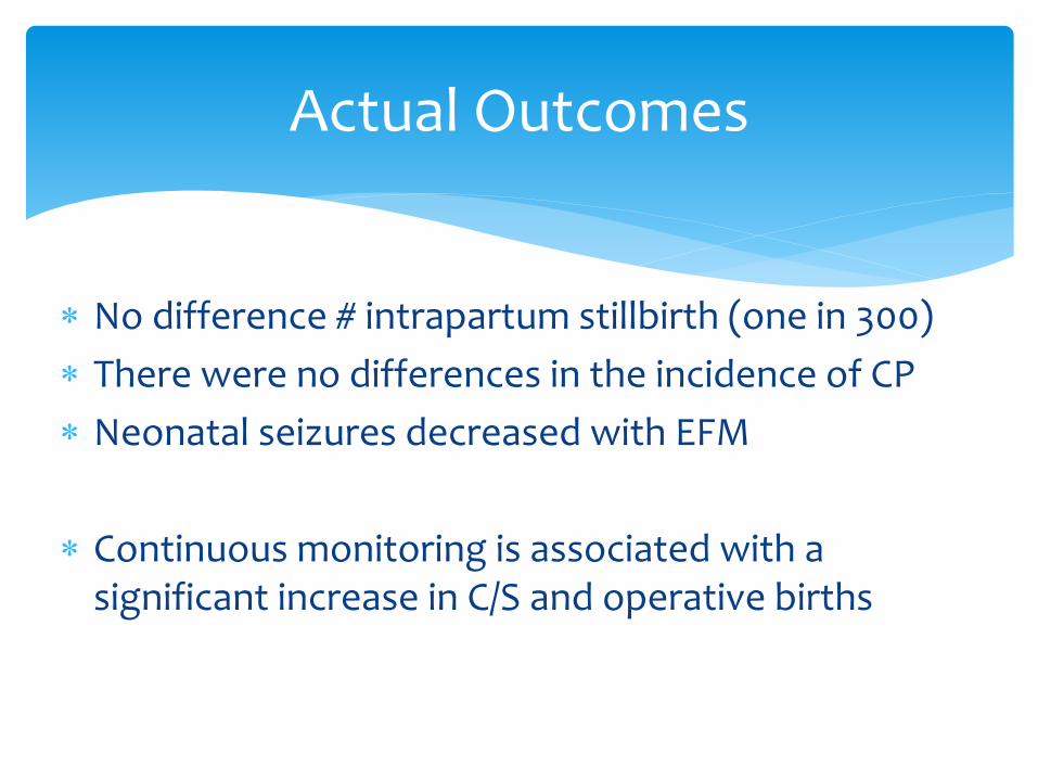
No difference # intrapartum stillbirth (one in 300)
There were no differences in the incidence of CP
Neonatal seizures decreased with EFM
Continuous monitoring is associated with a significant increase in C/S and operative births
Actual Outcomes
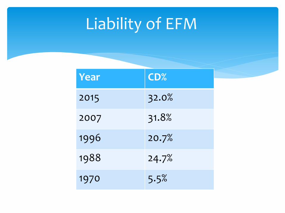
Year CD%
2015 32.0%
2007 31.8%
1996 20.7%
1988 24.7%
1970 5.5%
Liability of EFM
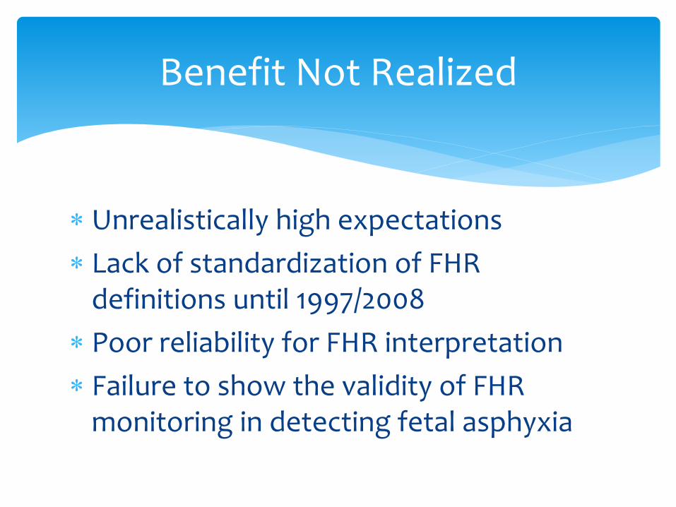
Unrealistically high expectations
Lack of standardization of FHR definitions until 1997/2008
Poor reliability for FHR interpretation
Failure to show the validity of FHR monitoring in detecting fetal asphyxia
Benefit Not Realized
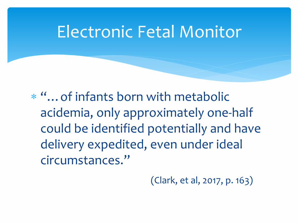
“…of infants born with metabolic acidemia, only approximately one-half could be identified potentially and have delivery expedited, even under ideal circumstances.”
(Clark, et al, 2017, p. 163)
Electronic Fetal Monitor
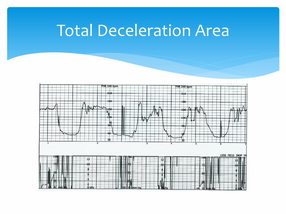
Total Deceleration Area
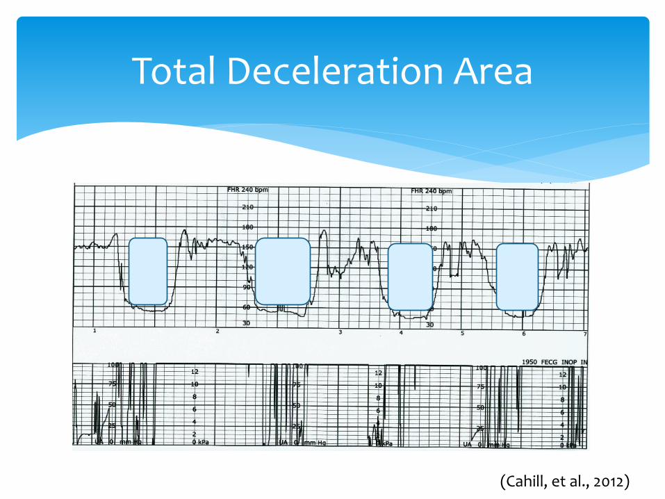
Total Deceleration Area
(Cahill, et al., 2012)

Category I Strip

Category III Strip
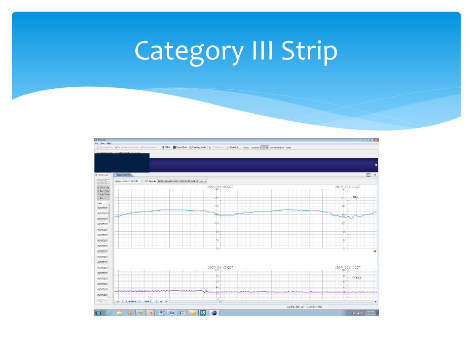
Category III Strip

Parer et al. (2006) reported that only 24% of fetuses with Category III strips have metabolic acidemia
Category III Strips
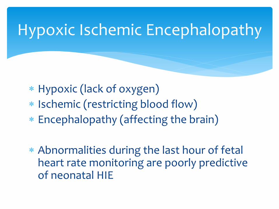
Hypoxic (lack of oxygen)
Ischemic (restricting blood flow)
Encephalopathy (affecting the brain)
Abnormalities during the last hour of fetal heart rate monitoring are poorly predictive of neonatal HIE
Hypoxic Ischemic Encephalopathy
“Hypoxic-ischemic encephalopathy is associated with significant increases in electronic fetal monitoring abnormalities, but the predictive ability to identify these conditions is low.”
Larma, et al., 2007
Hypoxic Ischemic Encephalopathy
Specificity of 98.9%
Percent of healthy pts appearing healthy
Sensitivity of 7.7%
Percent of unhealthy pts appearing unhealthy
Larma, et al., 2007
EFM Predicting Acidosis
UCG’s are an important parameter in the decision for brain cooling
Neonatal therapeutic hypothermia has been shown to help reduce significant brain damage in infants who suffered from lack of oxygen during labor/birth
Is Acidemia Important?
In 1958, James, et al. recognized the possibility of interpreting umbilical cord blood gases
History of UCBG

Intrapartum Event =/? Adverse Outcome
All Non-elective cesareans deliveries
5- minute Apgar 3
“Abnormal” Fetal Heart Rate Tracing
Severe IUGR
Intrapartum Fever
Maternal Thyroid Disease
Multiple Gestation
ACOG Committee Opinion
Cord blood analysis is the most objective way of assessing the fetal condition at birth
Allows for differentiation of respiratory and metabolic acidemia
Clinical Relevance
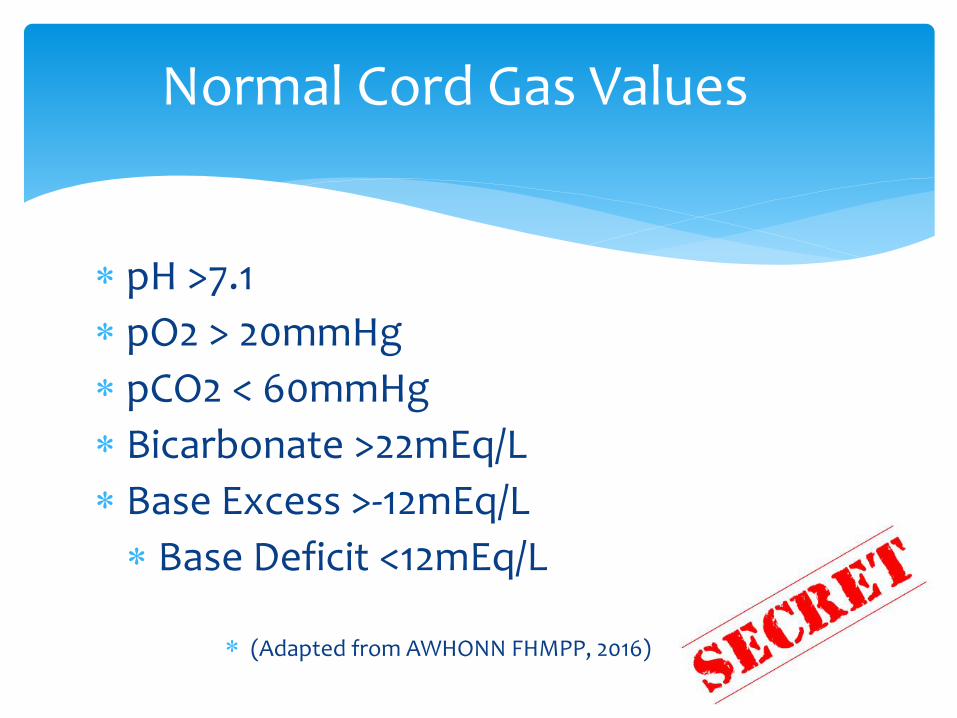
pH >7.1
pO2 > 20mmHg
pCO2 < 60mmHg
Bicarbonate >22mEq/L
Base Excess >-12mEq/L
Base Deficit <12mEq/L
(Adapted from AWHONN FHMPP, 2016)
Normal Cord Gas Values
Fetal neurologic injury does not occur without significant metabolic acidemia
pH <7.0
ACOG, 2010
Fetal Injury
Potential for Hydrogen (H+) Increasing H+, decreasing pH Decreasing H+, increasing pH
Fetal Normal = > 7.10ACOG recommends= <7.0 for severe acidemia
pH
The amount of dissolved oxygen in the blood
Normal umbilical gas values >20mmHg
pO2
Dissolved carbon dioxide in the blood
Normal umbilical gas value <60mmHg
Umbilical vein has lower pCO2
Umbilical artery has higher pCO2
pCO2
Buffering systems
Circulating acids are neutralized
Bicarbonate (HCO3) is the largest buffering system
Bicarbonate
The base excess is defined as the amount of H+ ions that would be required to return the pH of the blood to 7.35 expressed as a - number
Indirect measurement of anaerobic metabolism
Base Excess
If you had a high H+ content in the blood (metabolic acidemia) it would take a very low amount to return to 7.35
Base Excess
Maternal oxygenation is compromised
Maternal cardiac disease
Reduced perfusion of the placenta
Preeclampsia
Delivery of oxygenated blood from the placenta to the fetus is impeded
Abruption, cord entanglement
Poor Oxygenation
CO2 accumulates
pCO2 >60mmHg
pH falls
Develops quickly, clears quickly
Primary Apnea
Respiratory Acidemia
Delayed Cord Clamping (DCC)
Reperfusion of the peripheral tissues causes CO2 to be released and can be detected in the umbilical artery
Decreased pH
Increased CO2
“Hidden Acidemia”
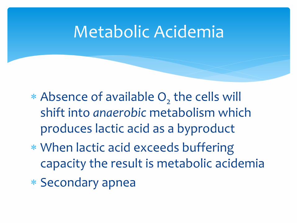
Absence of available O2 the cells will shift into anaerobic metabolism which produces lactic acid as a byproduct
When lactic acid exceeds buffering capacity the result is metabolic acidemia
Secondary apnea
Metabolic Acidemia
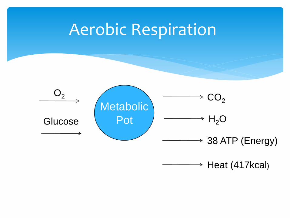
Aerobic Respiration
Metabolic
Pot
O2
Glucose
CO2
H2O
38 ATP (Energy)
Heat (417kcal)
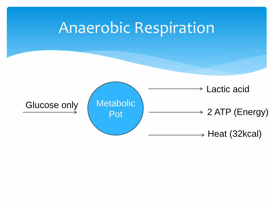
Anaerobic Respiration
Metabolic
PotGlucose only
Lactic acid
2 ATP (Energy)
Heat (32kcal)

Both necessary to ensure the biological validity of the samples obtained
Arterial or Venous or Both
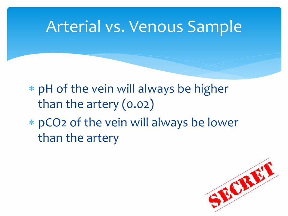
pH of the vein will always be higher than the artery (0.02)
pCO2 of the vein will always be lower than the artery
Arterial vs. Venous Sample
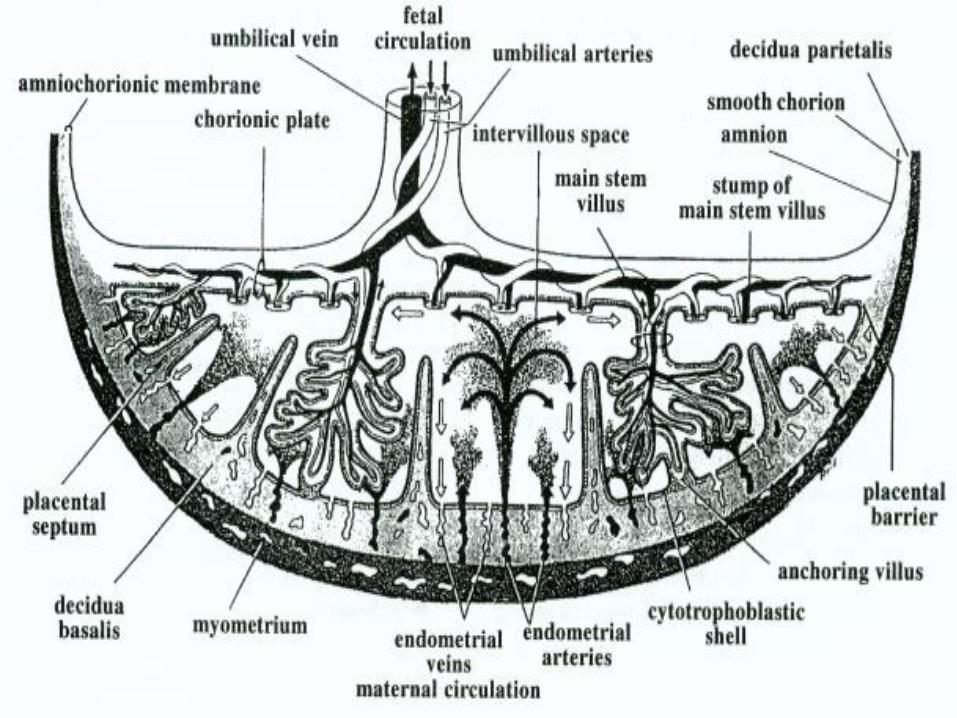
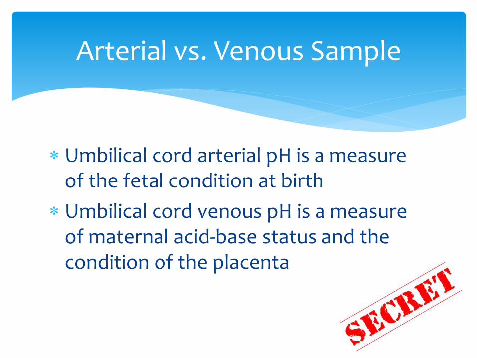
Umbilical cord arterial pH is a measure of the fetal condition at birth
Umbilical cord venous pH is a measure of maternal acid-base status and the condition of the placenta
Arterial vs. Venous Sample
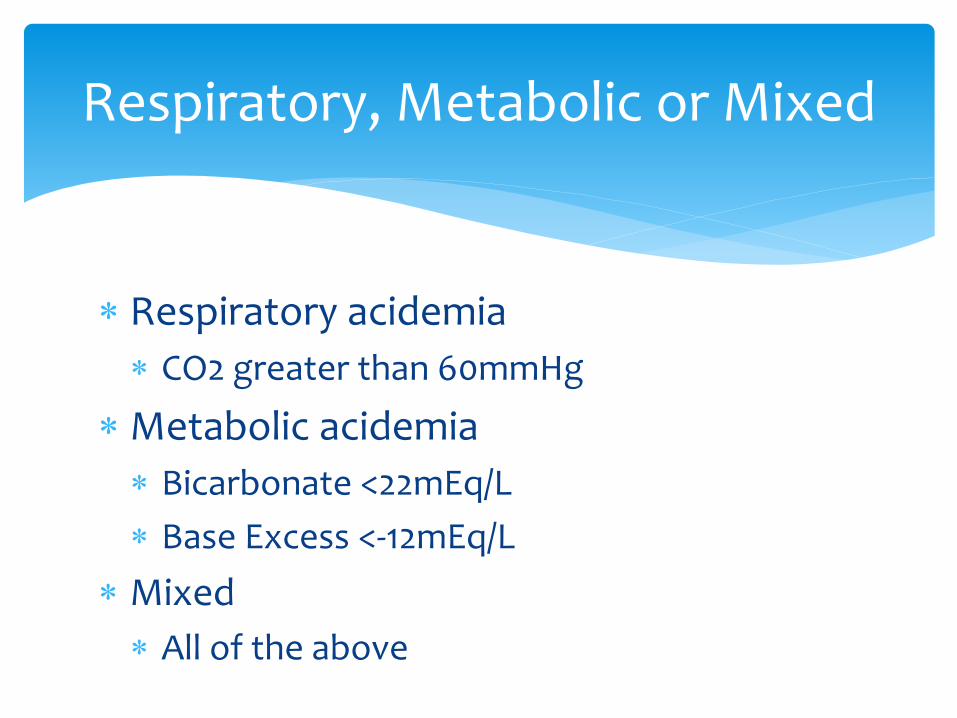
Respiratory acidemia
CO2 greater than 60mmHg
Metabolic acidemia
Bicarbonate <22mEq/L
Base Excess <-12mEq/L
Mixed
All of the above
Respiratory, Metabolic or Mixed
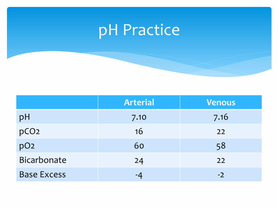
Arterial Venous
pH 7.10 7.16
pCO2 16 22
pO2 60 58
Bicarbonate 24 22
Base Excess -4 -2
pH Practice
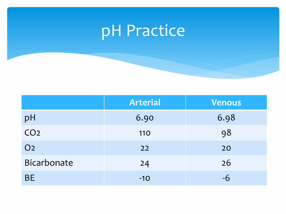
Arterial Venous
pH 6.90 6.98
CO2 110 98
O2 22 20
Bicarbonate 24 26
BE -10 -6
pH Practice
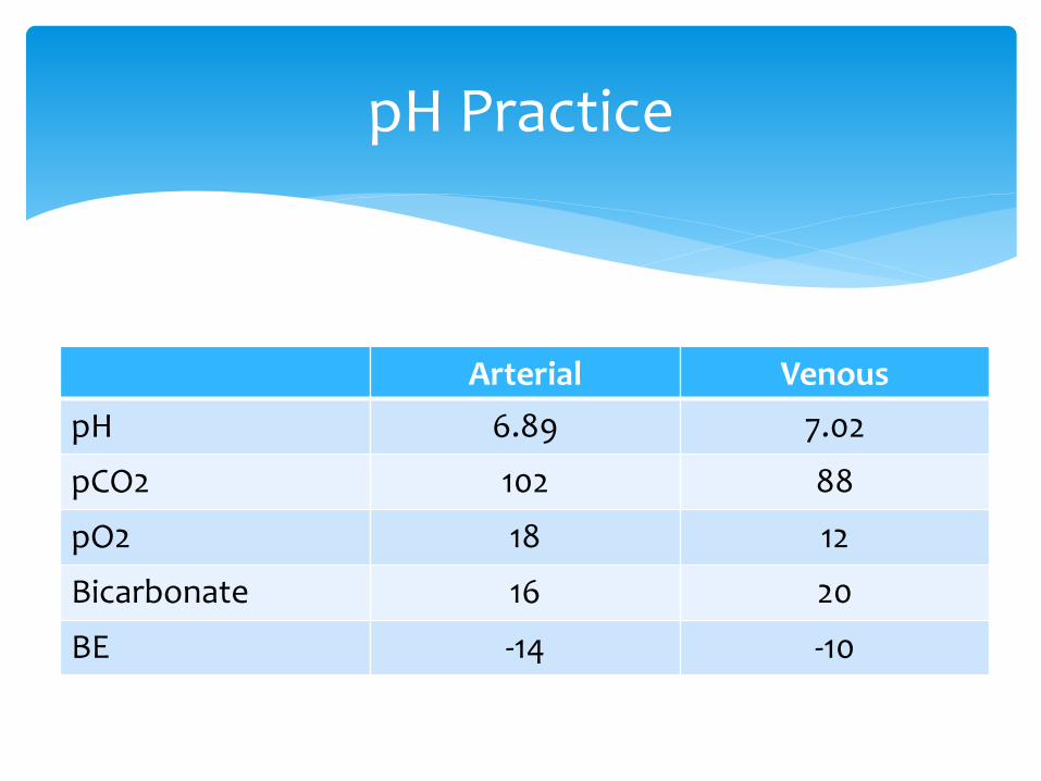
Arterial Venous
pH 6.89 7.02
pCO2 102 88
pO2 18 12
Bicarbonate 16 20
BE -14 -10
pH Practice
A wide difference in values is often due to obstructed O2 delivery
A small difference is most likely caused by impairment of maternal perfusion of the placenta
UA/UV Differences
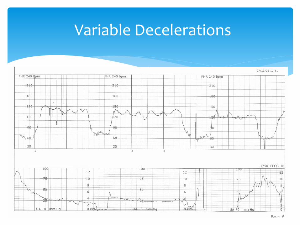
Variable Decelerations
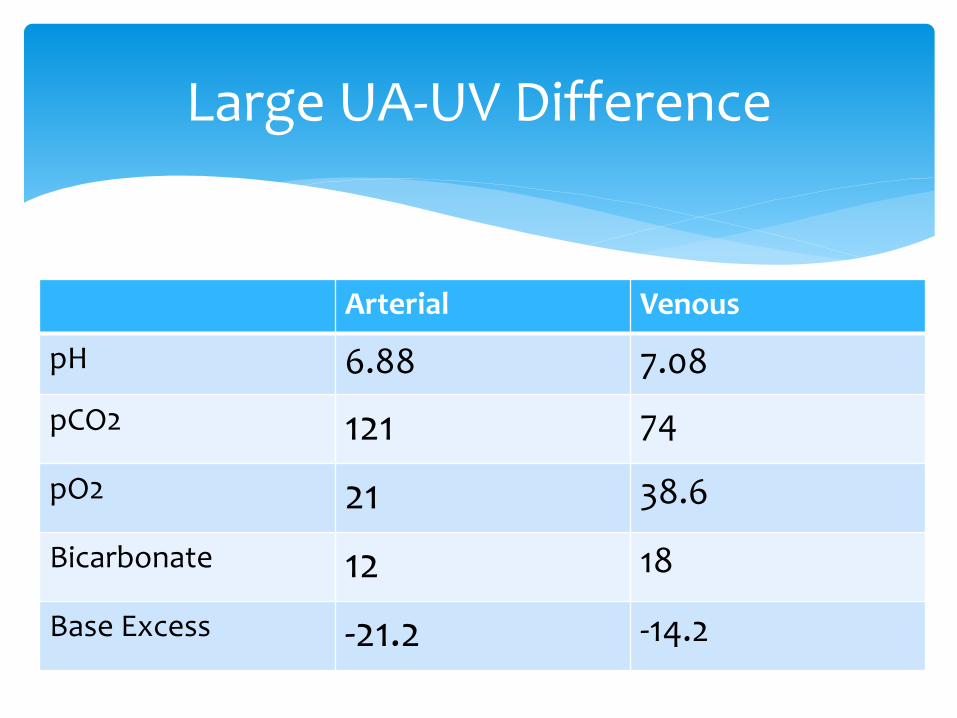
Arterial Venous
pH 6.88 7.08
pCO2 121 74
pO2 21 38.6
Bicarbonate 12 18
Base Excess -21.2 -14.2
Large UA-UV Difference
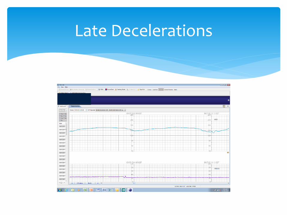
Late Decelerations
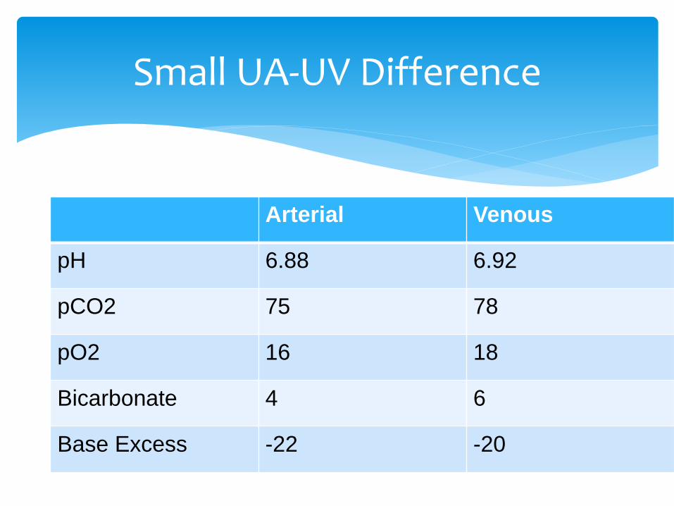
Small UA-UV Difference
Arterial Venous
pH 6.88 6.92
pCO2 75 78
pO2 16 18
Bicarbonate 4 6
Base Excess -22 -20

Double Clamped Cord Segment
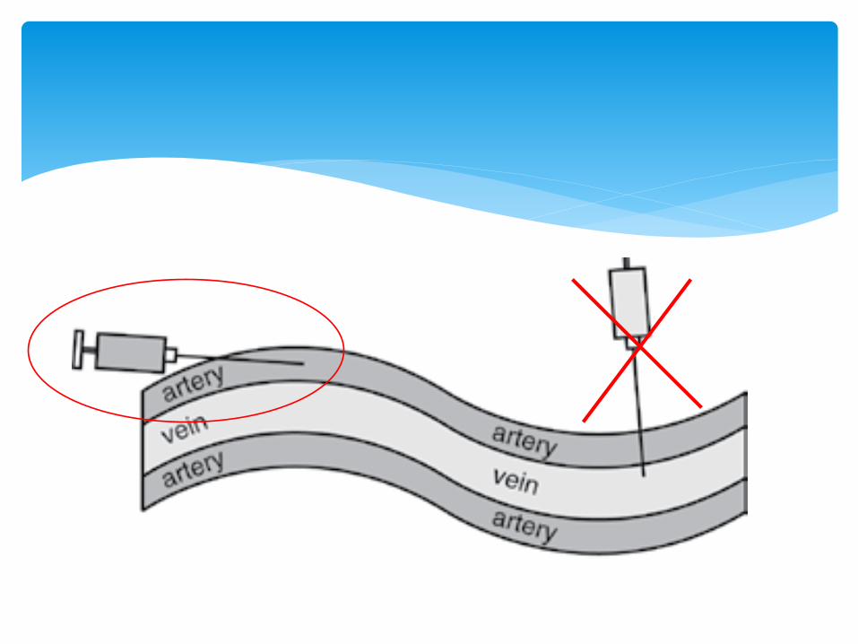
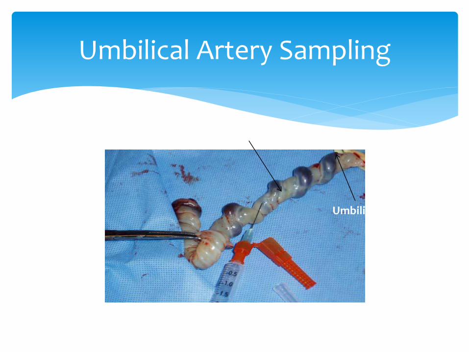
Umbilical Artery Sampling
Umbilical Artery
Umbilical Vein
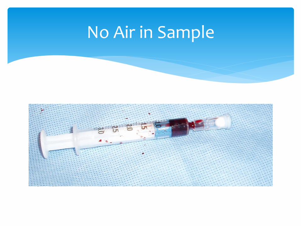
No Air in Sample

Clamped cord good for 60 minutes at room temperature
Stable in a plastic syringe for 30 minutes prior to sampling
Transport at room temperature
Wait Time & Transport


![hernia of the umbilical cord [وضع التوافق] of the umbilical cord.pdf · Umbilical cord hernia…cont Conclusion: ¾Hernia of the umbilical cord is a rare entityy, of the](https://img.pdfslide.net/doc/110x75/5ea7ce695a148409cd011fd0/hernia-of-the-umbilical-cord-of-the-umbilical-cordpdf.jpg)