Embed Size (px)
Citation preview
FIB-SEM: A new technique for investigating pollen walls
A. House and K. Balkwill School of Animal, Plant & Environmental Sciences, University of the Witwatersrand, Johannesburg, Private Bag 3, WITS,
2050, South Africa
The typically distinctive and elaborate external structures and sculpturing of pollen grains have been widely used in the taxonomy and systematics of the flowering plants. The family Acanthaceae is a striking example, where the widely diverse external pollen grain morphology has proved useful in determining relationships between taxa. Detailed information for external pollen wall morphology has traditionally been accumulated using light microscopy (LM), transmission electron microscopy (TEM) and scanning electron microscopy (SEM) techniques. Internal pollen wall features from which the external sculpturing is derived, also exhibit complex and diverse characteristics, but lack investigation due to the difficult and cumbersome methods required to examine the underlying exine. Advancing technology in the field of microscopy has made it possible to view the internal structure of pollen grain walls in far greater detail and in 3D. A new technique involving precise cross sectioning or slicing of pollen grains at a selected position for examining pollen wall ultra-structure, using a focused ion beam-scanning electron microscope (FIB-SEM), has been explored and is yielding data which will prove to be of taxonomic importance. The FIB-SEM affords high resolution, 3D views for investigating internal pollen wall structure.
Keywords: Acanthaceae; exine; focused ion beam-scanning electron microscope; FIB-SEM; palynology; pollen wall ultra-structure
1. Introduction
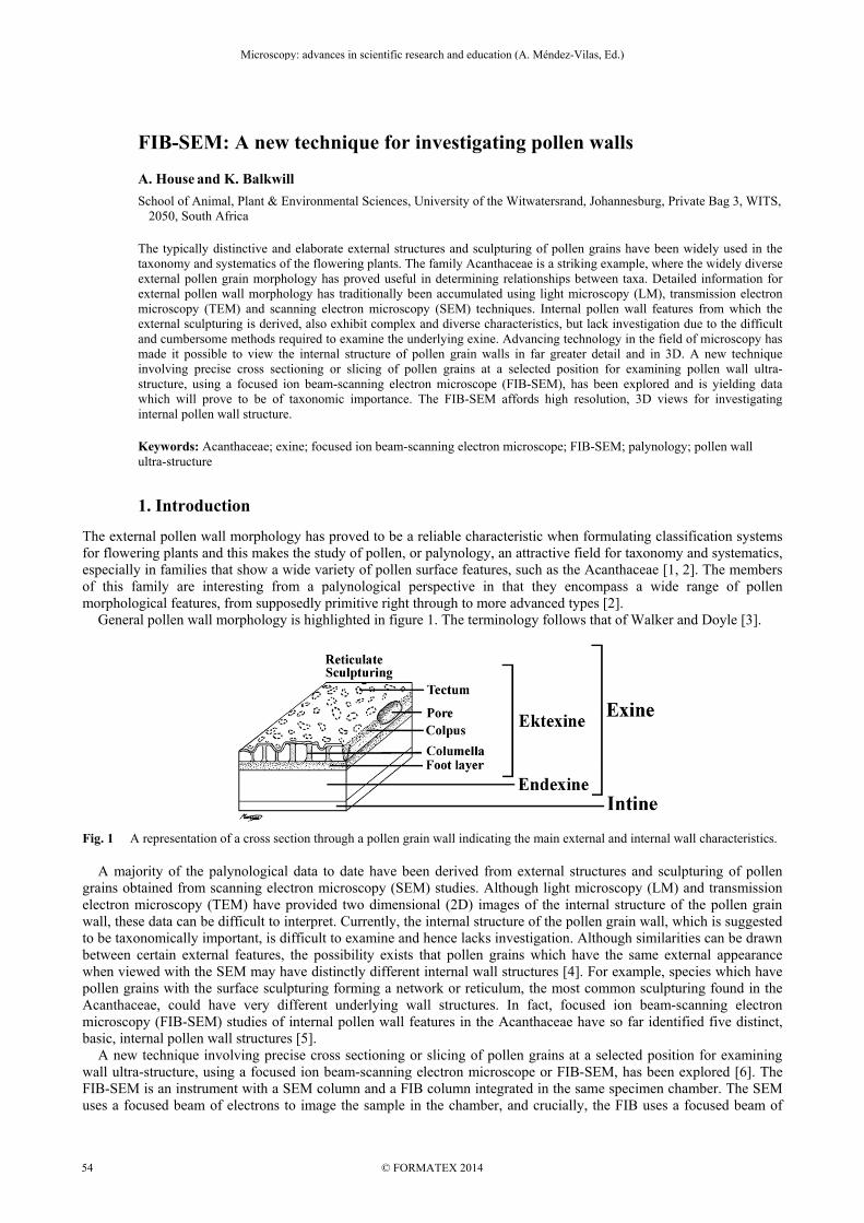
The external pollen wall morphology has proved to be a reliable characteristic when formulating classification systems for flowering plants and this makes the study of pollen, or palynology, an attractive field for taxonomy and systematics, especially in families that show a wide variety of pollen surface features, such as the Acanthaceae [1, 2]. The members of this family are interesting from a palynological perspective in that they encompass a wide range of pollen morphological features, from supposedly primitive right through to more advanced types [2]. General pollen wall morphology is highlighted in figure 1. The terminology follows that of Walker and Doyle [3].
Fig. 1 A representation of a cross section through a pollen grain wall indicating the main external and internal wall characteristics. A majority of the palynological data to date have been derived from external structures and sculpturing of pollen grains obtained from scanning electron microscopy (SEM) studies. Although light microscopy (LM) and transmission electron microscopy (TEM) have provided two dimensional (2D) images of the internal structure of the pollen grain wall, these data can be difficult to interpret. Currently, the internal structure of the pollen grain wall, which is suggested to be taxonomically important, is difficult to examine and hence lacks investigation. Although similarities can be drawn between certain external features, the possibility exists that pollen grains which have the same external appearance when viewed with the SEM may have distinctly different internal wall structures [4]. For example, species which have pollen grains with the surface sculpturing forming a network or reticulum, the most common sculpturing found in the Acanthaceae, could have very different underlying wall structures. In fact, focused ion beam-scanning electron microscopy (FIB-SEM) studies of internal pollen wall features in the Acanthaceae have so far identified five distinct, basic, internal pollen wall structures [5]. A new technique involving precise cross sectioning or slicing of pollen grains at a selected position for examining wall ultra-structure, using a focused ion beam-scanning electron microscope or FIB-SEM, has been explored [6]. The FIB-SEM is an instrument with a SEM column and a FIB column integrated in the same specimen chamber. The SEM uses a focused beam of electrons to image the sample in the chamber, and crucially, the FIB uses a focused beam of
Microscopy: advances in scientific research and education (A. Méndez-Vilas, Ed.)
© FORMATEX 2014
__________________________________________________________________
54
gallium ions to cut or mill a cross section while the sample remains in the chamber. Specimen material is milled away at a selected site to a chosen depth, with each pass of the beam, allowing for precise cross sectioning of the specimen. Although typically used for semiconductor research, the ultra-structure of fossilized pollen walls has previously been investigated using FIB techniques with promising results [7]. FIB techniques have also been used to characterize fragile and delicate biological specimens by cutting a window through the surface to observe subsurface layers [8], or by cutting an ultra-thin layer or lamella out of the specimen to view with a TEM [9], or by FIB tomography, which involves obtaining serial sections through a resin embedded specimen [10]. In these cases, the specimens are fixed and prepared in the same way as for TEM examination. The outer layer of the pollen wall or exine is composed of an extremely strong and resistant substance known as sporopollenin, which due to its nature, requires complex sample preparation to obtain results from TEM. With FIB sectioning, however, a single pollen grain may be successfully sectioned without using the extensive sample preparation required for TEM work. Sectioning pollen grains for TEM examination is a rather haphazard process where sectioning and viewing of samples are carried out on separate machines. Whereas, with the FIB technique, the exact position of the region to be sectioned can be selected. In addition serial sections of an entire pollen grain may be obtained where the grain is viewed and imaged with the SEM portion of the microscope after each cut, to prevent further damage to the specimen. This allows for high resolution 3D investigation of the pollen grain wall and as the sectioning is achieved inside the microscope chamber, the results are instantaneous.
2. Methodology
2.1 Test species
Pollen from a test species Isoglossa woodii C.B.Clarke was examined with LM, SEM and FIB-SEM and compared to TEM cross sections of pollen grains of this species, obtained from a previous study conducted at the University of the Witwatersrand.
2.2 Collection and preparation of pollen grains
Fresh, whole flowers with ripe anthers were collected and placed in small herbarium paper envelopes, and stored in silica gel granules. Individual pollen grains were collected directly from the anthers under a dissecting microscope and transferred to a glass slide or a carbon coated disc using fine insect pins.
2.3 Light microscopy
A small clump of Fuchsin jelly was placed on a glass slide and pollen grains were transferred onto the jelly. This was melted and a cover slip placed on top of the liquid and then viewed and imaged using a Zeiss Axio Imager.M2 light microscope.
2.4 FIB-SEM microscopy
A carbon coated disc with pollen grains was attached to a SEM stub and allowed to dry for 24 h. Samples were coated with a gold-palladium alloy at 20 mA for 1 min in order to lend stability and minimize charging. Samples were viewed with a Zeiss Auriga microscope with a dual-crossbeam FIB workstation and Gemini FE SEM column at the Council for Scientific and Industrial Research (CSIR), Brummeria, Gauteng, South Africa. Selected pollen grains were sectioned once at a predetermined site. A detailed method is explained in House and Balkwill [6].
3. Comparison of microscopy data
Light microscopy is very useful when identifying external and some internal features in the pollen grain wall. Extensive light microscope studies of pollen morphology in the Acanthaceae have been conducted [11, 12] in order to formulate a more comprehensive classification system for this diverse family. These studies have more recently prompted researchers to examine the external features of pollen grains in greater detail and in 3D with the aid of the SEM for the family as a whole [1] and for individual genera [e.g. 13, 14, 16]. External pollen morphological features have been used as characteristics for defining taxa and relationships between taxa. These studies not only highlighted the usefulness of pollen morphology in formulating classification systems, but also the shortfall of examining external features alone because the internal pollen wall characteristics have not, to date, been the focus of investigations. Transmission electron microscopy investigations have been conducted on a few individual genera [e.g. 13, 14, 15]. For the purposes of comparison in this study, pollen grains of Isoglossa woodii were examined.
Microscopy: advances in scientific research and education (A. Méndez-Vilas, Ed.)
© FORMATEX 2014
__________________________________________________________________
55
a) b)
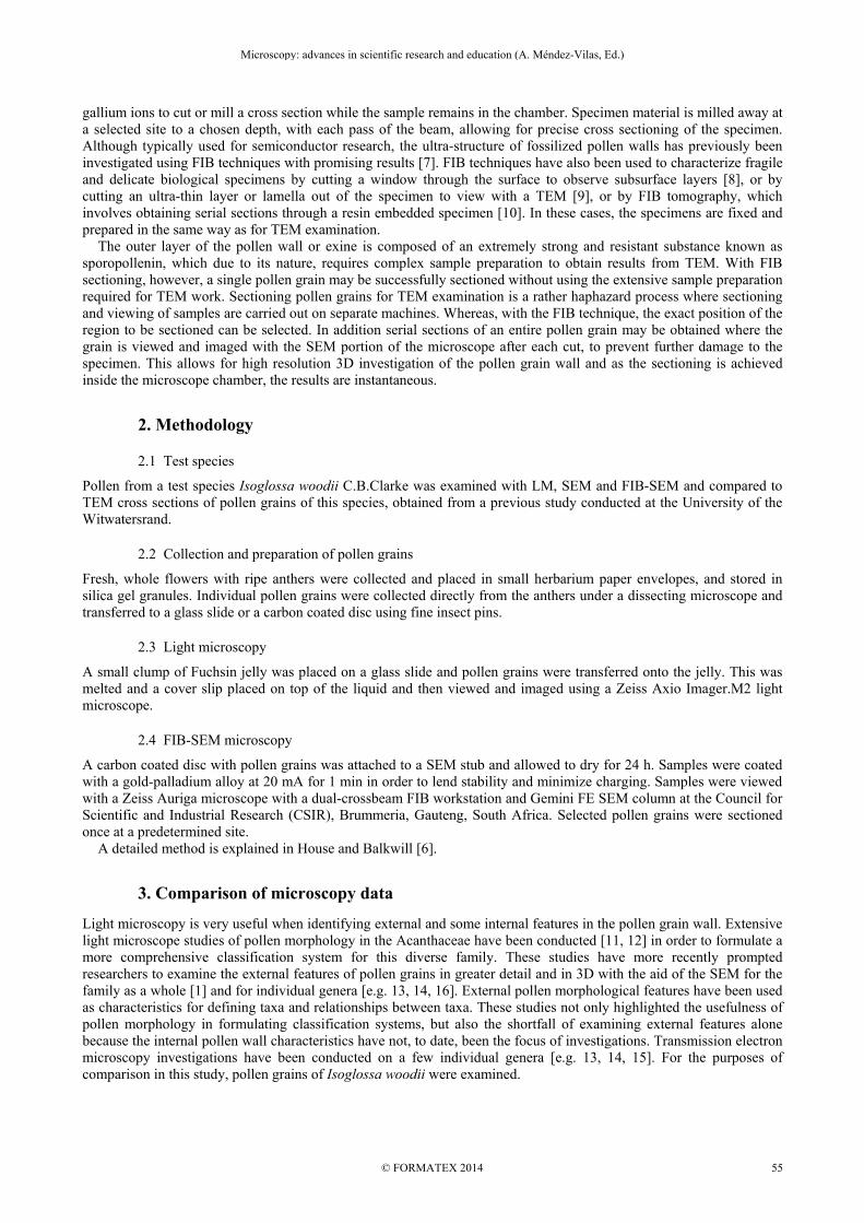
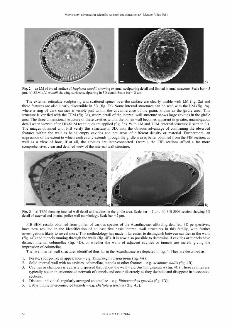
Fig. 2 a) LM of broad surface of Isoglossa woodii, showing external sculpturing detail and limited internal structure. Scale bar = 5 µm. b) SEM of I. woodii showing surface sculpturing in 3D detail. Scale bar = 2 µm. The external reticulate sculpturing and scattered spines over the surface are clearly visible with LM (fig. 2a) and these features are also clearly discernible in 3D (fig. 2b). Some internal structures can be seen with the LM (fig. 2a), where a ring of dark cavities is visible just within the circumference of the grain, known as the girdle area. This structure is verified with the TEM (fig. 3a), where detail of the internal wall structure shows large cavities in the girdle area. The three dimensional structure of these cavities within the pollen wall becomes apparent in greater, unambiguous detail when viewed after FIB-SEM techniques are applied (fig. 3b). With LM and TEM, internal structure is seen in 2D. The images obtained with FIB verify this structure in 3D, with the obvious advantage of confirming the observed features within the wall as being empty cavities and not areas of different density or material. Furthermore, an impression of the extent to which each cavity extends through the girdle area is better obtained from the FIB section, as well as a view of how, if at all, the cavities are inter-connected. Overall, the FIB sections afford a far more comprehensive, clear and detailed view of the internal wall structure.
a) b)
Fig. 3 a) TEM showing internal wall detail and cavities in the girdle area. Scale bar = 2 µm. b) FIB-SEM section showing 3D detail of external and internal pollen wall morphology. Scale bar = 2 µm. FIB-SEM results obtained from pollen of various species of the Acanthaceae, affording detailed, 3D perspectives, have now resulted in the identification of at least five basic internal wall structures in this family, with further investigations likely to reveal more. This methodology has made it far easier to distinguish between cavities in the walls (fig. 4C) and tunnels running through the walls (fig. 4E). It is now also possible to determine if cavities or tunnels have distinct internal columellae (fig. 4D), or whether the walls of adjacent cavities or tunnels are merely giving the impression of columellae. The five internal wall structures identified thus far in the Acanthaceae are depicted in fig. 4. They are described as:
1. Porate, sponge-like in appearance – e.g. Thunbergia atriplicifolia (fig. 4A). 2. Solid internal wall with no cavities, columellae, tunnels or other features – e.g. Acanthus mollis (fig. 4B). 3. Cavities or chambers irregularly dispersed throughout the wall – e.g. Justicia petiolaris (fig. 4C). These cavities are
typically not an interconnected network of tunnels and occur discretely as they dwindle and disappear in successive sections.
4. Distinct, individual, regularly arranged columellae – e.g. Rhinacanthus gracilis (fig. 4D). 5. Labyrinthine interconnected tunnels – e.g. Dicliptera leistneri (fig. 4E).
Microscopy: advances in scientific research and education (A. Méndez-Vilas, Ed.)
© FORMATEX 2014
__________________________________________________________________
56
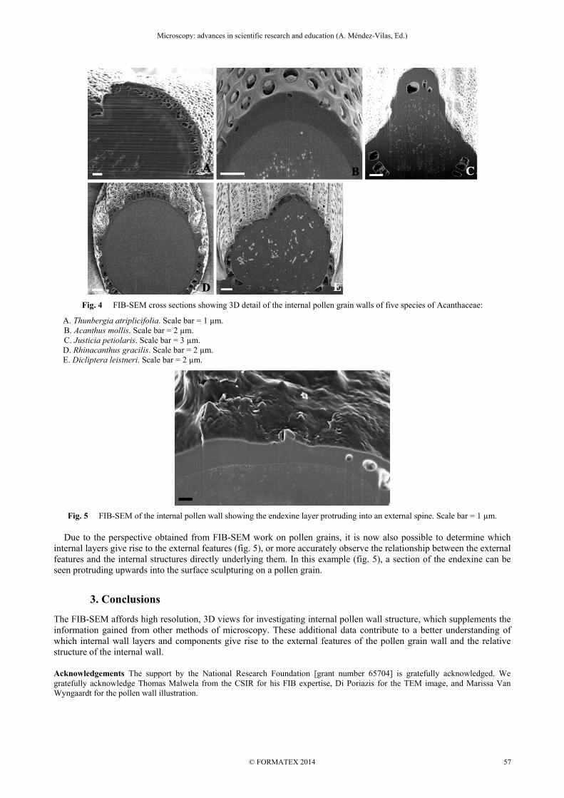
Fig. 4 FIB-SEM cross sections showing 3D detail of the internal pollen grain walls of five species of Acanthaceae:
A. Thunbergia atriplicifolia. Scale bar = 1 µm. B. Acanthus mollis. Scale bar = 2 µm.
C. Justicia petiolaris. Scale bar = 3 µm. D. Rhinacanthus gracilis. Scale bar = 2 µm. E. Dicliptera leistneri. Scale bar = 2 µm.
Fig. 5 FIB-SEM of the internal pollen wall showing the endexine layer protruding into an external spine. Scale bar = 1 µm. Due to the perspective obtained from FIB-SEM work on pollen grains, it is now also possible to determine which internal layers give rise to the external features (fig. 5), or more accurately observe the relationship between the external features and the internal structures directly underlying them. In this example (fig. 5), a section of the endexine can be seen protruding upwards into the surface sculpturing on a pollen grain.
3. Conclusions
The FIB-SEM affords high resolution, 3D views for investigating internal pollen wall structure, which supplements the information gained from other methods of microscopy. These additional data contribute to a better understanding of which internal wall layers and components give rise to the external features of the pollen grain wall and the relative structure of the internal wall.
Acknowledgements The support by the National Research Foundation [grant number 65704] is gratefully acknowledged. We gratefully acknowledge Thomas Malwela from the CSIR for his FIB expertise, Di Poriazis for the TEM image, and Marissa Van Wyngaardt for the pollen wall illustration.
Microscopy: advances in scientific research and education (A. Méndez-Vilas, Ed.)
© FORMATEX 2014
__________________________________________________________________
57

References [1] Scotland RW, Vollesen K. Classification of Acanthaceae. Kew Bulletin. 2000; 55:513-589. [2] Doyle JA. Evolutionary significance of granular exine structure in the light of phylogenetic analyses. microscopy. Review of
Palaeobotany. 2009; 156:98-210. [3] Walker JW, Doyle JA. The Bases of angiosperm phylogeny: Palynology. Annals of the Missouri Botanical Garden. 1975;
62:664-723. [4] Moore PD, Webb JA, Collinson ME. Pollen Analysis. 2nd ed. Oxford: Blackwell Science Ltd; 1991. p. 76-77. [5] House A, Balkwill K. Labyrinths, Columns and Cavities: New internal features of pollen grain walls in the Acanthaceae. In
preparation. 2014. [6] House A, Balkwill K. FIB-SEM: An additional technique for investigating internal structure of pollen walls. Microscopy and
Microanalysis. 2013; 19:1535-1541. [7] Villanueva-Amadoz U, Benedetti A, Mendez J, Sender LM, Diez JB. Focused ion beam nano-sectioning and imaging: a new
method in characterisation of palaeopalynological remains. Grana. 2012; 51:1-9. [8] Ishitani T, Hirose H, Tsuboi H. Focused-ion-beam digging of biological specimens. analyses. Journal of Electron Microscopy.
1995; 44:110-114. [9] Grandfield K, Engqvist H. Focused ion beam in the study of biomaterials and biological matter. Advances in Material Science
and Engineering. 2012; 2012:1-6. [10] Kizilyaprak C, Loussert C, Daraspe J, Humbel BM. Focussed ion beam scanning electron microscopy in biology. Microscopy
and Microanalysis. 2013; 19:874-875. [11] Lindau G. Acanthaceae. In: Engler A, Prantl K, editors. Die natuerlichen Pflanzenfamilien. 3b. Leipzig: Wilhelm Engelmann;
1895. p. 274-354. [12] Raj B. Pollen morphological studies in the Acanthaceae. Grana Palynologica. 1961; 3:3-108. [13] Furness CA. Pollen morphology of Crossandra Salisbury and Crossandrella C.B. Clarke (Acanthaceae: Acantheae). Grana.
1990; 29:161-176. [14] Furness CA. Pollen morphology of Sclerochiton (Acanthaceae: Acantheae). Kew Bulletin. 1991; 46:51-59. [15] Furness CA. Pollen morphology of Acanthopsis Harvey, Acanthus L. and Blepharis Jussieu (Acanthaceae: Acantheae). Review
of Palaeobotany and Palynology. 1996; 92:253-268. [16] Graham VAW. Delimitation and infra-generic classification of Justicia (Acanthaceae). Kew Bulletin. 1988; 43:551-624.
Microscopy: advances in scientific research and education (A. Méndez-Vilas, Ed.)
© FORMATEX 2014
__________________________________________________________________
58