Embed Size (px)
Citation preview
Thorax (1956), 11, 287.
MASSIVE PULMONARY FIBROSIS FROM THE INHALATIONOF TALC
BY
A. C. HUNTFrom the Department of Forensic Medicine, The London Hospital Medical College
(RECEIVED FOR PUBLICATION FEBRUARY 4, 1956)
It is generally accepted that the inhalation ofcommercial talc powders can occasionally pro-duce pulmonary disease. This is usually fibrosisof a nodular type, and only a few examples ofmassive fibrosis have been reported, most of themwithout pathological description. This is the re-port of one such case showing certain unusualfeatures.
CASE REPORTINDUSTRIAL HISTORY.-The patient was 57 years
old at his death. He joined the Army at the age of16, at the start of the first world war. During thiswar he was gassed, although not severely. When heleft the Army he became a storekeeper in a foodstore. In 1927, at the age of 29, he first entered theaccumulator industry. For the first six years he wasemployed in lead burning, and was not exposed todust. After this he joined another firm as a platecaster. The following 10 years, i.e., between the agesof 35 and 45, was the only period during which hewas exposed to dust. Lead accumulator plates werecast in a hand-operated mould consisting of twofaces hinged at the bottom. There were two men cast-ing in one small room, each with his own mould andwith a communal gas-fired lead pot. The mould wasat about waist height. It was opened, and then eachsurface was roughly dusted with talc from a looselywoven calico bag or an old sock. The bag wasbanged hard against the mould and a cloud of dustwould arise. Next, the mould was closed and thelead poured in. It was cooled with water and openedand the plate removed. The sequence was thenrepeated. The patient was a good workman and castup to 110 plates an hour, being paid piece rates. Thetalc bag contained about three-quarters of a poundof talc, and about three bags were used in two days.There was no dust extraction in the room, and every-thing was covered with talc dust. The source of the talcused cannot be traced, except for the last two yearsof this period, when it was obtained from Man-churia, Canada, or Egypt. The plates are now castautomatically in this factory and the mould faces aredressed with a cork compound; there is no dust riskwhatever.
After 10 years of this work the patient was engaged,for 12 years before his death, in battery assembly and
charging and occasionally in plate casting. Theamount of talc dust to which he was exposed forthese years was negligible.CLINICAL HISTORY.-Despite the gross radiological
changes his physical state was remarkably good. Hefirst came under observation as an out-patient of theMiller General Hospital, Greenwich, nine years beforehis death, when he was examined because his sondeveloped pulmonary tuberculosis. During the last10 years of his life he had several attacks of " bron-chitis," with cough and sputum and pyrexia. Thesewere treated at home with early recovery. Apartfrom these attacks he had a slight morning cough,productive of a jelly-like, greyish sputum. He wasnot breathless on normal exertion. Nine monthsbefore death all the metacarpo-phalangeal joints ofboth hands became swollen and painful, with slightulnar deviation of the fingers. The left ankle wastransiently swollen. There was no spindling of theinterphalangeal joints, and at this time there was earlyclubbing of the fingers. His hands were radiographedwhen he was first seen to exclude the bony changesof sarcoidosis, and again, together with other joints,when the knuckles became swollen. None of theseradiographs showed any bony abnormality. Clini-cally the joint changes were considered to be mani-festations of pulmonary osteoarthropathy. Unfor-tunately, no joints were examined histologically afterdeath, and it is impossible to exclude rheumatoidarthritis. It would have been of interest to makethis exclusion, in view of Caplan's (1953) observationof the modification of coal-miners' pneumoconiosis inmen with rheumatoid arthritis. The only physicalsigns recorded in the chest during the period ofobservation were occasional rales, especially over theright lung. Sputa were examined for tubercle bacilliby microscopy and culture repeatedly during thesenine years. Between four and six groups of sputawere examined yearly, and no tubercle bacilli wereever found. The sedimentation rate was estimatedat regular intervals, and was on most occasions moder-ately raised but sometimes normal. The Mantouxreaction was positive at a dilution of 1:10,000. Anelectrocardiograph taken three months before deathshowed no definite right ventricular preponderance.An axillary lymph node was removed for biopsy sixyears before death, to investigate the possibility ofsarcoidosis. There were a few doubly refractile
copyright. on F
ebruary 11, 2020 by guest. Protected by
http://thorax.bmj.com
/T
horax: first published as 10.1136/thx.11.4.287 on 1 Decem
ber 1956. Dow
nloaded from
A. C. HUNT
I
FIG. 1.-Radiograph of chest nine years before death.
particles in the lymphatic sinuses resembling the talcparticles found in the lung at necropsy, but no otherabnormality. During the last year of his life he hadoccasional fainting attacks on exertion. On the day
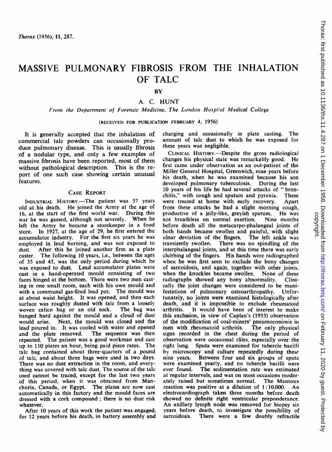
YFIG. 2.-Radiograph of chest seven years before death.
of his death he had dug potatoes in the morning andeaten a large lunch. Shortly after the meal he wasfound dead in a lavatory, having defaecated. He wasseen to be quite well immediately before this.
:
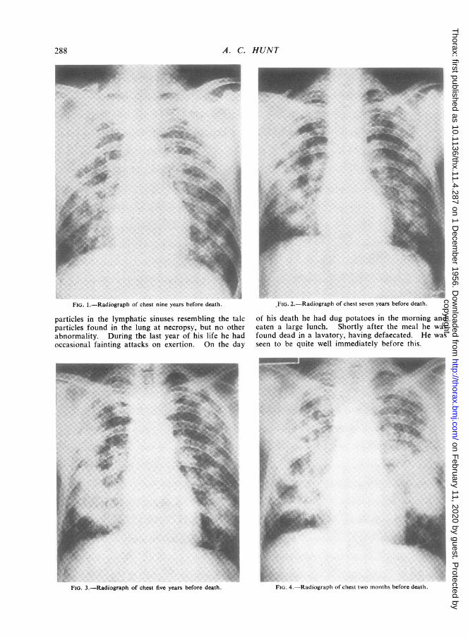
FIG. 3.-Radiograph of chest five years before death.
288
AOI
00.
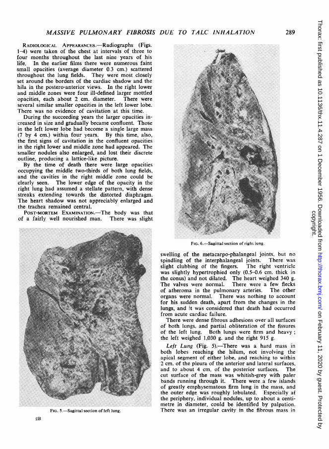
FIG. 4.-Radiograph of chest two months before death.
A
4
Ir
copyright. on F
ebruary 11, 2020 by guest. Protected by
http://thorax.bmj.com
/T
horax: first published as 10.1136/thx.11.4.287 on 1 Decem
ber 1956. Dow
nloaded from
MASSIVE PULMONARY FIBROSIS DUE TO TALC INHALATION
RADIOLOGICAL APPEARANCES.-Radiographs (Figs.1-4) were taken of the chest at intervals of three tofour months throughout the last nine years of hislife. In the earlier films there were numerous faintsmall opacities (average diameter 0.3 cm.) scatteredthroughout the lung fields. They were most closelyset around the borders of the cardiac shadow and thehila in the postero-anterior views. In the right lowerand middle zones were four ill-defined larger mottledopacities, each about 2 cm. diameter. There wereseveral similar smaller opacities in the left lower lobe.There was no evidence of cavitation at this time.
During the succeeding years the larger opacities in-creased in size and gradually became confluent. Thosein the left lower lobe had become a single large mass(7 by 4 cm.) within four years. By this time, also,the first signs of cavitation in the confluent opacitiesin the right lower and middle zone had appeared. Thesmaller nodules also enlarged, and lost their discreteoutline, producing a lattice-like picture.By the time of death there were large opacities
occupying the middle two-thirds of both lung fields,and the cavities in the right middle zone could beclearly seen. The lower edge of the opacity in theright lung had assumed a stellate pattern, with densestreaks extending towards the distorted diaphragm.The heart shadow was not appreciably enlarged andthe trachea remained central.POST-MORTEM ExAMINATION.-The body was that
of a fairly well nourished man. There was slight
FIG. 5.-Sagittal section of left lung.
FIG. 6.-Sagittal section of right lung.
swelling of the metacarpo-phalangeal joints, but nospindling of the interphalangeal joints. There wasslight clubbing of the fingers. The right ventriclewas slightly hypertrophied only (0.5-0.6 cm. thick inthe conus) and not dilated. The heart weighed 340 g.The valves were normal. There were a few flecksof atheroma in the pulmonary arteries. The otherorgans were normal. There was nothing to accountfor his sudden death, apart from the changes in thelungs, and .t was considered that death had occurredfrom acute cardiac failure.There were dense fibrous adhesions over all surfaces
of both lungs, and partial obliteration of the fissuresof the left lung. Both lungs were firm and heavy;the left weighed 1,030 g. and the right 915 g.
Left Lung (Fig. 5).-There was a hard mass inboth lobes reaching the hilum, not involving theapical segment of either lobe, and reaching to within2 cm. of the pleura of the anterior and lateral surfaces,and to about 4 cm. of the posterior surfaces. Thecut surface of the mass was whitish-grey with palerbands running through it. There were a few islandsof greatly emphysematous firm lung in the mass, andthe outer edge was roughly lobulated. Especially atthe periphery, individual nodules, up to about a centi-metre in diameter, could be identified by palpation.There was an irregular cavity in the fibrous mass in
2B
289
copyright. on F
ebruary 11, 2020 by guest. Protected by
http://thorax.bmj.com
/T
horax: first published as 10.1136/thx.11.4.287 on 1 Decem
ber 1956. Dow
nloaded from

A. C. HUNT
visible with the naked eye in the fibrosis in the upperlobe, and there were similar nodules in the remaininglung, as in the left lung, and the centres of many ofthese were also friable.HISTOLOGY.-Running throughout the confluent
masses were thick cellular seams of macrophages andwell-formed multinucleate giant cells of a foreignbody type (Fig. 7). The giant cells, and most of themacrophages, were packed with doubly refractileparticles when viewed by polarized light (Fig. 8).These particles were predominantly needle-shaped and5-10 a1 long. The seams were broadest at the peri-phery of the masses, where they were up to 0.5 cm. indiameter and varying from a few millimetres to acentimetre or more apart. The rest of each masswas fibrous tissue containing large areas of necrosis.Some of these areas were composed of amorphousfinely granular material stained yellow with vanGieson's stain and pink with eosin. Sometimessurviving macrophages were present at the edges, anda few poorly fuchsinophil strands were still visible.Doubly refractile particles were present in largenumbers, often greatest at the periphery (Fig. 9). Inplaces the amorphous material was in aggregates thesize and shape of necrotic cells, and these areasappeared to have been formed by necrosis in the
FIG. 7.-Giant-cell granulomatous seam. (Haematoxylin and eosin,x 130.)
the upper lobe (8 by 5 by 3 cm.), the long axis ofwhich ran antero-posteriorly. The wall of the cavitywas trabeculated and the lining rough and slightlygritty to the touch. The centre of the fibrosis in thelower lobe was breaking down in an area of about4 cm. diameter, where it was of similar colour to theremaining fibrosis, but friable. The remaining lungwas emphysematous, and scattered in it were ill-defined firm nodules varying from a pin's head to1.5 cm. in diameter. The centres of most of theselarger nodules were also soft and friable. There wasslight diffuse cylindrical bronchiectasis throughoutthe lung.Right Lung (Fig. 6).-There was a similar mass
extending from the hilum to within 3 cm. of thepleura of the posterior surface but reaching thepleura of the lateral surface of the lower lobe. Themass was more obviously nodular than on the left.The fibrosis appeared denser around the largerbronchi, especially in the lower lobe. There was acavity (9 by 3 cm.) in the mass in the lower lobe,mainly in the anterior basal segment. It consisted ofa honeycomb of interconnecting large spaces with asmooth, shiny black lining. There was a smallercavity in the right middle lobe which was similarlylined, except for a few areas in which the lining wasrough and granular. Small patches of necrosis were
.... _ ^ ' . . .:
Fir.. 8.-Giant cells laden with doubly refractile particles. Photo-graph obtained by exposure with and without polarization oflight. (Haematoxylin and eosin, x 650.)
290
E.AL-
AA... ........
copyright. on F
ebruary 11, 2020 by guest. Protected by
http://thorax.bmj.com
/T
horax: first published as 10.1136/thx.11.4.287 on 1 Decem
ber 1956. Dow
nloaded from
MASSIVE PULMONARY FIBROSIS DUE TO TALC INHALATION
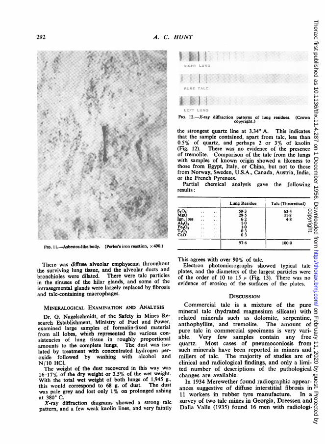
Perles's reaction, and for calcium by von Kossa'smethod. Throughout the fibrous parts of the lungswere a few macrophages containing free iron, andthere were small particles in the non-cavitated necrosis.The cavities in the right lung were similar to thosein the left upper lobe, except that there was a thinlayer of fuchsinophil collagen superficial to thenecrotic and cellular elements and forming the smoothlining. The earliest lesions in the remaining lung<
A were small interstitial cellular foci containing fine col-lagen strands, mostly related to small vessels (Fig. 10).There were also patches of diffuse interstitial fibrosis,often associated with smaller nodules. The larger
0`ts*sr 1 . N 2 ~< ffi w b ;A discrete nodules were similar to the confluent masses.The fibrous tissue forming them was not whorled, andthe centres were frequently necrotic. In these discrete
e nodules the foreign body giant-cell reaction was notso marked a feature.Curious bodies (McLaughlin, Rogers, and Dunham.a.: . d,* ; t.............1949),similar but not identical to asbestos bodies,
have been described in most cases of talc pneumo-1btW P!, ILSconiosis. They consist of a single fibre, with terminalrosettes, but without intermediate beading. Theywere present in this case, although in small numbers
,iMjE(Fig. 11). No acid-fast bacilli were seen in any of=11:> the sections.
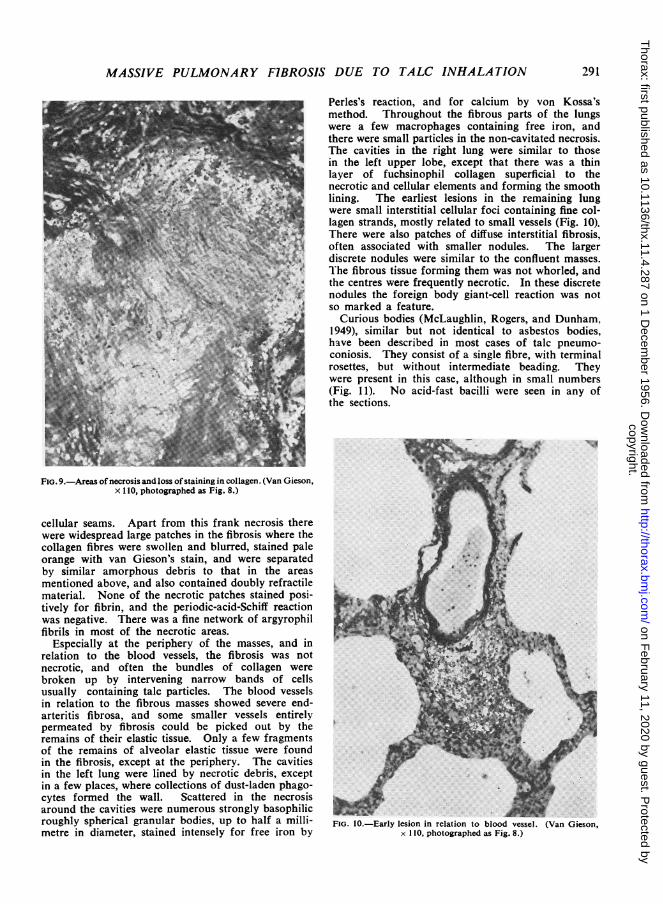
FIG. 9.-Areas of necrosis and loss ofstaining in collagen. (Van Gieson,x 10, photographed as Fig. 8.)
cellular seams. Apart from this frank necrosis therewere widespread large patches in the fibrosis where thecollagen fibres were swollen and blurred, stained paleorange with van Gieson's stain, and were separatedby similar amorphous debris to that in the areasmentioned above, and also contained doubly refractile *material. None of the necrotic patches stained posi-tively for fibrin, and the periodic-acid-Schiff reactioniwas negative. There was a fine network of argyrophil Afibrils in most of the necrotic areas.
Especially at the periphery of the masses, and in A4.relation to the blood vessels, the fibrosis was notnecrotic, and often the bundles of collagen werebroken up by intervening narrow bands of cellsusually containing talc particles. The blood vesselsin relation to the fibrous masses showed severe end-arteritis fibrosa, and some smaller vessels entirelypermeated by fibrosis could be picked out by theremains of their elastic tissue. Only a few fragmentsof the remains of alveolar elastic tissue were foundin the fibrosis, except at the periphery. The cavitiesin the left lung were lined by necrotic debris, exceptin a few places, where collections of dust-laden phago-cytes formed the wall. Scattered in the necrosis _ ,^around the cavities were numerous strongly basophilicroughly spherical granular bodies, up to half a milli- FIG. 10.-Early lesion in relation to blood vessel. (Van Gieson,metre in diameter, stained intensely for free iron by x 110, photographed as Fig. 8.)
291
copyright. on F
ebruary 11, 2020 by guest. Protected by
http://thorax.bmj.com
/T
horax: first published as 10.1136/thx.11.4.287 on 1 Decem
ber 1956. Dow
nloaded from
A. C. HUNT
3,.R i X
:.
A. B
.4t..
.:.:
....
..:a .#
*4w;.* a
a4I
A*A
A..v.
4.... -1,
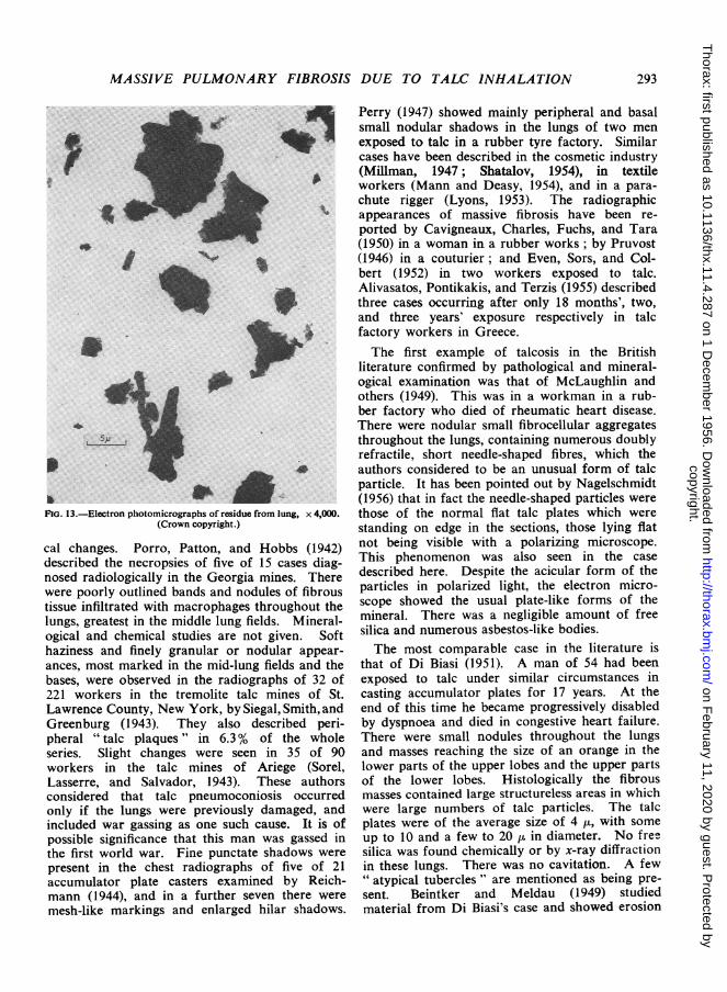
Pio. I .-Asbestos-like body. (Perles's iron reaction, x 490.)
'II
I !
FiG. 12.-X-ray diffraction patterns of lung residues. (Crowncopyright.)
the strongest quartz line at 3.34° A. This indicatesthat the sample contained, apart from talc, less than0.5% of quartz, and perhaps 2 or 3% of kaolin(Fig. 12). There was no evidence of the presenceof tremolite. Comparison of the talc from the lungswith samples of known origin showed a likeness tothose from Egypt, Italy, or China, but not to thosefrom Norway, Sweden, U.S.A., Canada, Austria, India,or the French Pyrenees.*6
Partial chemical analysis gave the followingresults:l~~~~~~~~~
Lung Residue Talc (Theoretical)
S102 59 3 63-4MgO 29-5 31-8Ign. loss . . 6-2 4.8Al20 * 1-0Fe2O3 1-0T102 0 3CaO 0 3
97-6 100-0
There was diffuse alveolar emphysema throughoutthe surviving lung tissue, and the alveolar ducts andbronchioles were dilated. There were talc particlesin the sinuses of the hilar glands, and some of theintrasegmental glands were largely replaced by fibrosisand talc-containing macrophages.
MINERALOGICAL EXAMINATION AND ANALYSIS
Dr. G. Nagelschmidt, of the Safety in Mines Re-search Establishment, Ministry of Fuel and Power,examined large samples of formalin-fixed materialfrom all lobes, which represented the various con-sistencies of lung tissue in roughly proportionalamounts to the complete lungs. The dust was iso-lated by treatment with concentrated hydrogen per-oxide followed by washing with alcohol andN/10 HCl.The weight of the dust recovered in this way was
16-17% of the dry weight or 3.5% of the wet weight.With the total wet weight of both lungs of 1,945 g.,this would correspond to 68 g. of dust. The dustwas pale grey and lost only 1% on prolonged ashingat 3800 C.
X-ray diffraction diagrams showed a strong talcpattern, and a few weak kaolin lines, and very faintly
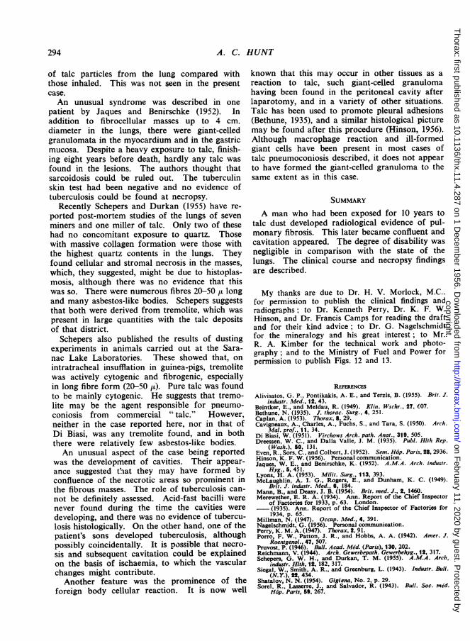
This agrees with over 90% of talc.Electron photomicrographs showed typical talc
plates, and the diameters of the largest particles wereof the order of 10 to 15 ,' (Fig. 13). There was noevidence of erosion of the surfaces of the plates.
DISCUSSIONCommercial talc is a mixture of the pure
mineral talc (hydrated magnesium silicate) withrelated minerals such as dolomite, serpentine,anthophyllite, and tremolite. The amount ofpure talc in commercial specimens is very vari-able. Very few samples contain any freequartz. Most cases of pneumoconiosis fromsuch minerals have been reported in miners andmillers of talc. The majority of studies are ofclinical and radiological findings, and only a limi-ted number of descriptions of the pathologicalchanges are available.
In 1934 Merewether found radiographic appear-ances suggestive of diffuse interstitial fibrosis in11 workers in rubber tyre manufacture. In asurvey of two talc mines in Georgia, Dreessen andDalla Valle (1935) found 16 men with radiologi-
292
...A:.4:
:;.F.A.
f.1 J.v
.9:.: .t a;
.v..*
copyright. on F
ebruary 11, 2020 by guest. Protected by
http://thorax.bmj.com
/T
horax: first published as 10.1136/thx.11.4.287 on 1 Decem
ber 1956. Dow
nloaded from
MASSIVE PULMONARY FIBROSIS DUE TO TALC INHALATION
^..
aI
U.
It'
oil461
FiG. 13.-Electron photomicrographs of residue from lung, x 4,000.(Crown copyright.)
cal changes. Porro, Patton, and Hobbs (1942)described the necropsies of five of 15 cases diag-nosed radiologically in the Georgia mines. Therewere poorly outlined bands and nodules of fibroustissue infiltrated with macrophages throughout thelungs, greatest in the middle lung fields. Mineral-ogical and chemical studies are not given. Softhaziness and finely granular or nodular appear-
ances, most marked in the mid-lung fields and thebases, were observed in the radiographs of 32 of221 workers in the tremolite talc mines of St.Lawrence County, New York, by Siegal, Smith, andGreenburg (1943). They also described peri-pheral "talc plaques" in 6.3% of the wholeseries. Slight changes were seen in 35 of 90workers in the talc mines of Ariege (Sorel,Lasserre, and Salvador, 1943). These authorsconsidered that talc pneumoconiosis occurredonly if the lungs were previously damaged, andincluded war gassing as one such cause. It is ofpossible significance that this man was gassed inthe first world war. Fine punctate shadows were
present in the chest radiographs of five of 21accumulator plate casters examined by Reich-mann (1944), and in a further seven there were
mesh-like markings and enlarged hilar shadows.
Perry (1947) showed mainly peripheral and basalsmall nodular shadows in the lungs of two menexposed to talc in a rubber tyre factory. Similarcases have been described in the cosmetic industry(Millman, 1947; Shatalov, 1954), in textileworkers (Mann and Deasy, 1954), and in a para-chute rigger (Lyons, 1953). The radiographicappearances of massive fibrosis have been re-ported by Cavigneaux, Charles, Fuchs, and Tara(1950) in a woman in a rubber works; by Pruvost(1946) in a couturier; and Even, Sors, and Col-bert (1952) in two workers exposed to talc.Alivasatos, Pontikakis, and Terzis (1955) describedthree cases occurring after only 18 months', two,and three years' exposure respectively in talcfactory workers in Greece.The first example of talcosis in the British
literature confirmed by pathological and mineral-ogical examination was that of McLaughlin andothers (1949). This was in a workman in a rub-ber factory who died of rheumatic heart disease.There were nodular small fibrocellular aggregatesthroughout the lungs, containing numerous doublyrefractile, short needle-shaped fibres, which theauthors considered to be an unusual form of talcparticle. It has been pointed out by Nagelschmidt(1956) that in fact the needle-shaped particles werethose of the normal flat talc plates which werestanding on edge in the sections, those lying flatnot being visible with a polarizing microscope.This phenomenon was also seen in the casedescribed here. Despite the acicular form of theparticles in polarized light, the electron micro-scope showed the usual plate-like forms of themineral. There was a negligible amount of freesilica and numerous asbestos-like bodies.The most comparable case in the literature is
that of Di Biasi (1951). A man of 54 had beenexposed to talc under similar circumstances incasting accumulator plates for 17 years. At theend of this time he became progressively disabledby dyspnoea and died in congestive heart failure.There were small nodules throughout the lungsand masses reaching the size of an orange in thelower parts of the upper lobes and the upper partsof the lower lobes. Histologically the fibrousmasses contained large structureless areas in whichwere large numbers of talc particles. The talcplates were of the average size of 4 /i, with someup to 10 and a few to 20 z in diameter. No freesilica was found chemically or by x-ray diffractionin these lungs. There was no cavitation. A few" atypical tubercles " are mentioned as being pre-sent. Beintker and Meldau (1949) studiedmaterial from Di Biasi's case and showed erosion
293
b'0411
copyright. on F
ebruary 11, 2020 by guest. Protected by
http://thorax.bmj.com
/T
horax: first published as 10.1136/thx.11.4.287 on 1 Decem
ber 1956. Dow
nloaded from
A. C. HUNT
of talc particles from the lung compared withthose inhaled. This was not seen in the presentcase.An unusual syndrome was described in one
patient by Jaques and Benirschke (1952). Inaddition to fibrocellular masses up to 4 cm.
diameter in the lungs, there were giant-celledgranulomata in the myocardium and in the gastricmucosa. Despite a heavy exposure to talc, finish-ing eight years before death, hardly any talc was
found in the lesions. The authors thought thatsarcoidosis could be ruled out. The tuberculinskin test had been negative and no evidence oftuberculosis could be found at necropsy.
Recently Schepers and Durkan (1955) have re-
ported post-mortem studies of the lungs of seven
miners and one miller of talc. Only two of thesehad no concomitant exposure to quartz. Thosewith massive collagen formation were those withthe highest quartz contents in the lungs. Theyfound cellular and stromal necrosis in the masses,
which, they suggested, might be due to histoplas-mosis, although there was no evidence that thiswas so. There were numerous fibres 20-50 Mu longand many asbestos-like bodies. Schepers suggeststhat both were derived from tremolite, which was
present in large quantities with the talc depositsof that district.
Schepers also published the results of dustingexperiments in animals carried out at the Sara-nac Lake Laboratories. These showed that, on
intratracheal insufflation in guinea-pigs, tremolitewas actively cytogenic and fibrogenic, especiallyin long fibre form (20-50 ,). Pure talc was foundto be mainly cytogenic. He suggests that tremo-lite may be the agent responsible for pneumo-
coniosis from commercial "talc." However,neither in the case reported here, nor in that ofDi Biasi, was any tremolite found, and in boththere were relatively few asbestos-like bodies.An unusual aspect of the case being reported
was the development of cavities. Their appear-ance suggested that they may have formed byconfluence of the necrotic areas so prominent inthe fibrous masses. The role of tuberculosis can-
not be definitely assessed. Acid-fast bacilli were
never found during the time the cavities were
developing, and there was no evidence of tubercu-losis histologically. On the other hand, one of thepatient's sons developed tuberculosis, althoughpossibly coincidentally. It is possible that necro-
sis and subsequent cavitation could be explainedon the basis of ischaemia, to which the vascularchanges might contribute.
Another feature was the prominence of theforeign body cellular reaction. It is now well
known that this may occur in other tissues as areaction to talc, such giant-celled granulomahaving been found in the peritoneal cavity afterlaparotomy, and in a variety of other situations.Talc has been used to promote pleural adhesions(Bethune, 1935), and a similar histological picturemay be found after this procedure (Hinson, 1956).Although macrophage reaction and ill-formedgiant cells have been present in most cases oftalc pneumoconiosis described, it does not appearto have formed the giant-celled granuloma to thesame extent as in this case.
SUMMARYA man who had been exposed for 10 years to
talc dust developed radiological evidence of pul-monary fibrosis. This later became confluent andcavitation appeared. The degree of disability wasnegligible in comparison with the state of thelungs. The clinical course and necropsy findingsare described.
My thanks are due to Dr. H. V. Morlock, M.C..for permission to publish the clinical findings andradiographs; to Dr. Kenneth Perry, Dr. K. F. W.Hinson, and Dr. Francis Camps for reading the draftand for their kind advice; to Dr. G. Nagelschmidtfor the mineralogy and his great interest; to Mr.R. A. Kimber for the technical work and photo-graphy; and to the Ministry of Fuel and Power forpermission to publish Figs. 12 and 13.
REFERENCESAlivisatos, G. P., Pontikakis, A. E., and Terzis, B. (1955). Brit. J.
industr. Med., 12, 43.Beintker, E., and Meldau, R. (1949). Klin. Wschr., 27, (07.Bethune, N. (1935). J. thorac. Surg., 4, 251.Caplan, A. (1953). Thorax, 8, 29.Cavigneaux, A., Charles, A., Fuchs, S., and Tara, S. (1950). Arch.
Mal. prof., 11, 34.Di Biasi, W. (1951). Virchows Arch. path. Anat., 319, 505.Dreessen, W. C., and Dalla Valle, J. M. (1935). Publ. Hlth Rep.
(Wash.), 50, 131.Even, R., Sors, C., and Colbert, J. (1952). Sem. H6p. Paris, 28, 2936.Hinson, K. F. W. (1956). Personal communication.Jaques, W. E., and Benirschke, K. (1952). A.M.A. Arch. industr.
Hjg., 5, 451.Lyons, H. A. (1953). Milit. Surg., 113, 393.McLaughlin, A. I. G., Rogers, E., and Dunham, K. C. (1949).
Brit. J. industr. Med., 6, 184.Mann, B., and Deasy, J. B. (1954). Brit. med. J., 2, 1460.Merewether, E. R. A. (1934). Ann. Report of the Chief Inspector
of Factories for 1933, p. 63. London.(1935). Ann. Report oF the Chief Inspector of Factories for1934, p. 65.
Millman, N. (1947). Occup. Med., 4, 391.Nagelschmidt, G. (1956). Personal communication.Perry, K. M. A. (1947). Thorax, 2, 91.Porro, F. W., Patton, J. R., and Hobbs, A. A. (1942). Amer. J.
Roentgenol., 47, 507.Pruvost, P. (1946). Bull. Acad. MJd. (Paris), 130, 202.Reichmann, V. (1944). Arch. Gewerbepath. Gewerbehyg., 12, 317.Schepers, G. W. H., and Durkan, T. M. (1955). A.M.A. Arch.
industr. Hlth, 12, 182, 317.Siegal, W., Smith, A. R., and Greenburg, L. (1943). Indusir. Bull.
(N.Y.), 22, 434.Shatalov, N. N. (1954). Gigiena, No. 2, p. 29.Sorel, R., Lasserre, J., and Salvador, R. (1943). Bull. Soc. med.
H6p. Paris, 51, 267.
294
copyright. on F
ebruary 11, 2020 by guest. Protected by
http://thorax.bmj.com
/T
horax: first published as 10.1136/thx.11.4.287 on 1 Decem
ber 1956. Dow
nloaded from