Embed Size (px)
Citation preview
262 Saso A, et al. Arch Dis Child Educ Pract Ed 2020;105:262–269. doi:10.1136/archdischild-2018-316603
Best practice and FiFteen- minute consultations
Fifteen- minute consultation: Management of the infant born to a mother with toxoplasmosis in pregnancy
anja saso,1,2 alasdair Bamford,3,4 Karen Grewal,5 muna noori,6 James Hatcher,7 Felice d'arco,8 edward Guy,9 Hermione lyall10
► Additional material is published online only. To view please visit the journal online (http:// dx. doi. org/ 10. 1136/ archdischild- 2018- 316603).
For numbered affiliations see end of article.
Correspondence toDr Anja Saso, Academic Paediatrics, Imperial College London, London W2 1PY, UK; anja. saso@ nhs. net
Received 26 January 2019Revised 12 September 2019Accepted 6 December 2019Published Online First 18 February 2020
To cite: Saso A, Bamford A, Grewal K, et al. Arch Dis Child Educ Pract Ed 2020;105:262–269.
© Author(s) (or their employer(s)) 2020. Re- use permitted under CC BY- NC. No commercial re- use. See rights and permissions. Published by BMJ.
AbstrActCongenital toxoplasmosis occurs following transplacental transfer of Toxoplasma gondii. Irrespective of symptom status at birth, infants with congenital infection may develop serious long- term sequelae, including learning disability, seizures, hydrocephalus, motor and hearing deficits, chorioretinitis and retinal scarring with impaired vision. Timely diagnosis facilitates early initiation of therapy, aimed at prevention or amelioration of adverse clinical consequences. Diagnosis can be difficult, however, since acutely infected mothers are often asymptomatic and laboratory testing can be complex. Moreover, any decision to start treatment in the newborn must include careful consideration of the benefits and risks. This paper outlines a structured approach for managing an infant born to a woman with possible or confirmed T. gondii infection during pregnancy, including key aspects of the antenatal history, interpretation and timing of investigations, treatment and appropriate follow- up. Our recommendations are based on current evidence in the literature, consensus from two UK paediatric infectious disease centres and the UK specialist Toxoplasma Reference Unit.
EpidEmiology And trAnsmissionCongenital toxoplasmosis (CT) occurs when Toxoplasma gondii, an intracel-lular protozoan parasite, infects the fetus transplacentally.1 Worldwide, the esti-mated incidence of CT ranges from 0.1 to 6 cases per 1000 live births, with the highest burden in the Middle East and South America.2 However, the current systems for monitoring CT vary across different settings, precluding accu-rate estimates; in England and Wales, for example, there are no prenatal
nor postnatal screening programmes, although enhanced surveillance reports approximately 7–10 confirmed cases annually (online supplementary table 1).3
T. gondii can be transmitted to humans by consumption of raw or poorly cooked meat containing viable tissue cysts, ingestion of water or food contaminated by oocysts excreted in cat faeces, vertical transmission of T. gondii during preg-nancy, or by transplantation of tissues from an infected donor to an uninfected recipient.1 4 5 The risk of vertical trans-mission is dependent on gestational age at the time of primary maternal infection and may be reduced by antenatal treat-ment. For untreated women, the trans-mission rate is approximately 10%–15% in the first trimester rising to 70%–80% by the third trimester6 7; severity of outcome to the infant, however, is greatest if transmission occurs in the first trimester.6 8 In immunocompromised women, CT may occur following reac-tivation of a previously latent infection.1
The differential diagnosis of CT mainly includes other congenital infections (eg, cytomegalovirus, rubella, syphilis, vari-cella) and conditions that cause retinal lesions and/or intracranial calcifications.1 5
A systEmAtic ApproAch to mAnAgEmEntIn cases where maternal serology is suggestive of recent toxoplasmosis (eg, positive T. gondii- specific IgG and IgM results) and there is, therefore, a risk of CT, a systematic strategy should be adopted to evaluate the evidence for neonatal (and fetal) disease and, if appropriate, initiate treatment promptly.
copyright. on D
ecember 25, 2021 by guest. P
rotected byhttp://ep.bm
j.com/
Arch D
is Child E
duc Pract E
d: first published as 10.1136/archdischild-2018-316603 on 18 February 2020. D
ownloaded from
copyright.
on Decem
ber 25, 2021 by guest. Protected by
http://ep.bmj.com
/A
rch Dis C
hild Educ P
ract Ed: first published as 10.1136/archdischild-2018-316603 on 18 F
ebruary 2020. Dow
nloaded from
copyright. on D
ecember 25, 2021 by guest. P
rotected byhttp://ep.bm
j.com/
Arch D
is Child E
duc Pract E
d: first published as 10.1136/archdischild-2018-316603 on 18 February 2020. D
ownloaded from
Saso A, et al. Arch Dis Child Educ Pract Ed 2020;105:262–269. doi:10.1136/archdischild-2018-316603 263
Best practice and Fifteen- minute consultations
Box 1 Fetal ultrasound features suggestive of congenital toxoplasmosis
► Intracranial calcifications. ► Ventriculomegaly. ► Hydrocephalus. ► Ascites. ► Hepatosplenomegaly±intrahepatic calcifications. ► Severe intrauterine growth restriction. ► Echogenic bowel. ► Pericardial and/or pleural effusions. ► Hydrops fetalis. ► Polyhydramnios.
Note: These findings are non- specific and overlap with other intrauterine infections.
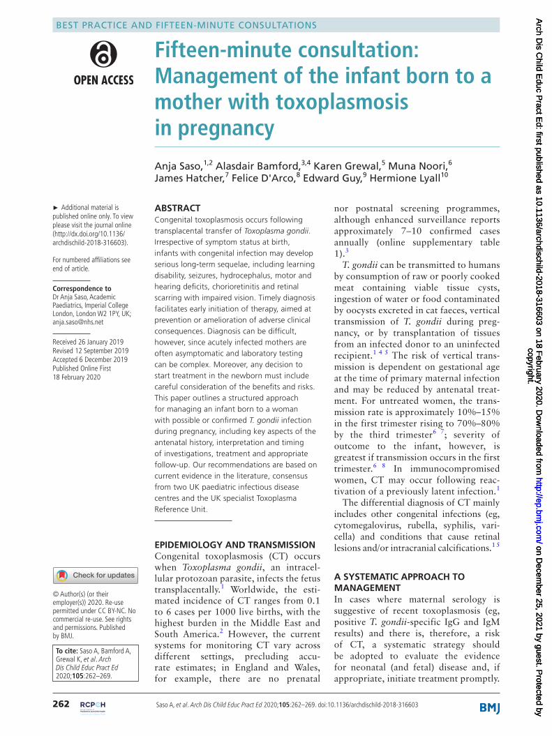
Table 1 Interpretation of common laboratory tests (maternal and infant) in suspected toxoplasmosis6 7 9–12
Laboratory test Significance
Screening test for presence of T. gondii- specific antibodies
► Often a latex agglutination or ELISA assay identifying combined (IgG and IgM) antibodies. ► Usually performed at the local hospital laboratory.
T.gondii- specific IgM ► First Ig to be detected: appears within 1–2 weeks of exposure. ► May be difficult to interpret because: (A) can remain detectable beyond 2 years after infection in some highly sensitive assays; (B) false
positive rate as high as 2%; (C) transplacental leakage of maternal T. gondii- specific IgM can lead to low positive T. gondii- specific IgM in an uninfected infant shortly after birth (should disappear within 1–2 weeks); (D) only 50%–60% of congenitally infected babies have detectable levels of T. gondii- specific IgM (and IgA) in the first month of life; levels may be undetectable even in severe disease, if maternal seroconversion occurs early in pregnancy, or if treatment is given in pregnancy or at or soon after birth.
► Consider congenital infection if T. gondii- specific IgM is present in infant and not in mother.
T.gondii- specific IgA ► IgA can cross the placenta (usually disappearing by 2 weeks of life), therefore: (A) passive transfer must be excluded; (B) only consider a positive result significant if the mother is T. gondii- specific IgA negative.
► May not be produced by congenitally infected infant in the first months of life.
T.gondii- specific IgG ► Negative neonatal T. gondii- specific IgG essentially excludes congenital toxoplasmosis. ► Appears from 2 weeks of primary infection and persists for life. ► Maternal: seroconversion of IgM, IgG or IgA during pregnancy suggests recent acute maternal toxoplasma infection. ► Neonatal: IgG crosses the placenta and therefore in the newborn may reflect past or current maternal infection. ► Transplacental maternal T. gondii- specific IgG has a half- life of around 3–4 weeks. Persistence beyond 12 months of age is considered
confirmatory of congenital infection.
Sabin- Feldman dye test
► Measures T. gondii- specific IgM and IgG antibody. ► Particularly helpful if: (A) initial dye tests are significantly raised or serial dye test results demonstrate increasing levels over time; (B)
infant dye test levels ≥4× maternal levels are suggestive of congenital infection.
Avidity test ► Measures functional affinity of T. gondii- specific IgG antibody. ► Useful when T. gondii- specific IgM samples are equivocal: may help discriminate between past (especially preconception) and recently
acquired infection. ► High avidity reliably excludes recent infection, but low avidity is not a reliable indicator of recent infection since rise in avidity may be
delayed in some individuals.
T.gondii- specific NAAT ► Molecular diagnostic techniques, for example, PCR. ► Can be done on amniotic fluid, blood or CSF samples. ► Detection of T. gondii in amniotic fluid or infant specimens confirms congenital infection.
CSF, cerebrospinal fluid; Ig, immunoglobulin; NAAT, nucleic acid amplification test; PCR, Polymerase chain reaction; T.gondii, Toxoplasma gondii.
review of maternal backgroundAntenatal historyAny symptoms or signs consistent with acute toxoplas-mosis should be noted, including date of onset. Ten to twenty per cent of all individuals develop cervical lymphadenopathy or a ‘flu- like’ illness, which has a benign, self- limiting clinical course.1 6 Generally, however, T. gondii infection is asymptomatic and may go unnoticed. Immunosuppression is a risk factor for
reactivation and/or more severe maternal infection and should also be identified.1
Antenatal investigationsReview of antenatal ultrasound and/or MRI findings is needed as there may be indications of evolving fetal toxoplasmosis, although these are non- specific (box 1).1 5 7
A positive T. gondii- specific IgM result during preg-nancy suggests acute T. gondii infection, probably acquired after conception, and an associated risk of transplacental transmission. Previous maternal spec-imens, such as booking bloods, should be retrospec-tively tested; however, there are important caveats to interpretation, summarised in table 1.6 7 9–11 In equiv-ocal cases, further testing should be undertaken either locally or at a national reference laboratory.6
T. gondii- specific IgG avidity can help discriminate between past and recently acquired infection; high avidity reliably excludes recent infection. Comparing serial blood results may demonstrate increasing anti-body titres (IgM and/or IgG dye test) or confirm sero-conversion during pregnancy (table 1).6 7 9–12 If infection acquired before conception can be confirmed, the risk to the pregnancy from T. gondii is considered low (in immunocompetent women), that is, T. gondii- specific
copyright. on D
ecember 25, 2021 by guest. P
rotected byhttp://ep.bm
j.com/
Arch D
is Child E
duc Pract E
d: first published as 10.1136/archdischild-2018-316603 on 18 February 2020. D
ownloaded from
Saso A, et al. Arch Dis Child Educ Pract Ed 2020;105:262–269. doi:10.1136/archdischild-2018-316603264
Best practice and Fifteen- minute consultations
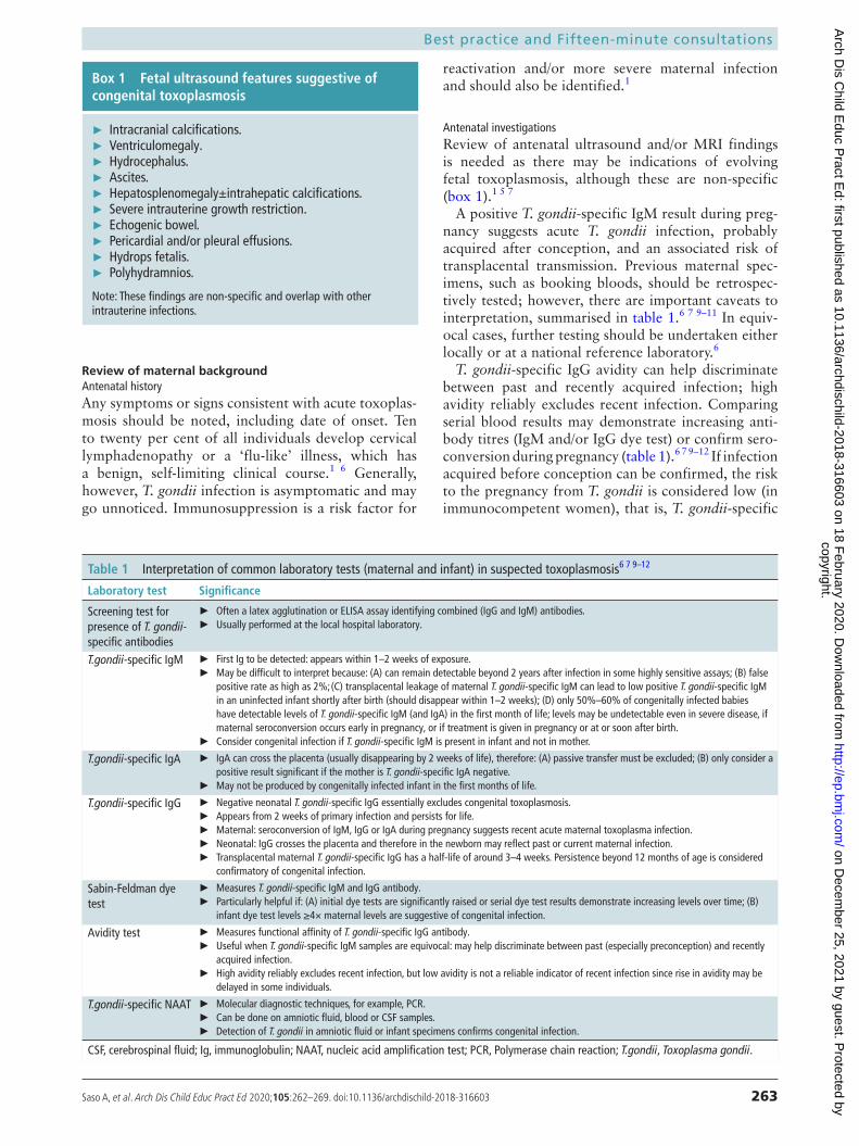
Table 2 Clinical features in the neonate suggesting congenital toxoplasmosis1 4 5 8 20
Central nervous system disease
MicrocephalyHearing impairmentSeizuresHydrocephalusMotor deficitsIntracranial calcifications
Ophthalmological disease with subsequent visual impairment
MicrophthalmiaChorioretinitis (usually bilateral)Retinal scarringStrabismusNystagmusCataract
Hepatic disease HepatosplenomegalyJaundice
Cardiorespiratory disease PneumonitisMyocarditis
Systemic features Rash (may be ‘blueberry- muffin’)FeverBone marrow suppression (thrombocytopenia, anaemia)
IgG positive, IgM negative, with or without high IgG avidity on booking or preconception bloods.
Some women may be offered amniocentesis: a posi-tive T. gondii nucleic acid amplification test (NAAT) result on amniotic fluid confirms intrauterine infec-tion.9 10 However, a negative result does not exclude fetal infection, particularly if maternal primary infec-tion occurred in the third trimester.7
Treatment during pregnancyTreatment given for acute toxoplasmosis in pregnancy should be recorded, including type and duration of therapy, since the interpretation of infant results may be affected.9 The two regimens often used are spiramycin (fetal prophylaxis preventing intrauterine infection) and combined pyrimethamine/sulfadiazine/folinic acid (treatment of evolving fetal infection).13 Cases should be discussed with a fetal medicine expert and local infection specialist (microbiologist or infec-tious diseases physician). The efficacy of maternal treatment remains unclear and should not preclude treating the infant if appropriate.14 15 However, obser-vational studies have demonstrated effective reduc-tion in transplacental transmission and/or severity of clinical manifestations in symptomatic infants16 17; less is known about the impact on non- severe congenital sequelae.
Assess clinical features in the newbornTable 2 identifies key clinical features suggestive of CT although no signs are pathognomonic. The classic triad is considered to be the presence of chorio-retinitis, intracranial calcifications and hydroceph-alus.4 5 However, up to 85% of congenitally infected newborns may have no findings on routine physical
examination1; more specific testing, primarily exam-ination of cerebrospinal fluid (CSF), neuroimaging and detailed ophthalmological review, is required to demonstrate subclinical end- organ abnormalities, as discussed below.18 One study demonstrated that up to 40% of infants with confirmed infection had retinal or central nervous system (CNS) abnormalities despite a normal routine initial neonatal examination.19
Arrange urgent postnatal investigationsBlood testsA basic understanding of the significance of different infant tests is useful (table 1). Paired T. gondii- specific serology from the infant and the mother must be taken at birth (not cord blood). This enables a comparison between their T. gondii- specific IgM, IgA and IgG levels. Significantly raised levels of T. gondii- specific IgM or IgG (more than four times) in the infant compared with the mother are suggestive of congen-ital infection.9 10 18 Detection of IgM in the neonate, however, should be interpreted with some caution, particularly where highly sensitive IgM tests are employed, since low levels of transplacental leakage can be detected. In such cases, these typically very low levels of maternal IgM disappear from the infant within 1–2 weeks. Detection of IgA in the newborn can also only be considered significant if the mother is IgA negative, since IgA readily crosses the placenta (disappearing by 2 weeks) and, therefore, passive transfer must be excluded (table 1).7 9
Conversely, only 50%–60% of congenitally infected infants will have positive T. gondii- specific IgM or IgA in the first month of life. Where results are negative, repeat testing should be carried out until at least 3 months of age, since seropositivity can be delayed.6 T. gondii- specific IgG testing should continue beyond this, for example, at 6 and 12 months. In uninfected infants, all maternally acquired IgG should have disap-peared by 12 months (table 1).9 In infected infants treated before, at or soon after birth, a serological response may not occur until treatment is withdrawn. Thus, negative serological findings in such cases cannot exclude congenital infection, and follow- up testing is required after completion of treatment.11
Infant specimens found positive on T. gondii NAAT confirm congenital infection. Nevertheless, a blood NAAT may not detect organ- specific disease (including ocular toxoplasmosis).18
Additional useful blood tests include full blood count, liver function and renal profile. Tests to exclude other congenital infections should also be undertaken, particularly in equivocal cases.
Lumbar punctureAny infant with findings consistent with CT should have a lumbar puncture and CSF sent for protein, glucose, cell count, culture and T. gondii NAAT.
copyright. on D
ecember 25, 2021 by guest. P
rotected byhttp://ep.bm
j.com/
Arch D
is Child E
duc Pract E
d: first published as 10.1136/archdischild-2018-316603 on 18 February 2020. D
ownloaded from
Saso A, et al. Arch Dis Child Educ Pract Ed 2020;105:262–269. doi:10.1136/archdischild-2018-316603 265
Best practice and Fifteen- minute consultations
Table 3 Evidence of congenital Toxoplasma gondii infection versus end- organ disease
Evidence of congenital infection Evidence of end- organ disease
Antenatal ► Positive T. gondii NAAT on amniotic fluid. ► Clinical features consistent with congenital toxoplasmosis on neonatal examination (table 2).
► Typical abnormal findings on: – Neuroimaging. – Ophthalmology review. – Audiology review.
These findings may indicate subclinical end- organ damage. ► Positive T. gondii NAAT on infant CSF.
Postnatal ► Positive infant T. gondii- specific IgM and/or IgA beyond 2–3 weeks.
► Significantly raised levels of T. gondii- specific IgM or IgG (≥4× titre) in the infant compared with the mother.
► Increasing infant serial T. gondii- specific IgG titres or persistence beyond 12 months.
► Positive T. gondii NAAT on infant specimens (including blood and CSF).
CSF, cerebrospinal fluid; Ig, immunoglobulin;NAAT, nucleic acid amplification test; T.gondii, Toxplasma gondii.
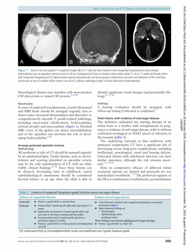
Figure 1 Axial T2 (A) and sagittal T1- weighted images (B) of a 1- day- old male newborn with congenital toxoplasmosis show marked hydrocephalus due to aqueduct stenosis (arrow in B) on a background of loss of cerebral white matter. Axial CT of an 11- week- old female infant with congenital toxoplasmosis (C) demonstrates typical periventricular and basal ganglia calcifications (arrows) and dilatation of the ventricles, mainly due to loss of cerebral white matter (‘ex- vacuo’), without radiological signs of acute obstructive hydrocephalus.
Neurological disease may manifest with mononuclear CSF pleocytosis or raised CSF protein.9 10 18
NeuroimagingIn cases of suspected toxoplasmosis, cranial ultrasound and MRI brain should be arranged urgently, first to detect major structural abnormalities and thereafter to comprehensively describe T. gondii- related pathology, including intracranial calcifications, hydrocephalus, cortical atrophy and microcephaly (figure 1). Detailed MRI views of the globes can detect microphthalmia and of the aqueduct can ascertain the risk of devel-oping hydrocephalus.4 18
Arrange postnatal specialist reviewsOphthalmologyAll newborns at risk of CT should be assessed urgently by an ophthalmologist. Ocular disease, such as chorio-retinitis and scarring identified on specialist review, may be the only manifestation of CT, with no other visible clinical findings.15 18 Since these lesions can be delayed, developing later in childhood, repeat ophthalmological assessments should be considered beyond infancy to an age when the child is able to
identify significant visual changes (and potentially life-long).6 7 11 12
AudiologyA hearing evaluation should be arranged, with follow- up testing if infection is confirmed.7
treat infants with evidence of end-organ diseaseThe definitive indication for starting therapy in an infant born to a mother with toxoplasmosis in preg-nancy is evidence of end- organ disease, with or without confirmed serological or NAAT proof of infection in the neonate (table 3).
The underlying rationale is that newborns with untreated symptomatic CT have a significant risk of developing severe long- term complications, including intellectual, neurological, visual and hearing deficits. Untreated infants with subclinical infection can have similar outcomes, although the risk remains uncer-tain.7 11 12 19
Data on comparative efficacy of different infant treatment options are limited and protocols are not standardised worldwide.7 13 The preferred regimen in the UK is a combination of sulfadiazine, pyrimethamine
copyright. on D
ecember 25, 2021 by guest. P
rotected byhttp://ep.bm
j.com/
Arch D
is Child E
duc Pract E
d: first published as 10.1136/archdischild-2018-316603 on 18 February 2020. D
ownloaded from
Saso A, et al. Arch Dis Child Educ Pract Ed 2020;105:262–269. doi:10.1136/archdischild-2018-316603266
Best practice and Fifteen- minute consultations
Box 2 Suggested treatment regimen for congenital toxoplasmosis5*
► Pyrimethamine 1 mg/kg TWICE daily for 2 days. – Then 1 mg/kg ONCE daily for 6 months. – Then 1 mg/kg THREE times weekly to complete 12
months. ► Sulfadiazine 50 mg/kg TWICE daily for 12 months.
– Only start after neonatal jaundice resolved. ► Folinic acid (calcium folinate, Leucovorin) given to counteract bone marrow toxicity of pyrimethamine.
– 5–20 mg THREE times weekly during and for 1 week after pyrimethamine therapy.
– Dose adjusted to age (5 mg in neonates, 10 mg in infants >1 month) and full blood count (table 3).
*As per the British National Formulary for Children (2019–2020) and Manual of Childhood Infections—The Blue Book, Fourth Edition (2016), endorsed by the Royal College of Paediatric and Child Health and European Society of Paediatric Infectious Diseases.
and folinic acid for 12 months (box 2), in keeping with recommendations made in many European countries, North America and Australia.6 7 11 12 G6PD deficiency must be excluded prior to administration and steroids may be added for severe ocular disease or high CSF protein (more than 1 g/dL).1 18
There are no randomised controlled trials (RCT) of this combination therapy in infants.13 Previous large cohort studies, particularly in North America, have reported that a 12- month therapeutic course is associated with significantly decreased incidence and severity of long- term sequelae and new- onset ocular disease when compared with untreated historical controls or those treated for a shorter duration.8 19 20 In European studies, treatment was often started ante-natally precluding reliable determination of the effects of postnatal therapy. Data predominantly demon-strated an improvement in ocular prognosis following early diagnosis and timely initiation of treatment.11 13 A French RCT is currently comparing the efficacy of triple therapy for 3 vs 12 months on the development of chorioretinitis over a 2- year period in infants with non- severe CT ( ClinicalTrials. gov NCT01202500).
Despite better outcomes, treated infants are still at risk of developing delayed complications.8 This is because current treatment targets T. gondii tachyzoites (rapidly proliferating developmental stage) but does not successfully eliminate slower- growing, latent bradyzoites established within tissue cysts, particularly in the eye and CNS.1 5 18 Therefore, relapse of ocular (including late- onset retinal lesions) and CNS disease can occur, in some cases even several years after treatment has been completed, potentially requiring retreatment.11 18
monitor infants with no evidence of end-organ diseaseGiven the paucity of available evidence, there is no clear consensus on the benefit of treating neonates
who have been born to a mother with toxoplasmosis in pregnancy and who are asymptomatic without confirmed end- organ disease.7 11–13 Positive fetal or infant serology or NAAT results, although not indi-cations for treatment, are still useful to diagnose T. gondii- specific infection in the infant (table 3) and, therefore, guide longer term follow- up (figure 2).
Most infectious disease paediatricians in the UK would monitor serology and defer treatment, bearing in mind the toxicity of treatment. Close outpatient follow- up is required, only starting treatment if clin-ically indicated. This decision may vary in other settings.
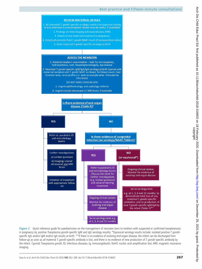
Figure 2 summarises the step- by- step considerations in the investigation and management of newborns at risk of CT. Interpretation of results and decisions about treatment should be made in conjunction with microbiologists and infectious disease specialists. Families should be fully informed about the risks and benefits of treatment, including limitations of current knowledge and diagnostic tests.
Follow-up of treated infantsClose monitoring of infants on treatment for adverse events, disease recurrence, symptom progression and therapeutic response is critical (table 4).7 11 12 Follow- up is recommended in a specialist paediatric infectious diseases clinic. Potential prognostic factors associated with poor outcomes include delay in initi-ation of treatment (and/or correction of hydroceph-alus); prolonged hypoxia or episodes of hypothermia, apnoea and bradycardia at presentation; CSF protein more than 1 g/dL and cerebral atrophy.8
Parents should be offered peer support through networking groups and access to helpful online resources. The UK Tommy’s charity provides online support and comprehensive information for parents (https://www. tommys. org/ pregnancy- information/ pregnancy- complications/ infections- pregnancy/ toxoplasmosis- pregnancy).
conclusionWhen faced with a case of suspected CT, the paediatric team must be aware of the workup required to confirm diagnosis, the significance of the results obtained and the importance of timely treatment to prevent or ameliorate adverse clinical consequences. The key learning points are:
► Positive T. gondii- specific IgM and IgG in pregnancy does not necessarily equate to subsequent congenital toxoplasmosis and further investigation and monitoring of the infant (and fetus) are required.
► A thorough postnatal examination is key, although the majority of infected newborns are asymptomatic on initial assessment but may still develop serious long- term sequelae; ophthalmology review and neuroimaging are necessary to detect subclinical end- organ abnormalities.
► T. gondii NAATs and serial infant T. gondii- specific serology are helpful to diagnose infection, although
copyright. on D
ecember 25, 2021 by guest. P
rotected byhttp://ep.bm
j.com/
Arch D
is Child E
duc Pract E
d: first published as 10.1136/archdischild-2018-316603 on 18 February 2020. D
ownloaded from
Saso A, et al. Arch Dis Child Educ Pract Ed 2020;105:262–269. doi:10.1136/archdischild-2018-316603 267
Best practice and Fifteen- minute consultations
Figure 2 Quick reference guide for paediatricians on the management of neonates born to mothers with suspected or confirmed toxoplasmosis in pregnancy (ie, positive Toxoplasma gondii- specific IgM and IgG serology results). *Equivocal serology results include: isolated positive T. gondii- specific IgG and/or IgM and/or IgA results at birth. **If there is no evidence of evolving end- organ disease, the infant can be discharged from follow- up as soon as all maternal T. gondii- specific antibody is lost, and there is no evidence of new production of T. gondii- specific antibody by the infant. T.gondii, Toxoplasma gondii; ID, infectious diseases; Ig, immunoglobulin; NAAT, nucleic acid amplification test; MRI, magnetic resonance imaging.
copyright. on D
ecember 25, 2021 by guest. P
rotected byhttp://ep.bm
j.com/
Arch D
is Child E
duc Pract E
d: first published as 10.1136/archdischild-2018-316603 on 18 February 2020. D
ownloaded from
Saso A, et al. Arch Dis Child Educ Pract Ed 2020;105:262–269. doi:10.1136/archdischild-2018-316603268
Best practice and Fifteen- minute consultations
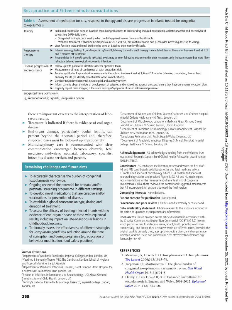
Table 4 Assessment of medication toxicity, response to therapy and disease progression in infants treated for congenital toxoplasmosis
Toxicity ► Full blood count to be done at baseline then during treatment to look for drug- induced neutropenia, aplastic anaemia and haemolysis (if co- existing G6PD deficiency).
– Suggested timing is once weekly when on daily pyrimethamine then monthly if stable. – Withhold treatment if absolute neutrophil count <0.5 x10∧9/L, but continue folinic acid (±consider increasing dose up to 20 mg).
► Liver function tests and renal profile to be done at baseline then monthly if stable.
Response to therapy
► Interval serology testing: T. gondii- specific IgG and IgM every 3 months until therapy is completed then at the end of treatment and at 1, 3 and 6 months off treatment.
► Rebound rise in T. gondii- specific IgM±IgG levels may be seen following treatment; this does not necessarily indicate relapse but more likely reflects a delayed serological response to infection.
Disease progression and recurrence
► Follow- up with paediatric infectious disease specialist team. ► Measurement of head circumference at each outpatient visit. ► Regular ophthalmology and vision assessments throughout treatment and at 3, 6 and 12 months following completion, then at least
annually for life (to identify potential late- onset complications). ► Consider neurodevelopmental, neurological and auditory review. ► Inform parents about the risks of development of seizures and/or raised intracranial pressure: ensure they have an emergency action plan. ► Urgently repeat brain imaging if there are any signs/symptoms of raised intracranial pressure.
Suggested time points only.Ig, immunoglobulin; T.gondii, Toxoplasma gondii.
there are important caveats to the interpretation of labo-ratory results.
► Treatment is indicated if there is evidence of end- organ disease.
► End- organ damage, particularly ocular lesions, can present beyond the neonatal period and, therefore, suspected cases must be followed up appropriately.
► Multidisciplinary care is recommended with clear communication encouraged between obstetric, fetal medicine, midwifery, neonatal, laboratory, specialist infectious disease services and parents.
Remaining challenges and future aims
► To accurately characterise the burden of congenital toxoplasmosis worldwide.
► Ongoing review of the potential for prenatal and/or postnatal screening programme in different settings.
► To develop novel medications that are curative and/or vaccinations for prevention of disease.
► To establish a global consensus on type, dosing and duration of treatment.
► To assess the efficacy of treating infected infants with no evidence of end- organ disease or those with equivocal results, including impact on late- onset ocular lesions in childhood/adolescence.
► To formally assess the effectiveness of different strategies for Toxoplasma gondii risk reduction around the time of conception and during pregnancy (eg, education on behaviour modification, food safety practices).
Author affiliations1Department of Academic Paediatrics, Imperial College London, London, UK2Vaccines & Immunity Theme, MRC The Gambia at London School of Hygiene and Tropical Medicine, Banjul, Gambia3Department of Paediatric Infectious Diseases, Great Ormond Street Hospital for Children NHS Foundation Trust, London, UK4Section of Infection, Inflammation and Rheumatology, UCL Great Ormond Street Institute of Child Health, London, UK5Tommy’s National Centre for Miscarriage Research, Imperial College London, London, UK
6Department of Women and Children, Queen Charlotte’s and Chelsea Hospital, Imperial College Healthcare NHS Trust, London, UK7Department of Microbiology, Laboratory Medicine, Great Ormond Street Hospital for Children NHS Trust, London, United Kingdom8Department of Paediatric Neuroradiology, Great Ormond Street Hospital for Children NHS Foundation Trust, London, UK9Toxoplasma Reference Unit, Public Health Wales, Swansea, UK10Department of Paediatric Infectious Diseases, St Mary’s Hospital, Imperial College Healthcare NHS Trust, London, UK
Acknowledgements AS acknowledges funding from the Wellcome Trust Institutional Strategic Support Fund Global Health Fellowship, award number 204834/Z/16/Z.
Contributors AS conducted the literature review and wrote the first draft. KG and MN contributed specialist obstetrics and fetal medicine advice. JH contributed specialist microbiology advice. FDA contributed specialist neuroradiology advice and provided Figure 1. EG, AB and HL made expert recommendations for the management of infants at risk of congenital toxoplasmosis. All authors reviewed the content and suggested amendments that AS incorporated. All authors approved the final version.
Competing interests None declared.
Patient consent for publication Not required.
Provenance and peer review Commissioned; externally peer reviewed.
Data availability statement All data relevant to the study are included in the article or uploaded as supplementary information.
Open access This is an open access article distributed in accordance with the Creative Commons Attribution Non Commercial (CC BY- NC 4.0) license, which permits others to distribute, remix, adapt, build upon this work non- commercially, and license their derivative works on different terms, provided the original work is properly cited, appropriate credit is given, any changes made indicated, and the use is non- commercial. See: http:// creativecommons. org/ licenses/ by- nc/ 4. 0/.
RefeRences 1 Montoya JG, Liesenfeld O, Toxoplasmosis LO. Toxoplasmosis.
The Lancet 2004;363:1965–76. 2 Torgerson PR, Mastroiacovo P. The global burden of
congenital toxoplasmosis: a systematic review. Bull World Health Organ 2013;91:501–8.
3 Halsby K, Guy E, Said B, et al. Enhanced surveillance for toxoplasmosis in England and Wales, 2008-2012. Epidemiol Infect 2014;142:1653–60.
copyright. on D
ecember 25, 2021 by guest. P
rotected byhttp://ep.bm
j.com/
Arch D
is Child E
duc Pract E
d: first published as 10.1136/archdischild-2018-316603 on 18 February 2020. D
ownloaded from
Saso A, et al. Arch Dis Child Educ Pract Ed 2020;105:262–269. doi:10.1136/archdischild-2018-316603 269
Best practice and Fifteen- minute consultations
4 McAuley JB, Toxoplasmosis C. Congenital toxoplasmosis. J Pediatric Infect Dis Soc 2014;3:S30–5.
5 Christopher B, Wilson MD, Victor Nizet MD, et al. Toxoplasmosis. 8th edn. Philadelphia: Elsevier Saunders, 2016.
6 Division PHWHP. Public Health Wales Health Protection Division - Toxoplasma Reference Unit. Available: http://www. wales. nhs. uk/ sites3/ page. cfm? orgId= 457& pid= 25359 [Accessed 18 Nov 2018].
7 Maldonado YA, Read JS. Committee on infectious diseases. diagnosis, treatment, and prevention of congenital toxoplasmosis in the United States. Pediatrics 2017;139:e20163860.
8 McAuley J, Boyer KM, Patel D, et al. Early and longitudinal evaluations of treated infants and children and untreated historical patients with congenital toxoplasmosis: the Chicago Collaborative treatment trial. Clinical Infectious Diseases 1994;18:38–72 http://www. ncbi. nlm. nih. gov/ pubmed/ 8054436
9 Pomares C, Montoya JG. Laboratory diagnosis of congenital toxoplasmosis. J Clin Microbiol 2016;54:2448–54.
10 Murat J- B, Fricker Hidalgo H, Brenier- Pinchart M- P, et al. Human toxoplasmosis: which biological diagnostic tests are best suited to which clinical situations? Expert Rev Anti Infect Ther 2013;11:943–56.
11 Peyron F, L’ollivier C, Mandelbrot L, et al. Maternal and congenital toxoplasmosis: diagnosis and treatment recommendations of a French multidisciplinary Working group. Pathogens 2019;8:24.
12 Paquet C, Yudin MH, No YMH. No. 285- Toxoplasmosis in pregnancy: prevention, screening, and treatment. J Obstet Gynaecol Can 2018;40:e687–93.
13 Robert- Gangneux F. It is not only the cat that did it: how to prevent and treat congenital toxoplasmosis. J Infect 2014;68 Suppl 1:S125–33.
14 Peyron F, Wallon M, Liou C, et al. Treatments for toxoplasmosis in pregnancy. Cochrane Database Syst Rev 2009;1999:CD001684.
15 Thiébaut R, Leproust S, Chêne G, et al. Effectiveness of prenatal treatment for congenital toxoplasmosis: a meta- analysis of individual patients' data. Lancet 2007;369:115–22.
16 Cortina- Borja M, Tan HK, Wallon M, et al. Prenatal treatment for serious neurological sequelae of congenital toxoplasmosis: an observational prospective cohort study. PLoS Med 2010;7:e1000351.
17 Hotop A, Hlobil H, Gross U. Efficacy of rapid treatment initiation following primary Toxoplasma gondii infection during pregnancy. Clin Infect Dis 2012;54:1545–52.
18 Khan K, Khan W. Congenital toxoplasmosis: an overview of the neurological and ocular manifestations. Parasitol Int 2018;67:715–21.
19 Guerina NG, Hsu HW, Meissner HC, et al. Neonatal serologic screening and early treatment for congenital Toxoplasma gondii infection. the new England regional Toxoplasma Working group. N Engl J Med 1994;330:1858–63.
20 McLeod R, Boyer K, Karrison T, et al. Outcome of treatment for congenital toxoplasmosis, 1981-2004: the National collaborative Chicago- Based, congenital toxoplasmosis study. Clin Infect Dis 2006;42:1383–94.
copyright. on D
ecember 25, 2021 by guest. P
rotected byhttp://ep.bm
j.com/
Arch D
is Child E
duc Pract E
d: first published as 10.1136/archdischild-2018-316603 on 18 February 2020. D
ownloaded from
SUPPLEMENTARY MATERIALS
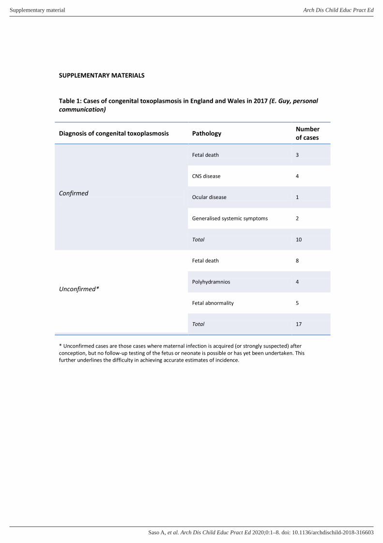
Table 1: Cases of congenital toxoplasmosis in England and Wales in 2017 (E. Guy, personal
communication)
Diagnosis of congenital toxoplasmosis Pathology Number
of cases
Confirmed
Fetal death 3
CNS disease 4
Ocular disease 1
Generalised systemic symptoms 2
Total 10
Unconfirmed*
Fetal death 8
Polyhydramnios 4
Fetal abnormality 5
Total 17
* Unconfirmed cases are those cases where maternal infection is acquired (or strongly suspected) after
conception, but no follow-up testing of the fetus or neonate is possible or has yet been undertaken. This
further underlines the difficulty in achieving accurate estimates of incidence.
Supplementary material Arch Dis Child Educ Pract Ed
doi: 10.1136/archdischild-2018-316603–8.:10 2020;Arch Dis Child Educ Pract Ed, et al. Saso A