Embed Size (px)
Citation preview

Nematodes from tissues
Filariae, Toxocara spp., Trichinellaspiralis
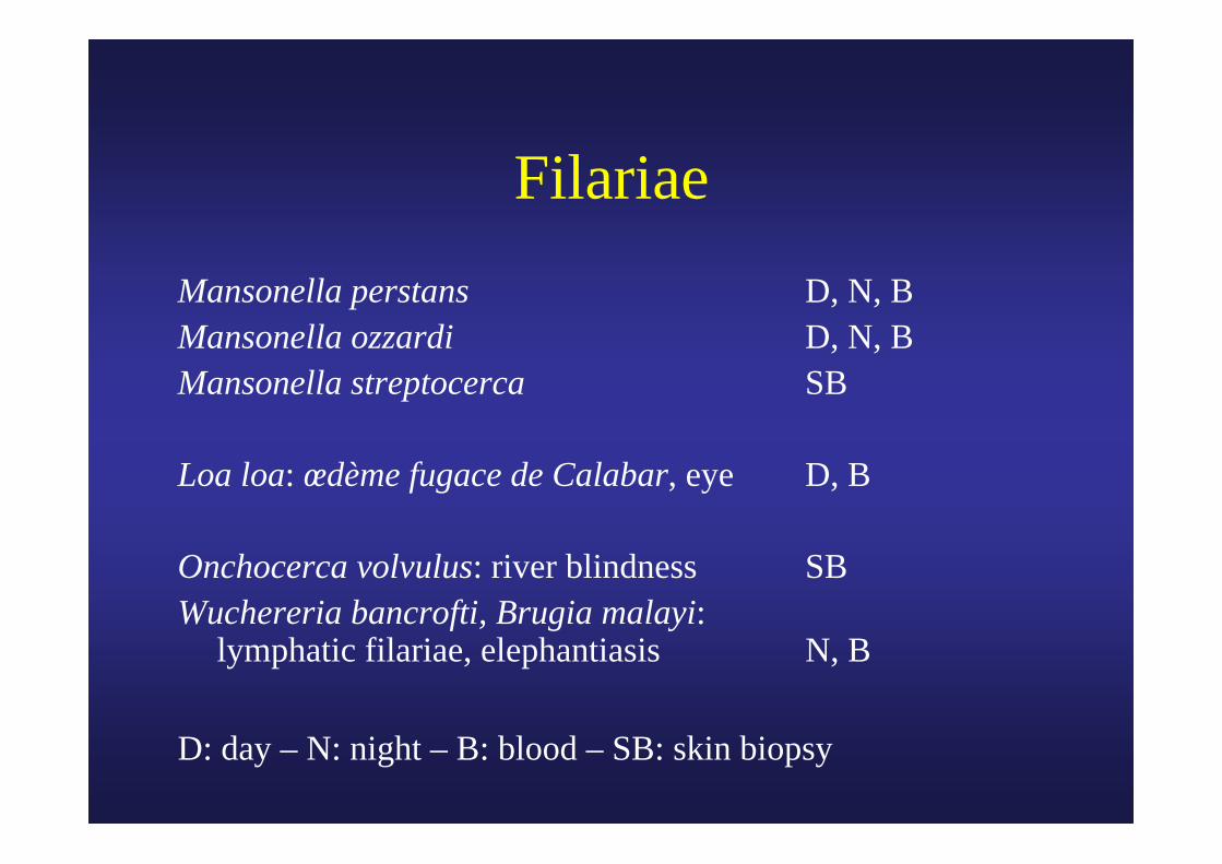
FilariaeMansonella perstans D, N, BMansonella ozzardi D, N, BMansonella streptocerca SB
Loa loa: œdème fugace de Calabar, eye D, B
Onchocerca volvulus: river blindness SBWuchereria bancrofti, Brugia malayi:
lymphatic filariae, elephantiasis N, B
D: day – N: night – B: blood – SB: skin biopsy

FilariaeMansonella perstans Africa, South
America, Surinam Mansonella ozzardi South AmericaMansonella streptocerca West Africa
Loa loa West and Central AfricaOnchocerca volvulus Africa, South America
Wuchereria bancrofti Africa, South America, Asia
Brugia malayi Asia
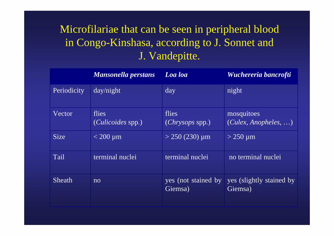
Microfilariae that can be seen in peripheral blood in Congo-Kinshasa, according to J. Sonnet and
J. Vandepitte.Mansonella perstans Loa loa Wuchereria bancrofti
Periodicity day/night day night
Vector flies(Culicoides spp.)
flies (Chrysops spp.)
mosquitoes (Culex, Anopheles, …)
Size < 200 µm > 250 (230) µm > 250 µm
Tail terminal nuclei terminal nuclei no terminal nuclei
Sheath no yes (not stained by Giemsa)
yes (slightly stained by Giemsa)
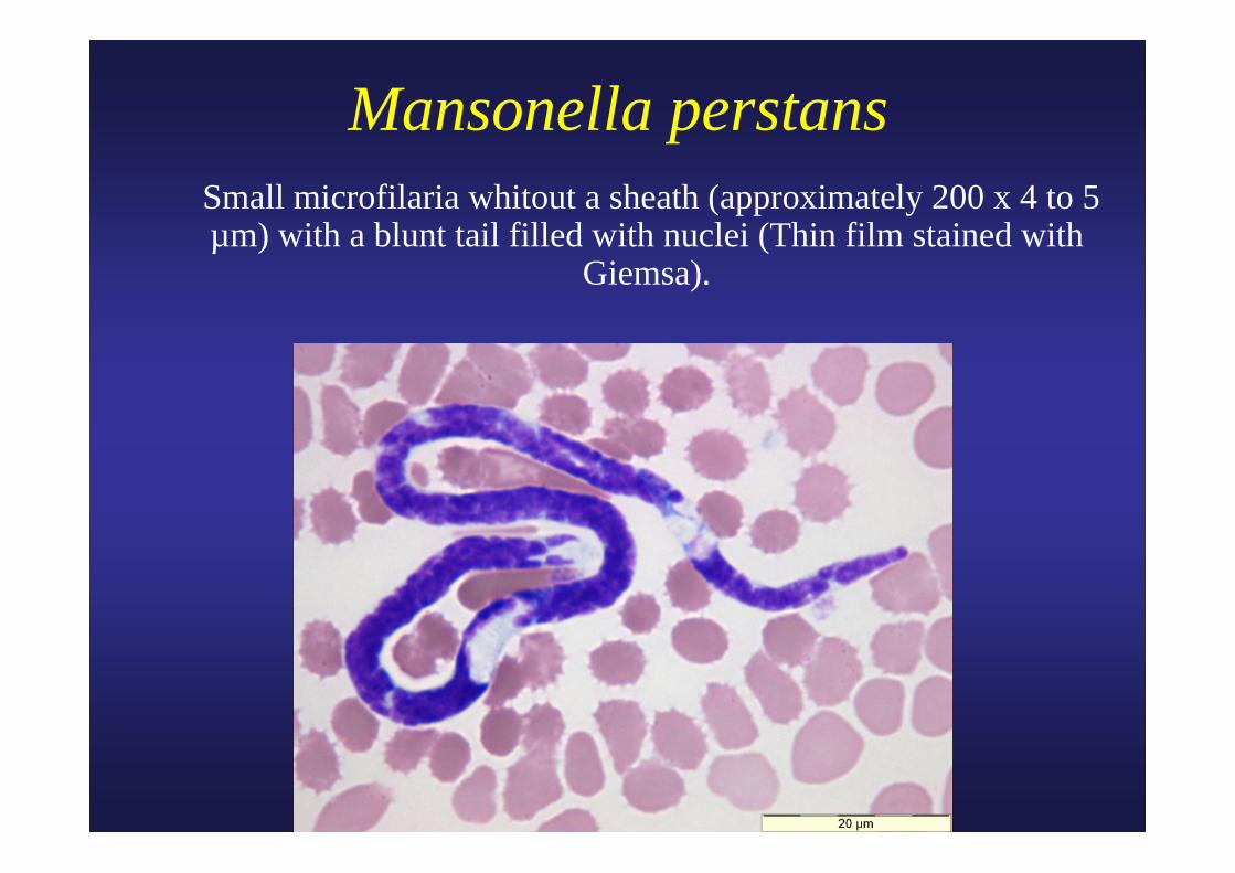
Mansonella perstans Small microfilaria whitout a sheath (approximately 200 x 4 to 5
µm) with a blunt tail filled with nuclei (Thin film stained with Giemsa).
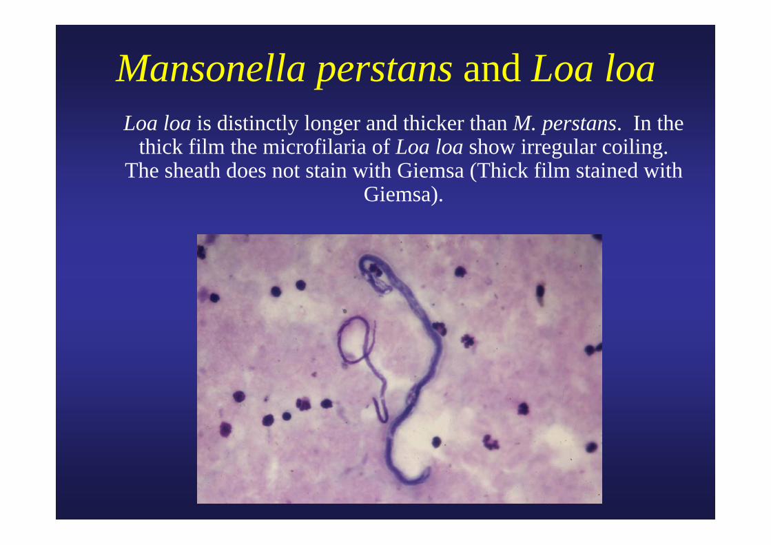
Mansonella perstans and Loa loa Loa loa is distinctly longer and thicker than M. perstans. In the
thick film the microfilaria of Loa loa show irregular coiling. The sheath does not stain with Giemsa (Thick film stained with
Giemsa).

Courtesy CDC
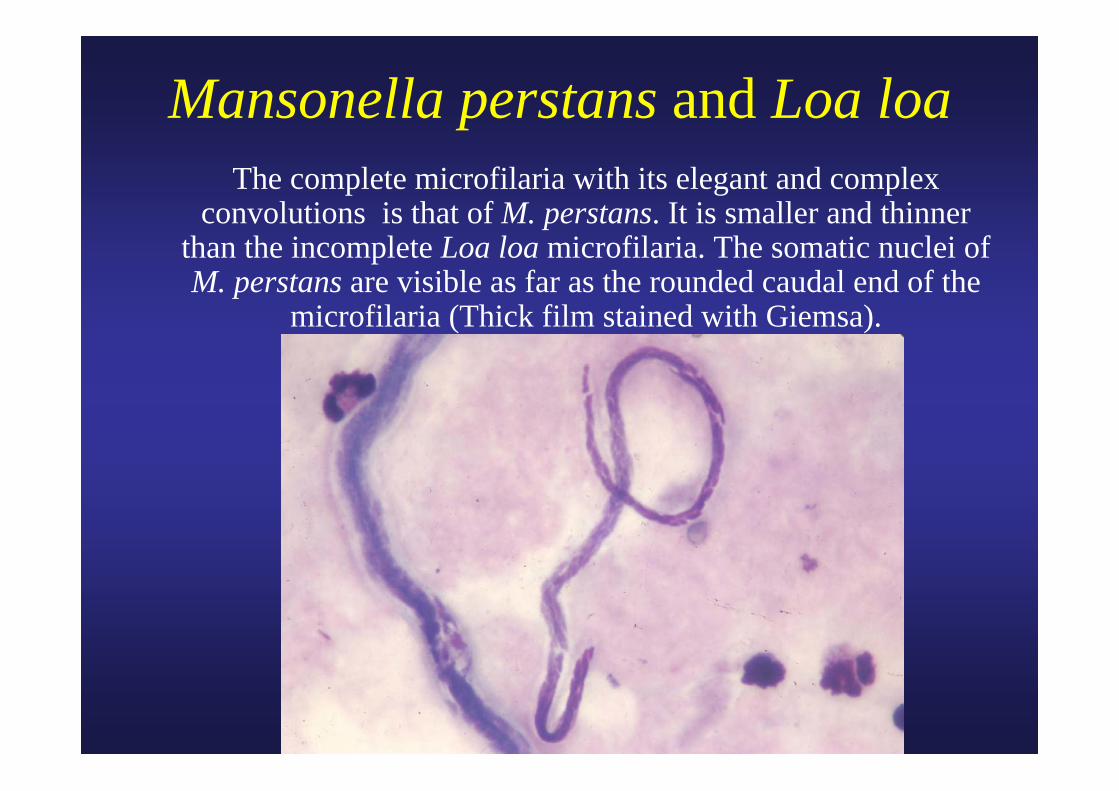
Mansonella perstans and Loa loa The complete microfilaria with its elegant and complex convolutions is that of M. perstans. It is smaller and thinner
than the incomplete Loa loa microfilaria. The somatic nuclei of M. perstans are visible as far as the rounded caudal end of the
microfilaria (Thick film stained with Giemsa).
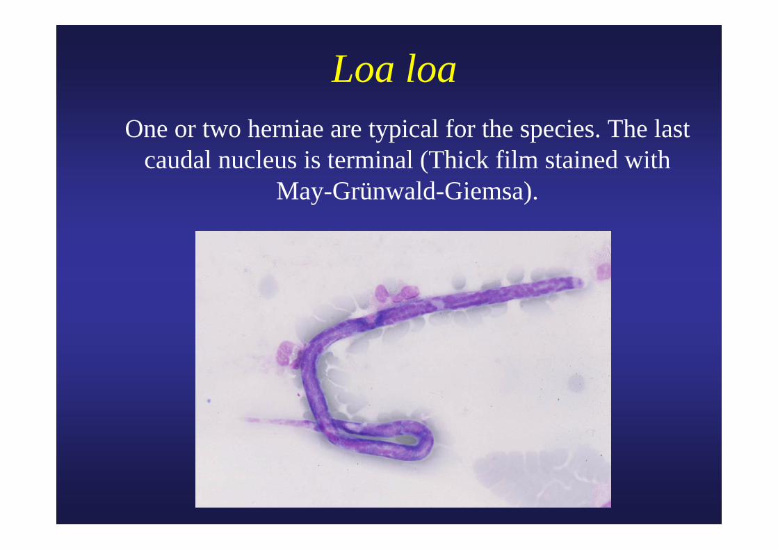
Loa loa One or two herniae are typical for the species. The last
caudal nucleus is terminal (Thick film stained with May-Grünwald-Giemsa).
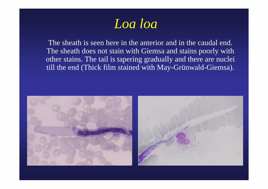
Loa loa The sheath is seen here in the anterior and in the caudal end.
The sheath does not stain with Giemsa and stains poorly with other stains. The tail is tapering gradually and there are nuclei till the end (Thick film stained with May-Grünwald-Giemsa).
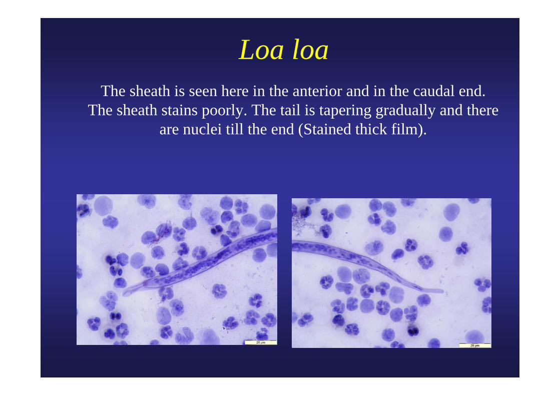
Loa loa The sheath is seen here in the anterior and in the caudal end. The sheath stains poorly. The tail is tapering gradually and there
are nuclei till the end (Stained thick film).

Loa loaThe sheath is seen here at the caudal end. The sheath stains poorly.
The tail is tapering gradually and there are nuclei till the end(Stained thick film).

Loa loa Adult male removed from the eye. The male filaria is
smaller (up to 3,4 cm) than the female (5-7 cm) (Unstained).
cmcm
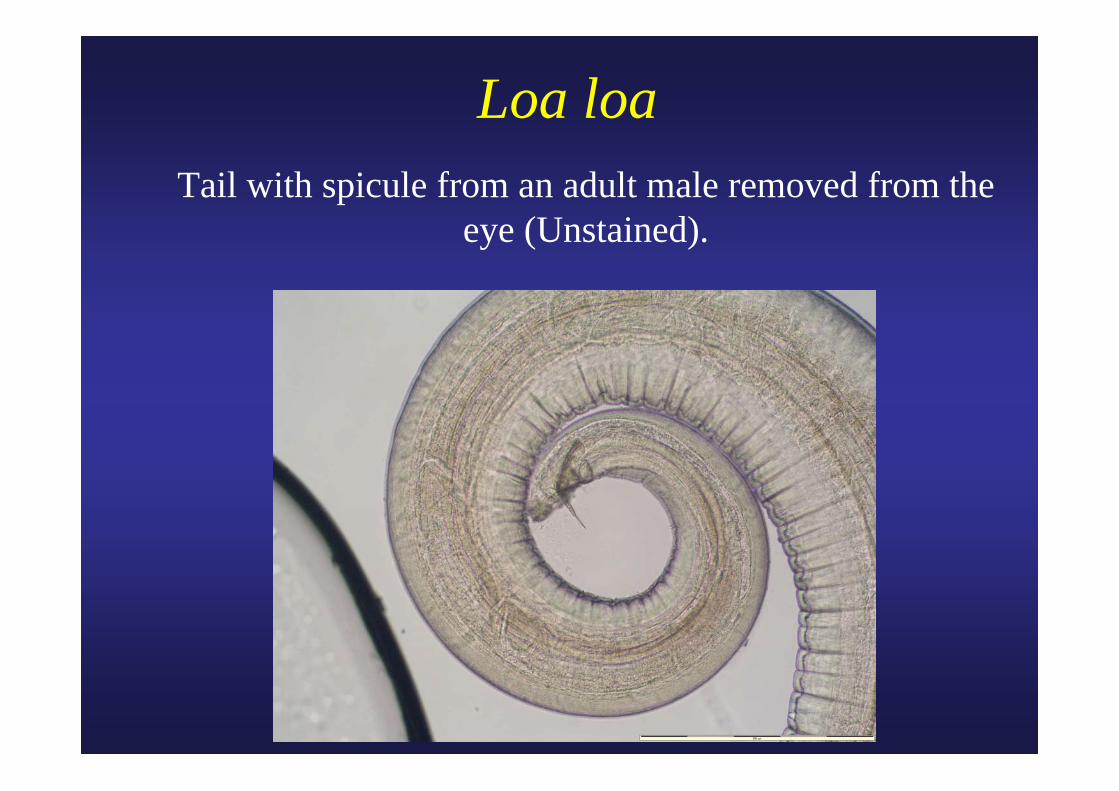
Loa loa Tail with spicule from an adult male removed from the
eye (Unstained).

Mansonella streptocercaSlender microfilaria with rounded tip curved to form a
hook (Skin scarification stained with Giemsa).
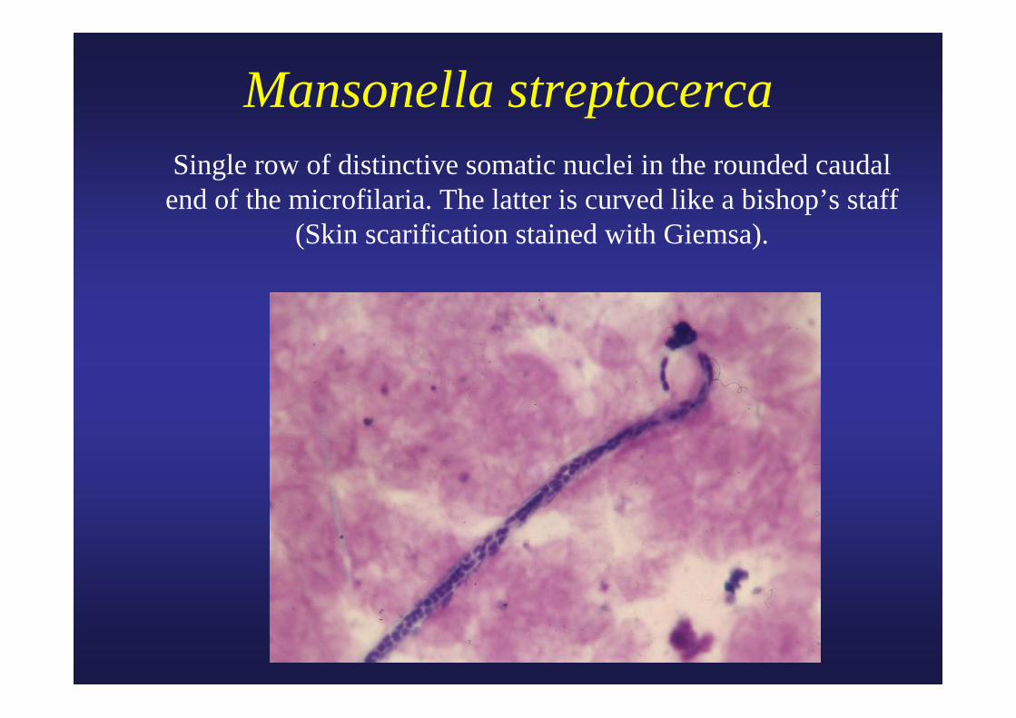
Mansonella streptocercaSingle row of distinctive somatic nuclei in the rounded caudal
end of the microfilaria. The latter is curved like a bishop’s staff (Skin scarification stained with Giemsa).
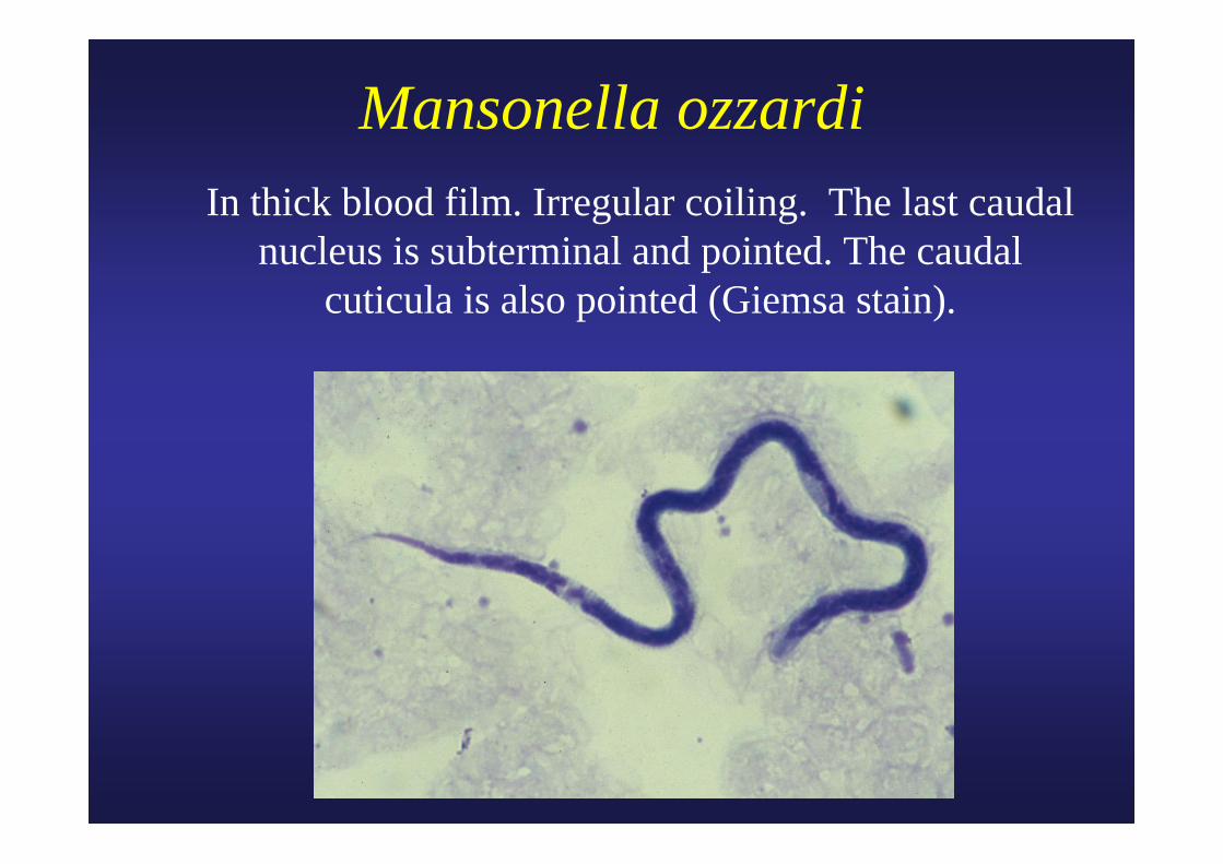
Mansonella ozzardi In thick blood film. Irregular coiling. The last caudal
nucleus is subterminal and pointed. The caudal cuticula is also pointed (Giemsa stain).
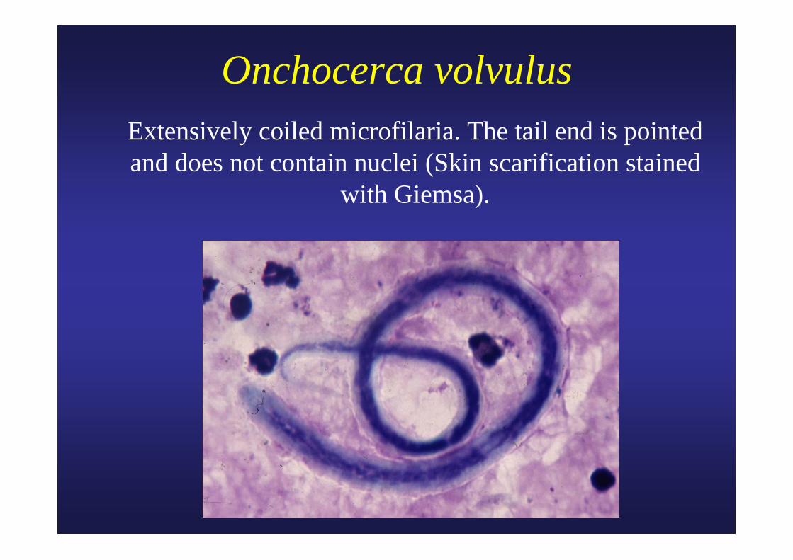
Onchocerca volvulus Extensively coiled microfilaria. The tail end is pointed
and does not contain nuclei (Skin scarification stained with Giemsa).
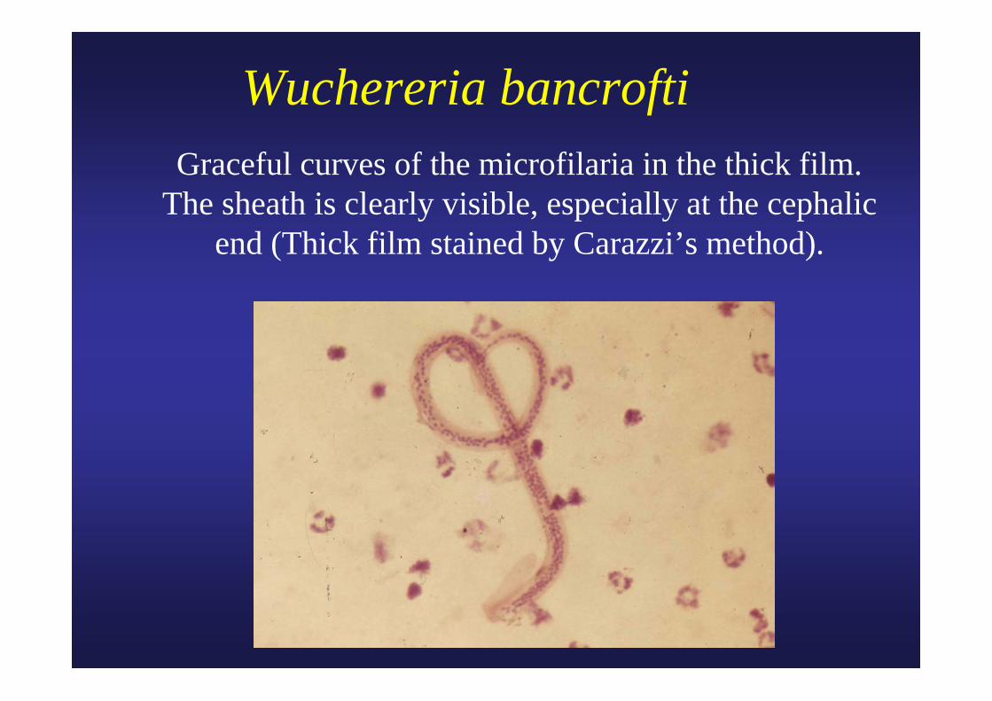
Wuchereria bancrofti Graceful curves of the microfilaria in the thick film.
The sheath is clearly visible, especially at the cephalic end (Thick film stained by Carazzi’s method).

Wuchereria bancrofti Cephalic end with sheath and caudal end of the
microfilaria. No nuclei in the pointed caudal end (Thick film stained by Carazzi’s method).
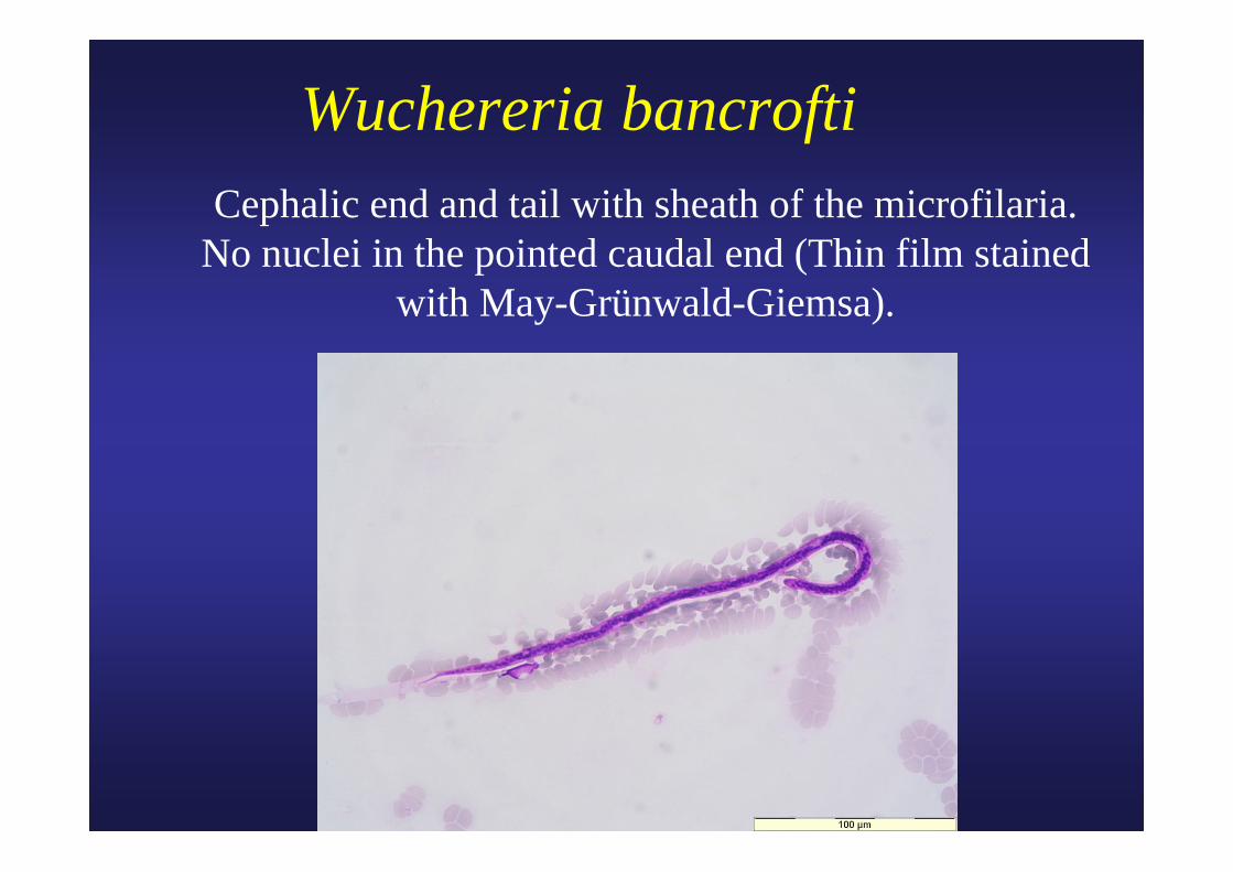
Wuchereria bancrofti Cephalic end and tail with sheath of the microfilaria.
No nuclei in the pointed caudal end (Thin film stained with May-Grünwald-Giemsa).

Wuchereria bancrofti Caudal end with sheath of the microfilaria. No nuclei
in the pointed caudal end (Thin film stained by May-Grünwald-Giemsa).

Wuchereria bancrofti Caudal end of the microfilaria. No nuclei in the
pointed caudal end (Thin film stained by May-Grünwald-Giemsa).
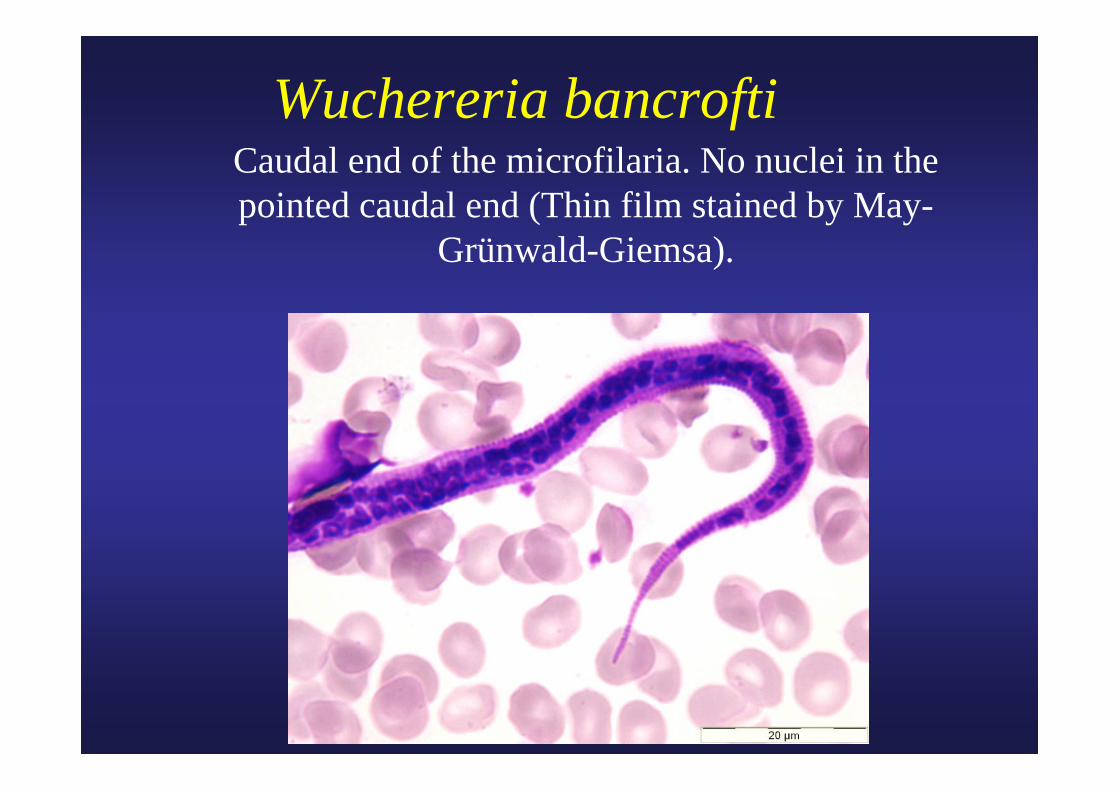
Wuchereria bancrofti Caudal end of the microfilaria. No nuclei in the
pointed caudal end (Thin film stained by May-Grünwald-Giemsa).
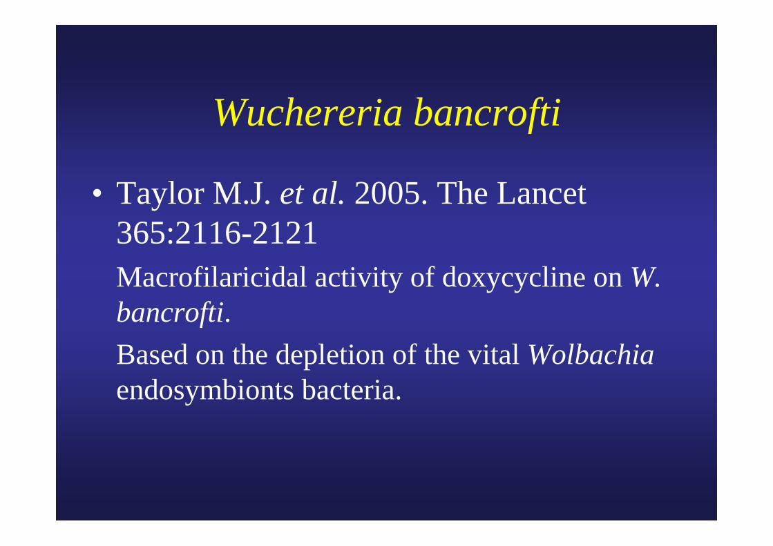
Wuchereria bancrofti
• Taylor M.J. et al. 2005. The Lancet 365:2116-2121Macrofilaricidal activity of doxycycline on W.bancrofti.Based on the depletion of the vital Wolbachiaendosymbionts bacteria.

Brugia malayi Coiled microfilaria in thick blood film. The nuclei are more or
less distinct. The last caudal nucleus is clearly separated fromthe others and extends into the tip of the tail. The sheath is well
developed and stained red (Giemsa stain).
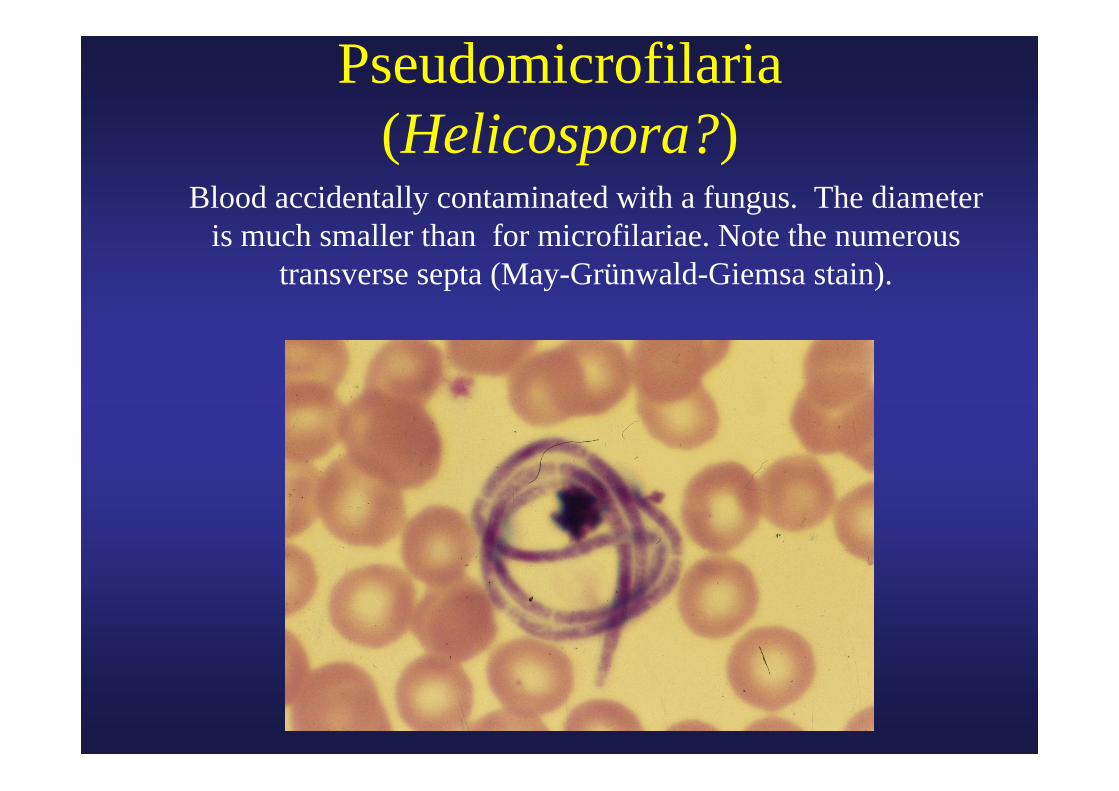
Pseudomicrofilaria (Helicospora?)
Blood accidentally contaminated with a fungus. The diameter is much smaller than for microfilariae. Note the numerous
transverse septa (May-Grünwald-Giemsa stain).

Global programme to eliminate lymphatic filariasis
• “Elephantiasis”• Resolution WHA50.29 in 1997• 120 million patients in 83 countries• More than 1 billion (20 % of world population)
are at risk of acquiring the infection• 90% Wuchereria bancrofti, most of the remainder
Brugia malayi• 2-drug combinations (DEC + albendazole or
ivermectin + albendazole)
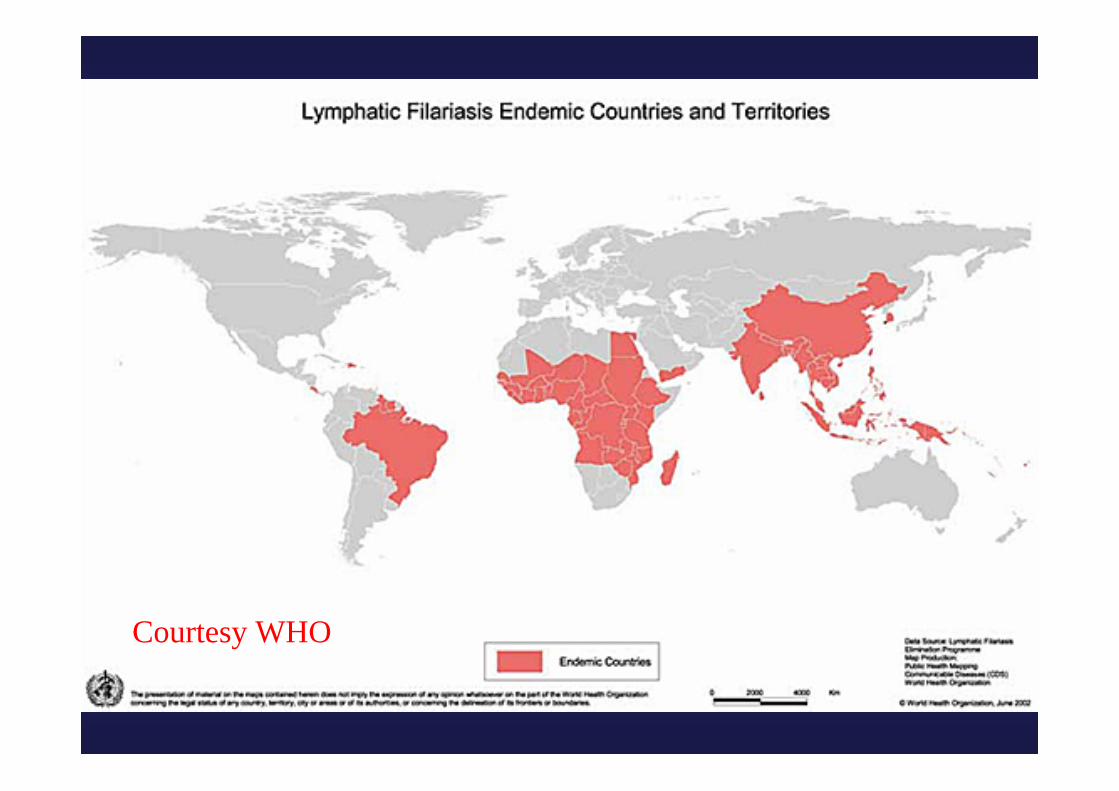
Courtesy WHO
Larva migrans
• Larva migrans cutanea: larbish, creeping eruption, caused by hookworms of animals (cat, dog …) Ancylostoma ceylanicum, Ancylostoma brasiliense, …
• Larva migrans visceralis: high eosinophilia with variable symptomatology (general, eye invasion), due to Toxocara canis, diagnosis by serology, …
• …

Toxocara canisSeveral adult worms, smaller than A. lumbricoides.
1 cm
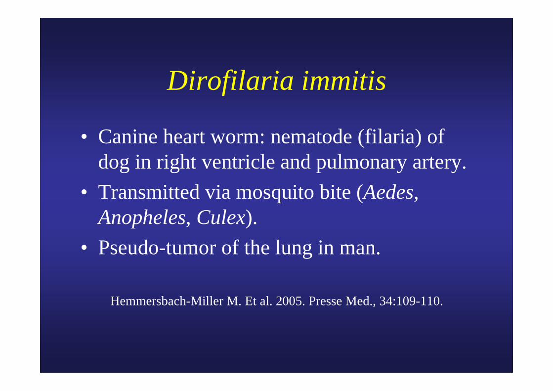
Dirofilaria immitis
• Canine heart worm: nematode (filaria) of dog in right ventricle and pulmonary artery.
• Transmitted via mosquito bite (Aedes, Anopheles, Culex).
• Pseudo-tumor of the lung in man.
Hemmersbach-Miller M. Et al. 2005. Presse Med., 34:109-110.

Trichinella spiralis Larva in muscle tissue following biopsy .
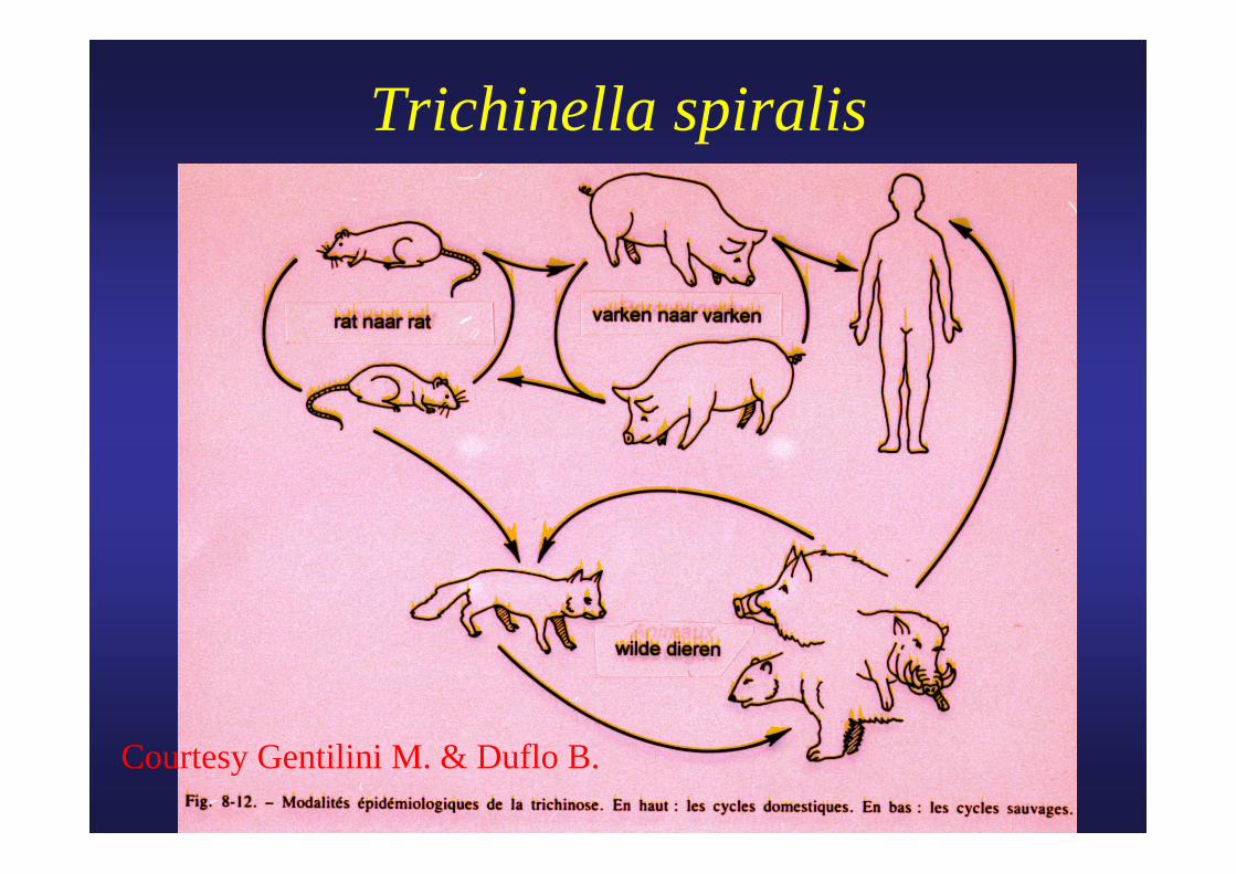
Trichinella spiralis
Courtesy Gentilini M. & Duflo B.
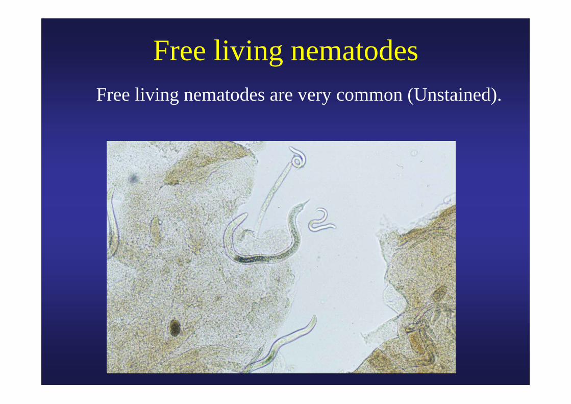
Free living nematodes Free living nematodes are very common (Unstained).