Embed Size (px)
Citation preview
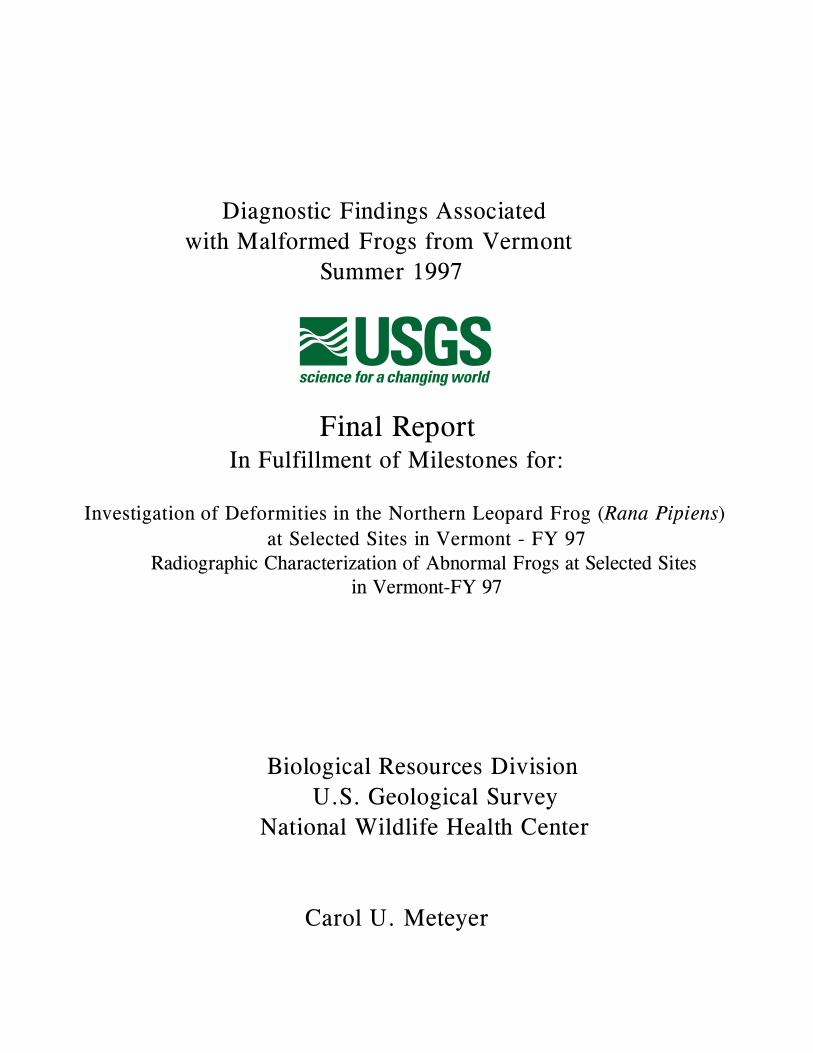
Diagnostic Findings Associated
with Malformed Frogs from Vermont
Summer 1997
Final Report In Fulfillment of Milestones for:
Investigation of Deformities in the Northern Leopard Frog (Rana Pipiens)
at Selected Sites in Vermont - FY 97Radiographic Characterization of Abnormal Frogs at Selected Sites
in Vermont-FY 97
Biological Resources Division
U.S. Geological Survey
National Wildlife Health Center
Carol U. Meteyer
Acknowledgments:
Report development: Cathy Pourchot-AckerTechnical assistance with radiographs: Nathan RamsayNecropsy technical assistance: Dottie Johnson, Matt McCollum, Nathan Ramsay, Kathleen Graber, Janna KottkeMicrobiological assistance: Doug Docherty, Rebecca Cole, Mark Wolcott, Connie Roderick, Brenda Berlowski, Tina Jaquish, Renee LongField assistance: Vermont Agency of Natural Resources, USFWS., Kathryn ConverseGraphics assistance and illustrations: Kathryn Converse, Harry Rihn, Cathy AckerData base and reference assistance: Kathy Wesenberg
Information presented here as well as the organizational schemes and table formats are not to be used or applied to
other field/pathology data without the permission of the pathologist.
3
TABLE OF CONTENTS
SUMMARY . . . . . . . . . . . . . . . . . . . . . . . . . . . . . . . . . . . . . . . . . . . . . . . . . . 4
BACKGROUND . . . . . . . . . . . . . . . . . . . . . . . . . . . . . . . . . . . . . . . . . . . . . . . 5
DEFINING THE PROBLEM . . . . . . . . . . . . . . . . . . . . . . . . . . . . . . . . . . . . . . 6
Morphogenesis . . . . . . . . . . . . . . . . . . . . . . . . . . . . . . . . . . . . . . . . . . . . 7
Malformation vs. Deformation . . . . . . . . . . . . . . . . . . . . . . . . . . . . . . . . . . 7
Teratogenesis . . . . . . . . . . . . . . . . . . . . . . . . . . . . . . . . . . . . . . . . . . . . . 7
FINDINGS . . . . . . . . . . . . . . . . . . . . . . . . . . . . . . . . . . . . . . . . . . . . . . . . . . 8
PHYSICAL CHARACTERISTICS OF FROGS WITH SIMILAR
MALFORMATIONS . . . . . . . . . . . . . . . . . . . . . . . . . . . . . . . . . . . . 8
Deformations . . . . . . . . . . . . . . . . . . . . . . . . . . . . . . . . . . . . . . . . . 8
Trauma (8)
Malformations . . . . . . . . . . . . . . . . . . . . . . . . . . . . . . . . . . . . . . . . 9
Amelia (9)
Polymelia (9)
Hemimelia (10)
Abnormal feet or missing digits (Ectrodactyly or Brachydactyly) (11)
Rotation of long bones and skin webbing (12)
Craniofacial abnormalities / Body conformation (12)
Normal (12)
INTERPRETATION OF DATA . . . . . . . . . . . . . . . . . . . . . . . . . . . . 13
MORPHOLOGIC FINDINGS IN FROGS FROM EACH STUDY SITE: . . . . 13
Alburg Dune . . . . . . . . . . . . . . . . . . . . . . . . . . . . . . . . . . . . . . . . 14
Poultney River . . . . . . . . . . . . . . . . . . . . . . . . . . . . . . . . . . . . . . . 15
Lapans Bay . . . . . . . . . . . . . . . . . . . . . . . . . . . . . . . . . . . . . . . . . 16
Missisquoi NWR . . . . . . . . . . . . . . . . . . . . . . . . . . . . . . . . . . . . . . 17
DETERMINATION OF BODY CONDITION . . . . . . . . . . . . . . . . . . . . . . . 17
HISTOPATHOLOGY FINDINGS . . . . . . . . . . . . . . . . . . . . . . . . . . . . . . 18
RADIOGRAPHIC FINDINGS . . . . . . . . . . . . . . . . . . . . . . . . . . . . . . . . . 18
RESULTS OF MICROBIOLOGICAL TESTING . . . . . . . . . . . . . . . . . . . . . 18
Parasitology . . . . . . . . . . . . . . . . . . . . . . . . . . . . . . . . . . . . . . . . 18
Bacteriology: . . . . . . . . . . . . . . . . . . . . . . . . . . . . . . . . . . . . . . . . 19
Virology . . . . . . . . . . . . . . . . . . . . . . . . . . . . . . . . . . . . . . . . . . . 19
FUTURE WORK . . . . . . . . . . . . . . . . . . . . . . . . . . . . . . . . . . . . . . . . . . . . . 19
4
APPENDICES
APPENDIX A: DEFINITIONS ................................................................20
APPENDIX B: Quick Response Grant Proposals
And Progress Report ...................................................22
APPENDIX C: STUDY PLAN.......... .......................................................27
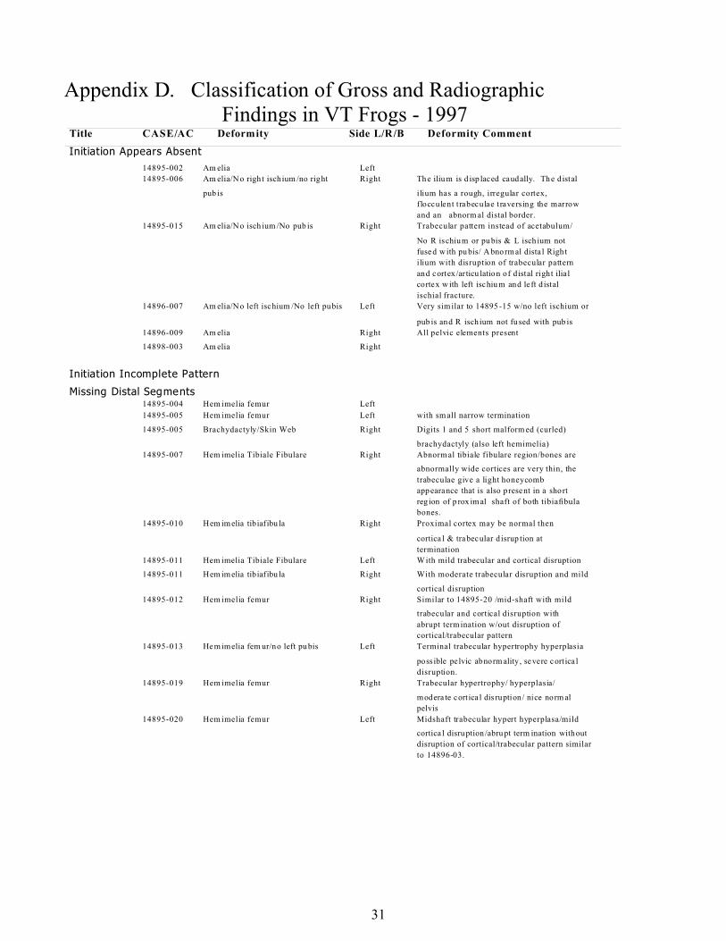
APPENDIX D: RADIOGRAPHIC/NECROPSY FINDINGS............................31
APPENDIX E: Enlargements of Report Radiographs and Tables.......................37
APPENDIX F: TABLE #1
Frog Body Condition/Average by Deformity..........................42
TABLE #2
Frog Body Condition Average by Collection Site....................45
APPENDIX G: HISTOPATHOLOGY FINDINGS........................................49
APPENDIX H: BACTERIOLOGY AND PARASITOLOGY
Raw Data Spread Sheets....................................................60
APPENDIX I: Limb Measurements
Raw Data Spread Sheets...................................................63
APPENDIX J: REFERENCES ...............................................................70
APPENDIX K: NECROPSY REPORTS.....................................................72
5
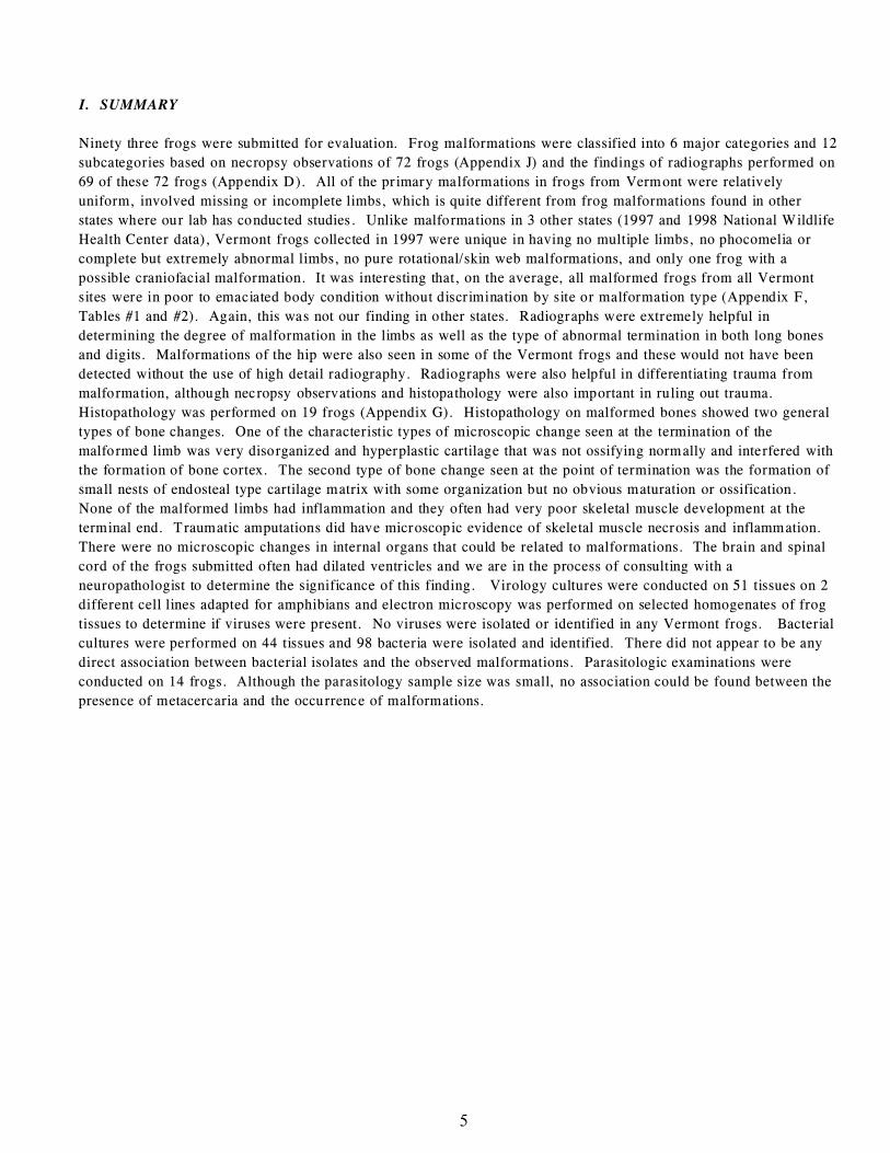
I. SUMMARY
Ninety three frogs were submitted for evaluation. Frog malformations were classified into 6 major categories and 12
subcategories based on necropsy observations of 72 frogs (Appendix J) and the findings of radiographs performed on
69 of these 72 frogs (Appendix D). All of the primary malformations in frogs from Vermont were relatively
uniform, involved missing or incomplete limbs, which is quite different from frog malformations found in other
states where our lab has conducted studies. Unlike malformations in 3 other states (1997 and 1998 National Wildlife
Health Center data), Vermont frogs collected in 1997 were unique in having no multiple limbs, no phocomelia or
complete but extremely abnormal limbs, no pure rotational/skin web malformations, and only one frog with a
possible craniofacial malformation. It was interesting that, on the average, all malformed frogs from all Vermont
sites were in poor to emaciated body condition without discrimination by site or malformation type (Appendix F,
Tables #1 and #2). Again, this was not our finding in other states. Radiographs were extremely helpful in
determining the degree of malformation in the limbs as well as the type of abnormal termination in both long bones
and digits. Malformations of the hip were also seen in some of the Vermont frogs and these would not have been
detected without the use of high detail radiography. Radiographs were also helpful in differentiating trauma from
malformation, although necropsy observations and histopathology were also important in ruling out trauma.
Histopathology was performed on 19 frogs (Appendix G). Histopathology on malformed bones showed two general
types of bone changes. One of the characteristic types of microscopic change seen at the termination of the
malformed limb was very disorganized and hyperplastic cartilage that was not ossifying normally and interfered with
the formation of bone cortex. The second type of bone change seen at the point of termination was the formation of
small nests of endosteal type cartilage matrix with some organization but no obvious maturation or ossification.
None of the malformed limbs had inflammation and they often had very poor skeletal muscle development at the
terminal end. Traumatic amputations did have microscopic evidence of skeletal muscle necrosis and inflammation.
There were no microscopic changes in internal organs that could be related to malformations. The brain and spinal
cord of the frogs submitted often had dilated ventricles and we are in the process of consulting with a
neuropathologist to determine the significance of this finding. Virology cultures were conducted on 51 tissues on 2
different cell lines adapted for amphibians and electron microscopy was performed on selected homogenates of frog
tissues to determine if viruses were present. No viruses were isolated or identified in any Vermont frogs. Bacterial
cultures were performed on 44 tissues and 98 bacteria were isolated and identified. There did not appear to be any
direct association between bacterial isolates and the observed malformations. Parasitologic examinations were
conducted on 14 frogs. Although the parasitology sample size was small, no association could be found between the
presence of metacercaria and the occurrence of malformations.
6
II. BACKGROUND
During the summer of 1995, a group of school children conducting a wetland nature study observed a high rate of
abnormal leopard frogs in Henderson County, Minnesota. Since 1995, occurrence of deformed frogs have been
reported in 42 states and 3 Canadian provinces (Gilbertson). The species reported have included northern leopard
frog (Rana pipiens), spring peeper (Hyla crucifer), American toad (Bufo americanus), gray treefrog (Hyla
versicolor), bullfrog (Rana catesbeiana), wood frog (Rana sylvatica), green frog (Rana clamitans), pickerel frog
(Rana palustris), and mink frog (Rana septentrionalis).
Studies, funded by the USGS Biological Resource Division’s Eastern Region in coordination with the Vermont
Agency of Natural Resources, and the U.S. Fish and Wildlife Service, were initiated through the National Wildlife
Health Center in the summer of 1997. The objectives of the study were to characterize frog abnormalities, document
the distribution of malformed frogs, determine the frequency of malformations, collect baseline morphometric
measurement of frogs, and collect water and sediment samples for analysis. The National Wildlife Health Center
(NWHC) was requested to assist in defining the developmental problems in Vermont frogs. The role of the NWHC
was to perform diagnostic assessments of abnormal and normal frogs from Vermont sites that were known to have
malformed frogs. The diagnostic evaluation included gross necropsy examination, histopathology, virology,
bacteriology, parasitology. In the spring of 1998 funding became available to perform radiology on a subset of the
Vermont frogs.
A report of preliminary findings was presented at the NIH/NIEHS Workshop on Strategies for Assessing the
Implications of Malformed Frogs for Environmental Health held December 4-5, 1997 at the National Institute of
Environmental Health Sciences, Research Triangle Park, North Carolina. The data we presented at the conference
made it clear that teratogenesis was occurring in tadpoles of the northern leopard frog. Factors that have been shown
in the lab to cause teratogenesis are numerous falling into the general categories of genetic or environmental. Some
environmental factors that can influence development are; chemical, thermal, infectious, or trauma-related factors
such as UV radiation which can cause cell damage.
Tadpoles are free-living postembryonic organisms that achieve all of their intricate developmental milestones without
the protection of a shell or uterus. The environment in which these tadpoles develop is complex and dynamic.
Sorting out the significant physical and chemical (molecular) factors that are contributing to teratogenesis in the frogs
from those factors that are coincidentally present in the environment without direct effect is important.
Malformations can indicate a general time frame during which factors are influencing the teratogenic process.
Knowledge of the possible developmental stages that have incorporated an error can provide guidance in choosing
appropriate times for collecting field samples for chemical analysis and guide researches in developing bioassays that
mimic the timing of factors present in the field. Careful characterization and categorization of malformations
occurring in the frogs can provide clues to potential causes through comparison with similar malformations that have
been produced in lab experiments. Some of these laboratory experiments involving development date back as far as
1929 (Vogt). However, associations based on morphologic evidence are circumstantial and without further field and
lab analyses, conclusions cannot be drawn regarding the causes of malformations.
To determine the causes of malformations, agents will need to be identified in the field at appropriate developmental
times, and in the appropriate geographic locations in relation to occurrence of malformations. The agents must be
capable of consistently producing comparable lesions in appropriate species of frogs. Lab experiments will need to
take into consideration the role played by such variables as timing of exposure, dose and duration of exposure, and
interactions between other environmental or host factors in the potential for producing the wide range of
malformations seen in the field. Laboratory results will need to be reproducible under field conditions.
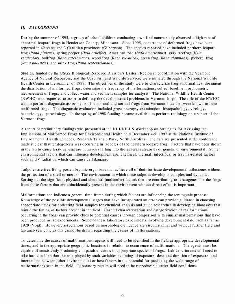
7
14897-011
III. DEFINING THE PROBLEM
The pathology of many of malformed frogs is obvious, the cause is not. Syndromes like
these have not been clearly defined in frogs and even the direction to look for answers is
under debate. These changes are not produced by a sudden and catastrophic insult.
Malformations represent the end point in a developmental process that incorporated an
error long before the external manifestation of pathology. What are the errors in
development and what questions can we ask to clarify the possible causes of these errors?
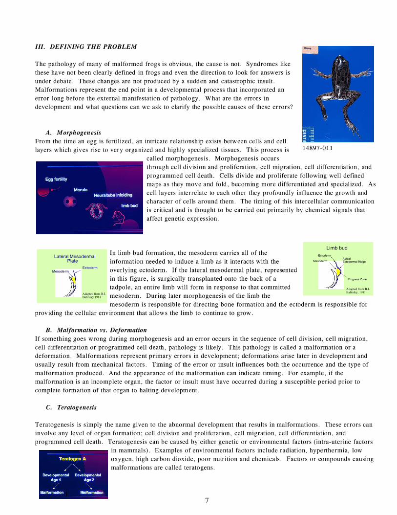
A. Morphogenesis
From the time an egg is fertilized, an intricate relationship exists between cells and cell
layers which gives rise to very organized and highly specialized tissues. This process is
called morphogenesis. Morphogenesis occurs
through cell division and proliferation, cell migration, cell differentiation, and
programmed cell death. Cells divide and proliferate following well defined
maps as they move and fold, becoming more differentiated and specialized. As
cell layers interrelate to each other they profoundly influence the growth and
character of cells around them. The timing of this intercellular communication
is critical and is thought to be carried out primarily by chemical signals that
affect genetic expression.
In limb bud formation, the mesoderm carries all of the
information needed to induce a limb as it interacts with the
overlying ectoderm. If the lateral mesodermal plate, represented
in this figure, is surgically transplanted onto the back of a
tadpole, an entire limb will form in response to that committed
mesoderm. During later morphogenesis of the limb the
mesoderm is responsible for directing bone formation and the ectoderm is responsible for
providing the cellular environment that allows the limb to continue to grow.
B. Malformation vs. Deformation
If something goes wrong during morphogenesis and an error occurs in the sequence of cell division, cell migration,
cell differentiation or programmed cell death, pathology is likely. This pathology is called a malformation or a
deformation. Malformations represent primary errors in development; deformations arise later in development and
usually result from mechanical factors. Timing of the error or insult influences both the occurrence and the type of
malformation produced. And the appearance of the malformation can indicate timing. For example, if the
malformation is an incomplete organ, the factor or insult must have occurred during a susceptible period prior to
complete formation of that organ to halting development.
C. Teratogenesis
Teratogenesis is simply the name given to the abnormal development that results in malformations. These errors can
involve any level of organ formation; cell division and proliferation, cell migration, cell differentiation, and
programmed cell death. Teratogenesis can be caused by either genetic or environmental factors (intra-uterine factors
in mammals). Examples of environmental factors include radiation, hyperthermia, low
oxygen, high carbon dioxide, poor nutrition and chemicals. Factors or compounds causing
malformations are called teratogens.
8
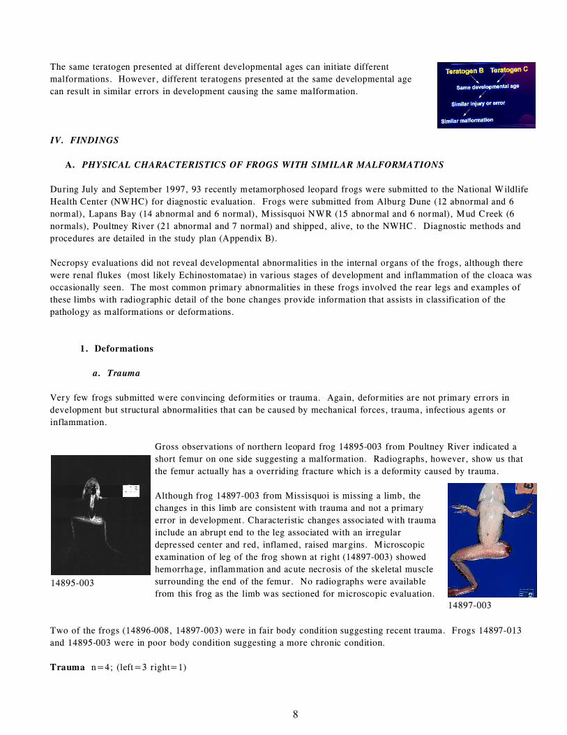
14897-003
14895-003
The same teratogen presented at different developmental ages can initiate different
malformations. However, different teratogens presented at the same developmental age
can result in similar errors in development causing the same malformation.
IV. FINDINGS
A. PHYSICAL CHARACTERISTICS OF FROGS WITH SIMILAR MALFORMATIONS
During July and September 1997, 93 recently metamorphosed leopard frogs were submitted to the National Wildlife
Health Center (NWHC) for diagnostic evaluation. Frogs were submitted from Alburg Dune (12 abnormal and 6
normal), Lapans Bay (14 abnormal and 6 normal), Missisquoi NWR (15 abnormal and 6 normal), Mud Creek (6
normals), Poultney River (21 abnormal and 7 normal) and shipped, alive, to the NWHC. Diagnostic methods and
procedures are detailed in the study plan (Appendix B).
Necropsy evaluations did not reveal developmental abnormalities in the internal organs of the frogs, although there
were renal flukes (most likely Echinostomatae) in various stages of development and inflammation of the cloaca was
occasionally seen. The most common primary abnormalities in these frogs involved the rear legs and examples of
these limbs with radiographic detail of the bone changes provide information that assists in classification of the
pathology as malformations or deformations.
1. Deformations
a. Trauma
Very few frogs submitted were convincing deformities or trauma. Again, deformities are not primary errors in
development but structural abnormalities that can be caused by mechanical forces, trauma, infectious agents or
inflammation.
Gross observations of northern leopard frog 14895-003 from Poultney River indicated a
short femur on one side suggesting a malformation. Radiographs, however, show us that
the femur actually has a overriding fracture which is a deformity caused by trauma.
Although frog 14897-003 from Missisquoi is missing a limb, the
changes in this limb are consistent with trauma and not a primary
error in development. Characteristic changes associated with trauma
include an abrupt end to the leg associated with an irregular
depressed center and red, inflamed, raised margins. Microscopic
examination of leg of the frog shown at right (14897-003) showed
hemorrhage, inflammation and acute necrosis of the skeletal muscle
surrounding the end of the femur. No radiographs were available
from this frog as the limb was sectioned for microscopic evaluation.
Two of the frogs (14896-008, 14897-003) were in fair body condition suggesting recent trauma. Frogs 14897-013
and 14895-003 were in poor body condition suggesting a more chronic condition.
Trauma n=4; (left=3 right=1)
9
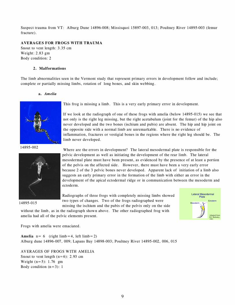
14895-002
14895-015
Suspect trauma from VT: Alburg Dune 14896-008; Missisquoi 15897-003, 013; Poultney River 14895-003 (femur
fracture).
AVERAGES FOR FROGS WITH TRAUMA
Snout to vent length: 3.35 cm
Weight: 2.83 gm
Body condition: 2
2. Malformations
The limb abnormalities seen in the Vermont study that represent primary errors in development follow and include;
complete or partially missing limbs, rotation of long bones, and skin webbing.
a. Amelia
This frog is missing a limb. This is a very early primary error in development.
If we look at the radiograph of one of these frogs with amelia (below 14895-015) we see that
not only is the right leg missing, but the right acetabulum (joint for the femur) of the hip also
never developed and the two bones (ischium and pubis) are absent. The hip and hip joint on
the opposite side with a normal limb are unremarkable. There is no evidence of
inflammation, fractures or vestigial bones in the regions where the right leg should be. The
limb never developed.
Where are the errors in development? The lateral mesodermal plate is responsible for the
pelvic development as well as initiating the development of the rear limb. The lateral
mesodermal plate must have been present, as evidenced by the presence of at least a portion
of the pelvis on the affected side. However, there must have been a very early error
because 2 of the 3 pelvic bones never developed. Apparent lack of initiation of a limb also
suggests an early primary error in the formation of the limb with either an error in the
development of the apical ectodermal ridge or in communication between the mesoderm and
ectoderm.
Radiographs of three frogs with completely missing limbs showed
two types of changes. Two of the frogs radiographed were
missing the ischium and the pubis of the pelvis only on the side
without the limb, as in the radiograph shown above. The other radiographed frog with
amelia had all of the pelvic elements present.
Frogs with amelia were emaciated.
Amelia n= 6 (right limb=4, left limb=2)
Alburg dune 14896-007, 009; Lapans Bay 14898-003; Poultney River 14895-002, 006, 015
AVERAGES OF FROGS WITH AMELIA
Snout to vent length (n=6): 2.93 cm
Weight (n=5): 1.76 gm
Body condition (n=3): 1
10
14896-003 14896-003
14896-014 14896-013
b. Polymelia
Multiple limbs (n=0)
None of the Vermont frogs examined by the NWHC in 1997 had multiple limbs or multiple elements associated with
the limb.
c. Hemimelia
Truncation of the limb suggests an error prior to the completion of limb development but an error that probably
occurred later in development than those errors occurring in frogs with amelia.
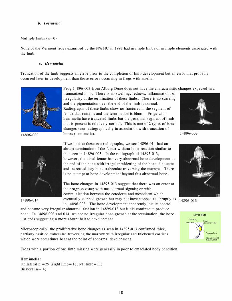
Frog 14896-003 from Alburg Dune does not have the characteristic changes expected in a
traumatized limb. There is no swelling, redness, inflammation, or
irregularity at the termination of these limbs. There is no scarring
and the pigmentation over the end of the limb is normal.
Radiographs of these limbs show no fractures in the segment of
femur that remains and the termination is blunt. Frogs with
hemimelia have truncated limbs but the proximal segment of limb
that is present is relatively normal. This is one of 2 types of bone
changes seen radiographically in association with truncation of
bones (hemimelia).
If we look at these two radiographs, we see 14896-014 had an
abrupt termination of the femur without bone reaction similar to
that seen in 14896-003. In the radiograph of 14895-013,
however, the distal femur has very abnormal bone development at
the end of the bone with irregular widening of the bone silhouette
and increased lacy bone trabeculae traversing the marrow. There
is no attempt at bone development beyond this abnormal bone.
The bone changes in 14895-013 suggest that there was an error at
the progress zone; with mesodermal signals; or with
communication between the ectoderm and mesoderm which
eventually stopped growth but may not have stopped as abruptly as
in 14896-003. The bone development apparently lost its control
and became very irregular abnormal fashion in 14895-013 but it did continue to produce
bone. In 14896-003 and 014, we see no irregular bone growth at the termination, the bone
just ends suggesting a more abrupt halt to development.
Microscopically, the proliferative bone changes as seen in 14895-013 confirmed thick,
partially ossified trabeculae traversing the marrow with irregular and thickened cortices
which were sometimes bent at the point of abnormal development.
Frogs with a portion of one limb missing were generally in poor to emaciated body condition.
Hemimelia:
Unilateral n =29 (right limb=18, left limb=11)
Bilateral n= 4;
11
14895-008
14895-008
14895-018
Lapans Bay 14898-002, 005, 007, 008, 009, 011, 012, 014.; Missisquoi 14897-006, 008, 009, 010, 011, 012, 014,
017, 022; Alburg dune 14896-003, 005, 006, 010, 014; Poultney River 14895-004, 005, 007, 010, 011, 012, 013,
019, 020, 021
Hemimelia of femur n=11
Hemimelia of tibiafibula n=16
Hemimelia of tibiale and or fibulare n=3
Adactyly n=3
Bilateral hemimelia received counts for each leg for the occurrence of hemimelia, hence the 3 extra malformation
designations above. In the bilateral hemimelia frogs, one had bilateral femoral hemimelia, one had a tibiafibula
hemimelia and a tibiale/fibulare hemimelia, and the other frog had bilateral adactyly.
AVERAGES OF FROGS WITH HEMIMELIA
Hemimelia of femur
Snout to vent length n=11: 3.25 cm
Weight n=11: 2.66 gm
Body condition n=7: 1.71
Hemimelia of tibiale, fibulare (n=3)
Snout to vent length: 3.53 cm
Weight n=: 2.73 gm
Body condition (n=2): 1.0
Hemimelia of tibiafibula = 16
Snout to vent length n=15: 3.27 cm
Weight: 2.45 gm
Body condition n=11: 1.82
Adactyly (n=3)
Snout to vent length n=2: 3.35 cm
Weight n=2: 2.50 gm
Body condition n=1: 1.0
d. Abnormal feet or missing digits (Ectrodactyly or Brachydactyly)
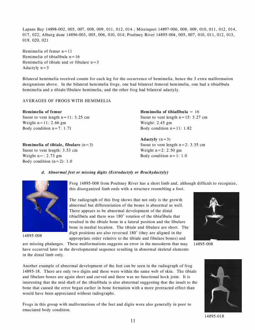
Frog 14895-008 from Poultney River has a short limb and, although difficult to recognize,
this disorganized limb ends with a structure resembling a foot.
The radiograph of this frog shows that not only is the growth
abnormal but differentiation of the bones is abnormal as well.
There appears to be abnormal development of the distal
tibiafibula and there was 180/ rotation of the tibiafibula that
resulted in the tibiale bone in a lateral position and the fibulare
bone in medial location. The tibiale and fibulare are short. The
digit positions are also reversed 180/ (they are aligned in the
appropriate order relative to the tibiale and fibulare bones) and
are missing phalanges. These malformations suggests an error in the mesoderm that may
have occurred later in the developmental sequence resulting in abnormal skeletal elements
in the distal limb only.
Another example of abnormal development of the feet can be seen in the radiograph of frog
14895-18. There are only two digits and these were within the same web of skin. The tibiale
and fibulare bones are again short and curved and there was no functional hock joint. It is
interesting that the mid-shaft of the tibiafibula is also abnormal suggesting that the insult to the
bone that caused the error began earlier in bone formation with a more protracted effect than
would have been appreciated without radiographs.
Frogs in this group with malformations of the feet and digits were also generally in poor to
emaciated body condition.
12
14895-010
Abnormal development of feet or digits; n= 24 (right limb=15, left limb=9)
Lapans Bay 14898-001, 006, 010; Missisquoi 14897-001, 004, 005, 007, 015(adult), 016, 018, 019, 020, 021, 023;
Alburg Dune 14896-001 (adult), 004, 009; Poultney River 14895-001, 005, 008, 009, 014, 016, 018.
AVERAGES OF FROGS WITH ECTRODACTYLY: (Not including adults)
Ectrodactyly (n=17)
Snout to vent length (n=12): 3.25 cm
Weight (n=12): 2.77 gm
Body condition (n=7 recorded): 1.57
Brachydactyly (n=4)
Snout to vent length: 2.88 cm
Weight: 1.88 gm
Body condition: 1
e. Rotation of long bones and skin webbing
The radiograph of the Vermont frog shown (14895-010), although missing the distal limb on one side has rotation of
the lower limb of the more normal leg with a bend in the bone that is traversed by a “bone bridge”. Rotational
abnormalities in the Minnesota frogs were very common. These rotational abnormalities
involved the limb proximal to the hock causing the flexed hock to have either a dorsal or
ventral orientation rather than pointing in the more normal medial direction. Although
there were no Vermont frogs with rotation as its primarily malformation rotations did occur
in relation with other malformations and did resemble some of the Minnesota rotations.
Rotations were most likely to occur in frogs with ectrodactyly or brachydactyly (missing or
shortened digits) suggesting that errors in the bone formation was occurring proximal to the
affected digits (4 frogs). Limb rotation also occurred in two frogs with femoral hemimelia
of the opposite limb and in the same limb as tibiafibular hemimelia in another frog. It
appeared that the more proximal the source of rotational abnormality (ie femur vs
tibiafibula), the more severely the propelling surface deviated from the norm.
Limb Rotation n=7; (right limb=2, left limb=5)
Missisquoi 14897- 004, 006, 011; Poultney River 14895-001, 005, 008, 010.
AVERAGES OF FROGS WITH ROTATION
Snout to vent length n=7: 3.32 cm
Weight n=7: 2.26 gm
Body condition n=6: 1.17
f. Craniofacial abnormalities / Body conformation
Although jaw and skull malformations have been common in other states, none were seen in the submissions from
Vermont.
One of the frogs from Alburg Dune (14896-011) was runted with possible compression of the vertebrae.
Short compressed (runted) body type (n=1; only with full assessments)
Snout to vent length: 2.6 cm
Weight: 1.3 gm
Body condition: 1
13
g. Normal
Normal frogs varied in size between sites. The average weight and length of normal frogs from Mud Creek, which
was the control site, were 4.35 gm and 3.97 cm respectively, average measures for weight and length of normal
frogs from sites with malformations were Alburg Dune: weight 2.0 gm and 3.97 cm and average weight and length
of normal frogs submitted from Lapans Bay were 1.4 gm and 2.6 cm respectively.
AVERAGES FOR NORMAL FROGS FROM CONTROL SITE
Mud Creek 14899-001, 002, 003, 004, 005, 006
Snout to vent length 3.97 cm
Weight 4.35 gm
Body condition 3.7
AVERAGES FOR NORMAL FROGS FROM SITES WITH MALFORMATIONS
Alburg Dune: 14896- 012, 013, 015
Snout to vent length: 3.13 cm
Weight 2.0 gm
Body condition 1 (recorded in 012 only)
Lapans Bay: 14898-015
Snout to vent length: 2.6 cm
Weight: 1.4 gm
Body condition: (not recorded)
Missisquoi NWR and Poultney River
No Normal frogs submitted
3. INTERPRETATION OF DATA
Although the number of frogs submitted from each site was low, it seems important to look at the data to see if there
was an association between malformation type and body condition. The potential impact a particular abnormalities
might have on frog success, as represented by general body condition or nutritional status, might also be accessible
through data analysis. Data from the field studies will be needed to determine if the malformations submitted from
each site for this study were truly representative of the population in both malformation type and frequency of
malformations present.
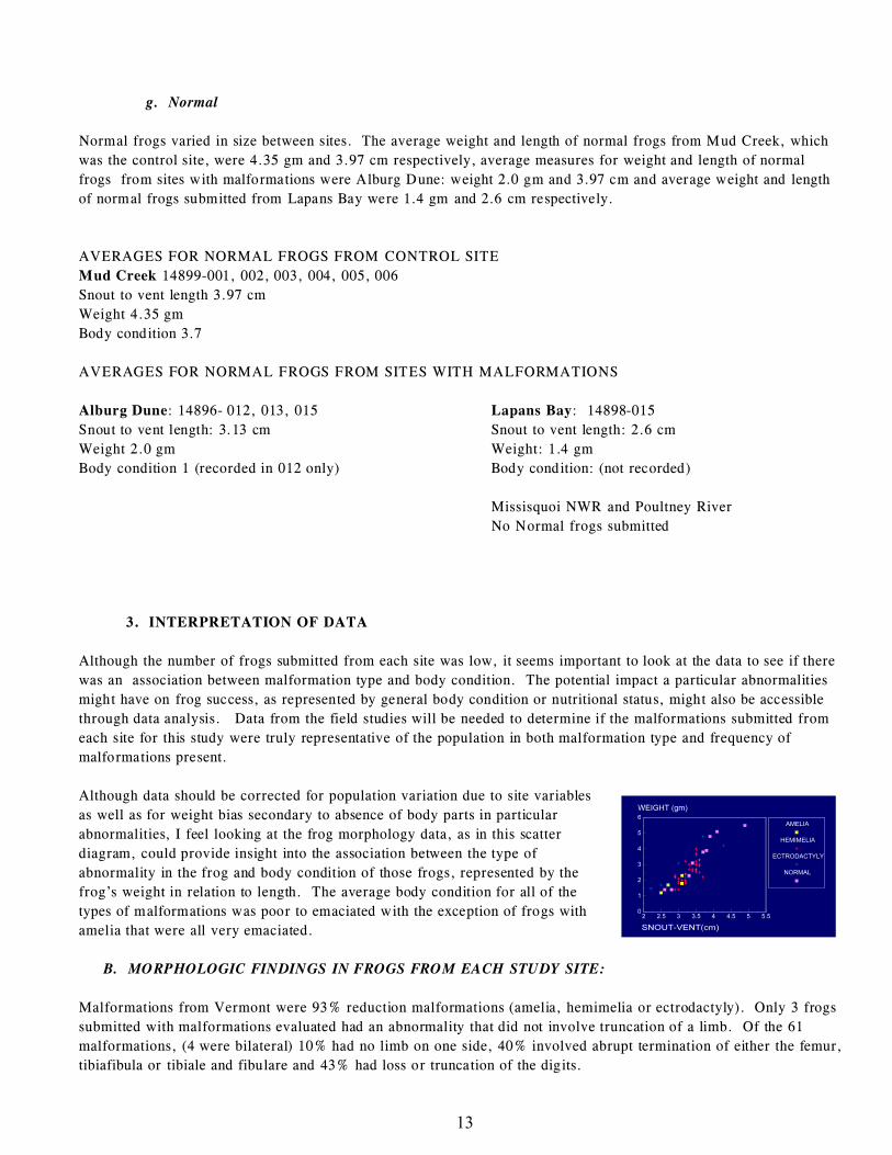
Although data should be corrected for population variation due to site variables
as well as for weight bias secondary to absence of body parts in particular
abnormalities, I feel looking at the frog morphology data, as in this scatter
diagram, could provide insight into the association between the type of
abnormality in the frog and body condition of those frogs, represented by the
frog’s weight in relation to length. The average body condition for all of the
types of malformations was poor to emaciated with the exception of frogs with
amelia that were all very emaciated.
B. MORPHOLOGIC FINDINGS IN FROGS FROM EACH STUDY SITE:
Malformations from Vermont were 93% reduction malformations (amelia, hemimelia or ectrodactyly). Only 3 frogs
submitted with malformations evaluated had an abnormality that did not involve truncation of a limb. Of the 61
malformations, (4 were bilateral) 10% had no limb on one side, 40% involved abrupt termination of either the femur,
tibiafibula or tibiale and fibulare and 43% had loss or truncation of the digits.

14
Frogs from Missisquoi had the most complete limb development with truncations involving the lower limb distal to
the femur. Frogs from Poultney River and Alburg Dune (although fewer number of total frogs were submitted from
Alburg Dune) had the most severe truncations with the primary abnormalities involving apparent lack of any
initiation of limb (amelia) or truncation of the limb at the femur (femoral hemimelia). Average body condition in
frogs from all of the study sites was poor to emaciated. Frogs with no apparent malformations (controls) collected at
the study sites also were in poor to fair body condition but the sample size was too small to make an extrapolation to
the overall population of leopard frogs at these sites. Lapans Bay had rear limb malformations primarily involving
the tibiafibula and the foot.
This chart of association of severity of malformation with site of
collection might suggest errors occurring either at different developmental
stages in frog development, or different dose or duration of exposure to a
teratogen.
1. Alburg Dune
ALBURG
DUNE (14896)
Total Frogs with malformed rear limbs: 11
Amelia: 2 = 18% of all malformations from Alburg
Dune
Hemimelia (one frog was bilateral): 6 = 55% of all
malformations from Alburg Dune and 67%
involved the femur
Abnormal feet or digits: 3 = 27% of all
malformations from Alburg Dune
Trauma: 1
Primary Site Malformation: Rear leg hemimelia/
ectromelia
4 femur, 2 tibiafibula, 3 digits or phalanges
This represented 27% of all long bone hemimelia
submitted from Vermont (femur or tibiafibula) and
13% of malformations submitted that had missing digits
or phalanges. Although only 2 frogs from Alburg Dune had completely missing limbs, this
represented 33% of all of the amelia submitted from
Vermont. One frog (14896-011) had stunted body
composition.
Average measurements for malformed frogs from Alburg Dune (n=9)
Snout to vent length: 2.99 cm
Weight: 1.93 gm
Body condition (n=7): 1.86
Average measurements for normal control frogs from Alburg Dune: (n=3)
Snout to vent length for normal frogs: 3.13 cm.
Weight of normal frogs: 2.03 gm
Body condition (n=1): 1
15
14895-010
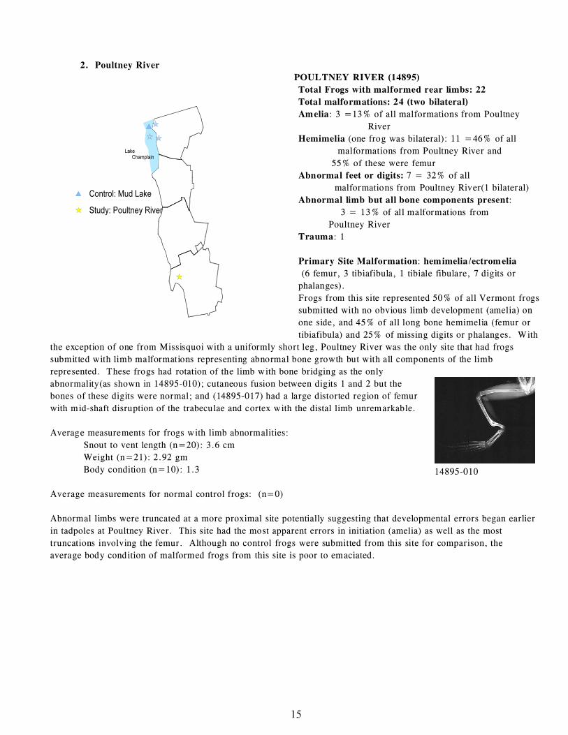
2. Poultney River
POULTNEY RIVER (14895)
Total Frogs with malformed rear limbs: 22
Total malformations: 24 (two bilateral)
Amelia: 3 =13% of all malformations from Poultney
River
Hemimelia (one frog was bilateral): 11 =46% of all
malformations from Poultney River and
55% of these were femur
Abnormal feet or digits: 7 = 32% of all
malformations from Poultney River(1 bilateral)
Abnormal limb but all bone components present:
3 = 13% of all malformations from
Poultney River
Trauma: 1
Primary Site Malformation: hemimelia/ectromelia
(6 femur, 3 tibiafibula, 1 tibiale fibulare, 7 digits or
phalanges).
Frogs from this site represented 50% of all Vermont frogs
submitted with no obvious limb development (amelia) on
one side, and 45% of all long bone hemimelia (femur or
tibiafibula) and 25% of missing digits or phalanges. With
the exception of one from Missisquoi with a uniformly short leg, Poultney River was the only site that had frogs
submitted with limb malformations representing abnormal bone growth but with all components of the limb
represented. These frogs had rotation of the limb with bone bridging as the only
abnormality(as shown in 14895-010); cutaneous fusion between digits 1 and 2 but the
bones of these digits were normal; and (14895-017) had a large distorted region of femur
with mid-shaft disruption of the trabeculae and cortex with the distal limb unremarkable.
Average measurements for frogs with limb abnormalities:
Snout to vent length (n=20): 3.6 cm
Weight (n=21): 2.92 gm
Body condition (n=10): 1.3
Average measurements for normal control frogs: (n=0)
Abnormal limbs were truncated at a more proximal site potentially suggesting that developmental errors began earlier
in tadpoles at Poultney River. This site had the most apparent errors in initiation (amelia) as well as the most
truncations involving the femur. Although no control frogs were submitted from this site for comparison, the
average body condition of malformed frogs from this site is poor to emaciated.
16
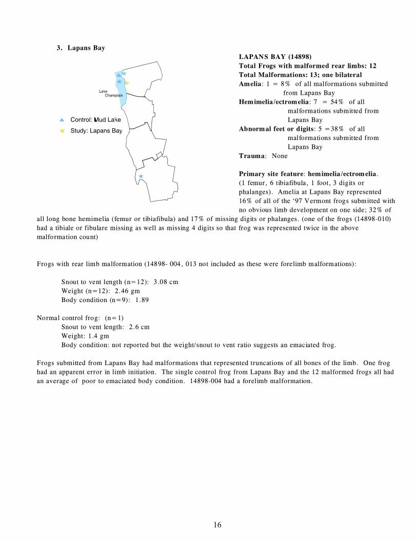
3. Lapans Bay
LAPANS BAY (14898)
Total Frogs with malformed rear limbs: 12
Total Malformations: 13; one bilateral
Amelia: 1 = 8% of all malformations submitted
from Lapans Bay
Hemimelia/ectromelia: 7 = 54% of all
malformations submitted from
Lapans Bay
Abnormal feet or digits: 5 =38% of all
malformations submitted from
Lapans Bay
Trauma: None
Primary site feature: hemimelia/ectromelia.
(1 femur, 6 tibiafibula, 1 foot, 3 digits or
phalanges). Amelia at Lapans Bay represented
16% of all of the ‘97 Vermont frogs submitted with
no obvious limb development on one side; 32% of
all long bone hemimelia (femur or tibiafibula) and 17% of missing digits or phalanges. (one of the frogs (14898-010)
had a tibiale or fibulare missing as well as missing 4 digits so that frog was represented twice in the above
malformation count)
Frogs with rear limb malformation (14898- 004, 013 not included as these were forelimb malformations):
Snout to vent length (n=12): 3.08 cm
Weight (n=12): 2.46 gm
Body condition (n=9): 1.89
Normal control frog: (n=1)
Snout to vent length: 2.6 cm
Weight: 1.4 gm
Body condition: not reported but the weight/snout to vent ratio suggests an emaciated frog.
Frogs submitted from Lapans Bay had malformations that represented truncations of all bones of the limb. One frog
had an apparent error in limb initiation. The single control frog from Lapans Bay and the 12 malformed frogs all had
an average of poor to emaciated body condition. 14898-004 had a forelimb malformation.
17
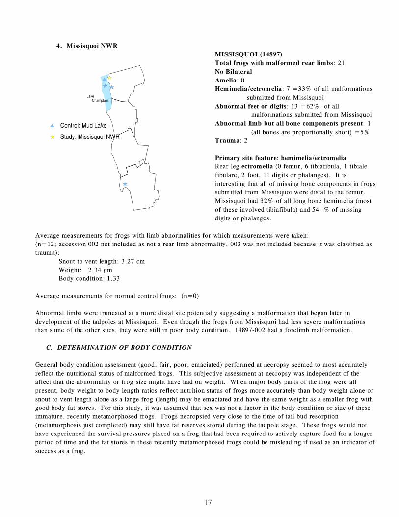
4. Missisquoi NWR
MISSISQUOI (14897)
Total frogs with malformed rear limbs: 21
No Bilateral
Amelia: 0
Hemimelia/ectromelia: 7 =33% of all malformations
submitted from Missisquoi
Abnormal feet or digits: 13 =62% of all
malformations submitted from Missisquoi
Abnormal limb but all bone components present: 1
(all bones are proportionally short) =5%
Trauma: 2
Primary site feature: hemimelia/ectromelia
Rear leg ectromelia (0 femur, 6 tibiafibula, 1 tibiale
fibulare, 2 foot, 11 digits or phalanges). It is
interesting that all of missing bone components in frogs
submitted from Missisquoi were distal to the femur.
Missisquoi had 32% of all long bone hemimelia (most
of these involved tibiafibula) and 54 % of missing
digits or phalanges.
Average measurements for frogs with limb abnormalities for which measurements were taken:
(n=12; accession 002 not included as not a rear limb abnormality, 003 was not included because it was classified as
trauma):
Snout to vent length: 3.27 cm
Weight: 2.34 gm
Body condition: 1.33
Average measurements for normal control frogs: (n=0)
Abnormal limbs were truncated at a more distal site potentially suggesting a malformation that began later in
development of the tadpoles at Missisquoi. Even though the frogs from Missisquoi had less severe malformations
than some of the other sites, they were still in poor body condition. 14897-002 had a forelimb malformation.
C. DETERMINATION OF BODY CONDITION
General body condition assessment (good, fair, poor, emaciated) performed at necropsy seemed to most accurately
reflect the nutritional status of malformed frogs. This subjective assessment at necropsy was independent of the
affect that the abnormality or frog size might have had on weight. When major body parts of the frog were all
present, body weight to body length ratios reflect nutrition status of frogs more accurately than body weight alone or
snout to vent length alone as a large frog (length) may be emaciated and have the same weight as a smaller frog with
good body fat stores. For this study, it was assumed that sex was not a factor in the body condition or size of these
immature, recently metamorphosed frogs. Frogs necropsied very close to the time of tail bud resorption
(metamorphosis just completed) may still have fat reserves stored during the tadpole stage. These frogs would not
have experienced the survival pressures placed on a frog that had been required to actively capture food for a longer
period of time and the fat stores in these recently metamorphosed frogs could be misleading if used as an indicator of
success as a frog.
18
D. HISTOPATHOLOGY FINDINGS
Histopathology was performed on 19 frogs (Appendix F). Histopathology on malformed bones showed two general
types of bone changes. One of the characteristics of bone at the termination of the malformed limb was very
disorganized and hyperplastic cartilage that was not ossifying normally and interfered with the formation of bone
cortex. The second type of bone change at the point of termination was the formation of small nests of endosteal type
cartilage matrix with some organization but no obvious maturation or ossification. None of the malformed limbs had
inflammation and they often had very poor skeletal muscle development. Traumatic amputations did have
microscopic evidence of skeletal muscle necrosis and inflammation. No consistent microscopic changes were seen in
internal organs (heart, lung, liver, stomach, intestine, pancreas, kidney, bladder, gonad, spleen, thyroid gland,
thymus, lymphoid aggregates) that could be related to malformations. The brain and spinal cord of the frogs
submitted often had dilated ventricles and we are in the process of consulting with a neuropathologist to determine
the significance of this finding.
E. RADIOGRAPHIC FINDINGS
Radiographs were extremely helpful in determining the degree of malformation in the limbs as well as the type of
termination in both long bones and digits.(Appendix D) Malformations of the hip were also seen in some of the
Vermont frogs and these would not have been detected without the use of high detail radiography. Radiographs were
also helpful in differentiating trauma from malformation, although necropsy observations and histopathology were
also important in ruling out trauma.
F. RESULTS OF MICROBIOLOGICAL TESTING
Lab tests were performed on the frogs sent into our lab so that information involving infectious agents could be
looked at in relation to malformation types.
1. Parasitology
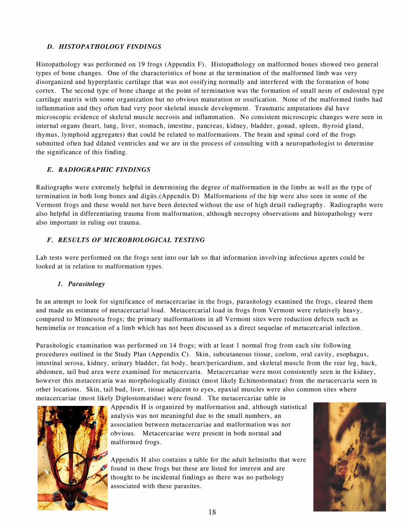
In an attempt to look for significance of metacercariae in the frogs, parasitology examined the frogs, cleared them
and made an estimate of metacercarial load. Metacercarial load in frogs from Vermont were relatively heavy,
compared to Minnesota frogs; the primary malformations in all Vermont sites were reduction defects such as
hemimelia or truncation of a limb which has not been discussed as a direct sequelae of metacercarial infection.
Parasitologic examination was performed on 14 frogs; with at least 1 normal frog from each site following
procedures outlined in the Study Plan (Appendix C). Skin, subcutaneous tissue, coelom, oral cavity, esophagus,
intestinal serosa, kidney, urinary bladder, fat body, heart/pericardium, and skeletal muscle from the rear leg, back,
abdomen, tail bud area were examined for metacercaria. Metacercariae were most consistently seen in the kidney,
however this metacercaria was morphologically distinct (most likely Echinostomatae) from the metacercaria seen in
other locations. Skin, tail bud, liver, tissue adjacent to eyes, epaxial muscles were also common sites where
metacercariae (most likely Diplostomatidae) were found. The metacercariae table in
Appendix H is organized by malformation and, although statistical
analysis was not meaningful due to the small numbers, an
association between metacercariae and malformation was not
obvious. Metacercariae were present in both normal and
malformed frogs.
Appendix H also contains a table for the adult helminths that were
found in these frogs but these are listed for interest and are
thought to be incidental findings as there was no pathology
associated with these parasites.
19
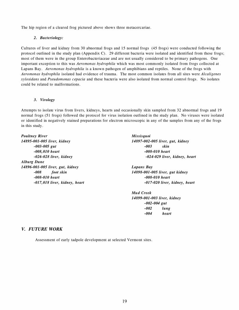
The hip region of a cleared frog pictured above shows three metacercariae.
2. Bacteriology:
Cultures of liver and kidney from 30 abnormal frogs and 15 normal frogs (45 frogs) were conducted following the
protocol outlined in the study plan (Appendix C). 29 different bacteria were isolated and identified from these frogs;
most of them were in the group Enterobacteriaceae and are not usually considered to be primary pathogens. One
important exception to this was Aeromonas hydrophila which was most commonly isolated from frogs collected at
Lapans Bay. Aeromonas hydrophila is a known pathogen of amphibians and reptiles. None of the frogs with
Aeromonas hydrophila isolated had evidence of trauma. The most common isolates from all sites were Alcaligenes
xylosidans and Pseudomonas cepacia and these bacteria were also isolated from normal control frogs. No isolates
could be related to malformations.
3. Virology
Attempts to isolate virus from livers, kidneys, hearts and occasionally skin sampled from 32 abnormal frogs and 19
normal frogs (51 frogs) followed the protocol for virus isolation outlined in the study plan. No viruses were isolated
or identified in negatively stained preparations for electron microscopic in any of the samples from any of the frogs
in this study.
Poultney River
14895-001-005 liver, kidney
-003-005 gut
-008,010 heart
-026-028 liver, kidney
Alburg Dune
14896-001-005 liver, gut, kidney
-008 foot skin
-008-010 heart
-017,018 liver, kidney, heart
Missisquoi
14897-002-005 liver, gut, kidney
-003 skin
-008-010 heart
-024-029 liver, kidney, heart
Lapans Bay
14898-001-005 liver, gut kidney
-008-010 heart
-017-020 liver, kidney, heart
Mud Creek
14899-001-003 liver, kidney
-002-004 gut
-002 lung
-004 heart
V. FUTURE WORK
Assessment of early tadpole development at selected Vermont sites.
APPENDICES
APPENDIX A: DEFINITIONS ................................................................20
APPENDIX B: Quick Response Grant Proposals
And Progress Report ...................................................22
APPENDIX C: STUDY PLAN.......... .......................................................27
APPENDIX D: RADIOGRAPHIC/NECROPSY FINDINGS............................31
APPENDIX E: Enlargements of Report Radiographs and Tables.......................37
APPENDIX F: TABLE #1
Frog Body Condition/Average by Deformity..........................42
TABLE #2
Frog Body Condition Average by Collection Site....................45
APPENDIX G: HISTOPATHOLOGY FINDINGS........................................49
APPENDIX H: BACTERIOLOGY AND PARASITOLOGY
Raw Data Spread Sheets....................................................60
APPENDIX I: Limb Measurements
Raw Data Spread Sheets...................................................63
APPENDIX J: REFERENCES ...............................................................70
APPENDIX K: NECROPSY REPORTS.....................................................72
20
APPENDIX A: DEFINITIONS
Description of frog malformations in this report are based on terminology used in human literature (Bolande, Robbins),
developmental biology (O’Rahilly; Carlson), and teratology (Wise). Many of these terms, defined in Appendix B, were
originally used to describe defects that are present in the mammalian fetus at birth. Direct application of these terms to
frogs that metamorphose in a free-living postembryonic state may or may not prove to be appropriate. However, common
terminology may allow recognition of similar conditions by individuals in related specialties involving developmental
anomalies across species and bring new interest and collaboration to the issues involving malformed frogs. The
terminology in reference to possible points of error in development (mesodermal versus ectodermal) may be simplistic, but
allows an initial attempt at looking at the pathogenesis of some of these malformations.
Case number: National Wildlife Health Center (NWHC) reference number given to a group of frogs submitted from the
same site and usually collected in a similar time frame.
Accession number: NWHC reference number given to individual frogs within a case.
Body condition (as defined for this report by Meteyer): Body condition reflects the nutritional state of the frog and is
determined by the pathologist by subjective assessment of relative quantity of muscle and body fat present in the frog at the
time of necropsy. Assessment of body condition is independent of absolute weight or snout to vent length. Body condition
has been scored in the following way so that averages can be made for comparison between sites and abnormalities;
good=4, fair=3, poor=2, emaciated=1.
Good body condition: Adequate skeletal muscle and abundant fat in the fat bodies.
Fair body condition: Adequate skeletal muscle with reduced fat in the fat bodies.
Poor body condition: Adequate skeletal muscle with scant or no body fat in the fat bodies.
Emaciated body condition: Skeletal muscle wasting and no body fat in the fat bodies.
Weight to length ratio (as defined for this report by Meteyer): The weight (gm) of the frog divided by the length (cm)
of the snout to vent length of the frog which provides an approximation of nutritional status or body condition as a
numerical index value which correlates with body condition as assessed at necropsy.
Morphogenesis: Developmental process which gives rise to highly organized and specialized tissues through cell division
and proliferation, cell migration, cell differentiation, and programmed cell death.
Pathogenesis: Cellular events, reactions and other pathologic mechanisms occurring in the development of disease
Mesoderm: The embryonic layer from which connective tissue, bone, cartilage, muscle, blood, vasculature, notochord,
pleura, pericardium, peritoneum, kidney, and gonads are derived.
Ectoderm: The embryonic layer from which epidermal tissues (skin, hair, etc.), mucous membranes, nervous tissue, and
external sense organs (eye, ear, etc.) are derived.
Malformations: Primary errors in any phase of morphogenesis including cell proliferation, cell migration,
differentiation, programmed cell death or regression of larval structures.
Deformations: Deformations arise later in fetal life and represent alterations in form or structure resulting from
mechanical factors.
There is no intrinsic defect in morphogenesis.
Malformation sequence: cascade of events leading to malformation (IE urethral obstruction may cause secondary affect
of renal morphogenesis and may lead to defects in the lower limbs due to compression of blood vessels).
21
Appendix A: Definitions Continued.
Malformation syndrome: single etiologic agent that simultaneously affects several tissues (IE viruses) RCK.
Teratogenesis: Abnormal development that gives rise to malformations
Teratogen: An agent or factor that causes the production of physical defects in the developing organism (embryo)
Hypoplasia: Incomplete development of an organ
Aplasia (agenesis): Lack of development of an organ or tissue often resulting from failure of appearance of the
primordium of an organ in embryonic development. For example, amelia is aplasia or agenesis of a limb.
Ectromelia: Hypoplasia or aplasia of one or more long bones of limbs. Types of ectromelia are amelia, hemimelia, and
phocomelia.
Ectrodactyly: Absence of a digit (toe or finger)
Brachydactyly: Absence of phalanx (a bone that comprise the digits)
Amelia: No obvious limb development
Hemimelia: Developmental anomaly characterized by absence of all or part of the distal half of a limb; fibular hemimelia,
tibial hemimelia
Phocomelia: Absence of the proximal portion of a limb, IE feet attached to the body by a single small irregularly shaped
bone
Polymelia: A developmental anomaly characterized by the presence of supernumerary limbs.
Sympodia: Fusion of lower extremities
Skin Web (as defined for this report by Meteyer): The skin covering the leg traverses at least one joint restricting
motion. Dorsal/ventral pigmentation maintained. Malformation or deformation of bone is often associated with restricted
limb development and is seen as rotation of the involved bone or joint, foreshortening of the bone, or fusion of joint.
Rotation (as defined for this report by Meteyer): Deviation of bone or joint causing distal limb to have abnormal
orientation.
22
APPENDIX B:
INVESTIGATION OF DEFORMITIES IN THE
NORTHERN LEOPARD FROG (RANA PIPIENS) AT SELECTED
SITES IN VERMONT
Background and Justification
In the fall of 1996, abnormalities in northern leopard frogs (Rana pipiens) were observed by the public at 12 sites adjacent
to Lake Champlain in Vermont. Incidences of abnormal leopard frogs were verified at four of these sites by staff of the
Vermont Agency of Natural Resources (VTANR) on October 9, 1996. The incidence of deformities averaged 16.5 %,
ranging from 5-23 % at the four sites (230 frogs observed). Several of the sites where deformities were observed were
near the borders of the Missisquoi National Wildlife Refuge in Swanton, Vermont. Most of the abnormalities observed
were missing legs or feet, but also included eye abnormalities. Similar observations have been made in at least seven
states and two Canadian provinces. The most extensive work to date has been conducted in Minnesota and in the St.
Lawrence Valley of Quebec. The mechanism(s) by which these deformities are occurring have not been identified,
however, the predominant theories include: UV-B radiation; xenobiotic contaminants; and viral, bacterial, fungal, or
parasitic diseases. The purpose of this investigation is to take the first steps in examining the abnormalities in the Vermont
frogs. Specifically, this investigation will examine affected frogs from four sites in Vermont, catalogue the external and
internal abnormalities, and compare to frogs of an unaffected site in Vermont, as well as abnormal frogs collected from
Minnesota in 1995 and 1996. Select samples will be examined for virology, bacteriology, and parasitology. The overall
information should verify whether the frogs in Vermont have developmental abnormalities, whether the abnormalities
observed in Vermont are the same as those observed in Minnesota, and to begin to determine whether viruses, bacteria,
and/or parasites could be responsible for the abnormalities.
Purpose
The purposes of this investigation are several fold: (1) to determine whether the abnormalities observed in northern
leopard frogs in Vermont are developmental in nature; (2) to compare abnormal frogs to normal-appearing frogs collected
from an expected control site to verify that there are differences; (3) to compare abnormalities found in Vermont to
abnormalities found in Minnesota to determine if they are the same phenomenon; and (4) to determine if a subsample of
frogs harbor viruses, bacteria, or parasites which could be responsible for the abnormalities observed in Vermont.
Methods
Forty northern leopard frog breeding sites have been identified using recent data from reptile and amphibian atlas work in
Vermont. Northern leopard frog metamorphs will be collected from these sites in July, 1997 as part of a larger study to
determine the distribution of deformed leopard frogs in Vermont. The 40 sites include Missisquoi National Wildlife
Refuge and the four sites where VTANR staff observed deformities in 1996. Fifteen abnormal-appearing frogs from up to
four sites, and five normal-appearing from a site expected to be unaffected will be shipped live to the National Wildlife
Health Center (NWHC) in Madison, Wisconsin. Frogs will be examined to characterize abnormalities and catalog them
using standardized nomenclature and morphologic diagrams. Routine microbiological samples will be run as necessary on
a subset of tissues. Catalogued data will be used to compare abnormalities in Vermont frogs to those recorded during 1995
and 1996 Minnesota frogs. Tissue samples will be archived for later use if funding becomes available.
Budget
Field trip for NWHC staff to document collection sites, demonstrate and coordinate methodology for classification of gross
abnormalities and collection and shipment of frogs.$1,000
23
Radiography, external examination, necropsy examination, microscopic evaluation of tissues, classification and cataloging
of abnormalities. Virology, bacteriology and/or parasitology on selected samples.$7,000
24
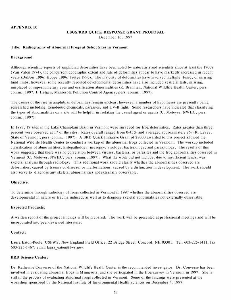
APPENDIX B:
USGS/BRD QUICK RESPONSE GRANT PROPOSAL
December 16, 1997
Title: Radiography of Abnormal Frogs at Select Sites in Vermont
Background
Although scientific reports of amphibian deformities have been noted by naturalists and scientists since at least the 1700s
(Van Valen 1974), the concurrent geographic extent and rate of deformities appear to have markedly increased in recent
years (DuBois 1996; Hoppe 1996; Tietge 1996). The majority of deformities have involved multiple, fused, or missing
hind limbs, however, some recently reported developmental deformities have also included vestigial tails, missing,
misplaced or supernumerary eyes and ossification abnormalities (R. Brannian, National Wildlife Health Center, pers.
comm., 1997; J. Helgen, Minnesota Pollution Control Agency, pers. comm., 1997).
The causes of the rise in amphibian deformities remain unclear, however, a number of hypotheses are presently being
researched including: xenobiotic chemicals, parasites, and UV-B light. Some researchers have indicated that classifying
the types of abnormalities on a site will be helpful in isolating the causal agent or agents (C. Meteyer, NWHC, pers.
comm., 1997).
In 1997, 19 sites in the Lake Champlain Basin in Vermont were surveyed for frog deformities. Rates greater than three
percent were observed at 17 of the sites. Rates overall ranged from 0-45% and averaged approximately 8% (R. Levey,
State of Vermont, pers. comm., 1997). A BRD Quick Initiative Grant of $8000 awarded to this project allowed the
National Wildlife Health Center to conduct a workup of the abnormal frogs collected in Vermont. The workup included
classification of abnormalities, histopathology, necropsy, virology, bacteriology, and parasitology. The results of this
work suggested that there was no correlation between viruses, bacteria, or parasites and the frog abnormalities observed in
Vermont (C. Meteyer, NWHC, pers. comm., 1997). What the work did not include, due to insufficient funds, was
skeletal analysis through radiology. This additional work should clarify whether the abnormalities observed are
deformities, caused by trauma or disease, or malformations, caused by a disfunction in development. The work should
also serve to diagnose any skeletal abnormalities not externally observable.
Objective:
To determine through radiology of frogs collected in Vermont in 1997 whether the abnormalities observed are
developmental in nature or trauma induced, as well as to diagnose skeletal abnormalities not externally observable.
Expected Products:
A written report of the project findings will be prepared. The work will be presented at professional meetings and will be
incorporated into peer-reviewed literature.
Contact:
Laura Eaton-Poole, USFWS, New England Field Office, 22 Bridge Street, Concord, NH 03301. Tel. 603-225-1411, fax
603-225-1467, email [email protected].
BRD Science Center:
Dr. Katherine Converse of the National Wildlife Health Center is the recommended investigator. Dr. Converse has been
involved in evaluating abnormal frogs in Minnesota, and she participated in the frog survey in Vermont in 1997. She is
still in the process of evaluating abnormal frogs collected in Vermont. Some of the findings were presented at the
workshop sponsored by the National Institute of Environmental Health Sciences on December 4, 1997.
25
Amount Requested:
70 frogs at $25.00/frog ..........................................................$1,750.00
Analysis and report ..........................................................$ 250.00
Total $2,000.00
Literature Cited
Dubois, R. B. 1996. Recent observations of deformed anurans in Wisconsin. NAAMP III - the North American
Amphibian Monitoring Program Third Annual Meeting: A Meeting to Present, Evaluate, and Discuss Amphibian
Monitoring Techniques for North America, World Wide Web Conference, November 14, 1996-February, 1997.
Hoppe, D.M. 1996. Historical observations and recent species diversity of deformed anurans in Minnesota. NAAMP
III - the North American Amphibian Monitoring Program Third Annual Meeting: A Meeting to Present, Evaluate, and
Discuss Amphibian Monitoring Techniques for North America, World Wide Web Conference, November 14, 1996-
February, 1997.
Tietge, J. 1996. National reporting center for amphibian deformities. NAAMP III - the North American Amphibian
Monitoring Program Third Annual Meeting: A Meeting to Present, Evaluate, and Discuss Amphibian Monitoring
Techniques for North America, World Wide Web Conference, November 14, 1996-February, 1997.
26
APPENDIX B:
PROGRESS REPORT FOR USGS-BRD PROJECTS FUNDED BY
EASTERN REGION QUICK RESPONSE MONEY
PROJECT LEADER: Dr. Carol U. Meteyer, Dr. Kathryn Converse
USGS-BRD ORGANIZATION: National Wildlife Health Center - Madison
PROJECT TITLE: Radiographic Characterization of Abnormal Frogs from
Selected Sites in Vermont
PROJECT COSTS: $ 2,000
Malformations of Vermont frogs collected during the summer of 1997 were characterized during necropsy examination,
and lab tests were performed to determine if there were associations between diagnostic findings and malformations.
Following that study, we obtained ultradetail radiographic equipment which provided very detailed and magnified images
of bone changes in the frogs. Given this new capability, quick response money was requested and provided in 1998 for a
retrospective radiographic study of these frogs to see if there were specific bone changes unique for frogs collected in
Vermont or at specific sites in Vermont. Radiographs of 33 frogs collected in 1997 from 4 Vermont sites have been
performed and interpretation of these radiographic changes is complete. Results of these findings are in the attached data
table which includes a unique classification system developed through insight gained from radiographic images. This table
combines description of necropsy and radiographic findings. At this time we are determining significance of the
radiographic findings based on malformation type and geographic distribution of the frogs. A final report will be sent by
December 31, 1998 to the USGS-BRD Eastern Regional Office, USFWS Region 5, and the Vermont Agency of Natural
Resources after the radiographic findings are incorporated into the overall final report for the Vermont study. We are
currently looking for funding to extend this study to include microscopic characterization of bone changes with comparison
to gross and radiographic findings in an effort to more completely delineate factors that might be contributing to the
production of malformations at the cellular level.
27
APPENDIX C:
U.S. Geological SurveyBiological Resources Division
NATIONAL WILDLIFE HEALTH CENTER6006 Schroeder Road
Madison, Wisconsin 53711Phone: 608-271-4640
STUDY PLAN
Study Plan: Investigation of deformities in the northern leopard frog (Rana pipiens) at selected sites in Vermont.
Background and Justification: In the fall of 1996, deformities in northern leopard frogs (Rana pipiens) were observed bythe public at 12 sites adjacent to Lake Champlain in Vermont. Several sites were near the border of the Missisquoi NWR inSwanton, Vermont. The presence of deformed leopard frogs were verified at four of these sites by staff of the VermontAgency of Natural Resources (VTDEC) on October 9, 1996. The prevalence of deformities averaged 16.5 %, ranging from 5-23 %at the four sites (230 frogs observed). Most of the frogs observed had missing legs or feet and some had eyeabnormalities. Similar observations have been made in at least seven states and two Canadian provinces. The mostextensive surveys and identification of deformities in frogs and environmental sampling to date has been conduced inMinnesota and the St. Lawrence Valley of Quebec. The mechanism(s) by which these deformities are occurring has not beenidentified; however, the popular theories include: UV-B radiation; xenobiotic contaminants; and viral, bacterial, fungal,or parasitic diseases. The purpose of this investigation is to take a first step by completing a clinical examination ofdeformed frogs collected from four sites in Vermont to complete the following objectives:
Objectives: 1. Characterize and catalog the external and internal deformities in leopard frogs in Vermont;
2. Compare deformed frogs to normal-appearing frogs from an expected control site;
3. Compare morphometric descriptions of abnormalities in Vermont leopard frogs to abnormalitiesconfirmed in Minnesota frogs to determine whether any similarities exist; and
4. Determine the presence or absence of viruses, bacteria, or parasites in deformed frogs.
Methods and approach: Forty site where northern leopard frogs breed are identified in the recent Vermont reptile andamphibian atlas (Andrews, 1995).). In 1996, deformed frogs were observed by the public on 12 of these sites; VTDEC staff confirmed the frog deformitieson four of these sites in 1996. A least 100 frogs will be collected from each of the 40 sites. Four sites with at least10% of the frogs appearing abnormal will be selected for this study. Fifteen abnormal-appearing frogs will be collectedfrom each of these four sites and five normal-appearing frogs will be collected from a control site where no deformitieswere present in a sample of at least 50 frogs.
Recently metamorphosed frogs will be collected and examined for the presence and type of deformities. Frogs withdeformities will be placed in 11 x 32 cm plastic boxes with lids containing 1 air-hole/inch in a chilled cooler or shadylocation. Approximately 3 cm of water from the site will be placed in the boxes along with sphagnum moss. At thecompletion of frog collection, the plastic boxes will be placed in hard-sided coolers, surrounded on four sides with frozenblue ice containers. The boxes will be held in place by a washable metal frame. Eight frogs will be placed live in eachcontainer and shipped via overnight service to the National Wildlife Health Center (NWHC). Upon arrival, frogs will beexamined to deter ermine how the frog responds to this abnormality, photographed, the deformities will be characterized andcataloged using nomenclature and morphologic diagrams developed at NWHC based on frogs submitted to NWHC, photographs bythe Minnesota Pollution Control Agency (MPCA, Helgen), and a Quebec study (Ouellet, 1997). Routine histopathological andmicrobiological samples will be collected and processed on a subset of tissues. Catalogued data will be used to compareabnormalities in Vermont frogs to those recorded during 1995 and 1996 Minnesota frogs. Tissue samples or whole frogs willbe archived for later use if funding becomes available or a potential agent causing the deformities is identified.
Characterization of abnormalities and Necropsy Examination
1. Describe clinical condition/function or movement of any deformed limbs, multiple limbs or other grosslyapparent deformities..
2. Photograph abnormalities before the frog is anesthetized or euthanized if possible.
3. Euthanize one frog at a time using inhalation of Halothane (see NWHC SOP).
4. Necropsy will be performed with the use of a dissecting scope and 2X head loop. Frogs are opened with anventral midline incision. The heart is collected and a blood smear made. Tissues for virology will be placed inviral transport media, tissues for bacteriology in vials of broth and tissue for histopathology in formalin. Verysmall tissues will be places in embedding bags in formalin.
28
5. Necropsy set-up and lab distribution of frogs is as follows:Sites 1-4 Case #’s 14895 - 14898
Accessions 001-005, Pathology:2 blood smears Multiple tissues in formalin and embedding bagTissues for bacteriology, virology
Accessions 006-007, Parasitology:Description of gross pathologySubmit whole carcasses in whirl-paks, in saline
Accessions 008-010, Virology:Description of gross pathologyBlood smearSubmit tissues in viral transport mediaCarcass saved in formalin jar - Hold
Accessions 011-013, Bacteriology:Description of gross pathologyBlood smearSubmit tissues bact broth vialCarcass saved in formalin jar - Hold
Accessions 014-015, Hold Frozen:Description of gross pathology Freeze back in whirl-paks
Normals, Case # 14899
Accession 001: Pathology (necropsy as above)Accessions 002-003: Virology (necropsy as above)Accessions 004-005: Bacteriology (necropsy as above)
Virology
1. Virus isolation will be attempted in amphibian and fish cell culture. Liver, spleen, kidney, gut and heartwere pooled by like tissues (liver with livers, spleen with spleens) with a maximum of three frogs per pool.
2. Viral isolates will be identified and characterized.
3. Electron microscopy will be performed on the pooled tissues in an attempt to detect any viruses that cannot beisolated in cell culture.
Bacteriology
Frog tissues consisting primarily of liver, heart, kidney and others will be placed directly into appropriately labeledtubes of tryptic soy broth (TSB) at necropsy and submitted to diagnostic bacteriology. The following procedure will befollowed:
1. The 10 ml tubes of TSB with tissue will be vortexes for 5-10 seconds and subcultured immediately onto bloodagar (BAP), eosine-methylene blue agar (EMB), (or MacConkey agar), Lowenstein-Jensen agar (LJ), sabouraud-dextroseagar (SAB), thioglycollate broth (THIO), and cooked-meat broth (CM).
2. Media (except LJ and SAB) will be examined at 18-24 hour intervals for up to 72 hours before being discarded asnegative. Positive cultures will be subcultured and identified to at least genus level. TSB will be subculturedafter 24 hours if growth appears. LJ and SAB will be held as appropriate until sufficient growth develops or thereis no growth.
3. Identification of bacterial isolates will be consistent with current capabilities and will include the use ofcommercial identification systems; predominately the API system, bioMerieux Vitex, St. Louis, Missouri. Referencelaboratories, primarily the National Veterinary Services Laboratory, Ames, Iowa will be used as needed.
Parasitology
To determine the presence and species of parasites or evidence of parasite infections, live or freshly euthanized frogswill be sent to the parasitology laboratory for examination under a dissection scope. The following procedures will befollowed:
1. Frogs will be skinned and the muscles and fascia examined under the dissecting microscope. Hind limb muscleswill be teased apart and examined for metacercaria.
2. Metacercaria in the subdermal layers will be dissected free and placed temporarily in ½ strength Locke’ssolution and refrigerated. In some cases, this will cause the metacercaria to encyst so that fixation and stainingcan proceed. Notes will be kept and vial labeled as to collection site for the metacercaria.
3. A ventral incision will be made from vent to lower mandible to expose internal organs.
29
4. The heart will be removed and a blood smear made according to DPL SOP.
5. All organs will be examined grossly, any parasites removed, prepped for fixation and fixed in AFA or 70%depending on the class of parasite. Most organs are small enough to examine via a squash preparation. Fat bodieswill be examined via squash preparation for mesocercariae.
6. The body cavity will be examined for immature helminths. If found they will be removed, relaxed and fixed.
7. Eviscerated carcasses will be placed in 10% buffered formalin and later cleared according to Hanken ansWassersug (1981). Following clearing frogs will be examined and the presence or absence of metacercaria noted.
Critical Data: 1. Specific characterization of external deformities,
2. Documentation of internal deformities,
3. Histopathological findings for Minnesota frogs for comparison with Vermont frogs.
Statistical Treatment: The research sites will be selected from areas with at least 15% of the frogs affected in a sampleof 100 frogs. No specific statistical methods will be applied the data from this study. Routine statistical methods maybe applied to determine the frequencies of certain lesions and establish similarities between sites.
Assessment Criteria: Frogs will be examined for external and internal morphological and histological lesions and theselesions characterized and cataloged. The lesions present in Vermont frogs in this study will compared to lesions inMinnesota frogs described by NWHC in 1996.
Funding and FTEs:
Operational Costs:Salaries and benefits: (NWHC staff) $ 5,000.00
Wildlife Disease Specialist FTE .05Pathologist FTE .05
Travel for field work 1,000Sample shipment 200Field equipment/supplies 350Laboratory equipment/supplies 450Histological preparation and radiography 1,000
Funding Source: $8,000 from USGS-BRD Quick Response Funds. Salaries are from NWHC base funding.
Project Investigators: Principal Investigators;
Kathryn Converse, Ph.D., NWHC, Wildlife Disease SpecialistCarol Meteyer, D.V.M., NWHC, Pathologist
Laura Eaton-Poole, USFWS, BiologistRick, Levey, Vermont Dept. Environmental Conservation Biologist
Special Safety Requirements: All necropsies and laboratory analysis will be conducted in a Level 3 bio-containment facilityto protect sample purity, quality, and eliminate the potential for exposure to any human pathogens. Protective clothingwill be worn during examinations and goggles when handling formalin. Follow standard operating procedure for use ofeuthanasia solution.
Completion Date: January 15, 1998
Schedule and milestones:
General schedule:6/9-13/97 Visit sample sites in Vermont and review collection protocol with USFWS and VTDEC.7/7/97 Ship coolers and supplies to Vermont7/21-30/97 Collect frogs, ship to NWHC and begin diagnostic evaluation9/1/97 Complete diagnostic tests12/15/97 Distribute first draft1/15/98 Distribute final report
Study Milestones:
1. Complete evaluation of field key to amphibian deformities2. Start key to internal deformities3. Complete comparative evaluation of Vermont and Minnesota frog deformities4. Document sites in Vermont with deformed frogs for use in this study. 5. Evaluate effectiveness of diagnostic procedures and sample size and type for amphibians
Relationship to Other Work:
This study is supported by ongoing surveys of frog populations in Vermont conducted by the Environmental Agency of Vermont,Middlebury College (Middlebury Vermont), the US Fish and Wildlife Service and the US Environmental Protection Agency.
Expected Products: Key to internal deformities to allow for standardization of diagnostic findings. Publication, BIB,
30
completion report and proposal for additional studies.
Literature Cited:
Andrews, S. James. 1995. A preliminary atlas of the reptiles and amphibians of Vermont. Vermont Department of Fish andWildlife, Natural Heritage Program. 64pp.
Hanken, James and R. Wassersug. 1981. The visible skeleton. Functional Photography 16:22-26, 44.
Ouellet, M., J. Bonin, J. Rodrique, J-L DesGranges and S. Lair. 1997. Hindlimb deformities (ectromelia, ectrodactyly) infree-living anurans from agricultural habitats. J. Wildlife Diseases 95-104.
Submitted by: ___________________________________________________________Kathryn Converse, Wildlife Disease Specialist Date
Principal Investigator
Approved: ____________________________________________________________Christopher Brand, Branch Chief Date
Field Investigations
Concurrence: ____________________________________________________________Milton Friend Director, NWHC Date
31
Appendix D. Classification of Gross and Radiographic Findings in VT Frogs - 1997
Title CASE/AC Deformity Side L/R/B Deformity Comment
Initiation Appears Absent
14895-002 Am elia Left
14895-006 Am elia/No righ t isch ium /no right Right Th e iliu m is d isp laced caudally. Th e d istal
pub is ilium has a rough, irregular cortex,
f locculent t rabeculae traversing the marrow
and an abnorm al distal border.
14895-015 Am elia/No isch ium /No pub is Right Trabecular pattern instead of acetabulum/
No R ischium or pu bis & L isch ium not
fuse d w ith pu bis/ A bnorm al dista l Righ t
ilium with disruption of trabecular pattern
and cortex/ar ticu lation of distal right ilia l
cortex w ith left ischiu m and left d istal
ischial fracture.
14896-007 Am elia/No left ischium /No left pubis Left Very sim ilar to 14895 -15 w/no left ischium or
pub is and R isch ium not fu sed with pub is
14896-009 Am elia Right All pelvic elements present
14898-003 Am elia Right
Initiation Incomplete Pattern
Missing Distal Segments14895-004 Hem imelia femur Left
14895-005 Hem imelia femur Left with sm all narrow termination
14895-005 Brachydactyly/Skin Web Right Digits 1 and 5 short malform ed (curled)
brachydactyly (also left hemimelia)
14895-007 Hem imelia Tibiale Fibulare Right Abnorm al tibiale fibulare region/bones are
abnormally wide cortices are very thin, the
trabeculae give a light honeycomb
appearance that is also p resent in a short
reg ion of p rox im al shaft of both tibiafibula
bones.
14895-010 Hem im elia tibiaf ibu la Right Proximal cortex may be normal then
cortica l & trabecular d isrup tion at
termination
14895-011 Hem imelia Tibiale Fibulare Left With mild trabecular and cortical disruption
14895-011 Hem im elia tibiaf ibu la Right With moderate trabecular disruption and mild
cortical disruption
14895-012 Hem imelia femur Right Similar to 14895-20 /mid-shaft with mild
trabecular and cortical disruption with
abrupt term ination w/out disruption of
cortical/trabecular pattern
14895-013 He mimelia fem ur/no left pu bis Left Terminal trabecular hypertrophy hyperplasia
possible pe lvic abnorm ality , severe cort ica l
disruption.
14895-019 Hem imelia femur Right Trabecular hypertrophy/ hyperplasia/
modera te cort ica l dis ruption/ nice norm al
pelvis
14895-020 Hem imelia femur Left Midshaft trabecular hypert hyperplasa/mild
cortica l disruption /abru pt term ination without
disruption of cortical/trabecular pattern similar
to 14896-03.
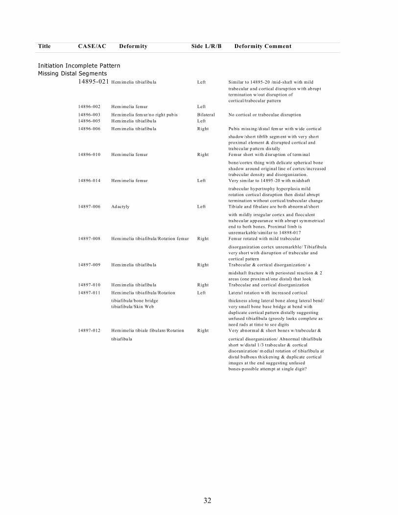
32
Title CASE/AC Deformity Side L/R/B Deformity Comment
Initiation Incomplete Pattern
Missing Distal Segments
14895-021 Hem im elia tibiaf ibu la Left Similar to 14895-20 /mid-shaft with mild
trabecular a nd cortical d isrup tion w ith ab rup t
termination w/out disrup tion of
cortical/trabecular pattern
14896-002 Hem imelia femur Left
14896-003 He mimelia fem ur/no righ t pub is Bilateral No cortical or trabeculae disruption
14896-005 Hem im elia tibiaf ibu la Left
14896-006 Hem im elia tibiaf ibu la Right Pu bis miss ing /dista l fem ur wi th w ide cortical
shadow /short tibfib segm ent w ith very short
proximal element & disrupted cortical and
trabecular p atte rn dis tally
14896-010 Hem imelia femur Right Femur short wi th d isrup tion of term ina l
bone/cortex thing with delicate spherical bone
shadow around original line of cortex/increased
trabecular density and disorganization.
14896-014 Hem imelia femur Left Very sim ilar to 14 895 -20 w ith m idsh aft
trabecular hypertrophy hyperplasia mild
rotation cortica l disruption then dista l abrupt
termination without cortical/trabecular change
14897-006 Ad actyly Left Tib iale and fibulare a re bo th abnorm al/short
with mildly irregu lar cortex and flocculent
trabecular appearan ce wi th abrup t symmetr ica l
end to both bones. Proximal limb is
unremarkable/similar to 14898-017
14897-008 Hem imelia tibiafibula/Rotation femur Right Fem ur rotated with m ild trabecular
disorganization cortex unremarkble/ Tibiafibula
very short with disruption of trabecular and
cortical pattern
14897-009 Hem im elia tibiaf ibu la Right Trabecular & cortical disorganization/ a
midshaft fracture with periosteal reaction & 2
areas (one proxim al/one distal) that look
14897-010 Hem im elia tibiaf ibu la Right Trabeculae and cortical disorganization
14897-011 Hem imelia tibiafibula/Rotation Left Latera l rotation w ith inc rea sed cort ica l
tibiafibula/bone bridge thickn ess a long lateral bone along latera l bend /
tibiafibula/Skin Web very small bone base bridge at bend with
duplicate cortical pattern distally suggesting
unfused tibiafibula (grossly looks complete as
need rad s at tim e to see digits
14897-012 Hem imelia tibiale fibulare/Rotation Right Very abnormal & short bones w/t rabecular &
tibiaf ibu la cortical disorganization/ Abnormal tibiafibula
short w/dis tal 1 /3 t rab ecular & cortical
disoranization/ m edia l rotation of tibia fibu la at
dis tal bulbous th ickening & duplicate cortical
images at the end suggesting unfused
bones-possib le attem pt at s ingle d igit?
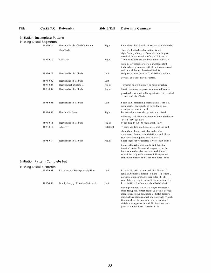
33
Title CASE/AC Deformity Side L/R/B Deformity Comment
Initiation Incomplete Pattern
Missing Distal Segments14897-014 Hem imelia tibiafibula/Rotation Right Latera l rotation & m ild increase cortica l dens ity
tibiaf ibu la laterally bu t trabecular p attern is not
significantly changed. Possible superimpose
terminal dorsal rotation of distal 0.1 cm of
14897-017 Ad actyly Right Tib iale and fibulare a re bo th abnorm al/short
with mildly irregu lar cortex and flocculent
trabecular appearan ce wi th abrup t symmetr ica l
end to both bones. Proximal limb is
14897-022 Hem im elia tibiaf ibu la Left Only very short (unfused? ) tibiafibula with no
cortical or trabecular disruption.
14898-002 Hem im elia tibiaf ibu la Left
14898-005 Hem im elia l tibiaf ibu la Right Term inal bulge that may be bone s tructure
14898-007 Hem im elia tibiaf ibu la Right Sh ort rem ain ing segm ent is abnorm al/norm al
proximal cortex with disorganization of terminal
cortex and tibiaf ibu la
14898-008 Hem im elia tibiaf ibu la Left Short thick remaining segment like 14898-07
wi th n orm al prox im al cortex and te rm ina l
disorganization-but mild.
14898-009 Hem imelia femur Right Pe riosteal reaction alon g shaft w ith dis tal
widening with delica te sphere of bone s imilar to
14896 -010, (do histo)
14898-011 Hem im elia tibiaf ibu la Right M uch like 14898 -08 radiog raph ically.
14898-012 Ad actyly Bilateral Tibiale and fibulare bones are short and end
abruptly without cortical or trabecular
disruption. Fractures in tibiafibula and tibiale
fibulare are thought to be artefacts.
14898-014 Hem im elia tibiaf ibu la Right Sh ort segm ent of tibiaf ibu la ve ry short norm al
bone. Silhouette proximally and then the
terminal cortex became disorganized with
increased trabecular pattern/distal femur is
folded dorsally with increased disorganized
trabecular pattern and a delicate dorsal bone
Initiation Pattern Complete but
Missing Distal Elements14895-001 Ectrodactyly/Brachydactyly/Skin Left Like 14895-018. Abnormal tibiafibula (1/2
length) A bnorm al tibiale fibulare (1/2 length),
dorsal rotation prob ably trian gular tib fib;
com plete w eb h ip to hock; 3 incom plete d igits
14895-008 Brachydactyly/ Rotation/Skin web Left Like 14895-18 w/abn distal-mid-tibfib/skin
web hip to hock/ tibfib 1/2 len gth w /midsh aft
mild d isrup tion of t rab eculae & doub le cort ica l
image su ggesting nonfusion of tibfib distal to
midshaft /rotation (dorsal hock) mshaft. Tibiale
fibu lare sh ort, bu t no trabecular d isrup tion/
tibiale now ap pears lateral. No function hock
joint w/medial dorsal rotation 180o
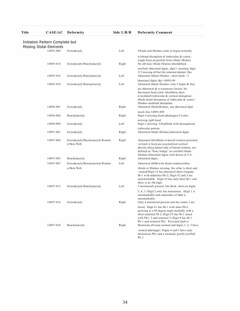
34
Title CASE/AC Deformity Side L/R/B Deformity Comment
Initiation Pattern Complete but
Missing Distal Elements14895-009 Ec trodactyly Left Tib iale and fibulare s eem to beg in norm ally
w/abrupt dis rup tion of trabeculae & cortex ;
single bony projection from tibiale fibulare
14895-014 Ec trodactyly/Brachyd actyly Right No obvious tibiale fibulare/short&thick
ossif ied/ abnorm al digits , digit 1 missing/ digit
2-5 missing all but the terminal phalan/ like
14895-016 Ec trodactyly/Brachyd actyly Left Abnormal tibiale fibulare - short thick / 3
abnormal digits like 14895-09
14895-018 Ec trodactyly/Brachyd actyly Left Abnorm al tibiale fib ulare/ only 2 d igits & they
are abnorm al & w/cutaneou s fusion/ No
functiona l hock joint/ tib iafibu la short
w/m idsh aft trab ecular & cortica l disruption /
tibiale d istal dis rup tion of trabecular & cortex /
fibulare midshaft disruption.
14896-001 Ec trodactyly Right Abnorm al tibialefib ulare, one abnorm al digit
much like 14895-009
14896-004 Brachyd actyly Right Digit 4 m issing distal phalanges/2-4 also
missing right hand
14896-009 Ec trodactyly Left Digit 1 missing. Tibiafibula with disorganised
trabecular pattern
14897-001 Ec trodactyly Right Abnorm al tibiale fib ulare/abnorm al digits
14897-004 Ectrodactyly/Brachydactyly/Rotatio Right Abnorm al tibiafibula w/lateral rotation proximal
n/Skin Web to hock w/increase assym etr ica l cor tica l
density along latera l side of lateral ro tation, not
defined as "bony bridge" no ossified tibiale
fibulare/Abnormal digits with fusion of 3-4.
14897-005 Brachyd actyly Right Abnorm al digits
14897-007 Ectrodactyly/Brachydactyly/Rotatio Left Abnorm al tibfib/with distal rotation/either
n/Skin Web tibiale or fibulare missing; the other is short and
rotated/Digit #1 has abnorm al short irregular
M-1 w ith indistinct Ph-2; Digit #2 and 3 are
unremarkable. Digit #4 has only short M-1 and
there is no 5th d igit.
14897-015 Ec trodactyly/Brachyd actyly Left 5 metatarsals present, but thick, short on digits
5, 4, 3. Digit 2 only has metatarsus. Digit 1 is
unremarkable and remainder of limb is
unremarkable.
14897-016 Ec trodactyly Right Only 4 metatarsal present and the center 2 are
fused. Digit #1 has M-1 with short Ph-2
growing at a 90 degree angle medially with a
short terminal Ph 2; Digit #3 has M-1 fused
with Ph1, 2 and terminal 3; Digit 4 has M-1
Ph-1 and terminal Ph2. Proximal limb is
14897-018 Brachyd actyly Right M etarsals all seem norm al and digits 1, 2, 3 have
norm al pha langes. D igits 4 a nd 5 have only
metatarsus Ph1 and a terminal, poorly ossified
Ph 2.
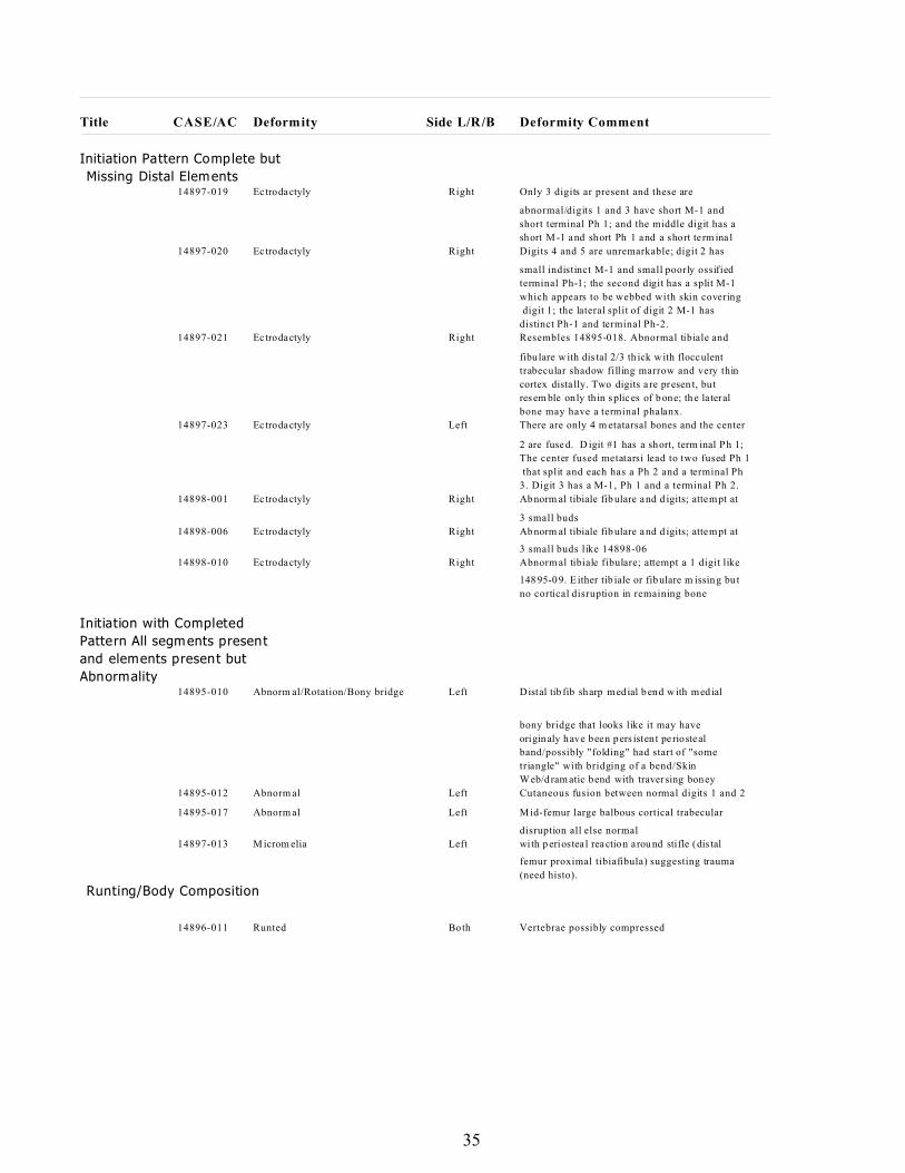
35
Title CASE/AC Deformity Side L/R/B Deformity Comment
Initiation Pattern Complete but
Missing Distal Elements14897-019 Ec trodactyly Right Only 3 digits ar present and these are
abnormal/digits 1 and 3 have short M-1 and
short terminal Ph 1; and the middle digit has a
short M -1 and short Ph 1 and a short te rm ina l
14897-020 Ec trodactyly Right Digits 4 and 5 are unremarkable; digit 2 has
small indistinct M-1 and small poorly ossified
terminal Ph-1; the second digit has a split M-1
which appears to be webbed with skin covering
digit 1; the lateral split of digit 2 M-1 has
distinct Ph-1 and terminal Ph-2.
14897-021 Ec trodactyly Right Resembles 14895-018. Abnormal tibiale and
fibu lare w ith dis tal 2/3 th ick w ith flocculent
trabecular shadow filling marrow and very thin
cortex dista lly. Two digits a re presen t, bu t
res em ble only thin s plices of b one; th e lateral
bone may have a terminal phalanx.
14897-023 Ec trodactyly Left There are only 4 m etatarsal bones and the center
2 are fuse d. D igit #1 has a short, term inal Ph 1;
The center fused metatarsi lead to two fused Ph 1
that split and each has a Ph 2 and a terminal Ph
3. Digit 3 has a M-1, Ph 1 and a terminal Ph 2.
14898-001 Ec trodactyly Right Abnorm al tibiale fib ulare a nd d igits; attempt at
3 small buds
14898-006 Ec trodactyly Right Abnorm al tibiale fib ulare a nd d igits; attempt at
3 small buds like 14898-06
14898-010 Ec trodactyly Right Abnorm al tibiale fibulare; attempt a 1 digit like
148 95-0 9. E ither tib iale or fib ulare m issing bu t
no cortical disruption in remaining bone
Initiation with Completed
Pattern All segments present
and elements present but
Abnormality14895-010 Abnorm al/Rotation/Bony bridge Left Distal tib fib sharp medial bend w ith medial
bony bridge that looks like it may have
originaly have been pers istent pe riosteal
band/possibly "folding" had start of "some
triangle" with bridging of a bend/Skin
Web/d ram atic bend with traversing boney
14895-012 Abnorm al Left Cutaneous fusion between normal digits 1 and 2
14895-017 Abnorm al Left M id-femur large balbous cortical trabecular
disruption all else normal
14897-013 M icrom elia Left wi th p eriostea l reaction arou nd stifle (dis tal
femur proximal tibiafibula) suggesting trauma
(need histo).
Runting/Body Composition
14896-011 Runted Both Vertebrae possibly compressed
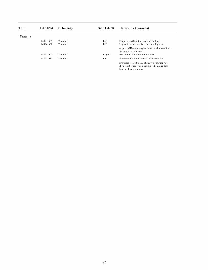
36
Title CASE/AC Deformity Side L/R/B Deformity Comment
Trauma
14895-003 Trauma Left Femur overiding fracture - no callous
14896-008 Trauma Left Leg soft tissue swelling, bu t deve lopm ent
appears OK-radiographs show no abnormalities
in pelvis or rear limbs.
14897-003 Trauma Right Rear limb traum atic ampu tation
14897-013 Trauma Left Increased react ion around distal femur &
proxim al tibiafibula at stifle. No fun ction to
dista l limb suggestin g trauma. The entire lef t
limb with microm elia
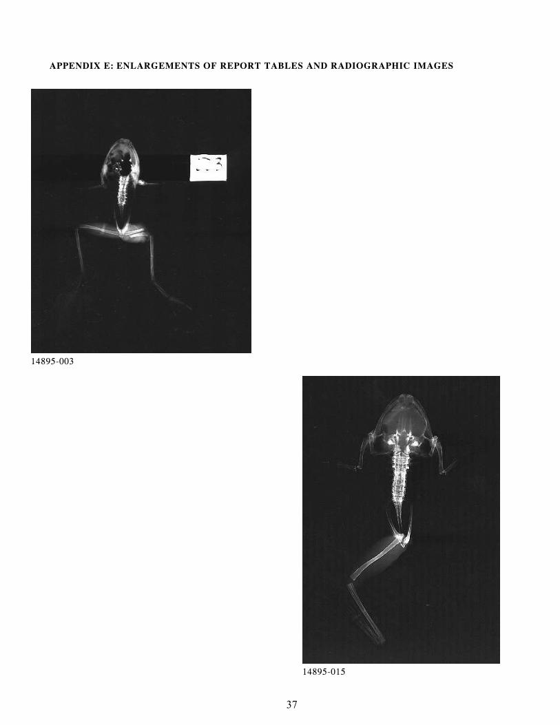
37
14895-003
14895-015
APPENDIX E: ENLARGEMENTS OF REPORT TABLES AND RADIOGRAPHIC IMAGES
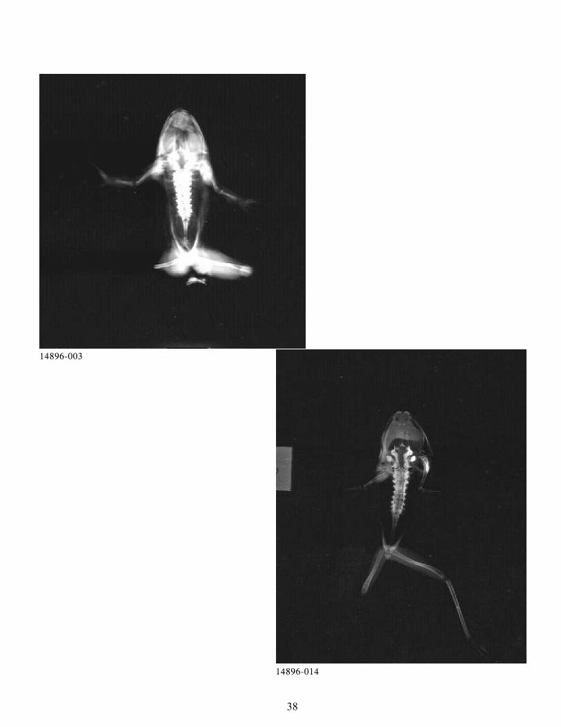
38
14896-003
14896-014
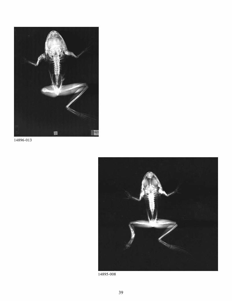
39
14896-013
14895-008
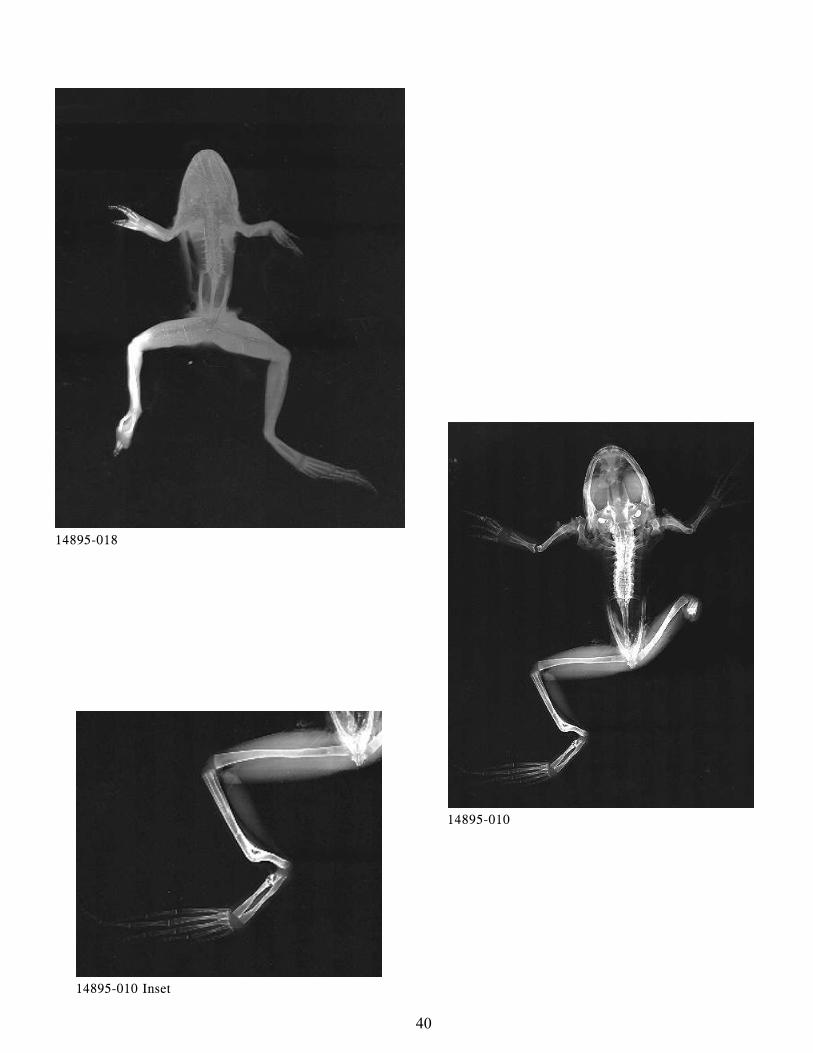
40
14895-018
14895-010
14895-010 Inset
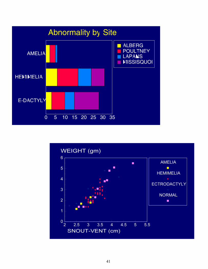
41
42
APPENDIX F: TABLE #1 Frog Body Condition/Average by Deformity
TABLE #2 Frog Body Condition/Average by Collection Site
Appendix F is available by contacting Dr. Carol Meteyer at:
National Wildlife Health Center
6006 Schroeder Road
Madison, WI 53711
608-270-2400
49
APPENDIX G: HISTOPATHOLOGY FINDINGS
Appendix G is available by contacting Dr. Carol Meteyer at:
National Wildlife Health Center
6006 Schroeder Road
Madison, WI 53711
608-270-2400
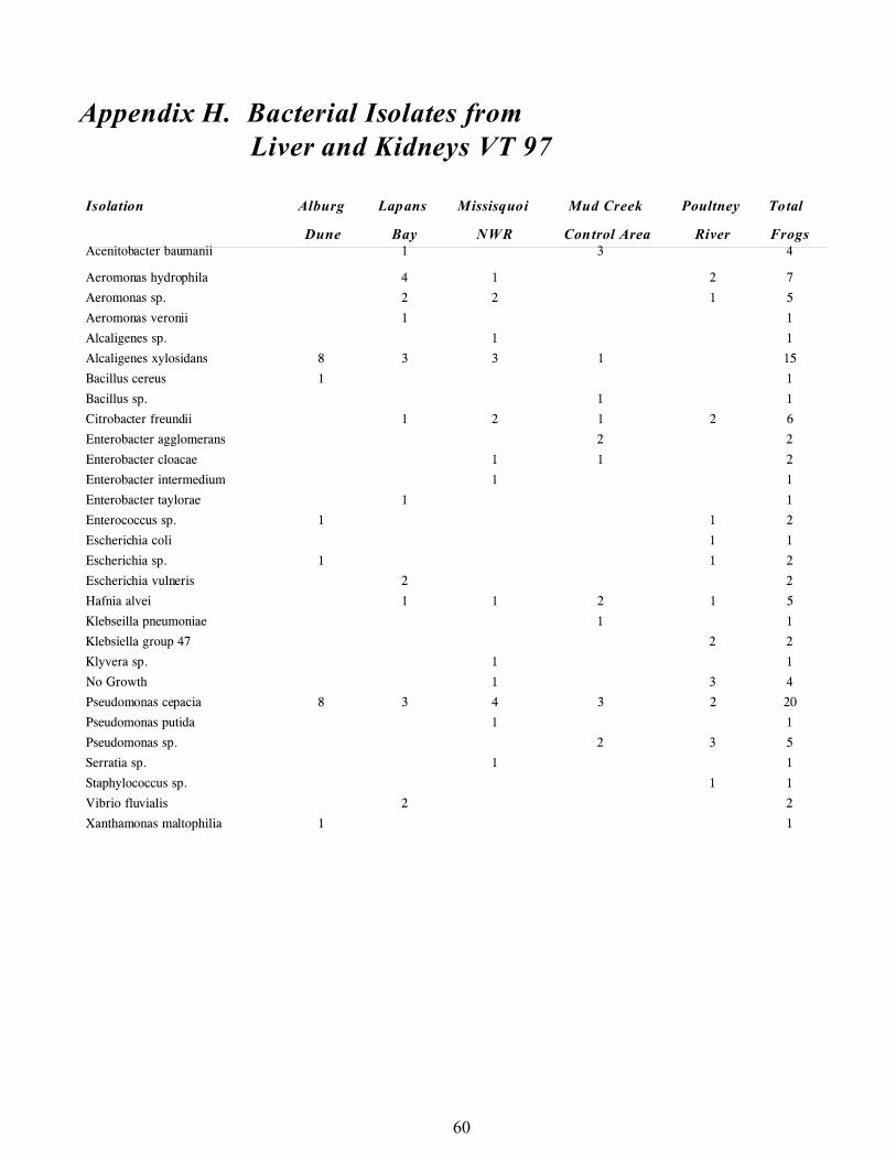
60
Appendix H. Bacterial Isolates fromLiver and Kidneys VT 97
Isolation Alburg Lapans Missisquoi Mud Creek Poultney Total
Dune Bay NWR Control Area River FrogsAcenitobacter baumanii 1 3 4
Aeromonas hydrophila 4 1 2 7
Aeromonas sp. 2 2 1 5
Aeromonas veronii 1 1
Alcaligenes sp. 1 1
Alcaligenes xylosidans 8 3 3 1 15
Bacillus cereus 1 1
Bacillus sp. 1 1
Citrobacter freundii 1 2 1 2 6
Enterobacter agglomerans 2 2
Enterobacter cloacae 1 1 2
Enterobacter intermedium 1 1
Enterobacter taylorae 1 1
Enterococcus sp. 1 1 2
Escherichia coli 1 1
Escherichia sp. 1 1 2
Escherichia vulneris 2 2
Hafnia alvei 1 1 2 1 5
Klebseilla pneumoniae 1 1
Klebsiella group 47 2 2
Klyvera sp. 1 1
No Growth 1 3 4
Pseudomonas cepacia 8 3 4 3 2 20
Pseudomonas putida 1 1
Pseudomonas sp. 2 3 5
Serratia sp. 1 1
Staphylococcus sp. 1 1
Vibrio fluvialis 2 2
Xanthamonas maltophilia 1 1
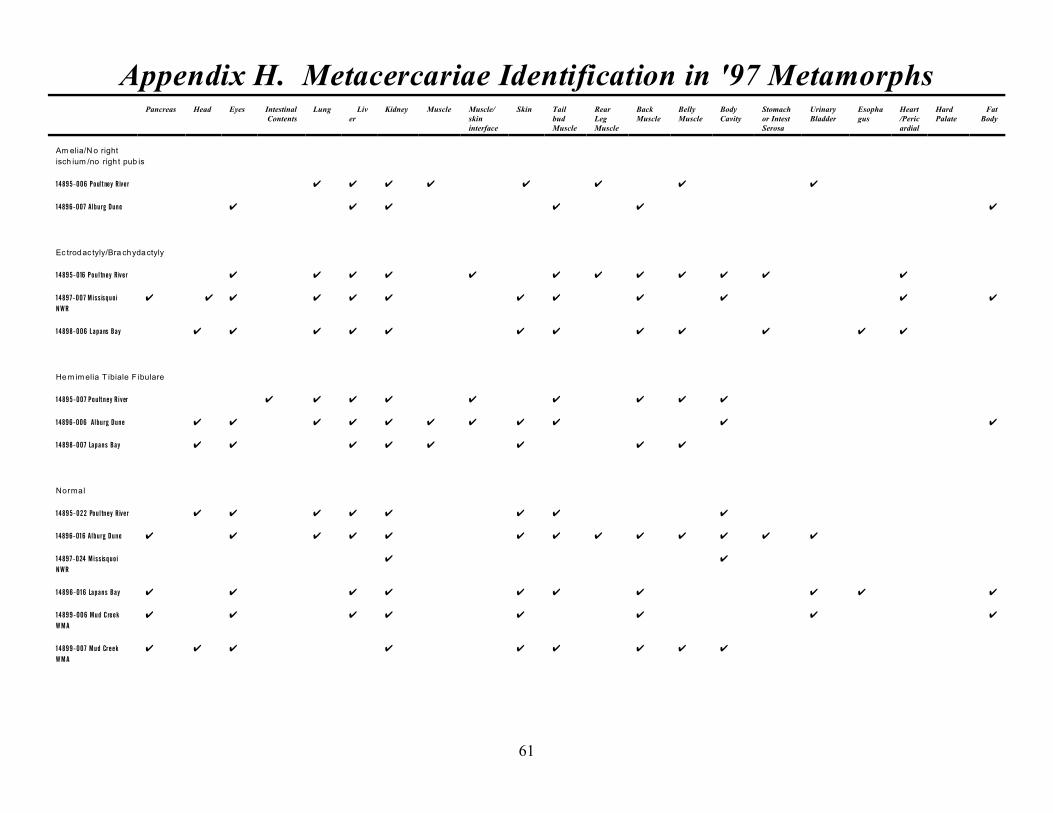
61
Appendix H. Metacercariae Identification in '97 MetamorphsPancreas Head Eyes Intestinal
ContentsLung Liv
erKidney Muscle Muscle/
skininterface
Skin TailbudMuscle
RearLegMuscle
BackMuscle
BellyMuscle
BodyCavity
Stomachor IntestSerosa
UrinaryBladder
Esophagus
Heart/Pericardial
HardPalate
FatBody
Am elia/N o right
isch ium /no righ t pub is
14895-006 Poultney River U U U U U U U U
14896-007 Alburg Dune U U U U U U
Ec trodac tyly/Bra chydactyly
14895-016 Poultney River U U U U U U U U U U U U
14897-007 Missisquoi
NWR
U U U U U U U U U U U U
14898-006 Lapans Bay U U U U U U U U U U U U
He m imelia T ibiale F ibulare
14895-007 Poultney River U U U U U U U U U
14896-006 Alburg Dune U U U U U U U U U U U
14898-007 Lapans Bay U U U U U U U U
Normal
14895-022 Poultney River U U U U U U U U
14896-016 Alburg Dune U U U U U U U U U U U U U
14897-024 Missisquoi
NWR
U U
14896-016 Lapans Bay U U U U U U U U U U
14899-006 Mud Creek
WMA
U U U U U U U U
14899-007 Mud Creek
WMA
U U U U U U U U U
62
Appendix H. Adult Helminth Identification in '97 MetamorphsIsolation Alburg Lapans Missisquoi Mud Creek Poultney Total
Dune Bay NWR Control Area River Frogs
Gorgoderidae
2 2 1 5
Nematode
1 2 1 1 5
Oxyurid
2 2
Rhabdias ranae
1 1 2
Trematode
1 1 2 4
63
APPENDIX I: LIMB MEASUREMENTS
1. REAR LEG MEASUREMENTS
2. FRONT LEG MEASUREMENTS
Appendix I is available by contacting Dr. Carol Meteyer at:
National Wildlife Health Center
6006 Schroeder Road
Madison, WI 53711
608-270-2400
70
APPENDIX J: References Cited
BALINSKY, B. I. 1960. Development of Mesodermal Organs in Vertebrates. Chapter 11, An Introduction to
Embryology, W.B. Saunders Company, Philadelphia, London, pp 285-306.
----------------------- 1981. Development of the Mesodermal Organs in Vertebrates, An Introduction to Embryology,
Fifth Edition, Saunders College Publishing, Philadelphia, p. 465.
BOLANDE, R. P. 1979. Developmental Pathology. American Journal of Pathology 94(3), pp 627-638.
CARLSON, B. M. 1994. Human Embryology and Developmental Biology. Chapter 11, Limb Development,
Mosby- Year Book, Inc. pp.182-203.
--------------- 1994. Human Embryology and Developmental Biology. Chapter 8, Developmental Disorders - Causes,
Mechanisms, and Patterns, Mosby - Year Book, Inc. pp.117-133.
COOK, T. W. 1978. The Migration of Fibricola cratera (Trematoda: Diplostomatidae) in Metamorphosing Rana
pipiens, The Journal of Parasitology, 64 (5), pp 938-939.
CUMMINGS, K. S., C. A. Mayer. 1992. Field guide to Freshwater Mussels of the Midwest. Champaign, Il.
Illinois Natural History Survey.
CUNNINGHAM, A. A., T. E. S. LANGTON, P. M. BENNETT, J. F. LEWIN, S. E. N. DRURY, R. E.
GOUGH AND S. .K. MACGREGOR. 1995. Investigations into Unusual Mortalities of the Common Frog
(Rana temporaria) in Britain.Zwart. P. and G Matz, Editors, Fifth International Colloquium on the
Pathology of Reptiles and Amphibians, Alphen Aan Den Run, The Netherlands pp.19-27.
----------------- 1996. Pathological and microbiological findings from incidents of unusual mortality of the common
frog (Rana temporaria), Philosophical Transactions of the Royal Society of London Biological Sciences 351:1539-
1557.
DICKMAN, S. 1997 Possible New Roles for HOX Genes. Science 278:1882-1883.
DUBOIS, R. B. 1996. Recent Observations of Deformed Anurans I Wisconsin, NAAMP III-The North American
Amphibian Monitoring Program Third Annual Meeting: A meeting to present, evaluate, and discuss amphibian
monitoring techniques for North America, World Wide Web Conference, November 14, 1996-February 1997.
DUELLMAN, W. E. AND L. TRUEB. 1996. Biology of Amphibians, The John Hopkins University Press,
Baltimore, pp. 173-192 and 289-414.
ELINSON, R. 1997. Amphibians. Embryology: Constructing the Organism, Sinauer Associates, Inc. Chapter 20,
pp. 409-436.
FRIED, B., P. L. PANE, AND A. REDDY. 1997. Experimental infection of Rana pipiens tadpoleswith
Echinostoma trivolvis cercariae, Parasitol Res 83:666-669.
GILBERSTON, H., R. DUBOIS, R. HAY. 1998. Wisconsin’s Amphibian Malformations: 1960-1997. Proceedings
of the Midwest Declining Amphibians Conference March 20-21, 1998.
HADORN, E. 1974. Experimental Studies of Amphibian Development. Translated by D. Turner. Springer Berlag
New York Heidelberg Berlin, pp. 114-131.
HANKEN, J. AND R. WASSERUG. 1981. The visible skeleton. Functional photograph 15: 22- 24.
HOPPE, D. M. 1996. Historical Observations and Recent Species Diversity of Deformed Anurans in Minnesota,
NAAMP III - The North American Amphibian Monitoring Program Third Annual Meeting: A meeting to
present, evaluate, and discuss amphibian monitoring techniques for North America, World Wide Web
Conference, November 14, 1996-February 1997.
HIRD, DIESCH et al.. 1983. Applied Environmental Microbiology. 46:1423-1425.
MARTIN, S. W., A. H. Meek, P. Willeberg. 1987. Veterinary epidemiology: principles and methods. 1st ed.
Ames, IA: Iowa State University Press.
MARTIN, T. R. AND D. B. CONN. 1990. The Pathogenicity, Localization, and Cyst Structure of
Echinostomatid Metacercariae (Trematoda) Infecting the Kidneys of the Frogs RanaClamitans and Rana
Pipiens, Journal of parasitology, 76(3) pp. 414-419.
71
References Cited
Page 2
MEYER-ROCHOW, V. B. AND J. KOEBKE. 1986. A Study of Extra Extremity in a Five-legged Rana
temporaria
Frog. Zoologischer Anzeiger 217: 1-13.
NIEDERREITHER, K., S. J. WARD, P. DOLLE, AND P. CHAMBON. 1996. Morphological and Molecular
Characterization of Retinoic Acid-Induced Limb Duplications in Mice. Developmental Biology 176, pp.185-
198.
O’RAHILLY. R., MULLER, F. 1996. Human Embryology & Teratology, Second Edition,Wiley-Liss, Inc. Pp. 10-
15.
O’SHEA, P. A. 1995. Congenital Defects and Their Causes. Human Reproduction: Growth and development, edited
by D. R. Coustan, R. V. Haning, Jr., and D. B. Singer. Chapter 8, pp. 121-138.
OUELLET, M., J. BONIN, J. RODRIQUE, J. L. DESGRANGES, AND S. LAIR. 1997. Hindlimb Deformities
(Ectromelia, Ectrodactyly) in Free-living Anurans from Agricultural Habitats, Journal of Wildlife Disease 33(1):
pp. 95-104.
REYNOLDS, T. D. AND T. D. STEPHENS. 1984. Multiple Ectopic Limbs in a Wild Populationof Hyla Regilla.
Great Basin Naturalist 44: 166-169.
ROBBINS, S. L., R. S. COTRAN, V. KUMAR. 1984. Congenital malformations. In Pathological Bases of
Disease, S. L. Robbins, R. S. Cotran, V. Kumar (eds), W.B. Saunders Company, Philadelphia, PA, pp. 479-
482.
RUTLEDGE, J. C., A. G. SHOURBAJI, L.A. HUGHES, J. E. POLIFKA, T. P. CRUZ, J. B. BISHOP, and W. M
GENEROSO. 1994. Limb and lower-body duplications induced by retinoic acid in mice. Proceeds National
Academy of Science, USA, Vol. 91, pp. 5436-5440.
SMITH, D. M., R. D. TORRES, AND T. D. STEPHENS. 1996. Mesonephros Has a Role in Limb Development
and Is Related to Thalidomide Embryopathy. Teratology 54: 126-134.
SESSIONS, S. K. AND RUTH, S. B. 1990. Explanation of Naturally Occurring Supernumerary Limbs in
Amphibians, The Journal of Experimental Zoology 254:38-47.
VOGT, W., 1929. Gestaltungsanalyse am Amphibienkeim mit ortlicher Vitalfarbung. II.Gastrulation and
Mesodermbildung bei Urodelen und Anuren. Roux Arch. 120, 385-706.
WISE, L. D., S. L. BECK, D. BELTRAME, B. K. BEYER, I. CHAHOUD, R. L. CLARK, R.CLARK, A.
M. DRUGA, M. H. FEUSTON, P. GUITTIN, S. M. HENWOOD, C. A. KIMMEL, P. LINDSTROM,
A. K. PALMER, J. A. PETRERE, H. M. SOLOMON, M.VASUDA AND R. G. YORK. 1997.
Terminology of Developmental Abnormalities in Common Laboratory Mammals (Version 1), Teratology 55:
249-292.
72
APPENDIX K: NECROPSY REPORTS
Necropsy reports are available by contacting Dr. Carol Meteyer at:
National Wildlife Health Center
6006 Schroeder Road
Madison, WI 53711
608-270-2400