Embed Size (px)
DESCRIPTION
Final Unit Histology Slides. Oral Epithelium. Buccal surface – stratified squamous non-keratinized epithelium. The Tongue & Gustatory Receptors. Filiform Papillae. Circumvallate Papillae. Gustatory Receptor. Fungiform Papillae. Esophagus. Muscularis. Mucosa. Submucosa. - PowerPoint PPT Presentation
Citation preview
Final Unit Histology Slides
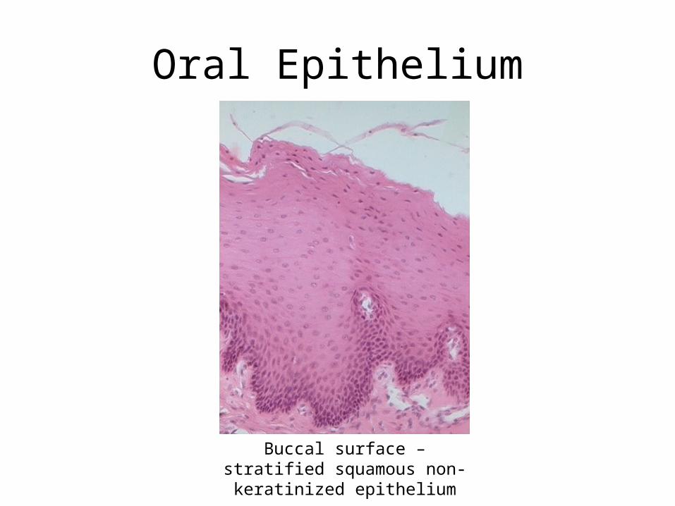
Oral Epithelium
Buccal surface – stratified squamous non-keratinized
epithelium
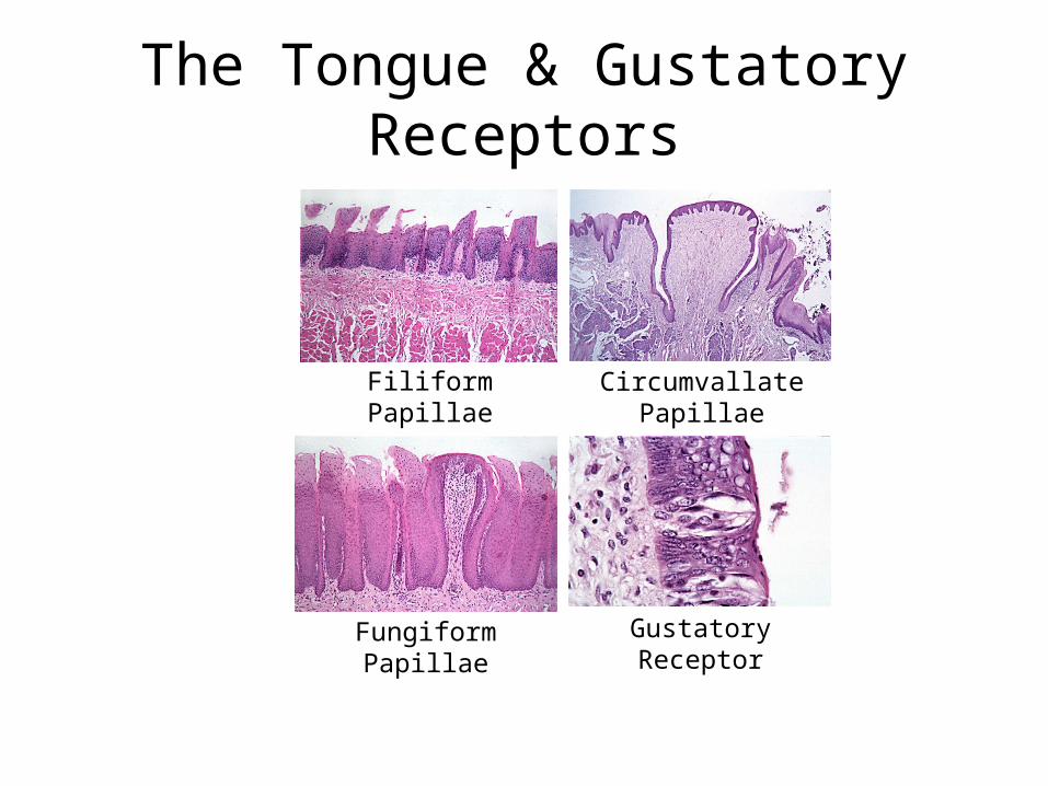
Filiform Papillae Circumvallate Papillae
Fungiform Papillae
Gustatory Receptor
The Tongue & Gustatory Receptors
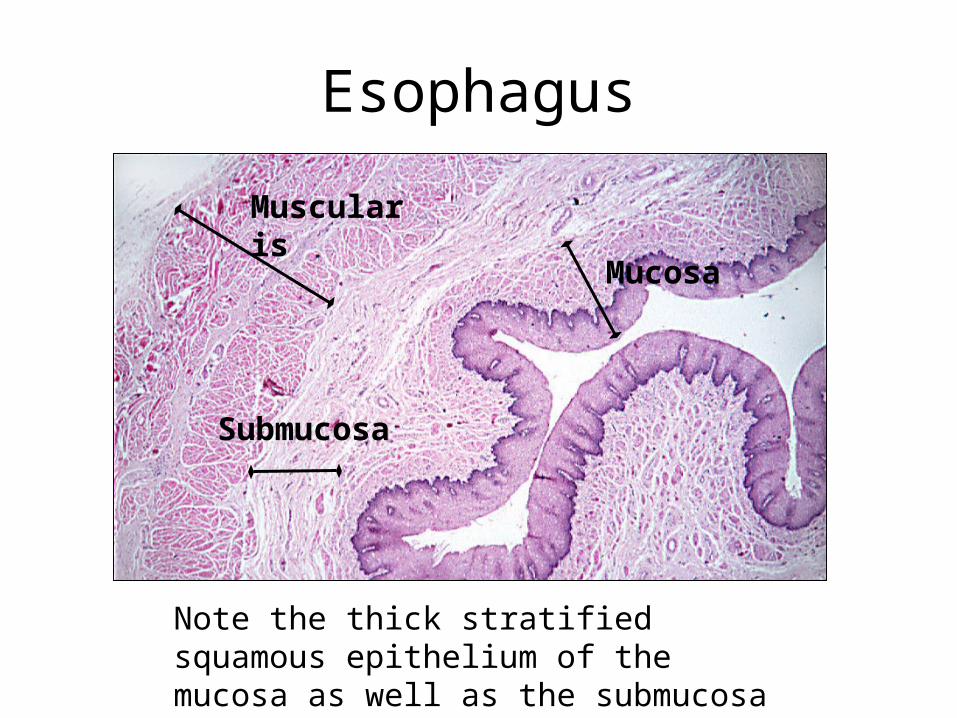
Esophagus
Note the thick stratified squamous epithelium of the mucosa as well as the submucosa and muscularis layers.
Muscularis
Submucosa
Mucosa
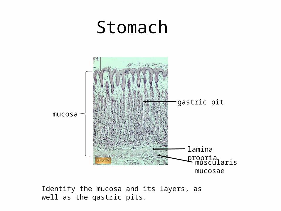
Stomach
mucosa
lamina propria
muscularis mucosae
gastric pit
Identify the mucosa and its layers, as well as the gastric pits.
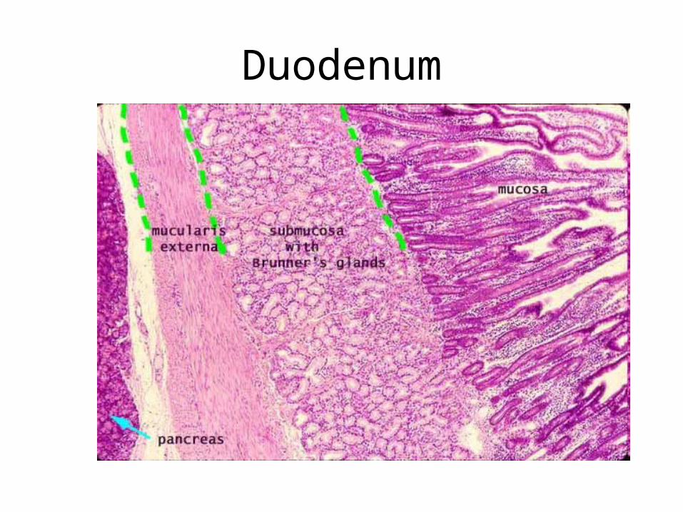
Duodenum
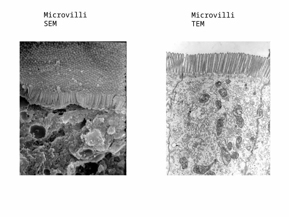
Microvilli SEM Microvilli TEM
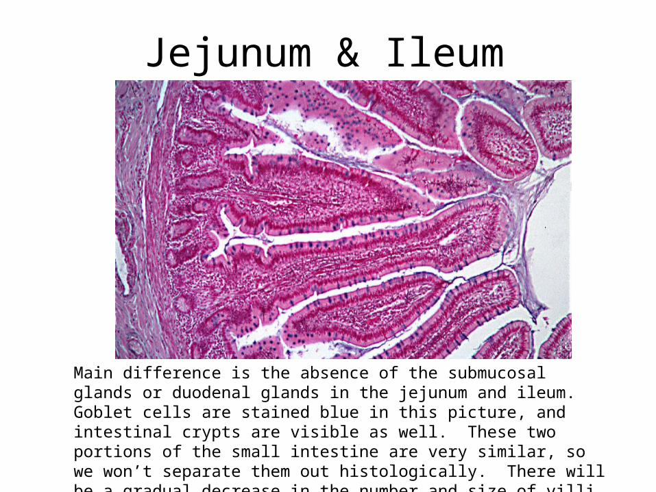
Jejunum & Ileum
Main difference is the absence of the submucosal glands or duodenal glands in the jejunum and ileum. Goblet cells are stained blue in this picture, and intestinal crypts are visible as well. These two portions of the small intestine are very similar, so we won’t separate them out histologically. There will be a gradual decrease in the number and size of villi as you progress towards the ileocecal valve, as well as an increase in the quantity of Peyer’s patches.
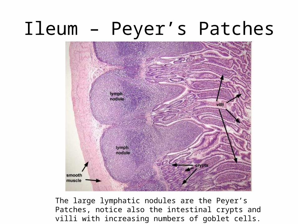
Ileum – Peyer’s Patches
The large lymphatic nodules are the Peyer’s Patches, notice also the intestinal crypts and villi with increasing numbers of goblet cells.
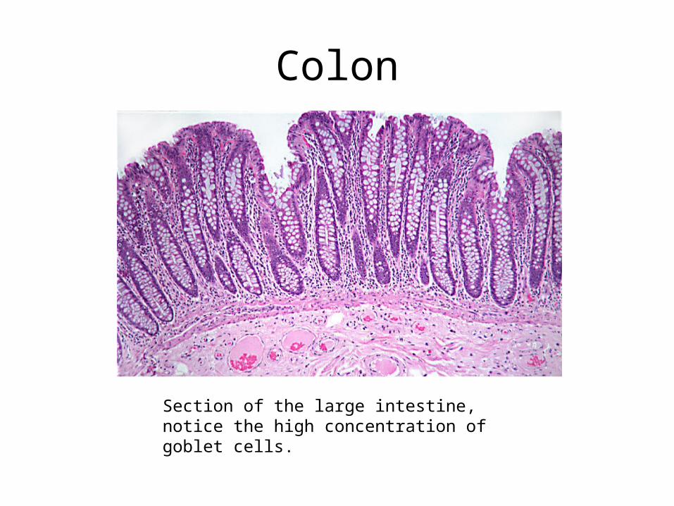
Colon
Section of the large intestine, notice the high concentration of goblet cells.
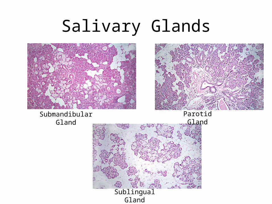
Salivary Glands
Parotid Gland
Sublingual Gland
Submandibular Gland
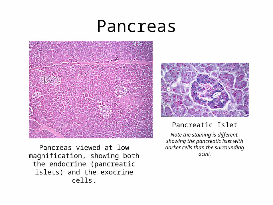
Pancreas
Pancreas viewed at low magnification, showing both the endocrine (pancreatic islets) and
the exocrine cells.
Pancreatic IsletNote the staining is different, showing the pancreatic islet with darker cells than the
surrounding acini.
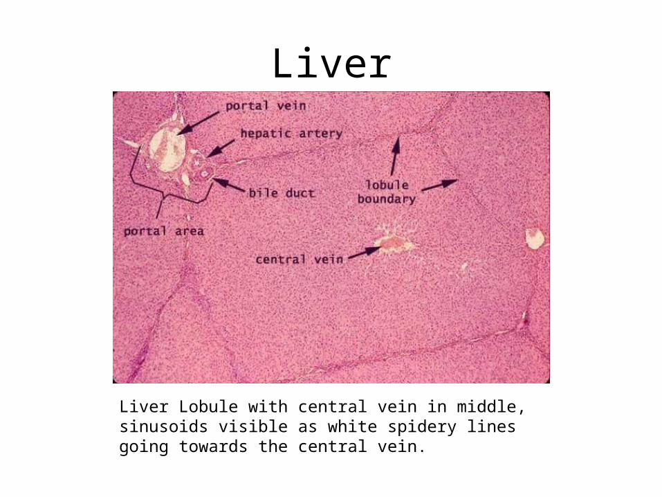
Liver
Liver Lobule with central vein in middle, sinusoids visible as white spidery lines going towards the central vein.
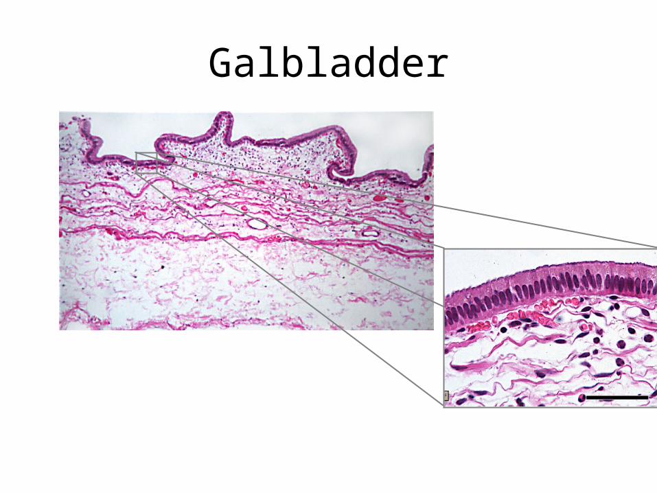
Galbladder
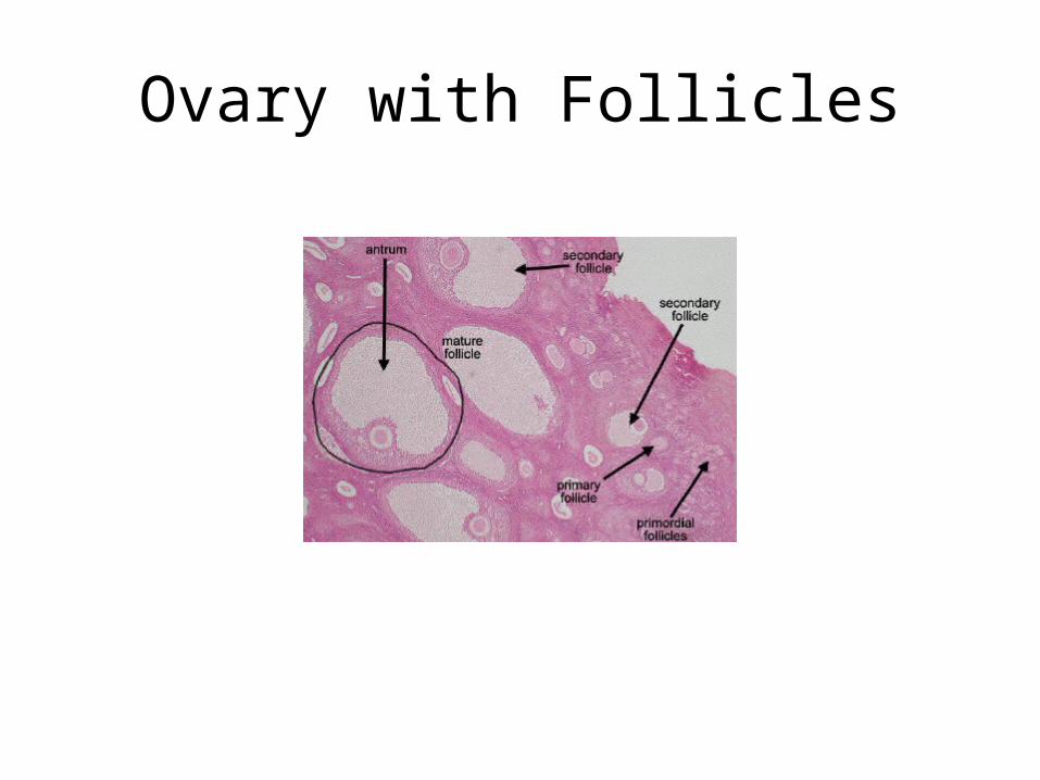
Ovary with Follicles
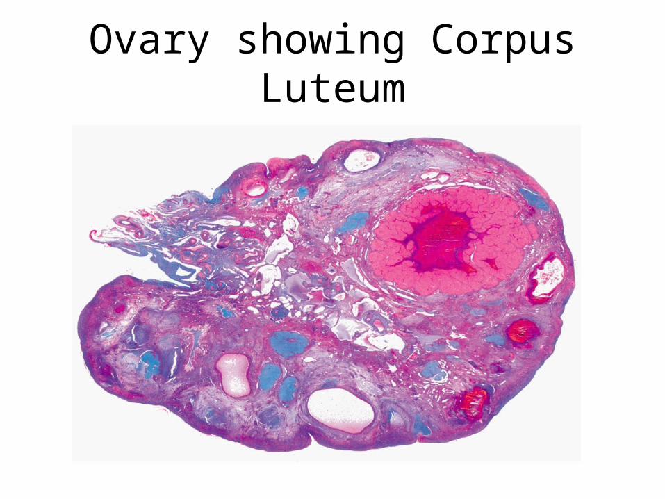
Ovary showing Corpus Luteum
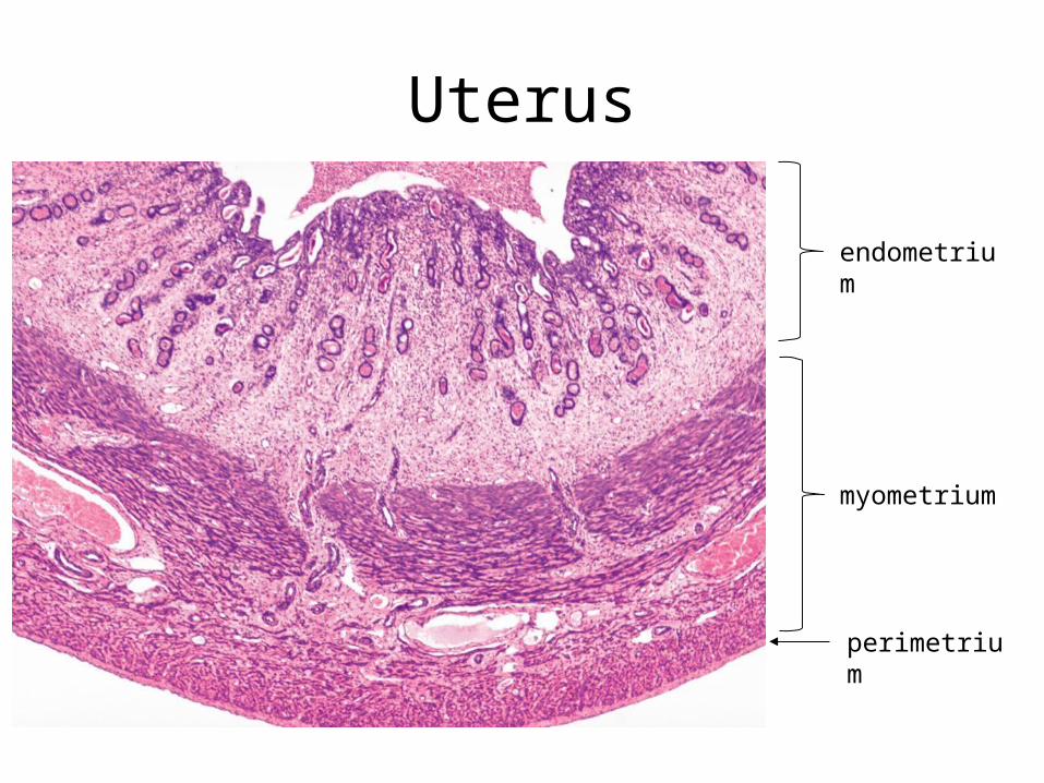
Uterus
endometrium
myometrium
perimetrium
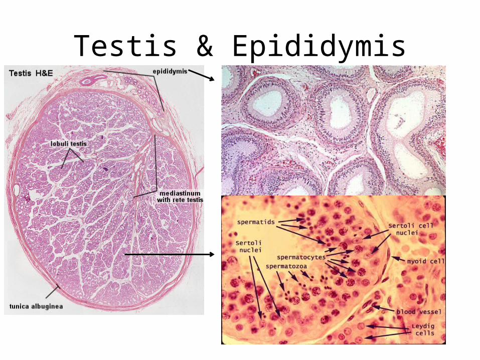
Testis & Epididymis
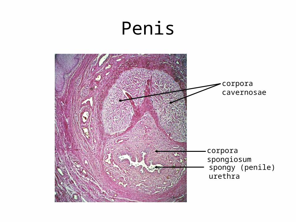
Penis
corpora cavernosae
corpora spongiosum
spongy (penile) urethra
![Histology Slides - mediconotes.commediconotes.com/freenotes/basic/histology_laboratory_slides.pdf[Histology] Histology Slides MedicoNotes provides real laboratory Histological slides](https://img.pdfslide.net/doc/110x75/5ae110e87f8b9a5a668e6aa3/histology-slides-histology-histology-slides-mediconotes-provides-real-laboratory.jpg)


![histology slides I [Kompatibilis mód]](https://img.pdfslide.net/doc/110x75/61bd122461276e740b0f0d53/histology-slides-i-kompatibilis-md.jpg)
