Embed Size (px)
Citation preview

Finger Injuries in BallSports
David T. Netscher, MDa,*, Dang T. Pham, MDb, Kimberly Goldie Staines, ORT, CHTcKEYWORDS
� Finger injuries � Ball sports � Athlete � Return to play � Orthoses
KEY POINTS
� Knowledge of bony and ligamentous anatomy of the digit is vital in treatment of finger injuries.
� Treatment of finger injuries in elite athletes remains a challenge for hand surgeons given the inherentdemand for an expeditious return to play.
� Communication with the athlete and the coach is important to understand the particular sport andposition of play, what orthosis may be allowable, and financial constraints.
� Occupational therapy and functional orthoses may greatly facilitate return to full activities.
INTRODUCTION
Ball sports are the leading cause of hand injuries inprofessional athletes.1 The use of the hand for ballcontrol and contact with the opponent leaves thefingers exposed to injury. Management of fingerinjuries in athletes often presents a challenge forhand surgeons. Multiple factors must be consid-ered, such as appropriate timing of treatment,long-term functional outcome, and (often themost difficult issue) return to play. This article dis-cusses the management of common finger injuriesin ball sports and provides return-to-play recom-mendations for professional athletes.
MALLET FINGER
Mallet finger injuries are common in ball sportssuch as baseball, basketball, and football.2 Theyare usually a result of forced hyperflexion of anextended finger on a ball or direct contact withanother player (Fig. 1). The middle, ring, and smallfingers are the most frequently involved digits.3
Disclosure Statement: The authors have nothing to discla Division of Plastic Surgery, Department of OrthopediStreet, Suite 2730, Houston, TX 77030, USA; b Departmenical College of Cornell University, 6550 Fannin Strec Department of Physical Medicine and Rehabilitation, M2002 Holcombe Boulevard, RCL117, Houston, TX 77030,* Corresponding author.E-mail address: [email protected]
Hand Clin 33 (2017) 119–139http://dx.doi.org/10.1016/j.hcl.2016.08.0180749-0712/17/Published by Elsevier Inc.
The athlete typically presents with a flexed postureat the distal interphalangeal (DIP) joint andimpaired active extension (extension lag). Plain ra-diographs including posterior-anterior and lateralviews of the affected finger are recommended toevaluate for fractures and joint subluxation. Malletinjuries can be categorized as soft tissue or bonymallet injuries.2,3 Soft tissue mallet injuries aredescribed as terminal extensor tendon avulsionfrom the insertion on the distal phalanx with noassociated bone fragment. When there is a frag-ment of bone associated with the terminalextensor tendon avulsion from the distal phalanx,it is a bony mallet injury. A bone fragment involvinggreater than a third of the articular surface mayresult in loss of joint congruency and volarsubluxation.
Closed mallet injuries that involve tendon only orthat have a small avulsed bone fragment can betreated nonoperatively with DIP joint extensionsplinting. The splint should span the width of thefinger and the length extend from fingertip to justdistal to the proximal interphalangeal (PIP) joint
ose and have no conflicts of interest.c Surgery, Baylor College of Medicine, 6624 Fannint of Surgery, Houston Methodist Hospital, Weill Med-et, Smith Tower 1661, Houston, TX 77030, USA;ichael E. DeBakey Veterans Affairs Medical Center,
USA
hand.th
eclinics.com

Fig. 1. Mechanism of injury of mallet finger. A flexionforce on the tip of an extended finger by a ball canresult in avulsion of the terminal extensor tendonon the dorsal lip of the distal phalanx base.
Netscher et al120
to allow for PIP joint motion.2 Care should be takento avoid skin ischemia, which can result fromdirect pressure on the skin by a tightly appliedsplint or by hyperextension of the DIP joint. Withregard to splint type, a systematic review revealedthat there are no substantial differences in treat-ment between prefabricated or custom-made or-thoses (Fig. 2).4 The key to treatment success ispatient compliance with splinting. It is recommen-ded to splint the DIP joint in full extension for 6 to8 weeks followed by a similar period of nighttimesplinting.3 Patient compliance with wearing andproperly positioning the splint is necessary for agood outcome.Operative management is considered when
there is volar subluxation of the distal phalanxwith significant joint incongruity. Closed reductionand percutaneous internal fixation of the DIP jointusing a Kirschner wire (K-wire) by extension blockpinning is the preferred technique.3 With the DIP
Fig. 2. (A) Stack splint. Prefabricated stack splint immobilizsplint. Dorsal aluminum splint for mallet finger should be san extended position.
joint maximally flexed, a K-wire is introduced at a45� angle into the head of the middle phalanx tocreate an extension block for the fragment andallow reduction of the volar fracture piece to thedorsal fragment (Fig. 3). Another K-wire is intro-duced axially from the distal to the middle phalanxto maintain the reduction. An alternative is toperform an open repair with pull-out button suture(Fig. 4). It can be used with either soft tissue malletor bony mallet. Open reduction has the advantageof direct access to the extensor tendon but hassignificant risk of complications, including skin ne-crosis, infection, nail dystrophy, osteoarthritis, andstiffness.5
Wehbe and Schneider6 advocated nonsurgicalmanagement of closed mallet fractures with largefracture fragments, even with volar subluxation ofthe distal phalanx. They found that subluxatedmallet fractures heal and remodel the DIP jointarticular surface with preservation of the jointspace. In a study with 22 closed mallet finger frac-tures, it was shown that even nonoperative treat-ment of mallet injuries involving greater thanone-third of the articular surface with or withoutsubluxation resulted in satisfactory outcomes.7
Based on the current literature, there is no clearindication for surgical treatment and insufficientevidence to support operative rather than nonop-erative treatment of mallet fractures. Most clini-cians consider operative intervention when thereis volar subluxation and loss of DIP joint congru-ency. Close follow-up with lateral radiographicviews is needed to guide treatment and ensuremaintenance of DIP joint congruency.
Return to Play
Immobilization of the DIP joint in full extension mayprevent optimal play for athletes and some mayopt to delay treatment or to have no treatment.8
Effective treatment of stable, closed mallet finger
es the DIP joint in full extension. (B) Dorsal aluminumecured with 2 strips of tape to maintain the DIP joint in

Fig. 3. Extension block pinning of mallet finger. A K-wire is inserted into the head of the middle phalanxat a 45� angle and proximal to the fractured fragmentto create an extension block. The volar fracture isreduced and a second K-wire is passed retrogradeacross the DIP joint to maintain reduction.
Finger Injuries in Ball Sports 121
injuries involves full-time splinting for at least 6 to8 weeks followed by nighttime splinting of a similarduration. Athletes are expected to adhere strictlyto the full treatment course and continue withimmobilization during strenuous activity, includingreturn to training.9–11 Splinting during training andplay runs the risk of maceration, loss of immobili-zation, and injury to other joints. During immobili-zation, consider including the adjacent joints inthe splint, using a circumferential cast (QuickCast,Patterson Medical) and buddy taping to limit risk ofinjury to PIP and metacarpophalangeal (MCP)joints (Fig. 5). Also, the use of Dynamic Tape(Vanuatu) or Kinesio Tape (Kinesio Holding Corpo-ration) in conjunction with the splint or cast cangreatly decrease the risk of maceration.12
If players cannot tolerate the external splintbecause of the demands of their positions, someinvestigators have advocated internal splinting bypercutaneous pins, also for at least 6 weeks.8
However, there is a risk of another jamming injurybreaking the pin in the joint of for pin migration.
Fig. 4. Pullout button suture. (A) Mallet finger with avulsphalanx. (B) A pullout button suture is used to secure twire inserted through the DIP joint is necessary to mainta
As with all injuries, options need to be tailored tothe urgency to return to play, the position andtype of sport, and the specific injured finger.
Following discontinuation of immobilization,players are permitted to return to play and toresume ball handling as tolerated. Athletes mustbe aware that even with strict compliance withsplinting, a residual 5� to 10� extensor lag and dor-sal joint prominence may be present. Withouttreatment, permanent flexion deformity, swanneck deformity, and DIP joint osteoarthritis candevelop.5
CHRONIC MALLET INJURIES AND SWAN NECKDEFORMITY
Chronic mallet deformities, categorized as morethan 4 weeks from injury, may be successfullytreated with extension splint even several weeksafter the injury.13 Swan neck deformity is charac-terized by hyperextension of the PIP joint andflexion of the DIP joint. It can result from failedtreatment of mallet finger injuries and is most com-mon in athletes with a hyperextensible PIP joint. Atrial of splinting of the DIP in extension and the PIPin flexion for 4 to 6 weeks is initiated. Adjustmentsto the splint are made as the deformity begins tocorrect itself. The duration of splinting may beswitched to part-time or nighttime splinting formaintenance. If there is a persistent deformity afternonoperative treatment of chronic mallet defor-mities, tendon rebalancing with a central sliptenotomy or spiral oblique retinacular ligament(SORL) reconstruction can be considered.3,13
The athlete should be examined for the degree of
ed bone fragment and volar subluxation of the distalhe terminal tendon to bone. (C) An axial or obliquein reduction.

Fig. 5. Mallet finger orthosis. (A) QuickCast (Patterson Medical) digital cast can be rapidly applied and easilyremoved for the duration of the sporting activity. (B) Custom dorsal mallet orthosis can be sufficiently rigid toavoid repeated jamming injuries. (C) Full-finger digital cast with buddy taping provides even better support pro-vided the positional play allows this. Full-finger immobilization, in contrast with only distal immobilization,avoids placing the PIP joint at risk for a new jamming injury. Splinting, where possible, should not only protectthe injured joint but also avoid the risk of loading adjacent joints and sustaining a new injury.
Netscher et al122
extension lag at the DIP and PIP joint. An exten-sion lag at the DIP joint less than 36� is best treatedwith Fowler central slip tenotomy.14 SORL proced-ure is recommended when there is an extensionlag at the DIP joint greater than 45�.3
The Fowler central slip tenotomy is designed toreduce the extensor tone at the PIP joint resultingfrom retraction of the extensor apparatus. It in-volves transecting the central slip proximal to itsinsertion into the middle phalanx base and allow-ing the extensor mechanism to slide proximally tocorrect the DIP extensor lag. Care is taken not toviolate the triangular ligament so that develop-ment of extensor lag at the PIP joint and bouton-niere deformity is avoided. The timing of theoperation is delayed until at least 6 months afterinjury to allow for tendon maturation.3 Fowler cen-tral slip tenotomy in several case series showed
good results. In a series of 20 patients withchronic mallet finger treated with central sliptenotomy, an average extensor lag of 37� beforethe operation corrected to 9� after the opera-tion.15 Houpt and colleagues16 found that, in 35patients with mallet finger and an averageextensor lag of 45�, 26 patients achieved fullextension after treatment with Fowler centraltendon tenotomy. The postoperative splintingprotocol involves splinting the PIP joint at 25� offlexion and the DIP joint in full extension for 10to 14 days, followed by a finger cast immobilizingthe DIP joint only for 2 additional weeks. After thepin is removed at 4 weeks, full active range ofmotion of the DIP joint is begun. The PIP joint isstarted on full active and passive range of motion.By 6 months, maximal range of motion of bothjoints is obtained.

Finger Injuries in Ball Sports 123
The SORL reconstruction is intended to restorethe tenodesis of the oblique retinacular ligamentsin promoting DIP joint extension with active PIPjoint extension (Fig. 6).3 A free tendon graft, typi-cally the palmaris longus or toe extensor tendon,is harvested and secured distally to the dorsalbase of the distal phalanx. The graft is then passedvolar in a spiral fashion around the radial aspect ofthe middle phalanx and is secured proximally tothe ulnar side of the flexor tendon sheath at thelevel of the proximal phalanx or directly to bone.Next, the DIP joint may be pinned in extensionand the PIP joint in 10� to 15� of flexion with anoblique K-wire.3 In a series of 12 patients who un-derwent the SORL reconstruction for chronicmallet finger caused by terminal tendon disruption,the mallet finger deformity was corrected in allcases.17 One patient required flexor tenolysis foradhesions to obtain full range of motion andanother patient required lengthening of the obliqueretinacular ligament graft to obtain full PIP jointextension. An alternative to SORL reconstructionis flexor digitorum superficialis (FDS) tenodesiswith combined flexor digitorum profundus jointfusion (Fig. 7).
Rehabilitation after SORL is challengingbecause the structures are so delicate. The goalsof rehabilitation are to dynamically facilitate DIP
Fig. 6. Spiral oblique retinacular ligament reconstruction.formities. (B) Dorsal capsulotomy of the PIP joint proximalis fixed to the distal phalanx, passed volar in a spiral fashishaft of the proximal phalanx, and secured with a bone antendon graft is shown in the (D) dorsal and (E) lateral vie
extension with PIP flexion, allowing for functionalgrasp and prehension in a tenodesis manner.18,19
Initial immobilization includes a dorsal gutter splintincorporating the PIP and DIP joints for 6 weeksfollowed by a figure-of-eight orthosis for an addi-tional 4 weeks. Return to play should be discour-aged for the initial 4 to 6 weeks for any teamsports. Immobilization at the time of return toplay should include the PIP in a flexed postureand DIP in an extended posture using a customorthosis or digital cast. Again, maceration is acommon complication, so consider using DynamicTape or Kinesio Tape under the cast or orthosis.Return to play with buddy taping can be enter-tained at 12 to 16 weeks as per tendon healingand strength assessment.
BOUTONNIERE DEFORMITY
A boutonniere deformity is characterized by flexionat the PIP joint and hyperextension at the DIPjoint.20 The precipitating injury leading to thischronic flexion deformity is disruption of the cen-tral slip, which causes continued flexion at thePIP joint and leads to attenuation of the triangularligament followed by volar subluxation of thelateral bands (Fig. 8).3 In athletes, disruption ofthe central slip can be caused by forced
(A) A 45-year-old woman with multiple swan neck de-to the central slip was performed. (C) The tendon grafton around the middle phalanx, tunneled through thechor or suture button. The direction and course of thews.

Fig. 7. FDS tenodesis and DIP joint fusion. (A) One slip of the FDS is transected proximal to the PIP joint, leavingthe distal attachment intact. With the PIP joint flexed 20� to 30�, the detached FDS slip is passed under and wrap-ped around the A1 pulley. (B) FDS tenodesis can be combined with an arthrodesis of the DIP joint if the flexion lagof the DIP joint is severe.
Netscher et al124
hyperflexion at the PIP joint, such as whencatching a ball with an outstretched hand. It canalso result from an occasional open injury withlaceration to the central slip or from crushinginjury, or volar dislocation at the PIP joint.Early recognition and intervention is key to pre-
venting boutonniere deformity. If the athlete re-ports jamming the finger or having a finger putback into place after any injury, the clinician shouldbe suspicious for central slip rupture.20 Volar dislo-cation of the PIP joint reduced on the field canresult in a boutonniere deformity if a central slipinjury is unrecognized and left untreated.21 TheElson test is a useful clinical assessment to deter-mine whether the patient has a central slip tearbefore the deformity develops (Fig. 9).22 It is per-formed by flexing the PIP joint to 90� and havingthe athlete extend the middle phalanx against
Fig. 8. Mechanism of injury of boutonniere deformity.A boutonniere deformity may develop in the acutesetting from forced hyperflexion of the PIP joint,causing disruption of the central slip at the base ofthe middle phalanx.
resistance. The test is positive if the DIP joint isable to extend because of the recruitment of thelateral bands in the setting of a central slip defi-cient finger. A digital block may be necessary toaccurately perform this test because pain mayprevent performance of the test. Plain radiographsshould be obtained to evaluate for dorsal fractureof the middle phalanx with or without volar sublux-ation or dislocation of the PIP joint.20
Early diagnosis of closed central slip injury canbe treated nonoperatively with splinting of thePIP joint in extension for 6 weeks to promote heal-ing of the central slip to the middle phalanx fol-lowed by 6 weeks of nighttime splinting. The DIPjoint is left free to allow flexion, which promotesdorsal translation of the lateral bands. Athletesare encouraged to perform DIP joint active andpassive flexion exercises hourly throughout thesplinting course.3
Operative management is recommended foropen injuries, large avulsion fractures, volar dislo-cation, or fracture-dislocation of the PIP joint.20
Central slip injuries associated with small avulsionfractures that are nondisplaced can be managednonoperatively. Larger, displaced fragments canbe repaired with K-wire or screw fixation. Centralslip laceration warrants open repair with reattach-ment of the tendon and placement of a transartic-ular K-wire to maintain the PIP joint in extensionduring healing.3
Return to Play
For closed central slip injury, athletes are generallyallowed to return to play as long as it does notobviate use of the splint and buddy taping is rec-ommended as an adjunct for game time protec-tion.23 Merritt24 advocates relative motionsplinting, which permits immediate full active mo-tion of the injured extensor tendon if it is placed in

Fig. 9. Elson test. (A) With the PIP joint extended, the DIP joint can actively extend. (B) With the PIP joint flexed,the DIP joint cannot actively extend. In the presence of central slip injury, the DIP joint can be extended when thePIP joint is flexed because of the recruitment of the lateral bands.
Finger Injuries in Ball Sports 125
15� to 20� less relative motion than adjacent ten-dons. The rationale for relative motion splinting isfor the injured tendon to experience less forcethan adjacent tendons during motion. In bouton-niere deformity, attenuation of the triangular liga-ment enables the lumbricals to act as flexors atthe PIP joint and to extend the DIP joint withoutan opposing force. Placing the injured digit into15� to 20� greater MCP flexion relative to adjacentdigits allows the lumbricals to relax, which in-creases the tension on the extensor hood and pro-motes dorsal migration of the lateral bands.24
Using the relative motion flexor splint when notplaying enables extensor tendon rehabilitationbut caution is needed when using this orthosisfor return to play because of the high risk of injuryto the adjacent fingers or unprotected hyperflexioninjury of the affected finger (Fig. 10). Return-to-play protection should include Dynamic Tape
Fig. 10. Relative motion splint. (A) Relative motion splin(B) The PIP joint is left unprotected for repeated hyperflexinjuries. For example, a forceful extension fall on an outstreRigid entrapment of adjacent fingers by the orthosis mafingers.
and/or circumferential digital casting with the PIPand DIP joints in full extension.
CHRONIC BOUTONNIERE DEFORMITY
Chronic boutonniere deformities can be catego-rized by the Burton classification. Stage I is a sup-ple, passively correctable deformity; stage IIconsists of extensor mechanism contracture thatis not passively correctable but does not involvethe joint; stage III is a fixed contracturewith involve-ment of the volar plate, collateral ligament, andintra-articular joint; and stage IV is stage III plusPIP joint arthritis.3 Stages I and II can undergo a trialof dynamic splint or serial casting to regain passiveextension of the PIP joint. If this is unsuccessful, anopen joint contracture release can be performed. Ifan active extensor lag persists but full passive PIPjoint was obtained, there are tendon rebalancing
t allows for neuromuscular retraining of extension.ion jamming injury. It also exposes the athlete to othertched hand may lead to proximal phalangeal fracture.y lead to susceptibility to ligament injuries in these

Netscher et al126
procedures that can be considered. Before under-going any surgical correction of a chronic bouton-niere deformity, patients should be fully informedof the risks and expected outcomes of the proced-ure, particularly thepossibility of jeopardizing flexorfunction in an attempt to gain extension.Terminal tendon tenotomy (distal Fowler or Dol-
phin tenotomy) was designed to decrease theextensor tone at the DIP joint and to allowthe extensor mechanism to retract and increasethe extensor tension to the PIP joint.25 It is mostvaluable in patients who report more difficultywith DIP joint hyperextension rather than the lackof PIP joint extension.26
Staged reconstruction of boutonniere deformity,described by Curtis and colleagues,27 is per-formed with wide-awake anesthesia to allowassessment of active extension intraoperativelyat each stage (Fig. 11). In stage I, an extensortenolysis is performed. If active extension is notpresent, the surgeon proceeds to stage II, in whichthe transverse retinacular ligament is transected toallow dorsal translation of the lateral bands. Instage III, if full correction is not achieved and thePIP joint extensor lag is less than 20�, a Fowlertenotomy is performed. If PIP joint extensor lag isgreater than 20�, the surgeon proceeds directlyto stage IV, in which the central tendon isdissected free from its insertion and advanced tothe middle phalanx base after 4 to 6 mm of scartissue are removed from its terminal end.
Fig. 11. Staged reconstruction for chronic boutonniere defthe transverse retinacular ligament enables the lateral baformed if full extension is not obtained.
A pseudoboutonniere deformity should bedifferentiated from a boutonniere deformity. Pseu-doboutonniere deformities are usually caused by ahyperextension injury to the PIP joint resulting involar plate and collateral ligament damage withno disruption of the central slip.28 Scarring of thedamaged structures leads to a PIP joint flexioncontracture with a secondary mild DIP joint hyper-extension deformity. Serial static or dynamicsplinting and casting techniques are used to grad-ually restore extension of the PIP joint. For persis-tent PIP joint flexion contracture, surgical releasemay be necessary.21
Return-to-play guidelines are the same for con-servative management of pseudoboutonniere andchronic boutonniere. Consider Dynamic Tape sup-port to prevent further injury with or withoutorthosis. Most athletic trainers or athletes can beeducated on independent application of this tapeon the field of play for long-term management.20
PULLEY RUPTURE
Injuries to the finger flexor pulley system are com-mon in rock climbers but have also been found inbaseball pitchers.29 The transfer of high forces tothe A2, A3, and A4 pulleys during the crimp-gripposition (MCP and DIP joints extended andresisted flexion at the PIP joint) is the likely injurymechanism for pulley rupture. Clinical findingsinclude localized swelling at the level of the injured
ormity. (A) Tenolysis of extensor tendon. (B) Release ofnd to swing dorsally. (C) Fowler tenotomy can be per-

Finger Injuries in Ball Sports 127
pulley and tendon bowstringing if the A2 and/or A4pulleys are involved.20 MRI can be useful in differ-entiating between a pulley strain and a partial orcomplete rupture (Fig. 12). The staging of pulleyinjuries and treatment protocol have been welldescribed by Schoffl and colleagues30 and thiscan be helpful in guiding treatment of professionalathletes.
Return to Play
Grade I injuries are pulley strains that can bemanaged conservatively with nonsteroidal antiin-flammatory medications. Easy sport activitiescan be started after 4 weeks and full sport activ-ities after 6 weeks. Grade 2 injuries are completeA4 or partial A2/A3 ruptures that can be treatedsimilarly to grade 1 injuries with the exceptionof a 10-day immobilization period. CompleteA2/A3 ruptures are grade 3 injuries that can stillbe treated conservatively, but easy sport activ-ities are restricted until 6 to 8 weeks and fullsport activities until 3 months. Grade 4 injuriesinvolve multiple pulley ruptures or single rupturecombined with lumbrical muscle or ligamenttrauma. These injuries require pulley reconstruc-tion, 14-day immobilization period after surgery,and 4 to 6 months of recovery before returningto sport activities. External pulley reinforcementcan facilitate early return to play for athletes(Fig. 13).
Fig. 12. Pulley rupture. (A) MRI of an uninjured finger. (Bwith associated bowstringing deformity. The patient has apseudoboutonniere hyperextension of the DIP joint. AsteriPIP joint.
PROXIMAL INTERPHALANGEAL JOINTDISLOCATION
Of the injuries in the hand, the PIP most commonlysustains ligamentous injuries.31,32 The collateralligaments and volar plate of the PIP joint create athree-dimensional ligament box that resists jointdisplacement until 2 sides of the box are disrupted(Fig. 14). These stabilizing structuresof thePIP jointare vulnerable to the axial loading and extensionforce that is applied on an outstretched finger. Ath-leteswhoparticipate in ball sports are at risk for PIPjoint injuries, either while catching a ball or falling.The spectrum of PIP joint injuries ranges from ami-nor volar plate strain to an irreducible dislocation ofthe joint. The joint should be inspected for an irre-ducible dislocation and any residual deformity orgross laxity.33 Plain radiographs must be obtainedto evaluate for avulsion fractures and the approxi-mate percentage of the involved articular surface.The dorsal V sign may be a subtle indication ofan incongruent or dorsally subluxated joint(Fig. 15).31 PIP joint dislocations are classified bythe direction of the middle phalanx in relation tothe proximal phalanx, which can occur in one of 3directions: dorsal, lateral, or volar.
Dorsal Dislocations
Dorsal dislocations are the most common type ofPIP joint dislocations and usually occur secondary
) MRI of a finger showing disruption of the A2 pulleylso developed a fixed PIP joint flexion contracture withsk shows scarring dorsal to the tendon and volar to the

Fig. 13. Pulley ring. (A) Taped pulley reinforcement can allow early return to play in grade 1 and 2 pulley ruptureinjuries. (B) For grade 3 and 4 pulley ruptures, the use of a custom pulley ring with tape and padding is recom-mended during play for 4 to 6 months.
Netscher et al128
to hyperextension injury. Pure dorsal dislocationsresult in an avulsion of the volar plate at its distalattachment with the collateral ligaments remainingintact and joint congruity maintained.31 With morelongitudinal force, one of the collateral ligamentscan be injured and result in a type I dorsal disloca-tion in which the joint surfaces are still touching or
Fig. 14. (A) PIP joint three-dimensional ligament box. Collup the ligament box. At least 2 sides of the box must be dbility. The 2 proximal phalangeal condyles provide 2 laterainherently provided by the congruency of the articular surto dorsal instability. (Adapted from [A] Merrell G, Slade JWolfe SW, Hotchkiss RN, Pederson WC, et al, editors. GreeElsevier Churchill-Livingstone; 2011. p. 292; with permissio
a type II dorsal dislocation in which there is a bay-onet appearance (Fig. 16). Type I dorsal disloca-tion can be reduced with volar translation andflexion. Type II dorsal dislocation requires hyper-extension of the middle phalanx followed bypalmar force because the volar plate acts as ablock to reduction with pure longitudinal traction.
ateral ligaments and volar plate of the PIP joint makeisrupted for joint displacement. (B) PIP joint bony sta-l columns for strength. The stability of the PIP joint isface. Fracture lines 1, 2, and 3 result in lateral or volarF. Dislocations and ligament injuries in the digits. In:n’s operative hand surgery. 6th edition. Philadelphia:n.)

Fig. 15. Dorsal V sign. On a lateral radiograph of ajoint, dorsal joint widening can be a sign of joint in-congruity and subluxation. (Adapted from Merrell G,Slade JF. Dislocations and ligament injuries in thedigits. In: Wolfe SW, Hotchkiss RN, Pederson WC,et al, editors. Green’s operative hand surgery. 6th edi-tion. Philadelphia: Elsevier Churchill-Livingstone;2011. p. 292; with permission.)
Finger Injuries in Ball Sports 129
Return to playIf the joint is stable and has full range of motion af-ter reduction, the athlete is allowed to return toplay immediately with buddy taping for protection.If the joint is unstable, the player is placed in anextension block orthosis for at least 4 weeks withweekly reassessment and splint adjustments(Fig. 17). For irreducible dislocations, open reduc-tion with extraction of the volar plate from the jointis the best option. Postoperative return to play re-quires 12 to 16 weeks with an extension blockorthosis during play.
Lateral Dislocations
Lateral dislocations of the PIP joint result fromrupture of the collateral ligament on one side andpartial avulsion of the volar plate on the side ofinjury.31 Assessment of lateral stability can tracewhich ligamentous structures are disrupted.
Fig. 16. Bayonet dislocation. (A) Anteroposterior and (B)importance of obtaining a true lateral view for PIP jointnormal.
Greater than 20� of deformity in extension sug-gests complete collateral ligament disruption andat least 1 of the secondary stabilizers. Once lateralPIP joint dislocations are reduced, they are oftenstable by virtue of the double-column bonysupport.
Return to playThe athlete is allowed early movement with buddytaping for protection.34 Open repair of the rupturedcollateral ligaments has been performed in ath-letes to expedite return to play, although there isno significant evidence that it expedites healingor improves motion.31 Occasionally, the collateralligament becomes trapped in the joint and requiresopen reduction.34 For unstable lateral dislocationsand high-level sporting dislocations, return to playcan include a ligament hinge brace with buddytaping (Fig. 18).
Volar Dislocations
Volar PIP joint dislocations are rare and usuallyoccur from a sudden torque to the digit causingthe base of the middle phalanx to dislocate volarlyand rupture the central slip.31 Even if the joint isreducible, clinicians should have a high index ofsuspicion for disruption of the extensor mecha-nism. Failure to immobilize the joint in extensionto allow the central slip to heal can lead to aboutonniere deformity. Complex volar dislocationsinvolve a rotatory component in which the condyleof the proximal phalanx buttonholes between thecentral slip and the lateral band (Fig. 19). Closedreduction can be attempted by relaxing the lateralbands through MCP joint and PIP joint flexion fol-lowed by gentle rotary manipulation. Most casesrequire open reduction because of failed reductionfrom the noose effect of the central slip and lateralband around the neck of the proximal phalanx.Less commonly, failed reduction can occur from
lateral radiographs of a bayonet dislocation show thefractures because the AP projection may look almost
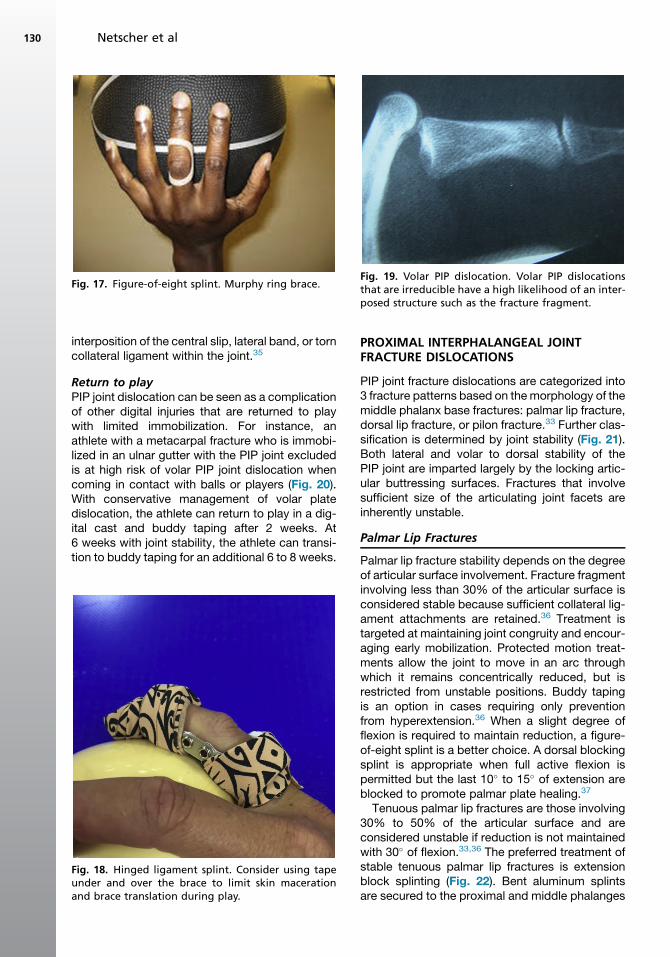
Fig. 17. Figure-of-eight splint. Murphy ring brace.Fig. 19. Volar PIP dislocation. Volar PIP dislocationsthat are irreducible have a high likelihood of an inter-posed structure such as the fracture fragment.
Netscher et al130
interposition of the central slip, lateral band, or torncollateral ligament within the joint.35
Return to playPIP joint dislocation can be seen as a complicationof other digital injuries that are returned to playwith limited immobilization. For instance, anathlete with a metacarpal fracture who is immobi-lized in an ulnar gutter with the PIP joint excludedis at high risk of volar PIP joint dislocation whencoming in contact with balls or players (Fig. 20).With conservative management of volar platedislocation, the athlete can return to play in a dig-ital cast and buddy taping after 2 weeks. At6 weeks with joint stability, the athlete can transi-tion to buddy taping for an additional 6 to 8 weeks.
Fig. 18. Hinged ligament splint. Consider using tapeunder and over the brace to limit skin macerationand brace translation during play.
PROXIMAL INTERPHALANGEAL JOINTFRACTURE DISLOCATIONS
PIP joint fracture dislocations are categorized into3 fracture patterns based on themorphology of themiddle phalanx base fractures: palmar lip fracture,dorsal lip fracture, or pilon fracture.33 Further clas-sification is determined by joint stability (Fig. 21).Both lateral and volar to dorsal stability of thePIP joint are imparted largely by the locking artic-ular buttressing surfaces. Fractures that involvesufficient size of the articulating joint facets areinherently unstable.
Palmar Lip Fractures
Palmar lip fracture stability depends on the degreeof articular surface involvement. Fracture fragmentinvolving less than 30% of the articular surface isconsidered stable because sufficient collateral lig-ament attachments are retained.36 Treatment istargeted at maintaining joint congruity and encour-aging early mobilization. Protected motion treat-ments allow the joint to move in an arc throughwhich it remains concentrically reduced, but isrestricted from unstable positions. Buddy tapingis an option in cases requiring only preventionfrom hyperextension.36 When a slight degree offlexion is required to maintain reduction, a figure-of-eight splint is a better choice. A dorsal blockingsplint is appropriate when full active flexion ispermitted but the last 10� to 15� of extension areblocked to promote palmar plate healing.37
Tenuous palmar lip fractures are those involving30% to 50% of the articular surface and areconsidered unstable if reduction is not maintainedwith 30� of flexion.33,36 The preferred treatment ofstable tenuous palmar lip fractures is extensionblock splinting (Fig. 22). Bent aluminum splintsare secured to the proximal and middle phalanges

Fig. 20. Ulnar gutter splint. (A) Ulnar gutter splint or padded forearm-based orthosis with buddy taping of meta-carpal fractures allows return to training. (B) Full-fist positioning should be considered for team practice and re-turn to play because of risk of hyperextension injury to the digits.
Finger Injuries in Ball Sports 131
in such a way that they allow PIP flexion but blockextension beyond the point at which subluxationoccurs.36 Confirmation of proper splint placementwith true lateral radiographs is recommended.Extension block pinning achieves a similar func-tion to extension block splinting (Fig. 23). TheK-wire is inserted retrograde into the proximalphalanx at a 30� angle to its long axis or at theangle that blocks unstable extension. Variousmethods of skeletal traction and dynamic externalfixation have been used to treat unstable PIP
Fig. 21. PIP fracture dislocations. (A) PIP joint stability isinvolvement. (B) The greater the percentage of articular sulizers are disrupted.
fracture dislocations (Fig. 24). The advantage tothese methods is that they allow for early motionand prevent joint stiffness. Athletes are notpermitted to return to play while they have thesedevices in place.
Unstable palmar lip fractures involve more than50% of the joint surface or involve 30% to 50%of the articular surface and require more than 30�
of flexion to maintain reduction of the PIP joint.33,36
Treatment is focused on restoring the palmarbuttress. Open reduction and internal fixation
classified based on the percentage of joint surfacerface involved, the more likely the ligamentous stabi-

Fig. 22. Extension block splinting. (A) Full active flexion is permitted. (B) Extension is blocked beyond the point atwhich the joint becomes unstable.
Netscher et al132
with screws and K-wires is recommended forlarger fracture fragments. For highly comminutedfractures of the base of the middle phalanx, cerc-lage wires can be used to stabilize the fragmentsby providing circumferential compression withwire tightening. When reconstructive proceduresare required to restore or reconstruct the base ofthe middle phalanx, palmar plate arthroplasty orhamate osteochondral autograft arthroplasty areconsidered.
Dorsal Lip Fractures
Dorsal lip fractures commonly involve disruption ofthe central slip and are maximally stable in fullextension.36 Stable dorsal lip fractures are usuallyminimally displaced avulsion fractures involvingless than 50% of the articular surface. Stable dor-sal lip fractures with less than 2 mm of fragmentdisplacement may initially be immobilized inextension by a splint or transarticular K-wire for3 weeks. The PIP joint only should be immobilizedand the DIP joint left free for active and passiverange-of-motion exercises. The patient is then
Fig. 23. Extension block pinning. Extension of the PIPjoint is blocked with a K-wire advanced retrogradedown the intramedullary canal of the middle phalanx.
transitioned to a dynamic extension splint for3 weeks to permit active flexion. At 6 weeks, pas-sive flexion and strengthening exercises arestarted. Dorsal lip fractures with fragmentdisplacement greater than 2 mmmay have associ-ated extensor lag and are best treated with openreduction and internal fixation with K-wires withor without a tension band wire, pullout suture, su-ture anchor, or lag screws.36
Dorsal lip fractures are categorized as unstableif there is any palmar subluxation or dislocationseen radiographically after closed reduction.Large fracture fragments are treated with openreduction and internal fixation. Highly comminutedfractures are better treated with closed reductionand percutaneous pinning of the PIP joint in fullextension to allow the fragments to consolidateand the central slip to heal.
Pilon Fractures
Pilon fractures involve both palmar and dorsal lipfractures and are considered unstable. Tractionis the treatment of choice because it providesalignment and permits early mobilization.36 Openreduction and internal fixation is often not recom-mended because of an association with loss ofreduction, stiffness, and infection.
Return to playProfessional athletes with stable PIP joint fracturedislocations who can function effectively with pro-tected motion splints in place can return to play,although close observation and serial radiographicevaluation are needed to confirm maintenance ofreduction.33 Athleteswith unstablePIP joint fracturedislocations are cautioned not to delay repairbecause it is associated with poorer outcomesand limited options. Return to play with functionalprotection is often permitted once stable fixation isobtained. Options for return-to-play immobilizationinclude digital casting, taping, custom orthotics, or

Fig. 24. Dynamic traction with TurnKey FCS device (Biomechanics Lab, Inc. Sacramento, CA). (A) A 54-year-oldman with an unstable PIP joint fracture-dislocation on lateral radiograph. (B) After the device is installed, lateralradiograph confirmed joint realignment. (C) Active flexion and (D) extension exercises are performed during frac-ture healing.
Finger Injuries in Ball Sports 133
reinforced personal protective equipment. All deci-sions regarding immobilization should beresearched for compliancewith thesports regulato-ry agency.
PROXIMAL INTERPHALANGEAL JOINTCONDYLAR FRACTURES
Condylar fractures of the PIP joint can be classifiedinto 3 categories: type I is a stable fracture withoutdisplacement; type II is an unstable, unicondylarfracture; and type III is a bicondylar or comminutedfracture.38 All 3 radiographic views are necessaryfor appropriate evaluation of joint stability. Ananteroposterior (AP) radiograph can assess prox-imal migration and articular step-off, a lateralview can evaluate for volar displacement, and anoblique view can provide fracture geometry.Although type I PIP joint condylar fractures areconsidered stable, they should be treated withcaution and with close follow-up because theyare inherently unstable. Type II and III condylarfractures are highly unstable and nearly all require
operative intervention. Fixation requires at least 2points of fixation to control rotation. For severecomminution, consider dynamic traction.
Return to Play
Depending on the stability of fixation, an athletemay be able to return to sports only if there is sta-ble external bracing to the finger. Otherwise, it maybe best to await fracture union. However, early PIPjoint range of motion is always encouraged if fixa-tion is secure enough in order to avoid jointstiffness.
SAGITTAL BAND INJURY
Sagittal band injury in athletes is more commonlyknown as boxer’s knuckle, although it can occurin any sports that involve a direct blow to theMCP joint, such as football and rugby.13 The mid-dle finger is most susceptible to injury because ofits prominent metacarpal head when making a fist.Athletes usually present with MCP joint swelling,

Netscher et al134
tenderness, inability to initiate extension, and oc-casionally central tendon subluxation or disloca-tion. In addition to standard radiographic views,the Brewerton view may be helpful to identifycollateral ligament avulsion injuries and occultfractures of the metacarpal head.39 It is an APview obtained with the MCP joint flexed 65� withthe dorsal aspect of the fingers flat against theradiograph cassette and the beam angled 15� ul-nar to radial. Rayan and Murray40 classified closedsagittal band injuries into 3 types: type I, injurywithout extensor tendon instability; type II, injurywith tendon subluxation; and type III, injury withtendon dislocation.39
Sagittal band injuries without extensor digitorumcommunis (EDC) subluxation can be managedwith buddy taping to an adjacent digit for4 weeks.40 Acute injuries (<3 weeks) with EDCsubluxation may be treated with an MCP jointflexion block splint, or sagittal band splint, for8 weeks.41 The splint is designed to hold theinjured MCP joint in 25� to 35� of hyperextensionrelative to the adjacent digit, which allows for im-mediate active mobilization of the MCP joint withthe splint in place. Catalano and colleagues41
used the sagittal band splint to treat 10 patientswith type III sagittal band injury. Eight patientshad no pain, 3 patients had residual EDC subluxa-tion, and 1 patient required sagittal bandreconstruction.For chronic sagittal band injuries (>3 weeks) or
those who have failed nonsurgical treatment, thereare several methods of sagittal band reconstruc-tion that consist of sagittal band repair withrealignment of the extensor tendon.3 Carroll andcolleagues42 described a technique wherein adistally based radial slip of EDC is looped aroundthe radial collateral ligament and sutured to itselffor centralization. Postoperative rehabilitationmay use a relative motion splint, as described byMerritt.24 This method facilitates recovery and isless cumbersome than an outrigger orthosis withflexion block.
Return to Play
Athletes with acute sagittal band injury withoutsubluxation can return to play with buddy tapingwithin 10 days of injury. When EDC subluxationis present, the immobilization orthosis limits func-tional return to play and increases risk of injury toother joints in the hand. As such, return to play isdiscouraged for 8 to 12 weeks with chronic andpostoperative sagittal band injuries. Once theathlete returns to play, buddy taping for 4 weeksis strongly encouraged. If the athlete returns toplay before full tissue healing and return of
strength, there is a significant risk of recurrentrupture of the sagittal band.
RADIAL COLLATERAL LIGAMENT RUPTURE OFTHE METACARPOPHALANGEAL JOINT
Athletes with isolated radial collateral ligament(RCL) rupture of the MCP joint often present withpersistent swelling and pain along the radialaspect of a previously injured finger.25 RCL ruptureof the MCP joint is typically caused by forced ulnardeviation while the MCP joint is flexed. A Brewer-ton radiographic view should be obtained to eval-uate for avulsed bone fragments.37 MRI may berequired and can help differentiate partial fromcomplete tears. Grade I injury is described aspain without laxity, grade II is laxity with an endpoint in 60� of flexion, and grade III has no endpoint. Treatment of grade I and II generally in-volves splinting the joint in 30� of flexion and reas-sessment for stability of the RCL in 3 weeks.31
However, athletes are very active and may benefitfrom early surgical repair because it has beenshown that most patients improve and have satis-factory results with surgery. Grade III injuries,when acute, can be managed nonoperativelyinitially with immobilization in radial deviation for6 weeks. Surgical repair may be reserved for thosewho fail nonoperative treatment or is sometimesthe primary treatment in an effort to return theplayer back to sports sooner.
Return to Play
Early management includes casting in 30� of MCPflexion for 4 weeks. During this period, athletes arenot encouraged to return to play. This period is fol-lowed by taping and custom orthosis wear for anadditional 4 weeks, at which time range-of-motion exercises begin (Fig. 25). Return to playat this point depends on the athlete’s level ofplay, sport, and the digit involved. At 8 weeks afterinjury, athletes are encouraged to wean from theorthosis by taping during practice and then tapingduring play (Fig. 26).
METACARPAL OSTEOCHONDRAL FRACTURES
Professional ball sport players are susceptible tointra-articular metacarpal head fractures involvingthe indexmetacarpal because of it being an unpro-tected, border digit.38 The middle finger may alsobe affected, being the longest finger and moresusceptible to axial impact than the adjacent fin-gers. Radiographic evaluation requires the stan-dard 3 views along with the Brewerton view toaccurately evaluate the articular contour.43 Dis-placed ligament avulsion fractures and

Fig. 25. Custom orthosis for MCP joint RCL rupture. (A) Lateral deviation and (B) MCP flexion can be restrictedwith a custom orthosis, which can be padded for return to play or taped for use in a glove.
Finger Injuries in Ball Sports 135
osteochondral fracture should be managed withopen reduction and internal fixation. Kumar andSatku44 recommended that small osteochondralfragments should be reduced by approximatingthe capsule to trap and hold the fragments inplace. Headless screws have facilitated intra-articular fracture fragment fixation.
Return to Play
Immobilization for metacarpal fracture healing in-cludes ulnar gutter casting or custom orthosis for
Fig. 26. Buddy taping for MCP joint RCL rupture. (A) BuddFor the small finger, a custom step-down strapping is req
10 to 21 days for callus formation. Although thesetypes of immobilization are anatomically appro-priate, care should be taken when immobilizingfor return to practice or play in order to limit therisk of injury to other fingers.
THUMB ULNAR COLLATERAL LIGAMENTINJURY
Injury to the ulnar collateral ligament (UCL) of thethumb MCP joint was historically referred to asgamekeepers’ thumb because of the chronic laxity
y taping permits early return to play and support. (B)uired because of digital length discrepancy.

Fig. 27. Stener lesion. (A) A 35-year-old woman with unstable ulnar collateral ligament of the MCP joint of thethumb. (B) Distal insertion of the collateral ligament has avulsed and is blocked from reattachment to its insertionby the interposed adductor aponeurosis.
Netscher et al136
of UCL of the thumb observed in Scottish game-keepers.45 It was later referred to as skier’s thumbbecause this injury is frequently noted in skierswho fall on an outstretched hand strapped to thehandle of a ski pole, causing forced abductionand extension of the thumb. Thumb MCP jointcollateral ligament injuries are increasingly recog-nized in professional athletes who participate inball-handling sports.46
Athletes typically describe a hyperabductioninjury to the thumb and present with pain andswelling at the ulnar aspect of the thumb MCPjoint. Radiographs may show an avulsion fractureat the attachment of the UCL to the base of theproximal phalanx or a widening of the ulnar aspectof the first MCP joint.46 A local nerve block beforevalgus stress testing of the thumb MCP joint canbe helpful to accurately assess stability. Tender-ness without laxity indicates a partial tear. The cri-terion for joint instability is typically greater than30� laxity with valgus stress or greater than 15�
of increased radial deviation compared with thecontralateral thumb.47
Acute partial ruptures of the thumb UCL can betreated with 4 weeks of continuous immobilizationin a thumb spica cast or splint followed by pro-tected range-of-motion exercises.31 A completetear generally warrants surgical repair with reat-tachment of the ligament to its bony insertion.However, large, avulsed, nondisplaced bone frag-ments may heal with immobilization alone. AStener lesion is another indication for surgicalrepair. It occurs when the adductor aponeurosisis interposed between the distally avulsed UCLand its insertion at the base of the proximal pha-lanx.48 Some clinicians are concerned that valgusstress testing of the thumb MCP joint can precipi-tate a Stener lesion that did not exist before(Fig. 27). Spontaneous ligament healing isinhibited because the ulnar ligament is not in con-tact with the bony insertion. Presence of a
persistent firm mass on the ulnar aspect of the firstmetacarpal head is suspicious for a Stener lesionand should be confirmed with MRI orultrasonography.
Return to Play
Return to play depends on the classification of thetear and the player’s dominant hand. In baseball,early return to sport with a thumb spica splint ispermitted for partial tears in the nonthrowingarm.49 For partial tears in the throwing arm,4 weeks of full-time immobilization followed by 2to 4 weeks of protective splint during play is rec-ommended. For complete tears that require oper-ative repair, additional caution is taken to avoidreinjury with early return to play. It is advised towait 6 to 8 weeks after surgery in the nonthrowingarm and 10 to 12 weeks in the throwing arm.
SUMMARY
Suggested orthoses, facilitations of return to play,and important considerations when returning anathlete to play are summarized in Table 1.50
Although tissue healing is the primary consider-ation when treating finger injuries, in elite athletesthere are several other factors to consider. Thesport, position of play, level of play, hand domi-nance, age of athlete, financial considerations,and player compliance are all serious points toconsider when determining the appropriatereturn-to-play style and timing. In addition, chronicuse of performance-enhancing drugs, previousinjury, and medical treatment to the area shouldbe considered when determining healing timeand prognosis for recovery. Communication be-tween the athlete, surgeon, hand therapist,training staff, and coaches is often imperative foran informed decision-making process. Thesemultifactorial considerations allow for great vari-ance among injuries and athletes.

Table 1Management of common finger injuries in ball sports
Injury Orthosis Return to Sport Considerations
Mallet finger � Full-finger cast ororthosis
� Buddy tape to adjacentfinger
� May return in orthosisbased on edema andpain
� Compliance with wear-ing and properly posi-tioning the splint isnecessary for a goodoutcome
� Hand dominance� Sport/equipment� Protect proximal andadjacent joints
� Add tape under orthosis/cast to limit macerationand translation oforthosis
Swan neckdeformity
� Full-finger cast/orthosisfor 6 wk
� Wean to figure-of-eightsplint for 4 wk
� Progress to PIP taping for12 wk
� May return to training infigure-of-eight splint at6 wk
� Consider full-finger castwith buddy tape forhigh-impact team sports
� Add tape under orthosis/cast to limit macerationand translation oforthosis
Boutonnieredeformity
� Closed injury: buddy tape� PIP extension orthosis for6 wk at all times
� Wean to nighttime splintfor 6 additional weeks
� 0–10 d for closed injurybased on comfort
� May return with full-finger cast and buddytape for nonoperativecases
� For postoperative, 6–8 wk in cast
� Be sure to protect prox-imal and distal joints
� Return to play in cast isbased on sport and tissueintegrity
� Add tape/cohesivebandage under orthosis/cast to limit macerationand translation oforthosis
Pulley rupture � Grade 1 and 2: tapesupport
� Grade 3 and 4: pulleyring over tape
� Light sport activities suchas training can be startedafter 4 wk
� Full sport activities after6 wk
� At all times, use tape tosupport the pulley
� Confirm custom ringaround sport equipment(bats, balls, rackets) andunder protective devices(gloves).
� Use tape under customring and over the fillfinger to decrease ringtranslation during play
Dorsal PIPdislocations
� Buddy taping� Full-finger extensioncast/orthosis
� Figure-of-eight exten-sion orthosis
� With stable joint and fullrange of motion, returnto play immediately withbuddy taping
� If the joint is unstable,return to play at >4 wkwith extension cast
� Postoperative: return toplay at 12–16 wk withbuddy tape
� Joint stability andintegrity
� Position� Light vs heavy demands� Key player vs supportplayer
Lateral PIPdislocations
� Buddy taping� Full-finger extensioncast/orthosis
� Hinged ligament splint
� Early movement withbuddy taping for protec-tion/deviation stability
� 2–4 wk return to playwith orthosis
� Joint stability andintegrity
� Position� Light vs heavy demands� Key player vs supportplayer
Volar PIPdislocations
� Buddy taping� Full-finger flexion cast/orthosis
� Figure-of-eight flexionorthosis
� In a digital cast andbuddy taping after 2 wk
� Continue buddy tape for6–8 wk
� Joint stability andintegrity
� Position� Light vs heavy demands� Key player vs supportplayer
Finger Injuries in Ball Sports 137

Netscher et al138
REFERENCES
1. Rettig AC. Epidemiology of hand and wrist injuries in
sports. Clin Sports Med 1998;17:401–6.
2. Yeh PC, Shin SS. Tendon ruptures: mallet, flexor dig-
itorum profundus. Hand Clin 2012;28(3):425–30.
3. Strauch RJ. Extensor tendon injury. In: Wolfe SW,
Hotchkiss RN, Pederson WC, et al, editors. Green’s
operative hand surgery. 6th edition. Philadelphia:
Elsevier Churchill-Livingstone; 2011. p. 159–88.
4. Witherow EJ, Peiris CL. Custom-made finger ortho-
ses have fewer skin complications than prefabri-
cated finger orthoses in the management of mallet
injury: a systematic review and meta-analysis. Arch
Phys Med Rehabil 2015;96(10):1913–23.
5. Botero SS, Hidalgo Diaz JJ, Benaida A, et al. Review
of acute traumatic closed mallet finger injuries in
adults. Arch Plast Surg 2016;43(2):134–44.
6. Wehbe MA, Schneider LH. Mallet fractures. J Bone
Joint Surg Am 1984;66(5):658–69.
7. Kalainov DM, Hoepfner PE, Hartigan BJ, et al.
Nonsurgical treatment of closed mallet finger frac-
tures. J Hand Surg Am 2005;30(3):580–6.
8. Shin SS. Baseball commentary–tendon ruptures:
mallet, FDP. Hand Clin 2012;28(3):431–2.
9. Simpson D, McQueen MM, Kumar P. Mallet defor-
mity in sport. J Hand Surg Br 2001;26(1):32–43.
10. Groth GN, Wilder DM, Young VL. The impact of
compliance on the rehabilitation of patients with
mallet finger injuries. J Hand Ther 1994;7(1):21–4.
11. Hovgaard C, Klareskov B. Alternate conservative
treatment of mallet-finger injuries by elastic double-
finger bandage. J Hand Surg Am 2005;30(3):580–6.
12. McMurtry JT, Isaacs J. Extensor tendon injuries. Clin
Sports Med 2015;34:167–80.
13. Chauhan A, Jacobs B, Andoga A, et al. Extensor
tendon injuries in athletes. Sports Med Arthrosc
2014;22(1):45–55.
14. Rozmaryn LM. Central slip tenotomy with distal
repair in the treatment of severe chronic mallet fin-
gers. J Hand Surg Am 2014;39(4):773–8.
15. Grundberg AB, Reagan DS. Central slip tenotomy
for chronic mallet finger deformity. J Hand Surg
Am 1987;12(4):545–7.
16. Houpt P, Dijkstra R, Storm van Leeuwen JB. Fowler’s
tenotomy for mallet deformity. J Hand Surg Br 1993;
18(4):499–500.
17. Kleinman WB, Petersen DP. Oblique retinacular liga-
ment reconstruction for chronic mallet finger defor-
mity. J Hand Surg Am 1984;9(3):399–404.
18. Thompson JS, Littler JW, Upton J. The spiral oblique
retinacular ligament (SORL). J Hand Surg Am 1978;
3(5):482–7.
19. Shrewsbury MM, Johnson RK. A systematic study of
the oblique retinacular ligament of the human finger:
its structure and function. J Hand Surg Am 1977;
2(3):194–9.
20. Marino JT, Lourie GM. Boutonniere and pulley rupture
in elite athletes. Hand Clin 2012;28(3):437–45.
21. Aronowitz ER, Leddy JP. Closed tendon injuries of
the hand and wrist in athletes. Clin Sports Med
1998;17(3):449–67.
22. Elson RA. Rupture of the central slip of the extensor
hood of the finger. A test for early diagnosis. J Bone
Joint Surg Br 1986;68(2):229–31.
23. Smith DW. Boutonniere and pulley rupture in elite
basketball. Hand Clin 2012;28(3):449–50.
24. Merritt WH. Relative motion splint: active motion af-
ter extensor tendon injury and repair. J Hand Surg
Am 2014;39(6):1187–94.
25. Dolphin JA. Extensor tenotomy for chronic bouton-
niere deformity of the finger; report of two cases.
J Bone Joint Surg Am 1965;47:161–4.
26. Meadows SE, Schneider LH, Sherwyn JH. Treatment
of the chronic boutonniere deformity by extensor te-
notomy. Hand Clin 1995;11(3):441–7.
27. Curtis RM, Reid RL, Provost JM. A staged technique
for the repair of the traumatic boutonniere deformity.
J Hand Surg Am 1983;8(2):167–71.
28. McCue FC, Honner R, Johnson MC, et al. Athletics
injuries of the proximal interphalangeal joint
requiring surgical treatment. J Bone Joint Surg Am
1970;52(5):937–56.
29. Lourie GM, Hamby Z, Raasch WG, et al. Annular
flexor pulley injuries in professional baseball pitchers:
a case series. Am J Sports Med 2011;39(2):421–4.
30. Schoffl V, Hochholzer T, Winkelmann HP, et al. Pulley
injuries in rock climbers. Wilderness Environ Med
2003;14(2):94–100.
31. Merrell G, Slade JF. Dislocations and ligament in-
juries in the digits. In: Wolfe SW, Hotchkiss RN,
Pederson WC, et al, editors. Green’s operative
hand surgery. 6th edition. Philadelphia: Elsevier
Churchill-Livingstone; 2011. p. 291–332.
32. Bindra RR, Foster BJ. Management of proximal inter-
phalangeal joint dislocations in athletes. Hand Clin
2009;25(3):423–35.
33. Williams CS 4th. Proximal interphalangeal joint frac-
ture dislocations: stable and unstable. Hand Clin
2012;28(3):409–16.
34. Vicar AJ. Proximal interphalangeal joint dislocations
without fractures. Hand Clin 1988;4(1):5–13.
35. Boden RA, Srinivasan MS. Rotational dislocation of
the proximal interphalangeal joint of the finger.
J Bone Joint Surg Br 2008;90(3):385–6.
36. Kang R, Stern PJ. Fracture dislocations of the prox-
imal interphalangeal joint. J Hand Surg Am 2002;
2(2):47–59.
37. McElfresh EC, Dobyns JH, O’Brien ET. Management
of fracture-dislocation of the proximal interphalan-
geal joints by extension-block splinting. J Bone Joint
Surg Am 1972;54(8):1705–11.
38. Day CS, Stern PJ. Fractures of the metacarpals and
phalanges. In: Wolfe SW, Hotchkiss RN,

Finger Injuries in Ball Sports 139
Pederson WC, et al, editors. Green’s operative hand
surgery. 6th edition. Philadelphia: Elsevier Churchill-
Livingstone; 2011. p. 239–90.
39. Kleinhenz BP, Adams BD. Closed sagittal band
injury of the metacarpal joint. J Am Acad Orthop
Surg 2015;23(7):415–23.
40. Rayan GM, Murray D. Classification and treatment of
closed sagittal band injuries. J Hand Surg Am 1994;
19(4):590–4.
41. Catalano LW 3rd, Gupta S, Ragland R 3rd, et al.
Closed treatment of nonrheumatoid extensor tendon
dislocations at the metacarpophalangeal joint.
J Hand Surg Am 2006;31(2):242–5.
42. Carroll CT, Moore JR, Weiland AJ. Posttraumatic ul-
nar subluxation of the extensor tendons: a recon-
structive technique. J Hand Surg Am 1987;12(2):
227–31.
43. Lane CS. Detecting occult fractures of the meta-
carpal head: the Brewerton view. J Hand Surg Am
1977;2(2):131–3.
44. Kumar VP, Satku K. Surgical management of osteo-
chondral fractures of the phalanges and
metacarpals: a surgical technique. J Hand Surg
Am 1995;20(6):1028–31.
45. Campbell CS. Gamekeeper’s thumb. J Bone Joint
Surg Br 1955;37(1):148–9.
46. Lee AT, Carlson MG. Thumb metacarpophalangeal
joint collateral ligament injury management. Hand
Clin 2012;28(3):361–70.
47. Heyman P. Injuries to the ulnar collateral ligament of
the thumb metacarpophalangeal joint. J Am Acad
Orthop Surg 1997;5(4):224–9.
48. Stener B. Displacement of the ruptured ulnar collat-
eral ligament of the metacarpophalangeal joint of the
thumb: a clinical and anatomical study. J Bone Joint
Surg Br 1962;44:869–79.
49. Chhor KS, Culp RW. Baseball commentary “thumb
ligament injuries: RCL and UCL”. Hand Clin 2012;
28(3):371–2.
50. Goldie Staines K, Collins ED. The athlete. In:
Jacobs MA, Austin NM, editors. Orthotic intervention
for the hand and upper extremity: splinting princi-
ples and process. Baltimore (MD): Lippincott Wil-
liams & Wilkins; 2014. p. 544–65.