Embed Size (px)
Citation preview

Stomatologija, Baltic Dental and Maxillofacial Journal, 2007, Vol. 9, No. 4 121
Finite element analysis of stresses in the maxillary andmandibular dental arches and TMJ articular discs during
clenching into maximum intercuspation, anterior andunilateral posterior occlusion
Gaivile Pileicikiene, Algimantas Surna, Rimantas Barauskas, Rimas Surna, Algidas Basevicius
SCIENTIFIC ARTICLES
SUMMARY
The objective of this study was to investigate distribution of stresses in the human TMJ discs,generated during clenching into various occlusal positions.
The work presents a biomechanical finite element model of interaction of mandibular andmaxillary dental arches and the TMJ discs of a particular person, based on real geometrical dataobtained from spiral computed tomography two-dimensional images. 3D contour coordinates - pointclouds were collected from these images and solid model was created. The system under investigationconsisted of eight basic parts: two rigid structures representing the mandibular and maxillary dentalarches, two mandibular condyles, two mandibular fossae of temporal bone, and solid models of twoarticular discs. The model of maxillary dental arch was fixed in space. The model of the mandibulardental arch was able to move in space synchronically with the mandibular condyles under actionof applied forces, which were considered as prescribed and known at insertion points of masticatorymuscles. The motion of the mandible was constrained by interdental contact interactions and contactinteraction with articular discs, which were situated in between mandibular condyles and mandibularfossae of temporal bone. The model was implemented by using LS-DYNA finite element software.
The obtained results presented a 3D view of stresses exhibited in the articular discs, as wellas the real contact points of dental interactions at given masticatory geometry of a particularsubject and the values of interaction forces. The expected practical value of the developed modelis the facilitation of biomechanical evaluations of the influence of tolerances of teeth shapes andocclusal areas together with the supporting areas on the final stress distribution in the dentalarches and articular discs.
Key words: biomechanics, mathematical model, finite element stress analysis,temporomandibular joint disc.
SCIENTIFIC ARTICLES Stomatologija, Baltic Dental and Maxillofacial Journal, 9:121-128, 2007
1Clinic of Prosthodontics, Kaunas Medical University, Lithuani2Kaunas University of Technology, Lithuania3Radiology Clinic, Kaunas Medical University, Lithuania
Gaivile Pileicikiene1 – D.D.S., assist. prof.Algimantas Surna1 – D.D.S., PhD, assoc. prof., Head of Clinic of
ProsthodonticsRimantas Barauskas2 – Dr. habil., prof., Head of System Analysis
DepartmentRimas Surna2 – PhD, assoc. prof.Algidas Basevicius3 – MD, PhD, prof., Head of Radiology Clinic,
Kaunas Medical University, Lithuania
Address correspondence to Gaivile Pileicikiene, Clinic ofProsthodontics, Sukileliu 51, Kaunas, Lithuania.E-mail: [email protected].
INTRODUCTION
The biomechanical functions of teeth generallyresult in stresses, which are transferred from the teeththrough the periodontal ligaments, mandible, maxilla andtemporomandibular joints and produce strains andstresses in all of them. Understanding the nature of strainand stress distribution is essential for better diagnosis and
treatment of stomatognathic diseases and reconstructionof masticatory function. Unfortunately, the stresses cannot be measured directly in a non-destructive way. Thenumber of direct studies on the masticatory system islimited, because its structures are difficult to reach andthe applications of experimental devices, such as straingauges, inside the structure introduce damage to itstissues, which interfere with normal function andinfluence their mechanical behavior. Dentalbiomechanics is an interdisciplinary approach in whichengineering principles are applied to dentistry [1](Asundi, 2000). During the latter decades numericalmethods and in particular the finite element methodbecame a powerful analysis instrument of structuralbehavior and interaction analysis of bodies, systems andenvironments of a very different physical nature. Inprinciple, most of the real physical can be more or lessadequately represented and modeled by usingappropriate computational software. Still persisting

122 Stomatologija, Baltic Dental and Maxillofacial Journal, 2007, Vol. 9, No. 4
simulation difficulties in biomechanics are related mainlywith the material models of investigated objects. Theyare always empirically determined, and often presentquite complex constitutive relations. A characteristicfeature of jaw and teeth interaction biomechanics iscomplex geometry of contacting surfaces. Finiteelements programs employing the explicit dynamicsapproach are most successfully used for simulation ofsuch systems. In this work we employed LSDYNA finiteelement software [2, 3]. Explicit dynamics programssimulate the processes in time or in pseudo-time byextrapolating the condition of the system at the next timestep on the base of the state of the system at current timestep and taking into account forces acting on the system.As calculations are performed step-by-step, complexcontact interactions are robustly managed andinteraction forces determined. Generally, numericalsimulation of physical systems and processes presentsa lot of interesting information about their condition andbehavior, which cannot be observed or measureddirectly. Often it can be used instead of makingexperiments on real physical systems. It considerablyreduces the price of the experiment. However, a certainamount of experience and attention is necessary in orderto obtain the simulation data that can be trusted as beingclose to reality. On the other hand, in process ofsimulation a lot of “non-quantified” understanding aboutreal physical behavior of the analyzed system is obtained.In this way, it can indirectly suggest the way to find theimproved practical solutions. A remarkable advantageof the finite element method is the chance to study areasthat are difficult or impossible to access without any risksto a living subject of investigation [4] (Jeon, 2001). Theuse of finite element method allows studying a single tooth,a set of teeth, or even the relationship between maxillaryand mandibular dental arches on a more solid and precisebiomechanical basis than other methods such asphotoelastic models and strain gauges [5] (Daegling,2000). Therefore, with this methodology it is possible tohave quantitative and qualitative representations ofdental and mandibular biomechanics to evaluatedisplacements, strains and stresses, which may occurin biomechanical structures.
The aims of this study were:1) to create a biomechanical three-dimensional
model of mandibular and maxillary dental arches and theTMJ discs of a particular person;
2) to verify the models ability for finite elementanalysis of stresses, generated in the temporomandibulardiscs during clenching.
MATERIALS AND METHODS
Skeletal morphologyObject of research was one cadaver of 20 year old
man. The research protocol of this study was approvedby Committee of Bioethics (Kaunas University ofMedicine). Computer tomography scanning (CTS) wasused to obtain two-dimensional images necessary for
creating 3D geometrical models. Multisection spiralcomputed tomography (General Electric) was performedin the area from infraorbital region to the base of mandibleand 1500 slices within thickness of 0.625 mm weregained. Case-oriented software was created to extractmorphological information of surfaces for eachcomponent of the biomechanical system independently.3D contour coordinates (point clouds) were collectedfrom the images and solid models were created. Three-dimensional geometrical models of all components com-prising the biomechanical system were created using the“Image Pro – Plus” software (Media Cybernetics,USA). Receiving of skeletal morphology and 3D geom-etry reconstruction was described, in detail, previously[6] (Pileicikiene et al, 2007).
The architecture of the modelThe geometry of parts comprising the model as
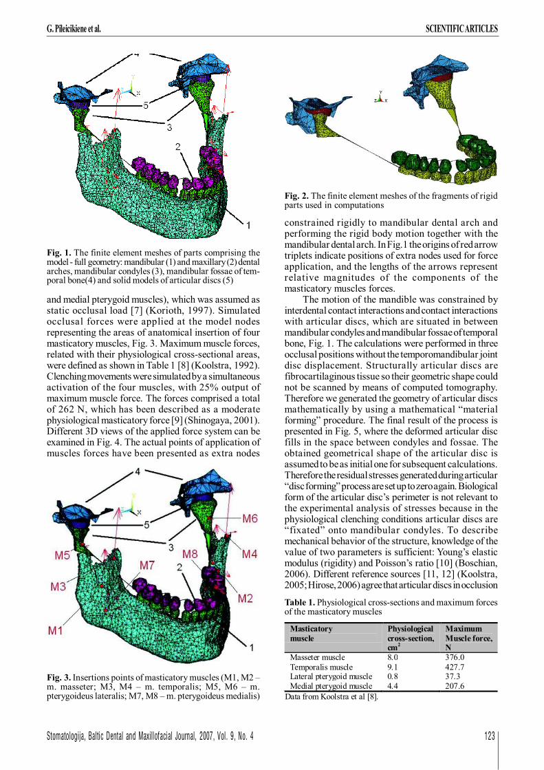
mandible, mandibular and maxillary dental arches,mandibular condyles and mandibular fossae of temporalbone have been obtained from spiral computedtomography two-dimensional images. By means ofsurfaces triangulation parts comprising the biomechanicalsystem have been created: mandibular dental arch,maxillary dental arch, right and left mandibular condylesand mandibular fossae of temporal bone. The finite elementmesh of parts comprising the model is presented in Fig. 1(parts 1 to 4). The precision of computations will notchange if the computational model includes only segmentsof rigid osseous structures, geometry of which is to betaken into account directly when modeling the contactinteraction, Fig. 2. The method of spiral computedtomography allows to obtain a very precise reconstructionof 3D view of scanned parts and to produce a highlyrefined finite element mesh. However, in this work wechose a modest refinement of the parts, containing ~13000nodes and ~18500 elements. The reasonable level ofrefinement of the model enabled to save computationalresources, simultaneously preserving all importantgeometrical properties of the investigated biomechanicalstructure. The models of mandibular and maxillary dentalarches were assumed to be rigid and therefore could bepresented by means of shell structures. The number ofnodes and elements necessary to present a shell structureis many times less than of a corresponding 3D solid body,and contributes to model size reduction. The value ofpenalty stiffness coefficient employed in the mathematicalcontact model of interdental contact interaction enabledto imitate the mobility of teeth comprising the dentalarches.
The model of maxillary dental arch was fixed inspace. The model of the mandibular dental arch was ableto move in space synchronically with the mandibularcondyles under action of applied forces, which wereconsidered as prescribed and known at points ofattachment of masticatory muscles. Clenching wassimulated by the action of resultant force vectors of fourbilateral masticatory muscles, responsible for theelevation of the mandible (masseter, temporalis, lateral
SCIENTIFIC ARTICLES G. Pileicikiene et al.
Stomatologija, Baltic Dental and Maxillofacial Journal, 2007, Vol. 9, No. 4 123
G. Pileicikiene et al. SCIENTIFIC ARTICLES
constrained rigidly to mandibular dental arch andperforming the rigid body motion together with themandibular dental arch. In Fig.1 the origins of red arrowtriplets indicate positions of extra nodes used for forceapplication, and the lengths of the arrows representrelative magnitudes of the components of themasticatory muscles forces.
The motion of the mandible was constrained byinterdental contact interactions and contact interactionswith articular discs, which are situated in betweenmandibular condyles and mandibular fossae of temporalbone, Fig. 1. The calculations were performed in threeocclusal positions without the temporomandibular jointdisc displacement. Structurally articular discs arefibrocartilaginous tissue so their geometric shape couldnot be scanned by means of computed tomography.Therefore we generated the geometry of articular discsmathematically by using a mathematical “materialforming” procedure. The final result of the process ispresented in Fig. 5, where the deformed articular discfills in the space between condyles and fossae. Theobtained geometrical shape of the articular disc isassumed to be as initial one for subsequent calculations.Therefore the residual stresses generated during articular“disc forming” process are set up to zero again. Biologicalform of the articular disc’s perimeter is not relevant tothe experimental analysis of stresses because in thephysiological clenching conditions articular discs are“fixated” onto mandibular condyles. To describemechanical behavior of the structure, knowledge of thevalue of two parameters is sufficient: Young’s elasticmodulus (rigidity) and Poisson’s ratio [10] (Boschian,2006). Different reference sources [11, 12] (Koolstra,2005; Hirose, 2006) agree that articular discs in occlusion
and medial pterygoid muscles), which was assumed asstatic occlusal load [7] (Korioth, 1997). Simulatedocclusal forces were applied at the model nodesrepresenting the areas of anatomical insertion of fourmasticatory muscles, Fig. 3. Maximum muscle forces,related with their physiological cross-sectional areas,were defined as shown in Table 1 [8] (Koolstra, 1992).Clenching movements were simulated by a simultaneousactivation of the four muscles, with 25% output ofmaximum muscle force. The forces comprised a totalof 262 N, which has been described as a moderatephysiological masticatory force [9] (Shinogaya, 2001).Different 3D views of the applied force system can beexamined in Fig. 4. The actual points of application ofmuscles forces have been presented as extra nodes
Fig. 1. The finite element meshes of parts comprising themodel - full geometry: mandibular (1) and maxillary (2) dentalarches, mandibular condyles (3), mandibular fossae of tem-poral bone(4) and solid models of articular discs (5)
Fig. 2. The finite element meshes of the fragments of rigidparts used in computations
Fig. 3. Insertions points of masticatory muscles (M1, M2 –m. masseter; M3, M4 – m. temporalis; M5, M6 – m.pterygoideus lateralis; M7, M8 – m. pterygoideus medialis)
Table 1. Physiological cross-sections and maximum forcesof the masticatory muscles
Masticatory muscle
Physiological cross-section, cm2
Maximum Muscle force, N
Masseter muscle 8.0 376.0 Temporalis muscle 9.1 427.7 Lateral pterygoid muscle 0.8 37.3 Medial pterygoid muscle 4.4 207.6
Data from Koolstra et al [8].

124 Stomatologija, Baltic Dental and Maxillofacial Journal, 2007, Vol. 9, No. 4
SCIENTIFIC ARTICLES G. Pileicikiene et al.
simulation can be modeled as elastic isotropic solids.Non-linear elasticity model with Young’s modulus 30.9-41.4 MPa up to stress level of 1.5 MPa, and Young’smodulus 92.4 MPa above stress level 1.5 MPa has beenemployed. Poisson’s ratio 0.4 of the material has beenused. The values of mechanical characteristics used inour study were estimated from compressive stress-relaxation tests using experimental animals andpreviously reported by Tanaka et al. [13] (Tanaka, 1999)(Table 2).
The system under investigation consisted of eight basicparts: two rigid structures representing the mandibular andmaxillary dental arches, two mandibular condyles, twomandibular fossae of temporal bone, and solid models oftwo articular discs. The final view of computational modelwith applied muscle force system and mathematicallygenerated articular discs, ready for numerical experimentsis presented in Fig. 6. To verify our model’s ability for finiteelement analysis of stresses, generated in thetemporomandibular discs during clenching, we simulatedvarious clenching conditions: clenching into maximum
intercuspation, biting with four frontal teeth and clenchingwith unilateral (left side) four posterior teeth.
RESULTS
Numerical experiment No. 1Simulation of clenching into maximum
intercuspation – contact between all the teeth in themaxillary and mandibular dental arches.
Rigid body to rigid body contact between dentalarches has been investigated under action of applied forcesystem at extra nodes as in Fig. 6. The sliding contact withfriction coefficient 0.2 has been assumed in condyle-articular disc and fossae-articular disc contact pairs.Distribution of stresses (von Misses) in thetemporomandibular joint discs in the full-arch clenchingcondition is presented in Fig. 7, A-C. The von Misses stressis a mathematical combination of all components of bothaxial and shear stresses. It is commonly used to representthe total stress a given region is experiencing [14](DeVocht JW). Fig. 7, A presents axonometric view ofthe system. Fig. 7, B presents the contour plots of von-Misses stresses in articular discs, where the articular fos-sae have been removed for visualization needs. Fig. 7, Cpresents the contour plots of von-Misses stresses in ar-ticular discs, where the condyles have been removed forvisualization needs. It is obvious that stress distribution inboth articular discs for this particular person in the condi-tion of full-arch clenching is slightly asymmetric. The larg-est stresses were located in the central part of articulardiscs, directly contacting with the mandibular condyles,
Fig. 4. The force system applied at extra nodes, whichrepresent the points of attachment of masticatory mus-cles: A – side view and B – bottom view
Fig. 5. Mathematically generated geometrical shapes of thearticular discs in between of mandibular condyles and man-dibular fossae of temporal bone
A
B

Stomatologija, Baltic Dental and Maxillofacial Journal, 2007, Vol. 9, No. 4 125
G. Pileicikiene et al. SCIENTIFIC ARTICLES
Fig. 6. The final computational model
Fig. 7. Distribution of von-Misses stresses in the TMJ discsin full-arch clenching condition
A
B
C
Table 2. Material properties of articular disc
their maximal values were 4.86 MPa. During the experi-ment articular discs were in the central position, typical ofthe maximum intercuspation.
Numerical experiment No. 2Simulation of anterior occlusion – biting with
four anterior teeth of the maxillary and mandibulardental arches.
Contact of only four frontal teeth was implementedby positioning a thin occlusion plate (Young’s modulus~1000 Mpa, yield stress 22 MPa) in-between frontalparts of mandibular and maxillary dental arches, Fig. 8,A. Fig. 8, B-C demonstrate the contour plots of vonMisses stresses in articular discs, where the articular fos-sae have been removed for visualization needs. It isevident that maximum stresses concentrated in the oc-clusion plate situated between the frontal teeth. Thestress distribution in the articular discs in condition offrontal teeth clenching was slightly asymmetric, whilethe values of stresses were approximately 30% lower(3.29 MPa) in comparison with full – arch clenchingcondition (4.86 MPa). During this part of experimentarticular discs stayed in the central position because bit-ing with four anterior teeth was simulated by placing athin occlusal plate between the anterior teeth withoutprotrusive movement of the mandible.
Numerical experiment No. 3Simulation of unilateral posterior occlusion –
clenching with four posterior teeth of the left side.Contact of only four left side posterior teeth wasimplemented by positioning a thin occlusion plate (Young’smodulus ~1000 Mpa, yield stress 22 MPa) in-between leftdistal parts of mandibular and maxillary dental arches,Fig.9, A. Fig. 9, B-C demonstrate the contour plots ofvon Misses stresses in articular discs, where the articu-
lar fossae have been removed for visualization needs. Itis obvious that maximum stresses are concentrated in theocclusion plate placed between the left side posterior teeth.The left articular disc was free of stresses, and moderatestresses (1.59 MPa) were generated in the right articulardisc, which comprised 33% of the strength of stresses,generated in the articular disc during clenching into maxi-mum intercuspation. During the last part of experimentarticular discs were located in the central position becausebiting with four posterior teeth was simulated by placing athin occlusal plate between the corresponding teeth with-out laterotrusive movement of the mandible.
Material Elastic modulus, E (MPa)
Poisson ratio
Articular disc 30.9 0.4 Data from Tanaka et al [13].

126 Stomatologija, Baltic Dental and Maxillofacial Journal, 2007, Vol. 9, No. 4
SCIENTIFIC ARTICLES G. Pileicikiene et al.
DISCUSSION
Finite element models have their current origin andreal use in mechanical engineering analysis and design.Biological applications have been successful wheremechanical principles would be of the most interest; forexample in modeling human joints (hip, knee, etc.) [15](Liau, 2001). In dentistry, models have been used to
determine the stresses in different biologic structures, suchas jaws, facial skeleton, dentition, periodontal ligament,temporomandibular joint [16, 17, 18, 19] (Cattaneo, 2003;Gross, 2001; Gomes de Oliveira, 2006; Toms, 2003;Hirose, 2006) and different dental restorative materials,for example, implants, composites etc. [20, 10] (Akpinar,2000; Boschian, 2006). Most of the surveyed FEM studiesanalyzed the biomechanical behavior of individual
Fig. 8. Distribution of von-Misses stresses in the TMJdiscs in four anterior teeth clenching condition
Fig. 9. Distribution of von-Misses stresses in the TMJ discsin unilateral (left side) four posterior teeth clenching condi-tion
A
B
C
A
B
C

Stomatologija, Baltic Dental and Maxillofacial Journal, 2007, Vol. 9, No. 4 127
G. Pileicikiene et al. SCIENTIFIC ARTICLES
structures or materials, while we have tried to investigateinteractions of stresses in the biomechanical system,representing the main elements of human masticatorysystem. Obviously, biomechanical models of the humanmasticatory system are not perfect, while they are basedon a number of assumptions and simplifications [12](Hirose, 2006). The adequacy of the FE computationalmodel to the real system depends on the correctness ofrepresentation of the geometry and material properties ofthe modeled object, the type and number of elements andthe boundary conditions imposed on the model [7] (Korioth,1997). General point is that the precision of finite elementcalculations increases as highly refined meshes of themodel parts are used. On the other hand, it is well knownthat highly refined meshes are necessary only at zoneswhere high stress gradients are expected and at zones ofcomplex and highly curved geometrical shapes of themodel. In other words, the adaptation of the computationalmesh density to the particular problem is always a goodpractice since it enables to achieve satisfactory accuracyand adequacy of the results by using reasonable modeldimensions. The error estimation of the results of nonlinearproblems solutions does always present a challenge,however, an „engineering“ way of checking theconvergence of the solution can always be employed. Thismeans a satisfactory coincidence of the simulation resultsobtained by using two different mesh densities the lineardimension of the elements of which differ approximatelytwo times.
In order to obtain the correct representation of thegeometry of the model we have chosen the minimal (0.625mm) slice thickness possible to be registered by the CTdevice. This increased X-ray radiation for the object andmade the research impossible to perform on the livingperson. In the future work the X-ray radiation could bereduced by scanning only the main parts of the masticatorysystem with high accuracy and after their geometryreconstruction integrating them into biomechanical systemtemplate, created from low accuracy (requiring lowradiation) computed scanning images. Magneticresonance imaging (MRI) could be used to get informationabout TMJ structures without introducing radiation to thepatient, but this method is more applicable for evaluatingthe soft structures of TMJ, especially the articular discs[21] (Kobs, 2004), while our study was focused on the hardtissues of masticatory system, such as jaws and teeth. Inreality the components of the biomechanical system aremade of different materials, which are described in termsof their individual mechanical characteristics. In order tosimplify the computations we assumed the models ofmandibular and maxillary dental arches as rigid and whole,not accentuating single teeth and their periodontalligaments. The simplification was performed because themain goal of this preliminary study was to evaluate the finalstress distributions in the temporomandibular joint discsin the clenching condition. Appropriate extensions of themodel would enable to investigate distribution of stressesand strains in all parts and in-between of them. Themechanical material properties of the articular disc were
obtained from the canine disc [13] (Tanaka, 1999), becauseno reliable material parameters for human are availablein the literature. The positions of the points, the directionsand magnitudes of forces exhibited by muscles werebased on the values presented in reference literature [7,8, 9] (Korioth, 1997; Koolstra, 1992; Shinogaya, 2001).As based on a previous studies [8] (Koolstra, 1992), a 25%activation of the masticatory muscles during clenching wasused for the stress analysis, although large inter-individualand intra-individual differences in masticatory muscleactivity may exist for the same task [12] (Hirose, 2006).Furthermore, the simultaneous activation of four differentmuscles at the same relative percentage of maximummuscle force was not real; for instance, there wasestimated that electromyogram activities of lateralpterygoid muscle, anterior temporalis, and massetermuscles were dependent on the velocity [22] (Huang,2005) and the phase of the closing jaw movement [23](Soboleva, 2005). Besides, movements of the mandibleare influenced not only by active muscle tensions generatedby contracting muscle fibers, but also by multiple passiveforces [24] (Pileicikiene, 2004). However, even with theabove-mentioned limitations, our model was able todemonstrate what effect clenching in different conditionshad on the stress distribution in the temporomandibular jointdiscs. The distribution of stresses in the models of rightand left TMJ articular discs of this particular cadaverduring full-arch clenching into maximal intercuspation wasslightly asymmetric; this could be appreciated as analogouswith stress distribution during clenching in physiologicconditions. During simulation of anterior biting or unilateralposterior clenching the maximum stresses concentrationwas found in the occlusion plate, while the articular discssustained only moderate stresses. Such distribution ofstresses is appropriate for the normal masticatory functionwith well balanced occlusion, where the major part ofocclusal load is directed to the dental arches and absorbedin the supporting tissues, while the temporomandibularjoints receive only minor part of the load. Comparison ofthe results from this study to previous reports [12, 25, 26,27] (Hirose, 2006; Chen, 1998; Radu, 2004; Naeije, 2003)was difficult because the applied load, material propertiesof the articular disc, boundary conditions and constraintsat the articulating surfaces varied among these models.However, the stress distributions in our study were similarto the previous reports [26, 27] (Radu, 2004; Naeije, 2003)and the stress levels from our model were within the rangeof reported stress magnitudes. Previous studies [12, 25](Hirose, 2006; Chen, 1998) reported that the maximumvon Misses stresses in the disc were 0,85 to 8,0 MPa ascompared to 4,86 MPa in the present study. Furthermore,previous reports were based on the partial models oftemporomandibular joints, while our model included all themain elements of masticatory system. Whereas we havereconstructed higher accuracy three-dimensionalgeometry of all the parts, comprising masticatory system,appropriate extensions of the model would enable toinvestigate distribution of stresses and strains in all partsand interaction between them in different simulated

128 Stomatologija, Baltic Dental and Maxillofacial Journal, 2007, Vol. 9, No. 4
SCIENTIFIC ARTICLES G. Pileicikiene et al.
situations, such as disturbed occlusal equilibrium or alveolarbone loss due to the periodontal disease. In the future workthe model could be supplemented with different prostheticdevices, such as periodontal splints or dental implants tosimulate prosthetic treatment and predict its efficiency.
CONCLUSIONS
1. A computer-based three-dimensional finiteelement model consisting of the mandibular and maxillarydental arches, mandibular condyles and mandibular fossaeof temporal bone, and two articular discs was created.
2. This initial FEM model has demonstrated that thecomputational modeling approach can be successfullyused in the biomechanical study of stresses distributionin the constituent parts of the masticatory system.
3. The model can be conveniently extended in orderto present the teeth mobility. This will cause the size of
the model to increase substantially because of the necessityof component models of every particular tooth in the dentalarch, inclusion of periodontal ligament, improvedrepresentation of masticatory muscles activity etc.
4. The expected practical value of the developedmodel is the facilitation of biomechanical evaluations ofthe influence of tolerances of teeth shapes and occlusalareas together with the supporting areas on the final stressdistribution in the dental arches and articular discs.
5. Future development of this model will enable theinclusion of differential material properties and simulationof the teeth, implants and prostheses in various pathologicalsituations with the objective of creating prosthetictreatment predictive models, for example, occlusalequilibration or splint therapy for dental arches, affectedby periodontitis.
6. Much more work is to be done in order to validatethe created models on the base of physical experiments.
Received: 07 08 2007Accepted for publishing: 21 12 2007
REFERENCES
1. Asundi A, Kishen A. A strain gauge and photoelastic analysisof in vivo strain and in vitro stress distribution in human dentalsupporting structures. Arch Oral Biol 2000;45:543-50.
2. LS-DYNA Keyword Users Manual.Version 960, vol.1, 2.Livermore Software Technology Corporation; March 2001.
3. LS-DYNA Theoretical Manual. Livermore SoftwareTechnology Corporation; May 1998.
4. Jeon PD, Turley PK, Ting K. Three-dimensional finite elementanalysis of stress in the periodontal ligament of the maxillaryfirst molar with simulated bone loss. Am J Orthod DentofacialOrthop 2001;119:498-504.
5. Daegling DJ, Hylander WL. Experimental observation,theoretical models, and biomechanical inference in the study ofmandibular form. Am J Phys Anthropol 2000;112:541–51.
6. Pileicikiene G, Varpiotas E, Surna R, Surna A. A three-dimensional model of the human masticatory system, includingthe mandible, the dentition and the temporomandibular joints.Stomatologija. Baltic Dent Maxillofac J 2007; 9:27-32.
7. Korioth TWP, Versluis A. Modeling the mechanical behaviorof the jaws and their related structures by finite element (FE)analysis. Crit Rev Oral Biol Med 1997;8:90-104.
8. Koolstra JH, Van Eijden TMGJ. Application and validation ofa three-dimensional mathematical model of the humanmasticatory system in vivo. J Biomech 1992; 25:175-87.
9. Shinogaya T, Bakke M, Thomsen CE, Vilmann A, SodeyamaA, Matsumoto M. Effects of ethnicity, gender and age onclenching force and load distribution. Clin Oral Invest2001;5:63-8.
10. Boschian PL, Guidotti S, Pietrabissa R, Gagliani M. Stressdistribution in a post-restored tooth using the three-dimensionalfinite element method. J Oral Rehabil 2006;33:690-7.
11. Koolstra JH, Van Eijden TMGJ. Combined finite-element andrigid-body analysis of human jaw joint dynamics. J Biomech2005;38:2431-9.
12. Hirose M, Tanaka E, Tanaka M, Fujita R, Kuroda Y, YamanoE, et al. Three-dimensional finite-element model of the humantemporomandibular joint disc during prolonged clenching. EurJ Oral Sci 2006; 114:441-8.
13. Tanaka E, Tanaka M, Miyawaki Y, Tanne K. Viscoelasticproperties of canine temporomandibular joint disc incompressive load-relaxation. Arch Oral Biol 1999;44:1021-6.
14. DeVocht JW, Goel VK, Zeitler DL, Lew D, Hoffman EA.Development of a Finite Element Model to Simulate and Studythe Biomechanics of the Temporomandibular Joint. Available
from: URL:http://www.nlm.nih.gov/research/visible/vhp_conf/devocht/vhppaper.htm.
15. Liau JJ, Cheng CK, Huang CH, Lo WH. The effect ofmalalignment on stresses in polyethylene component of totalknee prostheses – a finite element analysis. Clin Biomech2002;17:140-6.
16. Cattaneo PM, Dalstra M, Melsen B. The transfer of occlusalforces through the maxillary molars: A finite element study.Am J Orthod Dentofacial Orthop 2003; 123:367-73.
17. Gross MD, Arbel G, Hershkovitz I. Three-dimensional finiteelement analysis of the facial skeleton on simulated occlusalloading. J Oral Rehabil 2001;28:684-94.
18. Gomes de Oliveira S, Seraidarian PI, Landre J, Oliveira DD,Cavalcanti BN. Tooth displacement due to occlusal contacts: athree-dimensional finite element study. J Oral Rehabil2006;33:874-80.
19. Toms SR, Eberhard AW. A nonlinear finite element analysis ofthe periodontal ligament under orthodontic tooth loading. AmJ Orthod Dentofacial Orthop 2003; 123:657-65.
20. Akpinar I, Anil N, Parnas L. A natural tooth’s stress distributionin occlusion with a dental implant. J Oral Rehabil 2000;27:538-45.
21. Kobs G, Bernhardt O, Meyer G. Accuracy of computerizedaxiography controlled by MRI in detecting internalderangements of the TMJ. Stomatologija. Baltic Dent MaxillofacJ 2004; 6:7-10.
22. Huang BY, Whittle T, Murray GM. Activity of inferior headof human lateral pterygoid muscle during standardized lateraljaw movements. Arch Oral Biol 2005;50:49-64.
23. Soboleva U, Laurina L, Slaidina A. The masticatory system -an overview. Stomatologija. Baltic Dent Maxillofac J 2005;7:77-80.
24. Pileicikiene G, Surna A. The human masticatory system froma biomechanical perspective: a review. Stomatologija. BalticDent Maxillofac J 2004; 6:81-84.
25. Chen J, Akyuz U, Xu L and Pidaparti RMV. Stress analysis ofthe human temporomandibular joint. Med Eng Phys1998;20:565-72.
26. Radu M, Marandici M, Hottel TL. The effect of clenching oncondylar position: A vector analysis model. J Prosthet Dent2004;91:171-9.
27. Naeije M, Hofman N. Biomechanics of the humantemporomandibular joint during chewing. J Dent Res 2003;82:528-31.