Embed Size (px)
Citation preview
REVIEWpublished: 03 March 2016
doi: 10.3389/fmicb.2016.00272
Frontiers in Microbiology | www.frontiersin.org 1 March 2016 | Volume 7 | Article 272
Edited by:
Hector Mora Montes,
Universidad de Guanajuato, Mexico
Reviewed by:
Julian Naglik,
King’s College London, UK
Sven Krappmann,
Friedrich-Alexander-Universität
Erlangen-Nürnberg, University
Hospital Erlangen, Germany
*Correspondence:
Amariliz Rivera
Specialty section:
This article was submitted to
Fungi and Their Interactions,
a section of the journal
Frontiers in Microbiology
Received: 30 November 2015
Accepted: 19 February 2016
Published: 03 March 2016
Citation:
Espinosa V and Rivera A (2016) First
Line of Defense: Innate Cell-Mediated
Control of Pulmonary Aspergillosis.
Front. Microbiol. 7:272.
doi: 10.3389/fmicb.2016.00272
First Line of Defense: InnateCell-Mediated Control of PulmonaryAspergillosis
Vanessa Espinosa 1, 2 and Amariliz Rivera 1, 3*
1Center for Immunity and Inflammation, New Jersey Medical School, Rutgers-The State University of New Jersey, Newark,
NJ, USA, 2Graduate School of Biomedical Sciences, New Jersey Medical School, Rutgers-The State University of New
Jersey, Newark, NJ, USA, 3Department of Pediatrics, New Jersey Medical School, Rutgers-The State University of New
Jersey, Newark, NJ, USA
Mycotic infections and their effect on the human condition have been widely overlooked
and poorly surveilled by many health organizations even though mortality rates have
increased in recent years. The increased usage of immunosuppressive andmyeloablative
therapies for the treatment of malignant as well as non-malignant diseases has
contributed significantly to the increased incidence of fungal infections. Invasive fungal
infections have been found to be responsible for at least 1.5 million deaths worldwide.
About 90% of these deaths can be attributed toCryptococcus, Candida, Aspergillus, and
Pneumocystis. A better understanding of how the host immune system contains fungal
infection is likely to facilitate the development of much needed novel antifungal therapies.
Innate cells are responsible for the rapid recognition and containment of fungal infections
and have been found to play essential roles in defense against multiple fungal pathogens.
In this review we summarize our current understanding of host-fungi interactions with
a focus on mechanisms of innate cell-mediated recognition and control of pulmonary
aspergillosis.
Keywords: aspergillosis, innate cells, monocytes subsets, mechanisms of resistance, neutrophils, dendritic cells
(DC)
INTRODUCTION
Immunocompromised individuals comprise a growing population in today’s world. In part, thisis due to the increased use of immunosuppressive drugs as therapies for diverse disease states.Thus, a variety of patients are increasingly more susceptible to develop invasive fungal infections.Aspergillus fumigatus is the etiological agent of over 90% of the invasive aspergillosis (IA) casesand it is considered the most common inhaled fungal pathogen (Dixon et al., 1996; Hohl andFeldmesser, 2007; Lehrnbecher et al., 2010). Even with diagnosis and treatment, individualssuffering from IA rarely recover. Exposure to A. fumigatus spores is a daily event, and for mostindividuals exposure to this environmental fungus is without consequence (Ben-Ami et al., 2010).Immune responses to A. fumigatus are central in preventing IA, and are likely responsible for theabsence of disease manifestations in people with an intact immune system. Several recent reviewshave detailed the important contributions of adaptive immunity to antifungal defense (Wuthrichet al., 2012a; Rivera, 2014; Verma et al., 2015). In this review we will focus our discussion onthe recognition of the pathogen, the role of the innate immune system in response to respiratoryfungal infection, and how diverse innate cell populations orchestrate antifungal defense againstA. fumigatus.
Espinosa and Rivera Innate Immunity to Aspergillus
ASPERGILLUS FUMIGATUS AND RELATEDDISEASES
Aspergillus fumigatus is regarded as one of the most prevalentairborne fungal pathogens capable of causing severe to fatalinvasive infections in immunocompromised individuals (Dixonet al., 1996; Hohl and Feldmesser, 2007; Lehrnbecher et al., 2010).Once inhaled, the conidia of A. fumigatus are small enough (2–3 microns) to enter the terminal respiratory airways, and reachthe pulmonary alveoli (Ben-Ami et al., 2010). It is estimatedthat humans inhale several conidia per day, which are efficientlycleared by the pulmonary innate immune system (Margalitand Kavanagh, 2015). If not, they will germinate into hyphalstructures, which can damage lung tissue (Dagenais and Keller,2009). The innate immune system is the first line of defenseagainst metabolically active and swelling conidia. Importantinnate cells in defense against aspergillosis include macrophages,neutrophils, monocytes and dendritic cells (Margalit andKavanagh, 2015) (Table 1).
One of the most deleterious complications that can affectan immunocompromised individual is invasive aspergillosis(IA; Hohl and Feldmesser, 2007). Examples of susceptibleimmunocompromised patients include: those who areundergoing chemotherapy for acute leukemia, recipients ofallogeneic haematopoietic stem cell transplants as well assolid-organ transplants, those under corticosteroid treatment forgraft-vs.-host disease (GVHD), patients with aplastic anemiasand prolonged neutropenia, patients that suffer from neutrophildefects such as chronic granulomatous disease (CGD), andpatients suffering from advanced human immunodeficiencyvirus disease (HIV; Ben-Ami et al., 2010). Infection occursprimarily in the lungs of the patients, but dissemination topractically every organ can occur in the most severe of cases(Segal, 2009).
Some of the most prominent characteristics of IAinclude: filamentous growth in the pulmonary parenchyma,
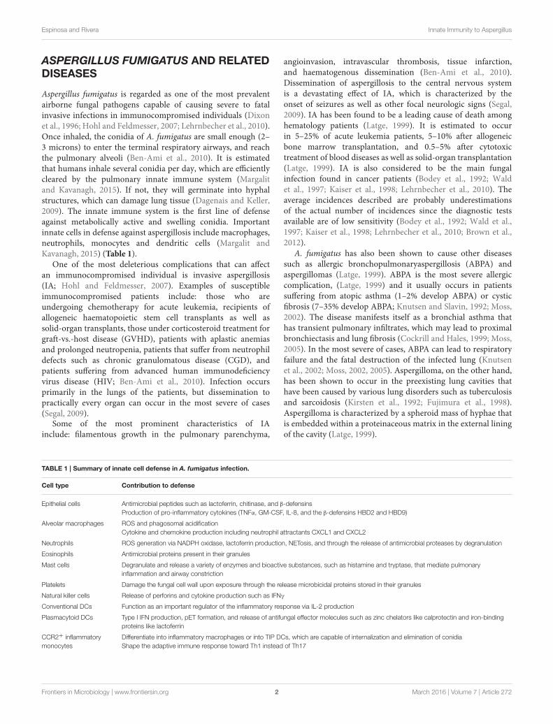
TABLE 1 | Summary of innate cell defense in A. fumigatus infection.
Cell type Contribution to defense
Epithelial cells Antimicrobial peptides such as lactoferrin, chitinase, and β-defensins
Production of pro-inflammatory cytokines (TNFα, GM-CSF, IL-8, and the β-defensins HBD2 and HBD9)
Alveolar macrophages ROS and phagosomal acidification
Cytokine and chemokine production including neutrophil attractants CXCL1 and CXCL2
Neutrophils ROS generation via NADPH oxidase, lactoferrin production, NETosis, and through the release of antimicrobial proteases by degranulation
Eosinophils Antimicrobial proteins present in their granules
Mast cells Degranulate and release a variety of enzymes and bioactive substances, such as histamine and tryptase, that mediate pulmonary
inflammation and airway constriction
Platelets Damage the fungal cell wall upon exposure through the release microbicidal proteins stored in their granules
Natural killer cells Release of perforins and cytokine production such as IFNγ
Conventional DCs Function as an important regulator of the inflammatory response via IL-2 production
Plasmacytoid DCs Type I IFN production, pET formation, and release of antifungal effector molecules such as zinc chelators like calprotectin and iron-binding
proteins like lactoferrin
CCR2+ inflammatory
monocytes
Differentiate into inflammatory macrophages or into TIP DCs, which are capable of internalization and elimination of conidia
Shape the adaptive immune response toward Th1 instead of Th17
angioinvasion, intravascular thrombosis, tissue infarction,and haematogenous dissemination (Ben-Ami et al., 2010).Dissemination of aspergillosis to the central nervous systemis a devastating effect of IA, which is characterized by theonset of seizures as well as other focal neurologic signs (Segal,2009). IA has been found to be a leading cause of death amonghematology patients (Latge, 1999). It is estimated to occurin 5–25% of acute leukemia patients, 5–10% after allogeneicbone marrow transplantation, and 0.5–5% after cytotoxictreatment of blood diseases as well as solid-organ transplantation(Latge, 1999). IA is also considered to be the main fungalinfection found in cancer patients (Bodey et al., 1992; Waldet al., 1997; Kaiser et al., 1998; Lehrnbecher et al., 2010). Theaverage incidences described are probably underestimationsof the actual number of incidences since the diagnostic testsavailable are of low sensitivity (Bodey et al., 1992; Wald et al.,1997; Kaiser et al., 1998; Lehrnbecher et al., 2010; Brown et al.,2012).
A. fumigatus has also been shown to cause other diseasessuch as allergic bronchopulmonaryaspergillosis (ABPA) andaspergillomas (Latge, 1999). ABPA is the most severe allergiccomplication, (Latge, 1999) and it usually occurs in patientssuffering from atopic asthma (1–2% develop ABPA) or cysticfibrosis (7–35% develop ABPA; Knutsen and Slavin, 1992; Moss,2002). The disease manifests itself as a bronchial asthma thathas transient pulmonary infiltrates, which may lead to proximalbronchiectasis and lung fibrosis (Cockrill and Hales, 1999; Moss,2005). In the most severe of cases, ABPA can lead to respiratoryfailure and the fatal destruction of the infected lung (Knutsenet al., 2002; Moss, 2002, 2005). Aspergilloma, on the other hand,has been shown to occur in the preexisting lung cavities thathave been caused by various lung disorders such as tuberculosisand sarcoidosis (Kirsten et al., 1992; Fujimura et al., 1998).Aspergilloma is characterized by a spheroid mass of hyphae thatis embedded within a proteinaceous matrix in the external liningof the cavity (Latge, 1999).
Frontiers in Microbiology | www.frontiersin.org 2 March 2016 | Volume 7 | Article 272
Espinosa and Rivera Innate Immunity to Aspergillus
RECOGNITION OF ASPERGILLUS
FUMIGATUS BY INNATE CELLRECEPTORS
C-Type Lectin Receptors (Dectin-1 andDectin-2)Upon inhalation, conidia mature and begin to swell, whichleads to the loss of their RodA hydrophobic layer exposingthe β-glucans in their cell wall (Aimanianda et al., 2009). β-glucans are recognized by the C-Lectin receptor, Dectin-1 (Hohlet al., 2005; Werner et al., 2009). Dectin-1 is expressed onmacrophages, neutrophils, and dendritic cells (Werner et al.,2009). In vitro, it has been shown that Dectin-1 dependentalveolar macrophage production of cytokine and chemokinesdoes not depend on the phagocytosis of the conidia, but on itsmorphology (Luther et al., 2007). Dectin-1 has been shown tobe activated only in the presence of swollen, but not restingconidia (Gersuk et al., 2006; Aimanianda et al., 2009). Dectin-1 signals through Syk kinase, leads to the activation of NFκB,and the production of tumor necrosis factor (TNFα), IL-10, IL-6,IL-1α, granulocyte macrophage colony stimulating factor (GM-CSF), macrophage inflammatory protein α and β (MIP-1α, andMIP-1β; Hohl et al., 2005; Steele et al., 2005; Faro-Trindadeet al., 2012). Dectin-1 has also been shown to have an importantrole in neutrophil recruitment (Werner et al., 2009). In Dectin-1 deficient mice, defects in neutrophil recruitment were due tounresponsive alveolar macrophages that were unable to producechemoattractants (Werner et al., 2009).
Dectin-2, in contrast, recognizes α-mannans, which are foundin the outer layer of the cell wall (Levitz, 2010; Sun et al., 2014).Dectin-2 has been shown to bemainly expressed onmacrophagesas well as dendritic cells (Sun et al., 2014). Detection of swollenconidia leads to the production of IL-1β, IL-10, IL-23p19, andTNFα via NFκB mediated by Syk (Sun et al., 2013, 2014).Blocking of Dectin-2 and Syk results in reduced conidial killingin macrophages differentiated from a human monocytic cell line(Sun et al., 2014).
Toll-Like Receptors (TLRs)Toll-like receptors (TLRs) are membrane receptors thathave a leucine-rich extracellular domain that recognizespathogen-associated molecular patterns (PAMPs) as well as anintracellular Toll/interleukin-1 receptor (TIR) domain neededfor downstream signaling (Kawai and Akira, 2007). OnceTLRs recognize the pathogen, the signaling cascade leads tothe activation of NFκB and other transcription factors, whichleads to cytokine and chemokine production (Kawai and Akira,2007).
TLRs have been found to play important roles in recognitionof A. fumigatus for host defense although there is conflictingdata, which could be attributed to differences in experimentaldesign (Steele et al., 2005). They are primarily expressed onthe cell surface of monocytes, macrophages, and dendritic cells(Takeda and Akira, 2005). TLR1−/− murine bone marrow-derived macrophages had reduced amounts of IL-6, TNFα,CXCL2, and IL-12p40 in response to A. fumigatus conidia
(Rubino et al., 2012). Cytokine production was diminishedin TLR2−/−, TLR4−/− and TLR6−/− macrophages, but notin TLR3−/− or wild-type macrophages. In terms of survival,TLR1−/−, TLR2−/−, TLR4−/−, and TLR6−/− have been shownto be non-essential, and do not make immunocompetent micemore susceptible to A. fumigatus infection (Dubourdeau et al.,2006; Rubino et al., 2012). In another study performed in vitro,TLR2−/− murine alveolar macrophages (AMs) infected withA. fumigatus were found to have an impaired inflammatoryresponse due to their deficiency in TNFα production (Steele et al.,2005). Because AMs were still able to produce some detectableTNFα, TLR2 was determined to be non-essential for TNFαproduction, but necessary for Dectin-1 mediated production ofTNFα (Steele et al., 2005). When AMs were treated with Dectin-1 blocking antibody, there was an observed 80% decrease incytokine production, which was consistent in both TLR2−/− andwild-type AMs (Steele et al., 2005). In addition, TLR2−/−TLR4-/-mice, were found to have deficiencies in neutrophil recruitmentcompared to the single knockouts indicating that both receptorsare required for an optimal immune response (Meier et al.,2003). TLR2 and TLR4 signaling requires the adaptor MyD88adapter-like (Mal)/TIRAP in the myeloid differentiation primaryresponse gene 88 (MyD88) dependent pathway (Horng et al.,2002). TLR4, on the other hand, can signal through anotheradaptor molecule, TIR-domain-containing adapter-inducinginterferon-β (TRIF)-related adapter molecule (TRAM/TICAM-2), in the MyD88 independent pathway (Yamamoto et al.,2003).
In contrast to previous studies (Meier et al., 2003; Bellocchioet al., 2004; Rubino et al., 2012), other work has recentlyimplicated a role for TLR3−/− in A. fumigatus infection.TLR3−/− mice were observed to have deficiencies in dendriticcell (DC) migration to the lymph node, which affected theirability to prime T cells (Carvalho et al., 2012). Consistent withthis finding, TLR3−/− mice lacked the ability to produce a CD8+
T cell response in response to A. fumigatus infection (Carvalhoet al., 2012). TLR3 as well as TLR4 can signal through theadaptor protein TRIF (Kawai and Akira, 2007), and TRIF−/−
mice displayed sustained inflammatory cell recruitment to thelungs in comparison to MyD88−/− and wild-type mice that werechemically immunocompromised to serve as models of IA (deLuca et al., 2010). In addition, TLR3-expressing lung epithelialcells were shown to activate indoleamine 2,3-dioxygenase, whichis an interferon (IFN)- inducible enzyme that degrades the aminoacid tryptophan and suppresses adaptive T cell immunity (deLuca et al., 2010).
TLR9 is expressed on a variety of cells such as macrophagesandmonocytes (Ramirez-Ortiz et al., 2008). During phagocytosisof A. fumigatus, TLR9 recognizes the exposed and unmethylatedCpG DNA (Ramirez-Ortiz et al., 2008). TLR9−/− neutropenicmice exhibited a decreased inflammatory response compared towild-type 2 days post infection, but was significantly increased4 days post infection indicating an immunoregulatory rolefor TLR9 in A. fumigatus infection (Ramaprakash et al.,2009). Dectin-1 expression was also found to be decreasedin TLR9−/− mice, which could explain why there is adelayed immune response since Dectin-1 is important for
Frontiers in Microbiology | www.frontiersin.org 3 March 2016 | Volume 7 | Article 272
Espinosa and Rivera Innate Immunity to Aspergillus
recognition of A. fumigatus swollen conidia (Ramaprakash et al.,2009).
Myeloid Differentiation Primary ResponseGene 88 (MyD88)MyD88, the universal adapter through which all TLRs exceptTLR3 signal, has been shown to play an important role early inthe inflammatory response against A. fumigatus (Ramaprakashet al., 2009). MyD88−/− mice were shown to have delayedfungal clearance for the first 2 days, but were comparableto wild-type mice at about 3 days (Bretz et al., 2008). Earlyon, MyD88−/− lungs appeared to have more necrotic tissue,and using a fluorescent A. fumigatus strain, a deficiency inmacrophage uptake was observed (Bretz et al., 2008). Also,there was decreased cytokine production of interleukin (IL)-1β,IL-6, keratinocyte-derived chemokine (KC/CXCL1), IFNγ, butincreased amounts of TNFα and MIP-1α in MyD88−/− mice(Bretz et al., 2008). The normalization observed at day threeindicates that there are alternative pathways involved in fungalclearance that are MyD88 independent (Margalit and Kavanagh,2015). Recent studies further suggest that MyD88 signaling indefense against IA is crucially active on lung epithelial cellsand is required for optimal production of neutrophil-recruitingchemokines (Jhingran et al., 2015).
INNATE CELL SUBSETS AND THEIRROLES IN DEFENSE AGAINSTASPERGILLOSIS
Epithelial CellsThe airway epithelium is the first point of contact for fungalspores upon inhalation, which leads to the initiation of the innateimmune response (Figure 1; Paris et al., 1997). The respiratoryepithelium consists of a variety of cell types such as mucous-secreting goblet cells, ciliated cells, and most importantly,respiratory epithelial cells (Paris et al., 1997). Respiratoryepithelial cells release a broad range of antimicrobial peptidessuch as lactoferrin, chitinase, and β-defensins (Alekseevaet al., 2009; Balloy and Chignard, 2009). The tracheobronchialepithelial cells, Type II alveolar epithelial cells, and endothelialcells have been shown to have the ability to internalize conidia,which are then trafficked to late endosomes for processing(Paris et al., 1997; Filler and Sheppard, 2006). In comparison toother phagocytosing cells such as macrophages and neutrophils,epithelial cells are less efficient in fungal elimination (Wasylnkaand Moore, 2003). Respiratory epithelial cells also expressrecognition receptors such as CLRs and TLRs (Sun et al., 2012).Upon challenge of the human bronchial epithelial cell line withswollen A. fumigatus conidia, Dectin-1 expression was induced
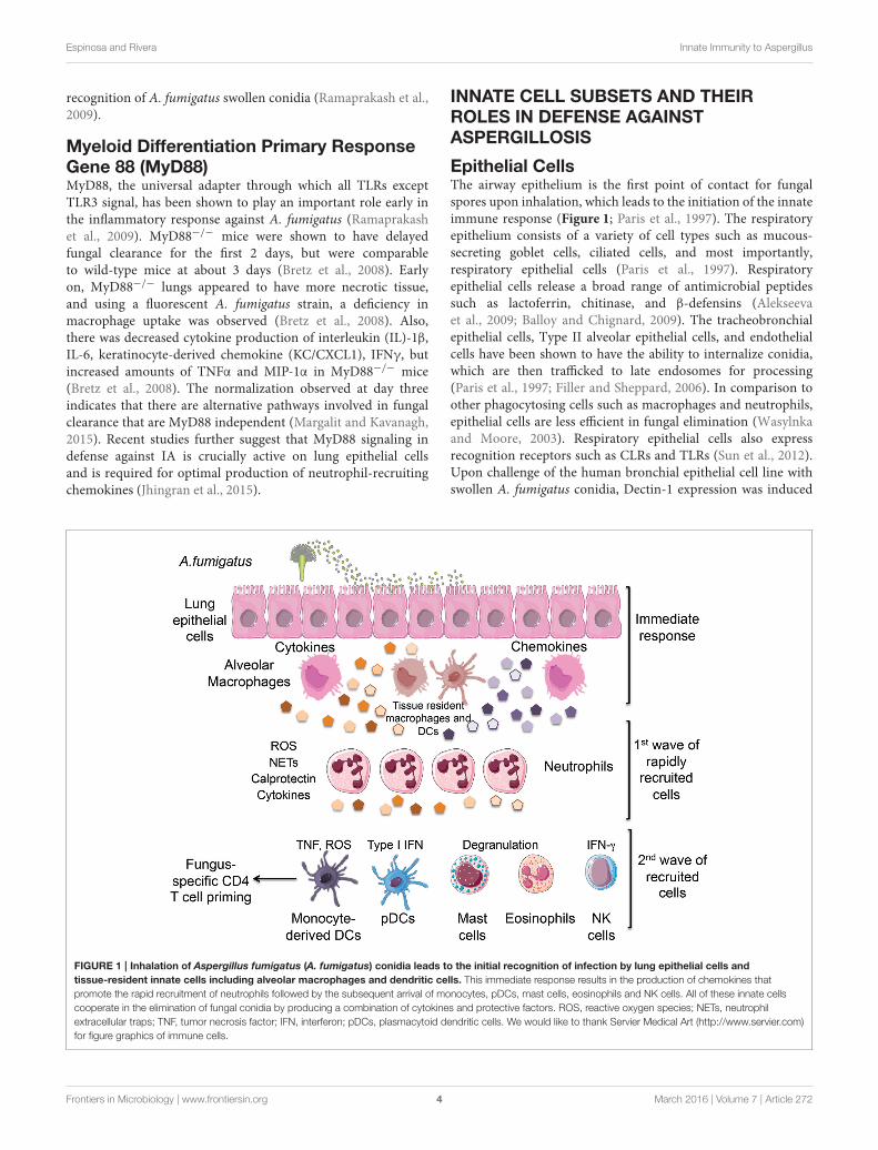
FIGURE 1 | Inhalation of Aspergillus fumigatus (A. fumigatus) conidia leads to the initial recognition of infection by lung epithelial cells and
tissue-resident innate cells including alveolar macrophages and dendritic cells. This immediate response results in the production of chemokines that
promote the rapid recruitment of neutrophils followed by the subsequent arrival of monocytes, pDCs, mast cells, eosinophils and NK cells. All of these innate cells
cooperate in the elimination of fungal conidia by producing a combination of cytokines and protective factors. ROS, reactive oxygen species; NETs, neutrophil
extracellular traps; TNF, tumor necrosis factor; IFN, interferon; pDCs, plasmacytoid dendritic cells. We would like to thank Servier Medical Art (http://www.servier.com)
for figure graphics of immune cells.
Frontiers in Microbiology | www.frontiersin.org 4 March 2016 | Volume 7 | Article 272
Espinosa and Rivera Innate Immunity to Aspergillus
in a TLR2-dependent manner, which induced the expressionof ROS as well as TNFα, GM-CSF, IL-8, and the β-defensinsHBD2 and HBD9 (Balloy et al., 2008; Sun et al., 2012). Dectin-1blockade inhibited the expression of these factors indicating thatairway epithelial cells require Dectin-1 for the upregulation ofpro-inflammatory cytokines as well as antimicrobial factors (Sunet al., 2012).
Alveolar Macrophages (AM)Alveolar macrophages have been shown to uptake as wellas kill conidia through two known mechanisms: Reactiveoxygen species (ROS) generation and phagosomal acidification(Ibrahim-Granet et al., 2003; Philippe et al., 2003). ROSgeneration occurs in response to swollen but not restingconidia, which leads to the recruitment of cytosolic proteins(p47phox, p67phox, p40phox, and Rac1/Rac2 GTPase) to theplasma membrane where they form a complex with membrane-bound flavocytochrome units, gp91phox and gp22phox, in orderto form an active nicotinamide adenine dinucleotide phosphate(NADPH) oxidase (Forman and Torres, 2002; Gersuk et al.,2006). In immunosuppressed mice through cyclophosphamidetreatment, mice were found to be less susceptible to IA thanp47phox deficient mice, which are defective in NADPH ROSgeneration and a model of CGD. In addition, mice transgenicfor p47phox under the control of the human CD68 that allowsfor targeted NADPH oxidase expression on macrophages andmonocytes had increased survival rates compared to the globalknockout illustrating the importance of oxidative mechanisms(Grimm et al., 2013). Specifically in AMs, p47phox deficient AMswere unable to control the growth of phagocytosed conidia incontrast to wild-type AMs (Grimm et al., 2013).
AMs play an important role in the inflammatory responsethrough the activation of PRRs and cytokine and chemokineproduction (Figure 1), which include neutrophil attractants suchas macrophage inflammatory protein-2 (MIP-2/CXCL2) andCXCL1(Bhatia et al., 2011). During phagosomal acidification, aphagosome containing conidia fuses with a lysosome in orderto form a phagolysosome, which leads to ATPase mediatedacidification and activation of enzymes such as chitinases thatleads to the degradation of the cell wall exposing ligandsfor pattern recognition receptors (PRR), TLRs, and Dectin-1(Ibrahim-Granet et al., 2003; Kasperkovitz et al., 2010; Faro-Trindade et al., 2012).
In vivo, clodronate treatment has been used as a method ofdepletion of AMs (Philippe et al., 2003; Bhatia et al., 2011).Clodronate treated mice were shown to have higher fungalburdens than wild-type mice even though there was an increasein neutrophil recruitment, which can indicate that there is someform of communication betweenAMs and neutrophils since bothseem to be needed in order to control the infection (Bhatia et al.,2011). The mechanism by which this occurs has yet to be fullyelucidated and warrants further study. It has been suggested thatAMs are able to eliminate low amounts of inocula, and thathigher amounts warrant neutrophil activation and recruitment(Philippe et al., 2003). These findings are controversial sincethere is also evidence that AMs are dispensable in A. fumigatusinfection, which could be attributed to their use of different
strains of mice as well as their use of diverse strains of A.fumigatus (Mircescu et al., 2009).
NeutrophilsA. fumigatus also produces immunosuppressive toxins suchas gliotoxin and fumagillin, which affects neutrophil functionby preventing the formation of a functional NADPH oxidase(Tsunawaki et al., 2004; Fallon et al., 2010). In a mutant strainof A. fumigatus in which the gliP gene is deleted, infectedimmunosuppressed mice through corticosteroid treatment hadan attenuated virulence compared to non-immunosuppressedmice (Sugui et al., 2007). GliP catalyzes the first biosynthetic stepin the synthesis of gliotoxin, and deletion prevents its synthesisas well as its effect on NADPH oxidase (Sugui et al., 2007).Neutrophils were found to be a primary target for gliotoxin sinceneutropenic mice did not differ in virulence when infected withthe mutant compared to wild-type A. fumigatus (Spikes et al.,2008).
During neutrophil degranulation, azurophil granules expelfungicidal hydrolytic enzymes into the phagocytic vacuole(Segal, 2005). There are two predominant types of granulespresent in neutrophils: azurophil (primary) granules andspecific (secondary) granules (Spitznagel, 1990; Segal, 2005). Asmentioned, azurophils contain hydrolytic enzymes for killingand digestion of pathogens whereas specific granules serve assources of replenishment for membrane components as well aslimiting free radical reactions (Segal, 2005). Azurophil granulescontain myeloperoxidase, cathepsin G, elastase, and proteinase3 (Segal, 2005). Specific granules contain lactoferrin (bindsand sequesters iron and copper), transcobalamin II, neutrophilgelatinase-associated lipocalin, and other membrane-associatedproteins including flavocytochrome b558 of the NADPH oxidase(Segal and Jones, 1979; Segal, 2005). NADPH oxidase derivedROS has been shown to promote degranulation and activation ofthese hydrolytic enzymes (Reeves et al., 2002; Feldmesser, 2006).Activation leads to an accumulation of ROS into the endocyticvacuole, which leads to the accumulation of potassium ions in thevacuole to compensate for the anionic charge from ROS (Reeveset al., 2002). The increase in ionic strength triggers the release ofthe granule proteins (Reeves et al., 2002). The importance of thesegranules is demonstrated in mice deficient in the serine proteasecathepsin G and/or neutrophil elastase that succumb earlier toStaphylococcal and Candida infections, but are competent in ROSproduction (Reeves et al., 2002).
Neutrophils have been shown to be essential innate effectors indefense against A. fumigatus (Mircescu et al., 2009; Margalit andKavanagh, 2015). Due to the high mortality rates of neutropenicmice and their inability to control fungal growth and hyphalformation, they are considered to be the most established modelof IA (Stephens-Romero et al., 2005; Mircescu et al., 2009).Neutrophils employ various mechanisms in the eliminationof A. fumigatus germinating spores such as: ROS generationvia NADPH oxidase, lactoferrin production, and through therelease of antimicrobial proteases by degranulation (Figure 1;Feldmesser, 2006; Sugui et al., 2007).
The importance of oxidative mediated conidiacidal activityby neutrophils is illustrated in patients suffering from CGD
Frontiers in Microbiology | www.frontiersin.org 5 March 2016 | Volume 7 | Article 272
Espinosa and Rivera Innate Immunity to Aspergillus
(Grimm et al., 2011). These patients possess mutations inp47phox, which leads to a defective ROS generation by NADPHoxidase (Grimm et al., 2011). In vivo, studies using NADPHoxidase deficient mice as a model for CGD demonstrated delayedrecruitment of neutrophils as well as their inability to containgerminating conidia (Bonnett et al., 2006). Histology from thelung tissue samples of the mice showed hyphal structures as wellas extensive damage to the lung tissue in contrast to their wild-type counterparts in both C57BL/6 and BALB/C backgrounds(Bonnett et al., 2006). In vitro, the addition of hydrogenperoxide and hypochlorous acid to the NADPH oxidase deficientcells prevented germination indicating the importance of ROS(Bonnett et al., 2006).
Neutrophil extracellular traps (NETs) are extracellularstructures that are made of chromatin with proteins fromneutrophilic granules attached including neutrophil elastase,myeloperoxidase, cathepsin G, lactoferrin, and gelatinase(Brinkmann et al., 2004). Chromatin is described as thebackbone of NETs due to the ability of DNases, but not proteasesto degrade it. NETs are formed in response to activation via IL-8,lipopolysaccharide (LPS), bacteria, fungi, or activated platelets(Brinkmann et al., 2004; Brinkmann and Zychlinsky, 2007).Once activated, a subset of neutrophils begin a “suicide” programthat leads to the their death and NETosis (Brinkmann andZychlinsky, 2007). NETs require a respiratory burst, which hasbeen experimentally supported by blocking ROS and preventingNET formation through the use of the oxidase inhibitordiphenylene iodonium (DPI; Brinkmann and Zychlinsky, 2007).
CGD patients, who are more susceptible to IA, haveneutrophils that are unable to form NETs when stimulatedwith bacteria or phorbol myristate acetate (PMA; Brinkmannand Zychlinsky, 2007; Fuchs et al., 2007). The importance ofNET formation is controversial, because there is also evidencethat neutrophil fungicidal activity is NET-independent (Margalitand Kavanagh, 2015). In vitro, human bronchoalveolar lavageneutrophils treated with micrococcal nuclease (MNase) in orderto degrade chromatin and prevent NET formation were stillcapable of fungicidal activity, which indicates that killing isNET-independent (Bianchi et al., 2009). This observation wasfurther supported by another group that treated neutrophilswith cytochalasin-D in order to block phagocytosis, and foundthat neutrophil killing was abrogated (Bruns et al., 2010). Thisindicates that phagocytosis and not NET formation is the primarykilling mechanism by neutrophils in response to fungi (Brunset al., 2010). The mechanisms as to whether a neutrophilundergoes NETosis upon contact or phagocytosis of fungalelements remain unclear, but in regards to killing, antimicrobialpeptides have been suggested to be one possible mechanism(Levitz et al., 1986; Bruns et al., 2010).
Human neutrophils have also recently been shown to produceNETs in response to A. fumigatus hyphal structures, but to alesser extent in response to resting and swollen conidia (Brunset al., 2010). An A. fumigatusmutant that lacks the hydrophobinRodA surface layer of swollen and resting conidia was shownto increase NET formation as compared to wild-type conidia(Bruns et al., 2010). This indicates that a NET inducing elementis exposed during hyphal formation when the RodA layer is
lost (McCormick et al., 2010). The RodA protein shields theconidia, and prevents the activation of an adaptive immuneresponse (Bruns et al., 2010; McCormick et al., 2010). AnotherNET associated protein is calprotectin, which has been shown tochelate zinc ions, and inhibit growth ofA. fumigatus (McCormicket al., 2010; Bianchi et al., 2011). Addition of zinc ions to culturemedium was able to rescue the growth inhibition. (McCormicket al., 2010) The effect seen was not A. fumigatus specific. Invitro, A. nidulans growth was inhibited by blocking calprotectinthrough S100A9 blocking antibodies or in the S100A9 deficientmouse strain (Bianchi et al., 2011).
Eosinophils and Mast CellsIn ABPA, there is enhanced eosinophil recruitment along withfungal enzymes that have been shown to contribute to epithelialdamage. In contrast, there is evidence that eosinophils possessfungicidal activity due to the antimicrobial proteins present intheir granules (Patterson and Strek, 2014). Eosinophils haverecently been shown to play a role in defense againstA. fumigatus(Lilly et al., 2014). Mice deficient in a high-affinity GATA-bindingsite in the GATA-1 promoter are depleted of eosinophils (notmast cells or platelets) have been shown to have deficiencies infungal clearance and increase in fungal burden in comparison totheir wild-type counterparts (Lilly et al., 2014). In addition, therewas impaired production of cytokines and chemokines such asIL-6, IL-17A, GM-CSF, IL-1β, and CXCL1 (Lilly et al., 2014).
Mast cells have been shown to be key mediators of thepathophysiology of asthma (Bradding et al., 2006). They havebeen shown to localize in the bronchial smooth muscle bundlesin patients with severe asthma such as ABPA patients, butnot in normal subjects or those with eosinophilic bronchitis(Bradding et al., 2006). Using the RBL-2H3 cell line andbone marrow-derived mast cell cultures to examine mast cellfunction, A. fumigatus hyphae was shown to adhere to mastcells, and induced their degranulation in an IgE- independentmanner unlike conidia and immature hyphae (Urb et al., 2009).Degranulation leads to the release of a variety of enzymaticproteins as well as bioactive substances such as histamineand tryptase, which are important in mediating pulmonaryinflammation and airway constriction (Bradding et al., 2006).Although exposure to A.fumigatus leads to degranulation of mastcells, mast cells cannot inhibit their growth or metabolic activity(Urb et al., 2009).
PlateletsSeveral studies have examined the role of platelets inA. fumigatusinfection since development of IA leads to hyphal invasion ofblood vessels, which can cause thrombosis as well as vascularinfarction (Lopes Bezerra and Filler, 2004). In vitro, it has beenshown that human platelets surround and adhere to hyphalstructures as well as conidia, but are unable to phagocytose fungalspores (Christin et al., 1998). In contrast, other studies haveindicated that it is A. fumigatus-derived serine proteases as wellas gliotoxin that lead to platelet activation, and is not contactdependent (Speth et al., 2013). Platelet activation is characterizedby the expression of CD62P and CD63 on the cell surface, whichare released by α-granules and δ- granules (Perkhofer et al.,
Frontiers in Microbiology | www.frontiersin.org 6 March 2016 | Volume 7 | Article 272
Espinosa and Rivera Innate Immunity to Aspergillus
2008; Rødland et al., 2010). Platelets incubated with fluoresceinisothiocyanate-labeled cell walls lead to the loss of hyphal surfaceproteins and hyphal cell wall integrity as indicated by the loss offluorescence via microscopy (Rødland et al., 2010). These resultsindicate a role for platelets in fungal containment by their abilityto damage the fungal cell wall upon exposure through the releaseplatelet microbicidal proteins stored in their granules.
Natural Killer (NK) CellsNatural Killer (NK) cells have been found to have fungicidalactivity, but the mechanisms in which this occurs, are poorlyunderstood (Bouzani et al., 2011; Schmidt et al., 2011, 2013). NKcells have been shown to be responsive to germinating but notresting conidia, and their release of perforins in vitro correlatedwith increased fungicidal activity (Schmidt et al., 2011). IFNγ
produced by NK cells could also contribute to fungal clearanceby preventing germinating conidia from growing into hyphalstructures, which suggests that in addition to functioning as animmunoregulatory molecule, IFNγ can function as an antifungaleffector against A. fumigatus directly (Park et al., 2009; Bouzaniet al., 2011). AMs incubated with NK cells in vitro have alsobeen shown to be more effective in killing the conidia thanwhen incubated alone or with IFNγ deficient NK cells. (Parket al., 2009) In neutropenic models that have been depletedof NK cells, the mortality rate is doubled in comparison toneutrophil depleted mice with wild-type NK cell function furthersuggesting a contribution of NK cells to antifungal defense in vivo(Morrison et al., 2003). Although NK cells have been found tohave antifungal activity, they do not seem to be essential. Inour previous work, mice that lack all lymphocytes includingNK cells, T cells, B cells, γδ T cells, iNKT cells, and innatelymphocytes (ILCs), RAG−/− γC−/−, do not develop IA uponinfection (Espinosa et al., 2014). These results indicate that theselymphocytes are not required for defense against A. fumigatusinfection.
Dendritic Cells (DCs)Dendritic Cells (DCs) have been shown to have multiple roles inresponse to A. fumigatus infection (Bhatia et al., 2011) (Table 1).DCs phagocytose conidia through PRRs including Dectin-1, Dendritic Cell-Specific Intercellular adhesion molecule-3-Grabbing Non-integrin (DC-SIGN), complement receptor 3(CR3), and FcγRIII (Bozza et al., 2002; Serrano-Gómez et al.,2005; Mezger et al., 2008). DCs also produce proinflammatorycytokines such as TNFα, IL-6, IL-12, IL-1α, and IL-1β in responseto A. fumigatus (Bozza et al., 2002; Mezger et al., 2008; Mortonet al., 2011). Differential cytokine production by DCs is observedwhen exposed to different forms of the fungus in vitro (Bozzaet al., 2002). When exposed to conidia and hyphae, TNFα isproduced. IL-12 is produced in response to conidia, and IL-4 andIL-10 in response to hyphae (Bozza et al., 2002). There are threemajor subtypes of DCs in the lung, conventional DCs (cDCs),plasmacytoid DCs (pDCs), and monocyte-derived DCs (moDCs;Margalit and Kavanagh, 2015).
DCs also produce a variety of chemokines that are neededfor the recruitment of a variety of innate and adaptive effectorcells to aid in fungal elimination (Scapini et al., 2000; Gafa et al.,
2007). DCs are recruited to sites of infection by the productionof MIP-1α and MIP-1β by neutrophils (Scapini et al., 2000). Invitro, infection of human dendritic cells by A. fumigatus conidiatriggers the secretion of chemokines for neutrophil and Th1lymphocyte recruitment (Gafa et al., 2007). DCs release increasedamounts of CXCL8, which results in neutrophil recruitment(Gafa et al., 2007). A. fumigatus infection resulted also inCCL3, CCL4, CXCL10, and CCL20 productions that induce themigration of effector memory Th1 cells (Gafa et al., 2007; Mortonet al., 2011). Together these results indicate a dual role for DCsin the innate as well as adaptive immune response against A.fumigatus (Margalit and Kavanagh, 2015).
Plasmacytoid Dendritic Cells (PDCs)pDCs are known as Type I IFN producing cells in responseto viral stimulation. They comprise about 0.2–0.8% of totalperipheral blood mononuclear cells (PBMCs) in humans, andexpress TLRs 7 and 9 (Colonna et al., 2004). pDCs link theinnate and adaptive immune systems by secreting IFNα andTNFα, and by differentiating into mature pDCs with upregulatedmajor histocompatilibility complex (MHC) and costimulatorymolecules capable of priming naive T cells (Colonna et al., 2004).
pDCs are a major source of type I IFN, but the role of pDCsas well as type I IFN has not been extensively examined inregards to fungal infections (Ramirez-Ortiz et al., 2011; Margalitand Kavanagh, 2015). Interferon αβ receptor deficient mice(IFNAR−/−) have been shown to have a higher susceptibilityto IA in comparison to wild-type mice, which is consistentwith the finding that antibody (120G8) mediated depletion ofpDCs make mice more vulnerable to A. fumigatus infection(Ramirez-Ortiz et al., 2011). When exposed to hyphae of A.fumigatus, human pDCs inhibited their growth, but did not killthe fungus (Ramirez-Ortiz et al., 2011). In addition, there arecontrasting studies as to whether pDCs are capable of engulfingA. fumigatus conidia, which could be attributed to differences inexperimental design for pDC isolation such as using CD303 asa marker for positive selection compared to CD123 (Ramirez-Ortiz et al., 2011; Lother et al., 2014). Apoptosis of pDCs isstimulated upon exposure to the release of cytotoxic moleculesby A. fumigatus such as gliotoxin, and their death results in therelease of antifungal effector molecules such as zinc chelators likecalprotectin and iron-binding proteins like lactoferrin (Ramirez-Ortiz et al., 2011). Similar to neutrophils, pDCs have alsorecently been shown to form Dectin-2 mediated extracellulartraps or pETs (plasmacytoid extracellular traps), which have beenobserved to form around A. fumigatus hyphae (Loures et al.,2015). Treatment with blocking antibodies against Dectin-2 ledto decreased association of pDCs with hyphae in contrast toDectin-1, which was similar to untreated pDCs exposed to A.fumigatus only (Loures et al., 2015). These results suggest thatpDCs can recognize A. fumigatus via Dectin-2, which results inantifungal activity through pET formation.
Conventional Dendritic Cells (cDCs)cDCs as well as moDCs are typically identified by their highexpression of the integrin CD11c and MHC class II. There aretwo types of cDCs: CD103+ cDCs and CD11b+ cDCs (Kopf
Frontiers in Microbiology | www.frontiersin.org 7 March 2016 | Volume 7 | Article 272
Espinosa and Rivera Innate Immunity to Aspergillus
et al., 2015). They can be distinguished from each other by themarkers CD207 (present on CD103+ cDCs) and MER proto-oncogene tyrosine kinase (MerTK; present on CD11b+ cDCs;Kopf et al., 2015). CD11b+ cDCs share common markers withmoDCs, but can be differentiated by using the marker for theFc receptor CD64 that is present on moDCs (Kopf et al., 2015).The development of cDCs as well as pDCs is dependent on theFMS-like tyrosine kinase 3 ligand (Flt3L), which is demonstratedby the lack of CD103+ DCs in Flt3L deficient mice (Ginhouxet al., 2009; Merad et al., 2013). Basic leucine zipper transcriptionfactor ATF-like 3 (BATF3) is also required for their steady-stategeneration similar to CD8α DCs (Edelson et al., 2010), but undercertain inflammatory conditions such as mycobacterial infection,other members of the BATF3 family have been shown to havecompensatory roles (Tussiwand et al., 2012). In an allergic asthmahouse dust mite model, it was demonstrated that moDCs as wellas CD11b+ cDCs are important for sensitization, but CD103+
DCs are dispensable (Plantinga et al., 2013). They were able todistinguish the contributions of the difference types of DCs byemploying the use of Flt3L deficient mice as well as Langerin-diphtheria toxin receptor (DTR) mice (Plantinga et al., 2013).In Langerin-DTR mice, lung CD103+ DCs and lymphoid tissueCD8α DCs are eliminated (Plantinga et al., 2013).
In a model of invasive aspergillosis, CD103+ DCs were shownto play an important role in shaping a Th17 response throughtheir IL-2 production via nuclear factor of activated T-cells(NFAT) signaling (Zelante et al., 2015). The absence of IL-2in pulmonary CD103+ DCs led to higher IL-17 productionin comparison to T cells cultured with IL-2 competent DCsin vitro in response to A. fumigatus germlings (Zelante et al.,2015). The impact of IL-2 production by DCs was also assessedusing mice lacking IL-2 in all tissues or lacking IL-2 in CD4+
T cells as well as mice lacking IL-2 specifically in the CD11c+
population (CD11ccreIL-2fl/fl; Zelante et al., 2015). Mice deficientin IL-2 in all tissues and in mice lacking IL-2 in CD4+ Tcells had significantly higher fungal burden, but had increasedsurvival in comparison to CD11ccre IL-2fl/fl. CD11ccre IL-2fl/fl
expressed higher levels of IL-17 and IL-23, which led to a fatalhyperinflammatory Th17 response (Zelante et al., 2015). Theseresults indicate that DCs can function as an important regulatorof the inflammatory response upon fungal infection.
CCR2+ Inflammatory Monocytes (CCR2+
Mo)Macrophage and dendritic cell precursors (MDP) in thebone marrow give rise to Ly6Chi monocytes or inflammatorymonocytes, which exit the bone marrow in a CC-chemokinereceptor 2 (CCR2)- dependent manner in response to infectionto the inflamed tissues (Serbina et al., 2008; Geissmann et al.,2010). They represent approximately 2–5% of circulating whiteblood cells in the bloodstream of a naïve mouse (Shi and Pamer,2011). The absence of CCR2 leads to deficiencies in monocyterecruitment to the site of infection (Serbina et al., 2008; Shiand Pamer, 2011). CCR2 is also expressed by other cells suchas hematopoietic stem cells (HSCs) as well as a subset of NKcells (Si et al., 2010). Once they reach infected tissues, CCR2+
Mo can differentiate into inflammatory macrophages or into
TNF and iNOS producing DCs (TIP DCs; Serbina et al., 2003;Auffray et al., 2009; Shi and Pamer, 2011). In the absence ofinflammation, CCR2+Mo can return to the bone marrow orto the spleen, which can function as a reservoir for circulatingmonocytes (Serbina et al., 2008; Geissmann et al., 2010; Shi andPamer, 2011). Humans also have a similar counterpart, which ischaracterized by CCR2+ CD14+ CD16− expression (Geissmannet al., 2003).
CCR2has twoprimary ligands,CCchemokine ligand2 (CCL2)and CCL7 that have been shown to be important for monocyterecruitment although the mechanism of action has yet to be fullyelucidated (Tsou et al., 2007; Shi and Pamer, 2011). In a theoreticalmodel, it was hypothesized that CC-chemokine ligands establishgradients, which guide monocytes to the site of infection bytheir association with glycosaminoglycans (Proudfoot et al., 2003;Allen et al., 2007). The finding that amino acid substitutionsin CCL2 affected monocyte recruitment supported this idea(Proudfoot et al., 2003). Other CC-chemokine ligands, CCL8and CCL12, have also been shown to bind to CCR2, but didnot have a significant role in monocyte trafficking (Tsou et al.,2007). Migration of monocytes is dependent on various integrinand adhesion molecules such as L-selectin (CD62L), P-selectinglycoprotein ligand 1 (PSGL1), lymphocyte function-associatedantigen 1 (LFA1; also known as αLβ2 integrin), macrophagereceptor 1 (MAC1; also known as integrin αMβ2), plateletendothelial cell adhesion molecule (PECAM1), and very lateantigen 4 (VLA4; also known as integrin α4β1; Ley et al., 2007).These molecules are necessary for proper rolling, adhesion, andmigrationofCCR2+Moto infected tissues by a variety of differentpathogens (Ley et al., 2007). CCR2+ Mo have been shown toplay important roles in bacterial, viral, protozoan, and fungalinfections (Shi and Pamer, 2011).
In the context of A. fumigatus infection our previous studiesfound an essential role for CCR2+ monocyte-derived dendriticcells in the activation of fungus-specific CD4+ T cells. Inthese studies, the role of CCR2 was investigated using adepleter mouse strain, in which the CCR2 promoter drives theexpression of the simian diphtheria toxin receptor (Hohl et al.,2009). Administration of diphtheria toxin (DT) leads to thetransient depletion of monocytes in the blood, bone marrow, andperipheral tissues (Hohl et al., 2009). Depletion of CCR2+ Mobefore infection leads to decreased transport of fungal spores tothe draining lymph nodes, which prevents A. fumigatus-specificCD4+ T cell priming (Rivera et al., 2011). Depletion of CCR2+
moDCs at later stages of infection, leads to a skewing from a Th1to a Th17 response in the lung, which indicates that recruitmentof monocytes has an influential role in shaping the adaptiveimmune response and are necessary to promote and sustainTh1 responses (Rivera et al., 2011). Studies with other fungalpathogens further support a conserved function for moDCs inshaping fungus-specific CD4+ T cell responses (Traynor et al.,2002; Szymczak and Deepe, 2009; Ersland et al., 2010; Szymczakand Deepe, 2010; Wüthrich et al., 2012b).
In addition to their importance in shaping fungus-specificCD4+ T cell responses, CCR2+ Mo and moDCs are importantdirect effectors necessary for prevention of IA (Espinosa et al.,2014). In recent studies, we found that sustained depletionof CCR2+ Mo results in the rapid development of IA. Our
Frontiers in Microbiology | www.frontiersin.org 8 March 2016 | Volume 7 | Article 272
Espinosa and Rivera Innate Immunity to Aspergillus
studies suggest that CCR2+ Mo and their derivative moDCsare important direct effectors of fungal spore killing. Moreover,they are necessary to sustain an inflammatory milieu in thelung and the antifungal activity of neutrophils (Espinosa et al.,2014; Caffrey et al., 2015). Altogether, these studies indicatethat CCR2+ Mo and their derivative moDCs are crucial, non-redundant cells in antifungal defense to A. fumigatus by actingboth as direct innate effectors and by shaping the response ofother innate and adaptive immune cells. The importance ofmonocytes in defense against IA is likely conserved in humans.Patients with monocytopenia are at increased risk for fungalinfections (Vinh et al., 2010; Hsu et al., 2011). Moreover,CD14+CD16− monocytes isolated from healthy allogeneichematopoietic stem cell transplantation donors were shown tophagocytose conidia, and inhibit conidial germination while theCD14+CD16+ subset was able to produce cytokines (Serbinaet al., 2009).
CONCLUDING REMARKS
The detrimental impact of fungal infections to diverse patientpopulations across the globe is likely to continue to rise. The
interactions of fungi with host innate cells crucially determinethe outcome of infection with multiple innate cells subsetscontributing essential protective functions. Future therapeuticinterventions aimed at boosting innate immunity are likelyto provide significant benefit and help improve the currentdetrimental outcomes associated with invasive aspergillosis andother invasive fungal infections.
AUTHOR CONTRIBUTIONS
The author confirms being the sole contributor of this work andapproved it for publication.
FUNDING
Research in the Rivera lab is supported by the NationalInstitutes of Health under awards granted by National Institute ofAllergy and Infectious Diseases R01AI114647-01A1 andNationalCancer Institute R21CA167238-01A1 to AR, and fellowship F31AI098408-01A1 to VE. The content of this article is solely theresponsibility of the authors and does not necessarily representthe official views of the NIH.
REFERENCES
Aimanianda, V., Bayry, J., Bozza, S., Kniemeyer, O., Perruccio, K., Elluru, S. R.,
et al. (2009). Surface hydrophobin prevents immune recognition of airborne
fungal spores. Nature 460, 1117–1121. doi: 10.1038/nature08264
Alekseeva, L., Huet, D., Femenia, F., Mouyna, I., Abdelouahab, M., Cagna, A., et al.
(2009). Inducible expression of beta defensins by human respiratory epithelial
cells exposed to Aspergillus fumigatus organisms. BMC Microbiol. 9:33. doi:
10.1186/1471-2180-9-33
Allen, S. J., Crown, S. E., and Handel, T. M. (2007). Chemokine: receptor
structure, interactions, and antagonism.Annu. Rev. Immunol. 25, 787–820. doi:
10.1146/annurev.immunol.24.021605.090529
Auffray, C., Sieweke, M. H., and Geissmann, F. (2009). Blood monocytes:
development, heterogeneity, and relationship with dendritic cells. Annu. Rev.
Immunol. 27, 669–692. doi: 10.1146/annurev.immunol.021908.132557
Balloy, V., and Chignard, M. (2009). The innate immune response to Aspergillus
fumigatus.Microbes Infect. 11, 919–927. doi: 10.1016/j.micinf.2009.07.002
Balloy, V., Sallenave, J. M., Wu, Y., Touqui, L., Latge, J. P., Si-Tahar, M., et al.
(2008). Aspergillus fumigatus-induced interleukin-8 synthesis by respiratory
epithelial cells is controlled by the phosphatidylinositol 3-kinase, p38 MAPK,
and ERK1/2 pathways and not by the toll-like receptor-MyD88 pathway. J. Biol.
Chem. 283, 30513–30521. doi: 10.1074/jbc.M803149200
Bellocchio, S., Moretti, S., Perruccio, K., Fallarino, F., Bozza, S., Montagnoli, C.,
et al. (2004). TLRs govern neutrophil activity in aspergillosis. J. Immunol. 173,
7406–7715. doi: 10.4049/jimmunol.173.12.7406
Ben-Ami, R., Lewis, R. E., and Kontoyiannis, D. P. (2010). Enemy of the
(immunosuppressed) state: an update on the pathogenesis of Aspergillus
fumigatus infection. Br. J. Haematol. 150, 406–417. doi: 10.1111/j.1365-
2141.2010.08283.x
Bhatia, S., Fei, M., Yarlagadda, M., Qi, Z., Akira, S., Saijo, S., et al. (2011). Rapid
host defense against Aspergillus fumigatus involves alveolar macrophages with
a predominance of alternatively activated phenotype. PLoS ONE 6:e15943. doi:
10.1371/journal.pone.0015943
Bianchi, M., Hakkim, A., Brinkmann, V., Siler, U., Seger, R. A., Zychlinsky, A.,
et al. (2009). Restoration of NET formation by gene therapy in CGD controls
aspergillosis. Blood 114, 2619–2622. doi: 10.1182/blood-2009-05-221606
Bianchi, M., Niemiec, M. J., Siler, U., Urban, C. F., and Reichenbach, J.
(2011). Restoration of anti-Aspergillus defense by neutrophil extracellular
traps in human chronic granulomatous disease after gene therapy is
calprotectin-dependent. J. Allergy Clin. Immunol. 127, 1243–52.e7. doi:
10.1016/j.jaci.2011.01.021
Bodey, G., Bueltmann, B., Duguid, W., Gibbs, D., Hanak, H., Hotchi, M.,
et al. (1992). Fungal infections in cancer patients: an international autopsy
survey. Eur. J. Clin. Microbiol. Infect. Dis. 11, 99–109. doi: 10.1007/BF01
967060
Bonnett, C. R., Cornish, E. J., Harmsen, A. G., and Burritt, J. B. (2006).
Early neutrophil recruitment and aggregation in the murine lung inhibit
germination of Aspergillus fumigatus Conidia. Infect. Immun. 74, 6528–6539.
doi: 10.1128/IAI.00909-06
Bouzani, M., Ok, M., McCormick, A., Ebel, F., Kurzai, O., Morton, C. O.,
et al. (2011). Human NK cells display important antifungal activity against
Aspergillus fumigatus, which is directly mediated by IFN-gamma release.
J. Immunol. 187, 1369–1376. doi: 10.4049/jimmunol.1003593
Bozza, S., Gaziano, R., Spreca, A., Bacci, A., Montagnoli, C., di Francesco, P.,
et al. (2002). Dendritic cells transport conidia and hyphae of Aspergillus
fumigatus from the airways to the draining lymph nodes and initiate
disparate Th responses to the fungus. J. Immunol. 168, 1362–1171. doi:
10.4049/jimmunol.168.3.1362
Bradding, P., Walls, A. F., and Holgate, S. T. (2006). The role of the mast cell in
the pathophysiology of asthma. J. Allergy Clin. Immunol. 117, 1277–1284. doi:
10.1016/j.jaci.2006.02.039
Bretz, C., Gersuk, G., Knoblaugh, S., Chaudhary, N., Randolph-Habecker,
J., Hackman, R. C., et al. (2008). MyD88 signaling contributes to early
pulmonary responses to Aspergillus fumigatus. Infect. Immun. 76, 952–958. doi:
10.1128/IAI.00927-07
Brinkmann, V., Reichard, U., Goosmann, C., Fauler, B., Uhlemann, Y.,Weiss, D. S.,
et al. (2004). Neutrophil extracellular traps kill bacteria. Science 303, 1532–1535.
doi: 10.1126/science.1092385
Brinkmann, V., and Zychlinsky, A. (2007). Beneficial suicide: why neutrophils die
to make NETs. Nat. Rev. Microbiol. 5, 577–582. doi: 10.1038/nrmicro1710
Brown, G. D., Denning, D. W., Gow, N. A., Levitz, S. M., Netea, M. G., and
White, T. C. (2012). Hidden killers: human fungal infections. Sci. Transl. Med.
4, 165rv13. doi: 10.1126/scitranslmed.3004404
Bruns, S., Kniemeyer, O., Hasenberg, M., Aimanianda, V., Nietzsche, S.,
Thywissen, A., et al. (2010). Production of extracellular traps againstAspergillus
fumigatus in vitro and in infected lung tissue is dependent on invading
Frontiers in Microbiology | www.frontiersin.org 9 March 2016 | Volume 7 | Article 272
Espinosa and Rivera Innate Immunity to Aspergillus
neutrophils and influenced by hydrophobin RodA. PLoS Pathog. 6:e1000873.
doi: 10.1371/journal.ppat.1000873
Caffrey, A. K., Lehmann, M. M., Zickovich, J. M., Espinosa, V., Shepardson,
K. M., Watschke, C. P., et al. (2015). IL-1alpha signaling is critical
for leukocyte recruitment after pulmonary Aspergillus fumigatus
challenge. PLoS Pathog. 11:e1004625. doi: 10.1371/journal.ppat.10
04625
Carvalho, A., De Luca, A., Bozza, S., Cunha, C., D’Angelo, C., Moretti,
S., et al. (2012). TLR3 essentially promotes protective class I-restricted
memory CD8(+) T-cell responses to Aspergillus fumigatus in hematopoietic
transplanted patients. Blood 119, 967–977. doi: 10.1182/blood-2011-06-
362582
Christin, L.,Wysong, D. R., Meshulam, T., Hastey, R., Simons, E. R., and Diamond,
R. D. (1998). Human platelets damage Aspergillus fumigatus hyphae and may
supplement killing by neutrophils. Infect. Immun. 66, 1181–1189.
Cockrill, B. A., and Hales, C. A. (1999). Allergic bronchopulmonary aspergillosis.
Annu. Rev. Med. 50, 303–316. doi: 10.1146/annurev.med.50.1.303
Colonna, M., Trinchieri, G., and Liu, Y. J. (2004). Plasmacytoid dendritic cells in
immunity. Nat. Immunol. 5, 1219–1226. doi: 10.1038/ni1141
Dagenais, T. R., and Keller, N. P. (2009). Pathogenesis of Aspergillus
fumigatus in invasive Aspergillosis. Clin. Microbiol. Rev. 22, 447–465. doi:
10.1128/CMR.00055-08
de Luca, A., Bozza, S., Zelante, T., Zagarella, S., D’Angelo, C., Perruccio, K.,
et al. (2010). Non-hematopoietic cells contribute to protective tolerance to
Aspergillus fumigatus via a TRIF pathway converging on IDO. Cell. Mol.
Immunol. 7, 459–470. doi: 10.1038/cmi.2010.43
Dixon, D. M., McNeil, M. M., Cohen, M. L., Gellin, B. G., and La Montagne,
J. R. (1996). Fungal infections: a growing threat. Public Health Rep. 111,
226–235.
Dubourdeau, M., Athman, R., Balloy, V., Huerre, M., Chignard, M., Philpott, D. J.,
et al. (2006).Aspergillus fumigatus induces innate immune responses in alveolar
macrophages through the MAPK pathway independently of TLR2 and TLR4.
J. Immunol. 177, 3994–4001. doi: 10.4049/jimmunol.177.6.3994
Edelson, B. T., Kc, W., Juang, R., Kohyama, M., Benoit, L. A., Klekotka, P.
A., et al. (2010). Peripheral CD103+ dendritic cells form a unified subset
developmentally related to CD8alpha+ conventional dendritic cells. J. Exp.
Med. 207, 823–836. doi: 10.1084/jem.20091627
Ersland, K., Wüthrich, M., and Klein, B. S. (2010). Dynamic interplay among
monocyte-derived, dermal, and resident lymph node dendritic cells during the
generation of vaccine immunity to fungi. Cell Host Microbe 7, 474–487. doi:
10.1016/j.chom.2010.05.010
Espinosa, V., Jhingran, A., Dutta, O., Kasahara, S., Donnelly, R., Du, P., et al.
(2014). Inflammatory monocytes orchestrate innate antifungal immunity in the
lung. PLoS Pathog. 10:e1003940. doi: 10.1371/journal.ppat.1003940
Fallon, J. P., Reeves, E. P., and Kavanagh, K. (2010). Inhibition of neutrophil
function following exposure to the Aspergillus fumigatus toxin fumagillin.
J. Med. Microbiol. 59(Pt 6), 625–633. doi: 10.1099/jmm.0.018192-0
Faro-Trindade, I., Willment, J. A., Kerrigan, A. M., Redelinghuys, P., Hadebe, S.,
Reid, D. M., et al. (2012). Characterisation of innate fungal recognition in the
lung. PLoS ONE 7:e35675. doi: 10.1371/journal.pone.0035675
Feldmesser, M. (2006). Role of neutrophils in invasive aspergillosis. Infect. Immun.
74, 6514–6516. doi: 10.1128/IAI.01551-06
Filler, S. G., and Sheppard, D. C. (2006). Fungal invasion of normally non-
phagocytic host cells. PLoS Pathog. 2:e129. doi: 10.1371/journal.ppat.0020129
Forman, H. J., and Torres, M. (2002). Reactive oxygen species and cell signaling:
respiratory burst inmacrophage signaling.Am. J. Respir. Crit. CareMed. 166(12
Pt 2), S4–S8. doi: 10.1164/rccm.2206007
Fuchs, T. A., Abed, U., Goosmann, C., Hurwitz, R., Schulze, I., Wahn, V., et al.
(2007). Novel cell death program leads to neutrophil extracellular traps. J. Cell
Biol. 176, 231–241. doi: 10.1083/jcb.200606027
Fujimura, M., Ishiura, Y., Kasahara, K., Amemiya, T., Myou, S., Hayashi, Y.,
et al. (1998). Necrotizing bronchial aspergillosis as a cause of hemoptysis in
sarcoidosis. Am. J. Med. Sci. 315, 56–58.
Gafa, V., Remoli, M. E., Giacomini, E., Gagliardi, M. C., Lande, R.,
Severa, M., et al. (2007). In vitro infection of human dendritic cells
by Aspergillus fumigatus conidia triggers the secretion of chemokines for
neutrophil and Th1 lymphocyte recruitment. Microb. Infect. 9, 971–980. doi:
10.1016/j.micinf.2007.03.015
Geissmann, F., Jung, S., and Littman, D. R. (2003). Blood monocytes consist of two
principal subsets with distinct migratory properties. Immunity 19, 71–82. doi:
10.1016/S1074-7613(03)00174-2
Geissmann, F., Manz, M. G., Jung, S., Sieweke, M. H., Merad, M., and Ley, K.
(2010). Development of monocytes, macrophages, and dendritic cells. Science
327, 656–661. doi: 10.1126/science.1178331
Gersuk, G. M., Underhill, D. M., Zhu, L., and Marr, K. A. (2006).
Dectin-1 and TLRs permit macrophages to distinguish between different
Aspergillus fumigatus cellular states. J. Immunol. 176, 3717–3724. doi:
10.4049/jimmunol.176.6.3717
Ginhoux, F., Liu, K., Helft, J., Bogunovic, M., Greter, M., Hashimoto, D., et al.
(2009). The origin and development of nonlymphoid tissue CD103+ DCs.
J. Exp. Med. 206, 3115–3130. doi: 10.1084/jem.20091756
Grimm, M. J., Vethanayagam, R. R., Almyroudis, N. G., Dennis, C. G., Khan, A.
N., D’Auria, A. C., et al. (2013). Monocyte- and macrophage-targeted NADPH
oxidase mediates antifungal host defense and regulation of acute inflammation
in mice. J. Immunol. 190, 4175–4184. doi: 10.4049/jimmunol.1202800
Grimm, M. J., Vethanayagam, R. R., Almyroudis, N. G., Lewandowski, D.,
Rall, N., Blackwell, T. S., et al. (2011). Role of NADPH oxidase in host
defense against aspergillosis. Med. Mycol. 49(Suppl. 1), S144–S149. doi:
10.3109/13693786.2010.487077
Hohl, T. M., and Feldmesser, M. (2007). Aspergillus fumigatus: principles
of pathogenesis and host defense. Eukaryotic Cell 6, 1953–1963. doi:
10.1128/EC.00274-07
Hohl, T. M., Rivera, A., Lipuma, L., Gallegos, A., Shi, C., Mack, M., et al.
(2009). Inflammatory monocytes facilitate adaptive CD4 T cell responses
during respiratory fungal infection. Cell Host Microbe 6, 470–481. doi:
10.1016/j.chom.2009.10.007
Hohl, T. M., Van Epps, H. L., Rivera, A., Morgan, L. A., Chen, P. L.,
Feldmesser, M., et al. (2005). Aspergillus fumigatus triggers inflammatory
responses by stage-specific beta-glucan display. PLoS Pathog. 1:e30. doi:
10.1371/journal.ppat.0010030
Horng, T., Barton, G. M., Flavell, R. A., and Medzhitov, R. (2002). The adaptor
molecule TIRAP provides signalling specificity for Toll-like receptors. Nature
420, 329–333. doi: 10.1038/nature01180
Hsu, A. P., Sampaio, E. P., Khan, J., Calvo, K. R., Lemieux, J. E., Patel, S. Y., et al.
(2011). Mutations in GATA2 are associated with the autosomal dominant and
sporadic monocytopenia and mycobacterial infection (MonoMAC) syndrome.
Blood 118, 2653–2655. doi: 10.1182/blood-2011-05-356352
Ibrahim-Granet, O., Philippe, B., Boleti, H., Boisvieux-Ulrich, E., Grenet, D.,
Stern, M., et al. (2003). Phagocytosis and intracellular fate of Aspergillus
fumigatus conidia in alveolar macrophages. Infect. Immun. 71, 891–903. doi:
10.1128/IAI.71.2.891-903.2003
Jhingran, A., Kasahara, S., Shepardson, K. M., Junecko, B. A., Heung, L.
J., Kumasaka, D. K., et al. (2015). Compartment-specific and sequential
role of MyD88 and CARD9 in chemokine induction and innate defense
during respiratory fungal infection. PLoS Pathog. 11:e1004589. doi:
10.1371/journal.ppat.1004589
Kaiser, L., Huguenin, T., Lew, P. D., Chapuis, B., and Pittet, D. (1998). Invasive
aspergillosis. Clinical features of 35 proven cases at a single institution.
Medicine (Baltimore). 77, 188–194. doi: 10.1097/00005792-199805000-
00004
Kasperkovitz, P. V., Cardenas, M. L., and Vyas, J. M. (2010). TLR9 is actively
recruited to Aspergillus fumigatus phagosomes and requires the N-terminal
proteolytic cleavage domain for proper intracellular trafficking. J. Immunol.
185, 7614–7622. doi: 10.4049/jimmunol.1002760
Kawai, T., and Akira, S. (2007). TLR signaling. Semin. Immunol. 19, 24–32. doi:
10.1016/j.smim.2006.12.004
Kirsten, D., Rieger, U., Amthor, M., and Magnussen, H. (1992). [Invasive
aspergillosis in cavitary lung sarcoidosis]. Pneumologie 46, 239–242.
Knutsen, A. P., Bellone, C., and Kauffman, H. (2002). Immunopathogenesis of
allergic bronchopulmonary aspergillosis in cystic fibrosis. J. Cyst. Fibros. 1,
76–89. doi: 10.1016/S1569-1993(02)00033-4
Knutsen, A., and Slavin, R. G. (1992). Allergic bronchopulmonary mycosis
complicating cystic fibrosis. Semin. Respir. Infect. 7, 179–192.
Kopf, M., Schneider, C., and Nobs, S. P. (2015). The development and function of
lung-resident macrophages and dendritic cells. Nat. Immunol. 16, 36–44. doi:
10.1038/ni.3052
Frontiers in Microbiology | www.frontiersin.org 10 March 2016 | Volume 7 | Article 272
Espinosa and Rivera Innate Immunity to Aspergillus
Latge, J. P. (1999). Aspergillus fumigatus and aspergillosis. Clin. Microbiol. Rev. 12,
310–350.
Lehrnbecher, T., Frank, C., Engels, K., Kriener, S., Groll, A. H., and Schwabe, D.
(2010). Trends in the postmortem epidemiology of invasive fungal infections at
a university hospital. J. Infect. 61, 259–265. doi: 10.1016/j.jinf.2010.06.018
Levitz, S. M. (2010). Innate recognition of fungal cell walls. PLoS Pathog.
6:e1000758. doi: 10.1371/journal.ppat.1000758
Levitz, S. M., Selsted, M. E., Ganz, T., Lehrer, R. I., and Diamond, R. D. (1986). In
vitro killing of spores and hyphae of Aspergillus fumigatus and Rhizopus oryzae
by rabbit neutrophil cationic peptides and bronchoalveolar macrophages.
J. Infect. Dis. 154, 483–489. doi: 10.1093/infdis/154.3.483
Ley, K., Laudanna, C., Cybulsky, M. I., and Nourshargh, S. (2007). Getting to
the site of inflammation: the leukocyte adhesion cascade updated. Nat. Rev.
Immunol. 7, 678–689. doi: 10.1038/nri2156
Lilly, L. M., Scopel, M., Nelson, M. P., Burg, A. R., Dunaway, C. W., and Steele,
C. (2014). Eosinophil deficiency compromises lung defense against Aspergillus
fumigatus. Infect. Immun. 82, 1315–1325. doi: 10.1128/IAI.01172-13
Lopes Bezerra, L. M., and Filler, S. G. (2004). Interactions of Aspergillus fumigatus
with endothelial cells: internalization, injury, and stimulation of tissue factor
activity. Blood 103, 2143–2149. doi: 10.1182/blood-2003-06-2186
Lother, J., Breitschopf, T., Krappmann, S., Morton, C. O., Bouzani, M., Kurzai, O.,
et al. (2014). Human dendritic cell subsets display distinct interactions with the
pathogenic mouldAspergillus fumigatus. Int. J. Med. Microbiol. 304, 1160–1168.
doi: 10.1016/j.ijmm.2014.08.009
Loures, F. V., Röhm, M., Lee, C. K., Santos, E., Wang, J. P., Specht, C. A., et al.
(2015). Recognition of Aspergillus fumigatus hyphae by human plasmacytoid
dendritic cells is mediated by dectin-2 and results in formation of extracellular
traps. PLoS Pathog. 11:e1004643. doi: 10.1371/journal.ppat.1004643
Luther, K., Torosantucci, A., Brakhage, A. A., Heesemann, J., and Ebel, F. (2007).
Phagocytosis of Aspergillus fumigatus conidia by murine macrophages involves
recognition by the dectin-1 beta-glucan receptor and Toll-like receptor 2. Cell.
Microbiol. 9, 368–381. doi: 10.1111/j.1462-5822.2006.00796.x
Margalit, A., and Kavanagh, K. (2015). The innate immune response to Aspergillus
fumigatus at the alveolar surface. FEMS Microbiol. Rev. 39, 670–687. doi:
10.1093/femsre/fuv018
McCormick, A., Heesemann, L., Wagener, J., Marcos, V., Hartl, D., Loeffler,
J., et al. (2010). NETs formed by human neutrophils inhibit growth of the
pathogenic mold Aspergillus fumigatus. Microbes Infect. 12, 928–936. doi:
10.1016/j.micinf.2010.06.009
Meier, A., Kirschning, C. J., Nikolaus, T., Wagner, H., Heesemann, J., and Ebel,
F. (2003). Toll-like receptor (TLR) 2 and TLR4 are essential for Aspergillus-
induced activation of murine macrophages. Cell. Microbiol. 5, 561–570. doi:
10.1046/j.1462-5822.2003.00301.x
Merad, M., Sathe, P., Helft, J., Miller, J., and Mortha, A. (2013). The dendritic
cell lineage: ontogeny and function of dendritic cells and their subsets in the
steady state and the inflamed setting. Annu. Rev. Immunol. 31, 563–604. doi:
10.1146/annurev-immunol-020711-074950
Mezger, M., Kneitz, S., Wozniok, I., Kurzai, O., Einsele, H., and Loeffler, J. (2008).
Proinflammatory response of immature human dendritic cells is mediated by
dectin-1 after exposure to Aspergillus fumigatus germ tubes. J. Infect. Dis. 197,
924–931. doi: 10.1086/528694
Mircescu, M. M., Lipuma, L., van Rooijen, N., Pamer, E. G., and Hohl, T. M.
(2009). Essential role for neutrophils but not alveolar macrophages at early time
points followingAspergillus fumigatus infection. J. Infect. Dis. 200, 647–656. doi:
10.1086/600380
Morrison, B. E., Park, S. J., Mooney, J. M., and Mehrad, B. (2003). Chemokine-
mediated recruitment of NK cells is a critical host defense mechanism
in invasive aspergillosis. J. Clin. Invest. 112, 1862–1870. doi: 10.1172/JCI
18125
Morton, C. O., Varga, J. J., Hornbach, A., Mezger, M., Sennefelder, H., Kneitz,
S., et al. (2011). The temporal dynamics of differential gene expression in
Aspergillus fumigatus interacting with human immature dendritic cells in vitro.
PLoS ONE 6:e16016. doi: 10.1371/journal.pone.0016016
Moss, R. B. (2002). Allergic bronchopulmonary aspergillosis. Clin. Rev. Allergy
Immunol. 23, 87–104. doi: 10.1385/CRIAI:23:1:087
Moss, R. B. (2005). Pathophysiology and immunology of allergic
bronchopulmonary aspergillosis. Med. Mycol. 43(Suppl. 1), S203–S206.
doi: 10.1080/13693780500052255
Paris, S., Boisvieux-Ulrich, E., Crestani, B., Houcine, O., Taramelli, D., Lombardi,
L., et al. (1997). Internalization of Aspergillus fumigatus conidia by epithelial
and endothelial cells. Infect. Immun. 65, 1510–1514.
Park, S. J., Hughes, M. A., Burdick, M., Strieter, R. M., and Mehrad, B.
(2009). Early NK cell-derived IFN-{gamma} is essential to host defense
in neutropenic invasive aspergillosis. J. Immunol. 182, 4306–4312. doi:
10.4049/jimmunol.0803462
Patterson, K. C., and Strek, M. E. (2014). Diagnosis and treatment of pulmonary
aspergillosis syndromes. Chest 146, 1358–1368. doi: 10.1378/chest.14-0917
Perkhofer, S., Kehrel, B. E., Dierich, M. P., Donnelly, J. P., Nussbaumer, W.,
Hofmann, J., et al. (2008). Human platelets attenuate Aspergillus species
via granule-dependent mechanisms. J. Infect. Dis. 198, 1243–1246. doi:
10.1086/591458
Philippe, B., Ibrahim-Granet, O., Prévost, M. C., Gougerot-Pocidalo, M. A.,
Sanchez Perez, M. A., Van der Meeren, A., et al. (2003). Killing of
Aspergillus fumigatus by alveolar macrophages is mediated by reactive oxidant
intermediates. Infect. Immun. 71, 3034–3042. doi: 10.1128/IAI.71.6.3034-
3042.2003
Plantinga, M., Guilliams, M., Vanheerswynghels, M., Deswarte, K., Branco-
Madeira, F., Toussaint, W., et al. (2013). Conventional and monocyte-
derived CD11b(+) dendritic cells initiate and maintain T helper 2 cell-
mediated immunity to house dust mite allergen. Immunity 38, 322–335. doi:
10.1016/j.immuni.2012.10.016
Proudfoot, A. E., Handel, T. M., Johnson, Z., Lau, E. K., LiWang, P., Clark-Lewis,
I., et al. (2003). Glycosaminoglycan binding and oligomerization are essential
for the in vivo activity of certain chemokines. Proc. Natl. Acad. Sci. U.S.A. 100,
1885–1890. doi: 10.1073/pnas.0334864100
Ramaprakash, H., Ito, T., Standiford, T. J., Kunkel, S. L., and Hogaboam, C.
M. (2009). Toll-like receptor 9 modulates immune responses to Aspergillus
fumigatus conidia in immunodeficient and allergic mice. Infect. Immun. 77,
108–119. doi: 10.1128/IAI.00998-08
Ramirez-Ortiz, Z. G., Lee, C. K., Wang, J. P., Boon, L., Specht, C. A., and Levitz, S.
M. (2011). A nonredundant role for plasmacytoid dendritic cells in host defense
against the human fungal pathogen Aspergillus fumigatus. Cell Host Microbe 9,
415–424. doi: 10.1016/j.chom.2011.04.007
Ramirez-Ortiz, Z. G., Specht, C. A., Wang, J. P., Lee, C. K., Bartholomeu, D.
C., Gazzinelli, R. T., et al. (2008). Toll-like receptor 9-dependent immune
activation by unmethylated CpG motifs in Aspergillus fumigatus DN. Infect.
Immun. A 76, 2123–2129. doi: 10.1128/IAI.00047-08
Reeves, E. P., Lu, H., Jacobs, H. L., Messina, C. G., Bolsover, S., Gabella, G.,
et al. (2002). Killing activity of neutrophils is mediated through activation of
proteases by K+ flux. Nature 416, 291–297. doi: 10.1038/416291a
Rivera, A. (2014). Protective immune responses to fungal infections. Parasite
Immunol. 36, 453–462. doi: 10.1111/pim.12098
Rivera, A., Hohl, T. M., Collins, N., Leiner, I., Gallegos, A., Saijo, S., et al. (2011).
Dectin-1 diversifiesAspergillus fumigatus-specific T cell responses by inhibiting
T helper type 1 CD4 T cell differentiation. J. Exp. Med. 208, 369–381. doi:
10.1084/jem.20100906
Rødland, E. K., Ueland, T., Pedersen, T. M., Halvorsen, B., Muller, F., Aukrust,
P., et al. (2010). Activation of platelets by Aspergillus fumigatus and potential
role of platelets in the immunopathogenesis of Aspergillosis. Infect. Immun. 78,
1269–1275. doi: 10.1128/IAI.01091-09
Rubino, I., Coste, A., Le Roy, D., Roger, T., Jaton, K., Boeckh, M., et al. (2012).
Species-specific recognition of Aspergillus fumigatus by Toll-like receptor 1
and Toll-like receptor 6. J. Infect. Dis. 205, 944–954. doi: 10.1093/infdis/
jir882
Scapini, P., Lapinet-Vera, J. A., Gasperini, S., Calzetti, F., Bazzoni, F., and
Cassatella, M. A. (2000). The neutrophil as a cellular source of chemokines.
Immunol. Rev. 177, 195–203. doi: 10.1034/j.1600-065X.2000.17706.x
Schmidt, S., Tramsen, L., Hanisch, M., Latge, J. P., Huenecke, S., Koehl, U., et al.
(2011). Human natural killer cells exhibit direct activity against Aspergillus
fumigatus hyphae, but not against resting conidia. J. Infect. Dis. 203, 430–435.
doi: 10.1093/infdis/jiq062
Schmidt, S., Zimmermann, S. Y., Tramsen, L., Koehl, U., and Lehrnbecher, T.
(2013). Natural killer cells and antifungal host response.Clin. Vaccine Immunol.
20, 452–458. doi: 10.1128/CVI.00606-12
Segal, A. W. (2005). How neutrophils kill microbes. Annu. Rev. Immunol. 23,
197–223. doi: 10.1146/annurev.immunol.23.021704.115653
Frontiers in Microbiology | www.frontiersin.org 11 March 2016 | Volume 7 | Article 272
Espinosa and Rivera Innate Immunity to Aspergillus
Segal, A. W., and Jones, O. T. (1979). The subcellular distribution and some
properties of the cytochrome b component of the microbicidal oxidase system
of human neutrophils. Biochem. J. 182, 181–188. doi: 10.1042/bj1820181
Segal, B. H. (2009). Aspergillosis. N. Engl. J. Med. 360, 1870–1884. doi:
10.1056/NEJMra0808853
Serbina, N. V., Cherny, M., Shi, C., Bleau, S. A., Collins, N. H., Young, J. W., et al.
(2009). Distinct responses of human monocyte subsets to Aspergillus fumigatus
conidia. J. Immunol. 183, 2678–2687. doi: 10.4049/jimmunol.0803398
Serbina, N. V., Jia, T., Hohl, T. M., and Pamer, E. G. (2008). Monocyte-mediated
defense against microbial pathogens. Annu. Rev. Immunol. 26, 421–452. doi:
10.1146/annurev.immunol.26.021607.090326
Serbina, N. V., Salazar-Mather, T. P., Biron, C. A., Kuziel, W. A., and Pamer,
E. G. (2003). TNF/iNOS-producing dendritic cells mediate innate immune
defense against bacterial infection. Immunity 19, 59–70. doi: 10.1016/S1074-
7613(03)00171-7
Serrano-Gómez, D., Leal, J. A., and Corbí, A. L. (2005). DC-SIGN mediates the
binding of Aspergillus fumigatus and keratinophylic fungi by human dendritic
cells. Immunobiology 210, 175–183. doi: 10.1016/j.imbio.2005.05.011
Shi, C., and Pamer, E. G. (2011). Monocyte recruitment during infection and
inflammation. Nat. Rev. Immunol. 11, 762–774. doi: 10.1038/nri3070
Si, Y., Tsou, C. L., Croft, K., and Charo, I. F. (2010). CCR2 mediates hematopoietic
stem and progenitor cell trafficking to sites of inflammation in mice. J. Clin.
Invest. 120, 1192–1203. doi: 10.1172/JCI40310
Speth, C., Hagleitner, M., Ott, H. W., Würzner, R., Lass-Flörl, C., and Rambach,
G. (2013). Aspergillus fumigatus activates thrombocytes by secretion of soluble
compounds. J. Infect. Dis. 207, 823–833. doi: 10.1093/infdis/jis743
Spikes, S., Xu, R., Nguyen, C. K., Chamilos, G., Kontoyiannis, D. P., Jacobson,
R. H., et al. (2008). Gliotoxin production in Aspergillus fumigatus contributes
to host-specific differences in virulence. J. Infect. Dis. 197, 479–486. doi:
10.1086/525044
Spitznagel, J. K. (1990). Antibiotic proteins of human neutrophils. J. Clin. Invest.
86, 1381–1386. doi: 10.1172/JCI114851
Steele, C., Rapaka, R. R., Metz, A., Pop, S. M., Williams, D. L., Gordon, S., et al.
(2005). The beta-glucan receptor dectin-1 recognizes specific morphologies
of Aspergillus fumigatus. PLoS Pathog. 1:e42. doi: 10.1371/journal.ppat.
0010042
Stephens-Romero, S. D., Mednick, A. J., and Feldmesser, M. (2005). The
pathogenesis of fatal outcome in murine pulmonary aspergillosis depends
on the neutrophil depletion strategy. Infect. Immun. 73, 114–125. doi:
10.1128/IAI.73.1.114-125.2005
Sugui, J. A., Pardo, J., Chang, Y. C., Zarember, K. A., Nardone, G., Galvez, E.
M., et al. (2007). Gliotoxin is a virulence factor of Aspergillus fumigatus: gliP
deletion attenuates virulence in mice immunosuppressed with hydrocortisone.
Eukaryotic Cell 6, 1562–1569. doi: 10.1128/EC.00141-07
Sun, H., Xu, X. Y., Shao, H. T., Su, X., Wu, X. D., Wang, Q., et al. (2013). Dectin-2
is predominately macrophage restricted and exhibits conspicuous expression
during Aspergillus fumigatus invasion in human lung. Cell. Immunol. 284,
60–67. doi: 10.1016/j.cellimm.2013.06.013
Sun, H., Xu, X. Y., Tian, X. L., Shao, H. T., Wu, X. D., Wang, Q., et al.
(2014). Activation of NF-kappaB and respiratory burst following Aspergillus
fumigatus stimulation of macrophages. Immunobiology 219, 25–36. doi:
10.1016/j.imbio.2013.06.013
Sun, W. K., Lu, X., Li, X., Sun, Q. Y., Su, X., Song, Y., et al. (2012). Dectin-1
is inducible and plays a crucial role in Aspergillus-induced innate immune
responses in human bronchial epithelial cells. Eur. J. Clin. Microbiol. Infect. Dis.
31, 2755–2764. doi: 10.1007/s10096-012-1624-8
Szymczak, W. A., and Deepe, G. S. Jr. (2010). Antigen-presenting dendritic
cells rescue CD4-depleted CCR2-/- mice from lethal Histoplasma capsulatum
infection. Infect. Immun. 78, 2125–2137. doi: 10.1128/IAI.00065-10
Szymczak, W. A., and Deepe, G. S. Jr. (2009). The CCL7-CCL2-CCR2 axis
regulates, IL-4 production in lungs and fungal immunity. J. Immunol. 183,
1964–1974. doi: 10.4049/jimmunol.0901316
Takeda, K., and Akira, S. (2005). Toll-like receptors in innate immunity. Int.
Immunol. 17, 1–14. doi: 10.1093/intimm/dxh186
Traynor, T. R., Herring, A. C., Dorf, M. E., Kuziel, W. A., Toews, G. B., and
Huffnagle, G. B. (2002). Differential roles of CC chemokine ligand 2/monocyte
chemotactic protein-1 and CCR2 in the development of T1 immunity.
J. Immunol. 168, 4659–4666. doi: 10.4049/jimmunol.168.9.4659
Tsou, C. L., Peters, W., Si, Y., Slaymaker, S., Aslanian, A. M., Weisberg, S. P.,
et al. (2007). Critical roles for CCR2 and MCP-3 in monocyte mobilization
from bone marrow and recruitment to inflammatory sites. J. Clin. Invest. 117,
902–909. doi: 10.1172/JCI29919
Tsunawaki, S., Yoshida, L. S., Nishida, S., Kobayashi, T., and Shimoyama, T. (2004).
Fungal metabolite gliotoxin inhibits assembly of the human respiratory burst
NADPH oxidase. Infect. Immun. 72, 3373–3382. doi: 10.1128/IAI.72.6.3373-
3382.2004
Tussiwand, R., Lee, W. L., Murphy, T. L., Mashayekhi, M., Kc, W., Albring, J. C.,
et al. (2012). Compensatory dendritic cell development mediated by BATF-IRF
interactions. Nature 490, 502–507. doi: 10.1038/nature11531
Urb, M., Pouliot, P., Gravelat, F. N., Olivier, M., and Sheppard, D. C. (2009).
Aspergillus fumigatus induces immunoglobulin E-independent mast cell
degranulation. J. Infect. Dis. 200, 464–472. doi: 10.1086/600070
Verma, A., Wüthrich, M., Deepe, G., and Klein, B. (2015). Adaptive
immunity to fungi. Cold Spring Harb. Perspect. Med. 5:a019612. doi:
10.1101/cshperspect.a019612
Vinh, D. C., Patel, S. Y., Uzel, G., Anderson, V. L., Freeman, A. F., Olivier,
K. N., et al. (2010). Autosomal dominant and sporadic monocytopenia with
susceptibility to mycobacteria, fungi, papillomaviruses, and myelodysplasia.
Blood 115, 1519–1529. doi: 10.1182/blood-2009-03-208629
Wald, A., Leisenring,W., van Burik, J. A., and Bowden, R. A. (1997). Epidemiology
of Aspergillus infections in a large cohort of patients undergoing bone marrow
transplantation. J. Infect. Dis. 175, 1459–1466. doi: 10.1086/516480
Wasylnka, J. A., and Moore, M. M. (2003). Aspergillus fumigatus conidia survive
and germinate in acidic organelles of A549 epithelial cells. J. Cell Sci. 116(Pt 8),
1579–1587. doi: 10.1242/jcs.00329
Werner, J. L., Metz, A. E., Horn, D., Schoeb, T. R., Hewitt, M. M., Schwiebert,
L. M., et al. (2009). Requisite role for the dectin-1 beta-glucan receptor in
pulmonary defense against Aspergillus fumigatus. J. Immunol. 182, 4938–4946.
doi: 10.4049/jimmunol.0804250
Wuthrich, M., Deepe, G. S. Jr., and Klein, B. (2012a). Adaptive immunity to fungi.
Annu. Rev. Immunol. 30, 115–148. doi: 10.1146/annurev-immunol-020711-
074958
Wüthrich, M., Ersland, K., Sullivan, T., Galles, K., and Klein, B. S. (2012b).
Fungi subvert vaccine T cell priming at the respiratory mucosa by preventing
chemokine-induced influx of inflammatory monocytes. Immunity 36, 680–692.
doi: 10.1016/j.immuni.2012.02.015
Yamamoto, M., Sato, S., Hemmi, H., Uematsu, S., Hoshino, K., Kaisho, T., et al.
(2003). TRAM is specifically involved in the Toll-like receptor 4-mediated
MyD88-independent signaling pathway. Nat. Immunol. 4, 1144–1150. doi:
10.1038/ni986
Zelante, T., Wong, A. Y., Ping, T. J., Chen, J., Sumatoh, H. R., Viganò, E., et al.
(2015). CD103(+) Dendritic Cells Control Th17 Cell Function in the Lung. Cell
Rep. 12, 1789–1801. doi: 10.1016/j.celrep.2015.08.030
Conflict of Interest Statement: The authors declare that the research was
conducted in the absence of any commercial or financial relationships that could
be construed as a potential conflict of interest.
Copyright © 2016 Espinosa and Rivera. This is an open-access article distributed
under the terms of the Creative Commons Attribution License (CC BY). The use,
distribution or reproduction in other forums is permitted, provided the original
author(s) or licensor are credited and that the original publication in this journal
is cited, in accordance with accepted academic practice. No use, distribution or
reproduction is permitted which does not comply with these terms.
Frontiers in Microbiology | www.frontiersin.org 12 March 2016 | Volume 7 | Article 272