Embed Size (px)
Citation preview
©FUNPEC-RP www.funpecrp.com.brGenetics and Molecular Research 8 (4): 1218-1230 (2009)
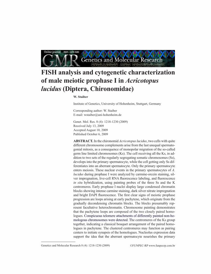
FISH analysis and cytogenetic characterization of male meiotic prophase I in Acricotopus lucidus (Diptera, Chirono midae)
W. Staiber
Institute of Genetics, University of Hohenheim, Stuttgart, Germany
Corresponding author: W. StaiberE-mail: [email protected]
Genet. Mol. Res. 8 (4): 1218-1230 (2009)Received July 13, 2009Accepted August 10, 2009Published October 6, 2009
ABSTRACT. In the chironomid Acricotopus lucidus, two cells with quite different chromo some complements arise from the last unequal spermato-gonial mitosis, as a consequence of monopolar migration of the so-called germ line limited chromosomes (Ks). The cell receiving all the Ks, in ad-dition to two sets of the regularly segregating somatic chromosomes (Ss), develops into the primary spermatocyte, while the cell getting only Ss dif-ferentiates into an aberrant spermatocyte. Only the primary spermatocyte enters meiosis. These nuclear events in the primary spermatocytes of A. lucidus during prophase I were analyzed by carmine-orcein staining, sil-ver impreg nation, live-cell RNA fluorescence labeling, and fluorescence in situ hybridization, using painting probes of the three Ss and the K centromeres. Early prophase I nuclei display large con densed chromatin blocks showing intense carmine staining, dark silver nitrate impregnation and bright DAPI fluores cence. The first clear signs of meiotic prophase progression are loops arising at early pachytene, which originate from the gradually decondensing chromatin blocks. The blocks presumably rep-resent facultative hetero chromatin. Chromosome painting demonstrates that the pachytene loops are composed of the two closely paired homo-logues. Conspicuous telo mere attachments of differently painted non-ho-mologous chromosomes were detected. The centromeres of the Ks group together, indicating a classical bouquet arrange ment of the paired homo-logues in pachytene. The clustered centromeres may function as pairing centers to initiate synapsis of the homologues. Nucleolus expression data support the idea that the aberrant spermatocyte nourishes the primary
1219
©FUNPEC-RP www.funpecrp.com.brGenetics and Molecular Research 8 (4): 1218-1230 (2009)
FISH analysis of male meiotic prophase I in A. lucidus
spermatocyte via a connecting cytoplasmic canal.
Key words: Chromosome painting; Germ line limited chromosomes; Heterochromatin; Meiosis; Prophase I; Spermatogenesis
INTRODUCTION
A programmed unequal chromosome segregation in the last spermatogonial mitosis of Acricotopus lucidus clearly shows in a monopolar migration of the so-called germ line limited chromosomes (=Ks). This system produces two cell types, the primary spermatocyte receiving all the Ks, together with two sets of somatic chromosomes (=Ss), and an aberrant spermato cyte receiving only two S sets (Bauer, 1970; White, 1973; Redi et al., 2001). Cyto kinesis is incomplete, and the daughter cells remain connected by a cytoplasmic canal, which is used to transport mito-chondria from the aberrant spermatocyte to the primary spermatocyte during premeiotic and early meiotic stages (Staiber, 2007). Only the primary spermatocyte passes through regular meiotic di-visions, while the develop ment of the con nected aberrant spermato cyte is blocked, with chromo-somes arrested in a condensed state at metaphase (Staiber, 2008). It is supposed that this chromo-some inactivation process arose during evolution to prevent the entry of the aberrant spermatocyte into meiosis, and con sequently to inhibit the formation of sperm that lack any Ks (Staiber, 2008).
Significant genetic events, namely recognition and pairing of homologues, and re-combination, take place during meiotic prophase I (McKee, 2004; Zickler, 2006). This is ac-companied by nuclear membrane attachment, movement and clustering of telomeres, and by a bouquet arrange ment of the chromosomes in zygotene and pachytene (Scherthan, 2001; Jor-dan, 2006). Little is known about early spermatogenesis in the Dipteran family Chironomidae, in particu lar about nuclear behavior during meiotic prophase I (Wolf, 1941). Neither is any cyto gene tic data available for early meiotic stages of species of the Orthocladiinae, a subfam-ily of the Chironomidae that possesses additional chromosomes restricted to the germ line.
We analyzed nuclear behavior and chromo some arrange ments during prophase I of primary spermatocytes of A. lucidus by classic carmine-orcein staining, silver impregnation and fluorescence in situ hybridization (FISH), using whole chromosome painting probes of the three Ss and probes of germ line specific repetitive DNA sequences located in the centromeric heterochromatin of the Ks.
MATERIAL AND METHODS
Carmine-orcein staining
Testes of pre-pupae taken from a laboratory stock of A. lucidus (Diptera, Chirono midae) were dissected in Ringer’s solution (100 mM NaCl, 3 mM KCl, 3 mM CaCl2, pH 7.2), immedi-ately fixed in freshly prepared ethanol:acetic acid (3:1; 5°C), stained in carmine-acetic acid, and squashed in a drop of 0.5% lacto-acetic orcein. Images of meiotic stages were captured with a Zeiss Standard microscope using a Neofluar 100/1.3 objective and a Canon 450D digital camera.
Silver staining
The staining with 50% AgNO3 and 2% gelatin in 1% formic acid followed the proce-dure of Howell and Black (1980).
1220
©FUNPEC-RP www.funpecrp.com.brGenetics and Molecular Research 8 (4): 1218-1230 (2009)
W. Staiber
Live cell imaging
Imaging of live spermatogonial cells was carried out as described earlier (Staiber, 2007). RNA was labeled using the SYTO® RNASelect green fluorescent cell stain (Invitrogen).
Chromosome painting and FISH
DNA specific for generating painting probes of the three somatic chromosomes of A. lucidus was obtained by microdissection of polytene chromosomes from larval salivary gland squash preparations made with the dry ice method. Two to five polytene chromosomes were dis sected using a mechanical Leitz micro manipulator equipped with fine glass needles, and were collected in a 500-µL polymerase chain reaction (PCR) tube in 5 µL 10 mM Tris-HCl buffer, pH 8.5. Protein digestion was performed by adding 5 μL proteinase K solution (2 mg/mL, PCR grade, Roche, Mann heim) for 48 h at room temperature. Proteinase K was inacti-vated by heating the PCR tube to 94°C for 30 min. The DNA of the digested chromo somes was amplified in a 50-µL degene rate oligonucleotide-primed (DOP)-PCR (Telenius et al., 1992), using the reagents of the Roche DOP-PCR Master Kit and the thermocycling protocol recom-mended by the manu facturer. The PCR products were checked on an agarose gel.
Painting probes of the three somatic chromosomes (SI, SII, SIII) were labeled in a second 50-µL PCR by pipetting 40 mM digoxigenin-11-dUTP or 66 mM fluorescein-12-dUTP (Roche) and 2.5-5 µL of the above chromosome-specific DNA stocks to the premixed reagents of the DOP-PCR kit (Roche), or via nick translation with biotin-16-dUTP using the Roche biotin-nick translation mix. Labeled probes were cleaned by ethanol precipitation and dis-solved in a hybridization mixture containing 50% deionized formamide, 10% dextrane sulfate, 2X SSC, 1X Denhardt solution and 0.1% SDS in 40 mM phosphate buffer, pH 7.0.
Meiotic preparations used for chromosome painting and FISH were made from testes incuba ted in hypotonic 0.5% sodium citrate solution for 20 min prior to ethanol-acetic acid fixation. For hybridization, the preparations were placed in a humidified chamber overnight at 37°C. They were then washed in 2X SSC, pH 7.0, at room temperature for 5 min and twice in 2X SSC at 37°C for 5 min each. The biotinylated SI probe was detected with Cy5-conjugated strept avidin (Dako), the digoxigenated SII probe with rhodamine-conjugated anti-digoxigenin Fab fragments (Roche) and the fluorescein-labeled SIII probe by layers of a rabbit anti-fluo-rescein antibody (Dako) and fluorescein conjugated goat anti-rabbit antibody (Vector Labo-ratories) as described in Staiber and Schiffkowski (2000). Chromosomes were counter stained with 0.2 µg/mL 4’,6-diamidino-2-phenylindole (DAPI) in phosphate-buffered saline, pH 7.4, for 5 min and mounted in Vectashield antifade solution (Vector Laboratories).
A digoxigenized probe of the germ line-specific tandem repetitive AlKeRe1 family (Staiber et al., 1997) localized in the centromeric heterochromatin of the Ks was used together with fluorescein-conjugated anti-digoxigenin Fab fragments (Roche) to label the centromeric re-gions of the Ks in the meiosis preparations. Chromosomes were stained by mounting the prepa-rations in Vectashield fluorescence antifade solution (Vector Laboratories) containing 2.5 μg/mL propidium iodide. Digital fluorescence images were taken with a Biorad MRC 1024 confocal laser scanning system on a Zeiss Axioscope using a Planapochromat 63/1.4 objective, and with a Zeiss Axiophot equipped with an epifluorescence system, a Plan-Neofluar 100/1.3 objective, and a Pixera digital camera. Images were further processed with a Corel Draw software package.
1221
©FUNPEC-RP www.funpecrp.com.brGenetics and Molecular Research 8 (4): 1218-1230 (2009)
FISH analysis of male meiotic prophase I in A. lucidus
RESULTS
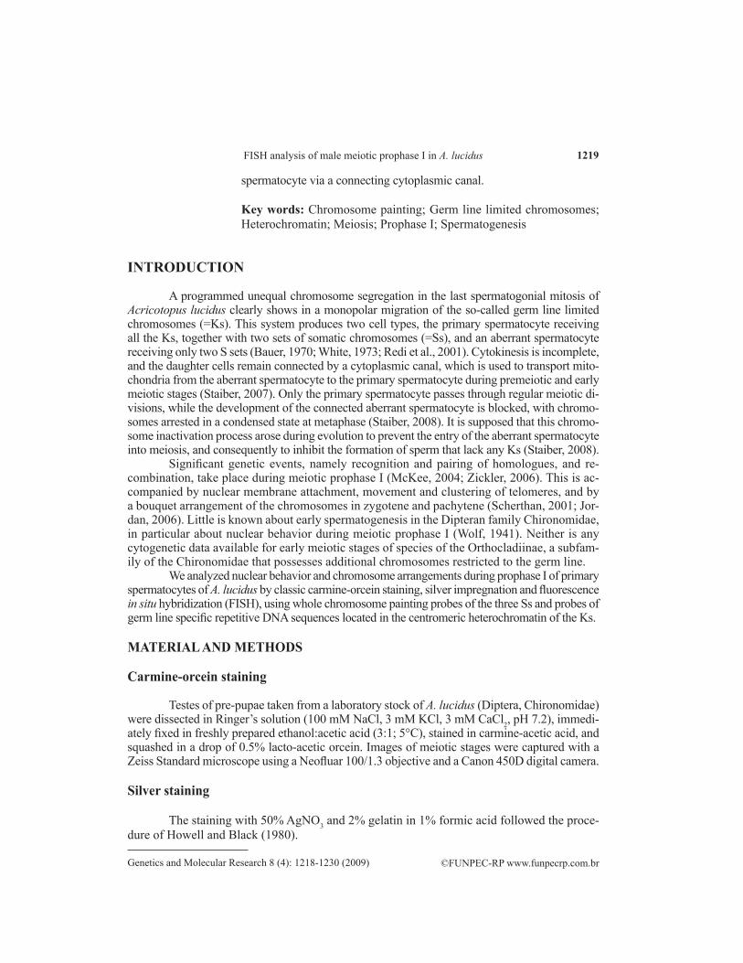
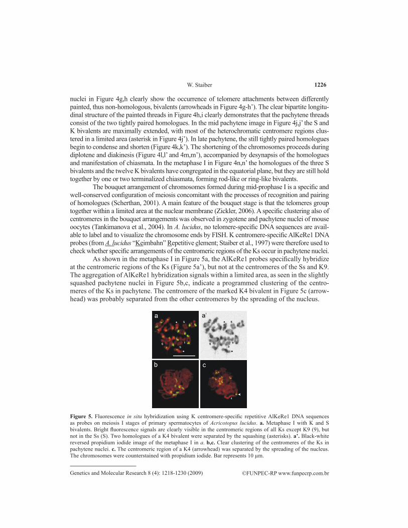
One of the unusual mitoses occurring in the complex chromosome cycle of A. lucidus is the last gonial mitosis. In this so-called differential mitosis, which proceeds in a similar way in both sexes, all germ line limited chromosomes (Ks; n = 6-16) migrate unseparated to one cell pole, as shown in the live cell in Figure 1a; while the somatic chromosomes (Ss; n = 3) first stay in the equatorial plane, and then segregate equally to both poles (Figure 1b). In the male, the daugh-ter cell that received all the Ks and Ss differentiates into the primary spermato cyte, while the other one containing only the Ss becomes an aberrant spermatocyte (Figure 1c). Both cells remain con-nected by a permanent cytoplasmic canal (arrowheads in Figure 1c, d). In the following premei-otic stage, the chromatin in the primary spermatocyte nucleus remains compact and condensed, while the chromosomes of the aberrant spermatocyte decondense and form an interphase nucleus, which expresses one large nucleo lus. During the following premeiotic growth phase, and until entry into meiosis, the primary sper matocyte receives most of the mitochondria of the aberrant spermato cyte via the cytoplasmic canal (Figure 1d; Staiber, 2007). The first indication of prophase I progression in live primary spermatocytes is the gradual appearance of threads and loops arising from the condensed nuclear chromatin masses (Figure 1d).
Figure 1. Live cell phase contrast images of the last unequal spermatogonial mitosis (a,b), and of the resulting primary spermatocyte (sp I) and aberrant spermatocyte (asp) in Acricotopus lucidus (c,d). a. All germ line limited chromosomes (K) move unseparated to only one cell pole (right), while the somatic chromosomes (S, 2n = 6) first remain in the equatorial plane. b. Just before arrival of the Ks at the pole the S sister chromatids segregate equally. c. The resulting daughter cells remain connected by a cytoplasmic canal (arrowhead). The aberrant spermatocyte receives most of the mitochondria (M) and forms one nucleolus (N) in the following interphase. The chromatin of the primary spermatocyte nucleus remains con densed. d. Pachytene threads and loops of paired homologues are the first signs of the beginning meiosis in the primary spermatocyte, and at that time most of the mitochondria (M) have been transported from the aberrant to the primary spermatocyte. Bar represents 10 µm.
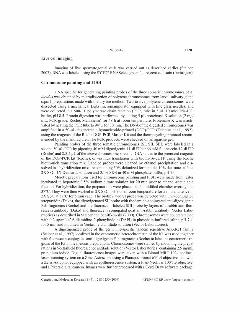
In order to analyze the nuclear changes occurring during prophase I in the primary spermato cyte, classic carmine-orcein staining was first applied to testis cells, which were fixed immediately after dissection (Figure 2a-l). In premeiotic and early prophase I stages large parts of the primary spermatocyte nuclei consist of darkly stained compact chromatin masses (Figure 2a). The first visible alteration of this status is the appearance of faintly stained strands
1222
©FUNPEC-RP www.funpecrp.com.brGenetics and Molecular Research 8 (4): 1218-1230 (2009)
W. Staiber
mostly developing on one side of the condensed chromatin blocks (Figure 2b,c). As shown later (Figure 4h,i), these strands represent the bivalents composed of the two tightly paired homologues, indicating that the primary spermatocyte has entered the pachytene stage (Figure 2c-i). Due to the condensed status of the chromatin, it is not possible to analyze the previous leptotene and zygotene stages by carmine-orcein staining. With the extending and enlarging threads and loops during mid-pachytene, the blocks of condensed chromatin are fragmented and gradually decrease in size (Figure 2e-g).
Figure 2. Carmine-orcein-stained spermatocyte prophase I stages of Acricotopus lucidus. The spermatocytes were immediately fixed without hypotonic pretreatment. a. Condensed early prophase nucleus. b. Probable zygotene stage, with some small fibrillar elements visible on one side of the condensed chromatin. c,d. Early pachytene. An increasing number of loops of paired homologues begin to extend from of the condensed chromatin blocks. e-g. Pachytene. The gradual reduction and fragmentation of the condensed chromatin areas correlates with the increasing number and size of the bivalent threads and loops. h,i. Pachytene. The maximal extended bivalents exhibit only a few condensed sections. j. Diplotene. k. Diakinesis. l. Meta phase I with stretched S and K bivalents. The scale bar in a is representative for a-g, the bar in h for h-k. Bars represent 10 µm.
In some nuclei, weaker stained vesicles of different size were observed within the condensed chromatin (Figure 2e). In late pachytene, only small parts of the condensed chro-matin are present, which probably represent chromosome sections consisting of centromeric, intercalary and terminal constitutive heterochromatin (Figure 2h,i) (Staiber, 1991a). In dip-lotene, the bivalents become successively more condensed (Figure 2j). The homologues are clearly separated, but held together by interstitial and terminal chiasmata. The telomeres of chro mosomes of different bivalents are frequently attached to each other (Figure 2j); while the process of chromosome condensation proceeds in diakinesis, often accompanied by a group-ing of telomeric-attached bivalents (Figure 2k). In metaphase I, the maximally condensed chiasmate homologues have congre gated in the equatorial plane, appearing as rod-like and ring-like bivalents due to pulling forces of the microtubules directed poleward from each centro mere (Figure 2l). Thereafter meiosis proceeds in the orthodox way.
In the condensed nuclei of live and carmine-orcein-stained primary spermato cytes, no clear nucleolus for mation could be observed during premeiotic and early meiotic stages. To analyze the nucleolus expression pattern from differential mitosis to early meiotic stages,
1223
©FUNPEC-RP www.funpecrp.com.brGenetics and Molecular Research 8 (4): 1218-1230 (2009)
FISH analysis of male meiotic prophase I in A. lucidus
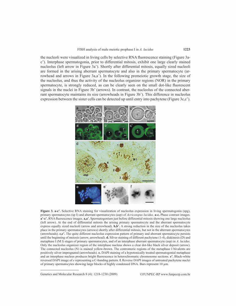
the nucleoli were visualized in living cells by selective RNA fluorescence staining (Figure 3a-c’). Inter phase spermatogonia, prior to differential mitosis, exhibit one large clearly stained nucleolus (left arrow in Figure 3a’). Shortly after differential mitosis, equally sized nucleoli are formed in the arising aberrant spermatocyte and also in the primary spermatocyte (ar-rowhead and arrows in Figure 3a,a’). In the following premeiotic growth stage, the size of the nucleolus, and thus the activity of the nucleolus organizer regions (NOR) in the primary spermatocyte, is strongly reduced, as can be clearly seen on the small dot-like fluorescent signals in the nuclei in Figure 3b’ (arrows). In contrast, the nucleolus of the connected aber-rant spermatocyte maintains its size (arrowheads in Figure 3b’). This difference in nucleolus expression between the sister cells can be detected up until entry into pachytene (Figure 3c,c’).
Figure 3. a-c’. Selective RNA staining for visualization of nucleolus expression in living sperma togonia (spg), primary spermatocytes (sp I) and aberrant spermatocytes (asp) of Acricotopus lucidus. a-c. Phase contrast images. a’-c’. RNA fluorescence images. a,a’. Spermatogonium just before differential mitosis showing one large nucleolus (left arrow). At the end of differential mitosis the arising primary spermatocyte and the aberrant spermatocyte express equally sized nucleoli (arrow and arrowhead). b,b’. A strong reduction in the size of the nucleolus takes place in the primary spermatocytes (arrows) shortly after differential mitosis, but not in the aberrant spermatocytes (arrowheads). c,c’. The quite different nu cleolus expression pattern of primary and aberrant spermatocyte persists until the beginning of meiosis (arrow, arrowhead). d. Silver staining of different pachytene (1-4), diakinesis (D) and metaphase I (M I) stages of primary spermatocytes, and of an interphase aberrant sper mato cyte (asp) in A. lucidus. Only the nucleolus organizer region of the interphase nucleus shows a clear dot-like black silver deposit (arrow). The connected nucleolus (N) is stained yellow-brown. The centromeric regions of the metaphase I bivalents are positively silver impregnated (arrow heads). e. DAPI staining of a hypotonically treated spermatogonial metaphase and an interphase nucleus produces bright fluorescence in heterochromatic chromosome sections. e’. Black-white reversed DAPI image of e representing a C-banding pattern. f. Reverse DAPI images of untreated pachytene nuclei of primary spermatocytes showing large blocks of highly condensed DNA. Bars represent 10 µm.
1224
©FUNPEC-RP www.funpecrp.com.brGenetics and Molecular Research 8 (4): 1218-1230 (2009)
W. Staiber
The selective silver-staining method of Howell and Black (1980) was used to detect and visualize NORs on chromosomes that were active during the previous inter-phase. Applying silver staining to testis squash preparations resulted in a clear black dot-like NOR staining only in interphase nuclei of aberrant spermatocytes (arrow in Figure 3d), but not in pachytene, diakinesis and metaphase I stages of primary spermatocytes, as shown in Figure 3d. The clear NOR-connected nucleolus of the aberrant spermatocyte in Figure 3d, which is expressed from the somatically paired NORs of the SIII chromo-somes (Staiber and Behnke, 1985), is im preg nated yellow-brown (Figure 3d). In addition the con densed chromatin blocks of early pro phase I nuclei are impreg nated dark-brown, as clearly seen in the four pachytene nuclei (1-4) in Figure 3d, exhibiting differently extended threads and loops. A specific silver staining also occurs in the centromeric regions of metaphase I bivalents (arrowheads in Figure 3d). To check whether the con-densed chromatin blocks in the early prophase I nuclei represent the total complement of constitutive heterochromatin of the Ks and Ss, a DAPI fluorescence staining producing a C-banding was performed on hypo tonically pretreated gonial metaphases and interphase nuclei (Figure 3e). The level of darkly stained heterochromatin sections in the metaphase chromosomes, and also in the interphase nucleus, is too low (reversed DAPI image in Figure 3e’), to visualize the large blocks of condensed chromatin in the early prophase I nuclei (Figures 3f and 2a-d).
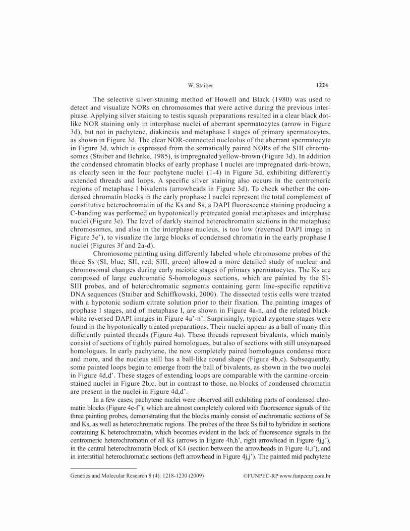
Chromosome painting using differently labeled whole chromosome probes of the three Ss (SI, blue; SII, red; SIII, green) allowed a more detailed study of nuclear and chromosomal changes during early meiotic stages of primary spermatocytes. The Ks are composed of large eu chroma tic S-homologous sections, which are painted by the SI-SIII probes, and of hetero chromatic segments containing germ line-specific repetitive DNA sequences (Staiber and Schiffkowski, 2000). The dissected testis cells were treated with a hypotonic sodium citrate solution prior to their fixation. The painting images of prophase I stages, and of meta phase I, are shown in Figure 4a-n, and the related black-white reversed DAPI images in Figure 4a’-n’. Surprisingly, typical zygotene stages were found in the hypotonically treated preparations. Their nuclei appear as a ball of many thin differently painted threads (Figure 4a). These threads represent bivalents, which mainly consist of sections of tightly paired homologues, but also of sections with still unsynapsed homologues. In early pachytene, the now com pletely paired homologues condense more and more, and the nucleus still has a ball-like round shape (Figure 4b,c). Subsequently, some painted loops begin to emerge from the ball of bivalents, as shown in the two nuclei in Figure 4d,d’. These stages of extending loops are comparable with the carmine-orcein-stained nuclei in Figure 2b,c, but in contrast to those, no blocks of condensed chromatin are present in the nuclei in Figure 4d,d’.
In a few cases, pachytene nuclei were observed still exhibiting parts of condensed chro-matin blocks (Figure 4e-f’); which are almost completely colored with fluores cence signals of the three painting probes, demonstrating that the blocks mainly consist of euchromatic sections of Ss and Ks, as well as heterochromatic regions. The probes of the three Ss fail to hybridize in sections containing K heterochromatin, which becomes evident in the lack of fluorescence signals in the centromeric heterochromatin of all Ks (arrows in Figure 4h,h’, right arrowhead in Figure 4j,j’), in the central heterochromatin block of K4 (section between the arrowheads in Figure 4i,i’), and in interstitial heterochromatic sections (left arrowhead in Figure 4j,j’). The painted mid pachytene
1225
©FUNPEC-RP www.funpecrp.com.brGenetics and Molecular Research 8 (4): 1218-1230 (2009)
FISH analysis of male meiotic prophase I in A. lucidus
Figure 4. Multicolor fluorescence in situ hybridization using whole chromosome painting probes of the three somatic chromosomes, SI (blue), SII (red) and SIII (green), of Acricotopus lucidus on spermatocyte prophase I stages. The spermatocytes were hypotonically treated prior to fixation. a-n. Painting images. a’-n’. The related black-white reversed DAPI images. a,a’. Zygotene nucleus composed of many differently painted thin threads representing paired but also still partially unpaired homologues. b-c’. Early pachytene stages with compact round nuclei, and condensing and shortening bivalents. d,d’. Two pachytene nuclei begin to extend loops of tightly paired homologues. e-f’. Pachytene nuclei still exhibiting parts of condensed chroma tin blocks (arrows), which are clearly decorated with the fluorescence signals of all three painting probes. g-h’. Pachytene nuclei showing clear telomere attachments between two differently colored bivalents (arrowheads). The hetero chromatic centromere regions of the Ks are not painted (arrows). i. In pachytene and metaphase I stages, the germ line limited chromosome K4 (inset) can be easily identified by its large non-painted central hetero chromatin segment (section between the arrowheads). j. Pachytene stage with maximal expanded bivalent threads. Arrowheads indicate centromeric (right) and intercalary (left) heterochromatic sections. k,k’. Late pachytene. The bivalents begin to shorten. l,l’. Diplo tene. m,m’. Diakinesis. n,n’. Metaphase I with three S bivalents (I-III) and twelve dif ferently painted K bivalents. The hetero chromatic sections of the Ks are not painted. Bars represent 10 µm.
1226
©FUNPEC-RP www.funpecrp.com.brGenetics and Molecular Research 8 (4): 1218-1230 (2009)
W. Staiber
nuclei in Figure 4g,h clearly show the occurrence of telomere attachments between differently painted, thus non-homologous, bivalents (arrowheads in Figure 4g-h’). The clear bipartite longitu-dinal structure of the painted threads in Figure 4h,i clearly demonstrates that the pachytene threads consist of the two tightly paired homologues. In the mid pachytene image in Figure 4j,j’ the S and K bivalents are maximally extended, with most of the heterochromatic centromere regions clus-tered in a limited area (asterisk in Figure 4j’). In late pachytene, the still tightly paired homologues begin to condense and shorten (Figure 4k,k’). The shortening of the chromo somes proceeds during diplotene and diakinesis (Figure 4l,l’ and 4m,m’), accompanied by desynapsis of the homologues and manifestation of chiasmata. In the metaphase I in Figure 4n,n’ the homologues of the three S bivalents and the twelve K bivalents have con gregated in the equatorial plane, but they are still hold together by one or two termina lized chiasmata, forming rod-like or ring-like bivalents.
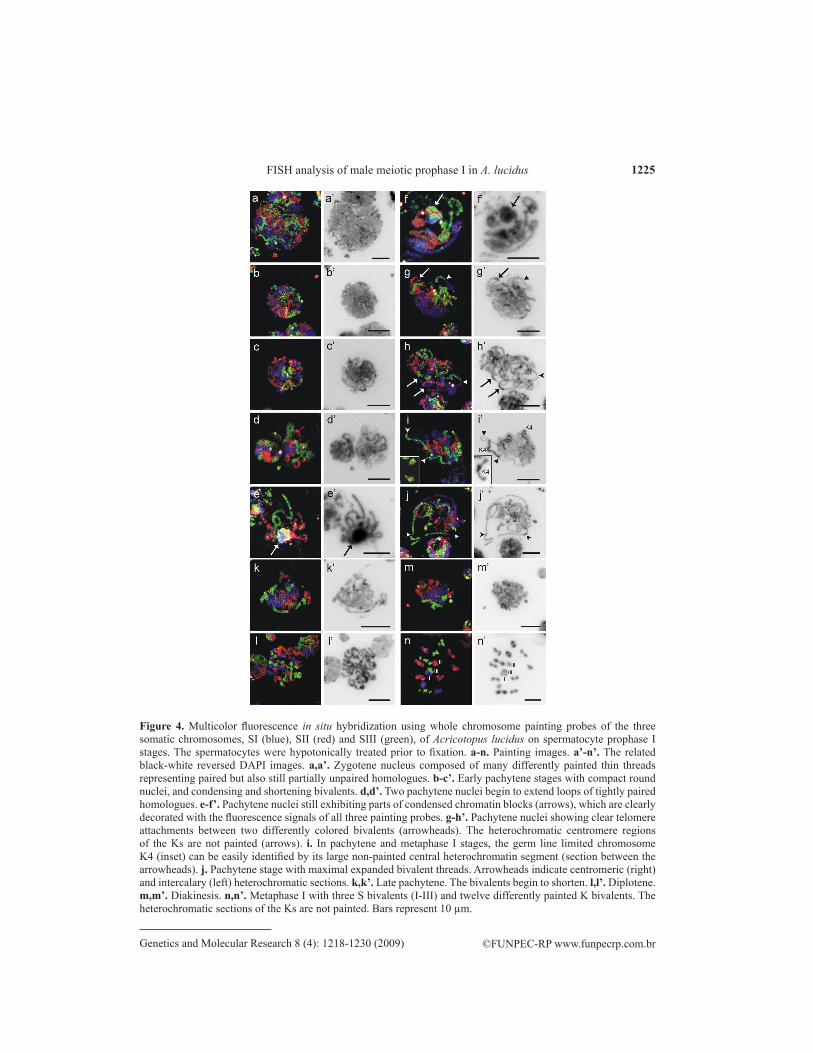
The bouquet arrangement of chromosomes formed during mid-prophase I is a specific and well-conserved configuration of meiosis concomitant with the processes of recog nition and pairing of homologues (Scherthan, 2001). A main feature of the bouquet stage is that the telomeres group together within a limited area at the nuclear membrane (Zickler, 2006). A specific clustering also of centromeres in the bouquet arrangements was observed in zygotene and pachytene nuclei of mouse oocytes (Tankimanova et al., 2004). In A. lucidus, no telomere-specific DNA sequences are avail-able to label and to visualize the chromosome ends by FISH. K centromere-specific AlKeRe1 DNA probes (from A. lucidus “Keimbahn” Repetitive element; Staiber et al., 1997) were therefore used to check whether specific arrangements of the centromeric regions of the Ks occur in pachytene nuclei.
As shown in the metaphase I in Figure 5a, the AlKeRe1 probes specifically hybridize at the centromeric regions of the Ks (Figure 5a’), but not at the centromeres of the Ss and K9. The aggregation of AlKeRe1 hybridization signals within a limited area, as seen in the slightly squashed pachytene nuclei in Figure 5b,c, indicate a programmed clustering of the centro-meres of the Ks in pachytene. The centromere of the marked K4 bivalent in Figure 5c (arrow-head) was probably separated from the other centromeres by the spreading of the nucleus.
Figure 5. Fluorescence in situ hybridization using K centromere-specific repetitive AlKeRe1 DNA sequences as probes on meiosis I stages of primary spermatocytes of Acricotopus lucidus. a. Metaphase I with K and S bivalents. Bright fluorescence signals are clearly visible in the centromeric regions of all Ks except K9 (9), but not in the Ss (S). Two homologues of a K4 bivalent were separated by the squashing (asterisks). a’. Black-white reversed propidium iodide image of the meta phase I in a. b,c. Clear clustering of the centromeres of the Ks in pachytene nuclei. c. The centromeric region of a K4 (arrowhead) was separated by the spreading of the nucleus. The chromosomes were counterstained with propidium iodide. Bar represents 10 µm.
1227
©FUNPEC-RP www.funpecrp.com.brGenetics and Molecular Research 8 (4): 1218-1230 (2009)
FISH analysis of male meiotic prophase I in A. lucidus
DISCUSSION
In A. lucidus, only one of the daughter cells of the last unequal spermatogonial mi-tosis, the future functional primary spermatocyte, which received all Ks in addition to the Ss, passes through meiosis. The other cell, the aberrant spermatocyte, receiving only Ss, is blocked from entering meiosis, presumably to prevent the formation of sperm containing no Ks (Staiber, 2008). To gain more insight into the processes, and the order of events taking place before and at the beginning of meiosis, a detailed analysis of nuclear changes and of behavior of chromosomes during early meiotic stages of primary spermatocytes was carried out, com bining classical cytogenetic methods, chromosome painting and FISH.
In carmine-orcein-stained testis preparations typical leptotene and zygotene nuclei were not observed. Early meiotic primary spermatocyte nuclei contained compact blocks of condensed chromatin. Threads and loops of paired homologues appearing out of the chroma-tin blocks are the first signs of prophase I progression to pachytene (Figure 2c,d). This cor-responds with the observation of Wolf (1941), who reported on male meiosis of Chironomus plumosus (a species of the subfamily Chironominae of the Chironomidae), which possesses no Ks, that “pachytene can be observed as the first clear stage of meiosis”. His sequence of line drawings of meiotic stages of C. plumosus begins with the pachytene (Wolf, 1941). In an elec-tron microscope study using hypotonically lysed early meiotic prophase nuclei to analyze the lampbrush structures of pachytene and diplotene chromosomes in primary spermato cytes of Chironomus pallidivi tatus, Keyl (1975) stated “The transparent nuclei originate during early pachytene stages”. This indicates that it was not possible to prepare and analyze the previous leptotene and zygotene stages; but neither Wolf (1941) nor Keyl (1975) reported on early meiotic nuclei containing large condensed chromatin masses, as detected here in A. lucidus.
Silver staining of testis preparation for the detection of previously active NORs re-vealed positive dot-like black silver deposits in interphase aberrant spermatocytes, but no labeling of NORs in premeiotic and prophase I stages, or in metaphase I of primary spermato-cytes. This agrees with the results of the analysis of the nucleolus expression patterns in live primary and aberrant spermatocytes obtained by selective RNA fluorescence staining. NOR activity is strongly reduced in the primary spermatocyte shortly after differential mitosis at the begin ning of the premeiotic growth phase, but not in the aberrant spermato cyte (Figure 3b-c’). This supports the idea that the aberrant spermatocyte supplies the primary spermatocyte, via the cytoplasmic canal, not only with mitochondria (Staiber, 2007) but also with other cyto-plasmic components, such as ribosomes. Intercellular transport of ribosomes via cytoplasmic bridges was established for mouse erythroblasts and early rat spermatids (Asano et al., 1991; Ventalä et al., 2003). In the present study, both DNA and proteins were stained yellow to dark-brown by silver staining (Figure 3d), so it cannot be determined by this method whether an accumulation of proteins or greater DNA condensation leads to the occurrence of these compact chromatin blocks in early prophase nuclei. The staining of early pachytene nuclei, immediately fixed without hypotonic pretreatment, with the DNA-specific fluorescence dye DAPI, clearly shows that a higher DNA density is responsible for the observed chroma tin compactness (Figure 3f); but the amount of constitutive heterochromatin in the Ss and Ks in the form of centromeric, inter stitial and telomeric hetero chroma tin segments, as determined by C-banding (Figure 3e,e’; Staiber, 1991a), is too low to explain the formation of such large heteropycnotic chroma tin blocks in the prepachytene and early pachytene nuclei.
1228
©FUNPEC-RP www.funpecrp.com.brGenetics and Molecular Research 8 (4): 1218-1230 (2009)
W. Staiber
There are only a few studies applying chromosome painting in insects. They have been carried out with probes generated by microdissection and DOP-PCR of the X and B chromosomes in Locusta migratoria (Teruel et al., 2009), of the W chromosome in the moth Cydia pomonella (Fuková et al., 2007), and of terminal parts of chromosomes X, 2 and 3 of Drosophila melanogas-ter (Fuchs et al., 1998); but the probes used painted only single chromo somes or parts of chromo-somes, and not the whole chromosome complement as in the present study, and in an earlier study on A. lucidus (Staiber and Wahl, 2002). Applying chro mo some painting with probes of the three Ss on hypo tonically treated primary spermatocyte nuclei of A. lucidus, it was possible to visualize chromosomes in zygotene. Probably the hypotonic treatment effects a swelling, decondensation and protein denaturation of the chromatin blocks, with concomitant transparency of the nuclei, similar to those which Keyl (1975) reported for hypotonically treated nuclei of C. pallidivitatus (see above). This would mean that the pairing of homo logues begins and proceeds in the compact zygo tene nuclei, or that the homologues are already paired before the primary spermatocyte enters meiosis. Wolf (1941) presumed that “leptotene and zygotene fail as a consequence of somatic pairing” in C. plumosus, which implies that the homologues are already paired prior to entry into meiosis. In A. lucidus, after duplication of the Ks due to their monopolar movement as unseparated sister chromatids during differen tial mitosis, each K type can be present two or four times, and one K type, K4, up to 10 times in a primary spermatocyte (Staiber, 1988). But, as demonstrated by X-ray-induced marker chromosomes, the overwhelming majority of the Ks form bivalents with their sister chromatid partner (Staiber, 1991b). So one can presume that the K sister chromatids stay together after differential mitosis and are already paired before entering into meiosis.
In some cases, residuals of the condensed chromatin blocks were preserved in hypotoni-cally pretreated pachytene nuclei (Figure 4e-f’). The clear hybridization signals of the painting probes covering these residuals definitely demonstrate that the condensed blocks contain large portions of euchromatic chromosome sections, in addition to some unstained hetero chromatic segments. It is possible that the condensed chromatin blocks in premeiotic or early meiotic nuclei of A. lucidus represent a special type of facultative heterochromatin, as reported for the entire pa-ternal haploid chromosome sets of mealybugs (Nur, 1990; Volpi et al., 2007), and for the unpaired X in pachytene spermato cytes of Locusta migratoria (Friedländer and Hauschteck-Jungen, 1986).
Chromosome painting of pachytene nuclei clearly demonstrates that telomere attach-ments between non-homologous chromosomes of different bivalents occur in A. lucidus. In carmine-orcein-stained preparations, also in diplotene and diakinesis, telomere attach-ments were observed (Figure 2j,k). Non-homologous chromosomes of different bivalents attached via telomeres at pachytene were also reported in the cricket Gryllus argentinus, the lepidopteran Sphinx ligustri, and the nematode Meloidogyne natalieli (Drets and Stoll, 1974; Goldstein and Triantaphyllou, 1986; Nokkala, 1987). In G. argentinus it was supposed that these end-to-end attachments of bivalents are initiated by terminal heterochro matic segments (Drets and Stoll, 1974); although the function of such telomere attachments between non-homologous chromo somes occurring in meiotic prophase remains unclear.
Centromeres and telomeres play an important role in the meiotic chromosome pair-ing process (Jordan, 2006; Scherthan, 2007). At the beginning of prophase I, the telomeres move along the inside of nuclear envelope mediated by actin (Trelles-Sticken et al., 2005), and they congregate in a distinct region near the microtubule-organizing center. This telo-mere clustering brings the homologues close together and probably initiates pairing and the formation of the synaptone mal complex between them, and consequently leads to the bouquet
1229
©FUNPEC-RP www.funpecrp.com.brGenetics and Molecular Research 8 (4): 1218-1230 (2009)
FISH analysis of male meiotic prophase I in A. lucidus
arrangement of the paired chromosomes (Bass, 2003; Scherthan, 2001). It was also recently demonstrated in budding yeast that centromeres can serve as pairing centers, and as preferred sites of synapsis initiation (Tsubouchi et al., 2008). Using CREST antibodies for visualization of centromere proteins a tight centromere clustering was detected during pachytene in mouse oocytes (Tankimanova et al., 2004). In A. lucidus, a clustering of the centromeres of the Ks in pachy tene could be demonstrated by FISH using probes of K-specific repetitive centromeric DNA, indicating that the formation of a bouquet configuration of the paired chromosomes also occurs in prophase I of A. lucidus. A typical bouquet was also reported and drawn from Wolf (1941), for pachytene spermatocyte nuclei of C. plumosus.
The present study highlights the importance of multicolor chromosome painting and FISH in analyses of nuclear changes and chromosome configuration during early meiosis in insects. In future investigations, it will be of interest to determine whether specific histone modifications, such as differential lysine methylation and acetylation, and/or the accumulation of specific hetero-chromatin proteins, are involved in the formation and the decondensation of the large condensed chromatin blocks occurring in premeiotic and early meiotic nuclei of primary spermatocytes.
ACKNOWLEDGMENTS
The author would like to thank Prof. Anette Preiss, University of Hohenheim, for sup-port, and Prof. Neil Jones, Biological Sciences, Aberystwyth University, Wales, for helpful suggestions and corrections to the manuscript.
REFERENCES
Asano H, Kobayashi M and Hoshino T (1991). Ultrastructural study of a cytoplasmic bridge connecting a pair of erythroblasts in mice. Cell Tissue Res. 264: 215-219.
Bass HW (2003). Telomere dynamics unique to meiotic prophase: formation and significance of the bouquet. Cell Mol. Life Sci. 60: 2319-2324.
Bauer H (1970). Rearrangements between germ-line limited and somatic chromosomes in Smittia parthenogenetica (Chironomidae, Diptera). Chromosoma 32: 1-10.
Drets ME and Stoll M (1974). C-banding and non-homologous associations in Gyrllus argentinus. Chromosoma 48: 367-390.Friedländer M and Hauschteck-Jungen E (1986). Facultative heterochromatin in Locusta migratoria spermatogenesis:
correlation between electron opacity and heteropycnosis. J. Cell Sci. 81: 299-306.Fuchs J, Kuhfittig S, Reuter G and Schubert I (1998). Chromosome painting in Drosophila. Chromosome Res. 6: 335-336.Fuková I, Traut W, Vítková M, Nguyen P, et al. (2007). Probing the W chromosome of the codling moth, Cydia pomonella,
with sequences from microdissected sex chromatin. Chromosoma 116: 135-145.Goldstein P and Triantaphyllou AC (1986). The synaptonemal complex of Meloidogyne nataliei and its relationship to that
of other Meloidogyne species. Chromosoma 93: 261-266.Howell WM and Black DA (1980). Controlled silver-staining of nucleolus organizer regions with a protective colloidal
developer: a 1-step method. Experientia 36: 1014-1015.Jordan P (2006). Initiation of homologous chromosome pairing during meiosis. Biochem. Soc. Trans. 34: 545-549.Keyl HG (1975). Lampbrush chromosomes in spermatocytes of Chironomus. Chromosoma 51: 75-91.McKee BD (2004). Homologous pairing and chromosome dynamics in meiosis and mitosis. Biochim. Biophys. Acta
1677: 165-180.Nokkala S (1987). Cytological characteristics of chromosome behaviour during female meiosis in Sphinx ligustri L.
(Sphingidae, Lepidoptera). Hereditas 106: 169-179.Nur U (1990). Heterochromatization and euchromatization of whole genomes in scale insects (Coccoidea: Homoptera).
Dev. Suppl. 29-34.Redi CA, Garagna S, Zacharias H, Zuccotti M, et al. (2001). The other chromatin. Chromosoma 110: 136-147.
1230
©FUNPEC-RP www.funpecrp.com.brGenetics and Molecular Research 8 (4): 1218-1230 (2009)
W. Staiber
Scherthan H (2001). A bouquet makes ends meet. Nat. Rev. Mol. Cell Biol. 2: 621-627.Scherthan H (2007). Telomere attachment and clustering during meiosis. Cell Mol. Life Sci. 64: 117-124.Staiber W (1988). G-banding of germ line limited chromosomes in Acricotopus lucidus (Diptera, Chironomidae).
Chromosoma 97: 231-234.Staiber W (1991a). Characterization of heterochromatin of germ line limited and soma chromosomes in Acricotopus
lucidus (Diptera, Chironomidae) by differential banding methods. Heredity 114: 91-96.Staiber W (1991b). Preferential pairing in the germ line limited chromosomes of Acricotopus lucidus (Diptera,
Chironomidae). Heredity 66: 197-201.Staiber W (2007). Asymmetric distribution of mitochondria and of spindle microtubules in opposite directions in
differential mitosis of germ line cells in Acricotopus. Cell Tissue Res. 329: 197-203.Staiber W (2008). Centrosome hyperamplification with the formation of multiple asters and programmed chromosome
inactivation in aberrant spermatocytes during male meiosis in Acricotopus. Cell Tissue Res. 334: 81-91.Staiber W and Behnke E (1985). Developmental puffing activity in the salivary gland and Malpighian tubule chromosomes
of Acricotopus lucidus (Diptera, Chironomidae). Chromosoma 93: 1-16.Staiber W and Schiffkowski C (2000). Structural evolution of the germ line-limited chromosomes in Acricotopus.
Chromosoma 109: 343-349.Staiber W and Wahl S (2002). Painting analysis of meiotic metaphase I configurations of the germ line-limited
chromosomes in Acricotopus. Chromosome Res. 10: 101-108.Staiber W, Wech I and Preiss A (1997). Isolation and chromosomal localization of a germ line-specific highly repetitive
DNA family in Acricotopus lucidus (Diptera, Chironomidae). Chromosoma 106: 267-275.Tankimanova M, Hultén MA and Tease C (2004). The initiation of homologous chromosome synapsis in mouse fetal oocytes
is not directly driven by centromere and telomere clustering in the bouquet. Cytogenet. Genome Res. 105: 172-181.Telenius H, Carter NP, Bebb CE, Nordenskjöld M, et al. (1992). Degenerate oligonucleotide-primed PCR: general
amplification of target DNA by a single degenerate primer. Genomics 13: 718-725.Teruel M, Cabrero J, Montiel EE, Acosta MJ, et al. (2009). Microdissection and chromosome painting of X and B
chromosomes in Locusta migratoria. Chromosome Res. 17: 11-18.Trelles-Sticken E, Adelfalk C, Loidl J and Scherthan H (2005). Meiotic telomere clustering requires actin for its formation
and cohesin for its resolution. J. Cell Biol. 170: 213-223.Tsubouchi T, Macqueen AJ and Roeder GS (2008). Initiation of meiotic chromosome synapsis at centromeres in budding
yeast. Genes Dev. 22: 3217-3226.Ventelä S, Toppari J and Parvinen M (2003). Intercellular organelle traffic through cytoplasmic bridges in early spermatids
of the rat: mechanisms of haploid gene product sharing. Mol. Biol. Cell 14: 2768-2780.Volpi S, Bongiorni S and Prantera G (2007). HP2-like protein: a new piece of the facultative heterochromatin puzzle.
Chromosoma 116: 249-258.White MJD (1973). Animal Cytology and Evolution. 3rd edn. Cambridge University Press, Cambridge.Wolf E (1941). Die Chromosomen in der Spermatogenese einiger Nematoceren. Chromosoma 2: 192-246.Zickler D (2006). From early homologue recognition to synaptonemal complex formation. Chromosoma 115: 158-174.