Embed Size (px)
Citation preview

FKBP52-PROGESTERONE RECEPTOR SIGNALING
DURING PREGNANCY
By
Susanne Tranguch
Dissertation
Submitted to the Faculty of the
Graduate School of Vanderbilt University
in fulfillment of the requirements
for the degree of
DOCTOR OF PHILOSOPHY
in
Cell and Development Biology
December, 2007
Nashville, Tennessee
Approved:
Sudhansu K. Dey
Richard M. Caprioli
Raymond N. DuBois
Sanjoy K. Das
Gary E. Olson

ii
To my parents, Lynn and Steve, for their unwavering belief in me

iii
ACKNOWLEDGEMENTS
This work would not have been possible without the support of my mentor,
Dr. Sudhansu K. Dey. I am forever indebted to Dr. Dey for teaching me how to
be a scientist, not only through his training but also by his example. I also want
to thank all the committee members, Dr. Gary Olson, Dr. Sanjoy K. Das, Dr.
Raymond N. DuBois, and Dr. Richard M. Caprioli, for their helpful discussions
and guidance in my continual journey to be a better student and scientist. I also
thank Dr. D regarding my FKBP52 experiments. I also thank Drs. Haibin Wang,
Takiko Daikoku, Huirong Xie, Hao Zhang and Yong Guo, who made the ample
hours in the lab so enjoyable with our chatting times, both socially and
scientifically. This entire lab truly became a second family to me, and each of
them contributed to the completion of my project. This work would not be
possible without the generous financial support from the Molecular Endocrinology
Training Program (METP) and NRSA pre-doctoral fellowship (NIH).
On a personal note, I could have not made it through twenty-eight years
as a student without the support of my best gs: Tara Tranguch, Grayson
Hudgins, Liz Hackman, Vanessa Hartley, Ashley Kaufman, Katy Kamp, and Jena
Fletcher. This work is dedicated to my parents, Lynn and Steve, who inspire me
every day to be a better person and more importantly, remind me to pay
attention, which is the best advice for any scientist.
iv
TABLE OF CONTENTS
Page
DEDICATION ........................................................................................................ii
ACKNOWLEDGEMENTS..................................................................................... iii
LIST OF TABLES ................................................................................................vii
LIST OF FIGURES ............................................................................................. viii
LIST OF ABBREVIATIONS .................................................................................. x
Chapter
I. INTRODUCTION ............................................................................................... 1
Stages of embryo implantation ............................................................... 2 Steroid hormonal regulation of pregnancy .............................................. 3 The window of uterine receptivity ........................................................... 5 The “ripple effect” theory......................................................................... 7 Progesterone (P4): discovering the ‘pregnancy hormone’....................... 7 Mechanism of progesterone signaling .................................................. 10 Progesterone-regulated target genes during early pregnancy.............. 13 Hoxa10 null mice reveal FKBP52 as a critical mediator of implantation .......................................................................................... 16
Uterine FKBP52 expression is cell-specific and down-regulated in Hoxa10 null mice .................................................................................. 21 FKBP52 is expressed in a spatiotemporal manner in the periimplantation uterus.......................................................................... 22 Progesterone and estrogen differentially regulate Fkbp52 expression in the uterus........................................................................ 25
II. COCHAPERONE IMMUNOPHILIN FKBP52 IS CRITICAL TO UTERINE RECEPTIVITY AND IMPLANTATION ........................................... 29
Abstract.................................................................................................. 29 Introduction ............................................................................................ 30 Methods ................................................................................................. 34 Generation of Fkbp52 null mice ................................................... 34 Mouse genotyping........................................................................ 35 Ovulation, fertilization, implantation, and blastocyst
v
transfer ......................................................................................... 36 Comparative RT-PCR and Southern blotting ............................... 36 Progesterone binding assay......................................................... 38 Transfection and PR transcription activity assay.......................... 38 In situ hybridization ...................................................................... 39 Northern hybridization .................................................................. 39 Isolation and culture of embryonic fibroblasts .............................. 39 Histology and immunostaining of Ki67 ......................................... 40 Results ................................................................................................... 40 Ovulation is normal in C57BL6/129 Fkbp52 null females............. 40 Fkbp52 null mice show implantation failure.................................. 44 FKBP52 and PR show overlapping uterine expression during implantation....................................................................... 46 Uterine PR activity is compromised in Fkbp52 null females......... 51 PR responsive genes and functions are aberrant in uteri of Fkbp52 null mice ...................................................................... 52 Aberrant expression of estrogen-responsive gene lactoferrin in Fkbp52 null uteri on day 4 of pregnancy................................... 54 Abnormal pattern of cell proliferation in Fkbp52 null uteri on day 4 of pregnancy.................................................................. 55 Discussion.............................................................................................. 56
III. FKBP52 DEFICIENCY-CONFERRED UTERINE PROGESTERONE RESISTANCE IS GENETIC BACKGROUND AND PREGNANCY STAGE SPECIFIC...................................................................................................... 58
Abstract.................................................................................................... 58 Introduction .............................................................................................. 59 Methods ................................................................................................... 60 Mice ............................................................................................... 60 Ovulation, fertilization, implantation, blastocyst transfer, and experimentally-induced decidualization ......................................... 61 Exogenous progesterone supplementation and other treatments...................................................................................... 62 Progesterone assay ....................................................................... 63 In situ hybridization ........................................................................ 63 Immunohistochemistry ................................................................... 63 Results..................................................................................................... 64 Implantation failure occurs in Fkbp52 null mice irrespective of genetic background.................................................................... 64 Progesterone supplementation rescues implantation in CD1 Fkbp52 null females............................................................... 66 Progesterone supplementation restores progesterone- and implantation-regulated gene expression in CD1 Fkbp52 null uteri ......................................................................................... 70
vi
Post-implantation defects in progesterone treated CD1 Fkbp52 null females....................................................................... 77 Experimentally-induced decidualization fails in Fkbp52 null mice irrespective of progesterone treatment.................................. 79 Progesterone delivery via silastic implants partially restores full-term pregnancy in CD1 Fkbp52 null females ........................... 80 Excessive estrogenic influence or complement activation does not contribute to pregnancy failure in progesterone- implanted Fkbp52 null females ...................................................... 83 Differential progesterone-PR signaling is required for successful full-term pregnancy ..................................................... 85 Discussion ............................................................................................... 87
REFERENCES................................................................................................... 95
vii
LIST OF TABLES
Table Page
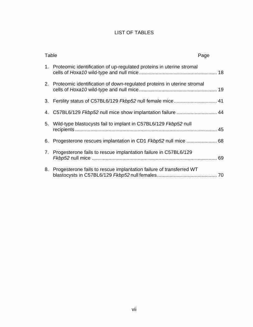
1. Proteomic identification of up-regulated proteins in uterine stromal cells of Hoxa10 wild-type and null mice........................................................ 18 2. Proteomic identification of down-regulated proteins in uterine stromal cells of Hoxa10 wild-type and null mice........................................................ 19 3. Fertility status of C57BL6/129 Fkbp52 null female mice............................... 41 4. C57BL6/129 Fkbp52 null mice show implantation failure ............................. 44 5. Wild-type blastocysts fail to implant in C57BL6/129 Fkbp52 null recipients ...................................................................................................... 45 6. Progesterone rescues implantation in CD1 Fkbp52 null mice ...................... 68 7. Progesterone fails to rescue implantation failure in C57BL6/129 Fkbp52 null mice .......................................................................................... 69 8. Progesterone fails to rescue implantation failure of transferred WT blastocysts in C57BL6/129 Fkbp52 null females........................................... 70
viii
LIST OF FIGURES
Figure Page
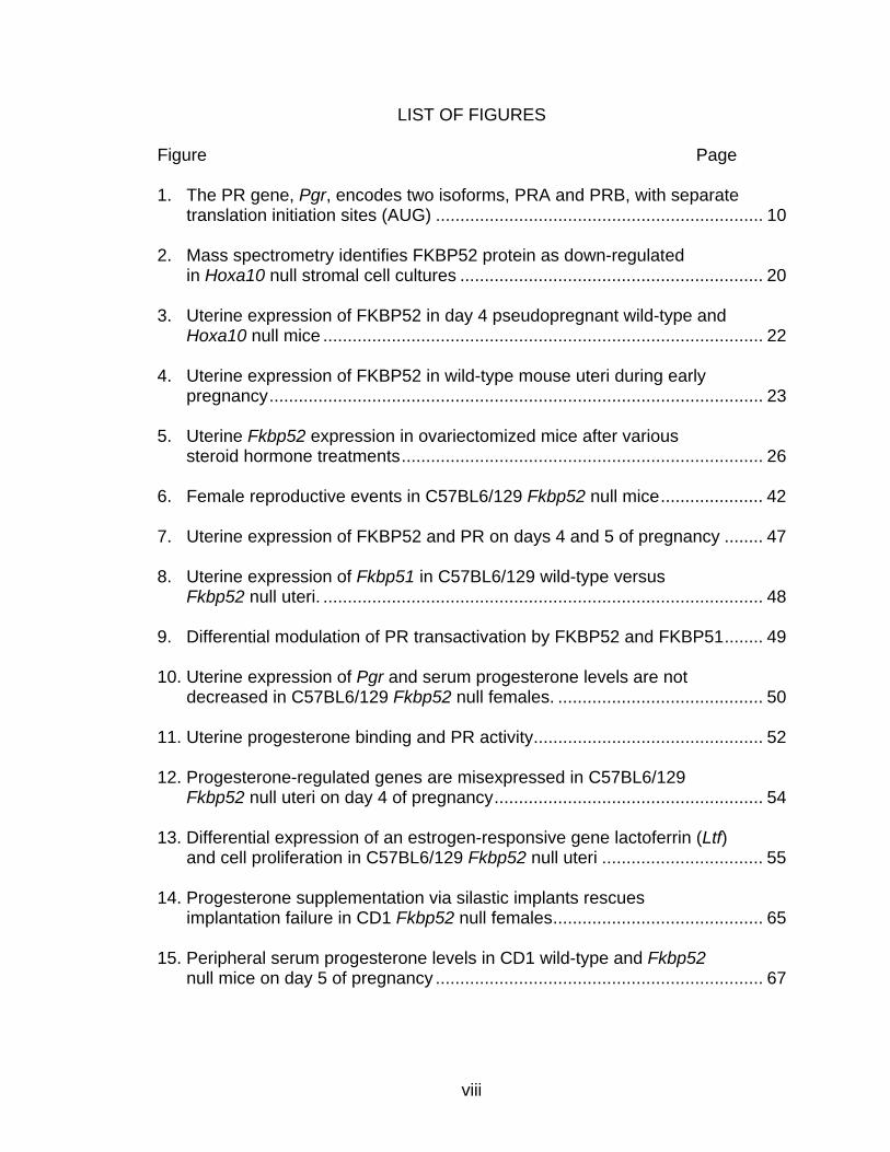
1. The PR gene, Pgr, encodes two isoforms, PRA and PRB, with separate translation initiation sites (AUG) ................................................................... 10 2. Mass spectrometry identifies FKBP52 protein as down-regulated in Hoxa10 null stromal cell cultures .............................................................. 20 3. Uterine expression of FKBP52 in day 4 pseudopregnant wild-type and Hoxa10 null mice .......................................................................................... 22 4. Uterine expression of FKBP52 in wild-type mouse uteri during early pregnancy..................................................................................................... 23 5. Uterine Fkbp52 expression in ovariectomized mice after various steroid hormone treatments.......................................................................... 26 6. Female reproductive events in C57BL6/129 Fkbp52 null mice..................... 42 7. Uterine expression of FKBP52 and PR on days 4 and 5 of pregnancy ........ 47 8. Uterine expression of Fkbp51 in C57BL6/129 wild-type versus Fkbp52 null uteri. .......................................................................................... 48 9. Differential modulation of PR transactivation by FKBP52 and FKBP51........ 49 10. Uterine expression of Pgr and serum progesterone levels are not decreased in C57BL6/129 Fkbp52 null females. .......................................... 50 11. Uterine progesterone binding and PR activity............................................... 52 12. Progesterone-regulated genes are misexpressed in C57BL6/129 Fkbp52 null uteri on day 4 of pregnancy....................................................... 54 13. Differential expression of an estrogen-responsive gene lactoferrin (Ltf) and cell proliferation in C57BL6/129 Fkbp52 null uteri ................................. 55 14. Progesterone supplementation via silastic implants rescues implantation failure in CD1 Fkbp52 null females........................................... 65 15. Peripheral serum progesterone levels in CD1 wild-type and Fkbp52 null mice on day 5 of pregnancy ................................................................... 67
ix
16. Progesterone supplementation via silastic implants corrects misexpression of progesterone-target genes in CD1 Fkbp52 null uteri ....... 71 17. Progesterone supplementation via silastic implants partially restores the expression of progesterone-target genes, but fails to counter an estrogen-responsive gene, lactoferrin, in C57BL6/129 Fkbp52 null uteri .... 73 18. Delivery of progesterone via silastic implants restores expression of implantation-related genes in CD1 Fkbp52 null females ............................. 76 19. Progesterone delivery via silastic implants rescues blastocyst-induced, but not oil-induced, decidualization in Fkbp52 null females......................... 78 20. Progesterone delivery by silastic implants fails to sustain full complement of term pregnancy in Fkbp52 null females .............................. 81 21. In situ hybridization of Pgr and Fkbp52 in sections of implantation sites on days 10 and 12 of pregnancy in CD1 WT uteri............................... 82 22. Anti-estrogen or heparin fails to rescue implantation in P4-treated CD1 Fkbp52 null females ............................................................................ 84 23. Daily progesterone injections restore full complement of term pregnancy in CD1 Fkbp52 null females....................................................... 86 24. Putative model for how exogenous P4 treatment rescues PR activity in the absence of FKBP52 ............................................................... 88 25. PR and FKBP52 expression in proliferative and secretory human endometrium.................................................................................... 93

x
ABBREVIATIONS
AREG, amphiregulin
BMP, bone morphogenetic protein
HDC, histidine decarboxylase
EGF, epidermal growth factor
E2, estrogen
HB-EGF, heparin-binding EGF-like growth factor
FKBP, FK506 binding protein
IHH, indian hedgehog
IS, implantation site
KO, knockout
LIF, leukemia inhibitory factor
P4, progesterone
PR, progesterone receptor
PDZ, primary decidual zone
SDZ, secondary decidual zone
WT, wild-type
1
CHAPTER I
INTRODUCTION
Implantation is a process by which the blastocyst comes into intimate
physical and physiological contact with the uterine endometrium. A two-way
interaction between the implantation-competent blastocyst and receptive uterine
luminal epithelium initiates this process (Dey et al., 2004; Wang and Dey, 2006).
If an appropriate embryo-uterine dialogue at the molecular and cellular level is
not established, the blastocyst will not implant leading to pregnancy failure.
Coordinated effects of the ovarian steroid hormones estrogen and progesterone
(P4) guide these embryo-uterine interactions that are not only critical for
determining correct implantation timing, but also for sustaining pregnancy. An
increased understanding of mammalian implantation and pregnancy
maintenance has been gained through the use of various genetically engineered
mouse models, and this information will potentially lead to strategies to correct
implantation failure and improve pregnancy rates in women.
The assembly of new life begins with the union between a sperm and an
egg through the process of fertilization. The one-cell fertilized egg, termed
embryo, undergoes several mitotic cell divisions, ultimately forming a ball of cells
(16 cells or more) termed morula. The morula then differentiates into a
blastocyst with two distinct cell populations, the inner cell mass (ICM) which will
become the embryo proper and a layer of trophectoderm cells surrounding the
2
ICM which will form the placenta and extraembryonic membranes (Dey et al.,
2004). Dysregulation of events prior, during, or after implantation can contribute
to poor pregnancy rates in eutherian mammals. For example, for a successful
pregnancy to occur, on the embryonic side, fertilized eggs must develop to
blastocysts, transit through the oviduct to the uterus, establish an intimate
contact with the uterine epithelium, be nurtured through embryogenesis, and
ultimately delivered at birth. On the maternal side, the uterus must be receptive
for implantation and undergo extensive remodeling for decidualization,
pregnancy maintenance, and ultimately parturition (Dey, 1996; Dey et al., 2004).
Therefore, studying implantation physiology and revealing the signaling pathways
involved are necessary for alleviating problems with human infertility and
ensuring the birth of quality offspring.
Stages of Embryo Implantation
Enders and Schlafke have classified the implantation process into three
stages: apposition, adhesion, and penetration (Enders, 1976; Enders and
Schlafke, 1969). Apposition is the stage when embryonic trophectoderm cells
become closely apposed to the uterine luminal epithelium. This is followed by
adhesion of the trophectoderm to the luminal epithelium. Ultimate attachment
and penetration involves the invasion of the luminal epithelium by the
trophectoderm.
The attachment reaction coincides with a localized increase in stromal
vascular permeability at the site of the blastocyst, which can be demarcated by
3
intravenous injection of a macromolecular blue dye, also referred to as the
uterine blue reaction (Psychoyos, 1973a). This is preceded by uterine luminal
closure, which contributes to the intimate apposition of the blastocyst with the
uterine epithelium (Enders, 1976; Psychoyos, 1973a). The first sign of the
attachment reaction in the mouse occurs on the evening (2000-2400 h) of day 4
of pregnancy (day 1 = vaginal plug) (Psychoyos, 1973a). In mice and humans,
while luminal epithelial cells undergo apoptosis after attachment (Parr et al.,
1987), stromal cells surrounding the implanting blastocyst undergo
decidualization eventually embedding the embryo within the antimesometrial
stroma. In mice, blastocysts are oriented with their ICMs directed mesometrially,
whereas in humans the ICM is directed antimesometrially (Dey et al., 2004). The
mechanism by which the blastocyst is directed to the antimesometrial luminal
epithelium or by which the orientation of the blastocyst is achieved at the time of
implantation remains unknown. This introduction highlights the more recently
defined signaling cascades involved in implantation, with specific regard to P4
signaling, therefore focusing specifically on steroid hormones, homeotic
transcription factors, and immunophilins.
Steroid hormonal regulation of early pregnancy
The uterus is comprised of heterogeneous cell types (luminal and
glandular epithelium, stroma, and myometrium) that each respond differentially to
estrogen and P4. In mice, the coordinated effects of estrogen and P4 regulate
proliferation and/or differentiation of uterine cells in a spatiotemporal manner to
4
govern events of early pregnancy (Huet-Hudson et al., 1989). For example, on
the first day of pregnancy (day 1) in mice, uterine epithelial cells undergo
proliferation under the influence of preovulatory estrogen secretion. Rising levels
of P4 secreted from freshly formed corpora lutea initiate stromal cell proliferation
from day 3 onward. This stromal cell proliferation is further stimulated by a small
secretion of ovarian estrogen on the morning of day 4 of pregnancy. These
coordinated effects of P4 and estrogen result in the cessation of uterine epithelial
cell proliferation, initiating differentiation (Huet-Hudson et al., 1989). During
normal pregnancy, the presence of an active blastocyst in the uterus is the
stimulus for the implantation reaction. After the attachment reaction is initiated on
day 4 (2000–2400 h), stromal cells surrounding the implanting blastocyst begin to
proliferate extensively and differentiate into decidual cells (Dey, 1996).
In pseudopregnant mice (female mice mated with vasectomized males),
the steroid hormonal milieu within the uterus is similarly maintained due to the
presence of corpora lutea. Therefore, the sensitivity of pseudopregnant uteri to
implantation on days 1 through 4 is quite similar to that of normal pregnancy, and
blastocyst transfer into the uterine lumen on day 4 of pseudopregnancy provokes
normal implantation reactions and subsequent decidualization. Although
blastocysts are the normal inducers of implantation events, various nonspecific
stimuli such as intraluminal infusion of oil, air, and mechanical stimuli can also
initiate certain aspects of the decidual cell reaction (deciduoma) in
pseudopregnant or steroid hormonally prepared uteri (Dey, 1996). However,
there is evidence that uterine reactions induced by nonspecific stimuli are
5
different than those induced by blastocysts (Lundkvist and Nilsson, 1982; Paria
et al., 2001; Tranguch et al., 2007).
The window of uterine receptivity
Uterine sensitivity with respect to steroid hormonal requirements and
implantation has been classified as prereceptive, receptive and nonreceptive
(refractory) (Dey, 1996; Psychoyos, 1973b). These phases have been defined
by blastocyst transfer experiments in pseudopregnant uteri. In the mouse, the
uterus is considered prereceptive on days 1 through 3 of pregnancy or
pseudopregnancy, fully receptive on day 4, and then progressively moves toward
nonreceptivity thereafter. Evidence suggests that the uterus is most receptive on
day 4 (Paria et al., 1993), and the efficiency of implantation decreases with time
as the uterus enters the nonreceptive phase (Song et al., 2002). By day 6, the
uterus is completely nonreceptive/refractory to blastocyst implantation.
The mouse uterus becomes receptive to blastocyst implantation only if
exposed to a small amount of estrogen 24-48 h following P4 priming (Huet-
Hudson and Dey, 1990). The concentration of estrogen within a very narrow
range determines the duration of the window of uterine receptivity in mice, i.e.
uterine receptivity remains open for an extended period at lower estrogen levels
but rapidly closes at higher levels. Uterine nonreceptivity induced at these higher
estrogen levels is accompanied by aberrant uterine expression of genes,
suggesting that careful regulation of estrogen levels could improve female fertility
in fertility clinics (Ma et al., 2003).
6
Another critical factor determining the window of receptivity is the state of
activity of the blastocyst. In mice, ovariectomy before the preimplantation
estrogen secretion on the morning of day 4 of pregnancy induces delayed
implantation (Paria et al., 1993; Yoshinaga and Adams, 1966). This status can
be maintained for many days if P4 treatment is continuously provided. Under this
condition, blastocysts undergo zona hatching at a slower pace, but become
dormant without initiating the attachment reaction and the P4-primed uterus
remains in the neutral stage. However, a single injection of estrogen promptly
induces blastocyst activation with the initiation of implantation in the P4-primed
uterus. A recent global gene expression study shows that active and dormant
blastocysts differ at the molecular and physiological level (Hamatani et al., 2004),
suggesting that a complex network of signaling pathways distinctly regulate
blastocyst dormancy and activation.
This model of delayed implantation also occurs naturally in many
vertebrate species, but the underlying mechanisms that direct this process differ
depending on species (Mead, 1993). For example, delayed implantation occurs
during lactation after postpartum ovulation and fertilization of the egg in mice and
rats (McLaren, 1968; Yoshinaga and Hosi, 1958). However, termination of the
suckling stimulus readily initiates implantation. While many other species
undergo obligatory seasonal delayed implantation, whether this process occurs
in humans remains unknown. Regardless, the delayed implantation model in
mice has been exploited in this and other studies to better understand the
7
molecular signaling events that emanate from the embryo to the uterus and vice
versa.
The “ripple effect” theory
Recent evidence suggests that a short delay in the attachment reaction
produces an adverse ripple effect throughout pregnancy with aberrant spacing of
embryos, defective placentation, increased number of resorptions, and retarded
fetal development, ultimately giving rise to a poor pregnancy outcome (Song et
al., 2002; Wilcox et al., 1999; Ye et al., 2005). This phenotype was shown in
mice lacking either cytoplasmic phospholipase A2α (cPLA2α) or G-protein-coupled
lysophosphatidic acid receptor (LPA3), illustrating the importance of these
signaling pathways in determining the window of uterine receptivity and on-time
implantation (Song et al., 2002; Ye et al., 2005). Interestingly, treatment of both
cPLA2α and Lpa3 null females with prostaglandins (PGs) resumes on-time
implantation, but embryo crowding persists (Song et al., 2002; Ye et al., 2005).
The phenotypic similarity between cPLA2α and Lpa3 null females and the
reduced levels of uterine COX-2 in Lpa3 null females identifies COX-2 as a
common signaling pathway in mediating these molecules. Overall, this concept
of timing as a crucial component of normal feto-placental development and
determinant of pregnancy outcome comprises a novel theme whereby early
embryo-uterine interaction directs developmental programming for the remainder
of the gestation.
8
Progesterone (P4): discovering the ‘pregnancy hormone’
While ovarian estrogen is essential for blastocyst implantation in the P4-
primed uterus in mice and rats, it is not essential for implantation in pigs, guinea
pigs, rabbits or hamsters (Dey et al., 2004). One reason for this lack of
requirement for estrogen could be that pig and rabbit blastocysts synthesize their
own estrogen, while mouse embryos lack such machinery. Whereas ovarian
estrogen is crucial in only some species, ovarian P4, on the other hand, is
required for pregnancy maintenance in most eutherians studied. The diverse
actions of P4 include balancing the effects of estrogen, regulating uterine smooth
muscle contraction until labor, and modulating maternal immunological
surveillance towards the embryo (Tranguch et al., 2006).
Since the discovery of the corpus luteum in 1672 by Regner DeGraaf, a
Dutch scientist whose namesake was given to the antral “Graafian” follicle, much
work followed to characterize the role of this structure during pregnancy.
DeGraaf recognized that the presence of the corpus luteum was associated with
pregnancy (Corner, 1943; Corner, 1974) and until 1900, many functions for the
corpus luteum were proposed. One group postulated that its function was to
prevent ovulation during pregnancy (August Prenant, France), while another
group implicated its involvement in preparing the endometrium for implantation
(Gustav Born, Germany) (Frobenius, 1999). Prior to his death, Gustav Born had
a revelation that the corpus luteum must secrete some substance to protect the
embryo and support implantation (Corner, 1974). In 1900, Ludwig Fraenkel, a
disciple of Born, examined this hypothesis and provided proof for an endocrine
9
function of the corpus luteum, an amazing triumph considering the terms
‘hormone’ or ‘endocrinology’ had not yet been coined. In these experiments,
Fraenkel used the rabbit as an animal model, a useful model since rabbits only
ovulate post-mating with embryos residing in the oviducts and uterus for about
seven days before implantation. Fraenkel found that pregnancy loss occurred if
he removed the corpora lutea within those seven days post-mating (Fraenkel,
1903). It was also at this time that Paul Ancel and Paul Bouin found that the
corpus luteum was responsible for changes occurring in the endometrium in both
pregnant and pseudopregnant rabbits (Bouin and Ancel, 1910). From these
experiments, it became clear that the corpus luteum does in fact secrete a
substance to influence both the fate of the embryo and the integrity of the uterus.
In 1934, the successful purification of this substance, progesterone, in its
crystalline form was independently reported by several groups (Karl Slotta, Hans
Ruschig, Eric Fels of Breslau; George W. Corner Sr. and Willard Allen of the
United States; and Karl Butenandt and Oskar Wintersteiner of Germany)
(Frobenius, 1999).
The discovery and isolation of P4 coincided with experiments by several
groups showing that pituitary or ovarian extracts stimulate ovulation in frogs
(Heilbrunn et al., 1939; Rugh, 1935; Ryan, 1940; Wright, 1945). In the late
1950s and 1960s, while researchers began to elaborate on various roles for P4
ligand, Gerald Mueller and Elwood Jensen discovered the estrogen receptor
(ER) (Fannon et al., 2001). The field of steroid hormone action then quickly
erupted when Jack Gorski and Angelo Notides linked estrogen action with uterine
10
protein synthesis (Notides and Gorski, 1966). Soon after, nuclear progesterone
receptor (PR) was the first steroid hormone receptor to be purified, first from the
chick oviduct (O'Malley et al., 1970) and later from humans (Smith et al., 1975).
After purification of PR, ER and androgen receptor (AR), it was soon realized that
steroid hormone receptors primarily affect gene transcription (Schwartz et al.,
1977). It was not until 1985, however, with the advent of DNA sequencing
technology, that a steroid hormone receptor, glucocorticoid receptor (GR), was
cloned (Weinberger et al., 1985); the cloning of the remaining steroid hormone
receptors followed shortly after (Arriza et al., 1987; Brinkmann et al., 1989;
Greene et al., 1986; Jeltsch et al., 1986).
Mechanism of progesterone signaling
P4 mediates its responses primarily through its nuclear PR to activate
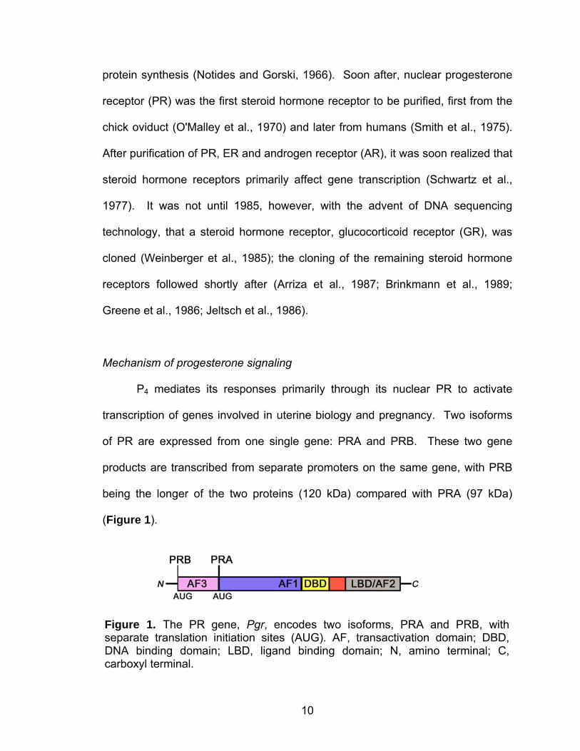
transcription of genes involved in uterine biology and pregnancy. Two isoforms
of PR are expressed from one single gene: PRA and PRB. These two gene
products are transcribed from separate promoters on the same gene, with PRB
being the longer of the two proteins (120 kDa) compared with PRA (97 kDa)
(Figure 1).
Figure 1. The PR gene, Pgr, encodes two isoforms, PRA and PRB, with separate translation initiation sites (AUG). AF, transactivation domain; DBD, DNA binding domain; LBD, ligand binding domain; N, amino terminal; C, carboxyl terminal.
11
The amino terminal portion of both isoforms is the most hypervariable region and
contains the transactivation domains, AF3 (PRB only) and AF1 (PRA and PRB),
responsible for the recruitment of coactivators and/or corepressors during
transcriptional regulation. The DNA-binding domain (DBD) is the most
conserved region, composed of two type II zinc finger structures responsible for
binding to specific cis-acting DNA sequences (Freedman, 1992; Luisi et al.,
1991). The carboxyl terminal region contains the ligand-binding domain (LBD)
with an additional site, AF2, for interaction with transcriptional coregulators. PR
interactions with heat shock proteins and other chaperones to localize to the
LBD/AF2 (Schowalter et al., 1991). The larger segment of PRB, referred to as
the B upstream sequence (Kelleher et al., 1999), contains the AF3 domain, which
is thought to attenuate PRB activity. For this reason, PRB tends to be less
transcriptionally active than the PRA isoform. In fact, it is thought that PRA acts
as a dominant repressor of PRB, evidenced by coexpression studies in cultured
cells (Kraus et al., 1995; Vegeto et al., 1993). PRA and PRB proteins also
respond differentially to P4 antagonists, e.g. RU-486, in that antagonist-bound
PRA is inactive, while antagonist-bound PRB has increased transcriptional
activity (Conneely et al., 2001). When expressed in equimolar concentrations in
cells, PRA and PRB can homodimerize or heterodimerize, essentially existing as
three entities: A:A, B:B, A:B. It is thought that each entity then will exhibit
differential activity in regulating downstream target genes.
Mice lacking the Pgr gene encoding both PR isoforms have illustrated the
essential roles of P4 signaling through its nuclear steroid hormone receptor
12
during reproductive processes. Female mice lacking both PR isoforms have
complete ovulation, fertilization, and implantation failure as well as impaired
sexual behavior, defective responses to gonadotropins, and defective ductal
branching morphogenesis and lobuloalveolar differentiation of the mammary
gland (Chappell et al., 1997; Lydon et al., 1995). P4 signaling through PR has
also been shown to play a role in decidualization and maintaining the uterus in a
quiescent state until delivery (Dey et al., 2004).
Mouse models lacking either PRA or PRB have been generated using the
CRE-loxP system to introduce a point mutation at either of the two ATG
translation initiation codons (Mulac-Jericevic et al., 2003; Mulac-Jericevic et al.,
2000). While PgrA null female mice are infertile due to severe abnormalities in
ovarian and uterine functions, PgrB null female mice show no apparent defects in
ovarian and uterine functions but have significantly reduced mammary ductal
side-branching and alveologenesis during pregnancy. These findings suggest
that PRA and PRB act in a tissue-selective manner to regulate their specific
actions, and also imply that PRA:PRB heterodimers are not required for uterine
reproductive events.
It is also thought that P4 can signal through a membrane receptor;
however, the contribution of this signaling pathway to specific physiological
processes has not yet been conclusively demonstrated. A role for this signaling
was first shown in Xenopus oocytes. Quiescent Xenopus oocytes remain
arrested in meiosis I until exposure to P4, which triggers rapid non-transcriptional
responses rather than the conventional transcriptional activation known to be a
13
signature of nuclear steroid hormone receptor signaling. Using the Xenopus
model, one study identified XPR1 as a membrane PR necessary for Xenopus
oocyte activation (Tian et al., 2000). Zhu and colleagues cloned a membrane
progestin receptor from spotted sea trout ovaries, characterizing it as a G-protein
coupled receptor expressed solely in the brain and reproductive organs that
activates mitogen-activated protein kinase (MAPK) signaling pathways upon
progestin binding (Zhu et al., 2003). Since then, three forms of putative
membrane-bound progestin receptors have been identified in channel catfish
(mPRα, β, and γ) (Kazeto et al., 2005), and another group has identified an
adrenal cortex protein, inner zone antigen (IZAg), as a putative membrane PR
(Raza et al., 2001). In the mouse, two potential membrane PRs are thought to
play roles particularly in ovarian cells: membrane progesterone receptor (MPR)
and PGR membrane component I (PGRMC1) (Peluso et al., 2006; Zhu et al.,
2003). While MPRs and PGRMC1 are expressed in various cell types of the
ovary, their roles in ovarian biology remain to be determined (Peluso et al.,
2006). Whether a role for these membrane PRs exists in uterine biology also
remains unexplored.
Progesterone-regulated target genes during early pregnancy
On day 4 of pregnancy in mice, the uterus is primarily under the influence
of P4, and P4-target genes activated through PR include homeobox transcription
factors, growth factors, and cytokines (Dey et al., 2004). These genes show
robust expression on this day of pregnancy. Specific examples of P4-regulated
14
or responsive genes include amphiregulin (Areg), histidine decarboxylase (Hdc),
Indian hedgehog (Ihh), and Hoxa-10.
Amphiregulin is a glycosylated heparin-binding protein and a member of
the epidermal growth factor (EGF) family. Under the influence of P4, Areg is
expressed in the luminal and glandular epithelia (Das et al., 1995) and is
regulated by P4 working via the PRA isoform (Mulac-Jericevic et al., 2003).
While amphiregulin is associated with uterine cell-specific differentiation and
proliferation, mice lacking the Areg gene appear to have normal fertility (Luetteke
et al., 1999). This could result from compensation by other EGF family
members, but precludes amphiregulin as an essential factor for implantation.
Another P4-regulated gene upregulated by P4 working through the PRA isoform is
histidine decarboxylase (Hdc) (Mulac-Jericevic et al., 2000). This gene is
expressed primarily in the epithelium on day 4 of pregnancy (Paria et al., 1998),
but its role in implantation biology has yet to be determined.
The components of the hedgehog signaling pathway are influenced by P4,
specifically those of Indian hedgehog (IHH) including the hedgehog-binding
receptor, Patched, and its downstream transcription factor Gli1-3 (Matsumoto et
al., 2002). Expression of Ihh increases in the luminal and glandular epithelium
from day 3 of pregnancy onward. During the same time, the expression of its
receptor and downstream transcription factors is upregulated in the underlying
stroma. This suggests that IHH functions as a paracrine growth factor for stromal
cell proliferation during early pregnancy. Roles for hedgehog signaling have
15
also been delineated in cancer, and recent evidence indicates roles for this
conserved signaling pathway in uterine biology (Lee et al., 2006).
Hoxa10 encodes a transcription factor that belongs to a multigene family.
Hox genes are developmentally regulated and share a common highly conserved
sequence element called the homeobox that encodes a helix-turn-helix DNA
binding domain (Krumlauf, 1994). Hox genes are organized into four clusters (A,
B, C, and D) on four different chromosomes in mice and humans and follow a
stringent pattern of spatial and temporal colinearity during embryogenesis
(Krumlauf, 1994). Specifically, genes at the 3’-end of each cluster are activated
during early embryogenesis in the anterior region of the developing embryo,
whereas genes located toward the 5’-end are restricted to posterior regions of
the embryo and are expressed during later stages of embryogenesis (Krumlauf,
1994). Hoxa10 is located at the 5’end of the A cluster and is classified as an
AbdB-like Hox gene because of its homology with the Drosophila AbdB gene.
The Abd genes constitute a distinct subfamily of homeobox genes that exhibit
posterior domains of expression including the genital imaginal disc in Drosophila
and the developing genitourinary system in mammals (Dolle et al., 1991; Izpisua-
Belmonte et al., 1991). Indeed, Hoxa10 is highly expressed in the developing
genitourinary tract and adult female reproductive tract, suggesting its roles in
reproductive events (Benson et al., 1996; Gendron et al., 1997; Hsieh-Li et al.,
1995).
16
Hoxa-10 null mice reveal FKBP52 as a critical mediator of implantation
Definitive evidence for roles of various genes in implantation and/or
decidualization has been demonstrated with an array of mouse gene knockout
models. Hoxa10 is strongly expressed in the mouse uterine stroma during the
receptive phase (day 4), implantation (day 5) and decidualization (day 6)
(Satokata et al., 1995). Indeed, failure of decidualization in Hoxa10 null uteri
leads to pregnancy failure (Benson et al., 1996). Uterine stromal cells isolated
from Hoxa10 null uteri show reduced proliferation in response to P4 contributing
to decidualization defects, while epithelial cell proliferation remains normal in
response to estrogen (Benson et al., 1996; Lim et al., 1999). Several P4-
responsive genes are dysregulated in uterine stroma of Hoxa10 null uteri (Lim et
al., 1999), suggesting that HOXA10 conveys P4 responsiveness by regulating
gene expression as a transcription factor. Furthermore, P4 is a strong inducer of
Hoxa10 in mouse uterine stroma, inducing its expression within 4 h post-P4
injection. This upregulation is attenuated by the PR antagonist RU-486,
suggesting a requirement for PR in Hoxa10 induction.
Since the mechanism by which HOXA10 regulates uterine stromal cell
proliferation and differentiation during uterine receptivity, implantation, and
decidualization remained unexplored, we used proteomics approach to identify
downstream targets of HOXA10 in the periimplantation uterus. Specifically, we
isolated stromal cells from wild-type and Hoxa10 null uteri, cultured them for 24
h, and then analyzed them by 2D DIGE (two-dimensional difference gel
electrophoresis). More than 1000 proteins were resolved with isoelectric points
17
between pH 4 and 7 and molecular masses between 10 and 150 kDa. Among
these, less than 4% (n=36) of the detected features displayed changes in
abundance that were consistent across all three measurements, supported by
Student’s t-test P values within the 99th percentile confidence interval (P<0.01,
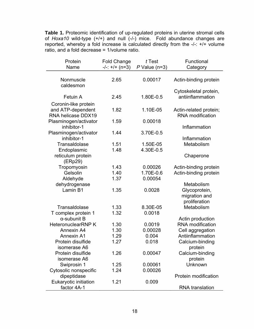
n=3) for the variance of mean changes. Twenty-two of the identified proteins
showed upregulation in Hoxa10 null stromal cells that primarily represented
cytoskeletal, metabolic, and actin-related proteins (Table 1) (Daikoku et al.,
2005).
18
Table 1. Proteomic identification of up-regulated proteins in uterine stromal cells of Hoxa10 wild-type (+/+) and null (-/-) mice. Fold abundance changes are reported, whereby a fold increase is calculated directly from the -/-: +/+ volume ratio, and a fold decrease = 1/volume ratio.
Protein Name
Fold Change-/-: +/+ (n=3)
t Test P Value (n=3)
Functional Category
Nonmuscle caldesmon
2.65
0.00017
Actin-binding protein
Fetuin A
2.45
1.80E-0.5
Cytoskeletal protein, antiinflammation
Coronin-like protein and ATP-dependent
RNA helicase DDX19
1.82
1.10E-05
Actin-related protein;
RNA modification Plasminogen/activator
inhibitor-1 1.59 0.00018
Inflammation Plasminogen/activator
inhibitor-1 1.44 3.70E-0.5
Inflammation Transaldolase 1.51 1.50E-05 Metabolism Endoplasmic
reticulum protein (ERp29)
1.48 4.30E-0.5 Chaperone
Tropomyosin 1.43 0.00026 Actin-binding protein Gelsolin 1.40 1.70E-0.6 Actin-binding protein
Aldehyde dehydrogenase
1.37 0.00054 Metabolism
Lamin B1 1.35 0.0028 Glycoprotein, migration and proliferation
Transaldolase 1.33 8.30E-05 Metabolism T complex protein 1
α-subunit B 1.32 0.0018
Actin production HeteronuclearRNP K 1.30 0.0019 RNA modification
Annexin A4 1.30 0.00028 Cell aggregation Annexin A1 1.29 0.004 Antiinflammation
Protein disulfide isomerase A6
1.27 0.018 Calcium-binding protein
Protein disulfide isomerase A6
1.26 0.00047 Calcium-binding protein
Swiprosin 1 1.25 0.00061 Unknown Cytosolic nonspecific
dipeptidase 1.24 0.00026
Protein modification Eukaryotic initiation
factor 4A-1 1.21 0.009
RNA translation
19
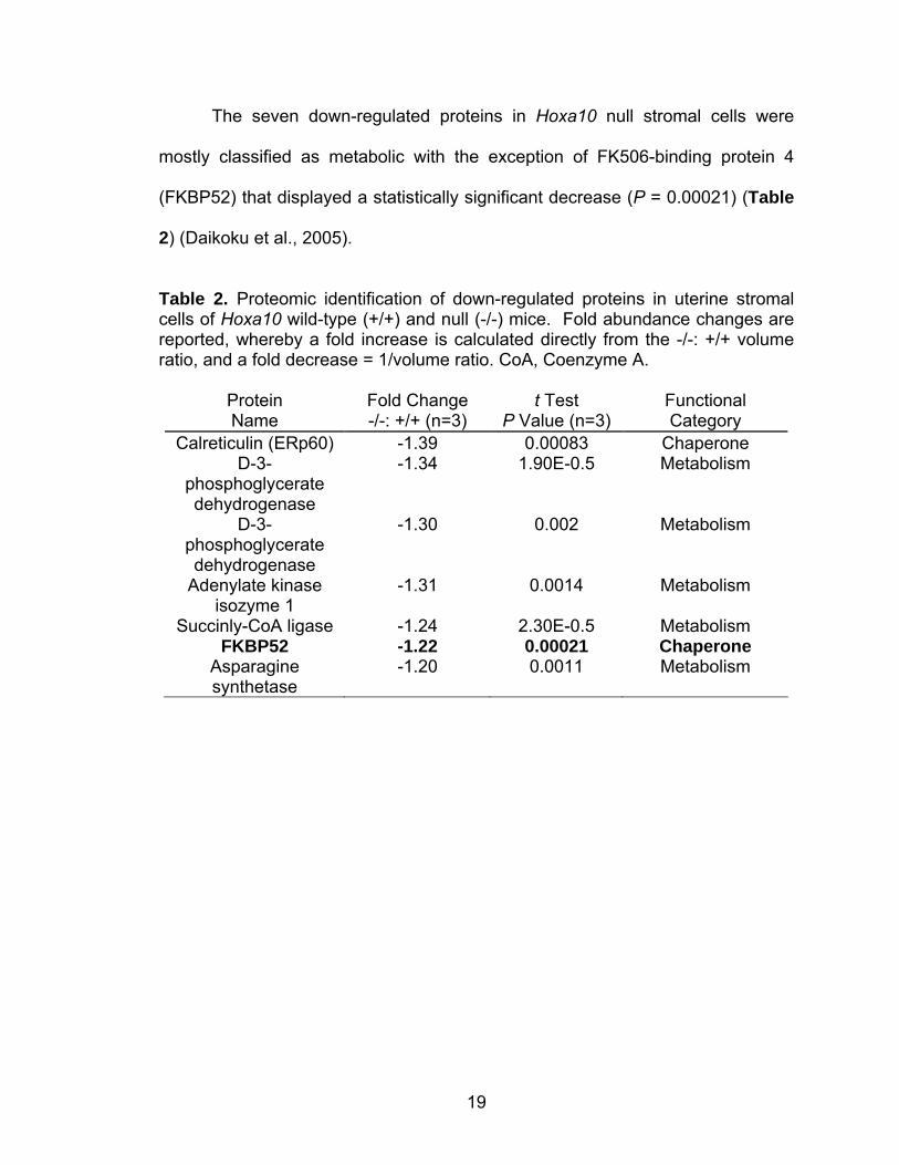
The seven down-regulated proteins in Hoxa10 null stromal cells were
mostly classified as metabolic with the exception of FK506-binding protein 4
(FKBP52) that displayed a statistically significant decrease (P = 0.00021) (Table
2) (Daikoku et al., 2005).
Table 2. Proteomic identification of down-regulated proteins in uterine stromal cells of Hoxa10 wild-type (+/+) and null (-/-) mice. Fold abundance changes are reported, whereby a fold increase is calculated directly from the -/-: +/+ volume ratio, and a fold decrease = 1/volume ratio. CoA, Coenzyme A.
Protein Name
Fold Change -/-: +/+ (n=3)
t Test P Value (n=3)
Functional Category
Calreticulin (ERp60) -1.39 0.00083 Chaperone D-3-
phosphoglycerate dehydrogenase
-1.34 1.90E-0.5 Metabolism
D-3-phosphoglycerate dehydrogenase
-1.30 0.002 Metabolism
Adenylate kinase isozyme 1
-1.31 0.0014 Metabolism
Succinly-CoA ligase -1.24 2.30E-0.5 Metabolism FKBP52 -1.22 0.00021 Chaperone
Asparagine synthetase
-1.20 0.0011 Metabolism
20
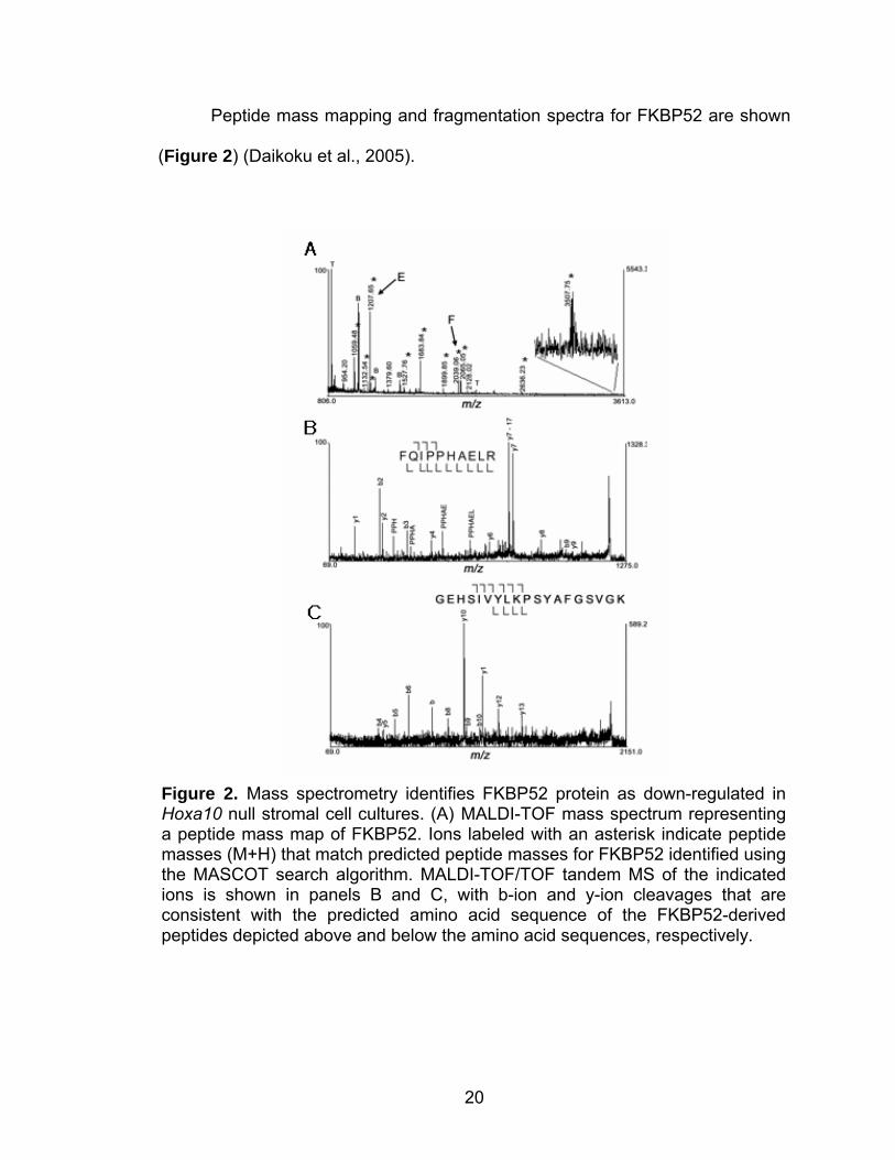
Peptide mass mapping and fragmentation spectra for FKBP52 are shown
(Figure 2) (Daikoku et al., 2005).
Figure 2. Mass spectrometry identifies FKBP52 protein as down-regulated in Hoxa10 null stromal cell cultures. (A) MALDI-TOF mass spectrum representing a peptide mass map of FKBP52. Ions labeled with an asterisk indicate peptide masses (M+H) that match predicted peptide masses for FKBP52 identified using the MASCOT search algorithm. MALDI-TOF/TOF tandem MS of the indicated ions is shown in panels B and C, with b-ion and y-ion cleavages that are consistent with the predicted amino acid sequence of the FKBP52-derived peptides depicted above and below the amino acid sequences, respectively.
21
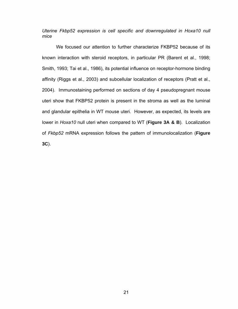
Uterine Fkbp52 expression is cell specific and downregulated in Hoxa10 null mice
We focused our attention to further characterize FKBP52 because of its
known interaction with steroid receptors, in particular PR (Barent et al., 1998;
Smith, 1993; Tai et al., 1986), its potential influence on receptor-hormone binding
affinity (Riggs et al., 2003) and subcellular localization of receptors (Pratt et al.,
2004). Immunostaining performed on sections of day 4 pseudopregnant mouse
uteri show that FKBP52 protein is present in the stroma as well as the luminal
and glandular epithelia in WT mouse uteri. However, as expected, its levels are
lower in Hoxa10 null uteri when compared to WT (Figure 3A & B). Localization
of Fkbp52 mRNA expression follows the pattern of immunolocalization (Figure
3C).
22
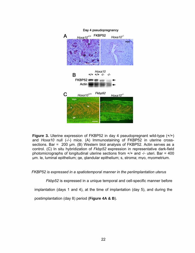
FKBP52 is expressed in a spatiotemporal manner in the periimplantation uterus
Fkbp52 is expressed in a unique temporal and cell-specific manner before
implantation (days 1 and 4), at the time of implantation (day 5), and during the
postimplantation (day 8) period (Figure 4A & B).
Figure 3. Uterine expression of FKBP52 in day 4 pseudopregnant wild-type (+/+) and Hoxa10 null (-/-) mice. (A) Immunostaining of FKBP52 in uterine cross-sections. Bar = 200 µm. (B) Western blot analysis of FKBP52. Actin serves as a control. (C) In situ hybridization of Fkbp52 expression in representative dark-field photomicrographs of longitudinal uterine sections from +/+ and -/- uteri. Bar = 400 µm. le, luminal epithelium; ge, glandular epithelium; s, stroma; myo, myometrium.
23
As mentioned, the uterus is under the influence of preovulatory estrogen
on day 1 of pregnancy or pseudopregnancy with heightened cell proliferation in
the epithelium. In contrast, the day 4 uterus is exposed to rising levels of P4 from
the newly formed corpora lutea and fortified with a small amount of estrogen that
results in epithelial cell differentiation with stromal cell proliferation. We observed
that Fkbp52 mRNA is detected mostly in the luminal epithelium on day 1 of
Figure 4. Uterine expression of FKBP52 in wild-type mouse uteri during early pregnancy. (A) In situ hybridization of Fkbp52. Hybridization signals in representative dark-field photomicrographs of longitudinal uterine sections on day 1 and day 4 and cross-sections on days 5 and 8 are shown. Bar = 400 µm. le, luminal epithelium; ge, glandular epithelium; s, stroma; myo, myometrium; m, mesometrial pole; am, anti-mesometrial pole; Dc, decidua. Arrows indicate location of embryo. (B) Western blotting of FKBP52 protein. Actin serves as a loading control.
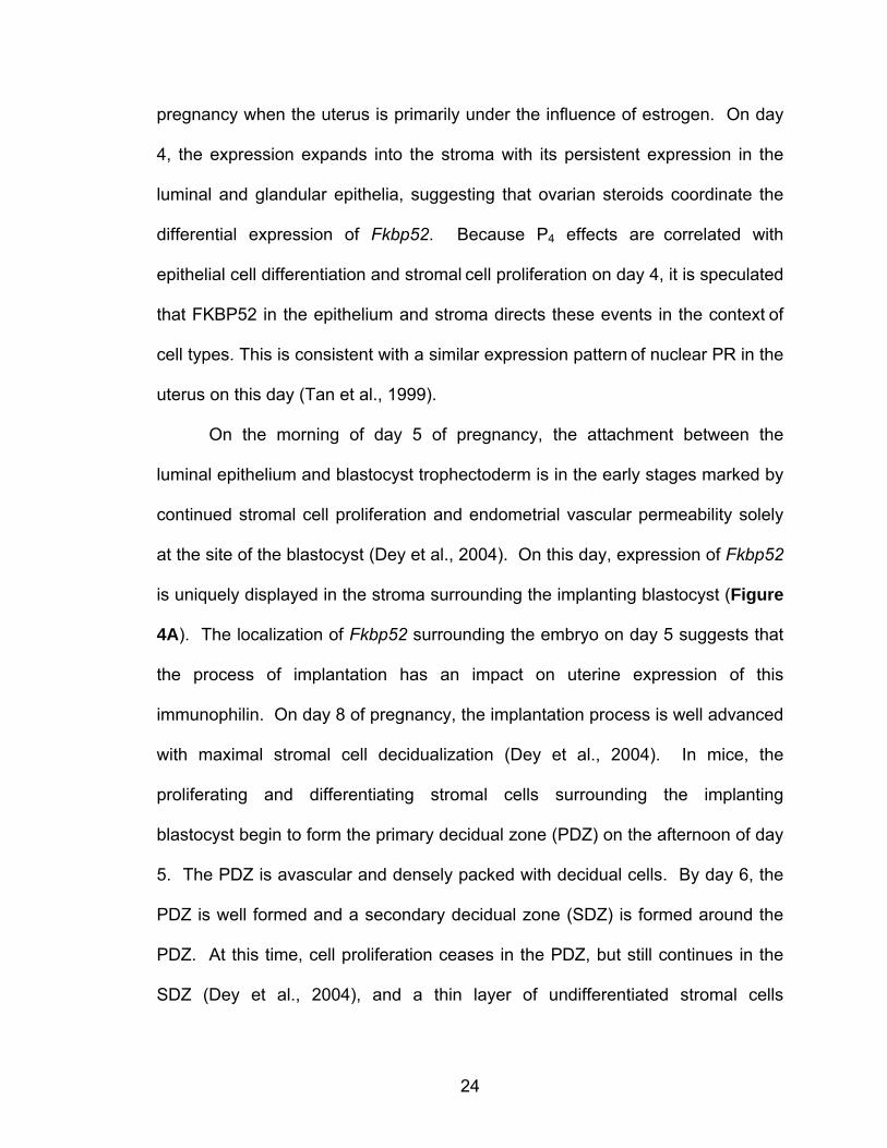
24
pregnancy when the uterus is primarily under the influence of estrogen. On day
4, the expression expands into the stroma with its persistent expression in the
luminal and glandular epithelia, suggesting that ovarian steroids coordinate the
differential expression of Fkbp52. Because P4 effects are correlated with
epithelial cell differentiation and stromal cell proliferation on day 4, it is speculated
that FKBP52 in the epithelium and stroma directs these events in the context of
cell types. This is consistent with a similar expression pattern of nuclear PR in the
uterus on this day (Tan et al., 1999).
On the morning of day 5 of pregnancy, the attachment between the
luminal epithelium and blastocyst trophectoderm is in the early stages marked by
continued stromal cell proliferation and endometrial vascular permeability solely
at the site of the blastocyst (Dey et al., 2004). On this day, expression of Fkbp52
is uniquely displayed in the stroma surrounding the implanting blastocyst (Figure
4A). The localization of Fkbp52 surrounding the embryo on day 5 suggests that
the process of implantation has an impact on uterine expression of this
immunophilin. On day 8 of pregnancy, the implantation process is well advanced
with maximal stromal cell decidualization (Dey et al., 2004). In mice, the
proliferating and differentiating stromal cells surrounding the implanting
blastocyst begin to form the primary decidual zone (PDZ) on the afternoon of day
5. The PDZ is avascular and densely packed with decidual cells. By day 6, the
PDZ is well formed and a secondary decidual zone (SDZ) is formed around the
PDZ. At this time, cell proliferation ceases in the PDZ, but still continues in the
SDZ (Dey et al., 2004), and a thin layer of undifferentiated stromal cells
25
establishes a boundary between the myometrium and the SDZ. The PDZ
progressively degenerates through day 8; at this point, placental and embryonic
growth gradually replaces the SDZ, which is reduced to a thin layer of cells called
the decidua capsularis. The mesometrial decidual cells eventually form the
decidua basalis (Dey et al., 2004). On day 8, expression of Fkbp52 dramatically
changes, lying in peripheral decidualizing cells more specifically at the
mesometrial SDZ and the undifferentiated stroma situated between the deciduum
and myometrium. Interestingly, Fkbp52 is also evident in the growing embryo on
this day (Figure 4A). Western blotting confirms translation of Fkbp52 mRNA
during these days of pregnancy (Figure 4B). Collectively, these results suggest
that ovarian steroid hormones and implantation events differentially affect uterine
Fkbp52 expression.
Progesterone and estrogen differentially regulate Fkbp52 expression in the uterus
Our results showing differential expression of Fkbp52 on days 1 and 4 of
pregnancy suggested that this gene is regulated in the uterus by ovarian steroids
in a cell-specific manner. Indeed, in situ hybridization was performed to
determine the uterine cell-specific expression of Fkbp52 in response to the
ovarian steroid hormones P4 and/or estrogen (Figure 5).
26
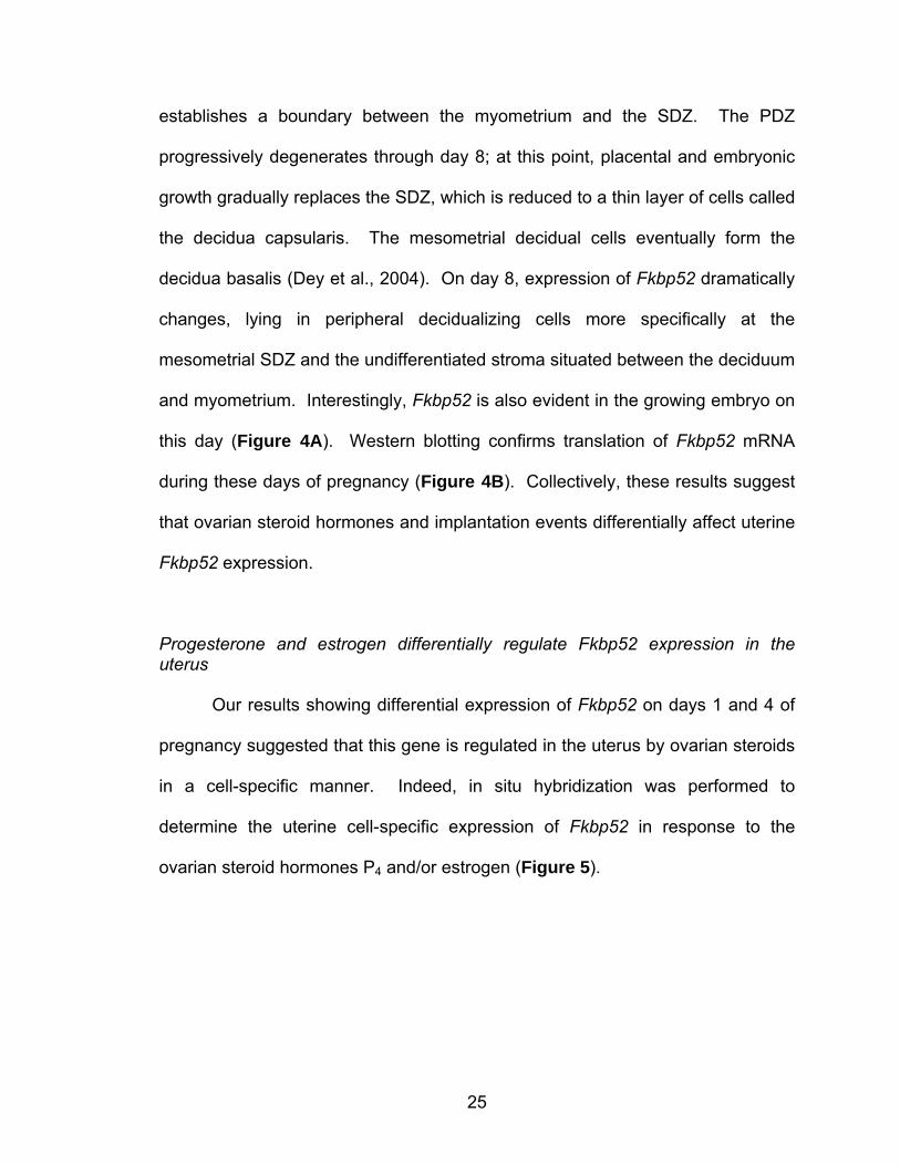
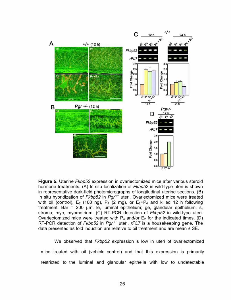
We observed that Fkbp52 expression is low in uteri of ovariectomized
mice treated with oil (vehicle control) and that this expression is primarily
restricted to the luminal and glandular epithelia with low to undetectable
Figure 5. Uterine Fkbp52 expression in ovariectomized mice after various steroid hormone treatments. (A) In situ localization of Fkbp52 in wild-type uteri is shown in representative dark-field photomicrographs of longitudinal uterine sections. (B) In situ hybridization of Fkbp52 in Pgr–/– uteri. Ovariectomized mice were treated with oil (control), E2 (100 ng), P4 (2 mg), or E2+P4 and killed 12 h following treatment. Bar = 200 µm. le, luminal epithelium; ge, glandular epithelium; s, stroma; myo, myometrium. (C) RT-PCR detection of Fkbp52 in wild-type uteri. Ovariectomized mice were treated with P4 and/or E2 for the indicated times. (D) RT-PCR detection of Fkbp52 in Pgr–/– uteri. rPL7 is a housekeeping gene. The data presented as fold induction are relative to oil treatment and are mean ± SE.
27
expression in the stroma. An injection of estradiol-17β (E2, 100 ng/0.1 ml/mouse)
upregulated Fkbp52 expression levels by 12 h with the pattern remaining
unaltered. In contrast, an injection of P4 (2 mg/0.1 ml/mouse) showed a
prominent shift in Fkbp52 expression from the luminal epithelium to the stromal
compartment at 12 h and expression was downregulated by 24 h (Figure 5A). A
combined injection of E2 and P4 showed an expression pattern similar to that of
P4 alone but at somewhat lower levels (Figure 5A). Interestingly, P4 treatment
markedly downregulated E2-induced epithelial Fkbp52 expression. Comparative
RT-PCR analysis of total RNA isolated from uteri harvested from similarly treated
mice confirmed our in situ hybridization results, showing an increase in Fkbp52
expression in E2, P4, and E2 + P4 treatment at 12 h with a return to basal levels at
24 h (Figure 5C).
We next used mice lacking nuclear PR to further define the mechanism of
steroidal regulation of Fkbp52. The induction of stromal Fkbp52 expression that
we observed in P4-treated wild-type mice was virtually abolished in Pgr null mice
(Figure 5B). However, the E2 response remained intact, showing a similar
increase in Fkbp52 expression in the luminal epithelium as was observed in WT
uteri (Figure 5B). In situ hybridization results were further confirmed by
comparative RT-PCR analysis of total RNA isolated from uteri harvested from
similarly treated Pgr null mice, showing an increase in Fkbp52 expression with E2
treatment, but no such increase in ovariectomized Pgr null mice treated with P4
(Figure 5D). Collectively, these results suggest that steroid hormones regulate
Fkbp52 expression differentially and in a cell-specific manner. This observation of

28
differential regulation of FKBP52 by estrogen and P4 points to a role for FKBP52
in epithelial-stromal cross-talk to mediate full complement of uterine functions in
response to ovarian steroid hormones.
29
CHAPTER II
COCHAPERONE IMMUNOPHILIN FKBP52 IS CRITICAL TO UTERINE RECEPTIVITY AND IMPLANTATION
Abstract
Embryo implantation in the uterus is a critical step in mammalian
reproduction, requiring preparation of the uterus receptive to blastocyst
implantation. Uterine receptivity lasts for a limited period, and it is during this
period that blastocysts normally implant. Ovarian steroid hormones estrogen and
P4 are the primary regulators of this process. FKBP52, an immunophilin, serves
as a cochaperone for steroid hormone nuclear receptors to govern appropriate
hormone action in target tissues. Here we show a critical role for FKBP52 in
mouse implantation. This immunophilin has unique spatiotemporal expression in
the uterus during implantation, and females missing the Fkbp52 gene have
complete implantation failure due to the lack of attainment of uterine receptivity.
The overlapping uterine expression of FKBP52 with nuclear PR in wild-type mice
together with reduced P4 binding to PR, attenuated PR transcriptional activity and
downregulation of several P4-regulated genes in uteri of Fkbp52 null mice,
establishes this cochaperone as a critical regulator of uterine P4 function.
Interestingly, ovulation, another P4-mediated event, remains normal in Fkbp52
null animals. Collectively, the present investigation provides evidence for an in
vivo role for this cochaperone in regulating tissue-specific hormone action and its
critical role in uterine receptivity for implantation.
30
Introduction
Immunophilins are aptly named due to their ability to bind and mediate
actions of certain immunosuppressive drugs. Immunophilins are grouped into two
protein families: the FK506-binding proteins (FKBPs) and the cyclosporin A
binding proteins termed cyclophilins (CyPs). FKBP and CyP typically exhibit
peptidylprolyl cis-trans isomerase (PPIase) activity, and this PPIase domain also
forms the drug-binding site (Fanghanel and Fischer, 2004). Some members of
the FKBP and CyP families contain a tetratricopeptide repeat (TPR) domain that
targets binding to the highly conserved C-terminal end of heat shock protein 90
(Hsp90), and thus participate as cochaperones with Hsp90 in its substrate
interactions.
FKBP52 and its related immunophilins FKBP51 and CyP40 were initially
characterized as Hsp90-binding proteins because of these conserved TPR
domains (Pratt and Toft, 1997). Hsp90 is bound with high affinity to the PR
complex in an ATP-bound state. ATP binding changes the conformation of the
protein and therefore, the ATP turnover rate determines the amount of time
Hsp90 remains bound to its target protein. This turnover rate is regulated by
Hsp90’s association with cochaperones, one being p23. This small, acidic co-
chaperone is known to inhibit the ATPase activity of human Hsp90, and this
inhibition is shown to be more robust in the presence of FKBP52 (McLaughlin et
al., 2002); however, while various roles for p23 in these complexes have been
proposed, its precise function and the molecular mechanism by which p23
mediates its functions are unknown.
31
The pathway for assembly of steroid receptor/chaperone complexes is
complex, involving a variety of Hsps and cochaperones. For PR specifically, it is
thought that this steroid hormone nuclear receptor first binds to Hsp40 and
Hsp70 in the cytoplasm of the cell. This is followed by the binding of Hop-Hsp90
to the bound Hsp70 (Hernandez et al., 2002). Hsp70 and Hop then dissociate
from this intermediate PR complex, while p23 is recruited to receptor-bound
Hsp90. The PR complex with Hsp90 and p23 bound then recruits one of the
cochaperone immunophilins (FKBP51, FKBP52, CyP40, or protein phosphatase
5 (PP5)) to an Hsp90 binding site. For PR, if this mature complex is bound to
FKBP52, then it is capable of optimally binding P4 ligand. The mature complex
containing Hsp90 must be maintained so that the receptor can bind P4 with high
affinity and efficiency. Recent reports indicate that each steroid hormone nuclear
receptor (GR, ER, and PR)-Hsp90 complex displays preferential binding with
specific immunophilins (Barent et al., 1998).
FKBP52 and FKBP51 share a common binding site on Hsp90 and
therefore compete for assembly with steroid receptor complexes (Riggs et al.,
2003). The PR heterocomplex, however, preferentially assembles with FKBP51
despite its lower concentration in the cell (20 nM) when compared to FKBP52
(100 nM) (Schiene-Fischer and Yu, 2001). FKBP52 elevates the hormone
binding affinity of PR in an Hsp90- and PPIase-dependent manner, while
FKBP51 antagonizes the actions of FKBP52 and reduces PR binding affinity for
ligand, thereby reducing secondary responses to hormones (Riggs et al., 2003).
32
It is surprising that functional differences exist between these two immunophilins,
given the 70% homology in their amino acid sequences (Nair et al., 1997).
It has been suggested that while FKBP52 is necessary for potentiating PR
activity through optimal ligand binding, basal PR activity can exist without
FKBP52 presence, albeit not as efficiently. The mechanism by which FKBP52
potentiates receptor hormone-binding ability is still unclear. Point mutation of the
FKBP52 TPR domain demonstrates that an association of FKBP52 with Hsp90 is
critical (Riggs et al., 2003), suggesting that FKBP52 is obligatorily recruited to the
receptor complex by Hsp90. In addition, point mutation of the FKBP52 PPIase
domain or addition of the PPIase inhibitor, FK506, blocks receptor potentiation,
which perhaps reflects a direct interaction between the PPIase domain and
receptor sites. FKBP52 and FKBP51 have similar PPIase activity towards small
peptide substrates (Pirkl and Buchner, 2001), and their respective PPIase
domains share 80% sequence similarity; yet, FKBP51 fails to potentiate receptor
activity. Chimeric constructs in which the PPIase domain has been exchanged
confirms that the FKBP52 PPIase domain is unique in its ability to stimulate
receptor activity (Riggs et al., 2003). However, the receptor site(s) putatively
targeted by the FKBP52 PPIase domain have not been identified although it is
known that they localize to the LBD (Riggs et al., 2003). Mapping of specific
amino acid differences within the PPIase domains of FKBP52 and FKBP51
should provide further insight into mechanism(s) of receptor potentiation or
inhibition.
33
There is additional evidence that FKBP52, through a dynein-binding ability
lacking in FKBP51, can enhance nuclear translocation of hormone-bound GR
(Davies et al., 2002; Pratt et al., 2004; Wochnik et al., 2005). This role for
FKBP52 is based on evidence that FKBP52 binds both a nuclear localization
signal (NLS) and dynein (Czar et al., 1995; Galigniana et al., 2004; Silverstein et
al., 1999), and on in vitro studies demonstrating that FKBP52 facilitates the
nuclear transport of both GR and p53 (Galigniana et al., 2004; Galigniana et al.,
2001; Wochnik et al., 2005). Evidence suggests that FKBP52 binds dynein
directly through its PPIase domain (Silverstein et al., 1999). It is debated
whether FKBP52 facilitates a similar translocation of PR (Silverstein et al., 1999),
and it is not yet clear what contribution dynein associations play relative to direct
FKBP-mediated effects on hormone binding, especially with PR and other
receptors that prior to hormone binding have a more predominant nuclear
localization than GR. Steroid hormone binding prevents reinitiation of the
heterocomplex assembly cycle in the cytoplasm, and while it is known that
hormone binding is followed by heterocomplex dissociation, it is not known
whether the complex dissociates during nuclear transport or within the nucleus.
Once in the nucleus, however, as described earlier, nuclear PR forms
homodimers to act as a transcription factor to either activate gene transcription
by binding to its cognate DNA elements or to inhibit transcription through
interaction with other transcription factors.
34
The purpose of this next study, therefore, was to examine a physiological
role for FKBP52 and FKBP51 in an in vivo context, since roles for these
cochaperones had not been examined with respect to P4-dependent processes.
Methods
Generation of Fkbp52 null mice
ES cells isolated from 129SvJ mouse were cultured in Knockout DMEM
(Invitrogen, Carlsbad, CA) supplemented with 10% FBS, penicillin/streptomycin,
essential amino acids, ESGRO (103 U/ml; Chemicon, Temecula, CA) with
irradiated embryonic fibroblast feeder cells. ES cells were electroporated at 0.2
kV, 950 µF (Gene Pulser II; Bio-Rad, Hercules, CA) with linearized targeting
vectors and selected with G418 (300 µg/ml). DNA from G418-resistant clones
was isolated for Southern blot analysis. A DNA probe was used to distinguish
EcoRI restriction fragments from wild-type (17 kb) and mutant (9 kb) alleles.
Appropriate homologous recombination in ES cell clones was confirmed by PCR
using primers complementary to sequences within the neomycin cassette and to
3' Fkbp52 sequences downstream from the recombination site. ES cell clones
containing a mutant Fkbp52 allele were injected into C57BL6/129 blastocysts and
implanted into pseudopregnant 129SvJ females. Chimeric offspring were
identified by coat patterns and mated to C57BL6/129 mice to obtain germline
transmission of the mutant allele. These mice were created by David F. Smith
(Mayo Clinic, Scottsdale, AZ) and are currently maintained in our animal facility
35
at Vanderbilt University (Nashville, TN). All mice were housed and used in the
current investigation in accordance with the National Institutes of Health and
institutional guidelines on the care and use of laboratory animals.
Mouse genotyping
To obtain genomic DNA, tail pieces (5–8 mm) collected from weaned mice
were placed in 200 µl DirectPCR Lysis Reagent (Viagen Biotech Inc., Los
Angeles, CA) containing freshly prepared 0.2-0.4 mg/ml Proteinase K (Sigma, St.
Louis, MO). Tails were placed in 55oC water bath for > 6 h. Lysates were then
incubated at 85oC for 45 min, spun down quickly for 10 sec, and stored at 4oC
until use. Genotypes were determined by PCR using specific primers: neoF2, 5’-
TCT ATC GCC TTC TTG ACG-3’; ex2F, 5’-AGA GAG GGT ACA GC-3’; ex3R,
5’-TAC AAG TGT GGC GTT GGG-3’; ex10R, 5’-ATG CAA CAG CGG TGT ACC
C-3’. These are specific for the wild-type (1 kb product) or mutant (700 bp
product) alleles. Cycle conditions are the following: 1 cycle of 95oC for 5 min,
followed by 40 cycles of 95oC for 30 sec, 55oC for 30 sec, 68oC for 2.5 min,
followed by 68oC for 10 min. Master mix contained 10x LA Taq PCR buffer, 2.5
mM dNTP mixture, 10 µM primer mix, and LA Taq polymerase (Takara Bio Inc,
Shiga, Japan). Amplified fragments were separated by electrophoresis on 2%
agarose gels and visualized by ethidium bromide staining.
36
Ovulation, fertilization, implantation, and blastocyst transfer
To examine ovulation and fertilization, wild-type or Fkbp52-/- mice were
bred with fertile males. The morning of finding a vaginal plug was designated day
1 of pregnancy. Mice were killed on day 2 of pregnancy and oviducts were
flushed with Whitten’s medium to recover eggs and embryos. Egg and embryo
morphology was examined under a dissecting microscope. Implantation sites on
days 5 and 6 of pregnancy were visualized by an intravenous injection (0.1 ml per
mouse) of a Chicago Blue B dye solution (1% solution in saline) and the number
of implantation sites, as demarcated by distinct blue bands, was recorded. For
blastocyst transfer, pseudopregnant recipients were generated by mating females
with vasectomized males. Day 4 wild-type blastocysts were transferred into day 4
uteri of wild-type, heterozygous or Fkbp52-/- pseudopregnant recipients, and
implantation sites were recorded 24 h later (day 5) by the blue dye method. All
mice used were between 2 and 4 months of age.
Comparative RT-PCR and southern hybridization
Total RNA was extracted from mouse uteri using Trizol according to the
manufacturer’s instruction. Reverse transcription with oligo(dT) priming was
performed to generate cDNAs from 4 µg total RNA using Superscript II following
the instruction provided by the manufacturer. DNA amplification was carried out
with Taq DNA polymerase (Invitrogen, San Diego, CA) using the following
primers: Fkbp52 (437 bp), 5'-AGT GTG GGG AAG GAG AGG TT-3' and 5'-GCT
CTT GCC AGG TCA AAG TC-3'; Pgr (461 bp), 5'-GCC ATC ACT TCC TGG TGT
37
CT -3' and 5'-GCA ATG GGA GAG TCT TGC TC-3'; Fkbp51 (402 bp), 5’-AAG
GTG TTG GCA GTC AAT CC-3’ and 5’-GGT GGT CAT TTG GGA AGC TA-3’; β-
actin (246 bp), 5’-CTC TTT GAT GTC ACG CAC GAT TTC-3’ and 5’-GTG GGC
CGC TCT AGG CAC CAA-3’. PCR conditions were 95oC for 5 min and then 23
cycles of 94oC for 30 sec, 60oC for 30 sec, and 72oC for 45 sec, followed by
incubation at 72oC for 10 min. Amplified fragments were separated by
electrophoresis on 2% agarose gels and visualized by ethidium bromide staining.
The intensity of each band was measured by Scion Image (Scion Corp.,
Frederick, MD), and intensities of Fkbp52, Pgr, and Fkbp51 were corrected by
the intensity of β-actin. Nested primers used to make hybridization probes were
as follows: Fkbp52, 5’-CAC GCT GAG CTG AGG TAT GA-3’; Pgr, 5’-GTC TCT
TTG GGC CAG AGC TT-3’; β-actin 5’-CCA CGG GCA TTG TGA TGG AC-3’.
Southern blots were prehybridized and hybridized at high stringency.
Hybridization was carried out for ~16 h at 37oC in 3x SET (1x SET=150 mM
NaCl, 5 mM EDTA and 10mM Tris-HCl, pH 8.0), 20 mM phosphate buffer (pH
7.2), 250 µg/ml tRNA, 10% dextran sulfate and ~2 x 10⁶ counts/min 32P-labeled
antisense RNA probe/ml of the hybridization buffer. After hybridization, the blots
were washed once in 1x SSC, 0.1% SDS for 1 h at 37oC, followed by a second
wash in 0.3x SSC, 0.1% SDS for 1 h at 37oC. Hybrids were detected by
autoradiography.
38
P4 binding assay
Ovariectomized wild-type or Fkbp52-/- mice were injected s.c with
estradiol-17β (1 µg/mouse) or vehicle (sesame oil) for 14 days to increase PR
levels. Aliquots of wild-type or Fkbp52-/- uterine cytosol (200 µg of total protein)
were incubated overnight at 4°C in the presence of indicated concentrations of
[3H]P4 (NEN, specific activity = 102.1 Ci/mmol; 1 Ci = 37 GBq), a 100-fold molar
excess of cortisol plus or minus a 100-fold molar excess of unlabeled P4.
Unbound ligand was removed by incubation with dextran-coated charcoal, and
bound radioactivity in the supernatant was measured by liquid scintillation
counting.
Transfection and PR transcription activity assay
Cells (8 x 10⁴ per well) were cotransfected with 20 ng of pCMV-
galactosidase plasmid (Clontech), 0.3 µg of pCR3.1-hPRB expression plasmid
(kindly provided by N. Weigel, Baylor College of Medicine, Houston, TX), 0.3 µg
of pMMTV-luc reporter plasmid (provided by J. Scammell, University of South
Alabama, Mobile, AL), and 0.3 µg of pCI-neo vector (Promega) lacking or
containing human Fkbp51 or Fkbp52 cDNA. Cells were incubated overnight with
P4 as indicated. After transfection, 10 nM DHT was added, and cells were
incubated an additional 24 h. Treated cells were washed three times with PBS
and lysed with 100 µl of M-PER reagent. For luciferase (Luciferase Assay
System; Promega) and β-galactosidase (Gal-Screen) assays, 30 µl and 20 µl,
39
respectively, of cell lysate were added to substrate mixtures and assayed
according to suppliers’ instructions.
In situ hybridization
In brief, frozen sections (12 µm) were mounted onto poly-L-lysine-coated
slides and fixed in cold 4% paraformaldehyde in PBS. After prehybridization,
sections were hybridized at 45°C for 4 h in 50% formamide hybridization buffer
containing the 35S-labeled antisense or sense cRNA probes. Ribonuclease A-
resistant hybrids were detected by autoradiography. Sections were poststained
with eosin and hematoxylin. Sections hybridized with sense probes did not exhibit
any positive signals and served as negative controls.
Northern hybridization
Total RNA (6 µg) was denatured, separated by formaldehyde-agarose gel
electrophoresis and transferred onto nylon membranes. Cross-linked blots were
prehybridized, hybridized, and washed at conditions for high stringency. Briefly,
hybridization was carried out at ~16 h at 68oC in 3x SET (1x SET=150 mM NaCl,
5 mM EDTA and 10mM Tris-HCl, pH 8.0), 20 mM phosphate buffer (pH 7.2), 250
µg/ml tRNA, 10% dextran sulfate and ~2 x 10⁶ counts/min 32P-labeled antisense
RNA probe/ml of the hybridization buffer. After hybridization, the blots were
washed once in 1x SSC, 0.1% SDS for 1 h at 68oC, followed by a second wash
in 0.3x SSC, 0.1% SDS for 1 h at 68oC. Hybrids were detected by
autoradiography.
40
Isolation and culture of embryonic fibroblasts
Day 13 embryos were isolated and genotyped by PCR as described. The
head, limbs, and liver were excluded from each embryo, and remaining tissues
were minced and digested with 0.25% trypsin at 37°C for 15 min, and plated in
60-mm plastic dishes in DMEM supplemented with 10% FBS plus essential
amino acids and penicillin/streptomycin. Fibroblasts were immortalized by the
3T3 protocol of Todaro and Green (Todaro and Green, 1963). These cultures
were performed in the laboratory of David F. Smith (Mayo Clinic, Scottsdale, AZ).
Histology and Immunostaining of Ki67
Frozen (10 µm) or formalin-fixed paraffin embedded uterine sections (5
µm) were subjected to hematoxylin and eosin staining or immunostaining by
using a Ki67 antigen staining kit according to the manufacturer’s instructions,
respectively (Novacastra, Newcastle, U.K.) Nuclear brown color indicates
positive Ki67 staining.
Results
Ovulation is normal in C57BL6/129 Fkbp52 null females
To delineate whether FKBP51 or FKBP52 plays a critical role in ovarian
and uterine functions, we generated mice with targeted deletion of either Fkbp51
or Fkbp52 genes. We found that, although Fkbp51 deficient mice show no overt
reproductive failures (unpublished observations), both males and females lacking
41
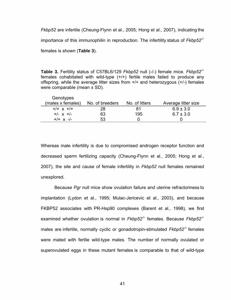
Fkbp52 are infertile (Cheung-Flynn et al., 2005; Hong et al., 2007), indicating the
importance of this immunophilin in reproduction. The infertility status of Fkbp52-/-
females is shown (Table 3).
Table 3. Fertility status of C57BL6/129 Fkbp52 null (-/-) female mice. Fkbp52-/- females cohabitated with wild-type (+/+) fertile males failed to produce any offspring, while the average litter sizes from +/+ and heterozygous (+/-) females were comparable (mean ± SD).
Genotypes
(males x females)
No. of breeders
No. of litters
Average litter size +/+ x +/+ 28 81 6.9 ± 3.0 +/- x +/- 63 195 6.7 ± 3.0 +/+ x -/- 53 0 0
Whereas male infertility is due to compromised androgen receptor function and
decreased sperm fertilizing capacity (Cheung-Flynn et al., 2005; Hong et al.,
2007), the site and cause of female infertility in Fkbp52 null females remained
unexplored.
Because Pgr null mice show ovulation failure and uterine refractoriness to
implantation (Lydon et al., 1995; Mulac-Jericevic et al., 2003), and because
FKBP52 associates with PR-Hsp90 complexes (Barent et al., 1998), we first
examined whether ovulation is normal in Fkbp52-/- females. Because Fkbp52-/-
males are infertile, normally cyclic or gonadotropin-stimulated Fkbp52-/- females
were mated with fertile wild-type males. The number of normally ovulated or
superovulated eggs in these mutant females is comparable to that of wild-type
42
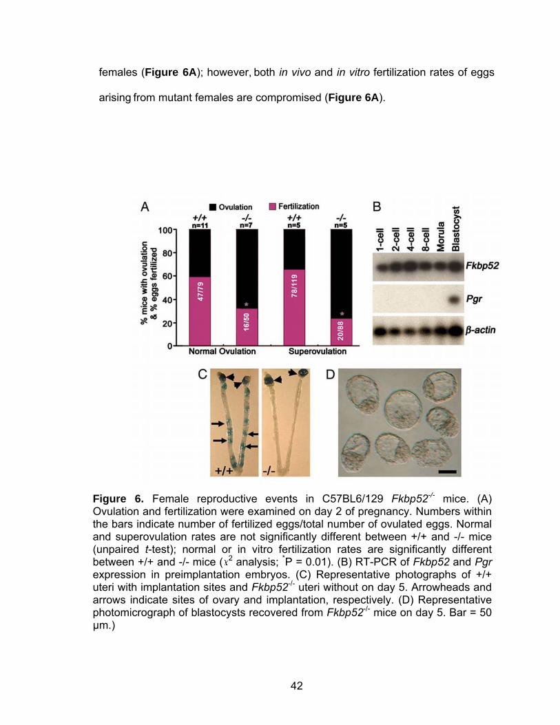
females (Figure 6A); however, both in vivo and in vitro fertilization rates of eggs
arising from mutant females are compromised (Figure 6A).
Figure 6. Female reproductive events in C57BL6/129 Fkbp52-/- mice. (A) Ovulation and fertilization were examined on day 2 of pregnancy. Numbers within the bars indicate number of fertilized eggs/total number of ovulated eggs. Normal and superovulation rates are not significantly different between +/+ and -/- mice (unpaired t-test); normal or in vitro fertilization rates are significantly different between +/+ and -/- mice ( 2 analysis; *P = 0.01). (B) RT-PCR of Fkbp52 and Pgr expression in preimplantation embryos. (C) Representative photographs of +/+ uteri with implantation sites and Fkbp52-/- uteri without on day 5. Arrowheads and arrows indicate sites of ovary and implantation, respectively. (D) Representative photomicrograph of blastocysts recovered from Fkbp52-/- mice on day 5. Bar = 50 µm.)
43
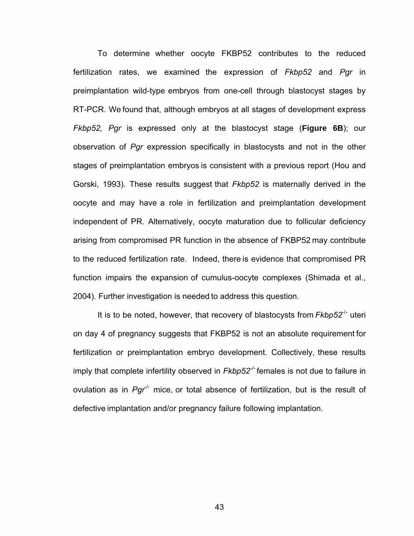
To determine whether oocyte FKBP52 contributes to the reduced
fertilization rates, we examined the expression of Fkbp52 and Pgr in
preimplantation wild-type embryos from one-cell through blastocyst stages by
RT-PCR. We found that, although embryos at all stages of development express
Fkbp52, Pgr is expressed only at the blastocyst stage (Figure 6B); our
observation of Pgr expression specifically in blastocysts and not in the other
stages of preimplantation embryos is consistent with a previous report (Hou and
Gorski, 1993). These results suggest that Fkbp52 is maternally derived in the
oocyte and may have a role in fertilization and preimplantation development
independent of PR. Alternatively, oocyte maturation due to follicular deficiency
arising from compromised PR function in the absence of FKBP52 may contribute
to the reduced fertilization rate. Indeed, there is evidence that compromised PR
function impairs the expansion of cumulus-oocyte complexes (Shimada et al.,
2004). Further investigation is needed to address this question.
It is to be noted, however, that recovery of blastocysts from Fkbp52-/- uteri
on day 4 of pregnancy suggests that FKBP52 is not an absolute requirement for
fertilization or preimplantation embryo development. Collectively, these results
imply that complete infertility observed in Fkbp52-/- females is not due to failure in
ovulation as in Pgr-/- mice, or total absence of fertilization, but is the result of
defective implantation and/or pregnancy failure following implantation.
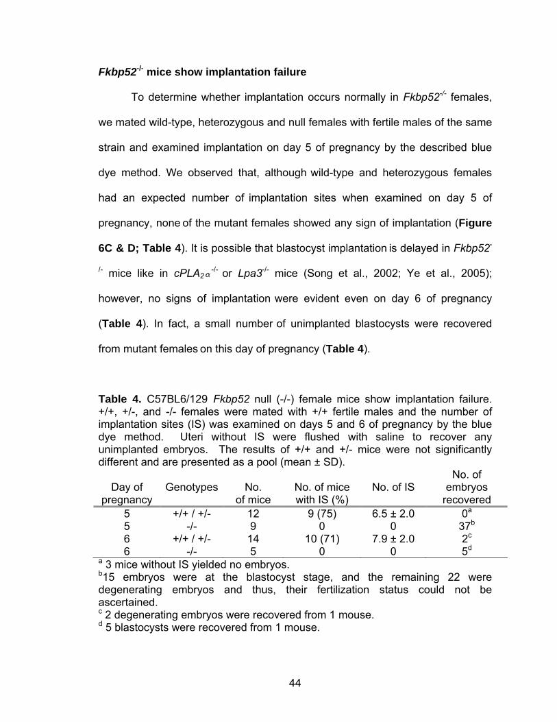
44
Fkbp52-/- mice show implantation failure
To determine whether implantation occurs normally in Fkbp52-/- females,
we mated wild-type, heterozygous and null females with fertile males of the same
strain and examined implantation on day 5 of pregnancy by the described blue
dye method. We observed that, although wild-type and heterozygous females
had an expected number of implantation sites when examined on day 5 of
pregnancy, none of the mutant females showed any sign of implantation (Figure
6C & D; Table 4). It is possible that blastocyst implantation is delayed in Fkbp52-
/- mice like in cPLA2-/- or Lpa3-/- mice (Song et al., 2002; Ye et al., 2005);
however, no signs of implantation were evident even on day 6 of pregnancy
(Table 4). In fact, a small number of unimplanted blastocysts were recovered
from mutant females on this day of pregnancy (Table 4).
Table 4. C57BL6/129 Fkbp52 null (-/-) female mice show implantation failure. +/+, +/-, and -/- females were mated with +/+ fertile males and the number of implantation sites (IS) was examined on days 5 and 6 of pregnancy by the blue dye method. Uteri without IS were flushed with saline to recover any unimplanted embryos. The results of +/+ and +/- mice were not significantly different and are presented as a pool (mean ± SD).
Day of
pregnancy
Genotypes
No.
of mice
No. of mice with IS (%)
No. of IS
No. of embryos
recovered 5 +/+ / +/- 12 9 (75) 6.5 ± 2.0 0a 5 -/- 9 0 0 37b 6 +/+ / +/- 14 10 (71) 7.9 ± 2.0 2c 6 -/- 5 0 0 5d
a 3 mice without IS yielded no embryos. b15 embryos were at the blastocyst stage, and the remaining 22 were degenerating embryos and thus, their fertilization status could not be ascertained. c 2 degenerating embryos were recovered from 1 mouse. d 5 blastocysts were recovered from 1 mouse.
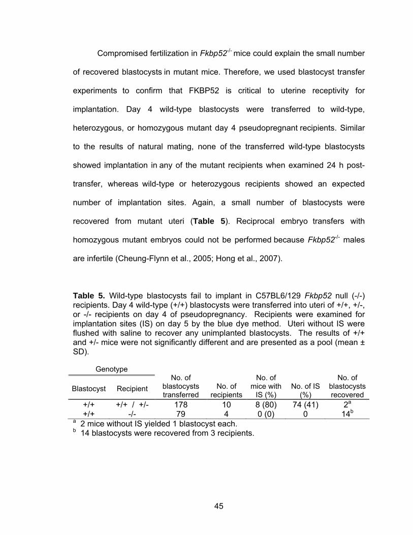
45
Compromised fertilization in Fkbp52-/- mice could explain the small number
of recovered blastocysts in mutant mice. Therefore, we used blastocyst transfer
experiments to confirm that FKBP52 is critical to uterine receptivity for
implantation. Day 4 wild-type blastocysts were transferred to wild-type,
heterozygous, or homozygous mutant day 4 pseudopregnant recipients. Similar
to the results of natural mating, none of the transferred wild-type blastocysts
showed implantation in any of the mutant recipients when examined 24 h post-
transfer, whereas wild-type or heterozygous recipients showed an expected
number of implantation sites. Again, a small number of blastocysts were
recovered from mutant uteri (Table 5). Reciprocal embryo transfers with
homozygous mutant embryos could not be performed because Fkbp52-/- males
are infertile (Cheung-Flynn et al., 2005; Hong et al., 2007).
Table 5. Wild-type blastocysts fail to implant in C57BL6/129 Fkbp52 null (-/-) recipients. Day 4 wild-type (+/+) blastocysts were transferred into uteri of +/+, +/-, or -/- recipients on day 4 of pseudopregnancy. Recipients were examined for implantation sites (IS) on day 5 by the blue dye method. Uteri without IS were flushed with saline to recover any unimplanted blastocysts. The results of +/+ and +/- mice were not significantly different and are presented as a pool (mean ± SD).
Genotype
Blastocyst
Recipient
No. of blastocysts transferred
No. of recipients
No. of mice with
IS (%)
No. of IS (%)
No. of blastocysts recovered
+/+ +/+ / +/- 178 10 8 (80) 74 (41) 2a +/+ -/- 79 4 0 (0) 0 14b
a 2 mice without IS yielded 1 blastocyst each. b 14 blastocysts were recovered from 3 recipients.
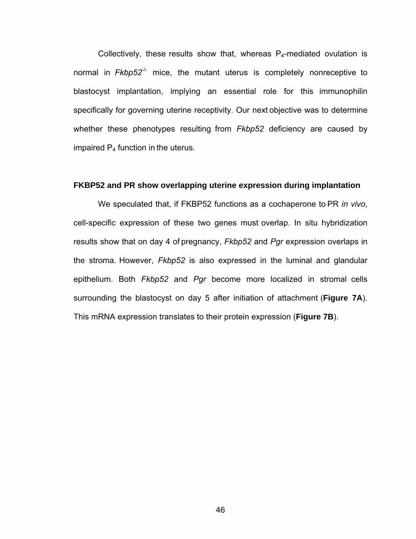
46
Collectively, these results show that, whereas P4-mediated ovulation is
normal in Fkbp52-/- mice, the mutant uterus is completely nonreceptive to
blastocyst implantation, implying an essential role for this immunophilin
specifically for governing uterine receptivity. Our next objective was to determine
whether these phenotypes resulting from Fkbp52 deficiency are caused by
impaired P4 function in the uterus.
FKBP52 and PR show overlapping uterine expression during implantation
We speculated that, if FKBP52 functions as a cochaperone to PR in vivo,
cell-specific expression of these two genes must overlap. In situ hybridization
results show that on day 4 of pregnancy, Fkbp52 and Pgr expression overlaps in
the stroma. However, Fkbp52 is also expressed in the luminal and glandular
epithelium. Both Fkbp52 and Pgr become more localized in stromal cells
surrounding the blastocyst on day 5 after initiation of attachment (Figure 7A).
This mRNA expression translates to their protein expression (Figure 7B).
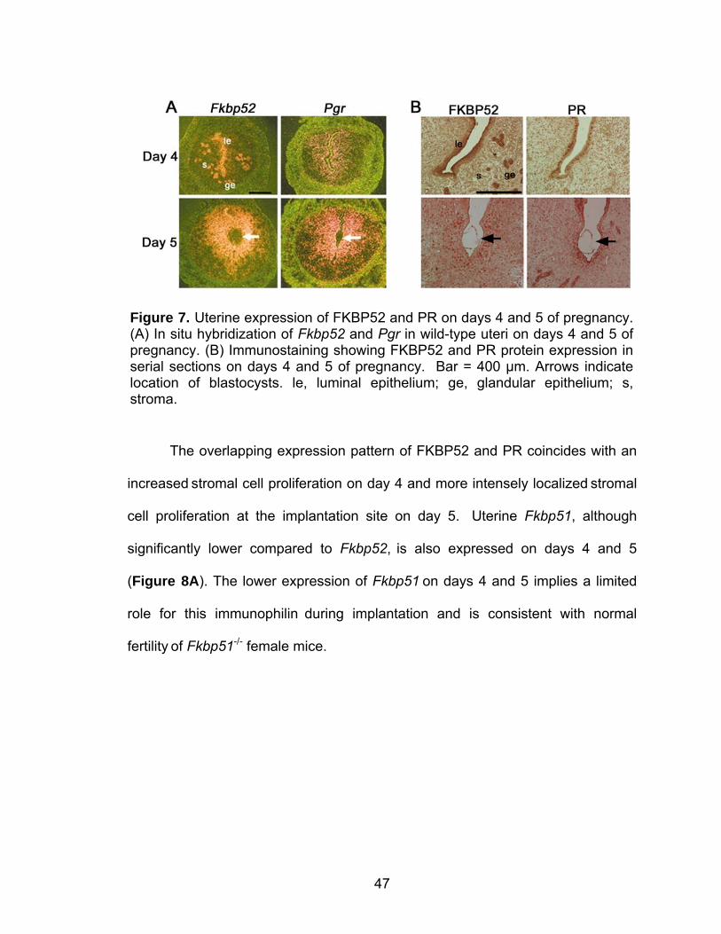
47
The overlapping expression pattern of FKBP52 and PR coincides with an
increased stromal cell proliferation on day 4 and more intensely localized stromal
cell proliferation at the implantation site on day 5. Uterine Fkbp51, although
significantly lower compared to Fkbp52, is also expressed on days 4 and 5
(Figure 8A). The lower expression of Fkbp51 on days 4 and 5 implies a limited
role for this immunophilin during implantation and is consistent with normal
fertility of Fkbp51-/- female mice.
Figure 7. Uterine expression of FKBP52 and PR on days 4 and 5 of pregnancy. (A) In situ hybridization of Fkbp52 and Pgr in wild-type uteri on days 4 and 5 of pregnancy. (B) Immunostaining showing FKBP52 and PR protein expression in serial sections on days 4 and 5 of pregnancy. Bar = 400 µm. Arrows indicate location of blastocysts. le, luminal epithelium; ge, glandular epithelium; s, stroma.
48
However, we wanted to determine whether an interaction between FKBP51 and
FKBP52 modulates PR transcriptional activity. Using mouse embryonic
fibroblasts (MEFs) in cellular assays, we found that FKBP51 is antagonistic to
FKBP52 in modulating PR transcriptional activity (Figure 9).
Figure 8. Uterine expression of Fkbp51 in C57BL6/129 wild-type versus Fkbp52-/- uteri. (A) In situ hybridization of Fkbp51 in wild-type uteri on days 4 and 5 of pregnancy. Arrow indicates blastocyst. Bar = 400 µm. (B) RT-PCR of Fkbp51 expression in +/+, +/-, and -/- uteri on day 4 of pregnancy. Data are presented as fold changes (mean ± SEM). Fold changes are not significantly different between +/+, +/-, or -/- mice (unpaired t-test).
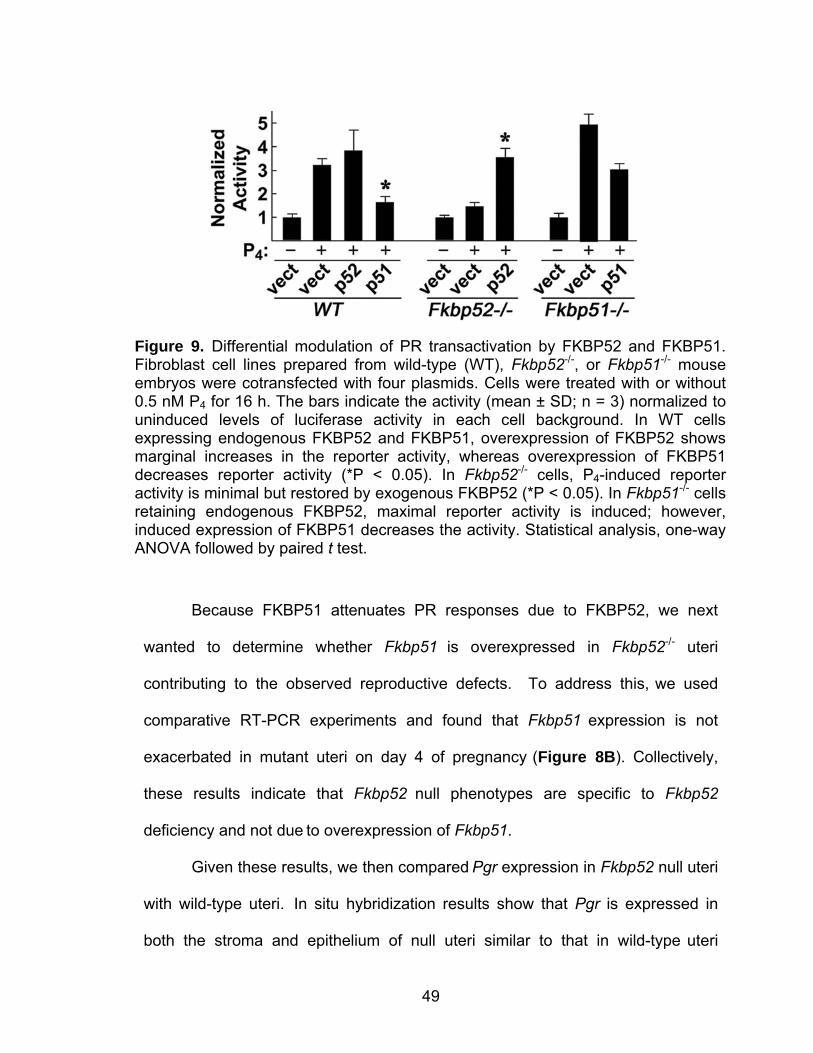
49
Because FKBP51 attenuates PR responses due to FKBP52, we next
wanted to determine whether Fkbp51 is overexpressed in Fkbp52-/- uteri
contributing to the observed reproductive defects. To address this, we used
comparative RT-PCR experiments and found that Fkbp51 expression is not
exacerbated in mutant uteri on day 4 of pregnancy (Figure 8B). Collectively,
these results indicate that Fkbp52 null phenotypes are specific to Fkbp52
deficiency and not due to overexpression of Fkbp51.
Given these results, we then compared Pgr expression in Fkbp52 null uteri
with wild-type uteri. In situ hybridization results show that Pgr is expressed in
both the stroma and epithelium of null uteri similar to that in wild-type uteri
Figure 9. Differential modulation of PR transactivation by FKBP52 and FKBP51. Fibroblast cell lines prepared from wild-type (WT), Fkbp52-/-, or Fkbp51-/- mouse embryos were cotransfected with four plasmids. Cells were treated with or without 0.5 nM P4 for 16 h. The bars indicate the activity (mean ± SD; n = 3) normalized to uninduced levels of luciferase activity in each cell background. In WT cells expressing endogenous FKBP52 and FKBP51, overexpression of FKBP52 shows marginal increases in the reporter activity, whereas overexpression of FKBP51 decreases reporter activity (*P < 0.05). In Fkbp52-/- cells, P4-induced reporter activity is minimal but restored by exogenous FKBP52 (*P < 0.05). In Fkbp51-/- cells retaining endogenous FKBP52, maximal reporter activity is induced; however, induced expression of FKBP51 decreases the activity. Statistical analysis, one-way ANOVA followed by paired t test.
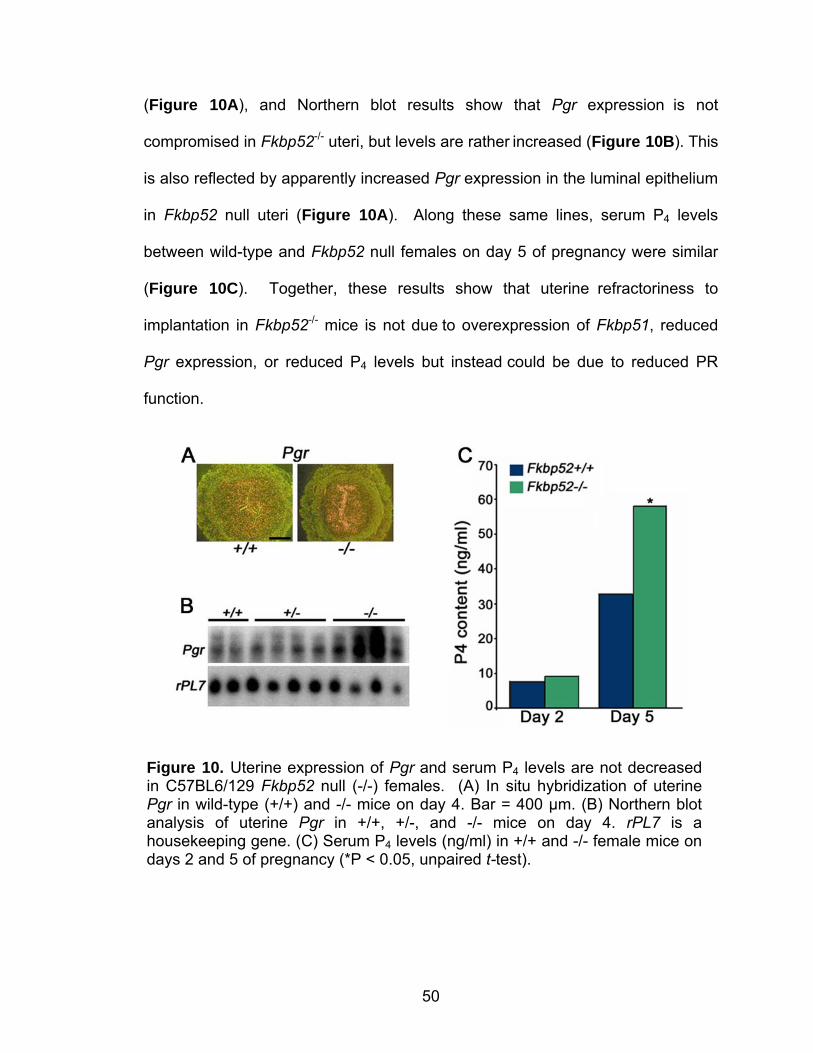
50
(Figure 10A), and Northern blot results show that Pgr expression is not
compromised in Fkbp52-/- uteri, but levels are rather increased (Figure 10B). This
is also reflected by apparently increased Pgr expression in the luminal epithelium
in Fkbp52 null uteri (Figure 10A). Along these same lines, serum P4 levels
between wild-type and Fkbp52 null females on day 5 of pregnancy were similar
(Figure 10C). Together, these results show that uterine refractoriness to
implantation in Fkbp52-/- mice is not due to overexpression of Fkbp51, reduced
Pgr expression, or reduced P4 levels but instead could be due to reduced PR
function.
Figure 10. Uterine expression of Pgr and serum P4 levels are not decreased in C57BL6/129 Fkbp52 null (-/-) females. (A) In situ hybridization of uterine Pgr in wild-type (+/+) and -/- mice on day 4. Bar = 400 µm. (B) Northern blot analysis of uterine Pgr in +/+, +/-, and -/- mice on day 4. rPL7 is a housekeeping gene. (C) Serum P4 levels (ng/ml) in +/+ and -/- female mice on days 2 and 5 of pregnancy (*P < 0.05, unpaired t-test).
51
Uterine PR activity is compromised in Fkbp52-/- mice
If FKBP52 is required for optimal PR function, then uterine PR activity
would be compromised in the absence of FKBP52. Indeed, binding assays using
radiolabeled P4 show that P4 binding to PR is reduced ~2-fold in Fkbp52-/- uterine
cytosol compared with that of wild-type mice. There is also a fractional reduction
in the number of P4 binding sites (Figure 11A). Reduced PR transcriptional
activity in the absence of FKBP52 is also noted in a transfected cell system in
vitro. Embryonic fibroblasts isolated from Fkbp52-/- mice were cotransfected with
human PR-B plasmid, a luciferase reporter plasmid, and either an empty vector
or a plasmid containing FKBP52 (p52). P4-dependent reporter activity was
measured over a range of P4 concentrations. Cells expressing exogenous
FKBP52 have enhanced P4-dependent reporter gene activation as compared to
cells lacking FKBP52 (Figure 11B).
PR-B results are shown because PR-A normally displays only modest
reporter transactivation in cell culture systems (Daikoku et al., 2003); however,
loss of FKBP52 did also reduce the modest activity of PR-A. These results
provide evidence that FKBP52 is required for optimal uterine PR activity; thus,
defects in uterine receptivity for implantation in Fkbp52-/- mice are likely caused
by compromised uterine P4 function.
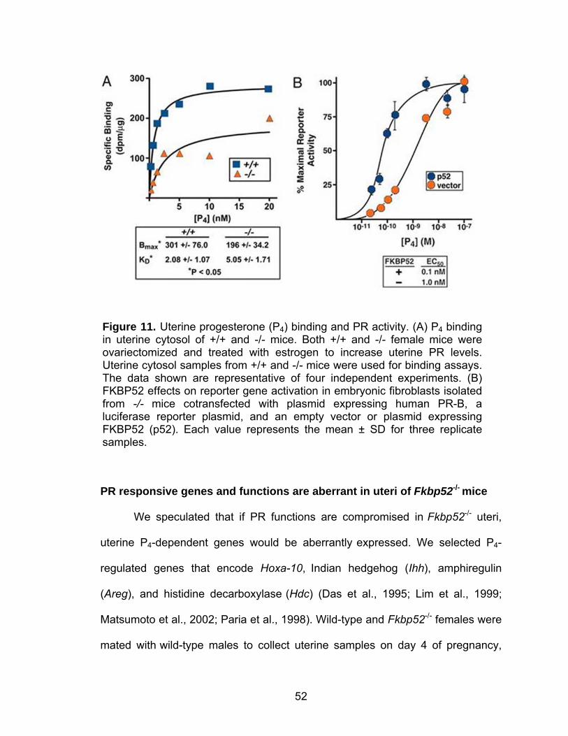
52
PR responsive genes and functions are aberrant in uteri of Fkbp52-/- mice
We speculated that if PR functions are compromised in Fkbp52-/- uteri,
uterine P4-dependent genes would be aberrantly expressed. We selected P4-
regulated genes that encode Hoxa-10, Indian hedgehog (Ihh), amphiregulin
(Areg), and histidine decarboxylase (Hdc) (Das et al., 1995; Lim et al., 1999;
Matsumoto et al., 2002; Paria et al., 1998). Wild-type and Fkbp52-/- females were
mated with wild-type males to collect uterine samples on day 4 of pregnancy,
Figure 11. Uterine progesterone (P4) binding and PR activity. (A) P4 binding in uterine cytosol of +/+ and -/- mice. Both +/+ and -/- female mice were ovariectomized and treated with estrogen to increase uterine PR levels. Uterine cytosol samples from +/+ and -/- mice were used for binding assays. The data shown are representative of four independent experiments. (B) FKBP52 effects on reporter gene activation in embryonic fibroblasts isolated from -/- mice cotransfected with plasmid expressing human PR-B, a luciferase reporter plasmid, and an empty vector or plasmid expressing FKBP52 (p52). Each value represents the mean ± SD for three replicate samples.
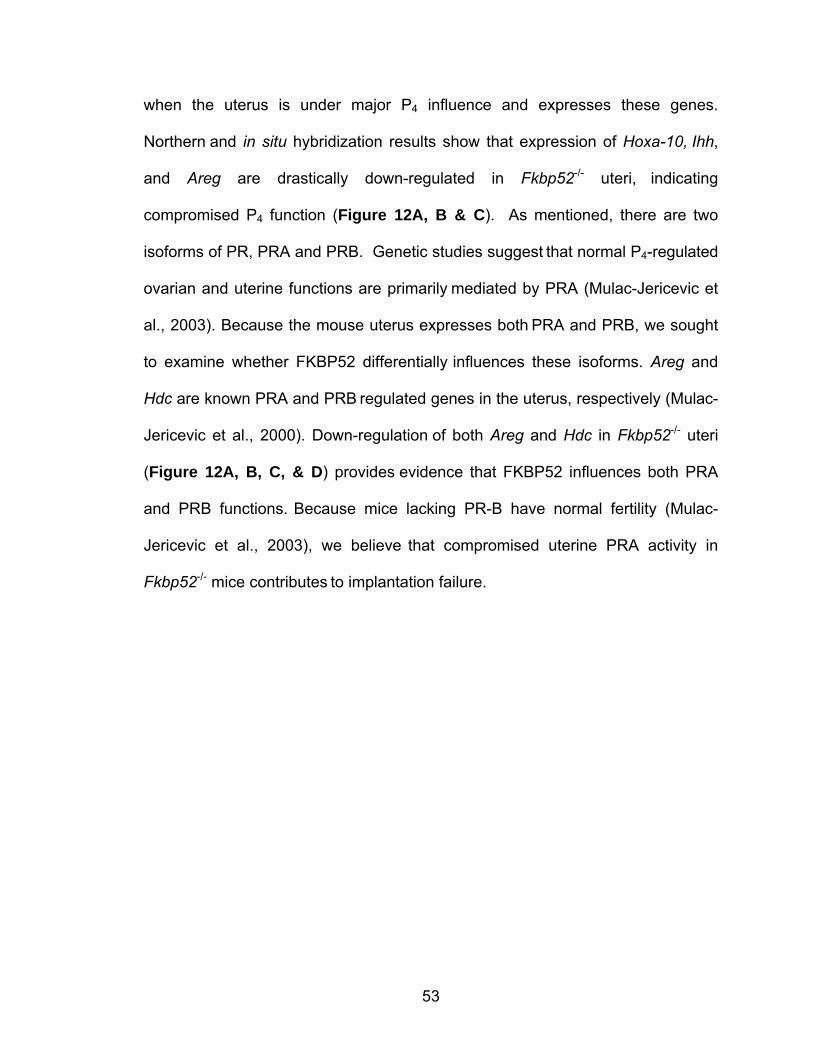
53
when the uterus is under major P4 influence and expresses these genes.
Northern and in situ hybridization results show that expression of Hoxa-10, Ihh,
and Areg are drastically down-regulated in Fkbp52-/- uteri, indicating
compromised P4 function (Figure 12A, B & C). As mentioned, there are two
isoforms of PR, PRA and PRB. Genetic studies suggest that normal P4-regulated
ovarian and uterine functions are primarily mediated by PRA (Mulac-Jericevic et
al., 2003). Because the mouse uterus expresses both PRA and PRB, we sought
to examine whether FKBP52 differentially influences these isoforms. Areg and
Hdc are known PRA and PRB regulated genes in the uterus, respectively (Mulac-
Jericevic et al., 2000). Down-regulation of both Areg and Hdc in Fkbp52-/- uteri
(Figure 12A, B, C, & D) provides evidence that FKBP52 influences both PRA
and PRB functions. Because mice lacking PR-B have normal fertility (Mulac-
Jericevic et al., 2003), we believe that compromised uterine PRA activity in
Fkbp52-/- mice contributes to implantation failure.
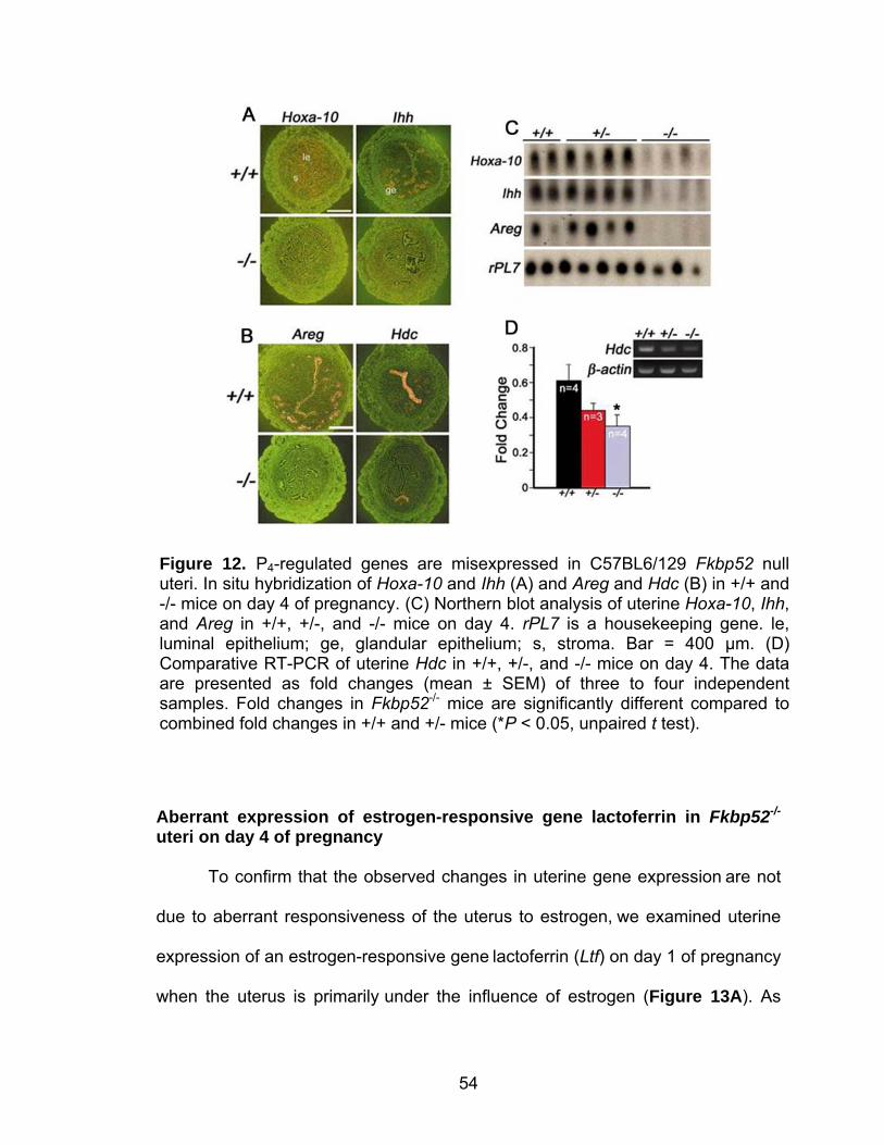
54
Aberrant expression of estrogen-responsive gene lactoferrin in Fkbp52-/- uteri on day 4 of pregnancy To confirm that the observed changes in uterine gene expression are not
due to aberrant responsiveness of the uterus to estrogen, we examined uterine
expression of an estrogen-responsive gene lactoferrin (Ltf) on day 1 of pregnancy
when the uterus is primarily under the influence of estrogen (Figure 13A). As
Figure 12. P4-regulated genes are misexpressed in C57BL6/129 Fkbp52 null uteri. In situ hybridization of Hoxa-10 and Ihh (A) and Areg and Hdc (B) in +/+ and -/- mice on day 4 of pregnancy. (C) Northern blot analysis of uterine Hoxa-10, Ihh, and Areg in +/+, +/-, and -/- mice on day 4. rPL7 is a housekeeping gene. le, luminal epithelium; ge, glandular epithelium; s, stroma. Bar = 400 µm. (D) Comparative RT-PCR of uterine Hdc in +/+, +/-, and -/- mice on day 4. The data are presented as fold changes (mean ± SEM) of three to four independent samples. Fold changes in Fkbp52-/- mice are significantly different compared to combined fold changes in +/+ and +/- mice (*P < 0.05, unpaired t test).
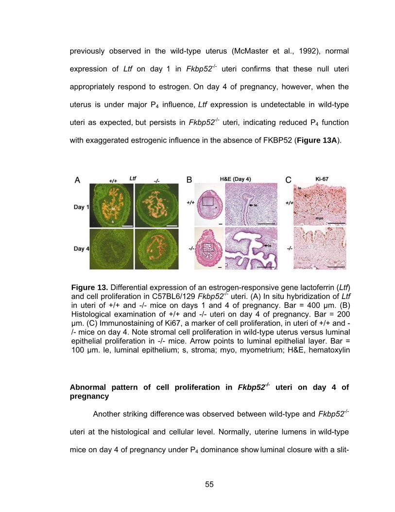
55
previously observed in the wild-type uterus (McMaster et al., 1992), normal
expression of Ltf on day 1 in Fkbp52-/- uteri confirms that these null uteri
appropriately respond to estrogen. On day 4 of pregnancy, however, when the
uterus is under major P4 influence, Ltf expression is undetectable in wild-type
uteri as expected, but persists in Fkbp52-/- uteri, indicating reduced P4 function
with exaggerated estrogenic influence in the absence of FKBP52 (Figure 13A).
Abnormal pattern of cell proliferation in Fkbp52-/- uteri on day 4 of pregnancy Another striking difference was observed between wild-type and Fkbp52-/-
uteri at the histological and cellular level. Normally, uterine lumens in wild-type
mice on day 4 of pregnancy under P4 dominance show luminal closure with a slit-
Figure 13. Differential expression of an estrogen-responsive gene lactoferrin (Ltf) and cell proliferation in C57BL6/129 Fkbp52-/- uteri. (A) In situ hybridization of Ltf in uteri of +/+ and -/- mice on days 1 and 4 of pregnancy. Bar = 400 µm. (B) Histological examination of +/+ and -/- uteri on day 4 of pregnancy. Bar = 200 µm. (C) Immunostaining of Ki67, a marker of cell proliferation, in uteri of +/+ and -/- mice on day 4. Note stromal cell proliferation in wild-type uterus versus luminal epithelial proliferation in -/- mice. Arrow points to luminal epithelial layer. Bar = 100 µm. le, luminal epithelium; s, stroma; myo, myometrium; H&E, hematoxylin
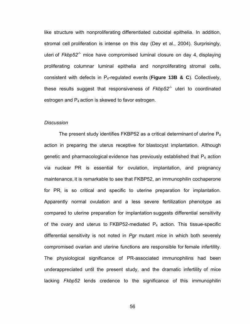
56
like structure with nonproliferating differentiated cuboidal epithelia. In addition,
stromal cell proliferation is intense on this day (Dey et al., 2004). Surprisingly,
uteri of Fkbp52-/- mice have compromised luminal closure on day 4, displaying
proliferating columnar luminal epithelia and nonproliferating stromal cells,
consistent with defects in P4-regulated events (Figure 13B & C). Collectively,
these results suggest that responsiveness of Fkbp52-/- uteri to coordinated
estrogen and P4 action is skewed to favor estrogen.
Discussion
The present study identifies FKBP52 as a critical determinant of uterine P4
action in preparing the uterus receptive for blastocyst implantation. Although
genetic and pharmacological evidence has previously established that P4 action
via nuclear PR is essential for ovulation, implantation, and pregnancy
maintenance, it is remarkable to see that FKBP52, an immunophilin cochaperone
for PR, is so critical and specific to uterine preparation for implantation.
Apparently normal ovulation and a less severe fertilization phenotype as
compared to uterine preparation for implantation suggests differential sensitivity
of the ovary and uterus to FKBP52-mediated P4 action. This tissue-specific
differential sensitivity is not noted in Pgr mutant mice in which both severely
compromised ovarian and uterine functions are responsible for female infertility.
The physiological significance of PR-associated immunophilins had been
underappreciated until the present study, and the dramatic infertility of mice
lacking Fkbp52 lends credence to the significance of this immunophilin

57
cochaperone in female fertility. This study places FKBP52 as a target for
improving female fertility or alternatively developing novel approaches to
contraception.
58
CHAPTER III
FKBP52 DEFICIENCY-CONFERRED UTERINE PROGESTERONE RESISTANCE IS GENETIC BACKGROUND AND PREGNANCY STAGE
SPECIFIC
Abstract
Immunophilin FKBP52 serves as a cochaperone to govern normal PR function.
Using Fkbp52-/- mice, here we show intriguing aspects of uterine P4-PR signaling
during pregnancy. Implantation failure is the major phenotype found in these null
females, which is conserved on both C57BL6/129 and CD1 backgrounds.
However, P4 supplementation rescues implantation and subsequent
decidualization in CD1, but not in C57BL6/129, null females. Surprisingly,
experimentally-induced decidualization in the absence of blastocysts fails in
Fkbp52-/- mice on either background even with P4 supplementation, suggesting
that embryonic signals complement uterine signaling for this event. Another
interesting finding is that while P4 at higher than normal pregnancy levels confers
PR signaling sufficient for implantation in CD1 null females, these levels are
inefficient in maintaining the full complement of term pregnancy. However,
elevating P4 levels further restores PR signaling optimal for successful term
pregnancy with normal litter size. Collectively, our results show that the
indispensability of FKBP52 in uterine P4-PR signaling is a function of genetic
disparity and pregnancy stage specific. Since there is evidence for a correlation
between P4 supplementation and reduced risks of P4-resistant recurrent
59
miscarriages and remission of endometriosis, these findings have clinical
implications for genetically diverse populations of women.
Introduction
As mentioned, P4 signaling is an absolute requirement for implantation
and pregnancy maintenance in most eutherian mammals studied (Dey et al.,
2004; Wang and Dey, 2006). The failure of ovulation and implantation,
decidualization and pregnancy maintenance (Lydon et al., 1995) precludes using
Pgr-/- mice to study potential new aspects of P4 function during pregnancy. In
contrast, the targeted deletion of the Fkbp52 gene that encodes FKBP52, an
immunophilin cochaperone that optimizes PR signaling, has allowed us to
address unique aspects of uterine P4-PR signaling during pregnancy in mice
since these mice display uterine-specific infertility (Tranguch et al., 2005).
Our finding that Fkbp52-/- females on a C57BL6/129 mixed background
have complete implantation failure with normal ovulation and slightly reduced
fertilization rates has recently been confirmed by another group in independently-
generated Fkbp52-/- mice on the same genetic background (Tranguch et al.,
2005; Yang et al., 2006). The fact that the infertile phenotype of Fkbp52-/-
females is primarily due to implantation defects suggests differential sensitivity of
the ovary and uterus to FKBP52-PR mediated P4 action, a tissue-specific
differential sensitivity not noted in Pgr-/- females with complete female infertility
(Lydon et al., 1995; Mulac-Jericevic et al., 2000).
60
Although P4 is commonly known as the “hormone of pregnancy”, various
aspects of its roles throughout pregnancy are not well understood. Therefore,
Fkbp52-/- mice provide a unique opportunity for studying such roles of P4
signaling throughout pregnancy. By transferring C57BL6/129 Fkbp52-/- mice to a
CD1 genetic background, we are able to exploit this mouse model to address
these roles for the first time.
Methods
Mice
The Fkbp52 gene was disrupted in mice by homologous recombination as
previously described (Cheung-Flynn et al., 2005). Tail genomic DNA was used
for PCR-based genotyping. Because genetic backgrounds of mice contribute to
different phenotypes (Threadgill et al., 1995; Wang et al., 2004), we introduced
Fkbp52 deficiency in CD1 mice by crossing C57BL6/129 Fkbp52 heterozygous
males to CD1 wild-type (WT) females producing an F1 generation. F1 Fkbp52+/-
males were then back-crossed to CD1 WT females, and the process was
continued for ten generations. Crossing heterozygous females with heterozygous
males of the same genetic background (CD1/F10) generated Fkbp52 WT and
null littermates for experiments. Mice on both genetic backgrounds were housed
and used in the present investigation in accordance with the National Institutes of
Health and institutional guidelines on the care and use of laboratory animals.
61
Ovulation, fertilization, implantation, blastocyst transfer and experimentally-induced decidualization
To examine ovulation and fertilization, CD1 WT or Fkbp52-/- mice were
mated with fertile WT males. On day 2 of pregnancy (day 1 = vaginal plug),
oviducts were flushed with Whitten's medium to recover ovulated eggs, and
fertilization was assessed by counting the number of 2-cell embryos. Implantation
sites on days 5 and 6 of pregnancy were visualized by an intravenous injection
(0.1 ml/mouse) of Chicago Blue B dye solution (1% in saline) and the number of
implantation sites as demarcated by distinct blue bands was recorded. For
blastocyst transfer, pseudopregnant recipients were generated by mating females
with vasectomized WT males. Day 4 WT blastocysts were transferred into day 4
uteri of C57BL6/129 Fkbp52 WT or null pseudopregnant recipients, and
implantation sites were observed 24 h (day 5) or 96 h (day 8) later by the blue
dye method (Paria et al., 2001).
To determine whether experimentally-induced decidualization occurs in
null females, Fkbp52 WT or null females were mated with vasectomized WT
males. On day 4, one uterine horn was infused with sesame oil (25 µl) and the
contralateral horn served as control. Mice were sacrificed on day 8 of
pseudopregnancy. Uterine weights of infused (oil) and non-infused (control)
horns were recorded, and fold increase in weight was used as an index of
decidualization. All mice used were between 2-5 months of age.
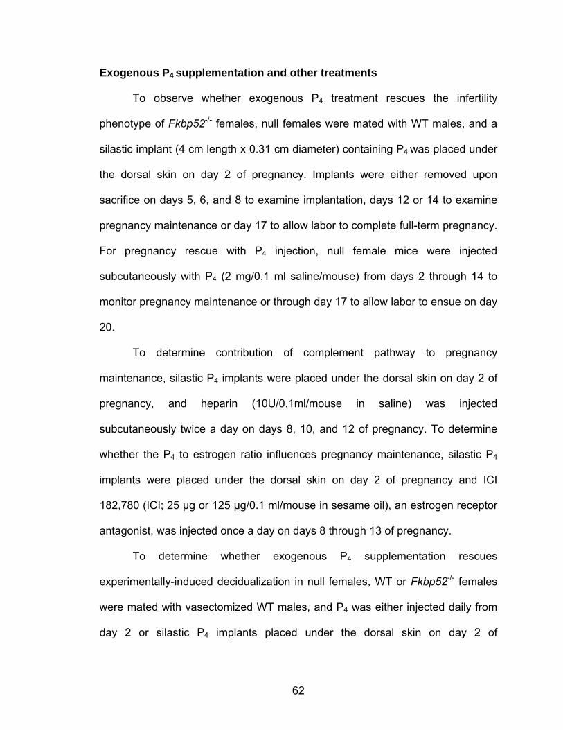
62
Exogenous P4 supplementation and other treatments
To observe whether exogenous P4 treatment rescues the infertility
phenotype of Fkbp52-/- females, null females were mated with WT males, and a
silastic implant (4 cm length x 0.31 cm diameter) containing P4 was placed under
the dorsal skin on day 2 of pregnancy. Implants were either removed upon
sacrifice on days 5, 6, and 8 to examine implantation, days 12 or 14 to examine
pregnancy maintenance or day 17 to allow labor to complete full-term pregnancy.
For pregnancy rescue with P4 injection, null female mice were injected
subcutaneously with P4 (2 mg/0.1 ml saline/mouse) from days 2 through 14 to
monitor pregnancy maintenance or through day 17 to allow labor to ensue on day
20.
To determine contribution of complement pathway to pregnancy
maintenance, silastic P4 implants were placed under the dorsal skin on day 2 of
pregnancy, and heparin (10U/0.1ml/mouse in saline) was injected
subcutaneously twice a day on days 8, 10, and 12 of pregnancy. To determine
whether the P4 to estrogen ratio influences pregnancy maintenance, silastic P4
implants were placed under the dorsal skin on day 2 of pregnancy and ICI
182,780 (ICI; 25 µg or 125 µg/0.1 ml/mouse in sesame oil), an estrogen receptor
antagonist, was injected once a day on days 8 through 13 of pregnancy.
To determine whether exogenous P4 supplementation rescues
experimentally-induced decidualization in null females, WT or Fkbp52-/- females
were mated with vasectomized WT males, and P4 was either injected daily from
day 2 or silastic P4 implants placed under the dorsal skin on day 2 of
63
pseudopregnancy. On day 4, one uterine horn was infused with sesame oil (25
µl), and the contralateral horn served as control. Mice were sacrificed on day 8 of
pseudopregnancy. Uterine weights of infused (oil) and non-infused (control)
horns were recorded, and fold increase in weight was used as an index of
decidualization.
P4 assay
Blood samples from mice were collected on the indicated days of pregnancy.
Serum was separated by centrifugation (3,000 rpm, 15 min) and stored at -80°C
until analysis. Serum P4 levels were measured by radioimmunoassay (University
of Virginia Center for Research in Reproduction, Ligand Assay & Analysis Core,
Charlottesville, VA).
In situ hybridization
Sense or antisense 35S-labeled cRNA probes for Areg, Ihh, Hoxa10, Ltf,
C3, Lif, Bmp2, Ptgs2, Pgr, and Fkbp52 generated by using appropriate
polymerases from respective cDNAs were used for hybridization. Sections
hybridized with sense probes showed no signal and served as negative controls.
Immunohistochemistry
Immunolocalization of cytokeratin was performed by using a polyclonal
rabbit anti-cow antibody (DakoCytomation, Carpinteria, CA). A Histostain-Plus
64
(DAB) kit (Zymed) was used to visualize antigen. Brown deposits indicate sites of
positive immunostaining.
Results
Implantation failure occurs in Fkbp52-/- mice irrespective of genetic background
There is increasing evidence that mutation of a gene often results in
altered phenotypes depending on the genetic background of mice (Threadgill et
al., 1995; Wang et al., 2004). These varying phenotypes are thought to be due to
differential expression and/or regulation of modifier genes (Bonyadi et al., 1997),
although identification of such modifiers remains largely unknown. There is also
evidence that compensatory function of genes among the same family is genetic
background dependent (Wang et al., 2004). We have recently shown that
C57BL6/129 Fkbp52-/- females have complete implantation failure, although
ovulation is normal (Tranguch et al., 2005). To determine whether this phenotype
is a function of genetic background, we established Fkbp52 deletion in CD1 mice
(see Methods). First, we examined ovulation and fertilization in CD1 Fkbp52-/-
females after mating with WT males since Fkbp52-/- males, irrespective of genetic
background, are infertile (Cheung-Flynn et al., 2005; Hong et al., 2007). We
found ovulation and fertilization in null females to be comparable with WT
(Figure 14A & B). CD1 Fkbp52-/- females were examined for implantation sites
on day 5 of pregnancy by the blue dye method. We observed that only 2 of 14
CD1 Fkbp52-/- females showed very faint blue bands (Figure 14C & Table 6).
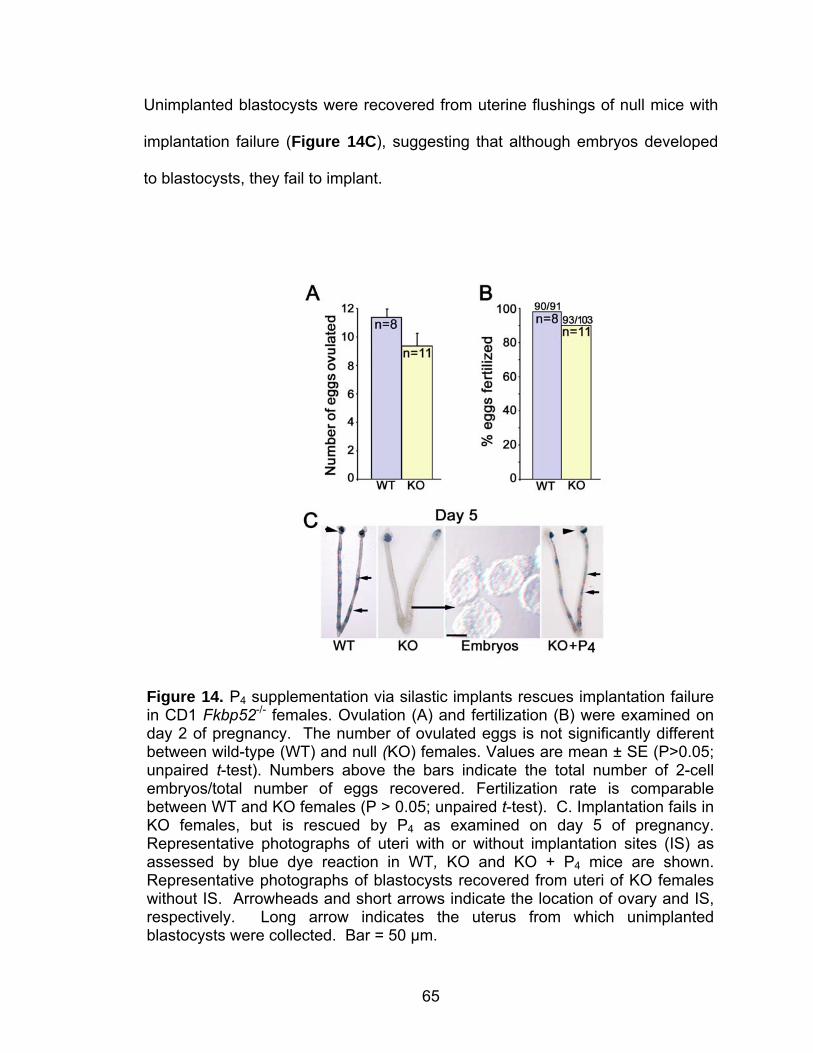
65
Unimplanted blastocysts were recovered from uterine flushings of null mice with
implantation failure (Figure 14C), suggesting that although embryos developed
to blastocysts, they fail to implant.
Figure 14. P4 supplementation via silastic implants rescues implantation failure in CD1 Fkbp52-/- females. Ovulation (A) and fertilization (B) were examined on day 2 of pregnancy. The number of ovulated eggs is not significantly different between wild-type (WT) and null (KO) females. Values are mean ± SE (P>0.05; unpaired t-test). Numbers above the bars indicate the total number of 2-cell embryos/total number of eggs recovered. Fertilization rate is comparable between WT and KO females (P > 0.05; unpaired t-test). C. Implantation fails in KO females, but is rescued by P4 as examined on day 5 of pregnancy. Representative photographs of uteri with or without implantation sites (IS) as assessed by blue dye reaction in WT, KO and KO + P4 mice are shown. Representative photographs of blastocysts recovered from uteri of KO females without IS. Arrowheads and short arrows indicate the location of ovary and IS, respectively. Long arrow indicates the uterus from which unimplanted blastocysts were collected. Bar = 50 µm.
66
Our data signify that optimal P4-PR signaling imparted by FKBP52 is
critical to uterine receptivity and implantation in mice, a phenotype conserved
across these genetic backgrounds. Because ovulation is normal in Fkbp52-/-
mice, our results suggest that uterine responsiveness to PR signaling differs from
that of ovarian responsiveness. It is possible that relatively high local P4 levels in
the ovary (Pointis et al., 1981), the site of its synthesis, enhance basal PR activity
sufficient for ovulation and fertilization processes.
Progesterone supplementation rescues implantation in CD1 Fkbp52-/- females
We have shown that PR activity, but not PR or P4 levels, is compromised
in C57BL6/129 Fkbp52-/- females (Tranguch et al., 2005; Tranguch et al., 2006).
It is possible that FKBP52 binding to the PR complex modulates the hormone-
responsiveness of PR not in an all-or-none fashion, but rather to fine-tune
physiological responses to P4. This tenet implies that while FKBP52 is necessary
for optimal PR activity, PR signaling can still operate, albeit not as efficiently, in
the absence of FKBP52.
Since serum P4 levels in CD1 Fkbp52-/- females are similar to those of WT
on day 5 of pregnancy (Figure 15), we speculated that exposing Fkbp52-/- uteri
to higher than normal P4 levels will enhance PR activity to rescue pregnancy
failure in the absence of FKBP52. This is consistent with our previous findings
that PR activity in Fkbp52-/- mouse embryonic fibroblasts (MEFs) reached levels
similar to those in WT MEFs exposed to higher P4 concentrations in culture
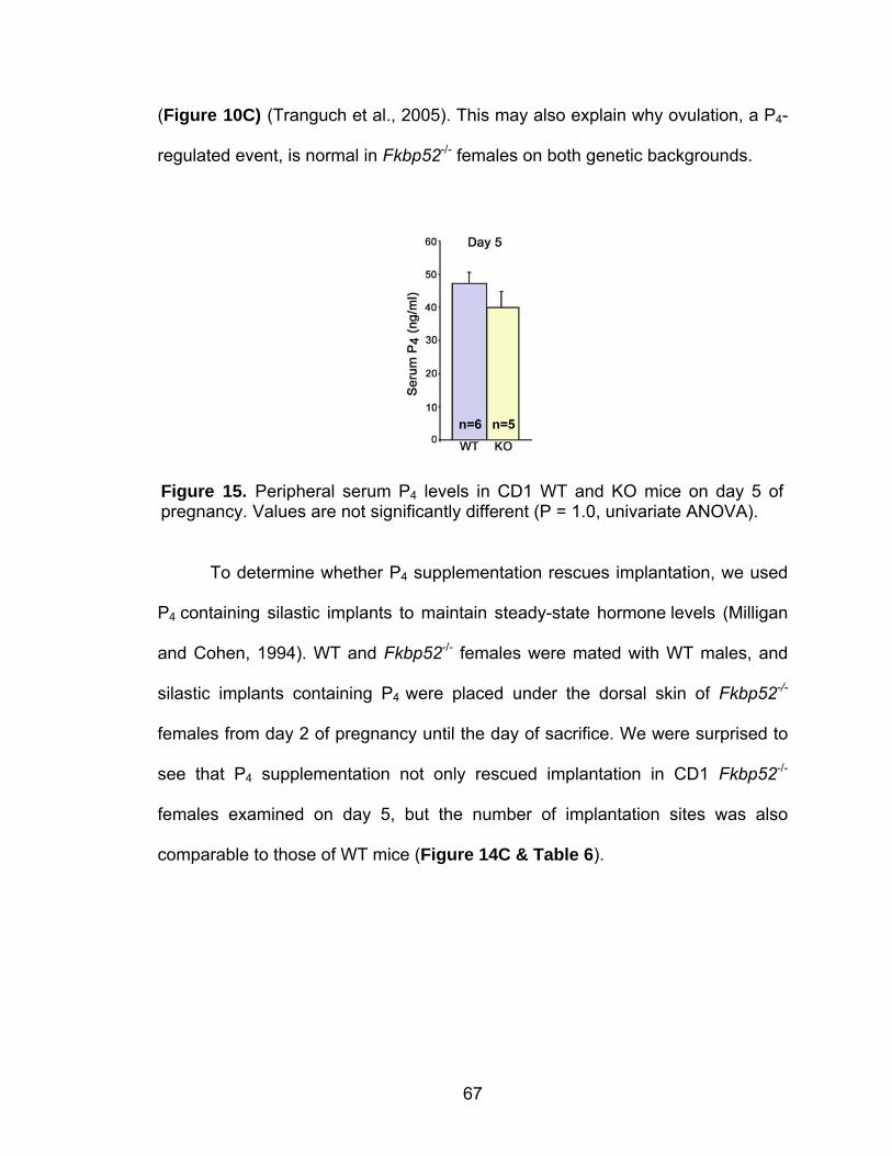
67
(Figure 10C) (Tranguch et al., 2005). This may also explain why ovulation, a P4-
regulated event, is normal in Fkbp52-/- females on both genetic backgrounds.
To determine whether P4 supplementation rescues implantation, we used
P4 containing silastic implants to maintain steady-state hormone levels (Milligan
and Cohen, 1994). WT and Fkbp52-/- females were mated with WT males, and
silastic implants containing P4 were placed under the dorsal skin of Fkbp52-/-
females from day 2 of pregnancy until the day of sacrifice. We were surprised to
see that P4 supplementation not only rescued implantation in CD1 Fkbp52-/-
females examined on day 5, but the number of implantation sites was also
comparable to those of WT mice (Figure 14C & Table 6).
Figure 15. Peripheral serum P4 levels in CD1 WT and KO mice on day 5 of pregnancy. Values are not significantly different (P = 1.0, univariate ANOVA).
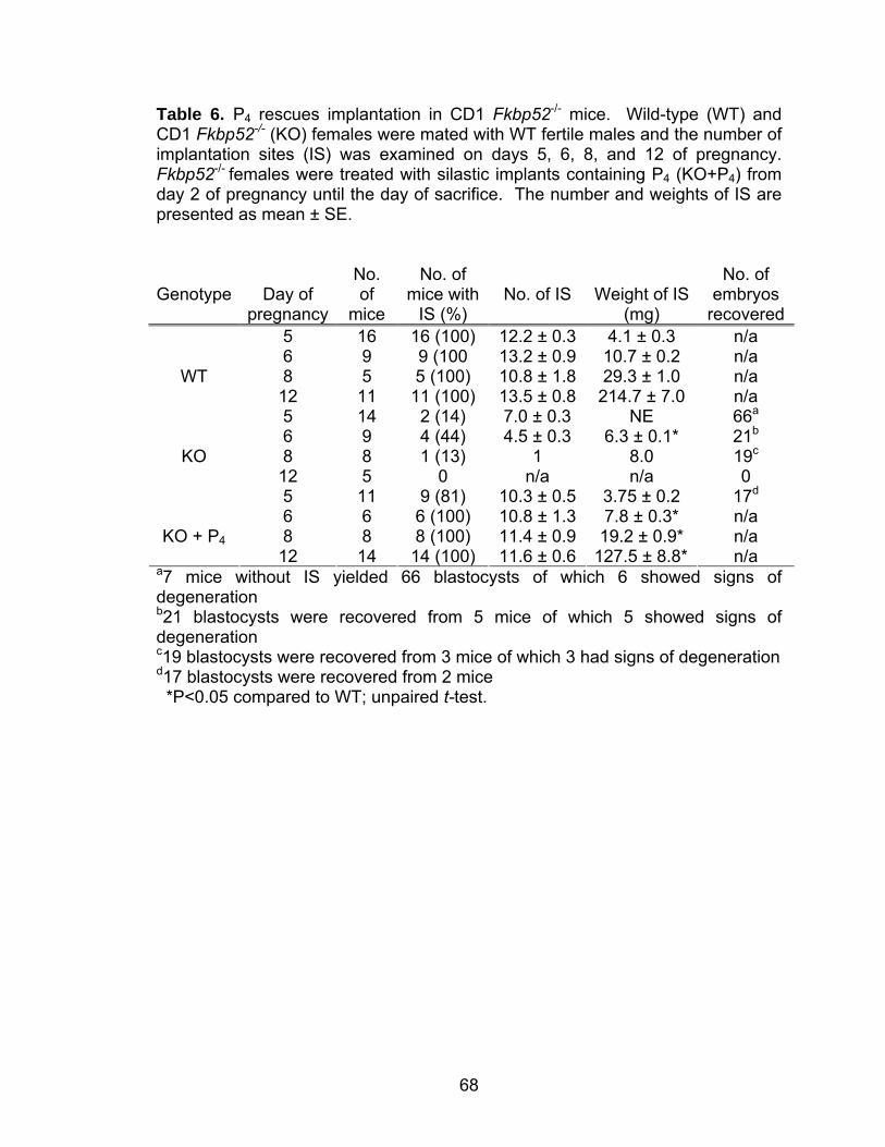
68
Table 6. P4 rescues implantation in CD1 Fkbp52-/- mice. Wild-type (WT) and CD1 Fkbp52-/- (KO) females were mated with WT fertile males and the number of implantation sites (IS) was examined on days 5, 6, 8, and 12 of pregnancy. Fkbp52-/- females were treated with silastic implants containing P4 (KO+P4) from day 2 of pregnancy until the day of sacrifice. The number and weights of IS are presented as mean ± SE.
Genotype
Day of pregnancy
No. of
mice
No. of
mice with IS (%)
No. of IS
Weight of IS (mg)
No. of
embryos recovered
5 16 16 (100) 12.2 ± 0.3 4.1 ± 0.3 n/a 6 9 9 (100 13.2 ± 0.9 10.7 ± 0.2 n/a 8 5 5 (100) 10.8 ± 1.8 29.3 ± 1.0 n/a
WT 12 11 11 (100) 13.5 ± 0.8 214.7 ± 7.0 n/a 5 14 2 (14) 7.0 ± 0.3 NE 66a 6 9 4 (44) 4.5 ± 0.3 6.3 ± 0.1* 21b 8 8 1 (13) 1 8.0 19c
KO 12 5 0 n/a n/a 0 5 11 9 (81) 10.3 ± 0.5 3.75 ± 0.2 17d 6 6 6 (100) 10.8 ± 1.3 7.8 ± 0.3* n/a 8 8 8 (100) 11.4 ± 0.9 19.2 ± 0.9* n/a
KO + P4 12 14 14 (100) 11.6 ± 0.6 127.5 ± 8.8* n/a
a7 mice without IS yielded 66 blastocysts of which 6 showed signs of degeneration b21 blastocysts were recovered from 5 mice of which 5 showed signs of degeneration c19 blastocysts were recovered from 3 mice of which 3 had signs of degeneration d17 blastocysts were recovered from 2 mice *P<0.05 compared to WT; unpaired t-test.
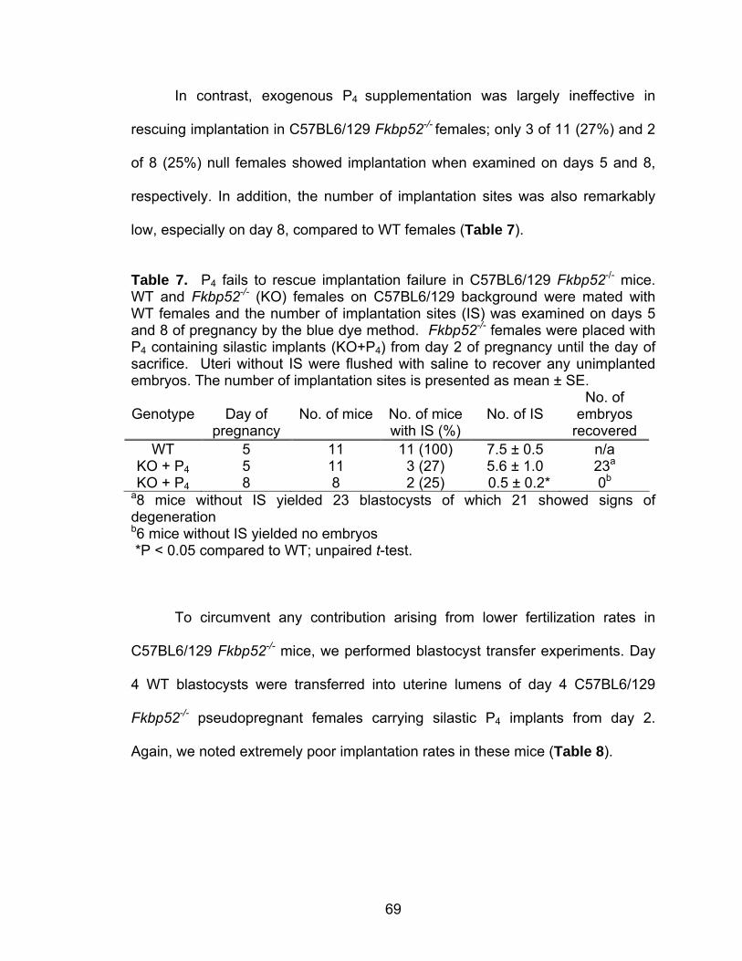
69
In contrast, exogenous P4 supplementation was largely ineffective in
rescuing implantation in C57BL6/129 Fkbp52-/- females; only 3 of 11 (27%) and 2
of 8 (25%) null females showed implantation when examined on days 5 and 8,
respectively. In addition, the number of implantation sites was also remarkably
low, especially on day 8, compared to WT females (Table 7).
Table 7. P4 fails to rescue implantation failure in C57BL6/129 Fkbp52-/- mice. WT and Fkbp52-/- (KO) females on C57BL6/129 background were mated with WT females and the number of implantation sites (IS) was examined on days 5 and 8 of pregnancy by the blue dye method. Fkbp52-/- females were placed with P4 containing silastic implants (KO+P4) from day 2 of pregnancy until the day of sacrifice. Uteri without IS were flushed with saline to recover any unimplanted embryos. The number of implantation sites is presented as mean ± SE.
Genotype
Day of
pregnancy
No. of mice
No. of mice with IS (%)
No. of IS
No. of embryos
recovered WT 5 11 11 (100) 7.5 ± 0.5 n/a
KO + P4 5 11 3 (27) 5.6 ± 1.0 23a KO + P4 8 8 2 (25) 0.5 ± 0.2* 0b
a8 mice without IS yielded 23 blastocysts of which 21 showed signs of degeneration b6 mice without IS yielded no embryos *P < 0.05 compared to WT; unpaired t-test.
To circumvent any contribution arising from lower fertilization rates in
C57BL6/129 Fkbp52-/- mice, we performed blastocyst transfer experiments. Day
4 WT blastocysts were transferred into uterine lumens of day 4 C57BL6/129
Fkbp52-/- pseudopregnant females carrying silastic P4 implants from day 2.
Again, we noted extremely poor implantation rates in these mice (Table 8).
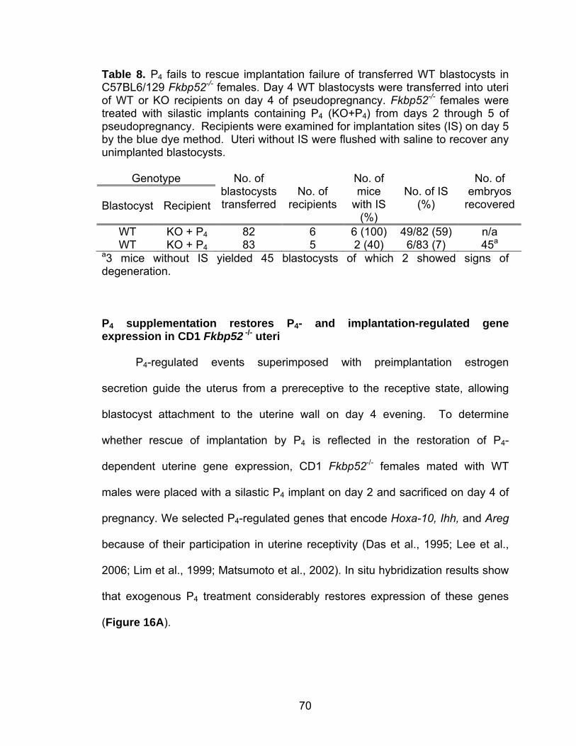
70
Table 8. P4 fails to rescue implantation failure of transferred WT blastocysts in C57BL6/129 Fkbp52-/- females. Day 4 WT blastocysts were transferred into uteri of WT or KO recipients on day 4 of pseudopregnancy. Fkbp52-/- females were treated with silastic implants containing P4 (KO+P4) from days 2 through 5 of pseudopregnancy. Recipients were examined for implantation sites (IS) on day 5 by the blue dye method. Uteri without IS were flushed with saline to recover any unimplanted blastocysts.
Genotype Blastocyst
Recipient
No. of blastocysts transferred
No. of
recipients
No. of mice
with IS (%)
No. of IS
(%)
No. of embryos
recovered
WT KO + P4 82 6 6 (100) 49/82 (59) n/a WT KO + P4 83 5 2 (40) 6/83 (7) 45a
a3 mice without IS yielded 45 blastocysts of which 2 showed signs of degeneration. P4 supplementation restores P4- and implantation-regulated gene expression in CD1 Fkbp52 -/- uteri
P4-regulated events superimposed with preimplantation estrogen
secretion guide the uterus from a prereceptive to the receptive state, allowing
blastocyst attachment to the uterine wall on day 4 evening. To determine
whether rescue of implantation by P4 is reflected in the restoration of P4-
dependent uterine gene expression, CD1 Fkbp52-/- females mated with WT
males were placed with a silastic P4 implant on day 2 and sacrificed on day 4 of
pregnancy. We selected P4-regulated genes that encode Hoxa-10, Ihh, and Areg
because of their participation in uterine receptivity (Das et al., 1995; Lee et al.,
2006; Lim et al., 1999; Matsumoto et al., 2002). In situ hybridization results show
that exogenous P4 treatment considerably restores expression of these genes
(Figure 16A).
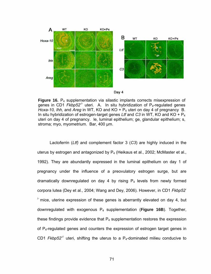
71
Lactoferrin (Ltf) and complement factor 3 (C3) are highly induced in the
uterus by estrogen and antagonized by P4 (Heikaus et al., 2002; McMaster et al.,
1992). They are abundantly expressed in the luminal epithelium on day 1 of
pregnancy under the influence of a preovulatory estrogen surge, but are
dramatically downregulated on day 4 by rising P4 levels from newly formed
corpora lutea (Dey et al., 2004; Wang and Dey, 2006). However, in CD1 Fkbp52-
/- mice, uterine expression of these genes is aberrantly elevated on day 4, but
downregulated with exogenous P4 supplementation (Figure 16B). Together,
these findings provide evidence that P4 supplementation restores the expression
of P4-regulated genes and counters the expression of estrogen target genes in
CD1 Fkbp52-/- uteri, shifting the uterus to a P4-dominated milieu conducive to
Figure 16. P4 supplementation via silastic implants corrects misexpression of genes in CD1 Fkbp52-/- uteri. A. In situ hybridization of P4-regulated genes Hoxa-10, Ihh, and Areg in WT, KO and KO + P4 uteri on day 4 of pregnancy B. In situ hybridization of estrogen-target genes Ltf and C3 in WT, KO and KO + P4 uteri on day 4 of pregnancy. le, luminal epithelium; ge, glandular epithelium; s, stroma; myo, myometrium. Bar, 400 µm.
72
uterine receptivity as opposed to one of estrogenic dominance that is detrimental
to uterine receptivity.
The fact that exogenous P4 treatment fully rescues implantation in CD1
Fkbp52-/- females but is largely ineffective in C57BL6/129 Fkbp52-/- mice
provoked us to examine whether P4 treatment restores the expression of P4-
regulated genes in C57BL6/129 Fkbp52-/- uteri similar to that observed in CD1
Fkbp52-/- mice. We found that although exogenous P4 treatment considerably
restores Hoxa-10 and Ihh expression in mice on this genetic background, Areg
expression remains low to undetectable with P4 treatment (Figure 17A). More
interestingly, exogenous P4, which normally inhibits estrogen-responsive Ltf
expression in CD1 Fkbp52-/- uteri on day 4, was not effective in attenuating Ltf
expression in C57BL6/129 Fkbp52-/- uteri (Figure 17B).
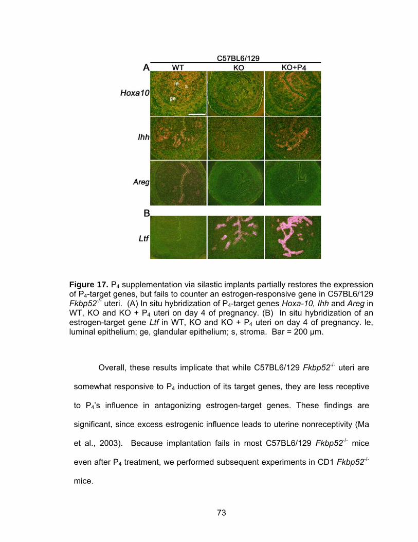
73
Overall, these results implicate that while C57BL6/129 Fkbp52-/- uteri are
somewhat responsive to P4 induction of its target genes, they are less receptive
to P4’s influence in antagonizing estrogen-target genes. These findings are
significant, since excess estrogenic influence leads to uterine nonreceptivity (Ma
et al., 2003). Because implantation fails in most C57BL6/129 Fkbp52-/- mice
even after P4 treatment, we performed subsequent experiments in CD1 Fkbp52-/-
mice.
Figure 17. P4 supplementation via silastic implants partially restores the expression of P4-target genes, but fails to counter an estrogen-responsive gene in C57BL6/129 Fkbp52-/- uteri. (A) In situ hybridization of P4-target genes Hoxa-10, Ihh and Areg in WT, KO and KO + P4 uteri on day 4 of pregnancy. (B) In situ hybridization of an estrogen-target gene Ltf in WT, KO and KO + P4 uteri on day 4 of pregnancy. le, luminal epithelium; ge, glandular epithelium; s, stroma. Bar = 200 µm.
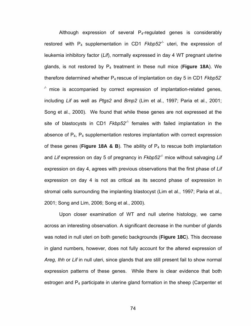
74
Although expression of several P4-regulated genes is considerably
restored with P4 supplementation in CD1 Fkbp52-/- uteri, the expression of
leukemia inhibitory factor (Lif), normally expressed in day 4 WT pregnant uterine
glands, is not restored by P4 treatment in these null mice (Figure 18A). We
therefore determined whether P4 rescue of implantation on day 5 in CD1 Fkbp52-
/- mice is accompanied by correct expression of implantation-related genes,
including Lif as well as Ptgs2 and Bmp2 (Lim et al., 1997; Paria et al., 2001;
Song et al., 2000). We found that while these genes are not expressed at the
site of blastocysts in CD1 Fkbp52-/- females with failed implantation in the
absence of P4, P4 supplementation restores implantation with correct expression
of these genes (Figure 18A & B). The ability of P4 to rescue both implantation
and Lif expression on day 5 of pregnancy in Fkbp52-/- mice without salvaging Lif
expression on day 4, agrees with previous observations that the first phase of Lif
expression on day 4 is not as critical as its second phase of expression in
stromal cells surrounding the implanting blastocyst (Lim et al., 1997; Paria et al.,
2001; Song and Lim, 2006; Song et al., 2000).
Upon closer examination of WT and null uterine histology, we came
across an interesting observation. A significant decrease in the number of glands
was noted in null uteri on both genetic backgrounds (Figure 18C). This decrease
in gland numbers, however, does not fully account for the altered expression of
Areg, Ihh or Lif in null uteri, since glands that are still present fail to show normal
expression patterns of these genes. While there is clear evidence that both
estrogen and P4 participate in uterine gland formation in the sheep (Carpenter et

75
al., 2003; Gray et al., 2001), our data also implicate a potential role for FKBP52-
PR signaling in mouse uterine gland formation and function. This is an exciting
finding but warrants further investigation.
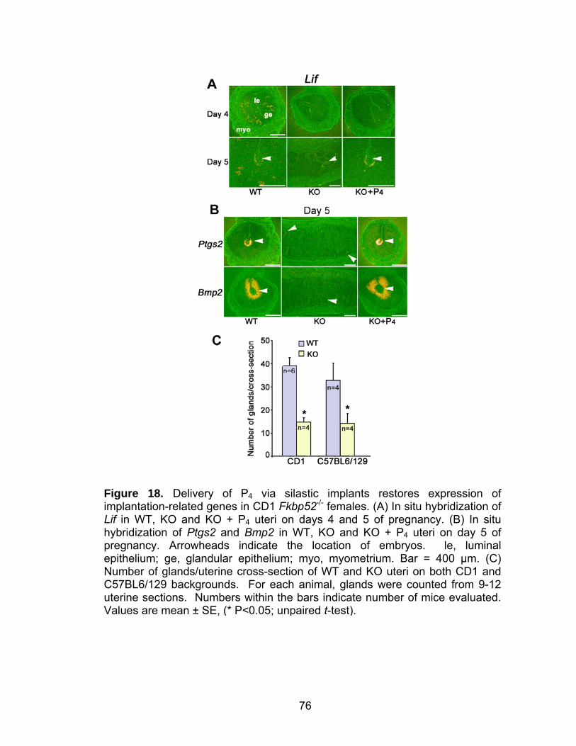
76
Figure 18. Delivery of P4 via silastic implants restores expression of implantation-related genes in CD1 Fkbp52-/- females. (A) In situ hybridization of Lif in WT, KO and KO + P4 uteri on days 4 and 5 of pregnancy. (B) In situ hybridization of Ptgs2 and Bmp2 in WT, KO and KO + P4 uteri on day 5 of pregnancy. Arrowheads indicate the location of embryos. le, luminal epithelium; ge, glandular epithelium; myo, myometrium. Bar = 400 µm. (C) Number of glands/uterine cross-section of WT and KO uteri on both CD1 and C57BL6/129 backgrounds. For each animal, glands were counted from 9-12 uterine sections. Numbers within the bars indicate number of mice evaluated. Values are mean ± SE, (* P<0.05; unpaired t-test).
77
Post-implantation defects in P4-treated CD1 Fkbp52-/- females
Our observation of unimplanted blastocysts recovered from CD1 Fkbp52-/-
females examined on day 5 of pregnancy suggested deferral of implantation
timing in these mice as is observed in cPLA2α-/- or Lpa3
-/- mice (Song et al., 2002;
Ye et al., 2005). To test for this possibility, CD1 Fkbp52-/- mice were sacrificed
on days 6 and 8 of pregnancy. We found that unlike cPLA2α-/- or Lpa3
-/- mice,
implantation timing is not altered; rather the process still drastically fails in
Fkbp52-/- females (Figure 19A, B & Table 6). Again, blastocysts were recovered
from CD1 Fkbp52-/- uteri on these days, confirming implantation as the major
defect in these mice (Figure 19A, B & Table 6).
78
Figure 19. P4 delivery via silastic implants rescues blastocyst-induced, but not oil-induced, decidualization in Fkbp52-/- females. Representative photographs of WT, KO and KO + P4 uteri on day 6 (A), day 8 (B) and day 12 (C) of pregnancy are shown. Arrowheads and short arrows indicate the location of ovary and IS, respectively. Representative images of recovered unimplanted blastocysts from uteri (long arrow) of KO females without IS are shown. Bar = 50 µm. (D) Experimentally induced decidualization fails in KO females on both genetic backgrounds. Representative photomicrographs of WT, KO and KO + P4 uteri on day 8 of pseudopregnancy. On day 4 of pseudopregnancy, 25 µl of oil was infused intraluminally in one uterine horn (oil); the contralateral horn without oil infusion served as a control (con). (E) Fold changes in weight between oil-infused and non-infused control uterine horns. Numbers above the bars indicate the number of mice with decidual response/total number of mice examined. Fold changes are presented as mean ± SE (* P<0.05; unpaired t-test).
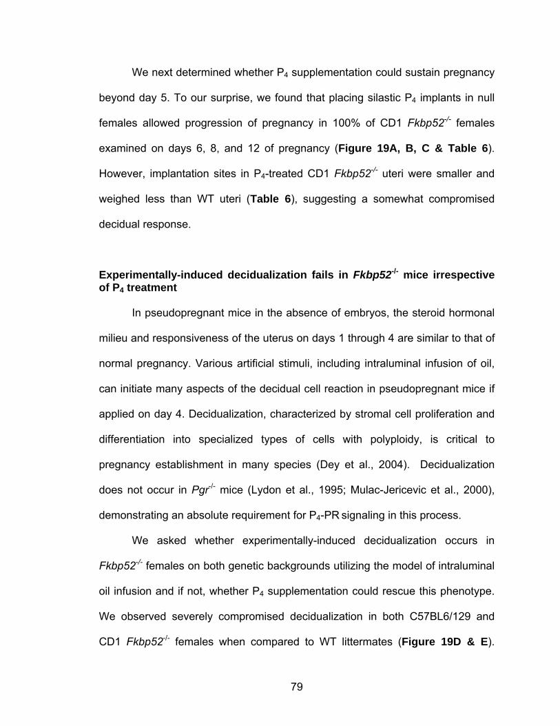
79
We next determined whether P4 supplementation could sustain pregnancy
beyond day 5. To our surprise, we found that placing silastic P4 implants in null
females allowed progression of pregnancy in 100% of CD1 Fkbp52-/- females
examined on days 6, 8, and 12 of pregnancy (Figure 19A, B, C & Table 6).
However, implantation sites in P4-treated CD1 Fkbp52-/- uteri were smaller and
weighed less than WT uteri (Table 6), suggesting a somewhat compromised
decidual response.
Experimentally-induced decidualization fails in Fkbp52-/- mice irrespective of P4 treatment In pseudopregnant mice in the absence of embryos, the steroid hormonal
milieu and responsiveness of the uterus on days 1 through 4 are similar to that of
normal pregnancy. Various artificial stimuli, including intraluminal infusion of oil,
can initiate many aspects of the decidual cell reaction in pseudopregnant mice if
applied on day 4. Decidualization, characterized by stromal cell proliferation and
differentiation into specialized types of cells with polyploidy, is critical to
pregnancy establishment in many species (Dey et al., 2004). Decidualization
does not occur in Pgr-/- mice (Lydon et al., 1995; Mulac-Jericevic et al., 2000),
demonstrating an absolute requirement for P4-PR signaling in this process.
We asked whether experimentally-induced decidualization occurs in
Fkbp52-/- females on both genetic backgrounds utilizing the model of intraluminal
oil infusion and if not, whether P4 supplementation could rescue this phenotype.
We observed severely compromised decidualization in both C57BL6/129 and
CD1 Fkbp52-/- females when compared to WT littermates (Figure 19D & E).
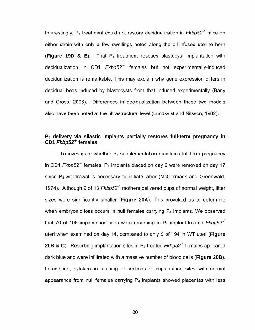
80
Interestingly, P4 treatment could not restore decidualization in Fkbp52-/- mice on
either strain with only a few swellings noted along the oil-infused uterine horn
(Figure 19D & E). That P4 treatment rescues blastocyst implantation with
decidualization in CD1 Fkbp52-/- females but not experimentally-induced
decidualization is remarkable. This may explain why gene expression differs in
decidual beds induced by blastocysts from that induced experimentally (Bany
and Cross, 2006). Differences in decidualization between these two models
also have been noted at the ultrastructural level (Lundkvist and Nilsson, 1982).
P4 delivery via silastic implants partially restores full-term pregnancy in CD1 Fkbp52-/- females
To investigate whether P4 supplementation maintains full-term pregnancy
in CD1 Fkbp52-/- females, P4 implants placed on day 2 were removed on day 17
since P4 withdrawal is necessary to initiate labor (McCormack and Greenwald,
1974). Although 9 of 13 Fkbp52-/- mothers delivered pups of normal weight, litter
sizes were significantly smaller (Figure 20A). This provoked us to determine
when embryonic loss occurs in null females carrying P4 implants. We observed
that 70 of 106 implantation sites were resorbing in P4 implant-treated Fkbp52-/-
uteri when examined on day 14, compared to only 9 of 194 in WT uteri (Figure
20B & C). Resorbing implantation sites in P4-treated Fkbp52-/- females appeared
dark blue and were infiltrated with a massive number of blood cells (Figure 20B).
In addition, cytokeratin staining of sections of implantation sites with normal
appearance from null females carrying P4 implants showed placentas with less
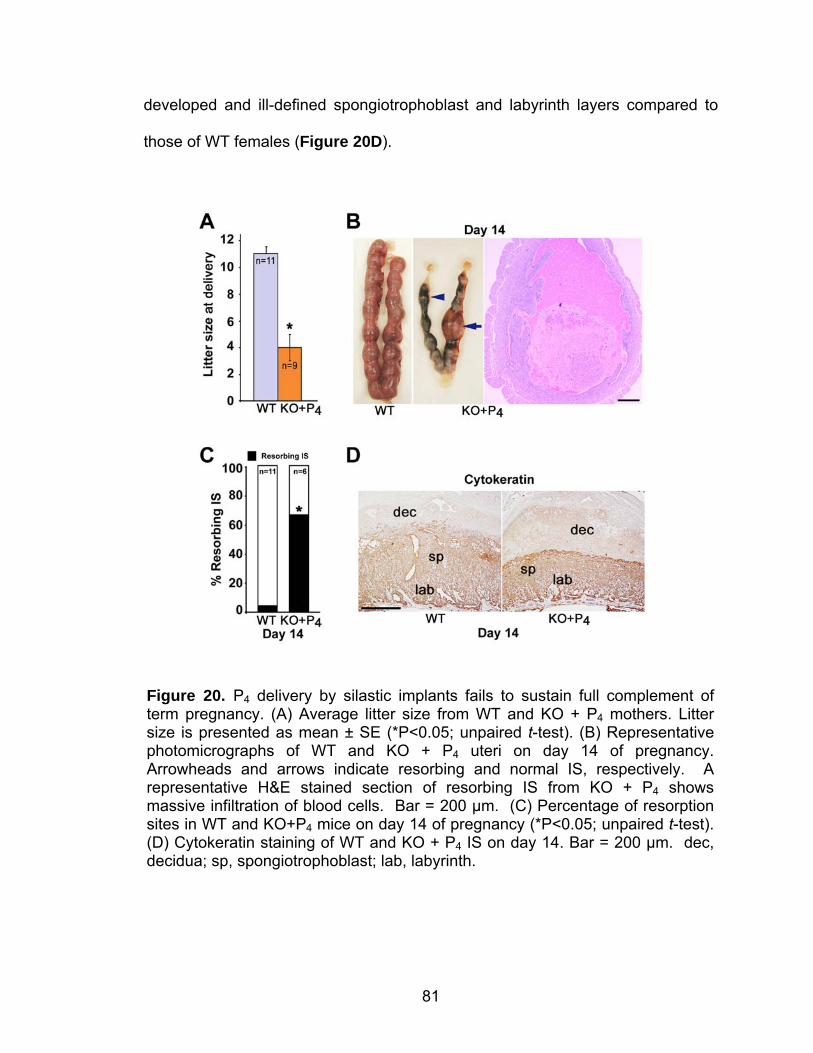
81
developed and ill-defined spongiotrophoblast and labyrinth layers compared to
those of WT females (Figure 20D).
Figure 20. P4 delivery by silastic implants fails to sustain full complement of term pregnancy. (A) Average litter size from WT and KO + P4 mothers. Litter size is presented as mean ± SE (*P<0.05; unpaired t-test). (B) Representative photomicrographs of WT and KO + P4 uteri on day 14 of pregnancy. Arrowheads and arrows indicate resorbing and normal IS, respectively. A representative H&E stained section of resorbing IS from KO + P4 shows massive infiltration of blood cells. Bar = 200 µm. (C) Percentage of resorption sites in WT and KO+P4 mice on day 14 of pregnancy (*P<0.05; unpaired t-test). (D) Cytokeratin staining of WT and KO + P4 IS on day 14. Bar = 200 µm. dec, decidua; sp, spongiotrophoblast; lab, labyrinth.
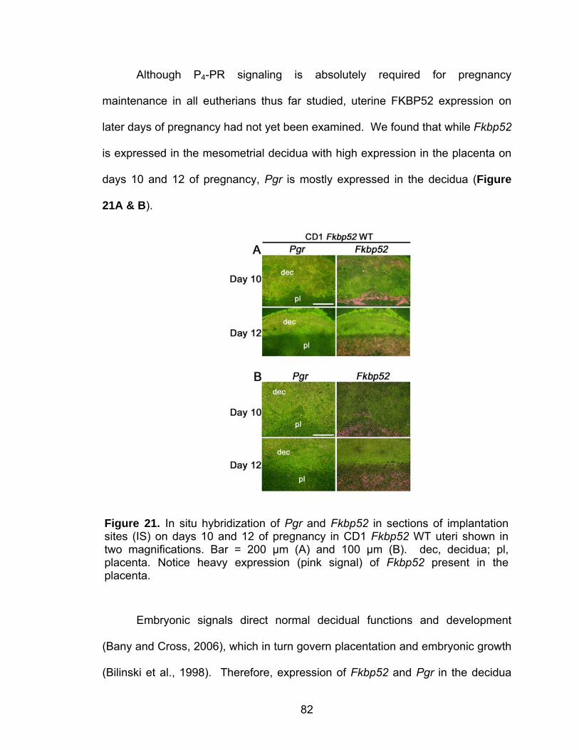
82
Although P4-PR signaling is absolutely required for pregnancy
maintenance in all eutherians thus far studied, uterine FKBP52 expression on
later days of pregnancy had not yet been examined. We found that while Fkbp52
is expressed in the mesometrial decidua with high expression in the placenta on
days 10 and 12 of pregnancy, Pgr is mostly expressed in the decidua (Figure
21A & B).
Embryonic signals direct normal decidual functions and development
(Bany and Cross, 2006), which in turn govern placentation and embryonic growth
(Bilinski et al., 1998). Therefore, expression of Fkbp52 and Pgr in the decidua
Figure 21. In situ hybridization of Pgr and Fkbp52 in sections of implantation sites (IS) on days 10 and 12 of pregnancy in CD1 Fkbp52 WT uteri shown in two magnifications. Bar = 200 µm (A) and 100 µm (B). dec, decidua; pl, placenta. Notice heavy expression (pink signal) of Fkbp52 present in the placenta.
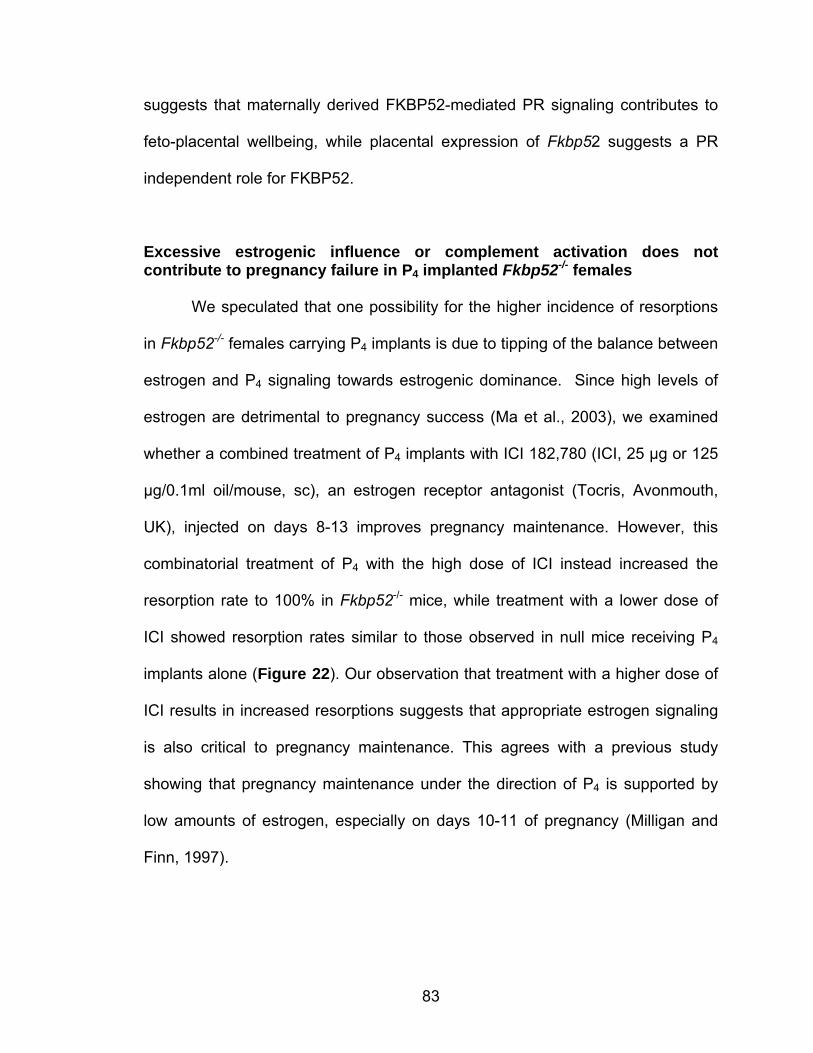
83
suggests that maternally derived FKBP52-mediated PR signaling contributes to
feto-placental wellbeing, while placental expression of Fkbp52 suggests a PR
independent role for FKBP52.
Excessive estrogenic influence or complement activation does not contribute to pregnancy failure in P4 implanted Fkbp52-/- females
We speculated that one possibility for the higher incidence of resorptions
in Fkbp52-/- females carrying P4 implants is due to tipping of the balance between
estrogen and P4 signaling towards estrogenic dominance. Since high levels of
estrogen are detrimental to pregnancy success (Ma et al., 2003), we examined
whether a combined treatment of P4 implants with ICI 182,780 (ICI, 25 µg or 125
µg/0.1ml oil/mouse, sc), an estrogen receptor antagonist (Tocris, Avonmouth,
UK), injected on days 8-13 improves pregnancy maintenance. However, this
combinatorial treatment of P4 with the high dose of ICI instead increased the
resorption rate to 100% in Fkbp52-/- mice, while treatment with a lower dose of
ICI showed resorption rates similar to those observed in null mice receiving P4
implants alone (Figure 22). Our observation that treatment with a higher dose of
ICI results in increased resorptions suggests that appropriate estrogen signaling
is also critical to pregnancy maintenance. This agrees with a previous study
showing that pregnancy maintenance under the direction of P4 is supported by
low amounts of estrogen, especially on days 10-11 of pregnancy (Milligan and
Finn, 1997).
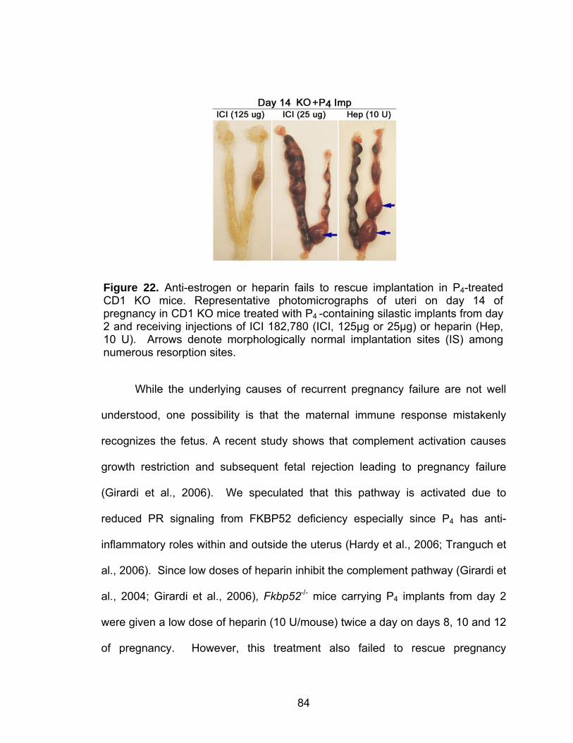
84
While the underlying causes of recurrent pregnancy failure are not well
understood, one possibility is that the maternal immune response mistakenly
recognizes the fetus. A recent study shows that complement activation causes
growth restriction and subsequent fetal rejection leading to pregnancy failure
(Girardi et al., 2006). We speculated that this pathway is activated due to
reduced PR signaling from FKBP52 deficiency especially since P4 has anti-
inflammatory roles within and outside the uterus (Hardy et al., 2006; Tranguch et
al., 2006). Since low doses of heparin inhibit the complement pathway (Girardi et
al., 2004; Girardi et al., 2006), Fkbp52-/- mice carrying P4 implants from day 2
were given a low dose of heparin (10 U/mouse) twice a day on days 8, 10 and 12
of pregnancy. However, this treatment also failed to rescue pregnancy
Figure 22. Anti-estrogen or heparin fails to rescue implantation in P4-treated CD1 KO mice. Representative photomicrographs of uteri on day 14 of pregnancy in CD1 KO mice treated with P4 -containing silastic implants from day 2 and receiving injections of ICI 182,780 (ICI, 125µg or 25µg) or heparin (Hep, 10 U). Arrows denote morphologically normal implantation sites (IS) among numerous resorption sites.
85
maintenance in these mice (Figure 22), suggesting that activation of the
complement pathway is not a major contributing factor for pregnancy failure in
P4-treated Fkbp52-/- mice.
Differential P4-PR signaling is required for successful full-term pregnancy
Normal serum P4 levels during pregnancy in CD1 WT mice on days 5 and
14 of pregnancy range between 40-47 ng/ml (Figure 15 & 23A). In our
experiments, silastic P4 implants provided an increase in serum P4 levels
sufficient to induce uterine receptivity and rescue implantation in CD1 Fkbp52-/-
females, but failed to maintain full complement of term pregnancy. We
speculated that increasing P4 levels even further would rectify this failure. We
injected P4 subcutaneously at a dose of 2 mg/ml per mouse daily to further
increase P4 serum levels. We observed that in null mice, serum P4 levels
increased to ~156 ng/ml by daily injection as compared to ~100 ng/ml in mice
treated with P4-implants, assessed on day 14 of pregnancy (Figure 23A). To our
surprise, these elevated P4 levels significantly improved pregnancy maintenance
in null females; the number and weights of implantation sites on day 14 were
comparable to those in WT mothers (Figure 23B, C & D). Pregnancy
maintenance in WT mice exposed to a similar P4 injection regimen was normal.
86
Our next objective was to examine whether daily P4 injections results in
full complement of term pregnancy in null females. CD1 Fkbp52-/- females mated
with WT males were injected daily with P4 from days 2 through 17 of pregnancy
and monitored for term delivery on day 20. We observed that all P4-injected null
females carried pups to term, and the average litter size was comparable to
those of WT mothers (Figure 23E). Pup weights from null and WT mothers at the
age of weaning and during early development were also similar (data not shown).
Figure 23. Daily P4 injections restore full complement of term pregnancy in CD1 Fkbp52-/- females. (A) Serum P4 levels in WT and KO + P4 mice on day 14 of pregnancy. KO mice were exposed daily to P4 either via silastic implants (Imp) or subcutaneous injection (Inj; 2 mg/ml) from day 2 of pregnancy. Bars with different letters are significantly different (P<0.05, univariate ANOVA). (B) Average number of IS between WT and KO + P4 Inj on day 14 is not significantly different (P>0.1; unpaired t-test). (C) Weights of IS from WT and KO + P4 Inj mice on day 14 of pregnancy are not significantly different (P>0.1;unpaired t-test). (D) A representative photomicrograph of WT and KO + P4 Inj uteri on day 14 of pregnancy is shown. (E) The average litter size from WT and KO + P4 Inj mice is not significantly different (P>0.05; unpaired t-test). All values are mean ± SE.
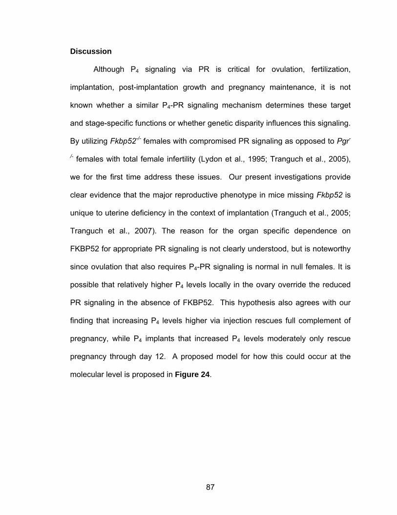
87
Discussion
Although P4 signaling via PR is critical for ovulation, fertilization,
implantation, post-implantation growth and pregnancy maintenance, it is not
known whether a similar P4-PR signaling mechanism determines these target
and stage-specific functions or whether genetic disparity influences this signaling.
By utilizing Fkbp52-/- females with compromised PR signaling as opposed to Pgr-
/- females with total female infertility (Lydon et al., 1995; Tranguch et al., 2005),
we for the first time address these issues. Our present investigations provide
clear evidence that the major reproductive phenotype in mice missing Fkbp52 is
unique to uterine deficiency in the context of implantation (Tranguch et al., 2005;
Tranguch et al., 2007). The reason for the organ specific dependence on
FKBP52 for appropriate PR signaling is not clearly understood, but is noteworthy
since ovulation that also requires P4-PR signaling is normal in null females. It is
possible that relatively higher P4 levels locally in the ovary override the reduced
PR signaling in the absence of FKBP52. This hypothesis also agrees with our
finding that increasing P4 levels higher via injection rescues full complement of
pregnancy, while P4 implants that increased P4 levels moderately only rescue
pregnancy through day 12. A proposed model for how this could occur at the
molecular level is proposed in Figure 24.
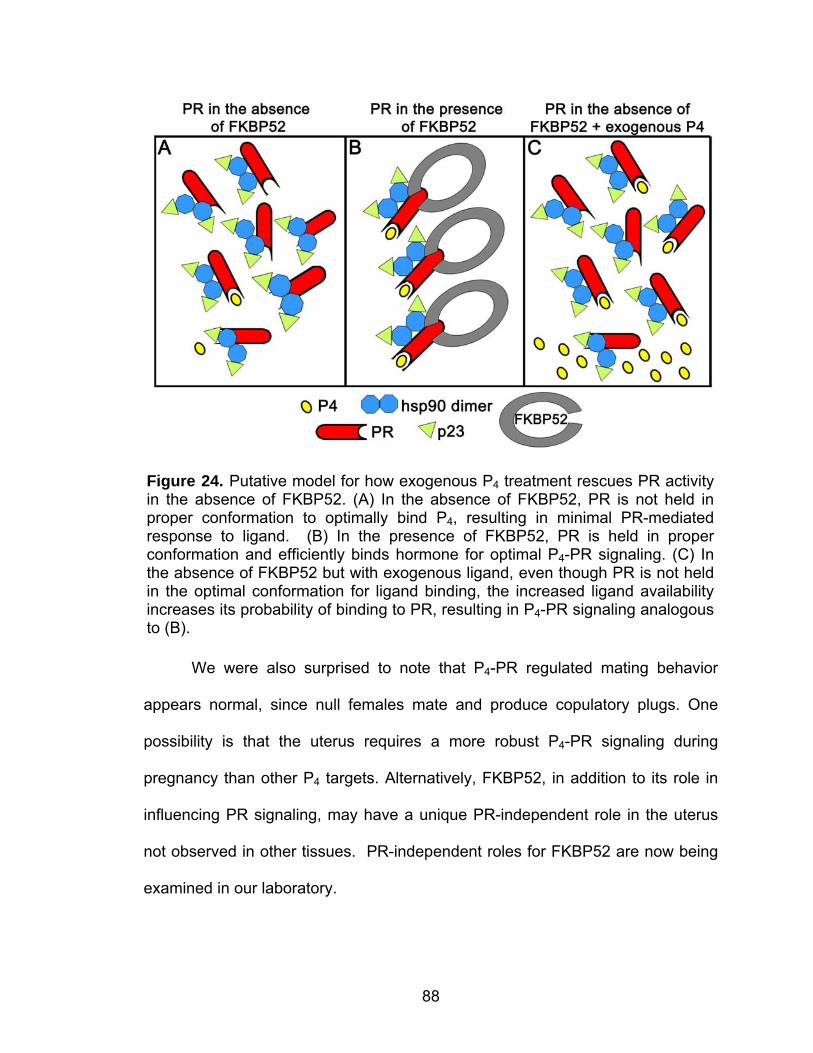
88
We were also surprised to note that P4-PR regulated mating behavior
appears normal, since null females mate and produce copulatory plugs. One
possibility is that the uterus requires a more robust P4-PR signaling during
pregnancy than other P4 targets. Alternatively, FKBP52, in addition to its role in
influencing PR signaling, may have a unique PR-independent role in the uterus
not observed in other tissues. PR-independent roles for FKBP52 are now being
examined in our laboratory.
Figure 24. Putative model for how exogenous P4 treatment rescues PR activity in the absence of FKBP52. (A) In the absence of FKBP52, PR is not held in proper conformation to optimally bind P4, resulting in minimal PR-mediated response to ligand. (B) In the presence of FKBP52, PR is held in proper conformation and efficiently binds hormone for optimal P4-PR signaling. (C) In the absence of FKBP52 but with exogenous ligand, even though PR is not held in the optimal conformation for ligand binding, the increased ligand availability increases its probability of binding to PR, resulting in P4-PR signaling analogous to (B).
89
P4 signaling via PR plays major roles at essentially all stages of pregnancy
from ovulation through parturition. It is surprising to see that genetic makeup of a
species alters such a fundamental signaling pathway. While FKBP52 is essential
to support implantation in both strains of mice in the absence of exogenous P4,
FKBP52’s role becomes less significant in CD1 mice exposed to high levels of
P4, while still remaining crucial in C57BL6/129 mice under similar treatment
conditions. The contrasting reproductive phenotypes of Fkbp52-/- mice on
C57BL6/129 and CD1 backgrounds with respect to P4 rescue indeed provides
first evidence that P4-PR-FKBP52 signaling is a function of genetic makeup.
Coordinated interactions of P4 and estrogen are essential to uterine
receptivity and implantation. While one aspect of P4 signaling is to correctly
orchestrate P4-responsive genes, another aspect is to appropriately constrain
and/or synergize estrogen-responsive genes in the uterus. One cause of
implantation failure in C57BL6/129, but not in CD1, Fkbp52-/- females
supplemented with P4 could be due to the failure of the uterus to attain optimal
receptivity arising from altered expression of Areg and/or Ltf. This inappropriate
gene expression in the presence of P4 may reflect even lower basal PR activity in
C57BL6/129 Fkbp52-/- uteri compared to CD1 Fkbp52-/- uteri. Alternatively,
differential expression of modifier genes in these two strains of mice could
contribute to differential uterine responsiveness to P4-PR signaling in the
absence of FKBP52.
There is evidence that rodent blastocysts synthesize P4 or structurally
similar steroids (Carson et al., 1982; Wu, 1987; Wu and Liu, 1990) and that gene
90
expression differs in embryo-induced decidua versus experimentally induced
deciduoma (Bany and Cross, 2006). Since our previous results show that
Fkbp52 and Pgr are expressed in blastocysts (Tranguch et al., 2005), it is
possible that P4 synthesized from blastocysts enhances PR-FKBP52 signaling
locally at their sites of apposition in the uterus. This could explain failure of oil-
induced decidualization but not of that induced by implanting blastocysts in CD1
Fkbp52-/- mice supplemented with P4. Alternatively, signaling arising from
blastocysts may influence other uterine functions. For example, the gene
encoding heparin-binding epidermal growth factor-like growth factor (Hbegf) is
induced in implantation-competent blastocysts which can induce uterine Hbegf to
initiate the implantation cascade. In fact, Affi-Gel Blue beads presoaked in rHB-
EGF, when transferred into uterine lumens of pseudopregnant mice on day 4,
show implantation-like responses similar to those induced by living blastocysts
including upregulation of Hbegf and Bmp2 (Hamatani et al., 2004; Paria et al.,
2001).
P4-PR signaling is critical throughout pregnancy until its downregulation for
the onset of parturition. Indeed, ovariectomizing mice practically at any stage of
pregnancy causes resorptions and/or abortion (Sharma and Bulmer, 1983).
However, the magnitude of this signaling during various stages of pregnancy
remains unknown. The use of Fkbp52-/- females has enabled us to show that the
requirement for P4-PR signaling is different for uterine receptivity, implantation
and post-implantation growth. This signaling appears tightly regulated since
blood levels of ~100 ng/ml of P4 are adequate to induce uterine receptivity and
91
implantation, but levels above 150 ng/ml are required for full complement of
pregnancy success in the absence of FKBP52. This suggests that a more robust
P4-PR signaling is required for pregnancy maintenance than is required for
uterine receptivity, implantation and decidualization. It is also possible that a
burst of P4 levels as provided by daily injections is more amenable to pregnancy
sustenance than the relatively constant levels maintained with silastic implants.
The observation that differential P4 levels are required for various stages
of pregnancy insinuates that signaling targets are different. We believe that P4-
PR signaling for uterine preparation, implantation and decidualization is primarily
targeted to uterine epithelial and stromal cells. This is consistent with P4’s known
roles in epithelial differentiation and stromal cell proliferation during the
periimplantation period. On the other hand, P4-PR signaling for pregnancy
maintenance is directed more towards keeping the myometrium quiescent until
parturition and providing sanctuary to the growing fetus from mother’s
immunological surveillance. P4-PR signaling is also known to regulate
angiogenesis (Ma et al., 2001), a process integral to placental development and
pregnancy maintenance. Since the events during the course of pregnancy are
very dynamic, P4-PR signaling at various targets could be overlapping. We
consider FKBP52’s influence on P4-PR signaling during pregnancy primarily to
be of maternal origin, since both PR and FKBP52 are expressed in the decidua
on days 10 and 12 of pregnancy. The high expression of FKBP52 in the
placenta implies a PR-independent role for FKBP52 in placentation. This
requires further investigation, but this putative PR-independent role is not critical
92
since pregnancies are completed to term with normal litter sizes in CD1 Fkbp52
null females receiving daily P4 injections.
An interesting observation is the successful nursing of pups to weaning by
P4-injected CD1 Fkbp52-/- mothers. Mammary morphogenesis during pregnancy
requires P4-PR signaling (Mulac-Jericevic et al., 2003; Mulac-Jericevic et al.,
2000), but the latter stages of lactogenesis and lactation correspond to
withdrawal of this signaling (Neville et al., 2002). It was recently reported that
FKBP52 may not be critical for P4-PR signaling in the mammary gland, since
exogenously provided P4 could stimulate mammary morphogenesis in Fkbp52-/-
mice (Yang et al., 2006). This is similar to our studies in which Fkbp52-/- mothers
were injected with exogenous P4 prior to parturition. These results suggest that
exogenously supplemented P4 overcomes the P4-resistant state in the mammary
gland and uterus in the absence of FKBP52.
The implanting blastocyst is the stimulus for normal decidualization in
mice. However, in humans, stromal cells undergo decidualization during the
receptive phase in each menstrual cycle in the absence of blastocysts. This pre-
decidualization is thought to be critical for blastocyst implantation in the pregnant
uterus (Chobotova et al., 2005). Our findings that P4 supplementation fails to
rescue oil-induced decidualization in Fkbp52 null uteri may therefore have
implications for pre-decidualization events in P4-resistant women.
P4 resistance is also a hallmark of endometriosis, a condition that affects
an estimated 5 million women in the United States alone (Berkley et al., 2005;
Bulun et al., 2006; Fang et al., 2004). In fact, a recent study examining global

93
gene expression profiles in endometria of women with or without endometriosis
found dysregulation of many known P4-target genes during the window of uterine
receptivity (Kao et al., 2003). While the mechanism(s) of P4 resistance remain
unclear, one speculation is that downregulation of PR contributes to P4
resistance, but other studies refute this concept (Bulun et al., 2006). Whether
FKBP52 expression differs in normal and endometriotic tissues has not been
examined. However, the finding that human FKBP52 interacts with and
potentiates human PR activity in mouse embryonic fibroblasts (Tranguch et al.,
2005), and our preliminary observation on a limited number of samples showing
FKBP52 and PR expression in human endometria in proliferative and secretory
phases (Figure 25A & B) suggest a potential role for uterine P4-PR-FKBP52
signaling in women.
In fact, three separate clinical trials found that P4 treatment resulted in a
statistically significant decrease in miscarriages in women with history of three or
Figure 25. PR and FKBP52 expression in human proliferative (A) and secretory (B) endometrium shown at two magnifications. Bar = 100 µm (A) and 200 µm (B).
94
more consecutive pregnancy losses (Oates-Whitehead et al., 2003). In this
respect, pregnancy rescue in CD1 Fkbp52-/- mice by daily P4 injections is
clinically relevant for women who are infertile due to P4 resistance. These are
exciting results especially since no significant differences in adverse effects were
found between P4 treatment and control groups. These findings in humans are
similar to our findings in mice that exogenous P4 injections in WT females do not
adversely affect pregnancy. Fkbp52 null mice with normal PR and P4 levels but
reduced PR activity constitute a unique model to study P4 resistance specifically
in uterine biology, and our findings hopefully will encourage human studies to
determine whether FKBP52 status influences P4 resistance in the uterus.
95
REFERENCES
Arriza, J. L., et al., 1987. Cloning of human mineralocorticoid receptor complementary DNA: structural and functional kinship with the glucocorticoid receptor. Science. 237, 268-75.
Bany, B. M., Cross, J. C., 2006. Post-implantation mouse conceptuses produce
paracrine signals that regulate the uterine endometrium undergoing decidualization. Dev Biol. 294, 445-56.
Barent, R. L., et al., 1998. Analysis of FKBP51/FKBP52 chimeras and mutants
for Hsp90 binding and association with progesterone receptor complexes. Mol Endocrinol. 12, 342-54.
Benson, G. V., et al., 1996. Mechanisms of reduced fertility in Hoxa-10 mutant
mice: uterine homeosis and loss of maternal Hoxa-10 expression. Development. 122, 2687-96.
Berkley, K. J., et al., 2005. The pains of endometriosis. Science. 308, 1587-9. Bilinski, P., et al., 1998. Maternal IL-11Ralpha function is required for normal
decidua and fetoplacental development in mice. Genes Dev. 12, 2234-43. Bonyadi, M., et al., 1997. Mapping of a major genetic modifier of embryonic
lethality in TGF beta 1 knockout mice. Nat Genet. 15, 207-11. Bouin, P., Ancel, P., 1910. Sur le determinisme de la preparation de l'uterus a la
fixtion de l'oeuf. J Physiol Path Gen. 12, 1-16. Brinkmann, A. O., et al., 1989. Structure and function of the androgen receptor.
Urol Res. 17, 87-93. Bulun, S. E., et al., 2006. Progesterone resistance in endometriosis: link to failure
to metabolize estradiol. Mol Cell Endocrinol. 248, 94-103. Carpenter, K. D., et al., 2003. Estrogen and antiestrogen effects on neonatal
ovine uterine development. Biol Reprod. 69, 708-17. Carson, D. D., et al., 1982. Synthesis of steroids in postimplantation mouse
embryos cultured in vitro. Dev Biol. 91, 402-12. Chappell, P. E., et al., 1997. Endocrine defects in mice carrying a null mutation
for the progesterone receptor gene. Endocrinology. 138, 4147-52.
96
Cheung-Flynn, J., et al., 2005. Physiological role for the cochaperone FKBP52 in androgen receptor signaling. Mol Endocrinol. 19, 1654-66.
Chobotova, K., et al., 2005. Heparin-binding epidermal growth factor and its
receptors mediate decidualization and potentiate survival of human endometrial stromal cells. J Clin Endocrinol Metab. 90, 913-9.
Conneely, O. M., et al., 2001. Reproductive functions of the progesterone
receptor isoforms: lessons from knock-out mice. Mol Cell Endocrinol. 179, 97-103.
Corner, G. W., Sr., 1943. On the female testes or ovaries. University of California
Press, Berkeley. Corner, G. W., Sr., 1974. The early history of progesterone. Gynecol Invest. 5,
106-12. Czar, M. J., et al., 1995. Evidence that the FK506-binding immunophilin heat
shock protein 56 is required for trafficking of the glucocorticoid receptor from the cytoplasm to the nucleus. Mol Endocrinol. 9, 1549-60.
Daikoku, T., et al., 2003. Expression of hypoxia-inducible factors in the peri-
implantation mouse uterus is regulated in a cell-specific and ovarian steroid hormone-dependent manner. Evidence for differential function of HIFs during early pregnancy. J Biol Chem. 278, 7683-91.
Daikoku, T., et al., 2005. Proteomic analysis identifies immunophilin FK506
binding protein 4 (FKBP52) as a downstream target of Hoxa10 in the periimplantation mouse uterus. Mol Endocrinol. 19, 683-97.
Das, S. K., et al., 1995. Amphiregulin is an implantation-specific and
progesterone-regulated gene in the mouse uterus. Mol Endocrinol. 9, 691-705.
Davies, T. H., et al., 2002. A new first step in activation of steroid receptors:
hormone-induced switching of FKBP51 and FKBP52 immunophilins. J Biol Chem. 277, 4597-600.
Dey, S. K., Implantation. In: E. Y. Adashi, et al., Eds.), Reproductive
endocrinology, surgery and technology. Lippincott-Raven, New York, 1996, pp. 421-434.
Dey, S. K., et al., 2004. Molecular cues to implantation. Endocr Rev. 25, 341-73. Dolle, P., et al., 1991. HOX-4 genes and the morphogenesis of mammalian
genitalia. Genes Dev. 5, 1767-7.
97
Enders, A. C., 1976. Anatomical aspects of implantation. J Reprod Fertil Suppl.
1-15. Enders, A. C., Schlafke, S., 1969. Cytological aspects of trophoblast-uterine
interaction in early implantation. Am J Anat. 125, 1-29. Fang, Z., et al., 2004. Intact progesterone receptors are essential to counteract
the proliferative effect of estradiol in a genetically engineered mouse model of endometriosis. Fertil Steril. 82, 673-8.
Fanghanel, J., Fischer, G., 2004. Insights into the catalytic mechanism of peptidyl
prolyl cis/trans isomerases. Front Biosci. 9, 3453-78. Fannon, S. A., et al., 2001. An abridged history of sex steroid hormone receptor
action. J Appl Physiol. 91, 1854-9. Fraenkel, L., 1903. Die funktion des corpus luteum. Archiv fur gynakologie. 67,
438-445. Freedman, L. P., 1992. Anatomy of the steroid receptor zinc finger region.
Endocr Rev. 13, 129-45. Frobenius, W., 1999. Ludwig Fraenkel: 'spiritus rector' of the early progesterone
research. Eur J Obstet Gynecol Reprod Biol. 83, 115-9. Galigniana, M. D., et al., 2004. Hsp90-binding immunophilins link p53 to dynein
during p53 transport to the nucleus. J Biol Chem. 279, 22483-9. Galigniana, M. D., et al., 2001. Evidence that the peptidylprolyl isomerase
domain of the hsp90-binding immunophilin FKBP52 is involved in both dynein interaction and glucocorticoid receptor movement to the nucleus. J Biol Chem. 276, 14884-9.
Gendron, R. L., et al., 1997. Abnormal uterine stromal and glandular function
associated with maternal reproductive defects in Hoxa-11 null mice. Biol Reprod. 56, 1097-105.
Girardi, G., et al., 2004. Heparin prevents antiphospholipid antibody-induced fetal
loss by inhibiting complement activation. Nat Med. 10, 1222-6. Girardi, G., et al., 2006. Complement activation induces dysregulation of
angiogenic factors and causes fetal rejection and growth restriction. J Exp Med. 203, 2165-75.
98
Gray, C. A., et al., 2001. Effects of neonatal progestin exposure on female reproductive tract structure and function in the adult ewe. Biol Reprod. 64, 797-804.
Greene, G. L., et al., 1986. Sequence and expression of human estrogen
receptor complementary DNA. Science. 231, 1150-4. Hamatani, T., et al., 2004. Global gene expression analysis identifies molecular
pathways distinguishing blastocyst dormancy and activation. Proc Natl Acad Sci U S A. 101, 10326-31.
Hardy, D. B., et al., 2006. Progesterone receptor plays a major antiinflammatory
role in human myometrial cells by antagonism of nuclear factor-kappaB activation of cyclooxygenase 2 expression. Mol Endocrinol. 20, 2724-33.
Heikaus, S., et al., 2002. Responsiveness of endometrial genes Connexin26,
Connexin43, C3 and clusterin to primary estrogen, selective estrogen receptor modulators, phyto- and xenoestrogens. J Mol Endocrinol. 29, 239-49.
Heilbrunn, L., et al., 1939. Initiation of maturation in the frog egg. Physiological
Zoology. 12, 97-100. Hernandez, M. P., et al., 2002. HSP40 binding is the first step in the HSP90
chaperoning pathway for the progesterone receptor. J Biol Chem. 277, 11873-81.
Hong, J., et al., 2007. Deficiency of co-chaperone immunophilin FKBP52
compromises sperm fertilizing capacity. Reproduction. 133, 395-403. Hou, Q., Gorski, J., 1993. Estrogen receptor and progesterone receptor genes
are expressed differentially in mouse embryos during preimplantation development. Proc Natl Acad Sci U S A. 90, 9460-4.
Hsieh-Li, H. M., et al., 1995. Hoxa 11 structure, extensive antisense transcription,
and function in male and female fertility. Development. 121, 1373-85. Huet-Hudson, Y. M., et al., 1989. Cell type-specific localization of c-myc protein
in the mouse uterus: modulation by steroid hormones and analysis of the periimplantation period. Endocrinology. 125, 1683-90.
Huet-Hudson, Y. M., Dey, S. K., 1990. Requirement for progesterone priming
and its long-term effects on implantation in the mouse. Proc Soc Exp Biol Med. 193, 259-63.
99
Izpisua-Belmonte, J. C., et al., 1991. Murine genes related to the Drosophila AbdB homeotic genes are sequentially expressed during development of the posterior part of the body. Embo J. 10, 2279-89.
Jeltsch, J. M., et al., 1986. Cloning of the chicken progesterone receptor. Proc
Natl Acad Sci U S A. 83, 5424-8. Kao, L. C., et al., 2003. Expression profiling of endometrium from women with
endometriosis reveals candidate genes for disease-based implantation failure and infertility. Endocrinology. 144, 2870-81.
Kazeto, Y., et al., 2005. Molecular characterization of three forms of putative
membrane-bound progestin receptors and their tissue-distribution in channel catfish, Ictalurus punctatus. J Mol Endocrinol. 34, 781-91.
Kelleher, N. L., et al., 1999. Localization of labile posttranslational modifications
by electron capture dissociation: the case of gamma-carboxyglutamic acid. Anal Chem. 71, 4250-3.
Kraus, W. L., et al., 1995. Inhibitory cross-talk between steroid hormone
receptors: differential targeting of estrogen receptor in the repression of its transcriptional activity by agonist- and antagonist-occupied progestin receptors. Mol Cell Biol. 15, 1847-57.
Krumlauf, R., 1994. Hox genes in vertebrate development. Cell. 78, 191-201. Lee, K., et al., 2006. Indian hedgehog is a major mediator of progesterone
signaling in the mouse uterus. Nat Genet. 38, 1204-9. Lim, H., et al., 1999. Hoxa-10 regulates uterine stromal cell responsiveness to
progesterone during implantation and decidualization in the mouse. Mol Endocrinol. 13, 1005-17.
Lim, H., et al., 1997. Multiple female reproductive failures in cyclooxygenase 2-
deficient mice. Cell. 91, 197-208. Luetteke, N. C., et al., 1999. Targeted inactivation of the EGF and amphiregulin
genes reveals distinct roles for EGF receptor ligands in mouse mammary gland development. Development. 126, 2739-50.
Luisi, B. F., et al., 1991. Crystallographic analysis of the interaction of the
glucocorticoid receptor with DNA. Nature. 352, 497-505. Lundkvist, O., Nilsson, B. O., 1982. Endometrial ultrastructure in the early uterine
response to blastocysts and artificial deciduogenic stimuli in rats. Cell Tissue Res. 225, 355-64.
100
Lydon, J. P., et al., 1995. Mice lacking progesterone receptor exhibit pleiotropic
reproductive abnormalities. Genes Dev. 9, 2266-78. Ma, W., et al., 2001. Adult tissue angiogenesis: evidence for negative regulation
by estrogen in the uterus. Mol Endocrinol. 15, 1983-92. Ma, W. G., et al., 2003. Estrogen is a critical determinant that specifies the
duration of the window of uterine receptivity for implantation. Proc Natl Acad Sci U S A. 100, 2963-8.
Matsumoto, H., et al., 2002. Indian hedgehog as a progesterone-responsive
factor mediating epithelial-mesenchymal interactions in the mouse uterus. Dev Biol. 245, 280-90.
McCormack, J. T., Greenwald, G. S., 1974. Progesterone and oestradiol-17beta
concentrations in the peripheral plasma during pregnancy in the mouse. J Endocrinol. 62, 101-7.
McLaren, A., 1968. A study of blastocysts during delay and subsequent
implantation in lactating mice. J Endocrinol. 42, 453-63. McLaughlin, S. H., et al., 2002. Stimulation of the weak ATPase activity of human
hsp90 by a client protein. J Mol Biol. 315, 787-98. McMaster, M. T., et al., 1992. Lactoferrin in the mouse uterus: analyses of the
preimplantation period and regulation by ovarian steroids. Mol Endocrinol. 6, 101-11.
Mead, R. A., 1993. Embryonic diapause in vertebrates. J Exp Zool. 266, 629-41. Milligan, S. R., Cohen, P. E., 1994. Silastic implants for delivering physiological
concentrations of progesterone to mice. Reprod Fertil Dev. 6, 235-9. Milligan, S. R., Finn, C. A., 1997. Minimal progesterone support required for the
maintenance of pregnancy in mice. Hum Reprod. 12, 602-7. Mulac-Jericevic, B., et al., 2003. Defective mammary gland morphogenesis in
mice lacking the progesterone receptor B isoform. Proc Natl Acad Sci U S A. 100, 9744-9.
Mulac-Jericevic, B., et al., 2000. Subgroup of reproductive functions of
progesterone mediated by progesterone receptor-B isoform. Science. 289, 1751-4.
101
Nair, S. C., et al., 1997. Molecular cloning of human FKBP51 and comparisons of immunophilin interactions with Hsp90 and progesterone receptor. Mol Cell Biol. 17, 594-603.
Neville, M. C., et al., 2002. Hormonal regulation of mammary differentiation and
milk secretion. J Mammary Gland Biol Neoplasia. 7, 49-66. Notides, A., Gorski, J., 1966. Estrogen-induced synthesis of a specific uterine
protein. Proc Natl Acad Sci U S A. 56, 230-5. O'Malley, B. W., et al., 1970. Progesterone "receptors" in the cytoplasm and
nucleus of chick oviduct target tissue. Proc Natl Acad Sci U S A. 67, 501-8.
Oates-Whitehead, R. M., et al., 2003. Progestogen for preventing miscarriage.
Cochrane Database Syst Rev. CD003511. Paria, B. C., et al., 1998. Histidine decarboxylase gene in the mouse uterus is
regulated by progesterone and correlates with uterine differentiation for blastocyst implantation. Endocrinology. 139, 3958-66.
Paria, B. C., et al., 1993. Blastocyst's state of activity determines the "window" of
implantation in the receptive mouse uterus. Proc Natl Acad Sci U S A. 90, 10159-62.
Paria, B. C., et al., 2001. Cellular and molecular responses of the uterus to
embryo implantation can be elicited by locally applied growth factors. Proc Natl Acad Sci U S A. 98, 1047-52.
Parr, E. L., et al., 1987. Apoptosis as the mode of uterine epithelial cell death
during embryo implantation in mice and rats. Biol Reprod. 36, 211-25. Peluso, J. J., et al., 2006. Progesterone membrane receptor component 1
expression in the immature rat ovary and its role in mediating progesterone's antiapoptotic action. Endocrinology. 147, 3133-40.
Pirkl, F., Buchner, J., 2001. Functional analysis of the Hsp90-associated human
peptidyl prolyl cis/trans isomerases FKBP51, FKBP52 and Cyp40. J Mol Biol. 308, 795-806.
Pointis, G., et al., 1981. Progesterone levels in the circulating blood of the
ovarian and uterine veins during gestation in the mouse. Biol Reprod. 24, 801-5.
Pratt, W. B., et al., 2004. Role of hsp90 and the hsp90-binding immunophilins in
signalling protein movement. Cell Signal. 16, 857-72.
102
Pratt, W. B., Toft, D. O., 1997. Steroid receptor interactions with heat shock
protein and immunophilin chaperones. Endocr Rev. 18, 306-60. Psychoyos, A., Endocrine control of egg implantation. In: R. O. Greep, et al.,
Eds.), Handbook of Physiology. American Physiology Society, Washington, D.C., 1973a, pp. 187-215.
Psychoyos, A., 1973b. Hormonal control of ovoimplantation. Vitam Horm. 31,
201-56. Raza, F. S., et al., 2001. Identification of the rat adrenal zona
fasciculata/reticularis specific protein, inner zone antigen (IZAg), as the putative membrane progesterone receptor. Eur J Biochem. 268, 2141-7.
Riggs, D. L., et al., 2003. The Hsp90-binding peptidylprolyl isomerase FKBP52
potentiates glucocorticoid signaling in vivo. Embo J. 22, 1158-67. Rugh, R., 1935. Ovulation in the frog. I. Pituitary relations in induced ovulation.
Journal of Experimental Zoology. 71, 149-162. Ryan, F., 1940. The stimulus for maturation and for ovulation of the frog's egg.
Physiological Zoology. 13, 383-390. Satokata, I., et al., 1995. Sexually dimorphic sterility phenotypes in Hoxa10-
deficient mice. Nature. 374, 460-3. Schiene-Fischer, C., Yu, C., 2001. Receptor accessory folding helper enzymes:
the functional role of peptidyl prolyl cis/trans isomerases. FEBS Lett. 495, 1-6.
Schowalter, D. B., et al., 1991. Characterization of progesterone receptor binding
to the 90- and 70-kDa heat shock proteins. J Biol Chem. 266, 21165-73. Schwartz, R. J., et al., 1977. Effect of progesterone receptors on transcription.
Ann N Y Acad Sci. 286, 147-60. Sharma, R., Bulmer, D., 1983. The effects of ovariectomy and subsequent
progesterone replacement on the uterus of the pregnant mouse. J Anat. 137 ( Pt 4), 695-703.
Shimada, M., et al., 2004. Down-regulated expression of A disintegrin and
metalloproteinase with thrombospondin-like repeats-1 by progesterone receptor antagonist is associated with impaired expansion of porcine cumulus-oocyte complexes. Endocrinology. 145, 4603-14.
103
Silverstein, A. M., et al., 1999. Different regions of the immunophilin FKBP52 determine its association with the glucocorticoid receptor, hsp90, and cytoplasmic dynein. J Biol Chem. 274, 36980-6.
Smith, D. F., 1993. Dynamics of heat shock protein 90-progesterone receptor
binding and the disactivation loop model for steroid receptor complexes. Mol Endocrinol. 7, 1418-29.
Smith, R. G., et al., 1975. Purification of human uterine progesterone receptor.
Nature. 253, 271-2. Song, H., Lim, H., 2006. Evidence for heterodimeric association of leukemia
inhibitory factor (LIF) receptor and gp130 in the mouse uterus for LIF signaling during blastocyst implantation. Reproduction. 131, 341-9.
Song, H., et al., 2000. Dysregulation of EGF family of growth factors and COX-2
in the uterus during the preattachment and attachment reactions of the blastocyst with the luminal epithelium correlates with implantation failure in LIF-deficient mice. Mol Endocrinol. 14, 1147-61.
Song, H., et al., 2002. Cytosolic phospholipase A2alpha is crucial [correction of
A2alpha deficiency is crucial] for 'on-time' embryo implantation that directs subsequent development. Development. 129, 2879-89.
Tai, P. K., et al., 1986. A 59-kilodalton protein associated with progestin,
estrogen, androgen, and glucocorticoid receptors. Biochemistry. 25, 5269-75.
Tan, J., et al., 1999. Differential uterine expression of estrogen and progesterone
receptors correlates with uterine preparation for implantation and decidualization in the mouse. Endocrinology. 140, 5310-21.
Threadgill, D. W., et al., 1995. Targeted disruption of mouse EGF receptor: effect
of genetic background on mutant phenotype. Science. 269, 230-4. Tian, J., et al., 2000. Identification of XPR-1, a progesterone receptor required for
Xenopus oocyte activation. Proc Natl Acad Sci U S A. 97, 14358-63. Todaro, G. J., Green, H., 1963. Quantitative studies of the growth of mouse
embryo cells in culture and their development into established lines. J Cell Biol. 17, 299-313.
Tranguch, S., et al., 2005. Cochaperone immunophilin FKBP52 is critical to
uterine receptivity for embryo implantation. Proc Natl Acad Sci U S A. 102, 14326-31.
104
Tranguch, S., et al., 2006. Progesterone receptor requires a co-chaperone for signalling in uterine biology and implantation. Reprod Biomed Online. 13, 651-60.
Tranguch, S., et al., 2007. FKBP52 deficiency-conferred uterine progesterone
resistance is genetic background and pregnancy stage specific. J Clin Invest. 117, 1824-1834.
Vegeto, E., et al., 1993. Human progesterone receptor A form is a cell- and
promoter-specific repressor of human progesterone receptor B function. Mol Endocrinol. 7, 1244-55.
Wang, H., Dey, S. K., 2006. Roadmap to embryo implantation: clues from mouse
models. Nat Rev Genet. 7, 185-99. Wang, H., et al., 2004. Rescue of female infertility from the loss of
cyclooxygenase-2 by compensatory up-regulation of cyclooxygenase-1 is a function of genetic makeup. J Biol Chem. 279, 10649-58.
Weinberger, C., et al., 1985. Identification of human glucocorticoid receptor
complementary DNA clones by epitope selection. Science. 228, 740-2. Wilcox, A. J., et al., 1999. Time of implantation of the conceptus and loss of
pregnancy. N Engl J Med. 340, 1796-9. Wochnik, G. M., et al., 2005. FK506-binding proteins 51 and 52 differentially
regulate dynein interaction and nuclear translocation of the glucocorticoid receptor in mammalian cells. J Biol Chem. 280, 4609-16.
Wright, P., 1945. Factors affecting in vitro ovulation in the frog. Journal of
Experimental Zoology. 100, 570-575. Wu, J. T., 1987. Metabolism of progesterone by preimplantation mouse
blastocysts in culture. Biol Reprod. 36, 549-56. Wu, J. T., Liu, Z. H., 1990. Conversion of pregnenolone to progesterone by
mouse morulae and blastocysts. J Reprod Fertil. 88, 93-8. Yang, Z., et al., 2006. FK506-binding protein 52 is essential to uterine
reproductive physiology controlled by the progesterone receptor A isoform. Mol Endocrinol. 20, 2682-94.
Ye, X., et al., 2005. LPA3-mediated lysophosphatidic acid signalling in embryo
implantation and spacing. Nature. 435, 104-8.
105
Yoshinaga, K., Adams, C. E., 1966. Delayed implantation in the spayed, progesterone treated adult mouse. J Reprod Fertil. 12, 593-5.
Yoshinaga, K., Hosi, T., 1958. On the delayed implantation in lactating pregnant
rat. I. The effect of estrogen. Jpn J Anim Reprod. 3, 93-94. Zhu, Y., et al., 2003. Cloning, expression, and characterization of a membrane
progestin receptor and evidence it is an intermediary in meiotic maturation of fish oocytes. Proc Natl Acad Sci U S A. 100, 2231-6.