Embed Size (px)
Citation preview

London, Berlin, Chicago, Tokyo, Barcelona, Beijing, Istanbul, Milan, Moscow, New Delhi, Paris, Prague, São Paulo, Seoul and Warsaw
Byung-Ho Choi, DDS, MS, PhDSeung-Mi Jeong, DDS, PhD
Jihun Kim, DDS, MSWilfried Engelke, DDS, MD, PhD
Flapless Implantology

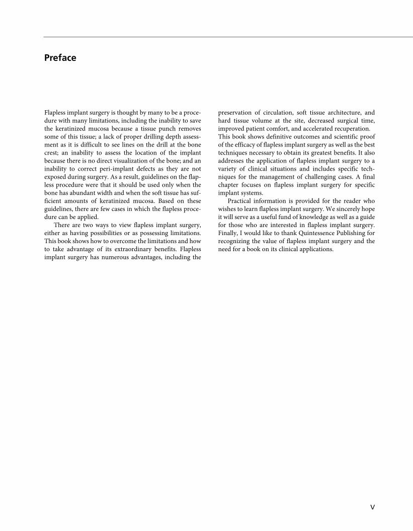
Flapless implant surgery is thought by many to be a proce-dure with many limitations, including the inability to savethe keratinized mucosa because a tissue punch removessome of this tissue; a lack of proper drilling depth assess-ment as it is difficult to see lines on the drill at the bonecrest; an inability to assess the location of the implantbecause there is no direct visualization of the bone; and aninability to correct peri-implant defects as they are notexposed during surgery. As a result, guidelines on the flap-less procedure were that it should be used only when thebone has abundant width and when the soft tissue has suf-ficient amounts of keratinized mucosa. Based on theseguidelines, there are few cases in which the flapless proce-dure can be applied.
There are two ways to view flapless implant surgery,either as having possibilities or as possessing limitations.This book shows how to overcome the limitations and howto take advantage of its extraordinary benefits. Flaplessimplant surgery has numerous advantages, including the
preservation of circulation, soft tissue architecture, andhard tissue volume at the site, decreased surgical time,improved patient comfort, and accelerated recuperation.This book shows definitive outcomes and scientific proofof the efficacy of flapless implant surgery as well as the besttechniques necessary to obtain its greatest benefits. It alsoaddresses the application of flapless implant surgery to avariety of clinical situations and includes specific tech-niques for the management of challenging cases. A finalchapter focuses on flapless implant surgery for specificimplant systems.
Practical information is provided for the reader whowishes to learn flapless implant surgery. We sincerely hopeit will serve as a useful fund of knowledge as well as a guidefor those who are interested in flapless implant surgery.Finally, I would like to thank Quintessence Publishing forrecognizing the value of flapless implant surgery and theneed for a book on its clinical applications.
V
Preface
VII
Byung-Ho Choi, DDS, MS, PhDProfessor Department of Oral and Maxillofacial SurgeryCollege of Dentistry, Wonju College of MedicineYonsei University, Korea
Seung-Mi Jeong, DDS, PhDProfessorDepartment of ProsthodonticsWonju College of MedicineYonsei University, Korea
Jihun Kim, DDS, MSLecturerDepartment of DentistryWonju College of MedicineYonsei University, Korea
Wilfried Engelke, DDS, MD, PhDProfessorDepartment of Oral SurgerySchool of DentistryGeorg-August-University, Göttingen, Germany
Authors

IX
Contents
Chapter 1 Comparison of Flap and Flapless Implant Surgeries . . . . . . . . . . . . . . . 1
Chapter 2 Possibilities and Limitations of Flapless Implantology . . . . . . . . . . . . 19
Chapter 3 Comparison of Soft Tissue Punch and Mini-incision Techniques. . . 37
Chapter 4 Diagnosis and Treatment Planning. . . . . . . . . . . . . . . . . . . . . . . . . . . . . 53
Chapter 5 Flapless Implant Surgery Technique . . . . . . . . . . . . . . . . . . . . . . . . . . . . 73
Chapter 6 Drilling in Flapless Implant Surgery. . . . . . . . . . . . . . . . . . . . . . . . . . . . 93
Chapter 7 Stage II Surgery . . . . . . . . . . . . . . . . . . . . . . . . . . . . . . . . . . . . . . . . . . . . 105
Chapter 8 Spontaneous Early Exposure of Flapless Submerged Implants. . . . . 123
Chapter 9 Early Plaque Control Following Flapless Implant Surgery . . . . . . . . 139
Chapter 10 Soft Tissue around Flapless Implants . . . . . . . . . . . . . . . . . . . . . . . . . . 155
Chapter 11 The Thick Mucosa. . . . . . . . . . . . . . . . . . . . . . . . . . . . . . . . . . . . . . . . . . 173
Chapter 12 The Deficient Alveolar Ridge. . . . . . . . . . . . . . . . . . . . . . . . . . . . . . . . . 191
Chapter 13 Simultaneous Socket Lift Procedure. . . . . . . . . . . . . . . . . . . . . . . . . . . 219
Chapter 14 Simultaneous Maxillary Sinus Membrane Elevation . . . . . . . . . . . . . 233
Chapter 15 Maxillary Sinus Bone Grafting . . . . . . . . . . . . . . . . . . . . . . . . . . . . . . . 251
Chapter 16 Clinical Cases . . . . . . . . . . . . . . . . . . . . . . . . . . . . . . . . . . . . . . . . . . . . . . 273
Chapter 17 Flapless Implant Surgery for Specific Implant Systems . . . . . . . . . . . 309
Index . . . . . . . . . . . . . . . . . . . . . . . . . . . . . . . . . . . . . . . . . . . . . . . . . . . . . . . . . . . . . . . . 327

Key point
Flap elevation on the alveolar crest can have a butterfly effect in implanttreatment.
C H A P T E R
1Comparison of Flap and
Flapless Implant Surgeries

mutual graft–implant stability, which facilitates simultan-eous implant placement in the posterior atrophic maxilla.When a Bio-Oss® block is required, this is so brittle andweak that it cannot provide sufficient mechanical force forthe primary stability of implants. Accordingly, when theremaining bone cannot ensure the primary stability of theimplants, flapless implant placement into the Bio-Oss®block should be delayed for 6–10 months.
Guidelines on the selection of technique for flapless implants
The choice of a soft tissue punch technique or a mini-incision technique is dependent on bone quality and primaryimplant stability. The following guidelines are intended tohelp clinicians to make the best choice (Fig 4-37).
Guideline 1: Select the soft tissue punch technique for a one-stage approachThe soft tissue punch technique is used for a one-stage surgical approach, whereas the mini-incision technique is
used for either a one-stage or a two-stage surgicalapproach. The two-stage surgical process places theimplant body below the soft tissue until bone healing hasoccurred. It is prudent to use the two-stage surgicalapproach when implants are not adequately stabilized or ifthe patient wears a soft tissue-borne partial denture.
Guideline 2: The mini-incision technique is preferred in areas with insufficient amounts of keratinized mucosaThe amount of keratinized tissue should be adequate, andideally patients need at least 1.5 mm of keratinized tissueto the facial aspect of the healing abutment. The mini-incision technique is beneficial in saving the keratinizedmucosa. Therefore, the soft tissue punch technique mustbe used in cases where at least 1.5 mm of keratinized tissueis left on the buccal side to this incision line of the punch.
Guideline 3: Select the mini-incision technique in the posterior maxillaOn rare occasions, an implant in the maxilla may notremain rigid after implant placement.10 A nonsubmerged,mobile implant may not heal predictably with a direct
Flapless Implantology
68
Chapter 4
Fig 4-36 (a) Block autogenous iliac bone grafting and simultaneous mini-incision implant placement. (b) Intraoral view immediately after surgery.(c) Postoperative radiographs after 6 months. (d) Illustration of block bone grafting and simultaneous mini-incision implant placement.
a b
c d

bone interface. Any implant that is not adequately stabi-lized should be submerged during healing, which reducesthe risk of micromovement and early implant failure.Therefore, the mini-incision technique is selected forimplants in the posterior maxilla in order to place theimplant body below the soft tissue (Fig 4-38). The mini-incision technique is the best approach in the posteriormaxilla where deficient osseous structures and an absenceof a cortical plate on the crest of the ridge further compro-mise the initial implant stability at the time of insertion.11
Guideline 4: In the mandible, a one-stageapproach offers more advantagesThe most common complication of a two-stage approachin the mandible is the risk of fistulas or gumboils, which
can develop in the mucosa covering the cover screws,because the mandible contains thick cortical bone,12-14 thinmucosa,15 and a 1–2 mm wide avascular zone in the crestalarea of the edentulous alveolar ridge16 (Figs 4-39 to 4-41).Fistulas or gumboils that develop in the mandible are dan-gerous because they can destroy bone (Fig 4-42). There-fore, when the implant is threaded into position with 20 Ncm or more, a one-stage approach is used in themandible (Fig 4-43). This approach eliminates the risk ofpostoperative infection and allows the peri-implant softtissue to be mature at the time of implant placement.When an implant in the mandible is not rigid after implantplacement, the implant should be submerged. Periodic,meticulous observation is necessary to check for the for-mation of a gumboil or fistula.
Diagnosis and Treatment Planning
69
Chapter 4
Fig 4-38 (a and b) On rare occasions, the implant in the maxilla may not be rigid after implant placement. Any implant that is not adequately sta-bilized should be submerged during healing, which reduces the risk of micromovement and early implant failure. Therefore, the mini-incision tech-nique is selected in the maxilla to place the implant body below the soft tissue.
Fig 4-37 Choice of a soft tissuepunch technique or a mini-incisiontechnique is dependent on bonequality and primary implant stabil-ity.
a b
Flapless Implant Surgery
Sufficient keratinized mucosa
Good quality and quantity of bone
Mandible
No bone graft
Insufficient keratinized mucosa
Poor quality and quantity of bone
Posterior maxilla
Bone graft
Soft tissue punch technique Mini-incision technique