Embed Size (px)
Citation preview

Fluid Mechanics of the eye:linear stability analysis of a two-liquid interface in the vitreouschamber and flow of aqueous humour with an intraocular lens
Jan Pralits
Department of Civil, Chemical and Environmental EngineeringUniversity of Genoa, Italy
December 9, 2014
The work presented has been carried out by:
Rodolfo Repetto DICCA, University of Genoa, Italy;
Jennifer Siggers Imperial College London, UK;
Jan Pralits DICCA, University of Genoa, Italy;
Alessandro Stocchino DICCA, University of Genoa, Italy;
Krystyna Isakova DICCA, University of Genoa, Italy;
Julia Meskauskas DISAT, University of L’Aquila, Italy;
Andrea Bonfiglio DICCA, University of Genoa, Italy.
Jan Pralits (University of Genoa) Fluid Mechanics of the eye December 9, 2014 1 / 38

1 Introduction
2 Stability of the interface between aqueous humor and vitreous substitutes after vitreoretinalsurgery
3 Flow of aqueous humour with an intraocular lens
4 References
Jan Pralits (University of Genoa) Fluid Mechanics of the eye December 9, 2014 2 / 38

Introduction
Anatomy of the eye
Jan Pralits (University of Genoa) Fluid Mechanics of the eye December 9, 2014 3 / 38

Introduction
Vitreous characteristics and functions
Vitreous composition
The main constituents are
Water (99%);
hyaluronic acid (HA);
collagen fibrils.
Its structure consists of long, thick, non-branching collagenfibrils suspended in hyaluronic acid.
Normal vitreous characteristicsThe healthy vitreous in youth is a gel-like material with visco-elastic mechanical properties,which have been measured by several authors (Lee et al., 1992; Nickerson et al., 2008;Swindle et al., 2008).
In the outermost part of the vitreous, named vitreous cortex, the concentration of collagenfibrils and HA is higher.
The vitreous cortex is in contact with the Internal Limiting Membrane (ILM) of the retina.
Physiological roles of the vitreousSupport function for the retina and filling-up function for the vitreous body cavity;
diffusion barrier between the anterior and posterior segment of the eye;
establishment of an unhindered path of light.
Jan Pralits (University of Genoa) Fluid Mechanics of the eye December 9, 2014 4 / 38
Introduction
Vitreous ageing
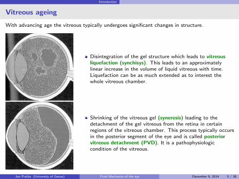
With advancing age the vitreous typically undergoes significant changes in structure.
Disintegration of the gel structure which leads to vitreousliquefaction (synchisys). This leads to an approximatelylinear increase in the volume of liquid vitreous with time.Liquefaction can be as much extended as to interest thewhole vitreous chamber.
Shrinking of the vitreous gel (syneresis) leading to thedetachment of the gel vitreous from the retina in certainregions of the vitreous chamber. This process typically occursin the posterior segment of the eye and is called posteriorvitreous detachment (PVD). It is a pathophysiologiccondition of the vitreous.
Jan Pralits (University of Genoa) Fluid Mechanics of the eye December 9, 2014 5 / 38

Introduction
Partial vitreous liquefaction
Jan Pralits (University of Genoa) Fluid Mechanics of the eye December 9, 2014 6 / 38

Introduction
Retinal detachment
Posterior vitreous detachment (PVD) andvitreous degeneration:
more common in myopic eyes;
preceded by changes in vitreousmacromolecular structure and invitreoretinal interface → possiblymechanical reasons.
If the retina detaches from the underlyinglayers → loss of vision;
Rhegmatogeneous retinal detachment:
fluid enters through a retinal break into thesub retinal space and peels off the retina.
Risk factors:
myopia;
posterior vitreous detachment (PVD);
lattice degeneration;
...
Jan Pralits (University of Genoa) Fluid Mechanics of the eye December 9, 2014 7 / 38
Introduction
Scleral buckling and vitrectomy
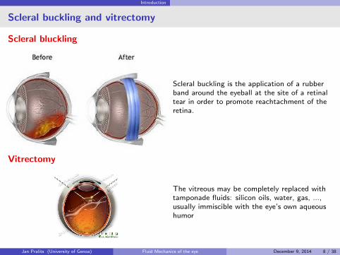
Scleral bluckling
Scleral buckling is the application of a rubberband around the eyeball at the site of a retinaltear in order to promote reachtachment of theretina.
Vitrectomy
The vitreous may be completely replaced withtamponade fluids: silicon oils, water, gas, ...,usually immiscible with the eye’s own aqueoushumor
Jan Pralits (University of Genoa) Fluid Mechanics of the eye December 9, 2014 8 / 38

Introduction
Intravitreal drug delivery
It is difficult to transport drugs to the retina from ’the outside’ due to the tight blood-retinalbarrier → use of intravitreal drug injections.
Diffusion is usually understood as the principal source for drug delivery, what about advection ?
Jan Pralits (University of Genoa) Fluid Mechanics of the eye December 9, 2014 9 / 38

Introduction
Motivations of the work
Why do research on vitreous motion?
Possible connections between the mechanism of retinal detachment andthe shear stress on the retina;flow characteristics.
Especially in the case of liquefied vitreous eye rotations may produce effective fluid mixing.In this case advection may be more important that diffusion for mass transport within thevitreous chamber.Understanding diffusion/dispersion processes in the vitreous chamber is important to predictthe behaviour of drugs directly injected into the vitreous.
Jan Pralits (University of Genoa) Fluid Mechanics of the eye December 9, 2014 10 / 38

Stability of the interface after vitreoretinal surgery
Stability of the interface between aqueous humor and vitreoussubstitutes after vitreoretinal surgery
Retinal detachment
Warning signs of retinal detachment:
Flashing lights.
Sudden appearance of floaters.
Shadows on the periphery of your vision.
Gray curtain across your field of vision.
Vitrectomy
The vitreous may be completely replaced withtamponade fluids: silicon oils, water, gas, ...
Denoted tamponade liquids
Purpose: Induce an instantaneousinterruption of an open communicationbetween the subretinal space/retinalpigment epithelial cells and the pre-retinalspace.
Healing: a scar should form as the cellsabsorb the remaining liquid.
Jan Pralits (University of Genoa) Fluid Mechanics of the eye December 9, 2014 11 / 38

Stability of the interface after vitreoretinal surgery
Fluids commonly used as a vitreous substitutes
Silicone oils;960 ≤ ρ∗ ≤ 1290 kg/m3
10−4 ≤ ν∗ ≤ 5× 10⁻³ m/s2
σ∗ ≈ 0.05 N/m
Perfluorocarbon liquids;1760 ≤ ρ∗ ≤ 2030 kg/m3
8× 10⁻⁷ ≤ ν∗ ≤ 8× 10⁻⁶ m/s2
σ∗ ≈ 0.05 N/m
Semifluorinated alkane liquids;1350 ≤ ρ∗ ≤ 1620 kg/m3
4.6× 10 ≤ ν∗ ≤ 10−3 m/s2
0.035 ≤ σ∗ ≤ 0.05 N/m
The choice of tamponade liquid depends on thespecific case
The tabulated fluids are immiscible withwater and commonly used in surgery
A lighter fluid (cf. water) is used totamponade in the upper part
A heavier fluid is used to tamponade in thelower part
High surface tension is preferred to a lowvalue (EXPERIENCE)
High value of viscosity (cf. water) ispreferred to a low value (EXPERIENCE)
What could happen otherwise ?
Jan Pralits (University of Genoa) Fluid Mechanics of the eye December 9, 2014 12 / 38

Stability of the interface after vitreoretinal surgery
Emulsification
Emulsification leads to loss of vision, not satisfactory
Figure: Emulsification of vitreous substitutes in the vitreous chamber
Jan Pralits (University of Genoa) Fluid Mechanics of the eye December 9, 2014 13 / 38
Stability of the interface after vitreoretinal surgery
Summary & Motivation
SummaryFrom experience it is known that tamponade fluids with high surface tension and highviscosity (compared to water) are less prone to emulsify
It is also know that initially ”good” tamponade fluids tend to change with time, for instancea decrease of surface tension due to surfactants, which leads to emulsification.
It is generally believed that shear stresses at the tamponade fluid-aqueous interfacegenerated during eye rotations play a crucial role in the generation of an emulsion.
The tamponade liquid needs to stay for a period of months so it is of interest to know howemulsification can be avoided.
Our analysisWe want to understand how emulsification, or the initial stages leading to emulsification, arerelated to the parameters (surface tension, viscosity, density, real conditions).
As a first study we focus on the stability characteristics of the interface in order to see if ithas any role.
A linear stability analysis, of wave like solutions, is used.
The evolution of the disturbance kinetic energy is analyzed.
Jan Pralits (University of Genoa) Fluid Mechanics of the eye December 9, 2014 14 / 38
Stability of the interface after vitreoretinal surgery
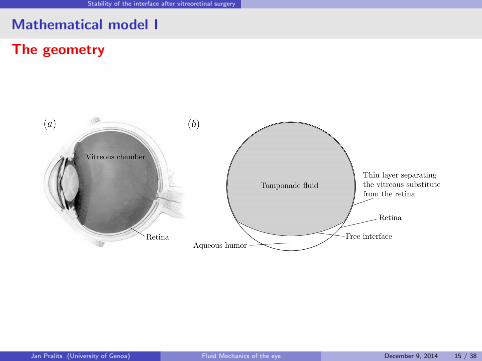
Mathematical model I
The geometry
Jan Pralits (University of Genoa) Fluid Mechanics of the eye December 9, 2014 15 / 38

Stability of the interface after vitreoretinal surgery
Mathematical model II
Underlying assumptions
Figure: Geometry of the problem
d∗ << R∗
2D-model;
flat wall oscillating harmonically;
semi-infinite domain;
small perturbations;
quasi-steady approach.
Stokes problem when 1 = 2
Jan Pralits (University of Genoa) Fluid Mechanics of the eye December 9, 2014 16 / 38
Stability of the interface after vitreoretinal surgery
Scaling and Dimensionless Parameters
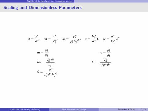
x =x∗
d∗, ui =
u∗iV ∗0
, pi =p∗i
ρ∗1V∗20
, t =V ∗0d∗
t, ω =d∗
V ∗0ω∗
m =µ∗2µ∗1
γ =ρ∗2ρ∗1
Re =V ∗0 d∗
ν∗1Fr =
V ∗0√g∗d∗
S =σ∗
ρ∗1d∗V ∗2
0
Jan Pralits (University of Genoa) Fluid Mechanics of the eye December 9, 2014 17 / 38

Stability of the interface after vitreoretinal surgery
Basic flow
Analytical solution
Parallel time-dependent flow
U1(y , t) = (c1e−ay + c2e
ey )e iωt + c.c.,
U2(y , t) = c3e−by e iωt + c.c.,
∂P1
∂y= −Fr−2,
∂P2
∂y= −γFr−2,
where
a =√iωR, b =
√iγωR
m.
and c1, c2, c3 are functions of a, b,m.
0
10
20
30
40
50
60
70
80
-1 -0.5 0 0.5 1
y
U
Jan Pralits (University of Genoa) Fluid Mechanics of the eye December 9, 2014 18 / 38

Stability of the interface after vitreoretinal surgery
Linear stability analysis
Flow decomposition:ui = Ui + ui
′, vi = vi′ pi = Pi + p′i
Boundary conditions:
u′1(0, t) = v ′1(0, t) = 0 and u′2(y , t)→ 0, v ′2(y , t)→ 0 as y →∞
Interface: (y∗ = d∗) introducing also the perturbation of the interface η′
Continuity of the perturbation velocity components across the interface
Continuity of the tangential stress of across the interface
The wall normal stress is balanced by the surface tension
A quasi-steady approach is assumed with two-dimensional wave-like solutions as:
ξi = e iα(x−Ωt)ξi (y , τ) + c.c
where0 ≤ τ ≤ 2π/ω
The system of equations is reduced introducing the perturbation stream function giving twoOrr-Sommerfeld equations, discretized using finite differences, solved using an inverse iterationalgorithm.
Jan Pralits (University of Genoa) Fluid Mechanics of the eye December 9, 2014 19 / 38

Stability of the interface after vitreoretinal surgery
Range of variability of the dimensionless parameters
0.1
1
10
0 0.02 0.04 0.06
Re
ω
(a)
0.1
1
10
10 100 1000
Re
S
(b)
0.1
1
10
0 5 10 15 20 25
Re
F r
(c)
Figure: Relationship between Re − ω, Re − S and Re − Fr obtained adopting feasible values of eye movement.From thin to thick curves: d = 1× 10⁻⁵m, d = 3× 10⁻⁵m, d = 1× 10⁻⁴m
Jan Pralits (University of Genoa) Fluid Mechanics of the eye December 9, 2014 20 / 38

Stability of the interface after vitreoretinal surgery
Neutral Curves
160
180
200
220
240
0.3 0.35 0.4 0.45 0.5
L=
2π/α
ωt/π
Unstable
Stable
m = 510
15
20
Figure: S = 14, γ = 1.0, R = 7, ω = 0.001
Jan Pralits (University of Genoa) Fluid Mechanics of the eye December 9, 2014 21 / 38

Stability of the interface after vitreoretinal surgery
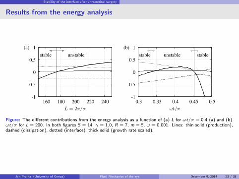
Energy analysis
The disturbance kinetic energy is given by 3 contributions: production, dissipation and interfacerelated terms
α
2π
dE
dt= −
∫ 1
0
u1v1U′1dy − γ
∫ +∞
1
u2v2U′2dy
−1
Re
∫ 1
0
[(∂u1
∂x
)2+(∂u1
∂y
)2+(∂v1
∂x
)2+(∂v1
∂y
)2]dy
−m
Re
∫ +∞
1
[(∂u2
∂x
)2+(∂u2
∂y
)2+(∂v2
∂x
)2+(∂v2
∂y
)2]dy
(v1
[(γ − 1)Fr−2 + α
2S]η −
v1
Re(∂v1
∂y− m
∂v2
∂y) +
1
Re(u1
∂u1
∂y− mu2
∂u2
∂y))∣∣∣
y=1. (4)
The different contributions of (4) are commonly presented in terms of growth rates. The sum can directly becompared with the solution of the eigenvalue problem governing the linear stability problem.
1
2αE
dE
dt= Im(Ω).
Jan Pralits (University of Genoa) Fluid Mechanics of the eye December 9, 2014 22 / 38
Stability of the interface after vitreoretinal surgery
Results from the energy analysis
-1
-0.5
0
0.5
1
160 180 200 220 240
L = 2π/α
(a)
stable unstable
-1
-0.5
0
0.5
1
0.3 0.35 0.4 0.45 0.5
ωt/π
(b)
unstable stablestable
Figure: The different contributions from the energy analysis as a function of (a) L for ωt/π = 0.4 (a) and (b)ωt/π for L = 200. In both figures S = 14, γ = 1.0, R = 7, m = 5, ω = 0.001. Lines: thin solid (production),dashed (dissipation), dotted (interface), thick solid (growth rate scaled).
Jan Pralits (University of Genoa) Fluid Mechanics of the eye December 9, 2014 23 / 38

Stability of the interface after vitreoretinal surgery
Shortes unstable wave length
The shortest unstable wave length as a function of S, Re and γ.
20
40
60
80
100
120
140
160
180
200
0 2 4 6 8 10 12 14
Lm
in
S
(a)
Unstable
Stable
20
40
60
80
100
120
140
160
180
200
0 20 40 60
Lm
in
Re
(b)
Unstable
Stable
20
40
60
80
100
120
140
160
180
200
0.5 1 1.5 2
Lm
in
γ
(c)
Unstable
Stable
Figure: Length of the shortest unstable perturbation Lmin versus S (a), Re (b), and γ (c) with ω = 0.001 andm = 5. The values of Re = 7 in (a) and (c), S = 14 in (b) and (c), and γ = 1 in (a) and (b), respectively.
Jan Pralits (University of Genoa) Fluid Mechanics of the eye December 9, 2014 24 / 38
Stability of the interface after vitreoretinal surgery
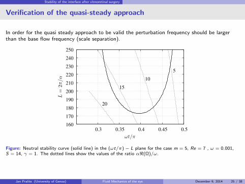
Verification of the quasi-steady approach
In order for the quasi steady approach to be valid the perturbation frequency should be largerthan the base flow frequency (scale separation).
160
170
180
190
200
210
220
230
240
250
0.3 0.35 0.4 0.45 0.5
L=
2π/α
ωt/π
10
15
20
5
Figure: Neutral stability curve (solid line) in the (ωt/π)− L plane for the case m = 5, Re = 7 , ω = 0.001,S = 14, γ = 1. The dotted lines show the values of the ratio α<(Ω)/ω.
Jan Pralits (University of Genoa) Fluid Mechanics of the eye December 9, 2014 25 / 38

Stability of the interface after vitreoretinal surgery
Conclusions and Continuation
Monitoring the shortest unstable wave length (critical wave length) we have seen that:
Increasing the viscosity, ratio the critical wave length increases (stabilizing for the Eye)
Increasing the surface tension, the critical wave length increases (stabilizing for the Eye)
Increasing the Reynolds no., the critical wave length decreases (destabilizing for the Eye)
Increasing the density ratio, the critical wave length decreases (destabilizing for the Eye)
The first two is ”in line” with realistic observations.
For realistic values of R,S , γ,m, ω, d∗ the critical wave length ≈ 5 mm, which is about halfthe Eye radius.
However, the growth rate is instantaneous and the waves unstable only during certainintervals of one period. (cf. turbulent burst in the classical Stokes II problem). No sustainedgrowth over one period is guaranteed.
This analysis is far from explaining the onset of emulsion but a first step to rule out (or not)different physical mechanisms.
Next step...
Floquet analysis
Non-modal analysis
Include curvature, ...., more realistic geometry (circle, sphere)
Jan Pralits (University of Genoa) Fluid Mechanics of the eye December 9, 2014 26 / 38

Flow of aqueous humour with an intraocular lens
Flow of aqueous humour with an intraocular lens
Phakic intraocular lenses
MotivationIntraocular lenses decrease endothelial cell density in some patients. Possible reasons include:
Excess shear stress due to altered flow of aqueous
Impaired transport of nutrients due to altered flow
Non-ideal placement of lens, with respect to iris and other tissues
Impact of external body on eye (e.g. accidental impact, patient touching eyes)
This study: Investigate the first possibility, by comparing the shear stress both in the presenceof and without the lens. Moreover, monitor the intra-ocular pressure (IOP).
Jan Pralits (University of Genoa) Fluid Mechanics of the eye December 9, 2014 27 / 38

Flow of aqueous humour with an intraocular lens
Description of the model
Governing equations:
∇ · u =0
ρ
(∂u
∂t+ u ·∇u
)=−∇p +∇2u + ρg
ρcp
(∂T
∂t+ u ·∇T
)=ka∇2T
Assumptions:
Boussinesq approximation (ρ is constant,except in the gravitational acceleration termin which ρ = ρ0(1− α(T − T0)))
Numerical solution:
All solutions are obtained with OpenFOAM
Solvers were first tested on cases whereanalytical solutions exist.
Definitions:
Symbol Definitionu velocityp pressureρ density of aqueousµ viscosity of aqueousg acceleration due to gravitycp specific heat at constant pressureT temperatureka thermal conductivity of aqueous
Jan Pralits (University of Genoa) Fluid Mechanics of the eye December 9, 2014 28 / 38

Flow of aqueous humour with an intraocular lens
Model geometry
Figure: (left) ”real” anterior chamber, (right) cross-section of the idealized anterior chamber used in thesimulations.
Figure: (a) Geometry of the pIOL consisting of a lens and two haptics that have claws that allow the lens to beattached to the iris, (b) pIOL placed in the anterior chamber.
Jan Pralits (University of Genoa) Fluid Mechanics of the eye December 9, 2014 29 / 38

Flow of aqueous humour with an intraocular lens
Flow mechanisms
Flow induced by aqueous production/drainage:Aqueous humor is produced by the ciliary body, and then flows throughthe posterior chamber, the pupil and the anterior chamber, from whereit is drained into the trabecular meshwork. (3 µl/min)
Flow induced during miosis:During pupil contraction (miosis), a flow from the posterior to theanterior chamber of the eye is generated, which is intense, although itonly lasts a short time, typically less than 1 s. (middle figure)
Buoyancy-driven flow:It is well known that, since the posterior surface of the cornea istypically cooler than the iris and lens. We prescribed a temperature of34 C on the cornea and 37 C on all other surfaces.
Flow induced by saccades of the eye:We consider the flow generated in the anterior chamber by rotations ofthe eye bulb by modeling isolated rotations using the analyticalrelationship proposed by Repetto et al. (2005) which provides theangular velocity of the eye as a function of time. (bottom figure)
0
5e-10
1e-09
1.5e-09
2e-09
2.5e-09
0 0.1 0.2 0.3 0.4 0.5 0.6 0.7 0.8
Inle
t flux [m
3/s
]
Time [s]
(a)
0
50
100
150
200
250
300
350
0 0.01 0.02 0.03 0.04 0.05 0.06
Angula
r velo
city [deg/s
]
Time [s]
(b)A= 10 deg
20 deg
Jan Pralits (University of Genoa) Fluid Mechanics of the eye December 9, 2014 30 / 38

Flow of aqueous humour with an intraocular lens
Flow induced by aqueous production and drainage
2e-5
4e-5
6e-5
p [mmHg]
0
7.53e-05(a)
(b)
2e-5
4e-5
U [m/s]
0
5.89e-05
Figure: Flow due to production/drainage of aqueous humorwith the device present: (a) excess pressure above IOP, (b)velocity magnitude.
0.00e+00
5.00e-05
1.00e-04
1.50e-04
2.00e-04
2.50e-04
0.06 0.07 0.08 0.09 0.1 0.11 0.12 0.13 0.14
Ave
rag
e in
let
pre
ssu
re [
mm
Hg
]
Minimum distance between the pIOL and the iris [mm]
without pIOL
pIOL without hapticspIOL
Figure: Average pressure drop between iris–lenschannel and trabecular meshwork as a function ofthe minimum distance between the pIOL and theiris (solid circles). No pIOL (horizontal line) andpIOL with haptics (empty circle).
Lubrication theory:
∆p =6µQ
π
∫ r2
r1
dr
rh3,
To get ∆p = 1mmHg you need h = 1 µm
Jan Pralits (University of Genoa) Fluid Mechanics of the eye December 9, 2014 31 / 38

Flow of aqueous humour with an intraocular lens
Flow induced during miosis
-0.0005
0
0.0005
0.001
0.0015
0.002
0.0025
0.003
0.0035
0.004
0 0.1 0.2 0.3 0.4 0.5 0.6 0.7 0.8
Ave
rag
e in
let
pre
ssure
[m
mH
g]
Time [s]
pIOLwithout pIOL
Figure: Spatially averaged pressure over the inlet as a function of time during miosis, in an eye both with thepIOL (solid circles) and without it (open circles).
Jan Pralits (University of Genoa) Fluid Mechanics of the eye December 9, 2014 32 / 38

Flow of aqueous humour with an intraocular lens
Buoyancy-driven flow I
0.0002
0.0004
U [m/s]
0
0.000592
(a)
0.0002
0.0004
U [m/s]
0
0.000585
Figure: Buoyancy-driven flow in the absence of the pIOL. Gravity acts in the vertical direction. Streamlines ofthe flow and the distribution of the velocity magnitude on selected horizontal planes, (a) without the lens, (b)with the lens.
Jan Pralits (University of Genoa) Fluid Mechanics of the eye December 9, 2014 33 / 38
Flow of aqueous humour with an intraocular lens
Buoyancy-driven flow II
0.0004
0.0008
WSS [Pa]
0
0.00119
(a)
0.001
0.002
WSS [Pa]
0
0.00255
(b)
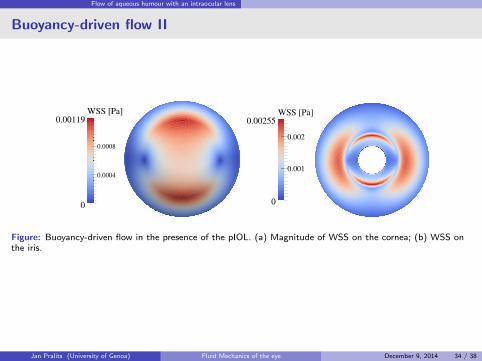
Figure: Buoyancy-driven flow in the presence of the pIOL. (a) Magnitude of WSS on the cornea; (b) WSS onthe iris.
Jan Pralits (University of Genoa) Fluid Mechanics of the eye December 9, 2014 34 / 38

Flow of aqueous humour with an intraocular lens
Buoyancy-driven flow III
0.0012
0.0013
0.0014
0.0015
0.0016
0.0017
0.1 0.15 0.2 0.25 0.3 0.35 0.4 0.45 0.5
Ma
xim
um
WS
S m
ag
nitu
de
[P
a]
Minimum distance between the pIOL and the iris [mm]
without pIOL
Figure: Spatial maximum of the WSS on the cornea as a function of the distance between the pIOL and the iris,neglecting the haptics (open circles). The maximum WSS with no pIOL is shown by the horizontal line, whilstthe maximum WSS with the pIOL in its normal position and including the haptics is shown by the solid circle.
Jan Pralits (University of Genoa) Fluid Mechanics of the eye December 9, 2014 35 / 38

Flow of aqueous humour with an intraocular lens
Flow induced by saccades of the eye
0.02
0.04
0.06
0.08
0.1
0.12
0.14
0.16
0.18
0.2
0 0.02 0.04 0.06 0.08 0.1
Ma
xim
um
WS
S m
ag
nitu
de
o
n t
he
co
rne
a [
Pa
]
Time [s]
(a)without pIOL
pIOL
0.02
0.04
0.06
0.08
0.1
0.12
0.14
0.16
0.18
0.2
0 0.02 0.04 0.06 0.08 0.1
Ma
xim
um
WS
S m
ag
nitu
de
o
n t
he
iris [
Pa
]
Time [s]
(b)without pIOL
pIOL
Figure: Time evolution of the spatial maximum of the WSS on the (a) cornea and (b) iris in an eye performinga saccade of angle 10 .
Jan Pralits (University of Genoa) Fluid Mechanics of the eye December 9, 2014 36 / 38
Flow of aqueous humour with an intraocular lens
Discussion and conclusions
(i) If the lens is properly placed there is a negligible influence on the pressure in the posterior chamber.
(ii) There is no significant increase of the WSS on the cornea.
(iii) The WSS on the iris is significantly greater than in the case with no pIOL, but the increase is not likely tobe sufficiently great so as to give a risk of cell detachment.
Endothelial cell loss:
Kaji et al.(2005) performed experiments on porcine corneal endothelial cells that were plated onto glassslides, and found that significant detachment was observed for shear stresses in excess of 0.03 Pa if thecells had had 1 hour of adhesion, rising to 0.1 Pa for 3 hours of adhesion. Our model predicts that theactual values are significantly smaller than this.
Other causes: rubbing the eye (mechanical forcing), insufficient delivery of oxygen and/or nutrients to thecorneal epithelium. The latter will be studied in the near future.
Jan Pralits (University of Genoa) Fluid Mechanics of the eye December 9, 2014 37 / 38

References
References I
B. Lee, M. Litt, and G. Buchsbaum. Rheology of the vitreous body. Part I: viscoelasticity ofhuman vitreous. Biorheology, 29:521–533, 1992.
C. S. Nickerson, J. Park, J. A. Kornfield, and H. Karageozian. Rheological properties of thevitreous and the role of hyaluronic acid. Journal of Biomechanics, 41(9):1840–6, 2008. doi:10.1016/j.jbiomech.2008.04.015.
K. Swindle, P. Hamilton, and N. Ravi. In situ formation of hydrogels as vitreous substitutes:Viscoelastic comparison to porcine vitreous. Journal of Biomedical Materials Research - Part A,87A(3):656–665, Dec. 2008. ISSN 1549-3296.
Jan Pralits (University of Genoa) Fluid Mechanics of the eye December 9, 2014 38 / 38