Embed Size (px)
Citation preview

Jpn J Ophthalmol 43, 25–30 (1999)© 1999 Japanese Ophthalmological Society 0021-5155/99/$–see front matterPublished by Elsevier Science Inc. PII S0021-5155(98)00059-8
Fluorescein and Indocyanine GreenVideoangiography of Choroidal Melanomas
Leyla S. Atmaca, Figen Batio lu and Pelin Atmaca
Eye Clinic, Ankara University Medical School, Ankara, Turkey
Purpose:
This study was performed to determine what role indocyanine green video angiog-raphy might play in the evaluation of choroidal melanomas, and to compare this role withthat of fluorescein angiography.
Methods:
Six patients with posterior segment uveal melanoma underwent digital fluoresceinand indocyanine green videoangiography. All patients were women and their mean age was50.7 years.
Results:
In all eyes with melanoma, fluorescein angiography revealed irregular hyperfluores-cence in the early phase and staining of the tumor in the late phase. A double circulation pat-tern was obvious in 1 eye with a mushroom-shaped melanoma. The patterns of indocyaninegreen videoangiography varied, depending on the degree of tumor pigmentation, thickness,and vascularity. Early frames of indocyanine green video angiography demonstrated hypof-luorescence in all eyes, and the intrinsic choroidal vasculature was obvious in 3 eyes. In thelate phase of indocyanine green videoangiograms, different patterns (hyperfluorescence,three-ring pattern) were observed.
Conclusion:
Indocyanine green videoangiography may be a useful adjunct to fluorescein an-giography in the evaluation of choroidal melanomas.
Jpn J Ophthalmol 1999;43:25–30
© 1999 Japanese Ophthalmological Society
Key Words:
Choroidal melanoma, fluorescein angiography, indocyanine green videoangiography.
g
Introduction
A choroidal melanoma in its earlier stage of devel-opment is more likely to be observed clinically whenit arises in or near the macular area. However, whenthe eye condition is complicated by hemorrhage, ret-inal detachment, glaucoma, or cataract, caution shouldbe exercised in making the exact diagnosis.
Like other choroidal tumors, choroidal melanomais usually diagnosed by indirect ophthalmoscopy.
1
Since McLean and Maumenee
2
described the intensestaining of choroidal hemangiomas after an intrave-nous injection of fluorescein, angiography has be-come essential in the investigation and diagnosis offundus mass lesions. In addition, fundus imagingtechniques, e.g., fundus photography and fluoresceinangiography, have the advantages of providing ob-
jective documentation of the growth of the tumor.However, it is not always possible to make a defini-tive diagnosis of a choroidal tumor on ophthalmo-scopic and fluorescein angiographic characteristicsalone, and other clinical modalities are often neces-sary.
3
Indocyanine green (ICG) diffuses much moreslowly from choroidal vessels because it is a morehighly bound protein than sodium fluorescein. Theseproperties make it suitable for imaging the choroidalvasculature. Several reports have stressed the useful-ness of this dye in the evaluation of different chori-oretinal disorders especially choroidal neovascular-ization.
4–6
Recently, different fluorescence patternshave been shown in choroidal tumors with ICG an-giography.
7,8
In this study, the ICG angiographic characteris-tics of choroidal melanomas are described and com-pared with the pattern obtained from fluorescein an-giography.
Received: November 20, 1997.Address correspondence and reprint requests to: Leyla S. AT-
MACA, MD, GMK Bulv. 23/1, 06440 Ankara, Turkey
26
Jpn J OphthalmolVol 43: 25–30, 1999
Materials and Methods
Six patients with choroidal melanoma were in-cluded in this study. All the patients were women.Their ages ranged from 39–65 years, with a mean ageof 50.7 years. Routine examination of each patientincluded indirect ophthalmoscopy as well as directophthalmoscopy with Goldmann’s three-mirror lens.Ultrasonography was done and color fundus photo-graphs were taken of each patient’s affected eye.The diagnosis was based on patient history and re-sults of the “classic” examinations mentioned above.
Topcon ImageNET Digital Imaging System linkedto a Topcon TRC 50 IA camera was used for angiog-raphy. Fluorescein angiography was performed byinjecting 5 mL of 20% sodium fluorescein (S. E. R. B.Lab Pharmaceutiques). After the images had beendownloaded to the optical disc, the ICG procedurewas begun. For this, 25 mg ICG was dissolved in 1.5mL aqueous solvent (S. E. R. B. Lab Pharmaceu-tiques) and injected, followed by a 5-mL sterile sa-line solution. The early, middle, and late phase an-giograms were taken. Images were reviewed, thendownloaded to the optical disc for storage and analy-sis. No complication occurred during the angiogra-phies.
Results
Fundus examination of the 6 patients revealed anonpigmented/hypopigmented tumor in 4 eyes, apigmented tumor in one eye, and a centrally pig-mented tumor in the sixth eye. The lesion was associ-ated with retinal detachment in 4 eyes. The tumorwas dome-shaped in 3 eyes and mushroom-shaped in
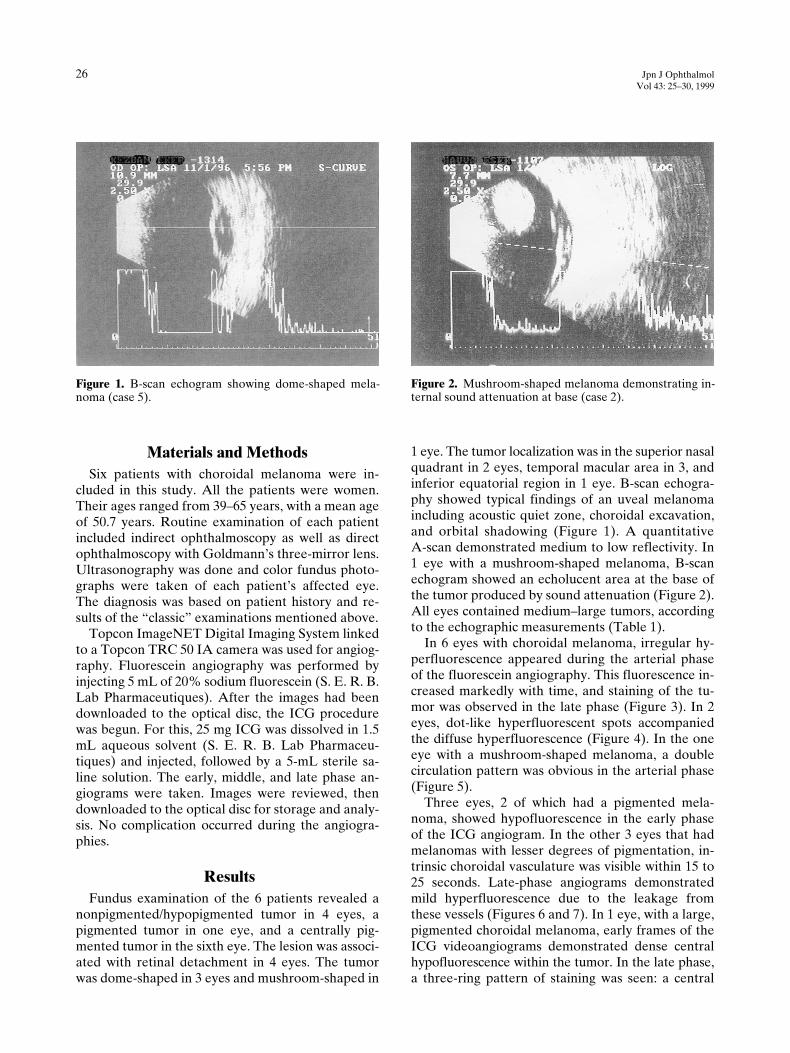
1 eye. The tumor localization was in the superior nasalquadrant in 2 eyes, temporal macular area in 3, andinferior equatorial region in 1 eye. B-scan echogra-phy showed typical findings of an uveal melanomaincluding acoustic quiet zone, choroidal excavation,and orbital shadowing (Figure 1). A quantitativeA-scan demonstrated medium to low reflectivity. In1 eye with a mushroom-shaped melanoma, B-scanechogram showed an echolucent area at the base ofthe tumor produced by sound attenuation (Figure 2).All eyes contained medium–large tumors, accordingto the echographic measurements (Table 1).
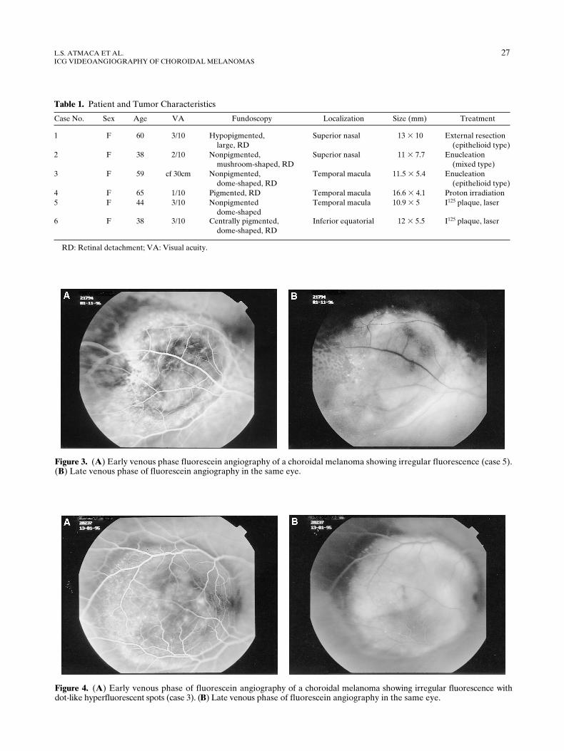
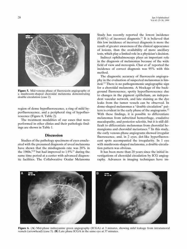
In 6 eyes with choroidal melanoma, irregular hy-perfluorescence appeared during the arterial phaseof the fluorescein angiography. This fluorescence in-creased markedly with time, and staining of the tu-mor was observed in the late phase (Figure 3). In 2eyes, dot-like hyperfluorescent spots accompaniedthe diffuse hyperfluorescence (Figure 4). In the oneeye with a mushroom-shaped melanoma, a doublecirculation pattern was obvious in the arterial phase(Figure 5).
Three eyes, 2 of which had a pigmented mela-noma, showed hypofluorescence in the early phaseof the ICG angiogram. In the other 3 eyes that hadmelanomas with lesser degrees of pigmentation, in-trinsic choroidal vasculature was visible within 15 to25 seconds. Late-phase angiograms demonstratedmild hyperfluorescence due to the leakage fromthese vessels (Figures 6 and 7). In 1 eye, with a large,pigmented choroidal melanoma, early frames of theICG videoangiograms demonstrated dense centralhypofluorescence within the tumor. In the late phase,a three-ring pattern of staining was seen: a central
Figure 1. B-scan echogram showing dome-shaped mela-noma (case 5).
Figure 2. Mushroom-shaped melanoma demonstrating in-ternal sound attenuation at base (case 2).
L.S. ATMACA ET AL.
27
ICG VIDEOANGIOGRAPHY OF CHOROIDAL MELANOMAS
Figure 3. (A) Early venous phase fluorescein angiography of a choroidal melanoma showing irregular fluorescence (case 5).(B) Late venous phase of fluorescein angiography in the same eye.
Figure 4. (A) Early venous phase of fluorescein angiography of a choroidal melanoma showing irregular fluorescence withdot-like hyperfluorescent spots (case 3). (B) Late venous phase of fluorescein angiography in the same eye.
Table 1.
Patient and Tumor Characteristics
Case No. Sex Age VA Fundoscopy Localization Size (mm) Treatment
1 F 60 3/10 Hypopigmented,large, RD
Superior nasal 13
3
10 External resection(epithelioid type)
2 F 38 2/10 Nonpigmented,mushroom-shaped, RD
Superior nasal 11
3
7.7 Enucleation(mixed type)
3 F 59 cf 30cm Nonpigmented,dome-shaped, RD
Temporal macula 11.5
3
5.4 Enucleation(epithelioid type)
4 F 65 1/10 Pigmented, RD Temporal macula 16.6
3
4.1 Proton irradiation5 F 44 3/10 Nonpigmented
dome-shapedTemporal macula 10.9
3
5 I
125
plaque, laser
6 F 38 3/10 Centrally pigmented,dome-shaped, RD
Inferior equatorial 12
3
5.5 I
125
plaque, laser
RD: Retinal detachment; VA: Visual acuity.
28
Jpn J OphthalmolVol 43: 25–30, 1999
region of dense hypofluorescence, a ring of mild hy-perfluorescence, and a peripheral ring of hypofluo-rescence (Figure 8, Table 2).
The treatment modalities of our cases that wereperformed in other clinics and their pathologic find-ings are shown in Table 1.
Discussion
Studies of the pathology specimens of eyes enucle-ated with the presumed diagnosis of uveal melanomahave shown that the misdiagnosis rate was 20% inthe 1960s,
9,10
but had improved to 1.9%
11
during thesame time period at a center with advanced diagnos-tic facilities. The Collaborative Ocular Melanoma
Study has recently reported the lowest incidence(0.48%) of incorrect diagnosis.
11
It is believed thatthis low incidence of incorrect diagnosis is more theresult of greater awareness of the clinical appearanceof lesions, than the availability of more ancillarytests, which play a limited role in a physician’s decision.
Indirect ophthalmoscopy plays an important rolein the diagnosis of melanomas because of the widefield of view and stereopsis. Char et al
3
reported theincidence of correct diagnosis was 95% with thismethod.
The diagnostic accuracy of fluorescein angiogra-phy in the evaluation of suspected melanomas is lim-ited.
13
There is no pathognomonic angiographic signfor a choroidal melanoma. A blockage of the back-ground fluorescence, spotty hyperfluorescence dueto changes in the pigment epithelium, an indepen-dent vascular network, and late staining as the dyeleaks from the tumor vessels can be observed. Indome-shaped melanomas a “double circulation” pat-tern is evident in the early phase of the angiograms.
12
With these findings, it is possible to differentiatemelanomas from subretinal hemorrhage, exudativemaculopathy, and posterior scleritis, but it is still dif-ficult to differentiate melanomas from choroidal he-mangioma and choroidal metastases.
12
In this study,the early venous-phase angiograms showed irregularfluorescence, and, in 2 eyes, dot-like hyperfluores-cent spots accompanied the irregularity. In 1 eyewith mushroom-shaped melanoma, a double-circula-tion pattern was obvious.
It has been more than 20 years since the initial in-vestigations of choroidal circulation by ICG angiog-raphy. Advances in imaging techniques have im-
Figure 6. (A) Mid-phase indocyanine green angiography (ICGA) at 3 minutes, showing mild leakage from intratumoralvessels (arrowhead) (case 5). (B) Late phase ICGA in the same eye at 37 minutes.
Figure 5. Mid-venous phase of fluorescein angiography ofa mushroom-shaped choroidal melanoma demonstratingdouble circulation (case 2).

L.S. ATMACA ET AL.
29
ICG VIDEOANGIOGRAPHY OF CHOROIDAL MELANOMAS
proved the resolution of the choroidal angiograms.Recently, the role of ICG angiography in diagnosisof choroidal melanomas was discussed by several au-thors.
7,8
Other reports have focused on the usefulnessof ICG in various choroidal disorders, especially poorlydefined choroidal neovascularizations.
4,6
The findings of ICG angiography in choroidal tu-mors were superficially described in the early devel-opmental years of dye technology.
13,14
It was foundthat vascularization of nonpigmented tumors couldbe studied and the angiogram could provide a topo-graphic parameter for delineating the size andgrowth of choroidal tumors. Due to the deficient res-olution of the early imaging systems, the details ofpigmented choroidal tumors were not clearly ob-served. In recent years, with the new technology ofICG videoangiography, various angiographic find-
ings have been identified in pigmented and nonpig-mented choroidal melanomas.
7,8
ICG fluorescence findings for choroidal melano-mas vary, depending on the tumor pigmentation,thickness, and vascularization.
15
In general, the lesspigmented and more vascular tumors demonstratemore fluorescence. The greater the thickness of thetumor, the greater fluorescence due to the large-cali-ber intrinsic vessels. In Shield’s study, the maximumfluorescence was achieved at an average time of 18.2minutes, and although this period varied, it was notdependent on tumor thickness and color.
8
In most cases, the intrinsic choroidal vasculatureof the tumor can be observed as well as the normalretinal and choroidal circulation. Tumor vessels aretortuous, have random direction, and show abnor-mal branching. These vessels are more pronouncedon ICG angiography, depending on the amount ofpigmentation.
7
In this study, these vessels wereclearly seen with ICG videoangiography in the 3eyes with dome-shaped, nonpigmented melanomas.In the late phase, diffuse hyperfluorescence due tovascular leakage was observed. The presence of ab-normal vascular leakage with late dye leakage onICG angiography may be considered an indicator ofmalignancy. It is better to follow these tumors withICG angiography.
7
The ICG videoangiographic image of pigmentedchoroidal melanomas was variable. These tumors usu-ally showed well-defined hypofluorescence through-out the angiogram. This hypofluorescence is causedby blockage from the tumor pigment, and the lack ofobvious intrinsic tumor vascularity, RPE prolifera-tion, hemorrhage, and necrosis.
8
In minimally ele-
Figure 7. (A) Early phase indocyanine green angiography(ICGA) at 29 seconds showing intratumoral choroidal cir-culation (case 3). (B) Late phase ICGA in the same eye at36 minutes.
Figure 8. Late phase indocyanine green angiography at 33minutes showing a “three-ring pattern” (case 4).

30
Jpn J OphthalmolVol 43: 25–30, 1999
Table 2.
Fluorescein and Indocyanine Green Angiographic Findings
vated pigmented melanomas, late phase ICG angio-grams often reveal hypofluorescence, but a fuzzy,mild fluorescence can be observed as well. In mostcases, the fluorescence is homogenous, but in somecases a three-ring pattern of staining is seen. We ob-served this pattern in 1 eye with a large, pigmentedmelanoma in the late frames at 30 minutes.
In this study, exudative retinal detachment accom-panied the tumor in 4 eyes, and 3 of these eyes had adome-shaped melanoma. In these eyes, delineationof tumor edges was difficult and the base of the lesioncould barely be visualized by fundoscopy or fluores-cein angiography. ICG videoangiography defined themargins of these tumors better than previous methods.
In conclusion, fluorescein and ICG angiographiesdemonstrated various fluorescence patterns, depend-ing on the characteristics of the tumor. None of thesefindings is pathognomic for a choroidal melanoma.The major advantage of ICG angiography is the bet-ter delineation of the tumor, providing an objectivedocumentation of tumor growth. This is especiallyimportant in the follow-up of suspected melanomas.ICG angiography should always be combined withfluorescein angiography as well as other current di-agnostic techniques, especially ultrasonography.
References
1. Shields JA, Shields CL. Intraocular tumors: a text and atlas.Philadelphia: WB Saunders, 1992:11–24.
2. McLean AL, Maumenee EA. Hemangioma of the choroid.Am J Ophthalmol 1960;50:3–11.
3. Char DH, Stone RD, Irvine AR, et al. Diagnostic modalitiesin choroidal melanoma. Am J Ophthalmol 1980;89:223–30.
4. Destro M, Puliafito CA. Indocyanine green videoangiographyof choroidal neovascularization. Ophthalmology 1989;96:846–53.
5. Guyer DR, Puliafito CA, Mones JM, Friedman E, Chang W,Verdooner SR. Digital indocyanine green angiography inchorioretinal disorders. Ophthalmology 1992;99:287–91.
6. Yannuzzi LA, Slakter JS, Sorenson JA, Guyer DR, Orlock,DA. Digital indocyanine green videoangiography and choroi-dal neovascularization. Retina 1992;12:191–223.
7. Sallet G, Amoaku WMK, Lafaut BA, Brabant P, De Laey JJ.Indocyanine green angiography of choroidal tumors. GraefesArch Clin Exp Ophthalmol 1995;233:677–89.
8. Shields CL, Shields JA, De Potter. Patterns of indocyaninegreen videoangiography of choroidal tumors. Br J Ophthal-mol 1995;79:237–45.
9. Shields JA, Zimmerman LE. Lesions simulating malignantmelanoma of the posterior uvea. Arch Ophthalmol1973;89:466–71.
10. Shields JA, McDonald PR. Improvements in the diagnosis ofposterior uveal melanomas. Arch Ophthalmol 1974;91:259–64.
11. Collaborative Ocular Melanoma Study Group. Accuracy ofdiagnosis of choroidal melanomas in the Collaborative OcularMelanoma Study. COMS report no 1. Arch Ophthalmol1990;108:1268–73.
12. Richard G. Fluorescein angiography textbook and atlas. NewYork: Thieme Medical Publishers, 1990:202–7.
13. Bischoff PM, Flower RW. Ten years experience with choroi-dal angiography using indocyanine green dye: a new routineexamination or an epilogue? Doc Ophthalmol 1985;60:235–91.
14. Chopdar A, Turk AK HU DW. Fluorescent infra-red angiog-raphy of the fundus oculi using indocyanine green dye. TransOphthalmol Soc UK 1978;98:142–6.
15. Shields CL. 1997 Clinical evaluation of choroidal tumors. In:Yannuzzi LA, Flower RW, Slakter JS, eds. Indocyanine greenangiography. St. Louis, MO: Mosby, 1997:279–95.


![Unilateral Choroidal Osteoma with Choroidal Neovascularization...Surgical evacuation of the choroidal neovascular membrane has been reported [12] but the visual outcome was not favorable](https://img.pdfslide.net/doc/110x75/6053732923e31173be575e28/unilateral-choroidal-osteoma-with-choroidal-neovascularization-surgical-evacuation.jpg)