Embed Size (px)
Citation preview
Fluorescence-based surface magnifyingchromoendoscopy and optical coherencetomography endoscope
R. Andrew WallJennifer K. Barton
Downloaded From: https://www.spiedigitallibrary.org/journals/Journal-of-Biomedical-Optics on 28 Apr 2019Terms of Use: https://www.spiedigitallibrary.org/terms-of-use
Fluorescence-based surface magnifying chromoendoscopyand optical coherence tomography endoscope
R. Andrew Walla and Jennifer K. Bartona,b
aThe University of Arizona, College of Optical Sciences, 1630 E. University Boulevard, Tucson, Arizona 85721bThe University of Arizona, Department of Biomedical Engineering, 1127 E. James E. Rogers Way, Tucson, Arizona 85721
Abstract. A side-viewing, 2.3-mm diameter, surface magnifying chromoendoscopy-optical coherence tomography(SMC-OCT) endoscope has been designed for simultaneous, nondestructive surface fluorescence visualization andcross-sectional imaging. We apply this endoscope to in vivo examination of the mouse colon. A 30,000 elementfiber bundle is combined with single mode fibers, for SMC and OCT imaging, respectively. The distal optics consistof a gradient-index lens and spacer to provide a 1×magnification at a working distance of 1.58 mm in air, necessaryto image the sample through a 0.23-mm thick outer glass envelope, and an aluminized right-angle prism fixed tothe distal end of the gradient-index lens assembly. The resulting 1∶1 imaging system is capable of 3.9-μm lateraland 2.3-μm axial resolution in the OCT channel, and 125-lp∕mm resolution across a 0.70-mm field of view in theSMC channel. The endoscope can perform high contrast crypt visualization, molecular imaging, and cross-sectional imaging of colon microstructure. © 2012 Society of Photo-Optical Instrumentation Engineers (SPIE). [DOI: 10.1117/1.JBO.17
.8.086003]
Keywords: optical coherence tomography; surface magnifying chromoendoscopy; endoscope; catheter, gradient-index; gastrointestinal;colon; aberrant crypt foci.
Paper 12161P received Mar. 8, 2012; revised manuscript received Jul. 6, 2012; accepted for publication Jul. 12, 2012; published onlineAug. 3, 2012.
1 IntroductionColonoscopy is the most commonly used technique for earlydetection of colorectal cancer, which is the third most frequentlydiagnosed malignancy in the United States. In 2012, it is esti-mated that colorectal cancer will be responsible for 9% of newlydiagnosed cancers as well as cancer related deaths in men andwomen. While the five-year survival rate is 90% when thesecancers are detected at an early, localized stage, only 39% ofpatients are diagnosed early.1 A need exists for rapid, nondes-tructive visualization of tissues in vivo, for clinical diagnostics aswell as for scientific study in order to make significant advancesin the basic science of chemoprevention and chemotherapy.
Optical coherence tomography (OCT) is a noninvasive inter-ferometric imaging technique capable of imaging up to 2 mmdeep in tissue. OCT creates cross-sectional images usingnear-infrared light back scattered from index of refraction mis-matches.2,3 Recently, OCT has been used to image the humanand mouse colon and rectum with micron-scale resolution.4,5
Various mouse models exist which are excellent platforms forstudying colon carcinogenesis, preventatives and treatmentmethods.6
We have previously constructed dual modality endoscopesthat provide OCT information about tissue boundaries, struc-ture, and thickness, as well as laser-induced fluorescence (LIF)spectra information about the biochemical composition of thetissue. These endoscopes can provide a heightened sensitivityand specificity to tumor detection when compared to either mod-ality alone.7,8 However, our current systems cannot reliablyvisualize the subtle alterations in mouse mucosal crypt structure
that signify the earliest stages of disease. Our OCT systems havesufficient resolution, but resolution is generally measured witha 100% contrast target. The innate contrast of the mouse colonis very low, which when combined with the very small cryptsizes, lead to challenges in colon visualization in this diminu-tive species. Exogenous contrast agents would mitigate thisproblem. We have utilized nanoshells as an OCT contrastagent in the mouse colon but they enhanced regions of tissuerather than crypt structure.9 Exogenous dyes could improvecontrast, but our previous LIF systems have emphasized obtain-ing high resolution spectral rather than high resolution spatialinformation.
Surface-magnifying chromoendoscopy (SMC) has beendeveloped to visualize the surface mucosal structure of thegastrointestinal (GI) tract, and has gained wide-ranging appli-cation for early detection of colorectal cancer and its pre-cursors.10,11 Methylene blue (MB) is a dye clinically used toenhance contrast for SMC. MB can be visualized either byits significant absorption or fluorescence emission in the redto near-infrared wavelength range. With SMC, it is possibleto discriminate normal colonic crypt structure from aberrantcrypt foci (ACF). ACF are clinically interesting because theconventional paradigm is that they are the first event in color-ectal carcinogenesis, although this paradigm has recently beendebated.12,13 While studies to prove or disprove the role of ACFin humans are difficult, mouse models offer an opportunity todetermine the causal relationship between ACF and colorectalneoplasia.
Combining SMC and OCT in one endoscopic system willallow the possibility for a large scale serial study to be doneon the murine model, with SMC providing surface visualizationof ACF and OCT allowing cross-sectional imaging for the
Address all correspondence to: Jennifer K. Barton, The University of Arizona,College of Optical Sciences, 1630 E. University Boulevard, Tucson, Arizona85721. Tel: +520-621-4116; E-mail: [email protected] 0091-3286/2012/$25.00 © 2012 SPIE
Journal of Biomedical Optics 086003-1 August 2012 • Vol. 17(8)
Journal of Biomedical Optics 17(8), 086003 (August 2012)
Downloaded From: https://www.spiedigitallibrary.org/journals/Journal-of-Biomedical-Optics on 28 Apr 2019Terms of Use: https://www.spiedigitallibrary.org/terms-of-use
detection of mucosal thickening and adenoma. We havedesigned a dual modality fluorescence-based SMC and OCTendoscope capable of resolving crypt structure in the mousecolon. In this paper, we describe our instrument for obtainingsimultaneous fluorescence SMC and OCT data and presentpreliminary results obtained from methylene blue-stainedmouse colon.
2 Materials and Methods
2.1 Proximal SMC-OCT System
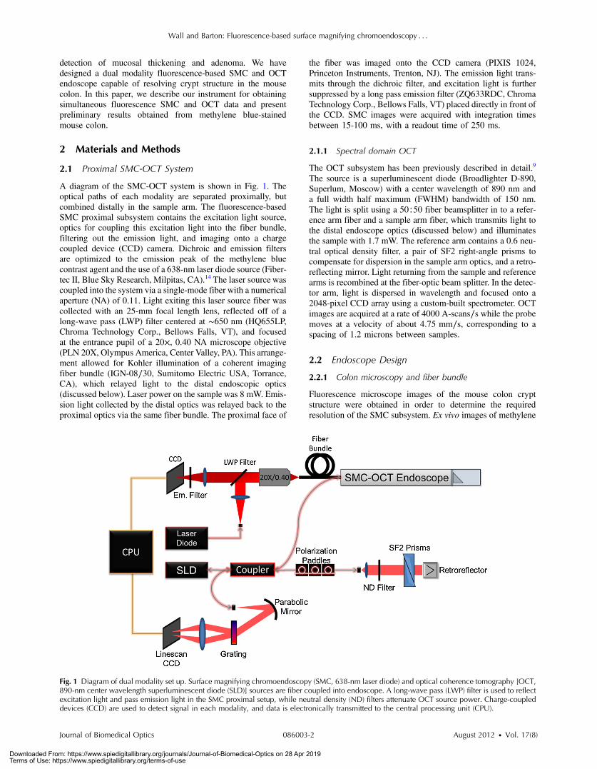
A diagram of the SMC-OCT system is shown in Fig. 1. Theoptical paths of each modality are separated proximally, butcombined distally in the sample arm. The fluorescence-basedSMC proximal subsystem contains the excitation light source,optics for coupling this excitation light into the fiber bundle,filtering out the emission light, and imaging onto a chargecoupled device (CCD) camera. Dichroic and emission filtersare optimized to the emission peak of the methylene bluecontrast agent and the use of a 638-nm laser diode source (Fiber-tec II, Blue Sky Research, Milpitas, CA).14 The laser source wascoupled into the system via a single-mode fiber with a numericalaperture (NA) of 0.11. Light exiting this laser source fiber wascollected with an 25-mm focal length lens, reflected off of along-wave pass (LWP) filter centered at ∼650 nm (HQ655LP,Chroma Technology Corp., Bellows Falls, VT), and focusedat the entrance pupil of a 20×, 0.40 NA microscope objective(PLN 20X, Olympus America, Center Valley, PA). This arrange-ment allowed for Kohler illumination of a coherent imagingfiber bundle (IGN-08∕30, Sumitomo Electric USA, Torrance,CA), which relayed light to the distal endoscopic optics(discussed below). Laser power on the sample was 8 mW. Emis-sion light collected by the distal optics was relayed back to theproximal optics via the same fiber bundle. The proximal face of
the fiber was imaged onto the CCD camera (PIXIS 1024,Princeton Instruments, Trenton, NJ). The emission light trans-mits through the dichroic filter, and excitation light is furthersuppressed by a long pass emission filter (ZQ633RDC, ChromaTechnology Corp., Bellows Falls, VT) placed directly in front ofthe CCD. SMC images were acquired with integration timesbetween 15-100 ms, with a readout time of 250 ms.
2.1.1 Spectral domain OCT
The OCT subsystem has been previously described in detail.9
The source is a superluminescent diode (Broadlighter D-890,Superlum, Moscow) with a center wavelength of 890 nm anda full width half maximum (FWHM) bandwidth of 150 nm.The light is split using a 50∶50 fiber beamsplitter in to a refer-ence arm fiber and a sample arm fiber, which transmits light tothe distal endoscope optics (discussed below) and illuminatesthe sample with 1.7 mW. The reference arm contains a 0.6 neu-tral optical density filter, a pair of SF2 right-angle prisms tocompensate for dispersion in the sample arm optics, and a retro-reflecting mirror. Light returning from the sample and referencearms is recombined at the fiber-optic beam splitter. In the detec-tor arm, light is dispersed in wavelength and focused onto a2048-pixel CCD array using a custom-built spectrometer. OCTimages are acquired at a rate of 4000 A-scans∕s while the probemoves at a velocity of about 4.75 mm∕s, corresponding to aspacing of 1.2 microns between samples.
2.2 Endoscope Design
2.2.1 Colon microscopy and fiber bundle
Fluorescence microscope images of the mouse colon cryptstructure were obtained in order to determine the requiredresolution of the SMC subsystem. Ex vivo images of methylene
Fig. 1 Diagram of dual modality set up. Surface magnifying chromoendoscopy (SMC, 638-nm laser diode) and optical coherence tomography [OCT,890-nm center wavelength superluminescent diode (SLD)] sources are fiber coupled into endoscope. A long-wave pass (LWP) filter is used to reflectexcitation light and pass emission light in the SMC proximal setup, while neutral density (ND) filters attenuate OCT source power. Charge-coupleddevices (CCD) are used to detect signal in each modality, and data is electronically transmitted to the central processing unit (CPU).
Journal of Biomedical Optics 086003-2 August 2012 • Vol. 17(8)
Wall and Barton: Fluorescence-based surface magnifying chromoendoscopy : : :
Downloaded From: https://www.spiedigitallibrary.org/journals/Journal-of-Biomedical-Optics on 28 Apr 2019Terms of Use: https://www.spiedigitallibrary.org/terms-of-use
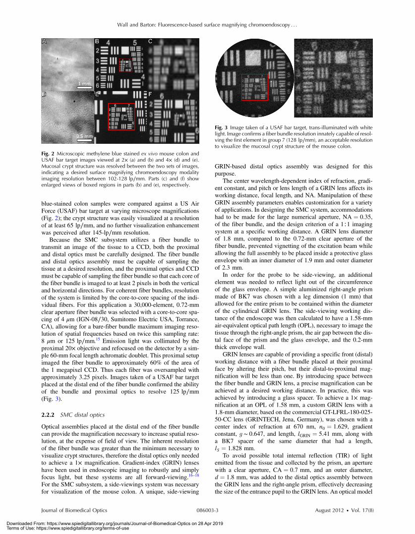
blue-stained colon samples were compared against a US AirForce (USAF) bar target at varying microscope magnifications(Fig. 2); the crypt structure was easily visualized at a resolutionof at least 65 lp∕mm, and no further visualization enhancementwas perceived after 145-lp∕mm resolution.
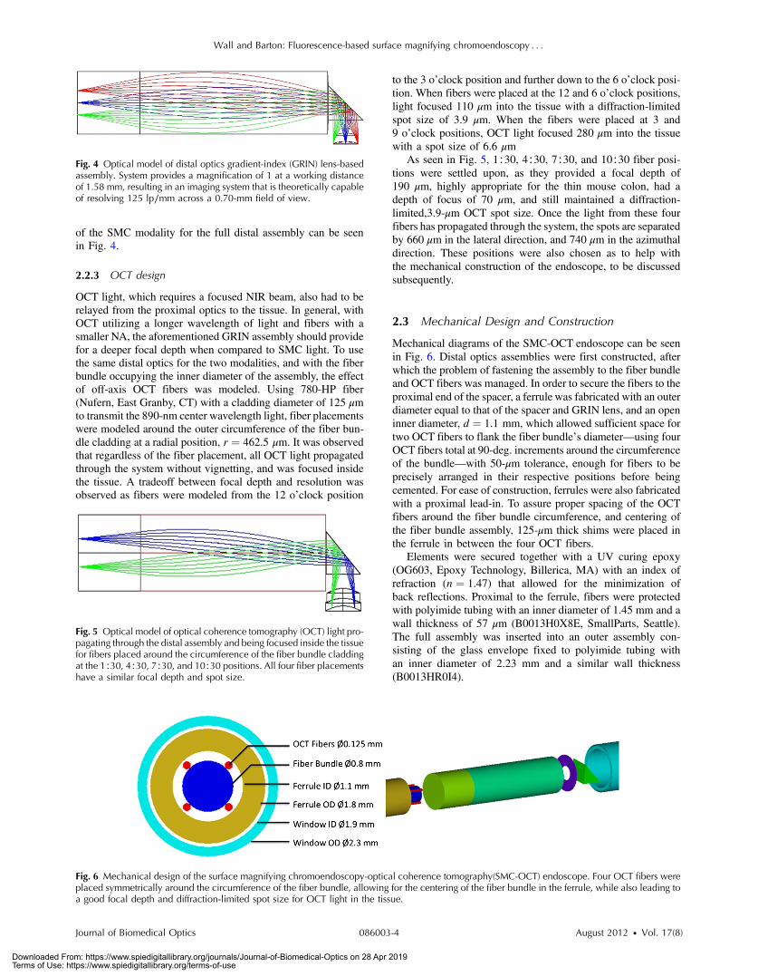
Because the SMC subsystem utilizes a fiber bundle totransmit an image of the tissue to a CCD, both the proximaland distal optics must be carefully designed. The fiber bundleand distal optics assembly must be capable of sampling thetissue at a desired resolution, and the proximal optics and CCDmust be capable of sampling the fiber bundle so that each core ofthe fiber bundle is imaged to at least 2 pixels in both the verticaland horizontal directions. For coherent fiber bundles, resolutionof the system is limited by the core-to-core spacing of the indi-vidual fibers. For this application a 30,000-element, 0.72-mmclear aperture fiber bundle was selected with a core-to-core spa-cing of 4 μm (IGN-08∕30, Sumitomo Electric USA, Torrance,CA), allowing for a bare-fiber bundle maximum imaging reso-lution of spatial frequencies based on twice this sampling rate:8 μm or 125 lp∕mm.15 Emission light was collimated by theproximal 20× objective and refocused on the detector by a sim-ple 60-mm focal length achromatic doublet. This proximal setupimaged the fiber bundle to approximately 60% of the area ofthe 1 megapixel CCD. Thus each fiber was oversampled withapproximately 3.25 pixels. Images taken of a USAF bar targetplaced at the distal end of the fiber bundle confirmed the abilityof the bundle and proximal optics to resolve 125 lp∕mm(Fig. 3).
2.2.2 SMC distal optics
Optical assemblies placed at the distal end of the fiber bundlecan provide the magnification necessary to increase spatial reso-lution, at the expense of field of view. The inherent resolutionof the fiber bundle was greater than the minimum necessary tovisualize crypt structures, therefore the distal optics only neededto achieve a 1× magnification. Gradient-index (GRIN) lenseshave been used in endoscopic imaging to robustly and simplyfocus light, but these systems are all forward-viewing.16–18
For the SMC subsystem, a side-viewings system was necessaryfor visualization of the mouse colon. A unique, side-viewing
GRIN-based distal optics assembly was designed for thispurpose.
The center wavelength-dependent index of refraction, gradi-ent constant, and pitch or lens length of a GRIN lens affects itsworking distance, focal length, and NA. Manipulation of theseGRIN assembly parameters enables customization for a varietyof applications. In designing the SMC system, accommodationshad to be made for the large numerical aperture, NA ¼ 0.35,of the fiber bundle, and the design criterion of a 1∶1 imagingsystem at a specific working distance. A GRIN lens diameterof 1.8 mm, compared to the 0.72-mm clear aperture of thefiber bundle, prevented vignetting of the excitation beam whileallowing the full assembly to be placed inside a protective glassenvelope with an inner diameter of 1.9 mm and outer diameterof 2.3 mm.
In order for the probe to be side-viewing, an additionalelement was needed to reflect light out of the circumferenceof the glass envelope. A simple aluminized right-angle prismmade of BK7 was chosen with a leg dimension (1 mm) thatallowed for the entire prism to be contained within the diameterof the cylindrical GRIN lens. The side-viewing working dis-tance of the endoscope was then calculated to have a 1.58-mmair-equivalent optical path length (OPL), necessary to image thetissue through the right-angle prism, the air gap between the dis-tal face of the prism and the glass envelope, and the 0.2-mmthick envelope wall.
GRIN lenses are capable of providing a specific front (distal)working distance with a fiber bundle placed at their proximalface by altering their pitch, but their distal-to-proximal mag-nification will be less than one. By introducing space betweenthe fiber bundle and GRIN lens, a precise magnification can beachieved at a desired working distance. In practice, this wasachieved by introducing a glass spacer. To achieve a 1× mag-nification at an OPL of 1.58 mm, a custom GRIN lens with a1.8-mm diameter, based on the commercial GT-LFRL-180-025-50-CC lens (GRINTECH, Jena, Germany), was chosen with acenter index of refraction at 670 nm, n0 ¼ 1.629, gradientconstant, g ∼ 0.647, and length, lGRIN ¼ 5.41 mm, along witha BK7 spacer of the same diameter that had a length,lS ¼ 1.828 mm.
To avoid possible total internal reflection (TIR) of lightemitted from the tissue and collected by the prism, an aperturewith a clear aperture, CA ¼ 0.7 mm, and an outer diameter,d ¼ 1.8 mm, was added to the distal optics assembly betweenthe GRIN lens and the right-angle prism, effectively decreasingthe size of the entrance pupil to the GRIN lens. An optical model
Fig. 2 Microscopic methylene blue stained ex vivo mouse colon andUSAF bar target images viewed at 2× (a) and (b) and 4× (d) and (e).Mucosal crypt structure was resolved between the two sets of images,indicating a desired surface magnifying chromoendoscopy modalityimaging resolution between 102-128 lp∕mm. Parts (c) and (f) showenlarged views of boxed regions in parts (b) and (e), respectively.
Fig. 3 Image taken of a USAF bar target, trans-illuminated with whitelight. Image confirms a fiber bundle resolution innately capable of resol-ving the first element in group 7 (128 lp∕mm), an acceptable resolutionto visualize the mucosal crypt structure of the mouse colon.
Journal of Biomedical Optics 086003-3 August 2012 • Vol. 17(8)
Wall and Barton: Fluorescence-based surface magnifying chromoendoscopy : : :
Downloaded From: https://www.spiedigitallibrary.org/journals/Journal-of-Biomedical-Optics on 28 Apr 2019Terms of Use: https://www.spiedigitallibrary.org/terms-of-use
of the SMC modality for the full distal assembly can be seenin Fig. 4.
2.2.3 OCT design
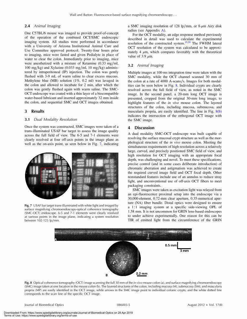
OCT light, which requires a focused NIR beam, also had to berelayed from the proximal optics to the tissue. In general, withOCT utilizing a longer wavelength of light and fibers with asmaller NA, the aforementioned GRIN assembly should providefor a deeper focal depth when compared to SMC light. To usethe same distal optics for the two modalities, and with the fiberbundle occupying the inner diameter of the assembly, the effectof off-axis OCT fibers was modeled. Using 780-HP fiber(Nufern, East Granby, CT) with a cladding diameter of 125 μmto transmit the 890-nm center wavelength light, fiber placementswere modeled around the outer circumference of the fiber bun-dle cladding at a radial position, r ¼ 462.5 μm. It was observedthat regardless of the fiber placement, all OCT light propagatedthrough the system without vignetting, and was focused insidethe tissue. A tradeoff between focal depth and resolution wasobserved as fibers were modeled from the 12 o’clock position
to the 3 o’clock position and further down to the 6 o’clock posi-tion. When fibers were placed at the 12 and 6 o’clock positions,light focused 110 μm into the tissue with a diffraction-limitedspot size of 3.9 μm. When the fibers were placed at 3 and9 o’clock positions, OCT light focused 280 μm into the tissuewith a spot size of 6.6 μm
As seen in Fig. 5, 1∶30, 4∶30, 7∶30, and 10∶30 fiber posi-tions were settled upon, as they provided a focal depth of190 μm, highly appropriate for the thin mouse colon, had adepth of focus of 70 μm, and still maintained a diffraction-limited,3.9-μm OCT spot size. Once the light from these fourfibers has propagated through the system, the spots are separatedby 660 μm in the lateral direction, and 740 μm in the azimuthaldirection. These positions were also chosen as to help withthe mechanical construction of the endoscope, to be discussedsubsequently.
2.3 Mechanical Design and Construction
Mechanical diagrams of the SMC-OCT endoscope can be seenin Fig. 6. Distal optics assemblies were first constructed, afterwhich the problem of fastening the assembly to the fiber bundleand OCT fibers was managed. In order to secure the fibers to theproximal end of the spacer, a ferrule was fabricated with an outerdiameter equal to that of the spacer and GRIN lens, and an openinner diameter, d ¼ 1.1 mm, which allowed sufficient space fortwo OCT fibers to flank the fiber bundle’s diameter—using fourOCT fibers total at 90-deg. increments around the circumferenceof the bundle—with 50-μm tolerance, enough for fibers to beprecisely arranged in their respective positions before beingcemented. For ease of construction, ferrules were also fabricatedwith a proximal lead-in. To assure proper spacing of the OCTfibers around the fiber bundle circumference, and centering ofthe fiber bundle assembly, 125-μm thick shims were placed inthe ferrule in between the four OCT fibers.
Elements were secured together with a UV curing epoxy(OG603, Epoxy Technology, Billerica, MA) with an index ofrefraction (n ¼ 1.47) that allowed for the minimization ofback reflections. Proximal to the ferrule, fibers were protectedwith polyimide tubing with an inner diameter of 1.45 mm and awall thickness of 57 μm (B0013H0X8E, SmallParts, Seattle).The full assembly was inserted into an outer assembly con-sisting of the glass envelope fixed to polyimide tubing withan inner diameter of 2.23 mm and a similar wall thickness(B0013HR0I4).
Fig. 4 Optical model of distal optics gradient-index (GRIN) lens-basedassembly. System provides a magnification of 1 at a working distanceof 1.58 mm, resulting in an imaging system that is theoretically capableof resolving 125 lp∕mm across a 0.70-mm field of view.
Fig. 6 Mechanical design of the surface magnifying chromoendoscopy-optical coherence tomography(SMC-OCT) endoscope. Four OCT fibers wereplaced symmetrically around the circumference of the fiber bundle, allowing for the centering of the fiber bundle in the ferrule, while also leading toa good focal depth and diffraction-limited spot size for OCT light in the tissue.
Fig. 5 Optical model of optical coherence tomography (OCT) light pro-pagating through the distal assembly and being focused inside the tissuefor fibers placed around the circumference of the fiber bundle claddingat the 1∶30, 4∶30, 7∶30, and 10∶30 positions. All four fiber placementshave a similar focal depth and spot size.
Journal of Biomedical Optics 086003-4 August 2012 • Vol. 17(8)
Wall and Barton: Fluorescence-based surface magnifying chromoendoscopy : : :
Downloaded From: https://www.spiedigitallibrary.org/journals/Journal-of-Biomedical-Optics on 28 Apr 2019Terms of Use: https://www.spiedigitallibrary.org/terms-of-use
2.4 Animal Imaging
One C57BL/6 mouse was imaged to provide proof-of-conceptof the operation of the combined OCT/SMC endoscopicimaging system. All studies were performed in accordancewith a University of Arizona Institutional Animal Care andUse Committee approved protocol. Twenty-four hours priorto imaging, mice were fasted and given Pedialyte in place ofwater to clear the colon. Immediately prior to imaging, micewere anesthetized with a mixture of Ketamine (0.33 mg∕ml,100 mg∕kg) and Xylazine (0.033 mg∕ml, 10 mg∕kg) adminis-tered by intraperitoneal (IP) injection. The colon was gentlyflushed with 3-9 mL of warm saline to clear excess mucous.Methylene blue (MB) solution (1%, 0.2 ml) was lavaged inthe colon and allowed to incubate for 2 min, after which thecolon was gently flushed again with warm saline. The SMC-OCT endoscope was coated with a thin layer of a biocompatiblewater-based lubricant and inserted approximately 32 mm insidethe colon, and sequential SMC and OCT images obtained.
3 Results
3.1 Dual Modality Resolution
Once the system was constructed, SMC images were taken of atrans-illuminated USAF bar target to assess the image qualityacross the full field of view. The 6-5 and 7-1 elements wereclearly resolved at four off-axis points in the image plane aswell as the on-axis point, as seen below in Fig. 7, indicating
a SMC imaging resolution of 128 lp∕mm, or 8-μm Airy diskradius (see Appendix A).
For the OCT modality, an edge response method previouslydescribed in detail was used to calculate the experimentalresolution of the constructed system.19,20 The FWHM lateralOCT resolution of the system was calculated to be approxi-mately 4 μm, which compares favorably with the theoreticalvalue of 3.9 μm.
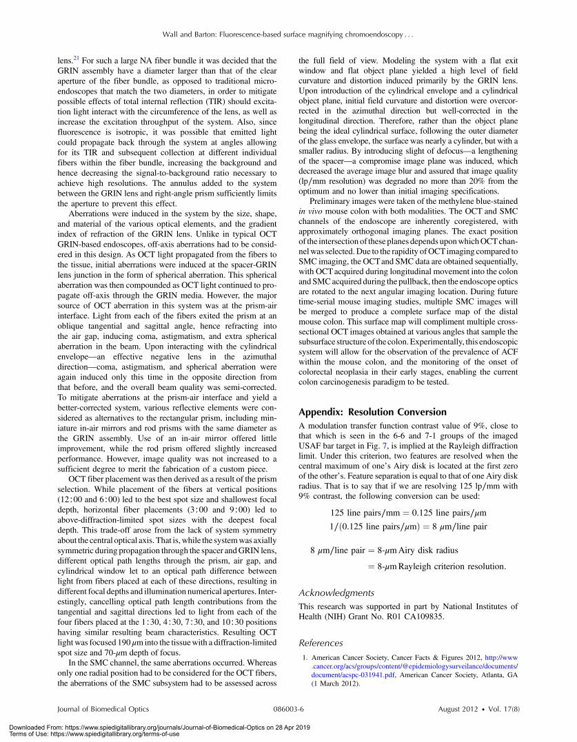
3.2 Animal Imaging
Multiple images at 100-ms integration time were taken with theSMC modality, while the OCT channel scanned 30 mm ofthe colon at a rate of 4000 A-scans∕s. Images for both modal-ities can be seen below in Fig. 8. Individual crypts are clearlyresolved across the full field of view, as noted in the SMCimage. In the second panel, a 20-mm long OCT image ispresented, cropped from the original 30-mm long image, tohighlight features of the in vivo mouse colon. The layeredstructures of the colon, including mucosa, submucosa, andmuscularis propria, are easily identified. The line in Fig. 8(b)indicates the intersection of the orthogonal OCT image withthe SMC image.
4 DiscussionA dual modality SMC-OCT endoscope was built capable ofresolving the surface mucosal crypt structure as well as the mor-phological structure of the in vivo mouse colon. Meeting thesimultaneous requirements of high resolution across a relativelylarge, curved, and precisely positioned SMC field of view, andhigh resolution for OCT imaging with an appropriate focaldepth, was challenging and novel. To meet these specifications,precise control (and in some cases deliberate introduction) ofchromatic aberration and astigmatism was achieved to createthe required curved image field and OCT focal depth. Othernonstandard features include use of an annulus to reduce straylight, and unconventional use of off-axis OCT fibers to meetpackaging constraints.
SMC images were taken as excitation light was relayed froman epi-fluorescence proximal setup into the endoscope via a30,000-element, 0.72-mm clear aperture, 0.35-numerical aper-ture (NA) fiber bundle. Distal optics were designed to ensurea 1∶1 imaging system at a specific side-viewing OPL of1.58 mm. It is not uncommon for GRIN lens-based endoscopesto under achieve experimentally. One reason for this can beTIR of emitted light from the circumference of the GRIN
Fig. 8 Optical coherence tomography (OCT) image scanning the full 30 mm of the in vivomouse colon (a), and surface magnifying chromoendoscopy(SMC) image taken at one location in the mouse colon (b). The layered structures of the colon, including mucosa (M), submucosa (SM), and muscularispropria (MP) are easily identified in the OCT image, while arrows in the SMC image point to individual colonic crypts, and the white dotted linecorresponds to the scan line of the specific OCT image.
Fig. 7 USAF bar target trans-illuminated with white light and imaged bysurface magnifying chromoendoscopy-optical coherence tomography(SMC-OCT) endoscope. 6-5 and 7-1 elements were clearly resolvedat various points in the image plane, indicating a system resolutionbetween 102-125 lp∕mm.
Journal of Biomedical Optics 086003-5 August 2012 • Vol. 17(8)
Wall and Barton: Fluorescence-based surface magnifying chromoendoscopy : : :
Downloaded From: https://www.spiedigitallibrary.org/journals/Journal-of-Biomedical-Optics on 28 Apr 2019Terms of Use: https://www.spiedigitallibrary.org/terms-of-use
lens.21 For such a large NA fiber bundle it was decided that theGRIN assembly have a diameter larger than that of the clearaperture of the fiber bundle, as opposed to traditional micro-endoscopes that match the two diameters, in order to mitigatepossible effects of total internal reflection (TIR) should excita-tion light interact with the circumference of the lens, as well asincrease the excitation throughput of the system. Also, sincefluorescence is isotropic, it was possible that emitted lightcould propagate back through the system at angles allowingfor its TIR and subsequent collection at different individualfibers within the fiber bundle, increasing the background andhence decreasing the signal-to-background ratio necessary toachieve high resolutions. The annulus added to the systembetween the GRIN lens and right-angle prism sufficiently limitsthe aperture to prevent this effect.
Aberrations were induced in the system by the size, shape,and material of the various optical elements, and the gradientindex of refraction of the GRIN lens. Unlike in typical OCTGRIN-based endoscopes, off-axis aberrations had to be consid-ered in this design. As OCT light propagated from the fibers tothe tissue, initial aberrations were induced at the spacer-GRINlens junction in the form of spherical aberration. This sphericalaberration was then compounded as OCT light continued to pro-pagate off-axis through the GRIN media. However, the majorsource of OCT aberration in this system was at the prism-airinterface. Light from each of the fibers exited the prism at anoblique tangential and sagittal angle, hence refracting intothe air gap, inducing coma, astigmatism, and extra sphericalaberration in the beam. Upon interacting with the cylindricalenvelope—an effective negative lens in the azimuthaldirection—coma, astigmatism, and spherical aberration wereagain induced only this time in the opposite direction fromthat before, and the overall beam quality was semi-corrected.To mitigate aberrations at the prism-air interface and yield abetter-corrected system, various reflective elements were con-sidered as alternatives to the rectangular prism, including min-iature in-air mirrors and rod prisms with the same diameter asthe GRIN assembly. Use of an in-air mirror offered littleimprovement, while the rod prism offered slightly increasedperformance. However, image quality was not increased to asufficient degree to merit the fabrication of a custom piece.
OCT fiber placement was then derived as a result of the prismselection. While placement of the fibers at vertical positions(12∶00 and 6∶00) led to the best spot size and shallowest focaldepth, horizontal fiber placements (3∶00 and 9∶00) led toabove-diffraction-limited spot sizes with the deepest focaldepth. This trade-off arose from the lack of system symmetryabout the central optical axis. That is,while the systemwas axiallysymmetric during propagation through the spacer andGRIN lens,different optical path lengths through the prism, air gap, andcylindrical window let to an optical path difference betweenlight from fibers placed at each of these directions, resulting indifferent focal depths and illumination numerical apertures. Inter-estingly, cancelling optical path length contributions from thetangential and sagittal directions led to light from each of thefour fibers placed at the 1∶30, 4∶30, 7∶30, and 10∶30 positionshaving similar resulting beam characteristics. Resulting OCTlight was focused 190 μm into the tissuewith a diffraction-limitedspot size and 70-μm depth of focus.
In the SMC channel, the same aberrations occurred. Whereasonly one radial position had to be considered for the OCT fibers,the aberrations of the SMC subsystem had to be assessed across
the full field of view. Modeling the system with a flat exitwindow and flat object plane yielded a high level of fieldcurvature and distortion induced primarily by the GRIN lens.Upon introduction of the cylindrical envelope and a cylindricalobject plane, initial field curvature and distortion were overcor-rected in the azimuthal direction but well-corrected in thelongitudinal direction. Therefore, rather than the object planebeing the ideal cylindrical surface, following the outer diameterof the glass envelope, the surface was nearly a cylinder, but with asmaller radius. By introducing slight of defocus—a lengtheningof the spacer—a compromise image plane was induced, whichdecreased the average image blur and assured that image quality(lp∕mm resolution) was degraded no more than 20% from theoptimum and no lower than initial imaging specifications.
Preliminary images were taken of the methylene blue-stainedin vivo mouse colon with both modalities. The OCT and SMCchannels of the endoscope are inherently coregistered, withapproximately orthogonal imaging planes. The exact positionof the intersection of these planesdependsuponwhichOCTchan-nelwas selected.Due to the rapidity ofOCTimaging compared toSMC imaging, the OCTand SMC data are obtained sequentially,with OCTacquired during longitudinal movement into the colonand SMCacquired during the pullback, then the endoscope opticsare rotated to the next angular imaging location. During futuretime-serial mouse imaging studies, multiple SMC images willbe merged to produce a complete surface map of the distalmouse colon. This surface map will compliment multiple cross-sectional OCT images obtained at various angles that sample thesubsurface structure of the colon.Experimentally, this endoscopicsystem will allow for the observation of the prevalence of ACFwithin the mouse colon, and the monitoring of the onset ofcolorectal neoplasia in their early stages, enabling the currentcolon carcinogenesis paradigm to be tested.
Appendix: Resolution ConversionA modulation transfer function contrast value of 9%, close tothat which is seen in the 6-6 and 7-1 groups of the imagedUSAF bar target in Fig. 7, is implied at the Rayleigh diffractionlimit. Under this criterion, two features are resolved when thecentral maximum of one’s Airy disk is located at the first zeroof the other’s. Feature separation is equal to that of one Airy diskradius. That is to say that if we are resolving 125 lp∕mm with9% contrast, the following conversion can be used:
125 line pairs∕mm ¼ 0.125 line pairs∕μm1∕ð0.125 line pairs∕μmÞ ¼ 8 μm∕line pair
8 μm∕line pair ¼ 8-μmAiry disk radius
¼ 8-μmRayleigh criterion resolution:
AcknowledgmentsThis research was supported in part by National Institutes ofHealth (NIH) Grant No. R01 CA109835.
References1. American Cancer Society, Cancer Facts & Figures 2012, http://www
.cancer.org/acs/groups/content/@epidemiologysurveilance/documents/document/acspc-031941.pdf, American Cancer Society, Atlanta, GA(1 March 2012).
Journal of Biomedical Optics 086003-6 August 2012 • Vol. 17(8)
Wall and Barton: Fluorescence-based surface magnifying chromoendoscopy : : :
Downloaded From: https://www.spiedigitallibrary.org/journals/Journal-of-Biomedical-Optics on 28 Apr 2019Terms of Use: https://www.spiedigitallibrary.org/terms-of-use
2. B. E. Bouma and G. J. Yearney, Eds., Handbook of Optical CoherenceTomography, Marcel Dekker, New York (2002).
3. D. Huang et al., “Optical coherence tomography,” Science 254(5035),1178–1181 (1991).
4. D. C. Adler et al., “Three-dimensional endomicroscopy of the humancolon using optical coherence tomography,” Opt. Express 17(2),784–796 (2009).
5. L. P. Hariri et al., “Ex vivo optical coherence tomography and laser-induced fluorescence spectroscopy imaging of murine gastrointestinaltract,” Comp. Med. 57, 175–185 (2007).
6. E. W. Gerner, N. A. Ignatenko, and D. G. Besselsen, “Preclinical modelsfor chemoprevention of colon cancer,” Recent Res. Cancer 163,264–266 (2003).
7. L. P. Hariri et al., “Serial endoscopy in azoxymethane treated mice usingultra-high resolution optical coherence tomography,” Cancer Biol. Ther.6, 1752–1762 (2007).
8. J. K. Barton, A. R. Tumlinson, and U. Utzinger, “Combined endoscopicoptical coherence tomography and laser induced fluorescence,” inOptical Coherence Tomography: Technology and Applications, Eds.,W. Drexler and J. Fujimoto, Springer, New York, NY (2008).
9. A. M. Winkler et al., “Quantitative tool for rapid disease mapping usingoptical coherence tomography images of azoxymethane-treated mousecolon,”.J. Biomed Opt. 15, 041512 (2010).
10. K. Ohnita et al., “Magnifying chromoendoscopic findings of earlygastric cancer and gastric adenoma,” Digest. Dis. Sci. 56(9),2715–2722 (2011).
11. H. Chichoz-Lach and K. Celinnski, “Modern methods of endoscopicdiagnosis of gastrointestinal tract,” J. Physiol. Pharmacol. 58, 21–31(2007).
12. J. C. Anderson et al., “Aberrant crypt foci as predictors of colorectalneoplasia on repeat colonoscopy,” Cancer Cause. Control 23(2),355–361 (2012).
13. N. L. Cho et al., “Aberrant crypt foci in the adenoma prevention withcelecoxib trial,” Cancer Prev. Res. 1, 21–31 (2008).
14. T. J. Muldoon et al., “Subcellular-resolution molecular imaging withinliving tissue by fiber microendoscopy,” Opt. Express 15(25),16413–16423 (2007).
15. J. B. Pawley, “Points pixels and gray levels: digitizing image data,” inThe Handbook of Biological Confocal Microscopy, 3rd ed., SpringerScience+Business Media, New York (2006).
16. M. Pierce, D. Yu, and R. Richards-Kortum, “High-resolution fiber-opticmicroendoscopy for in situ cellular imaging,” J. Vis. Express 47, 3–6(2011).
17. A. Latrive and A. C. Boccara, “In vivo and in situ cellular imaging full-field optical coherence tomography with a rigid endoscopic probe,”Opt. Express 2(10), 2897–2904 (2011).
18. S. Schenkl et al., “Rigid and high NA multiphoton fluorescence GRIN-endoscopes,” Proc. ECBO 6631, 1–7 (2007).
19. R. A. Wall, G. T. Bonnema, and J. K. Barton, “Focused OCT and LIFendoscope,” Proc. SPIE 7558, 75580Q (2010).
20. S. W. Smith, “Special imaging techniques,” in The Scientist andEngineer’s Guide to Digital Signal Processing, California TechnicalPublishing, San Diego (1997).
21. A. L. Kano, “Ultrathin single and multi-channel fiberscopes forbiomedical imaging,” PhD dissertation, (The University of Arizona2009).
Journal of Biomedical Optics 086003-7 August 2012 • Vol. 17(8)
Wall and Barton: Fluorescence-based surface magnifying chromoendoscopy : : :
Downloaded From: https://www.spiedigitallibrary.org/journals/Journal-of-Biomedical-Optics on 28 Apr 2019Terms of Use: https://www.spiedigitallibrary.org/terms-of-use