Embed Size (px)
Citation preview
An Bras Dermatol. 2006;81(5 Supl 3):S293-6.
Received on May 29, 2003.Approved by the Consultive Council and accepted for publication on July 06, 2006.* Work done at Discipline of Dermatology at Faculdade de Medicina da Fundação ABC - Santo André (SP), Brazil.Conflict of interest: None.
1 Specialist in Dermatology, member of the Brazilian Society of Dermatology.
©2006 by Anais Brasileiros de Dermatologia
Focal acral hyperkeratosis: case report and discussion onmarginal keratodermas*
Hiperceratose focal acral: relato de caso e discussão sobre asceratodermias marginais*
Maurício Zanini1
Abstract: The author reports a case of focal acral hyperkeratosis, an affection of the group ofkeratodermas, characterized by papular or plaque eruption in the palmoplantar borderswhose onset is usually before 20 years of age, and predominates among black-skinnedpatients. The main differential diagnosis is acrokeratoelastoidosis of Costa, differing from theformer only for not presenting elastorrhexis in histopathology. Keywords: Keratin/genetic; Keratoderma palmoplantar/classification; Keratoderma palmo-plantar/diagnosis; Keratosis
Resumo: O autor relata um caso de hiperceratose focal acral, afecção do grupo das cera-todermias marginais caracterizada por erupção papulosa ou em placa nas bordas palmo-plantares que se inicia geralmente antes dos 20 anos de idade, com predomínio na raçanegra. Seu principal diagnóstico diferencial é a acroceratoelastoidose de Costa que dela sedistingue apenas por não apresentar elastorrexe na histopatologia.Palavras-chave: Ceratodermia palmar e plantar/classificação; Ceratodermia palmar eplantar/diagnóstico; Ceratose; Queratina/genética
Case Report
INTRODUCTIONPalmoplantar keratoderma syndrome is charac-
terized by palm and/or sole skin thickening, and maybe either acquired or hereditary. There are three cli-nical presentations, according to lesion distribution:diffuse, focal and punctate.1,2 The author reports acase of focal acral hyperkeratosis, a form of punctatekeratoderma, which, owing to the characteristic affec-tion of hands and feet borders, it is also called margi-nal palmoplantar keratoderma.1
CASE REPORTTwenty-year-old male patient, student, pho-
totype IV, who had complained of warts in the handsfor two years. Eruption was asymptomatic and hadslow evolution. He had no important past medicalhistory and denied similar cases in the family.
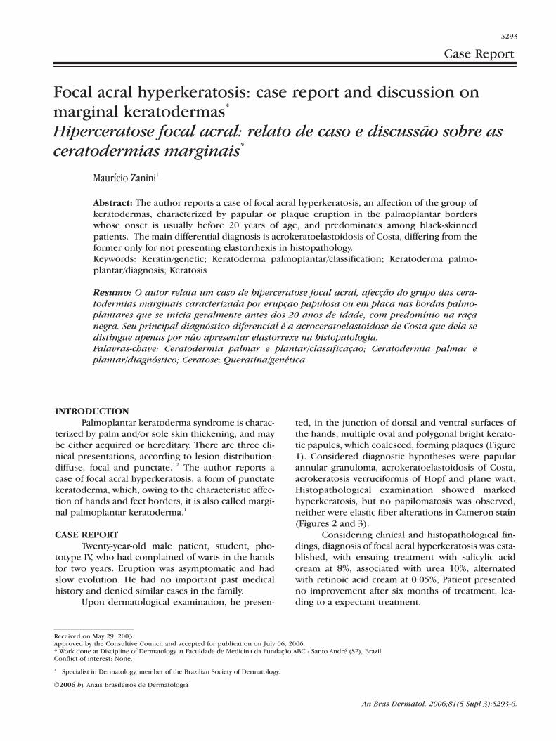
Upon dermatological examination, he presen-
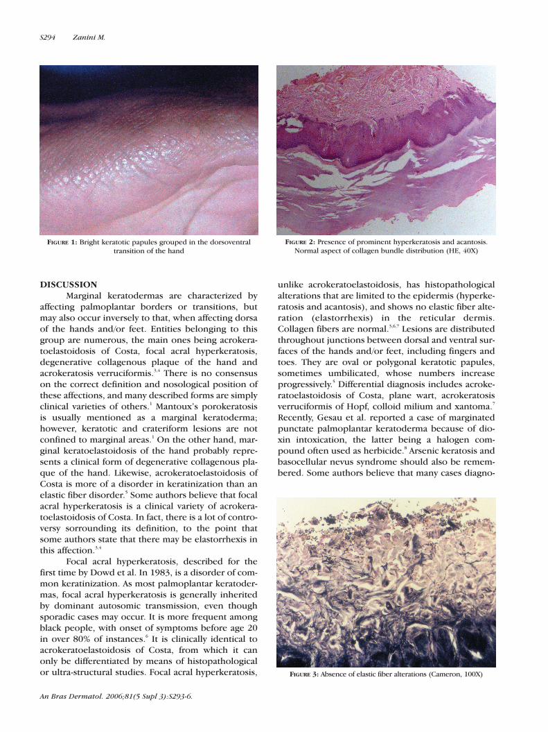
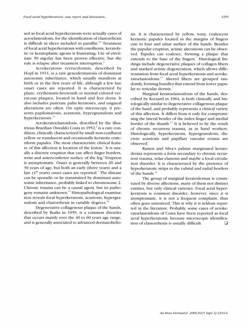
ted, in the junction of dorsal and ventral surfaces ofthe hands, multiple oval and polygonal bright kerato-tic papules, which coalesced, forming plaques (Figure1). Considered diagnostic hypotheses were papularannular granuloma, acrokeratoelastoidosis of Costa,acrokeratosis verruciformis of Hopf and plane wart.Histopathological examination showed markedhyperkeratosis, but no papilomatosis was observed,neither were elastic fiber alterations in Cameron stain(Figures 2 and 3).
Considering clinical and histopathological fin-dings, diagnosis of focal acral hyperkeratosis was esta-blished, with ensuing treatment with salicylic acidcream at 8%, associated with urea 10%, alternatedwith retinoic acid cream at 0.05%, Patient presentedno improvement after six months of treatment, lea-ding to a expectant treatment.
S293
An Bras Dermatol. 2006;81(5 Supl 3):S293-6.
S294 Zanini M.
DISCUSSIONMarginal keratodermas are characterized by
affecting palmoplantar borders or transitions, butmay also occur inversely to that, when affecting dorsaof the hands and/or feet. Entities belonging to thisgroup are numerous, the main ones being acrokera-toelastoidosis of Costa, focal acral hyperkeratosis,degenerative collagenous plaque of the hand andacrokeratosis verruciformis.3,4 There is no consensuson the correct definition and nosological position ofthese affections, and many described forms are simplyclinical varieties of others.1 Mantoux’s porokeratosisis usually mentioned as a marginal keratoderma;however, keratotic and crateriform lesions are notconfined to marginal areas.1 On the other hand, mar-ginal keratoelastoidosis of the hand probably repre-sents a clinical form of degenerative collagenous pla-que of the hand. Likewise, acrokeratoelastoidosis ofCosta is more of a disorder in keratinization than anelastic fiber disorder.5 Some authors believe that focalacral hyperkeratosis is a clinical variety of acrokera-toelastoidosis of Costa. In fact, there is a lot of contro-versy sorrounding its definition, to the point thatsome authors state that there may be elastorrhexis inthis affection.3,4
Focal acral hyperkeratosis, described for thefirst time by Dowd et al. In 1983, is a disorder of com-mon keratinization. As most palmoplantar keratoder-mas, focal acral hyperkeratosis is generally inheritedby dominant autosomic transmission, even thoughsporadic cases may occur. It is more frequent amongblack people, with onset of symptoms before age 20in over 80% of instances.6 It is clinically identical toacrokeratoelastoidosis of Costa, from which it canonly be differentiated by means of histopathologicalor ultra-structural studies. Focal acral hyperkeratosis,
unlike acrokeratoelastoidosis, has histopathologicalalterations that are limited to the epidermis (hyperke-ratosis and acantosis), and shows no elastic fiber alte-ration (elastorrhexis) in the reticular dermis.Collagen fibers are normal.3,6,7 Lesions are distributedthroughout junctions between dorsal and ventral sur-faces of the hands and/or feet, including fingers andtoes. They are oval or polygonal keratotic papules,sometimes umbilicated, whose numbers increaseprogressively.5 Differential diagnosis includes acroke-ratoelastoidosis of Costa, plane wart, acrokeratosisverruciformis of Hopf, colloid milium and xantoma.7
Recently, Gesau et al. reported a case of marginatedpunctate palmoplantar keratoderma because of dio-xin intoxication, the latter being a halogen com-pound often used as herbicide.8 Arsenic keratosis andbasocellular nevus syndrome should also be remem-bered. Some authors believe that many cases diagno-
FIGURE 1: Bright keratotic papules grouped in the dorsoventraltransition of the hand
FIGURE 2: Presence of prominent hyperkeratosis and acantosis.Normal aspect of collagen bundle distribution (HE, 40X)
FIGURE 3: Absence of elastic fiber alterations (Cameron, 100X)
sed as focal acral hyperkeratosis were actually cases ofacroelastoidosis, for the identification of elastorrhexisis difficult in slices included in paraffin.3,6 Treatmentof focal acral hyperkeratosis with emollients, keratoly-tic or keratoplastic agents is frustrating. Use of etreti-nate 50 mg/day has been proven effective, but therule is relapse after treatment interruption.5
Acrokeratosis verruciformis, described byHopf in 1931, is a rare genodermatosis of dominantautosomic inheritance, which usually manifests atbirth or in the first years of life, although a few lateonset cases are reported. It is characterized byplane, erythematic-brownish or normal colored ver-rucous plaques, located in hand and feet dorsa. Italso includes punctate palm keratoses, and unguealalterations are often. On optic microscopy, it pre-sents papilomatosis, acantosis, hypergranulosis andhyperkeratosis.1,4
Acrokeratoelastoidosis, described by the illus-trious Brazilian Oswaldo Costa in 1952,9 is a rare con-dition, clinically characterized by small non-confluentyellow or translucent and occasionally keratotic crate-riform papules. The most characteristic clinical featu-re of this affection is location of the lesion.7 It is usu-ally a discrete eruption that can affect finger borders,wrist and antero-inferior surface of the leg.5 Eruptionis asymptomatic. Onset is generally between 20 and30 years of age, but both an early (three years) and alate (47 years) onset cases are reported.7 The diseasecan be sporadic or be transmitted by dominant auto-somic inheritance, probably linked to chromosome 2.Chronic trauma can be a causal agent, but its patho-geny remains unknown.5-7 Histopathological examina-tion reveals focal hyperkeratosis, acantosis, hypergra-nulosis and elastorrhexis in variable degrees.3,6
Degenerative collagenous plaque of the hands,described by Burks in 1959, is a common disorderthat occurs mainly over the 40 to 60 years age range,and is generally associated to advanced dermatohelio-
sis. It is characterized by yellow, waxy, coalescentkeratotic papules located in the margins of fingersone to four and ulnar surface of the hands. Besidesthe papular eruption, actinic alterations can be obser-ved. Papules can coalesce, forming a plaque thatextends to the base of the fingers.7 Histological fin-dings include degenerative plaques of collagen fibersand marked actinic degeneration, which allows diffe-rentiation from focal acral hyperkeratosis and acroke-ratoelastoidosis.5-7 Altered fibers are grouped ran-domly, forming bundles that extend from lower papu-lar to reticular dermis.7
Marginal keratoelastoidosis of the hands, des-cribed by Kocsard in 1964, is both clinically and his-tologically similar to degenerative collagenous plaqueof the hand, and probably represents a clinical varietyof this affection. It differs from it only for compromi-sing the lateral border of the index finger and medialborder of the thumb.5,7 It is believed to be the resultof chronic recurrent trauma, as in hand workers.Histologically, hyperkeratosis, hypergranulosis, dis-crete acantosis and papillary vascular ectasia areobserved.7
Ramos and Silva’s palmar marginated kerato-derma represents a form secondary to chronic recur-rent trauma, solar elastosis and maybe a local circula-tion disorder. It is characterized by the presence ofhyperkeratotic strips in the cubital and radial bordersof the hands.10
The group of marginal keratodermas is consti-tuted by diverse affections, many of them not distinctentities, but only clinical varieties. Focal acral hyper-keratosis is common disorder; however, since it isasymptomatic, it is not a frequent complaint, thusoften goes unnoticed. This is why it is seldom repor-ted in the literature. Probably, some cases of acroke-ratoelastoidosis of Costa have been reported as focalacral hyperkeratosis, because microscopic identifica-tion of elastorrhexis is usually difficult. �
An Bras Dermatol. 2006;81(5 Supl 3):S293-6.
Focal acral hyperkeratosis: case report and discussion... S295
An Bras Dermatol. 2006;81(5 Supl 3):S293-6.
women. Int J Dermatol. 1998;37:532-7.7. Nelson-Adesokan P, Mallory SB, Lombardi C, Lund R.
Acrokeratoelastoidosis of Costa. Int J Dermatol. 1995;34:431-3.
8. Gesau A, Jurecka W, Nahavandi H, Schmidt JB, Stingl G, Tschachler E. Punctate keratoderma-like lesions on the palms and sole in a patient with chloracne: a new clinicalmanifestation of dioxin intoxication? Br J Dermatol. 2000;143:1067-71.
9. Costa OG. Acrokeratoelastosis. Arch Dermatol. 1954;70:228-31.
10. Sampaio SAP, Rivitti EA. Dermatologia. São Paulo: Artes Médicas; 1998. p.780-1.
REFERENCES1. Griffiths WAD, Judge MR, Leigh IM. Disorders of
keratinization. In: Rook A, Wilkinson DS, Ebling FJG. Textbook of Dermatology. 6 ed. Oxford: Blackwell Science; 1998. p.1483-588.
2. Stevens HP, Leigh IM. The inherited keratodermas of palms and soles. In: Freedberg IM, Eisen AZ, Wolff K, Austen KF, Goldsmith LA, Katz S, et al. Fitzpatrick's Dermatology in General Medicine. 5 ed. Philadelphia: MacGraw-Hill; 1998. p.604-12.
3. Rubegni P, De Aloe G, Romano C, Flori ML, Fimiani M. Acrokeratoelastoidosis: a report of two sporadic cases. Clin Exp Dermatol. 1997;22:54-64.
4. Rongioletti F, Betti R, Crosti Rebora A. Marginal papular acrokeratodermas: a unified nosography for focal acral hyperkeratosis, acrokeratoelastoidosis and relateddisorders. Dermatology. 1994;188:28-31.
5. Shbaklo Z, Jamaleddine NF, Kibbi AG, Salman SM, Zaynoun ST. Acrokeratoelastoidosis. Int J Dermatol. 1990;29:333-6.
6. Waxtein-Morgenstern L, Teixeira F, Cortes-Franco R, Veja-Memije ME, Ortiz-Plata A, Zamora-Hernandez C, Dominguez-Soto L. Lenticular acral keratosis in washer
MAILING ADDRESS:Maurício ZaniniRua Marechal Floriano Peixoto, 245 – Sala 8789010-500 - Blumenau - SC - BrazilTel.: +55 47 3326-5326E-mail: [email protected]
S296 Zanini M.


![Clinical Study Evaluation of Hemodynamics in Focal Steatosis and … · 2019. 7. 31. · focal steatosis and focal spared lesion [ ]. Some cases of focal steatosis and focal spared](https://img.pdfslide.net/doc/110x75/612bf41f63871b38801ecb60/clinical-study-evaluation-of-hemodynamics-in-focal-steatosis-and-2019-7-31.jpg)