Embed Size (px)
Citation preview

July 7, 2011
1
Harvard Medical School/Harvard School of Dental Medicine Format for the Curriculum Vitae
Date Prepared: July 7, 2011
Name: Simon Keith Warfield, Ph.D.
Office Address: Children's Hospital Dept of Radiology, Computational Radiology Laboratory 300 Longwood Avenue Boston, MA 02115 United States
Home Address: 173 Winchester St Brookline, MA 02446 United States
Work Phone: 617-355-4566
Work E-Mail: [email protected]
Work FAX: 617-730-4644
Place of Birth: Parramatta, Australia
Education 1991 B.Sc. Computer Science University of New South Wales,
Australia 1993 B.E. Honors
Class 1 Electrical Engineering University of New South Wales,
Australia 1997 Ph.D. Computer Science and
Engineering (Advisor: John Hiller, Ph.D.)
University of New South Wales, Australia
Postdoctoral Training 1996-1998
Research Fellow
Radiology Brigham & Women’s Hospital, Harvard Medical School, Boston, MA USA
1997-1998
Post-doc Medical Imaging (Advisor: Ron Kikinis, M.D.)
Brigham & Women’s Hospital, Harvard Medical School, Boston, MA USA
Faculty Academic Appointments
1998-2001
Instructor Radiology Brigham & Women’s Hospital, Harvard Medical School, Boston, MA USA
2001-2004
Assistant Professor
Radiology Brigham & Women’s Hospital, Harvard Medical School, Boston, MA USA

July 7, 2011
2
2002-2007
Research Affiliate
CSAIL Massachusetts Institute of Technology, Cambridge, MA USA
2004-2007
Associate Professor
Radiology Brigham & Women’s Hospital, Harvard Medical School, Boston, MA USA
2007-2010
Associate Professor
Radiology Children's Hospital Boston, Harvard Medical School, Boston, MA USA
2010- Professor Radiology Children's Hospital Boston, Harvard Medical School, Boston, MA USA
Appointments at Hospitals/Affiliated Institutions 1994, 1996-1998
Research Fellow Radiology Brigham & Women’s Hospital, Harvard Medical School, Boston, MA USA
1998-2001
Research Associate
Radiology Brigham & Women’s Hospital, Harvard Medical School, Boston, MA USA
2001-2007
Director Computational Radiology Laboratory
Brigham & Women’s Hospital, Harvard Medical School, Boston, MA USA
2007- Director Computational Radiology Laboratory
Children's Hospital Boston, Harvard Medical School, Boston, MA USA
Other Professional Positions 2006- Member, Research and Investment Advisory
Council (RIAC) CSIRO eHealth Research Centre
Major Administrative Leadership Positions 2008- Director of Radiology Research
Children's Hospital Boston, Harvard Medical School, Boston, MA
2008- Site Core Leader, Harvard Medical School CTSC Translational Technologies Imaging Consortium
Harvard Medical School, Children’s Hospital Boston, Boston, MA
Committee Service Local 2005-2006
Bioinformatics in Functional and Molecular Imaging Committee
Brigham & Women’s Hospital, Boston, MA
2005-2007
Division of Newborn Medicine Scholarship Advisory Committee
Children's Hospital Boston, MA
2007- MRI Research Committee Children's Hospital Boston, MA

July 7, 2011
3
2008-2010
Harvard Neonatal-Perinatal Fellowship Program Thesis Scholarship Oversight Committee
Harvard Medical School, Boston, MA
2009- Scientific and Resource Review Committee, Harvard Catalyst Participant and Clinical Interactions Resource (PCIR)
Harvard Medical School, Boston, MA
National 2004- NiFTI Geometry Format Working Group NIH 2008 Advisory Committee Member Osteoarthritis Initiative (OAI) International 1998 Program Committee Member MICCAI 98 1st International
Conference on Medical Image Computing and Computer Assisted Intervention
1999 Scientific Review Committee Member MICCAI 99 2nd International Conference on Medical Image Computing and Computer Assisted Intervention
2001 Scientific Review Committee Member MICCAI 01 3rd International Conference on Medical Image Computing and Computer Assisted Intervention
2002 Scientific Review Committee Member ISBI 2002: International Symposium on Biomedical Imaging
2002 Scientific Review Committee Member IS4TM 2003: International Symposium on Surgical Simulation and Soft Tissue Modeling
2002 Scientific Review Committee Member MICCAI 02 5th International Conference on Medical Image Computing and Computer Assisted Intervention
2002 Scientific Review Committee Member ISMRM 2003: International Society for Magnetic Resonance in Medicine Eleventh Scientific Meeting
2003 Scientific Review Committee Member ISBI 2004: International Symposium on Biomedical Imaging
2003 Scientific Review Committee Secretary MICCAI 03 6th International Conference on Medical Image Computing and Computer Assisted Intervention
2003 Scientific Review Committee Member WBIR 2003: Workshop on

July 7, 2011
4
Biomedical Image Registration 2003 Scientific Review Committee Member ISMRM 2004: International Society
for Magnetic Resonance in Medicine Twelfth Scientific Meeting
2004 Scientific Review Committee Member Second International Symposium on Medical Simulation
2004 Scientific Review Committee Member MICCAI 04 7th International Conference on Medical Image Computing and Computer Assisted Intervention
2005 Program Committee Member ISBI 2006: International Symposium on Biomedical Imaging
2005 Program Committee Member Computer Vision for Biomedical Image Applications
2005 Scientific Review Committee Member WBIR 2005: Workshop on Biomedical Image Registration
2005 Scientific Review Committee Member MICCAI 05 8th International Conference on Medical Image Computing and Computer Assisted Intervention
2006 Scientific Review Committee Member 3rd Symposium on Biomedical Simulation
2006 MICCAI 06 Workshop on Joint Disease Program Committee Member
MICCAI 06 9th International Conference on Medical Image Computing and Computer Assisted Intervention
2006 Scientific Review Committee Member MICCAI 06 9th International Conference on Medical Image Computing and Computer Assisted Intervention
2006 Scientific Review Committee Member International Conference on Pattern Recognition 2006
2007 International Program Committee for Visual Communications (VC 2008) Member
IASTED VC 2008
2007 Program Committee Member MICCAI 07 10th International Conference on Medical Image Computing and Computer Assisted Intervention
2008 International Program Committee Member IASTED International Conference on Internet and Multimedia Systems/Visual Communications
2008 Program Committee Member MICCAI 08 11th International Conference on Medical Image Computing and Computer Assisted Intervention
2008 Program Committee Member International Symposium on

July 7, 2011
5
Computational Models for Biomedical Simulation (ISBMS)
2008-2011
Annual Meeting Program Committee (AMPC) Member
International Society for Magnetic Resonance in Medicine (ISMRM)
2009 Program Committee Member MICCAI 09 12th International Conference on Medical Image Computing and Computer Assisted Intervention
2010 Program Committee Member MICCAI 10 13th International Conference on Medical Image Computing and Computer Assisted Intervention
2011 Program Committee Member MICCAI 11 14th International Conference on Medical Image Computing and Computer Assisted Intervention
Professional Societies 1998- International Society for Magnetic Resonance in Medicine 1998- Member 1998- Institute for Electrical and Electronics Engineers 1998- Member 2007- Senior Member 2001- IEEE Computer Society 2001- Member 2001- IEEE Signal Processing Society 2001- Member 2001- American Association for the Advancement of Science 2001- Member 2004- MICCAI 2004- Member Grant Review Activities 2004 NIDA: Design Evaluation, and Integration
of Image Analysis NIH
2004 Member 2004
ZRG1 SBIB-Q 50 Study Section NIH
2004 Member 2005-2007 BDCN K-10 Study Section NIH 2005- 2007
Member
2005 NIDA review panel RFC No.: N43DA-5-4403 (Topic 067)
NIH

July 7, 2011
6
2005 Ad-hoc Member 2006 ZRG1 SBIB-L (40) MR P41 NIH 2006 Member 2006 Australian Research Council
Australian Research Council
2006 Reviewer 2006-2007 ZRG1 BDCN-K 50M NIH 2006- 2007
Member
2007 Discovery Project Australian Research Council 2007 Reviewer 2007 European Young Investigator Award European Science Foundation 2007 Reviewer 2007 Sheffield Hospitals Charitable Trust Sheffield Hospitals Charitable Trust 2007 Reviewer 2007 ZRG1 BDCN-E (10) B Clinical
Neurophysiology, Devices & Neuroprosthetics
NIH
2007 Member 2007 ZRG1 BDCN-F (03) S Clinical
Neurophysiology, Devices & Neuroprosthetics
NIH
2007 Member 2008 ZRG1 SBIB U(91) Innovative Ultrasound
and Imaging NIH
2008 Member 2008 New Research Project Proposal Research Foundation – Flanders
(Belgium) (FWO) 2008 Referee 2008 Swiss National Science Foundation Swiss National Science Foundation 2008 Reviewer 2008 CFI Expert Review Committee Canada Foundation for Innovation 2008 Member 2009 SBIB-D 53 Peer Review NIH 2009 Member 2010 Human Connectome Project (HCP) RFA-
MH-10-020 NIH/NIMH
2010 Member 2011 ESAB Meeting - VCU P01 NIH 2011 Member 2011 BDMA Study Section NIH 2011 Member 2011 NIH BDCN N02 Special Emphasis Panel NIH 2011 Member Editorial Activities

July 7, 2011
7
Ad-hoc Reviewer 1996 Ad-hoc Reviewer Pattern Recognition Letters 1999- Ad-hoc Reviewer IEEE Transactions on Medical Imaging 1999- Ad-hoc Reviewer NeuroImage 1999- Ad-hoc Reviewer Medical Image Analysis 2000- Ad-hoc Reviewer Graphical Models 2000- Ad-hoc Reviewer Journal of Biomedical Informatics 2001- Ad-hoc Reviewer Signal Processing 2001- Ad-hoc Reviewer IEEE Transactions on Biomedical
Engineering 2001- Ad-hoc Reviewer IEEE Transactions on Image Processing 2002- Ad-hoc Reviewer International Journal of Image and Graphics 2003- Ad-hoc Reviewer Human Brain Mapping 2003- Ad-hoc Reviewer Medical and Biological Engineering and
Computing 2005- Ad-hoc Reviewer Cerebral Cortex 2005- Ad-hoc Reviewer Image and Vision Computing 2006- Ad-hoc Reviewer International Journal of Radiation Oncology
Biology Physics 2006- Ad-hoc Reviewer Pattern Analysis and Applications 2007- Ad-hoc Reviewer Pediatrics Research 2007- Ad-hoc Reviewer Nature Clinical Practice Neurology 2008- Ad-hoc Reviewer Pediatrics 2008- Ad-hoc Reviewer Neuroinformatics Other Editorial Roles 2005- Associate Editor IEEE Transactions on Medical Imaging 2005- Editor Medical Image Analysis 2007- Editor The Open Biomedical Engineering Journal Honors and Prizes 1993 Australian Postgraduate Research Award University Of New South Wales,
Australia 1993 B.E. Honors Class 1 (Electrical
Engineering) University Of New South Wales, Australia
1993 UNSW Faculty of Engineering Postgraduate Award
University Of New South Wales, Australia
1997-1998
National Multiple Sclerosis Society Postdoctoral Fellowship Award
National Multiple Sclerosis Society
1998 ISMRM Student/Postdoctoral Fellow Stipend Award
ISMRM
2000 CIMIT New Concept Award CIMIT

July 7, 2011
8
2005 CIMIT New Concept Award CIMIT 2005 Ferrant et al. Med Imag Anal 2002 - Top
1% Most Cited Paper in the Field Thompson/ISI
2006 Edward M. Kennedy Award for Health Care Innovation
CIMIT
2006-2008
International Fellow CSIRO
2006 Fast Breaking Paper - Warfield et al. IEEE TMI 2004 -Top 1% Most Cited Paper in the Field
Thomson/Essential Science Indicators
2008 Australia-Harvard Fellowship Harvard Club of Australia

July 7, 2011
9
Report of Funded and Unfunded Projects Funding Information Past 1993-1996 PI UNSW Faculty of
Engineering
Segmentation of Magnetic Resonance Images of the Brain 1993-1996 PI Australian
Government
Segmentation of Magnetic Resonance Images of the Brain 1998-2000 PI NMSS
RG 3094A1/T
Characterization of Multiple Sclerosis Lesions from MRI Successfully developed algorithms to automatically and accurately characterize multiple sclerosis lesions as seen on MRI, and to segment multiple sclerosis lesions with high sensitivity and specificity. 1998-2003 Investigator NIH
P41 RR013218 Project
High Performance Computing for Neuroimaging Center: 3D MRI Data The main research focus of the NAC High Performance Computing Project is to develop post-processing methods for digital medical imaging data and to use these algorithms for clinical applications. 1999-2001 Investigator NIH
R21 CA80945
Virtual Cystoscopy for Detection of Small Bladder Tumors The project was a 2-year feasibility study to develop and test the potential of virtual cystoscopy as a non-invasive technique for the detection of small (<2cm) bladder tumors. Developed high sensitivity and specificity algorithms for detecting small bladder tumors from high resolution CT. 1999-2002 Investigator NIH Visible Human Project Image Processing Tools The main goal of this project is to perform software engineering, validation, algorithm integration, and test bed application. 2000-2002 PI Center for Innovative
Minimally Invasive Therapy
Intraoperative MRI Guided Liver Cryotherapy Testbed to Develop Technologies for the
July 7, 2011
10
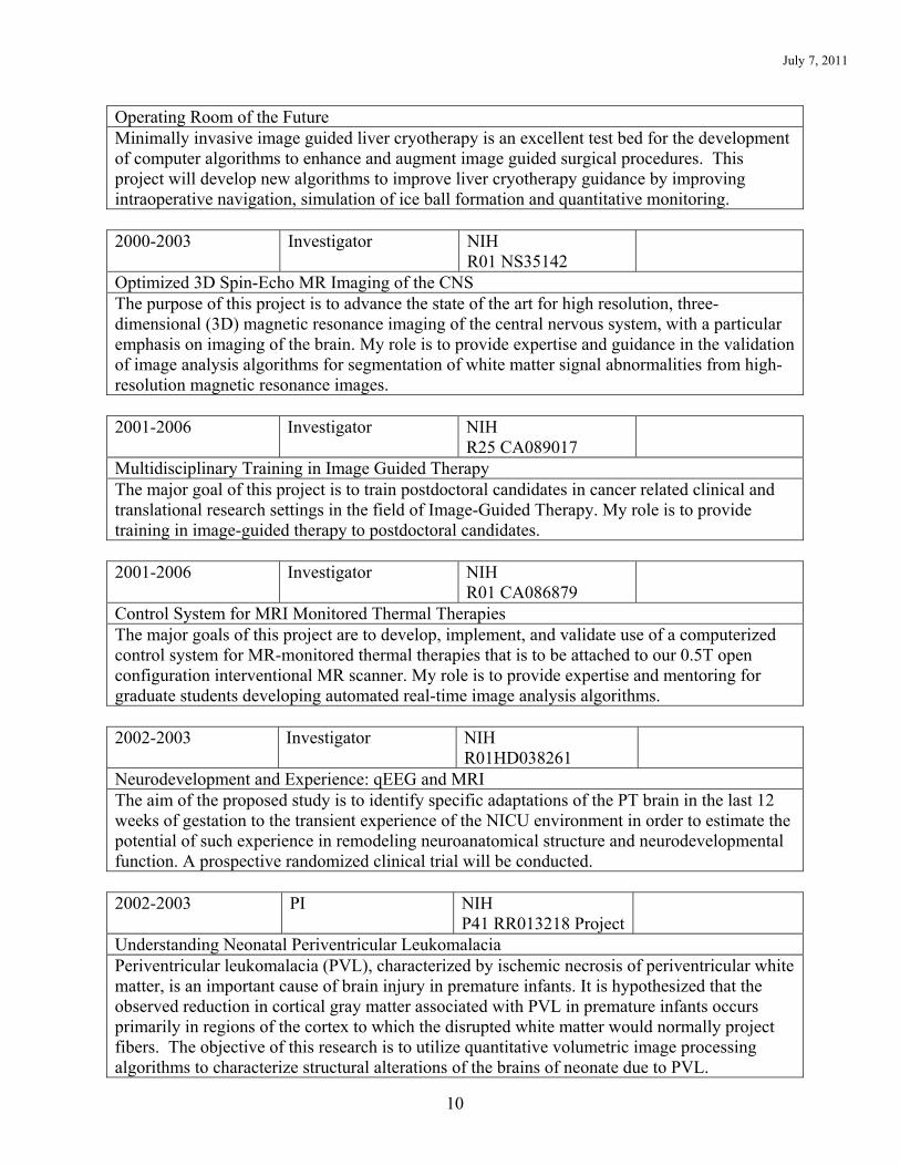
Operating Room of the Future Minimally invasive image guided liver cryotherapy is an excellent test bed for the development of computer algorithms to enhance and augment image guided surgical procedures. This project will develop new algorithms to improve liver cryotherapy guidance by improving intraoperative navigation, simulation of ice ball formation and quantitative monitoring. 2000-2003 Investigator NIH
R01 NS35142
Optimized 3D Spin-Echo MR Imaging of the CNS The purpose of this project is to advance the state of the art for high resolution, three-dimensional (3D) magnetic resonance imaging of the central nervous system, with a particular emphasis on imaging of the brain. My role is to provide expertise and guidance in the validation of image analysis algorithms for segmentation of white matter signal abnormalities from high-resolution magnetic resonance images. 2001-2006 Investigator
NIH R25 CA089017
Multidisciplinary Training in Image Guided Therapy The major goal of this project is to train postdoctoral candidates in cancer related clinical and translational research settings in the field of Image-Guided Therapy. My role is to provide training in image-guided therapy to postdoctoral candidates. 2001-2006 Investigator NIH
R01 CA086879
Control System for MRI Monitored Thermal Therapies The major goals of this project are to develop, implement, and validate use of a computerized control system for MR-monitored thermal therapies that is to be attached to our 0.5T open configuration interventional MR scanner. My role is to provide expertise and mentoring for graduate students developing automated real-time image analysis algorithms. 2002-2003 Investigator NIH
R01HD038261
Neurodevelopment and Experience: qEEG and MRI The aim of the proposed study is to identify specific adaptations of the PT brain in the last 12 weeks of gestation to the transient experience of the NICU environment in order to estimate the potential of such experience in remodeling neuroanatomical structure and neurodevelopmental function. A prospective randomized clinical trial will be conducted. 2002-2003 PI NIH
P41 RR013218 Project
Understanding Neonatal Periventricular Leukomalacia Periventricular leukomalacia (PVL), characterized by ischemic necrosis of periventricular white matter, is an important cause of brain injury in premature infants. It is hypothesized that the observed reduction in cortical gray matter associated with PVL in premature infants occurs primarily in regions of the cortex to which the disrupted white matter would normally project fibers. The objective of this research is to utilize quantitative volumetric image processing algorithms to characterize structural alterations of the brains of neonate due to PVL.
July 7, 2011
11
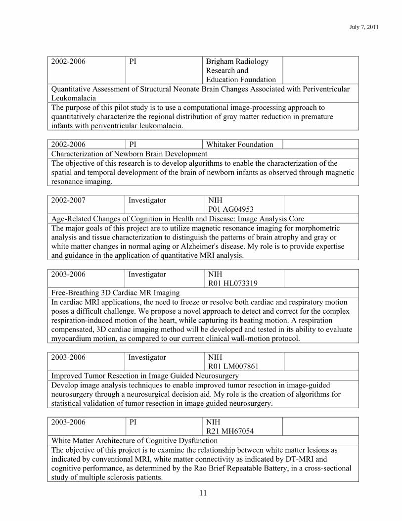
2002-2006 PI Brigham Radiology
Research and Education Foundation
Quantitative Assessment of Structural Neonate Brain Changes Associated with Periventricular Leukomalacia The purpose of this pilot study is to use a computational image-processing approach to quantitatively characterize the regional distribution of gray matter reduction in premature infants with periventricular leukomalacia. 2002-2006 PI Whitaker Foundation Characterization of Newborn Brain Development The objective of this research is to develop algorithms to enable the characterization of the spatial and temporal development of the brain of newborn infants as observed through magnetic resonance imaging. 2002-2007 Investigator NIH
P01 AG04953
Age-Related Changes of Cognition in Health and Disease: Image Analysis Core The major goals of this project are to utilize magnetic resonance imaging for morphometric analysis and tissue characterization to distinguish the patterns of brain atrophy and gray or white matter changes in normal aging or Alzheimer's disease. My role is to provide expertise and guidance in the application of quantitative MRI analysis. 2003-2006 Investigator NIH
R01 HL073319
Free-Breathing 3D Cardiac MR Imaging In cardiac MRI applications, the need to freeze or resolve both cardiac and respiratory motion poses a difficult challenge. We propose a novel approach to detect and correct for the complex respiration-induced motion of the heart, while capturing its beating motion. A respiration compensated, 3D cardiac imaging method will be developed and tested in its ability to evaluate myocardium motion, as compared to our current clinical wall-motion protocol. 2003-2006 Investigator NIH
R01 LM007861
Improved Tumor Resection in Image Guided Neurosurgery Develop image analysis techniques to enable improved tumor resection in image-guided neurosurgery through a neurosurgical decision aid. My role is the creation of algorithms for statistical validation of tumor resection in image guided neurosurgery. 2003-2006 PI NIH
R21 MH67054
White Matter Architecture of Cognitive Dysfunction The objective of this project is to examine the relationship between white matter lesions as indicated by conventional MRI, white matter connectivity as indicated by DT-MRI and cognitive performance, as determined by the Rao Brief Repeatable Battery, in a cross-sectional study of multiple sclerosis patients.

July 7, 2011
12
2003-2008 PI NIH
P41 RR013218 Project
Developmental Neuroinformatics at the Neuroimaging Analysis Center (NAC) The Neuroimaging Analysis Center (NAC) is a National Research Resource Center operating in an application-oriented, clinical environment with the mission of focused computer-science based technology research and development. This proposal represents a continuation and expansion of ongoing efforts, with a shift in focus from generic image analysis capabilities to neuroimage informatics techniques tightly coupled to support particular applications. These neuroscientific and clinical applications provide demanding neuroimage informatics challenges which require new technology research and development, which, when solved, will have widespread applicability. The proposed core activities include algorithm development for the analysis of white matter architecture using diffusion tensor MRI and characterization of the spatial and temporal development of the structures in the infant brain, as well as the development of image informatics tools that are aimed at facilitating the exploitation of fMRI-derived information in neurosurgical and neuroscientific applications. In addition, novel methods of medical image representation and visualization will be explored and developed, as well as a new multi-modal digital anatomical atlas. 2004-2006 Project Director NIH
P41 RR013218 Project
Grid Enabling the Insight Toolkit The Insight Toolkit (ITK) has become the de facto standard platform for advanced segmentation and registration research at many laboratories. At the same time, there is an increasing trend to deploy grid-computing infrastructures to support computations on extremely large data sets like those associated with the Visible Human Project. The architecture of ITK is not designed to support such efforts. We believe it is important to revisit and refine critical aspects of the architecture of ITK to support the emerging standards in the grid-computing community and to develop example applications to demonstrate the power of the ITK/grid combination in real-world research computing scenarios. 2004-2007 Co-PI NIH
R01 HD046855
Preterm Fetal Growth Restriction and Developmental Care This project will test the effectiveness of an in-NICU intervention for FGR infants. The study will be significant in understanding ways to reduce long-term functional morbidities in FGR infants, as well as in identifying opportunities for enhancing last trimester brain development. 2004-2007 (extended to 2008)
PI NSF NSF ITR 0426558
$250,000/year
ITR: Collaborative Research - \(ASE\) - \(DMC\): DDDAS: A Novel Grid Architecture Integrating Real-Time Data and Intervention During Image Guided Therapy The aim of this project is the development and deployment of an integrated and practical grid architecture for data driven intra-operative volumetric simulation of brain deformation during image guided therapy (IGT) and specifically for image guided neurosurgery (IGNS) to be

July 7, 2011
13
employed in the operating room of the future. 2005-2006 PI CIMIT Improved Analysis for Patient-Specific Epilepsy Surgical Planning The goal of this work is to develop an optimized MRI acquisition protocol, and post-acquisition analysis strategy to enable improved pediatric epilepsy surgical planning (ESP). 2005-2007 Investigator NIH
U41 RR019703
Image Guided Therapy Center The IGT Center proposed under this application will provide a unique, centralized infrastructure for clinical investigators, biomedical engineers, and basic scientists in promoting and advancing IGT methods and related clinical applications. The center will develop and make available new innovative technologies in five discrete TRD Core Projects: 1) the Computational Core; 2) the Imaging Core; 3) the MRI-guided Therapy Core; 4) the Image-Guided Neurosurgery Core; and 5) the Focused Ultrasound Therapy Core.
2006-2008 (extended to 2009)
PI NIH R03 CA126466
$50,000/year
Total Lagrangian Explicit Dynamics Finite Element Method for Brain Registration
The aims of this proposal are to 1) develop a very efficient finite element solver using Total Lagrangian formulation and explicit time integration scheme, suited to computing brain deformation in real time; 2) implement the new constitutive model of brain tissue, accounting for brain tissue higher stiffness in compression than in extension in finite element code; 3) carry out extensive validation and evaluation of the proposed model in the setting of intraoperative MRI alignment. 2006-2008 Co-Investigator NIH
R01 HL074942 $368,850/year
Ventilation Model and CNS Injury in Baboons with BPD In this study we propose to investigate the nature of cerebral injury in a prematurely born primate model (Papio sp) developed as a model of bronchopulmonary dysplasia, utilizing both magnetic resonance imaging (MR) and histopathology.
2008-2009 PI NIH R01 EB008015-S1
$50,000/year
GUI and Tutorial for Software for Validation of Image Segmentations
This is an administrative supplement to the NIH R01 EB008015 grant entitled “Assessment of Improved Navigation for Pediatric Brain Tumor Surgery.” The overall objective of this supplement is to enhance our existing software and disseminate a new graphical user interface together with enhanced training materials for users in the form of a tutorial description of our STAPLE algorithm and its implementation.
2008-2010 Co-Investigator NMSS RG4032A1

July 7, 2011
14
From Probable to Definite Multiple Sclerosis: an Imaging Based Predictive Model (PI: D. Goldberg-Zimring)
The goal of this project is the detection, delineation and modeling of major white matter fiber tract segments in a healthy volunteer (WMFTS) and the identification of disrupted WMFTS in the study population to determine the relationship between the decrease of cognitive performance and disrupted WMFTS. This will enable us to model the architecture of white matter and assess the relationship between disrupted WMFTS and cognitive dysfunction.
2004-2007 (extended to 2010)
PI NMSS RG3478A2
$110,000/year
Disruption of White Matter Circuits and Cognitive Deficits in Multiple Sclerosis
This study will construct statistical atlases of conventional MRI and Diffusion Tensor MRI utilizing 3.0T MRI of healthy controls and early diagnosis multiple sclerosis patients. Patterns of white matter alteration associated with multiple sclerosis will be determined.
2006-2010 PI NIH R01GM074068
$130,000/year
Bioinformatics Tools for Multi-Center Diagnostic Trials
Over the past decade, multi-center clinical trials utilizing diagnostic imaging modalities have been conducted and sponsored by the National Institutes of Health. The long-term goal is to develop efficient ways for better analyzing clustered data and utilizing prior knowledge in multi-center clinical trials.
2008-2009 (extended to 2010)
PI CIMIT 08-293
$100,000/year
Bayesian Source Imaging of Pediatric Epilepsy
The goal of this project is to create a new device capable of locating epileptogenic foci and thereby make curative surgery available to a larger population at an earlier age. This will be demonstrated through significant impact on clinical surgical planning in pediatric epilepsy.
2008-2010 Mentor William Randolph Hearst Fund
Study of cerebral perfusion using arterial spin labeling in term newborn infants with hypoxic-ischemic encephalopathy (PI: P. Wintermark) I served as mentor for Pia Wintermark during the period of her Hearst Fund award. This study developed an effective arterial spin labeling MRI strategy for characterizing perfusion in newborns with and without hypoxic-ischemic encephalopathy.
2008-2010 Mentor Thrasher Research Fund
Study of cerebral perfusion using arterial spin labeling in term newborn with hypoxic-ischemic encephalopathy (PI: P. Wintermark)

July 7, 2011
15
I served as mentor for Pia Wintermark during the period of her Early Career award from the Thrasher. The main purpose of the study was to measure the temporal evolution of perfusion in newborns with underlying HI encephalopathy. We acquired critically important data to guide the application of tailored neuroprotective strategies to specific infants, especially those targeted to prevent reperfusion injury, with the potential to decrease brain injury associated with HIE.
2009-2011 Mentor NIH
KL2 RR025757
$69,900/year
Improved Source Localization for Pediatric Epilepsy (PI: D. Hyde)
I am serving as mentor for Damon Hyde during the period of his training program. This research project seeks to dramatically increase the number of pediatric epilepsy patients who are cured by surgical intervention by developing `computational electrocorticography', a non-invasive alternative to electrocorticography, now made possible for the first time by a combination of major advances in electroencephalography, magnetic resonance imaging, and sophisticated patient-specific numerical simulations of bioelectromagnetic field propagation. Current
2006-2010 (extended to 2011)
PI NIH R01 RR021885
$200,000/year
Bioinformatics Software for MRI of Brain Development
The major goals of this project are the enhancement of an existing software package for quantitative analysis of MRI of the developing brain by the implementation, as open-source software, of existing validated and proven algorithms, and the creation of a user-friendly graphical user interface to enable end users to easily apply these methods.
2009-2010 (extended to 2011)
PI NIH R01 RR021885-S1
$190,000/year
Bioinformatics Software for MRI of Brain Development This is an administrative supplement to the NIH R01 RR021885 grant. The overall goal is to improve care of preterm newborns by providing quantitative MRI tools for the identification of high-risk infants. This research supplement proposes the development of a battery of tests, based on the tools in the parent grant, which will predict later neurodevelopmental outcome in infants based on MRI taken at term.
2006-2011 Co-Investigator NIHCD R01HD047730
Does Early Experience Improve Preterm Neurodevelopment? (PI: H. Als)
About fifty percent of prematurely born infants develop learning/behavior problems and school failure. The study will test the primary hypotheses, that preterm infants (PT) randomized to

July 7, 2011
16
developmental care in the Newborn Intensive Care Unit (NICU) will be superior in cognitive performance at school age when compared to their peers, who did not receive the intervention.
2007-2011 (extended to 2012)
PI NIH R01 EB008015
$225,000/year
Assessment of Improved Navigation for Pediatric Brain Tumor Surgery
This research proposal aims to apply and evaluate novel surgical navigation technology to improve outcomes in pediatric brain tumor surgery. The specific aims of this research are to 1) Evaluate target registration error in nonrigid registration algorithms for pediatric brain tumor surgery, (2) Significantly improve the duration of precise alignment and data fusion during pediatric brain tumor surgery, and 3)Evaluate the efficacy of enhanced navigation by assessing post-operative tumor resection volume.
2008-2013 Site Co-Director NIH/NCRR
UL1 RR025758
Harvard Clinical and Translational Science Center (PI: L. Nadler)
Provide enriched resources to educate and develop the next generation of researchers trained in the complexities of translating research discoveries into clinical trials and ultimately into practice. Design new and improved clinical research informatics tools for analyzing research data and managing clinical trials. Support outreach to underserved populations, local community and advocacy organizations, and health care providers. Assemble interdisciplinary teams and forge new partnerships with private and public health care organizations.
2009-2010 (extended to 2011)
PI NIH R03 EB008680
$100,000/year
Improved Interoperability and Dissemination of Software for Simultaneous Truth and Performance Level Estimation
This is an R03 grant for one year of funding to develop and to disseminate image analysis an enhanced and extended implementation of the algorithm called STAPLE (Simultaneous Truth and Performance Level Estimation). Our objective is to enable scientists to utilize the software for neuroimage analysis, by providing the software, example data and tutorial explanation of how to use the software effectively.
2009-2011 Co-PI NIH R41 MH086984
$102,160/year
Prospective/Retrospective Motion Correction System for Motion Robust Pediatric MR
This project aims at the development and evaluation of an integrated hardware/software system for motion robust pediatric MRI in order to minimize or eliminate the need for sedation. The integration of the aims will be quantitatively and critically evaluated in this project through controlled experiments and statistical hypotheses testing.
July 7, 2011
17
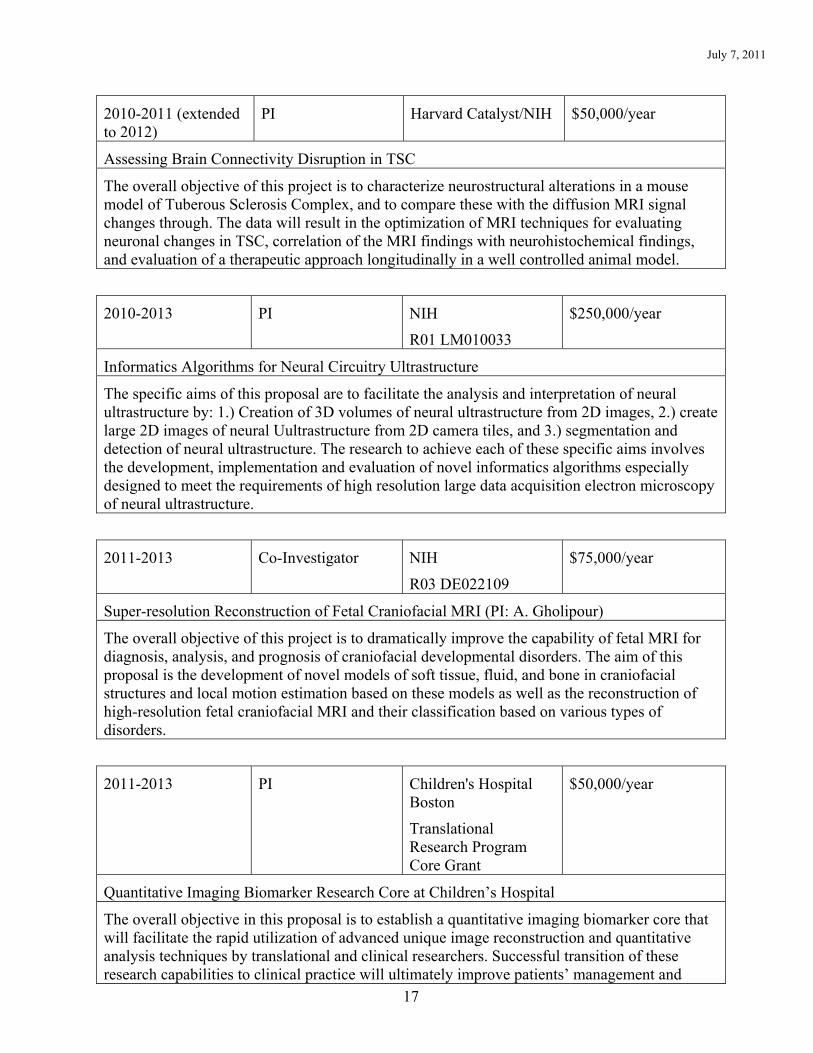
2010-2011 (extended to 2012)
PI Harvard Catalyst/NIH $50,000/year
Assessing Brain Connectivity Disruption in TSC
The overall objective of this project is to characterize neurostructural alterations in a mouse model of Tuberous Sclerosis Complex, and to compare these with the diffusion MRI signal changes through. The data will result in the optimization of MRI techniques for evaluating neuronal changes in TSC, correlation of the MRI findings with neurohistochemical findings, and evaluation of a therapeutic approach longitudinally in a well controlled animal model.
2010-2013 PI NIH
R01 LM010033
$250,000/year
Informatics Algorithms for Neural Circuitry Ultrastructure
The specific aims of this proposal are to facilitate the analysis and interpretation of neural ultrastructure by: 1.) Creation of 3D volumes of neural ultrastructure from 2D images, 2.) create large 2D images of neural Uultrastructure from 2D camera tiles, and 3.) segmentation and detection of neural ultrastructure. The research to achieve each of these specific aims involves the development, implementation and evaluation of novel informatics algorithms especially designed to meet the requirements of high resolution large data acquisition electron microscopy of neural ultrastructure.
2011-2013 Co-Investigator NIH
R03 DE022109
$75,000/year
Super-resolution Reconstruction of Fetal Craniofacial MRI (PI: A. Gholipour)
The overall objective of this project is to dramatically improve the capability of fetal MRI for diagnosis, analysis, and prognosis of craniofacial developmental disorders. The aim of this proposal is the development of novel models of soft tissue, fluid, and bone in craniofacial structures and local motion estimation based on these models as well as the reconstruction of high-resolution fetal craniofacial MRI and their classification based on various types of disorders.
2011-2013 PI Children's Hospital Boston
Translational Research Program Core Grant
$50,000/year
Quantitative Imaging Biomarker Research Core at Children’s Hospital
The overall objective in this proposal is to establish a quantitative imaging biomarker core that will facilitate the rapid utilization of advanced unique image reconstruction and quantitative analysis techniques by translational and clinical researchers. Successful transition of these research capabilities to clinical practice will ultimately improve patients’ management and

July 7, 2011
18
outcome.
2011-2016 Mentor NIH
K25 NS067068
$161,775/year
Improved Source Localization for Pediatric Epilepsy (PI: D. Hyde)
I am serving as mentor for Damon Hyde during the period of this research career development award. This proposal will use structural and functional information extracted from MR images to help improve the accuracy of source localization techniques. Improved maps of seizure activity will have a significant impact upon human health by allowing neurosurgeons to perform curative surgery in a larger portion of patients whose epilepsy is poorly controlled by current drug therapy.
2011-2012 Mentor Thrasher Research Fund
Thrasher Early Career Award
$25,000/year
Three-dimensional High-resolution Fetal MRI for Enhanced In-vivo Analysis of Congenital Anomalies (PI: A. Gholipour)
I am serving as mentor for Ali Gholipour during the period of this research career development award. The objective of this research is the development of advanced technology for 3D high-resolution (HR) motion-compensated fetal MRI to dramatically improve the diagnosis, analysis, and prognosis of congenital anomalies, specifically anomalies of the brain and lung.
Current Unfunded Projects
1996- PI Assessment of Knee Cartilage from MRI This project is developing imaging and image analysis technologies to improve our ability to quantitatively characterize cartilage of the knee from MRI.
Report of Local Teaching and Training Teaching of Students in Courses
2008 Planning for Image Guided Pediatric Neurosurgery (HST Course) Post-graduate students Lecturer 1 hour 2010 Interventional Imaging (HST Course) Post-graduate students Lecturer 1 hour
Clinical Supervisory and Training Responsibilities

July 7, 2011
19
2004-2007 Harvard Neonatal-Perinatal Medicine Fellowship Thesis Scholarship Oversight Committee – Deirdre O’Reilly, M.D.
5%
2008-2010 Harvard Neonatal-Perinatal Medicine Fellowship Thesis Scholarship Oversight Committee – Pia Wintermark, M.D.
5%
Laboratory and Other Research Supervisory and Training Responsibilities
2001-2007 Director, Computational Radiology Laboratory, BWH - Mentor 20% 2007- Director, Computational Radiology Laboratory, CHB - Mentor 20% 2008- Director of Radiology Research, Children’s Hospital - Mentor 20% Formally Supervised Trainees 1997-1998 Chahin Pachai, Ph.D. President & CEO, THERALYS, Lyon, France 1997-2000 Michael Kaus, Ph.D. Director, Research and Advanced Development at
Philips Healthcare, Madison, WI 1998-2002 Matthieu Ferrant, Ph.D. Product Manager, Clinical Applications, Agfa
Healthcare, Belgium 1999-2000 Olivier Cuisenaire, Ph.D. Staff Scientist, Philips Medical Systems, Paris, France 1999-2000 Torsten Butz, Ph.D. Staff Scientist, ImaSys SA, PSE, Lausanne,
Switzerland 1999-2002 Xingchang Wei Clinical Assistant Professor, University of Calgary,
Alberta, Canada 2000-2001 Aditya Bharatha, M.D. Resident, Sunnybrook Health Sciences Centre,
University of Toronto, Canada 2000-2001 Alida Tei Senior Associate, CATALYSIS, London, UK 2000-2002 Ying Wu Research Associate, Northwestern University Feinberg
School of Medicine, Evanston, IL 2001 Jan Rexilius, Ph.D. Computer Scientist, MeVis, Bremen, Germany 2001-2002 Sylvain Jaume, Ph.D. Staff Scientist, Siemens CRD Princeton, NJ 2001-2006 Andrea Mewes, M.D. Resident, Charite Hospital, Berlin, Germany 2002-2003 Vicente Grau-Colomer, Ph.D. Academic Fellow, Oxford, UK

July 7, 2011
20
2003- Neil Weisenfeld, Ph.D. Postdoctoral Fellow, Computational Radiology Lab,
Children's Hospital Boston 2003-2004 Lara Vigneron, Ph.D. Staff Scientist, Materialise 2003-2005 Aloys du Bois d'Aische, Ph.D. Staff Scientist, LTMA, Université Catholique de
Louvain (UCL), Belgium 2003-2006 Mathieu De Craene, Ph.D. Post-Doctoral Fellow, Universitat Pompeu Fabra,
Spain 2003-2006 Mahnaz Maddah, Ph.D. Staff Scientist, General Electric 2003-2007 Daniel Goldberg-Zimring,
Ph.D Assistant Professor of Neurology, Brigham & Women’s Hospital, Harvard Medical School, Boston
2004 Annika Berger, M.D. Resident, University Hospital Regensburg, Germany 2005-2006 Neculai Archip, Ph.D. Assistant Professor of Radiology, Brigham &
Women’s Hospital, Harvard Medical School, Boston 2005-2007 Julien Dauguet, Ph.D. Staff Scientist, Philips Medical Systems in Paris,
France 2007-2008 Michelle Krishnan, M.D. Medical Training, London, UK 2007-2009 Olivier Commowick, Ph.D. Research Scientist, INRIA-Rennes, France 2007- Arne Hans, Ph.D. Postdoctoral Fellow, Computational Radiology Lab,
Children's Hospital Boston 2008- Xavier Tomas-Fernandez,
M.Sc. Graduate Student, Computational Radiology Lab, Children's Hospital Boston
2008- Ayelet Akselrod-Ballin, Ph.D. Postdoctoral Fellow, Computational Radiology Lab,
Children's Hospital Boston 2008- Ali Gholipour, Ph.D. Postdoctoral Fellow, Computational Radiology Lab,
Children's Hospital Boston 2008- Damon Hyde, Ph.D. Postdoctoral Fellow, Computational Radiology Lab,
Children's Hospital Boston 2008 Žiga Špiclin, M.Sc. Graduate Student, Faculty of Electrical Engineering,
University of Ljubljana, Slovenia

July 7, 2011
21
2009-2010 Julien de Siebenthal, Ph.D. Research Scientist, University of Applied Sciences of Western Switzerland and Engineer, Symbios
2009- Ralph Suarez, Ph.D. Instructor in Radiology, Computational Radiology
Lab, Children's Hospital Boston 2009- Benoit Scherrer, Ph.D. Postdoctoral Fellow, Computational Radiology Lab,
Children's Hospital Boston 2010- Martin Polak, M.D. Postdoctoral Fellow, Computational Radiology Lab,
Children's Hospital Boston 2010- Alireza Akhondi-Asl, Ph.D. Postdoctoral Fellow, Computational Radiology Lab,
Children's Hospital Boston 2010- Signe Thorup, M.Sc. Graduate Student, Technical University of
Denmark/Computational Radiology Lab, Children's Hospital Boston
2010- Michael Sass Hansen, Ph.D. Postdoctoral Fellow, Computational Radiology Lab,
Children's Hospital Boston 2010- Jurriaan Peters, M.D. Resident, Neurology Department, Children's Hospital
Boston 2010- Moti Freiman, Ph.D. Postdoctoral Fellow, Computational Radiology Lab,
Children's Hospital Boston 2010- Maxime Taquet, Ph.D. Postdoctoral Fellow, Computational Radiology Lab,
Children's Hospital Boston Formal Teaching of Peers (e.g., CME and other continuing education courses) 2007 Algorithms for Planning for Pediatric
Neurosurgery One
New Horizon: Biomedical Engineering, Cancer Modeling, Virtual Reality & Simulation in Image Guided Therapy
Washington, D.C. International Brain Mapping and Intraoperative Surgical Planning Society
2007 Algorithms for Assessing Pediatric Brain MRI One
Knowledge-Based Image Analysis
Banff, AB, Canada Mathematical Methods in Medical

July 7, 2011
22
Image Analysis
2008 Segmentation One
Image Processing Toronto, ON, Canada ISMRM
2008 Clinical and Methodological Issues in Pediatric Neuroimaging
One
Melbourne, Australia Organization for Human Brain Mapping
2008 Imaging the Early Developing Brain: Challenges and Potential Impact
One
New York, NY MICCAI Society
2009 Image Segmentation One
Quantitative Imaging and Data Analysis
Honolulu, HI ISMRM
2009 Algorithms and software for image segmentation
One
Image Analysis Honolulu, HI ISMRM
2010 Quantitative MRI Approaches in Clinical Imaging
One
Image Segmentation Stockholm, Sweden ISMRM
2010 Evaluation of Image Segmentation One
Image and Signal Analysis
Hólar, Iceland
Summer School on Sparsity
2010 Accelerated Feature Based Registration for Electron Microscopy Images
One
Image and Signal Analysis
Hólar, Iceland
Summer School on Sparsity
2010 Quantitative Assessment of Brain Development in Tuberous Sclerosis Complex
One
Image and Signal Analysis
Hólar, Iceland
Summer School on Sparsity
2010 Translation of Neuroimaging Technologies to Advance Clinical Care
One
Image and Signal Analysis
Hólar, Iceland
Summer School on Sparsity
2010 Biomarkers from Images with Segmentation and Validation
One
Lifecycle of an Imaging Biomarker: From Validation to
Chicago, IL
RSNA 2010

July 7, 2011
23
Dissemination 2011 Image Analysis One
Montreal, Canada
ISMRM 2011
Local Invited Presentations 2007 MRI Biomarkers of Early Neurodevelopment Grand Rounds
Children's Hospital Boston None
2007 Planning for Pediatric Epilepsy Surgery Seminar
Children's Hospital Boston None
2008 Advances in Imaging and Image Analysis of Neonates
Seminar
Children's Hospital Boston None
2009 Image Analysis Algorithms for Pediatric Brain MRI
Seminar
Harvard School of Public Health None
Report of Regional, National and International Invited Teaching and Presentations Regional 1997 Segmentation of Cartilage of the Knee Plenary Presentation
Orthopedics and Arthritis Center
1998 Neonate MRI analysis Seminar
Pediatric Neurology, Massachusetts General Hospital
1998 Template Moderated Segmentation and Applications
Invited Lecture
Massachusetts Institute of Technology
2001 Nonrigid Registration and Segmentation Seminar
Center for Neurological Imaging
2002 Exploiting Atlases for Medical Image Segmentation
Invited Lecture
Northeastern University
2004 Biomechanical Simulation for Neurosurgery Seminar
July 7, 2011
24
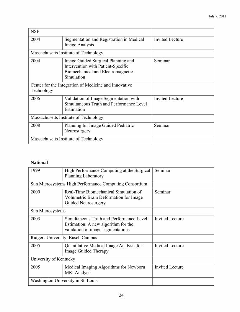
NSF
2004 Segmentation and Registration in Medical Image Analysis
Invited Lecture
Massachusetts Institute of Technology
2004 Image Guided Surgical Planning and Intervention with Patient-Specific Biomechanical and Electromagnetic Simulation
Seminar
Center for the Integration of Medicine and Innovative Technology
2006 Validation of Image Segmentation with Simultaneous Truth and Performance Level Estimation
Invited Lecture
Massachusetts Institute of Technology
2008 Planning for Image Guided Pediatric Neurosurgery
Seminar
Massachusetts Institute of Technology
National
1999 High Performance Computing at the Surgical Planning Laboratory
Seminar
Sun Microsystems High Performance Computing Consortium
2000 Real-Time Biomechanical Simulation of Volumetric Brain Deformation for Image Guided Neurosurgery
Seminar
Sun Microsystems
2003 Simultaneous Truth and Performance Level Estimation: A new algorithm for the validation of image segmentations
Invited Lecture
Rutgers University, Busch Campus
2005 Quantitative Medical Image Analysis for Image Guided Therapy
Invited Lecture
University of Kentucky
2005 Medical Imaging Algorithms for Newborn MRI Analysis
Invited Lecture
Washington University in St. Louis
July 7, 2011
25
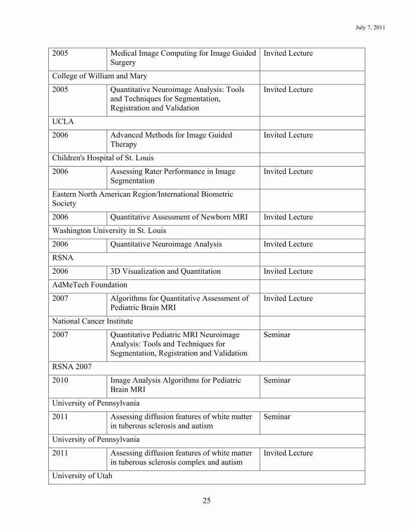
2005 Medical Image Computing for Image Guided Surgery
Invited Lecture
College of William and Mary
2005 Quantitative Neuroimage Analysis: Tools and Techniques for Segmentation, Registration and Validation
Invited Lecture
UCLA
2006 Advanced Methods for Image Guided Therapy
Invited Lecture
Children's Hospital of St. Louis
2006 Assessing Rater Performance in Image Segmentation
Invited Lecture
Eastern North American Region/International Biometric Society
2006 Quantitative Assessment of Newborn MRI Invited Lecture
Washington University in St. Louis
2006 Quantitative Neuroimage Analysis Invited Lecture
RSNA
2006 3D Visualization and Quantitation Invited Lecture
AdMeTech Foundation
2007 Algorithms for Quantitative Assessment of Pediatric Brain MRI
Invited Lecture
National Cancer Institute
2007 Quantitative Pediatric MRI Neuroimage Analysis: Tools and Techniques for Segmentation, Registration and Validation
Seminar
RSNA 2007
2010 Image Analysis Algorithms for Pediatric Brain MRI
Seminar
University of Pennsylvania
2011 Assessing diffusion features of white matter in tuberous sclerosis and autism
Seminar
University of Pennsylvania
2011 Assessing diffusion features of white matter in tuberous sclerosis complex and autism
Invited Lecture
University of Utah

July 7, 2011
26
International
1995 Segmentation of MRI of the Brain Seminar
University of New South Wales, Australia
1999 Template Driven Segmentation Invited Lecture
MICCAI 1999
1999 Template Moderated Classification Seminar
Universite de Louvain, Belgium
2001 Coupling Segmentation and Nonrigid Registration
Seminar
EPFL, Lausanne, Switzerland
2001 Segmentation and Nonrigid Registration Seminar
University Hospital of Geneva
2002 Simultaneous Truth and Performance Level Estimation
Seminar
Howard Florey Institute of Experimental Medicine and Physiology
2002 A new algorithm for judging image segmentations
Seminar
University of Technology, Sydney, Australia
2002 Quantitative Analysis of Medical Images Seminar
Howard Florey Institute of Experimental Medicine and Physiology
2002 Quantitative Medical Image Analysis Seminar
University of New South Wales, Australia
2003 A statistical estimation algorithm for validation of image segmentation
Seminar
EPFL, Lausanne, Switzerland
2003 Image Segmentation and Validation: Unifying Statistical Classification and Geometric Models
Invited Lecture
MICCAI 2003
2003 Capturing Brain Deformation Plenary Presentation
International Symposium on Surgery Simulation and Soft Tissue Modeling
July 7, 2011
27
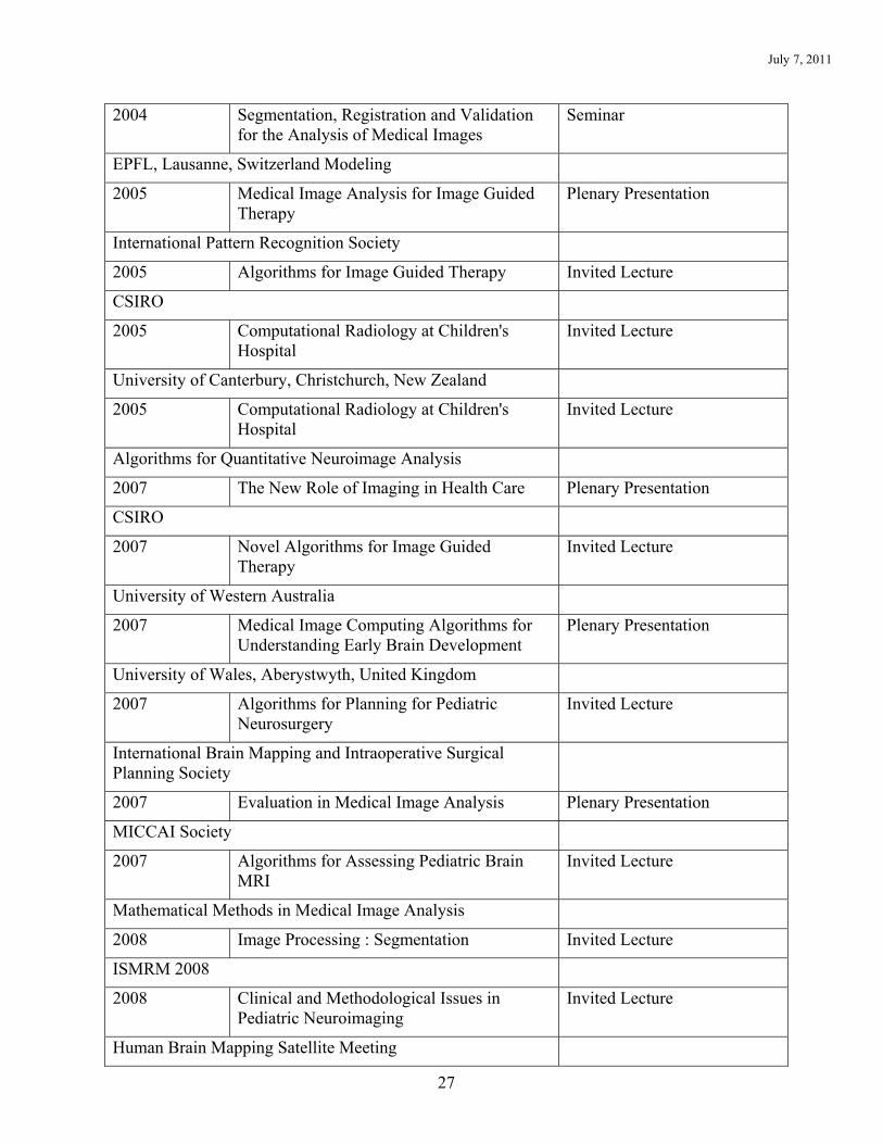
2004 Segmentation, Registration and Validation for the Analysis of Medical Images
Seminar
EPFL, Lausanne, Switzerland Modeling
2005 Medical Image Analysis for Image Guided Therapy
Plenary Presentation
International Pattern Recognition Society
2005 Algorithms for Image Guided Therapy Invited Lecture
CSIRO
2005 Computational Radiology at Children's Hospital
Invited Lecture
University of Canterbury, Christchurch, New Zealand
2005 Computational Radiology at Children's Hospital
Invited Lecture
Algorithms for Quantitative Neuroimage Analysis
2007 The New Role of Imaging in Health Care Plenary Presentation
CSIRO
2007 Novel Algorithms for Image Guided Therapy
Invited Lecture
University of Western Australia
2007 Medical Image Computing Algorithms for Understanding Early Brain Development
Plenary Presentation
University of Wales, Aberystwyth, United Kingdom
2007 Algorithms for Planning for Pediatric Neurosurgery
Invited Lecture
International Brain Mapping and Intraoperative Surgical Planning Society
2007 Evaluation in Medical Image Analysis Plenary Presentation
MICCAI Society
2007 Algorithms for Assessing Pediatric Brain MRI
Invited Lecture
Mathematical Methods in Medical Image Analysis
2008 Image Processing : Segmentation Invited Lecture
ISMRM 2008
2008 Clinical and Methodological Issues in Pediatric Neuroimaging
Invited Lecture
Human Brain Mapping Satellite Meeting

July 7, 2011
28
2008 Image Analysis in Planning for Pediatric Surgery
Plenary Presentation
University of Liege, Belgium
2008 Image Analysis Algorithms for Pediatric Brain MRI
Invited Lecture
University of Melbourne, Australia
2008 Neuroimage Informatics to Understand the Developing Brain
Plenary Presentation
The 4th International Conference on Intelligent Sensors, Sensor Networks and Information Processing (ISSNIP), Sydney, Australia
2008 Imaging the Early Developing Brain: Challenges and Potential Impact
Invited Lecture
MICCAI 2008
2009 Image Segmentation Plenary Presentation
ISMRM 2009
2009 Algorithms and software for image segmentation
Invited Lecture
ISMRM 2009
2009 A Survey of Validation Techniques for Image Segmentation and Registration, with a focus on the STAPLE algorithm
Invited Lecture
NITRC-OHBM
2010 Image Analysis Algorithms for Targeting Treatment and Assessing Response To Therapy
Invited Lecture
MICCAI Workshop on Computational Imaging Biomarkers for Tumors: From Qualitative to Quantitative (CIBT) – MICCAI 2010
2011 Methodologies for inferring a shape model from multiple template images
Invited Lecture
MICCAI Tutorial on Shape Models for Biomedical Image Segmentation – MICCAI 2011

July 7, 2011
29
Report of Technological and Other Scientific Innovations
Provisional Patent “Improved Registration Methods and Apparatus Using Random Projections” serial number 61/238,942 was filed on 09/18/09. (Co-Inventors: Simon K. Warfield, Ph.D. and Ayelet Akselrod-Ballin, Ph.D.) Many of today’s imaging applications utilize large sets of high-resolution images which require alignment through image registration. As the size of the typical data set becomes larger, the computations involved in conventional registration processes quickly become very costly. Typically, the most computationally expensive operation in the registration process is the search for feature correspondences between the target image and the reference image. Dr. Warfield demonstrated that the search for correspondences in feature-based image registration may be dramatically accelerated, while at the same time preserving robustness and accuracy of alignment, by utilizing randomized dimension reduction under the Johnson-Lindenstrauss (JL) lemma. It was demonstrated that dimensional reduction of image patches using random projections enables even further reductions in computation costs through incorporation of efficient lower dimensional correspondence search strategies. Computationally expensive brute-force pairwise computations of similarity measures between all projected patches may be replaced by accelerated search techniques such as approximate nearest neighbor (ANN) formulations. Finally, a transform can be estimated based on the rapidly identified correspondences in a robust manner using a novel expectation maximization iterative closest point search strategy. This has enabled 3D volume reconstruction from extremely large electron microscopy images, providing a unique new capability to assess and visualize detailed connectivity of neural ultrastructure.

July 7, 2011
30
Report of Scholarship Peer Reviewed Publications in print or other media Research Investigations
1. Warfield S, Dengler J, Zaers J, Guttmann CR, Wells WM, Ettinger GJ, Hiller J, Kikinis R. Automatic identification of gray matter structures from MRI to improve the segmentation of white matter lesions. J Imag Guided Surg 1995; 1(6):326-338.
2. Warfield S. Fast k-NN Classification for multichannel image data. Pattern Recog Lett 1996; 17(7):713-721.
3. Iosifescu DV, Shenton ME, Warfield SK, Kikinis R, Dengler J, Jolesz FA, McCarley RW. An automated registration algorithm for measuring MRI subcortical brain structures. Neuroimage 1997; 6(1):13-25.
4. Huppi PS, Warfield S, Kikinis R, Barnes PD, Zientara GP, Jolesz FA, Tsuji MK, Volpe JJ. Quantitative magnetic resonance imaging of brain development in premature and mature newborns. Ann Neurol 1998; 43(2):224-235.
5. Warfield SK, Jolesz FA, Kikinis R. A high performance computing approach to the registration of medical imaging data. Parallel Computing 1998; 24(9-10):1345-1368.
6. Guttmann CR, Kikinis R, Anderson MC, Jakab M, Warfield SK, Killiany RJ, Weiner HL, Jolesz FA. Quantitative follow-up of patients with multiple sclerosis using MRI: reproducibility. J Magn Reson Imaging 1999; 9(4):509-518.
7. Inder TE, Huppi PS, Warfield S, Kikinis R, Zientara GP, Barnes PD, Jolesz F, Volpe JJ. Periventricular white matter injury in the premature infant is followed by reduced cerebral cortical gray matter volume at term. Ann Neurol 1999; 46(5):755-760.
8. Warfield SK, Kaus M, Jolesz FA, Kikinis R. Adaptive, template moderated, spatially varying statistical classification. Med Image Anal 2000; 4(1):43-55.
9. Guttmann CR, Benson R, Warfield SK, Wei X, Anderson MC, Hall CB, Abu-Hasaballah K, Mugler JP, Wolfson L. White matter abnormalities in mobility-impaired older persons. Neurology 2000; 54(6):1277-1283.
10. Warfield SK, Mulkern RV, Winalski CS, Jolesz FA, Kikinis R. An image processing strategy for the quantification and visualization of exercise-induced muscle MRI signal enhancement. J Magn Reson Imaging 2000; 11(5):525-531.
11. Schreyer AG, Fielding JR, Warfield SK, Lee JH, Loughlin KR, Dumanli H, Jolesz FA, Kikinis R. Virtual CT cystoscopy: color mapping of bladder wall thickness. Invest Radiol 2000; 35(5):331-334.
12. Hata N, Nabavi A, Wells WM, Warfield SK, Kikinis R, Black PM, Jolesz FA. Three-dimensional optical flow method for measurement of volumetric brain deformation from intraoperative MR images. J Comput Assist Tomogr 2000; 24(4):531-538.
13. Sperling RA, Guttmann CR, Hohol MJ, Warfield SK, Jakab M, Parente M, Diamond EL, Daffner KR, Olek MJ, Orav EJ, Kikinis R, Jolesz FA, Weiner HL. Regional magnetic resonance imaging lesion burden and cognitive function in multiple sclerosis: a longitudinal study. Arch Neurol 2001; 58(1):115-121.
14. Kaus MR, Warfield SK, Nabavi A, Black PM, Jolesz FA, Kikinis R. Automated segmentation of MR images of brain tumors. Radiology 2001; 218(2):586-591.
15. Murphy BP, Inder TE, Huppi PS, Warfield S, Zientara GP, Kikinis R, Jolesz FA, Volpe JJ. Impaired cerebral cortical gray matter growth after treatment with dexamethasone for neonatal chronic lung disease. Pediatrics 2001; 107(2):217-221.
July 7, 2011
31
16. Nabavi A, Black PM, Gering DT, Westin CF, Mehta V, Pergolizzi RS, Ferrant M, Warfield SK, Hata N, Schwartz RB, Wells WM, Kikinis R, Jolesz FA. Serial intraoperative magnetic resonance imaging of brain shift. Neurosurgery 2001; 48(4):787-797; discussion 797-798.
17. Bharatha A, Hirose M, Hata N, Warfield SK, Ferrant M, Zou KH, Suarez-Santana E, Ruiz-Alzola J, D'Amico A, Cormack RA, Kikinis R, Jolesz FA, Tempany CM. Evaluation of three-dimensional finite element-based deformable registration of pre- and intraoperative prostate imaging. Med Phys 2001; 28(12):2551-2560.
18. Ferrant M, Nabavi A, Macq B, Jolesz FA, Kikinis R, Warfield SK. Registration of 3-D intraoperative MR images of the brain using a finite-element biomechanical model. IEEE Trans Med Imag 2001; 20(12):1384-1397.
19. Benson RR, Guttmann CR, Wei X, Warfield SK, Hall C, Schmidt JA, Kikinis R, Wolfson LI. Older people with impaired mobility have specific loci of periventricular abnormality on MRI. Neurology 2002; 58(1):48-55.
20. Wei X, Warfield SK, Zou KH, Wu Y, Li X, Guimond A, Mugler JP, Benson RR, Wolfson L, Weiner HL, Guttmann CR. Quantitative analysis of MRI signal abnormalities of brain white matter with high reproducibility and accuracy. J Magn Reson Imaging 2002; 15(2):203-209.
21. Fielding JR, Hoyte LX, Okon SA, Schreyer A, Lee J, Zou KH, Warfield S, Richie JP, Loughlin KR, O'Leary MP, Doyle CJ, Kikinis R. Tumor detection by virtual cystoscopy with color mapping of bladder wall thickness. J Urol 2002; 167(2 Pt 1):559-562.
22. Ruiz-Alzola J, Westin CF, Warfield SK, Alberola C, Maier S, Kikinis R. Nonrigid registration of 3D tensor medical data. Med Image Anal 2002; 6(2):143-161.
23. Warfield SK, Talos F, Tei A, Bharatha A, Nabavi A, Ferrant M, Black PM, Jolesz FA, Kikinis R. Real-time registration of volumetric brain MRI by biomechanical simulation of deformation during image guided neurosurgery. Comput Visual Sci 2002; 5(1):3-11.
24. Hirose M, Bharatha A, Hata N, Zou KH, Warfield SK, Cormack RA, D'Amico A, Kikinis R, Jolesz FA, Tempany CM. Quantitative MR imaging assessment of prostate gland deformation before and during MR imaging-guided brachytherapy. Acad Radiol 2002; 9(8):906-912.
25. Ferrant M, Nabavi A, Macq B, Black PM, Jolesz FA, Kikinis R, Warfield SK. Serial registration of intraoperative MR images of the brain. Med Image Anal 2002; 6(4):337-359.
26. Chinzei K, Warfield SK, Hata N, Tempany CMC, Jolesz FA, Kikinis R. Planning, simulation and assistance with intraoperative MRI. Minimally Invasive Therapy 2003; 12(1-2):59-64.
27. Jaume S, Ferrant M, Macq B, Hoyte L, Fielding JR, Schreyer A, Kikinis R, Warfield SK. Tumor detection in the bladder wall with a measurement of abnormal thickness in CT scans. IEEE Trans Biomed Eng 2003; 50(3):383-390.
28. Hunt RW, Warfield SK, Wang H, Kean M, Volpe JJ, Inder TE. Assessment of the impact of the removal of cerebrospinal fluid on cerebral tissue volumes by advanced volumetric 3D-MRI in posthaemorrhagic hydrocephalus in a premature infant. J Neurol Neurosurg Psychiatry 2003; 74(5):658-660.
29. Zou KH, Warfield SK, Fielding JR, Tempany CM, William MW, Kaus MR, Jolesz FA, Kikinis R. Statistical validation based on parametric receiver operating characteristic analysis of continuous classification data. Acad Radiol 2003; 10(12):1359-68.

July 7, 2011
32
30. Anderson NG, Warfield SK, Wells S, Spencer C, Balasingham A, Volpe JJ, Inder TE. A limited range of measures of 2-D ultrasound correlate with 3-D MRI cerebral volumes in the premature infant at term. Ultrasound Med Biol 2004; 30(1):11-18.
31. Wiegand LC, Warfield SK, Levitt JJ, Hirayasu Y, Salisbury DF, Heckers S, Dickey CC, Kikinis R, Jolesz FA, McCarley RW, Shenton ME. Prefrontal cortical thickness in first-episode psychosis: a magnetic resonance imaging study. Biol Psychiatry 2004; 55(2):131-140.
32. Zou KH, Warfield SK, Bharatha A, Tempany CM, Kaus MR, Haker SJ, Wells WM, Jolesz FA, Kikinis R. Statistical validation of image segmentation quality based on a spatial overlap index. Acad Radiol 2004; 11(2):178-189.
33. Als H, Duffy FH, McAnulty GB, Rivkin MJ, Vajapeyam S, Mulkern RV, Warfield SK, Huppi PS, Butler SC, Conneman N, Fischer C, Eichenwald EC. Early experience alters brain function and structure. Pediatrics 2004; 113(4):846-857.
34. Grau V, Mewes AU, Alcaniz M, Kikinis R, Warfield SK. Improved watershed transform for medical image segmentation using prior information. IEEE Trans Med Imaging 2004; 23(4):447-458.
35. Zou KH, Wells WM, Kikinis R, Warfield SK. Three validation metrics for automated probabilistic image segmentation of brain tumours. Stat Med 2004; 23(8):1259-1282.
36. Goldberg-Zimring D, Achiron A, Warfield SK, Guttmann CR, Azhari H. Application of spherical harmonics derived space rotation invariant indices to the analysis of multiple sclerosis lesions' geometry by MRI. Magn Reson Imaging 2004; 22(6):815-825.
37. Tolsa CB, Zimine S, Warfield SK, Freschi M, Sancho Rossignol A, Lazeyras F, Hanquinet S, Pfizenmaier M, Huppi PS. Early alteration of structural and functional brain development in premature infants born with intrauterine growth restriction. Pediatr Res 2004; 56(1):132-138.
38. Warfield SK, Zou KH, Wells WM. Simultaneous truth and performance level estimation (STAPLE): an algorithm for the validation of image segmentation. IEEE Trans Med Imaging 2004; 23(7):903-921.
39. Wei X, Guttmann CR, Warfield SK, Eliasziw M, Mitchell JR. Has your patient's multiple sclerosis lesion burden or brain atrophy actually changed? Mult Scler 2004; 10(4):402-406.
40. Hoyte L, Jakab M, Warfield SK, Shott S, Flesh G, Fielding JR. Levator ani thickness variations in symptomatic and asymptomatic women using magnetic resonance-based 3-dimensional color mapping. Am J Obstet Gynecol 2004; 191(3):856-861.
41. Silverman SG, Sun MR, Tuncali K, Morrison PR, vanSonnenberg E, Shankar S, Zou KH, Warfield SK. Three-dimensional assessment of MRI-guided percutaneous cryotherapy of liver metastases. AJR Am J Roentgenol 2004; 183(3):707-712.
42. Wiegand LC, Warfield SK, Levitt JJ, Hirayasu Y, Salisbury DF, Heckers S, Bouix S, Schwartz D, Spencer M, Dickey CC, Kikinis R, Jolesz FA, McCarley RW, Shenton ME. An in vivo MRI study of prefrontal cortical complexity in first-episode psychosis. Am J Psychiatry 2005; 162(1):65-70.
43. Inder TE, Warfield SK, Wang H, Huppi PS, Volpe JJ. Abnormal cerebral structure is present at term in premature infants. Pediatrics 2005; 115(2):286-294.
44. Limperopoulos C, Soul JS, Gauvreau K, Huppi PS, Warfield SK, Bassan H, Robertson RL, Volpe JJ, du Plessis AJ. Late gestation cerebellar growth is rapid and impeded by premature birth. Pediatrics 2005; 115(3):688-695.

July 7, 2011
33
45. Verhey JF, Wisser J, Warfield SK, Rexilius J, Kikinis R. Non-rigid registration of a 3D ultrasound and a MR image data set of the female pelvic floor using a biomechanical model. Biomed Eng Online 2005; 4(1):19.
46. Zou KH, Tuncali K, Warfield SK, Zentai CP, Worku D, Morrison PR, Silverman SG. Three-dimensional assessment of MR imaging-guided percutaneous cryotherapy using multi-performer repeated segmentations: the value of supervised learning. Acad Radiol 2005; 12(4):444-450.
47. du Bois d'Aische A, Craene MD, Geets X, Gregoire V, Macq B, Warfield SK. Efficient multi-modal dense field non-rigid registration: alignment of histological and section images. Med Image Anal 2005; 9(6):538-546.
48. Wolfson L, Wei X, Hall CB, Panzer V, Wakefield D, Benson RR, Schmidt JA, Warfield SK, Guttmann CR. Accrual of MRI white matter abnormalities in elderly with normal and impaired mobility. J Neurol Sci 2005; 232(1-2):23-27.
49. Tsai A, Wells WM, Warfield SK, Willsky AS. An EM algorithm for shape classification based on level sets. Med Image Anal 2005; 9(5):491-502.
50. Goldberg-Zimring D, Mewes AU, Maddah M, Warfield SK. Diffusion tensor magnetic resonance imaging in multiple sclerosis. J Neuroimaging 2005; 15(4 Suppl):68S-81S.
51. Limperopoulos C, Soul JS, Haidar H, Huppi PS, Bassan H, Warfield SK, Robertson RL, Moore M, Akins P, Volpe JJ, du Plessis AJ. Impaired trophic interactions between the cerebellum and the cerebrum among preterm infants. Pediatrics 2005; 116(4):844-850.
52. Clatz O, Sermesant M, Bondiau PY, Delingette H, Warfield SK, Malandain G, Ayache N. Realistic simulation of the 3-D growth of brain tumors in MR images coupling diffusion with biomechanical deformation. IEEE Trans Med Imaging 2005; 24(10):1334-1346.
53. Haidar H, Warfield SK, Soul JS. Talairach-based parcellation of neonatal brain magnetic resonance imaging data: validation of a new approach. J Neuroimaging 2005; 15(4):305-314.
54. Clatz O, Delingette H, Talos IF, Golby AJ, Kikinis R, Jolesz FA, Ayache N, Warfield SK. Robust nonrigid registration to capture brain shift from intraoperative MRI. IEEE Trans Med Imaging 2005; 24(11):1417-1427.
55. Zou KH, Greve DN, Wang M, Pieper SD, Warfield SK, White NS, Manandhar S, Brown GG, Vangel MG, Kikinis R, Wells WM, Reproducibility of functional MR imaging: preliminary results of prospective multi-institutional study performed by Biomedical Informatics Research Network. Radiology 2005; 237(3):781-789.
56. Zacharia A, Zimine S, Lovblad KO, Warfield S, Thoeny H, Ozdoba C, Bossi E, Kreis R, Boesch C, Schroth G, Huppi PS. Early assessment of brain maturation by MR imaging segmentation in neonates and premature infants. AJNR Am J Neuroradiol 2006; 27(5):972-977.
57. Shah DK, Anderson PJ, Carlin JB, Pavlovic M, Howard K, Thompson DK, Warfield SK, Inder TE. Reduction in cerebellar volumes in preterm infants: relationship to white matter injury and neurodevelopment at two years of age. Pediatr Res 2006; 60(1):97-102.
58. Mewes AU, Huppi PS, Als H, Rybicki FJ, Inder TE, McAnulty GB, Mulkern RV, Robertson RL, Rivkin MJ, Warfield SK. Regional brain development in serial magnetic resonance imaging of low-risk preterm infants. Pediatrics 2006; 118(1):23-33.
59. Shah DK, Guinane C, August P, Austin NC, Woodward LJ, Thompson DK, Warfield SK, Clemett R, Inder TE. Reduced occipital regional volumes at term predict impaired

July 7, 2011
34
visual function in early childhood in very low birth weight infants. Invest Ophthalmol Vis Sci 2006; 47(8):3366-3373.
60. Wu Y, Warfield SK, Tan IL, Wells WM, Meier DS, van Schijndel RA, Barkhof F, Guttmann CR. Automated segmentation of multiple sclerosis lesion subtypes with multichannel MRI. Neuroimage 2006; 32(3):1205-1215.
61. Dimaio SP, Archip N, Hata N, Talos IF, Warfield SK, Majumdar A, Mcdannold N, Hynynen K, Morrison PR, Wells WM, Kacher DF, Ellis RE, Golby AJ, Black PM, Jolesz FA, Kikinis R. Image-guided neurosurgery at Brigham and Women's Hospital. IEEE Eng Med Biol Mag 2006; 25(5):67-73.
62. Goldberg-Zimring D, Warfield SK. Novel image processing techniques to better understand white matter disruption in multiple sclerosis. Autoimmun Rev 2006; 5(8):544-548.
63. Wittek A, Miller K, Kikinis R, Warfield SK. Patient-specific model of brain deformation: application to medical image registration. J Biomech 2007; 40(4):919-929.
64. Alayon S, Robertson R, Warfield SK, Ruiz-Alzola J. A fuzzy system for helping medical diagnosis of malformations of cortical development. J Biomed Inform 2007; 40(3):221-235
65. Thompson DK, Warfield SK, Carlin JB, Pavlovic M, Wang HX, Bear M, Kean MJ, Doyle LW, Egan GF, Inder TE. Perinatal risk factors altering regional brain structure in the preterm infant. Brain 2007; 130(Pt 3):667-677.
66. Fripp J, Crozier S, Warfield SK, Ourselin S. Automatic segmentation of the bone and extraction of the bone-cartilage interface from magnetic resonance images of the knee. Phys Med Biol 2007; 52(6):1617-1631.
67. Archip N, Clatz O, Whalen S, Kacher D, Fedorov A, Kot A, Chrisochoides N, Jolesz F, Golby A, Black PM, Warfield SK. Non-rigid alignment of pre-operative MRI, fMRI, and DT-MRI with intra-operative MRI for enhanced visualization and navigation in image-guided neurosurgery. Neuroimage 2007; 35(2):609-624.
68. Mewes AU, Zollei L, Huppi PS, Als H, McAnulty GB, Inder TE, Wells WM, Warfield SK. Displacement of brain regions in preterm infants with non-synostotic dolichocephaly investigated by MRI. Neuroimage 2007; 36(4):1074-1085
69. Dauguet J, Peled S, Berezovskii V, Delzescaux T, Warfield SK, Born R, Westin CF. Comparison of fiber tracts derived from in-vivo DTI tractography with 3D histological neural tract tracer reconstruction on a macaque brain. Neuroimage 2007; 37(2):530-538.
70. Archip N, Jolesz FA, Warfield SK. A validation framework for brain tumor segmentation. Acad Radiol 2007; 14(10):1242-1251.
71. Downing KT, Hoyte LP, Warfield SK, Weidner AC. Racial differences in pelvic floor muscle thickness in asymptomatic nulliparas as seen on magnetic resonance imaging-based three-dimensional color thickness mapping. Am J Obstet Gynecol 2007; 197(6):625.e1-4.
72. Wisco JJ, Killiany RJ, Guttmann CR, Warfield SK, Moss MB, Rosene DL. An MRI study of age-related white and gray matter volume changes in the rhesus monkey. Neurobiol Aging 2008; 29(10):1563-1575.
73. Maddah M, Grimson WE, Warfield SK, Wells WM. A unified framework for clustering and quantitative analysis of white matter fiber tracts. Med Image Anal 2008; 12(2):191-202.
74. Lodygensky GA, Seghier ML, Warfield SK, Tolsa CB, Sizonenko S, Lazeyras F, Huppi PS. Intrauterine growth restriction affects the preterm infant's hippocampus. Pediatr Res 2008; 63(4):438-443.

July 7, 2011
35
75. Rivkin MJ, Davis PE, Lemaster JL, Cabral HJ, Warfield SK, Mulkern RV, Robson CD, Rose-Jacobs R, Frank DA. Volumetric MRI study of brain in children with intrauterine exposure to cocaine, alcohol, tobacco, and marijuana. Pediatrics 2008; 121(4):741-750.
76. Thompson DK, Wood SJ, Doyle LW, Warfield SK, Lodygensky GA, Anderson PJ, Egan GF, Inder TE. Neonate hippocampal volumes: Prematurity, perinatal predictors, and 2-year outcome. Ann Neurol 2008; 63(5):642-651.
77. Warfield SK, Zou KH, Wells WM. Validation of image segmentation by estimating rater bias and variance. Philos Transact A Math Phys Eng Sci 2008; 366(1874):2361-2375.
78. Archip N, Clatz O, Whalen S, Dimaio SP, Black PM, Jolesz FA, Golby A, Warfield SK. Compensation of geometric distortion effects on intraoperative magnetic resonance imaging for enhanced visualization in image-guided neurosurgery. Neurosurgery 2008; 62(3 Suppl 1):209-215; discussion 215-216.
79. Wisco JJ, Rosene DL, Killiany RJ, Moss MB, Warfield SK, Egorova S, Wu Y, Liptak Z, Warner J, Guttmann CR. A rhesus monkey reference label atlas for template driven segmentation. J Med Primatol 2008; 37(5):250-260.
80. Hoyte L, Damaser MS, Warfield SK, Chukkapalli G, Majumdar A, Choi DJ, Trivedi A, Krysl P. Quantity and distribution of levator ani stretch during simulated vaginal childbirth. Am J Obstet Gynecol 2008; 199(2):198.e1-5.
81. Dubois J, Benders M, Borradori-Tolsa C, Cachia A, Lazeyras F, Ha-Vinh Leuchter R, Sizonenko SV, Warfield SK, Mangin JF, Huppi PS. Primary cortical folding in the human newborn: an early marker of later functional development. Brain 2008; 131(Pt 8):2028-2041.
82. De Craene MS, Macq B, Marquesc F, Salembierc P, Warfield SK. Unbiased group-wise alignment by iterative central tendency estimation. Math Model Nat Phenom 2008; 3(6):2-32.
83. Thompson DK, Wood SJ, Doyle LW, Warfield SK, Egan GF, Inder TE. MR-determined hippocampal asymmetry in full-term and preterm neonates. Hippocampus 2009; 19(2):118-123.
84. Rullmann M, Anwander A, Dannhauer M, Warfield SK, Duffy FH, Wolters CH. EEG source analysis of epileptiform activity using a 1 mm anisotropic hexahedra finite element head model. Neuroimage 2009; 44(2):399-410.
85. Commowick O, Warfield SK. A continuous STAPLE for scalar, vector and tensor Images: An Application to DTI Analysis. IEEE Trans Med Imaging 2009; 28:838-846.
86. Weisenfeld NI, Warfield SK. Automatic segmentation of newborn brain MRI. Neuroimage 2009; 47:564-572.
87. Schaap M, Metz CT, van Walsum T, van der Giessen AG, Weustink AC, Mollet NR, Bauer C, Bogunović H, Castro C, Deng X, Dikici E, O'Donnell T, Frenay M, Friman O, Hernández Hoyos M, Kitslaar PH, Krissian K, Kühnel C, Luengo-Oroz MA, Orkisz M, Smedby O, Styner M, Szymczak A, Tek H, Wang C, Warfield SK, Zambal S, Zhang Y, Krestin GP, Niessen WJ. Standardized evaluation methodology and reference database for evaluating coronary artery centerline extraction algorithms. Med Image Anal. 2009; 13(5):701-14.
88. Vigneron LM, Duflot MP, Robe PA, Warfield SK, Verly JG. 2D XFEM-based modeling of retraction and successive resections for preoperative image update. Comput Aided Surg. 2009; Jul 27:1-20.

July 7, 2011
36
89. Benders MJ, Groenendaal F, van Bel F, Ha Vinh R, Dubois J, Lazeyras F, Warfield SK, Hüppi PS, de Vries LS. Brain development of the preterm neonate after neonatal hydrocortisone treatment for chronic lung disease. Pediatr Res. 2009; 66(5):555-9.
90. Krishnan M, Commowick O, Jeste SS, Weisenfeld N, Hans A, Gregas M, Sahin M, and Warfield SK. Diffusion features of white matter in Tuberous Sclerosis Complex assessed with tractography. Pediatric Neurology, 2010; 42(2):101-6.
91. Commowick O and Warfield SK. Estimation of inferential uncertainty in assessing expert segmentation performance from STAPLE. IEEE Trans Med Imaging 2010;9(3):771-80.
92. Lee JW, Wen PY, Hurwitz S, Black P, Kesari S, Drappatz J, Golby AJ, Wells WM 3rd, Warfield SK, Kikinis R, Bromfield EB. Morphological characteristics of brain tumors causing seizures. Arch Neurol. 2010;67 (3):336-42.
93. Gholipour A, Estroff JA, Warfield SK. Robust Super-resolution Volume Reconstruction from Slice Acquisitions: Application to Fetal Brain MRI. IEEE Trans Med Imaging., 2010; 29(10):1739-58.
94. Vigneron LM, Boman RC, Ponthot J-P, Robe PA, Warfield SK and Verly JG. Enhanced FEM-based modeling of brain shift deformation in Image-Guided Neurosurgery. Journal of Computational and Applied Mathematics, 2010; 234(7):2046-2053.
95. Wintermark P, Labrecque M, Warfield SK, Dehart S, Hansen A. Can induced hypothermia be assured during brain MRI in neonates with hypoxic-ischemic encephalopathy? Pediatr. Radiol., 2010; 40(12):1950-4.
96. Wittek A, Joldes G, Couton M, Warfield SK, Miller K. Patient-Specific Non-Linear Finite Element Modelling for Predicting Soft Organ Deformation in Real-Time; Application to Non-Rigid Neuroimage Registration. Progress in Biophysics and Molecular Biology. 2010; 103(2-3):292-303.
97. Gholipour A, Estroff JA, Warfield SK. Fetal brain volumetry through MRI volumetric reconstruction and segmentation. Int J Computer Assisted Radiology and Surgery, 2011 May;6(3):329-39.
98. Wintermark P, Hansen A, Soul J, Labrecque M, Robertson RL, Warfield SK. Early versus late MRI in asphyxiated newborns treated with hypothermia. Arch Dis Child Fetal Neonatal Ed. 2011 Jan;96(1):F36-44.
99. Thompson DK, Inder TE, Faggian N, Johnston L, Warfield SK, Anderson PJ, Doyle LW, Egan GF. Characterization of the corpus callosum in very preterm and full-term infants utilizing MRI. Neuroimage. 2011 Mar 15;55(2):479-90.
100. Akselrod-Ballin A, Bock D, Clay RC and Warfield SK. Accelerating image registration with the Johnson-Lindenstrauss Lemma: Application to imaging 3D neural ultrastructure with electron microscopy. IEEE Trans Med Imaging. 2011;30(7):1427-38.
101. Vigneron LM, Warfield SK, Robe PA, Verly JG. 3D XFEM-based modeling of retraction for preoperative image update. Comput Aided Surg. 2011;16(3):121-34.
102. Hoyte L, Ye W, Brubaker L, Fielding JR, Lockhart ME, Heilbrun ME, Brown MB, Warfield SK; Pelvic Floor Disorders Network. Segmentations of MRI images of the female pelvic floor: a study of inter- and intra-reader reliability. J Magn Reson Imaging. 2011 Mar;33(3):684-91.
103. Clouchoux C, Kudelski D, Gholipour A, Warfield SK, Viseur S, Bouyssi-Kobar M, Mari JL, Evans AC, du Plessis AJ, Limperopoulos C. Quantitative in vivo MRI measurement of cortical development in the fetus. Brain Struct Funct. 2011, in press. May 12. [Epub ahead of print]

July 7, 2011
37
Non-peer reviewed scientific or medical publications/materials in print or other media
Book Chapters
1. Warfield SK, Robatino A, Dengler J, Jolesz FA, Kikinis R. Nonlinear Registration and Template Driven Segmentation. In: Toga AW, editor. Brain Warping. San Diego: Academic Press; 1999. p. 67-84.
2. Mulkern RV, Winalski C, Zengingonul HP, Warfield S, Holtzman D. Proton Magnetic Resonance Imaging of Exercised Muscle. In: Pandolf KB, Takeda N, Singal PK. Adaptation Biology and Medicine. New Delhi, India: Narosa Publishing House; 1999. p. 170-185.
3. Kikinis R, Mehta NR, Nabavi A, Chatzidakis E, Warfield SK, Gering D, Weisenfeld N, Pergolizzi RS, Schwarz RB, Hata N, Wells W, Grimson E, Black PM, Jolesz FA. Intraoperative Visualization. In: Mazziotta JC, Toga AW, Frackowiak RSJ. Brain Mapping: The Disorders. San Diego: Academic Press; 2000. p. 107-129.
4. Warfield SK, Guimond A, Roche A, Bharatha A, Tei A, Talos F, Rexilius J, Ruiz-Alzola J, Westin CF, Haker S, Angenent S, Tannenbaum A, Jolesz FA, Kikinis R. Advanced Nonrigid Registration Algorithms for Image Fusion. In: Toga AW, Mazziotta JC. Brain Mapping: The Methods. San Diego: Academic Press; 2002. p. 661-690.
5. Goldberg-Zimring D, Meier DS, Bouix S, Warfield SK. Studying anatomy and disease in medical images using shape analysis. In: Leondes CT, editor. Medical Imaging Systems Technology: Methods in Diagnosis Optimization. Singapore: World Scientific Publishing Co; 2005. p. 329-361.
Reviews
1. Warfield SK, Haker SJ, Talos IF, Kemper CA, Weisenfeld N, Mewes AU, Goldberg-Zimring D, Zou KH, Westin CF, Wells WM, Tempany CM, Golby A, Black PM, Jolesz FA, Kikinis R. Capturing intraoperative deformations: research experience at Brigham and Women's Hospital. Med Image Anal 2005;9(2):145-162.
2. Jannin P, Krupinski E, Warfield S. Validation in medical image processing. In: IEEE Transactions on Medical Imaging. New York: IEEE; 2006. p. 1405-1409.
Case reports
1. Wintermark P, Boyd T, Parast MM, Van Marter LJ, Warfield SK, Robertson RL, Ringer SA. Fetal Placental Thrombosis and Neonatal Implications. Am J Perinatol. 2010; 27(3): 251-6.
Proceedings of meetings
1. Warfield S, Sachdev P, Aniss A, Hiller J. Validation of Automatic Classification Strategies for the Measurement of Subcortical Hyperintensities on T-weighted Magnetic Resonance Images. In: Boehme JM, Rowberg AH, Wolfman NT. S/CAR 94 Symposium for Computer Assisted Radiology; 1994 Jun; Winston-Salem, USA. Carlsbad, USA: Symposia Foundation; 1994, p. 319–324.
2. Hiller J, Zrimec T, Mankovich NJ, Warfield S. Visualising Cerebral Vasculature: Imaging Limitations from 2D and 3D Data. In: Kim Y. SPIE Medical Imaging 1994.

July 7, 2011
38
Image Capture, Formatting and Display; 1994 Feb 13–14; Newport Beach, USA. Washington, USA: SPIE; 1994, p. 74–82.
3. Warfield S, Dengler J, Zaers J, Guttmann CR, Wells III WM, Ettinger GJ, Hiller J, Kikinis R. Automatic identification of Grey Matter Structures from MRI to Improve the Segmentation of White Matter Lesions. In: MRCAS’95 Second International Symposium on Medical Robotics and Computer Assisted Surgery; 1995 Nov 4–7; Baltimore, USA. New York: John Wiley & Sons, Ltd; 1995, p. 140–147.
4. Warfield SK, Kaus M, Jolesz FA, Kikinis R. Adaptive Template Moderated Spatially Varying Statistical Classification. In: Wells WM, Colchester A, Delp S. MICCAI 98: First International Conference on Medical Image Computing and Computer-Assisted Intervention; 1998 Oct 11–13; Boston, USA. Heidelberg, Germany: Springer-Verlag; 1998, p. 231–238.
5. Hata N, Dohi T, Warfield SK, Wells III W, Kikinis R, Jolesz FA. Multimodality Deformable Registration of Pre- and Intraoperative Images for MRI-Guided Brain Surgery. In: Wells WM, Colchester A, Delp S. MICCAI 98: First International Conference on Medical Image Computing and Computer-Assisted Intervention; 1998 Oct 11–13; Boston, USA. Heidelberg, Germany: Springer-Verlag; 1998, p. 1067–1074.
6. Warfield SK, Jolesz FA, Kikinis R. Real-Time Image Segmentation for Image-Guided Surgery. In: SC 1998: High Performance Networking and Computing Conference; 1998 Nov 7–13; Orlando, USA. New York: IEEE; 1998, p. 1–14.
7. Westin CF, Warfield SK, Bhalerao A, Mui L, Richolt J, Kikinis R. Tensor controlled local structure enhancement of CT images for bone segmentation. In: Wells WM, Colchester A, Delp S. MICCAI 98: First International Conference on Medical Image Computing and Computer-Assisted Intervention; 1998 Oct 11–13; Boston, USA. Heidelberg, Germany: Springer-Verlag; 1998, p. 1205–1212.
8. Ferrant M, Warfield SK, Guttmann CRG, Mulkern RV, Jolesz FA, Kikinis R. 3D Image Matching Using a Finite Element Based Elastic Deformation Model. In: Taylor C, Colchester A. MICCAI 99: Second International Conference on Medical Image Computing and Computer-Assisted Intervention; 1999 Sep 19–22; Cambridge, England. Heidelberg, Germany: Springer-Verlag; 1999, p. 202–209.
9. Hata N, Nabavi A, Warfield S, Wells WM, Kikinis R, Jolesz FA. A volumetric optical flow method for measurement of brain deformation from intraoperative magnetic resonance images. In: Taylor C, Colchester A. MICCAI 99: Second International Conference on Medical Image Computing and Computer-Assisted Intervention; 1999 Sep 19–22; Cambridge, England. Heidelberg, Germany: Springer-Verlag; 1999, p. 928–935.
10. Warfield SK, Westin CF, Guttmann CRG, Albert M, Jolesz FA, Kikinis R. Fractional Segmentation of White Matter. In: Taylor C, Colchester A. MICCAI 99: Second International Conference on Medical Image Computing and Computer-Assisted Intervention; 1999 Sep 19–22; Cambridge, England. Heidelberg, Germany: Springer-Verlag; 1999, p. 62–71.
11. Kaus MR, Warfield SK, Nabavi A, Chatzidakis E, Black PM, Jolesz FA, Kikinis R. Segmentation of MRI of meningiomas and low grade gliomas. In: Taylor C, Colchester A. MICCAI 99: Second International Conference on Medical Image Computing and Computer-Assisted Intervention; 1999 Sep 19–22; Cambridge, England. Heidelberg, Germany: Springer-Verlag; 1999, p. 1–10.
12. Ferrant M, Nabavi A, Macq B, Warfield SK. Deformable Modeling for Characterizing Biomedical Shape Changes. In: Borgefors G, Nystrom I, Sanniti di Baja G. DGCI2000:

July 7, 2011
39
Discrete Geometry for Computer Imagery; 2000 Dec 13–15; Uppsala, Sweden. Heidelberg, Germany: Springer; 2000, p. 235–248.
13. Warfield SK, Nabavi A, Butz T, Tuncali K, Silverman SG, Black PM, Jolesz FA, Kikinis R. Intraoperative Segmentation and Nonrigid Registration For Image Guided Therapy. In: DiGioia AM, Delp S. MICCAI 2000: Third International Conference on Medical Robotics, Imaging And Computer Assisted Surgery; 2000 Oct 11–14; Pittsburgh, USA. Heidelberg, Germany: Springer-Verlag; 2000, p. 176–185.
14. Ruiz-Alzola J, Westin CF, Warfield SK, Nabavi A, Kikinis R. Nonrigid Registration of 3D Scalar, Vector and Tensor Medical Data. In: DiGioia AM, Delp S. MICCAI 2000: Third International Conference on Medical Robotics, Imaging And Computer Assisted Surgery; 2000 Oct 11–14; Pittsburgh, USA. Heidelberg, Germany: Springer-Verlag; 2000, p. 541–550.
15. Butz T, Warfield SK, Tuncali K, Silverman SG, van Sonnenberg E, Jolesz FA, Kikinis R. Pre- and Intra-operative Planning and Simulation of Percutaneous Tumor Ablation. In: DiGioia AM, Delp S. MICCAI 2000: Third International Conference on Medical Robotics, Imaging And Computer Assisted Surgery; 2000 Oct 11–14; Pittsburgh, USA. Heidelberg, Germany: Springer-Verlag; 2000, p. 317–326.
16. Warfield SK, Ferrant M, Gallez X, Nabavi A, Jolesz FA, Kikinis R. Real-Time Biomechanical Simulation of Volumetric Brain Deformation for Image Guided Neurosurgery. In: SC 2000: High Performance Networking and Computing Conference; 2000 Nov 4–10; Dallas, USA; 2000, p. 1–16.
17. Ferrant M, Warfield SK, Nabavi A, Macq B, Kikinis R. Registration of 3D Intraoperative MR Images of the Brain Using a Finite Element Biomechanical Model. In: DiGioia AM, Delp S. MICCAI 2000: Third International Conference on Medical Robotics, Imaging and Computer Assisted Surgery; 2000 Oct 11–14; Pittsburgh, USA. Heidelberg, Germany: Springer-Verlag; 2000, p. 19–28.
18. Kaus MR, Nabavi A, Mamisch CT, Wells WM, Jolesz FA, Kikinis R, Warfield SK. Simulation of Corticospinal Tract Displacement in Patients with Brain Tumors. In: DiGioia AM, Delp S. MICCAI 2000: Third International Conference on Medical Robotics, Imaging And Computer Assisted Surgery; 2000 Oct 11–14; Pittsburgh, USA. Heidelberg, Germany: Springer-Verlag; 2000, p. 9–18.
19. Warfield SK, Rexilius J, Huppi PS, Inder TE, Miller EG, Wells III WM, Zientara GP, Jolesz FA, Kikinis R. A Binary Entropy Measure to Assess Nonrigid Registration Algorithms. In: Niessen WJ, Viergever MA. MICCAI 2001: Fourth International Conference on Medical Image Computing and Computer-Assisted Intervention; 2001 Oct 14–17; Utrecht, The Netherlands. Heidelberg, Germany: Springer-Verlag; 2001, p. 266–274.
20. Rexilius J, Warfield SK, Guttmann CRG, Wei X, Benson R, Wolfson L, Shenton M, Handels H, Kikinis R. A Novel Nonrigid Registration Algorithm and Applications. In: Niessen WJ, Viergever MA. MICCAI 2001: Fourth International Conference on Medical Image Computing and Computer-Assisted Intervention; 2001 Oct 14–17; Utrecht, The Netherlands. Heidelberg, Germany: Springer-Verlag; 2001, p. 923–931.
21. Pachai C, Zhu YM, Guttmann CRG, Kikinis R, Jolesz FA, Gimenez G, Froment JC, Confavreux C, Warfield SK. Unsupervised and Adaptive Segmentation of Multispectral 3D Magnetic Resonance Images of Human Brain: a Generic Approach. In: Niessen WJ, Viergever MA. MICCAI 2001: Fourth International Conference on Medical Image Computing and Computer-Assisted Intervention; 2001 Oct 14–17; Utrecht, The Netherlands. Heidelberg, Germany: Springer-Verlag; 2001, p. 1067–1074.

July 7, 2011
40
22. Jaume S, Macq B, Warfield SK. Automated Labeling of the Cortical Surface Using a Deformable Multiresolution Mesh. In: MICCAI 2002: Fifth International Conference on Medical Image Computing and Computer-Assisted Intervention; 2002 Sep 25–28; Tokyo, Japan. Heidelberg, Germany: Heidelberg, Germany; 2002, p. 451–458.
23. Guimond A, Guttmann CRG, Warfield SK, Westin CF. Deformable Registration of DTMRI Data Based On Transformation Invariant Tensor Characteristics. In: Fessler J, Vannier M. International Symposium on Biomedical Imaging: Macro to Nano; 2002 Jul 7–10; Washington D.C., USA. New York, USA: IEEE; 2002, p. 1-4.
24. Pohl KM,Wells WM, Guimond A, Kasai K, Shenton ME, Kikinis R, Grimson WEL, Warfield SK. Incorporating Non-rigid Registration into Expectation Maximization Algorithm to Segment MR Images. In: MICCAI 2002: Fifth International Conference on Medical Image Computing and Computer-Assisted Intervention; 2002 Sep 25–28; Tokyo, Japan. Heidelberg, Germany: Heidelberg, Germany; 2002, p. 564–571.
25. Warfield SK, Zou KH, Kaus MR, Wells WM. Simultaneous Validation of Image Segmentation and Assessment of Expert Quality. In: Fessler J, Vannier M. International Symposium on Biomedical Imaging: Macro to Nano; 2002 Jul 7–10; Washington D.C., USA. New York, USA: IEEE; 2002, p. 1–4.
26. Zou KH, Wells WM, Kaus MR, Kikinis R, Jolesz FA, Warfield SK. Statistical Validation of Automated Probabilistic Segmentation Against Composite Latent Expert Ground Truth in MR Imaging of Brain Tumors. In: MICCAI 2002: Fifth International Conference on Medical Image Computing and Computer-Assisted Intervention; 2002 Sep 25–28; Tokyo, Japan. Heidelberg, Germany; 2002, p. 315–322.
27. Warfield SK, Zou KH, Wells WM. Validation of Image Segmentation and Expert Quality with an Expectation-Maximization Algorithm. In: MICCAI 2002: Fifth International Conference on Medical Image Computing and Computer-Assisted Intervention; 2002 Sep 25–28; Tokyo, Japan.Heidelberg, Germany; 2002 p. 298–306.
28. Warfield SK, Talos F, Kemper CA, O’Donnell L, Westin CF, Wells WM, Black PM, Jolesz FA, Kikinis R. Capturing Brain Deformation. In: International Symposium on Surgical Simulation and Soft Tissue Modeling; 2003, p. 203–217.
29. Talos IF, O’Donnell L, Westin CF, Warfield SK, Wells III W, Yoo SS, Panych LP, Golby A, Mamata H, Maier SS, Ratiu P, Guttmann CR, Black PM, Jolesz FA, Kikinis R. Diffusion Tensor and Functional MRI Fusion with Anatomical MRI for Image-Guided Neurosurgery. In: MICCAI 2003: Sixth International Conference on Medical Image Computing and Computer-Assisted Intervention; 2003 Nov 15-18; Montreal, Canada. Heidelberg, Germany: Springer-Verlag; 2003, p. 407–415.
30. Ruiz-Alzola J, Suarez E, Alberola-Lopez C, Warfield SK, Westin CF. Geostatistical Medical Image Registration. Geostatistical Medical Image Registration. In: MICCAI 2003: Sixth International Conference on Medical Image Computing and Computer-Assisted Intervention; 2003 Nov 15-18; Montreal, Canada. Heidelberg, Germany: Springer-Verlag; 2003, p. 894 – 901.
31. Mahnaz M, Zou KH, Wells WM, Kikinis R, Warfield SK. Automatic Optimization of Segmentation Algorithms Through Simultaneous Truth and Performance Level Estimation (STAPLE). In: Christian Barillot David R. Haynor Pierre Hellier. 09/26/2004-09/29/2004; Saint-Malo, France. Heidelberg: Springer Verlag; 2004, p. 274-282.
32. Pohl KM, Warfield SK, Kikinis RG, Grimson WE, Wells WM. Coupling Statistical Segmentation and PCA Shape Modeling. In: 09/26/2004-09/29/2004; Saint-Malo, France. Heidelberg: Springer Verlag; 2004, p. 151-159.
July 7, 2011
41
33. Craene MD, du Bois d’Aische A, Talos IF, Ferrant M, Black PM, Jolesz F, Kikinis R, Macq B, Warfield SK. Dense Deformation Field Estimation for Brain Intra-Operative Images Registration. In: SPIE Medical imaging: Image Processing; 2004. p. 1216-1224.
34. Tsai A, Wells WM, Warfield SK, Willsky AS,. Level Set Methods in an EM Framework for Shape Classification and Estimation. In: Barillot, Christian Haynor, David R. Hellier, Pierre. 09/26/2004-09/29/2004; Saint-Malo, France. Heidelberg, Germany: Springer Verlag; 2004, p. 1-9.
35. Vigneron L, Verly JG, Warfield SK. Modelling Surgical Cuts, Retractions, and Resections via Extended Finite Element Method. In: 09/26/2004-09/29/2004; Saint-Malo, France. Heidelberg, Germany: Springer Verlag; 2004, p. 311-318.
36. Weisenfeld NI, Warfield SK. Normalization of Joint Image-Intensity Statistics in MRI using the Kullback-Leibler Divergence. In: International Symposium on Biomedical Imaging; 2004; Arlington, VA. IEEE; 2004, p. 101- 104.
37. Fripp J, Bourgeat P, Mewes AJU, Warfield SK , Crozier S, Ourselin S. 3D Statistical Shape Models to Embed Spatial Relationship Information. In: Computer Vision for Biomedical Image Applications. CVBIA. Heidelberg, Germany: Springer Verlag; 2005, p. 51-60.
38. Majumdar A, Birnbaum A, Choi DJ, Trivedi A, Warfield SK, Baldridge K, Krysl P. A Dynamic Data Driven Grid System for Intra-operative Image Guided Neurosurgery. In: Book Computational Science – ICCS 2005; Heidelberg, Germany: Springer Verlag; 2005, p. 672-679.
39. du Bois d’Aische A, de Craene M, Macq B, Warfield SK. An articulated registration method. In: IEEE International Conference on Image Processing, 2005. ICIP; 2005, p. I - 21-24.
40. Maddah M, Mewes AUJ, Haker S, Grimson WEL, Warfield SK. Automated Atlas-Based Clustering of White Matter Fiber Tracts from DTMRI. In: MICCAI 2005: Eighth International Conference on Medical Image Computing and Computer-Assisted Intervention; 2005 Oct 26-29; Palm Springs, FL. Heidelberg, Germany: Springer-Verlag; 2005, p. 188-185.
41. Fripp F, Crozier S, Warfield S, Ourselin S. Automatic Initialization of 3D Deformable Models for Cartilage Segmentation. In: Digital Image Computing: Technqiues and Applications, 2005 Dec 06-08; Cairns, Australia. DICTA ' 05 Proceedings; 2005, p. 513- 518.
42. Wittek A, Kikinis R, Warfield SK, Miller K. Brain Shift Computation Using a Fully Nonlinear Biomechanical Model. In: MICCAI 2005: Eighth International Conference on Medical Image Computing and Computer-Assisted Intervention; 2005 Oct 26-29;Palm Springs, FL. Heidelberg, Germany: Springer-Verlag; 2005, p. 583-590.
43. Clatz O, Delingette H, Talos I-F, Golby AJ, Kikinis R, Jolesz FA, Ayache N, Warfield SK. Hybrid Formulation of the Model-Based Non-rigid Registration Problem to Improve Accuracy and Robustness. In: MICCAI 2005: Eighth International Conference on Medical Image Computing and Computer-Assisted Intervention; 2005 Oct 26-29; Palm Springs, FL. Heidelberg, Germany: Springer-Verlag; 2005, p. 295-302.
44. Haker S, Wells III WM, Warfield SK, Talos I-F, Bhagwat JG, Goldberg-Zimring D, Mian A, Ohno-Machado L, Zou KH. Combining Classifiers Using Their Receiver Operating Characteristics and Maximum Likelihood Estimation. In: MICCAI 2005: Eighth International Conference on Medical Image Computing and Computer-Assisted Intervention; 2005 Oct 26-29; Palm Springs, FL. Heidelberg, Germany: Springer-Verlag; 2005, p. 506-514.

July 7, 2011
42
45. Warfield SK. Medical Image Analysis for Image Guided Therapy. In: Digital Image Computing: Techniques and Applications, 2005 Dec 06-08; Cairns, Australia. DICTA '05. Proceedings; 2005, p. 42.
46. Clatz O, Sermesant M, Bondiau P-Y, Delingette H, Warfield SK, Malandain G, Ayache N. Realistic simulation of the 3-D growth of brain tumors in MR images coupling diffusion with biomechanical deformation. In: IEEE Transactions on Medical Imaging; 2005, p. 1334-1346.
47. Clatz O, Delingette H, Talos I-F, Golby AJ, Kikinis R, Jolesz FA, Ayache N, Warfield SK. Robust nonrigid registration to capture brain shift from intraoperative MRI. In: IEEE Transactions on Medical Imaging; 2005, p. 1417-1427.
48. Archip N, Rohling R, Cooperberg P, Tahmasebpour H, Warfield SK. Spectral Clustering Algorithms for Ultrasound Image Segmentation. In: MICCAI 2005: Eighth International Conference on Medical Image Computing and Computer-Assisted Intervention; 2005 Oct 26-29;Palm Springs, FL. Heidelberg, Germany: Springer-Verlag; 2005, p. 862-869.
49. Dauguet J, Peled S, Berezovskii V, Delzescaux T, Warfield SK, Born R, Westin C-F. 3D Histological Reconstruction of Fiber Tracts and Direct Comparison with Diffusion Tensor MRI Tractography. In: MICCAI 2006: Ninth International Conference on Medical Image Computing and Computer-Assisted Intervention; 2006 Oct 1-6;Copenhangen, Denmark. Heidelberg, Germany: Springer-Verlag; 2006, p. 109-116.
50. Fedorov A, Chrisochoides N, Kikinis R, Warfield SK. An evaluation of three approaches to tetrahedral mesh generation for deformable registration of brain MR images. In: 3rd IEEE International Symposium on Biomedical Imaging: Nano to Macro 2006 Apr 6-9, p. 658-661.
51. Fripp J, Warfield SK, Crozier S, Ourselin S. Automatic Segmentation of the Knee Bones using 3D Active Shape Models. In: 18th International Conference on Pattern Recognition (ICPR'06) 2006 Aug 20-24; Hong Kong, China. 2006; p. 171-174.
52. Weisenfeld NI, Mewes AUJ, Warfield SK. Highly Accurate Segmentation of Brain Tissue and Subcortical Gray Matter from Newborn MRI. In: MICCAI 2006: Ninth International Conference on Medical Image Computing and Computer-Assisted Intervention; 2006 Oct 1-6;Copenhangen, Denmark. Heidelberg, Germany: Springer-Verlag 2006; p. 199-206.
53. Dimaio SP, Archip N, Hata N, Talos I, Warfield SK, Majumdar A, Mcdannold N, Hynynen K, Morrison PR, Wells WM, Kacher DF, Ellis RE, Golby AJ, Black PM, Jolesz FA, Kikinis R. Image-guided neurosurgery at Brigham and Women's Hospital. In: IEEE Engineering in Medicine and Biology Magazine; 2006 p. 67-73.
54. Chrisochoides N, Fedorov A, Kot A, Archip N, Black P, Clatz O, Golby A, Kikinis R, Warfield SK. Imaging and visual analysis - Toward real-time image guided neurosurgery using distributed and grid computing. In: Conference on High Performance Networking and Computing. Proceedings of the 2006 ACM/IEEE conference on Supercomputing. Tampa, FL;2006. Article 76.
55. Devadithya T, Baldridge K, Birnbaum A, Majumdar A, Choi DJ, Wolski R, Warfield S. On-Demand High Performance Computing: Image Guided Neuro-Surgery Feasibility Study. In: 12th Annual Conference on Parallel and Distributed Systems, 2006. ICPADS 2006. Jul 12-15 Minneapolis, MN; 2006 p. 97-102.
56. Weisenfeld NI, Mewes AUJ, Warfield SK. Segmentation of newborn brain MRI. In: 3rd IEEE International Symposium on Biomedical Imaging: Nano to Macro, 2006. Apr 6-9 Arlington, VA; 2006, p. 766-769.

July 7, 2011
43
57. Maddah M, Crimson WEL, Warfield SK. Statistical modeling and EM clustering of white matter fiber tracts. In: 3rd IEEE International Symposium on Biomedical Imaging: Nano to Macro, 2006. Apr 6-9 Arlington, VA; 2006, p. 53-56.
58. Warfield SK, Zou KH, Wells WM. Validation of Image Segmentation by Estimating Rater Bias and Variance. In: MICCAI 2006: Ninth International Conference on Medical Image Computing and Computer- Assisted Intervention; 2006 Oct 1-6;Copenhangen, Denmark. Heidelberg, Germany: Springer-Verlag; 2006 p. 839-847.
59. Dauguet J, Bock D, Reid RC, Warfield SK. Alignment of Large Image Series Using Cubic B-Splines Tessellation: Application to Transmission Electron Microscopy Data. In: MICCAI 2007: Tenth International Conference on Medical Image Computing and Computer- Assisted Intervention; 2007 Oct 29- Nov 2; Brisbane Australia. Heidelberg, Germany: Springer-Verlag; 2007 p. 710-717.
60. Fripp J, Crozier S, Warfield SK, Ourselin S. Automatic Segmentation of Articular Cartilage in Magnetic Resonance Images of the Knee. In: MICCAI 2007: Tenth International Conference on Medical Image Computing and Computer- Assisted Intervention; 2007 Oct 29- Nov 2;Brisbane Australia. Heidelberg, Germany: Springer-Verlag; 2007 p. 186-194.
61. Chrisochoides N, Fedorov A, Kot A, Archip N, Goldberg-Zimring D, Kacher D, Whalen S, Kikinis R, Jolesz F, Clatz O, Warfield SK, Black PM, Golby A. Grid-Enabled Software Environment for Enhanced Dynamic Data-Driven Visualization and Navigation During Image-Guided Neurosurgery. In: MICCAI 2007: Tenth International Conference on Medical Image Computing and Computer- Assisted Intervention; 2007 Oct 29- Nov 2;Brisbane Australia. Heidelberg, Germany: Springer-Verlag; 2007, p. 980-987.
62. Archip N, Tatli S, Morrison P, Jolesz F, Warfield SK, Silverman S. Non-rigid Registration of Pre-procedural MR Images with Intra-procedural Unenhanced CT Images for Improved Targeting of Tumors During Liver Radiofrequency Ablations. In: MICCAI 2007: Tenth International Conference on Medical Image Computing and Computer- Assisted Intervention; 2007 Oct 29- Nov 2;Brisbane Australia. Heidelberg, Germany: Springer-Verlag; 2007, p. 969-977.
63. Maddah M, Wells III WM, Warfield SK, Westin C-F, Grimson WEL. Probabilistic Clustering and Quantitative Analysis of White Matter Fiber Tracts. In: Information Processing in Medical Imaging: 20th International Conference (IPMI 2007) Jul 2-6 Kerkrade, Netherlands; 2007. p. 372-383.
64. Dauguet J, Warfield SK, Bromfield E, Golby A, Lee JW. Comparison of the deformations of brain tissues caused by tumor in seizure and non-seizure patients. In: Biomedical Imaging: From Nano to Macro, 2008. ISBI 2008; p. 1143-1146.
65. Spiclin Z, Hans A, Duffy FH, Warfield SK, Likar B, Pernus F. EEG to MRI registration based on global and local similarities of MRI intensity distributions. In: MICCAI 2008: Eleventh International Conference on Medical Image Computing and Computer- Assisted Intervention; 2008 Sept 6-10; New York, NY. Heidelberg, Germany: Springer-Verlag; 2008. p. 762-70.
66. Commowick O, Fillard P, Clatz O, Warfield SK. Detection of DTI white matter abnormalities in multiple sclerosis patients. In: MICCAI 2008: Eleventh International Conference on Medical Image Computing and Computer-Assisted Intervention; 2008 Sept 6-10; New York, NY. Heidelberg, Germany: Springer-Verlag; 2008 p. 975-82.
67. Akselrod-Ballin A, Bock D, Reid RC, and Warfield SK. Accelerating Feature Based Registration using the Johnson-Lindenstrauss Lemma. In MICCAI 2009: Twelfth

July 7, 2011
44
International Conference on Medical Image Computing and Computer-Assisted Intervention; 2009 Sept 20-24; London, UK. Heidelberg, Germany: Springer Verlag; 2009 p. 632-9.
68. Gholipour A, Weisenfeld N and Warfield SK. Retrospective reconstruction of 3D fetal brain MRI. In: International Brain Mapping & Intraoperative Surgical Planning Society Conference; 2009 Aug 26-29; Boston, MA. IBMISPS 2009.
69. Commowick O, Warfield SK, and Malandain M. Using Frankenstein's Creature Paradigm to Build a Patient Specific Atlas In MICCAI 2009: Twelfth International Conference on Medical Image Computing and Computer-Assisted Intervention; 2009 Sept 20-24; London, UK. Heidelberg, Germany: Springer Verlag; 2009 p. 993-1000.
70. Joldes G, Wittek A, Couton M, Warfield SK, and Miller K. Real-Time Prediction of Brain Shift Using Nonlinear Finite Element Algorithms. In MICCAI 2009: Twelfth International Conference on Medical Image Computing and Computer-Assisted Intervention; 2009 Sept 20-24; London, UK. Heidelberg, Germany: Springer Verlag; 2009 p. 300-7.
71. Akselrod-Ballin A, Bock D, Reid RC, and Warfield SK. Improved registration for large electron microscopy images. In: Biomedical Imaging: From Nano to Macro, 2009. ISBI 2009; p. 433.
72. Weisenfeld and Warfield SK. A data-driven approach to discovering common brain anatomy. In: Biomedical Imaging: From Nano to Macro, 2009. ISBI 2009; p. 217-220.
73. Commowick O, Warfield SK. Estimation of inferential uncertainty in assessing expert segmentation performance from STAPLE. Inf Process Med Imaging. 2009; 21:701-12.
74. Scherrer B and Warfield SK. Why Multiple B-Values are Required for Multi-Tensor Models. Evaluation with a Constrained Log-Euclidean Model. In: Biomedical Imaging: From Nano to Macro. ISBI 2010; p. 1389-92.
75. Hyde DE and Warfield SK. Spatio-Temporal Smoothness Constraints for EEG Source Localization. In: Biomedical Imaging: From Nano to Macro. ISBI 2010; p. 828-831.
76. Gholipour A, Estroff JA, Barnewolt CE, Connolly SA and Warfield SK. Fetal brain volumetry through MRI volumetric reconstruction and segmentation. Computer Assisted Radiology and Surgery (CARS) 2010 June 23-26; Geneva, Switzerland. 2010.
77. Commowick O and Warfield SK. Incorporating Priors on Expert Performance Parameters for Segmentation Validation and Label Fusion: a Maximum A Posteriori STAPLE. In MICCAI 2010: Thirteenth International Conference on Medical Image Computing and Computer-Assisted Intervention; 2010 Sept 20-24; Beijing, China. Heidelberg, Germany: Springer Verlag; 2010 Part III p 25-32.
78. Gholipour A, Estroff JA, Sahin M, Prabhu SP, and Warfield SK. Maximum A Posteriori Estimation of Isotropic High-Resolution Volumetric MRI from Orthogonal Thick-Slice Scans. Thirteenth International Conference on Medical Image Computing and Computer-Assisted Intervention; 2010 Sept 20-24; Beijing, China. Heidelberg, Germany: Springer Verlag; 2010, Part II p. 109-116.
79. Freiman M, Voss S, Warfield SK. Demons registration with local affine adaptive regularization: application to registration of abdominal structures. In: Biomedical Imaging: From Nano to Macro. ISBI 2011; p. 1219-1222.
80. Akhondi-Asl A and Warfield SK. Cortical Brain Structures Segmentation Using Constrained Optimization and Intensity Coupling. In: Biomedical Imaging: From Nano to Macro. ISBI 2011; p. 1945-7928.

July 7, 2011
45
81. Hyde DE and Warfield SK. Anisotropic Equivalent Conductivity Tensors for Bioelectric Modeling of Partial Volume Effects in Cerebrospinal Fluid Space. In: Biomedical Imaging: From Nano to Macro. ISBI 2011; p.1109-1112.
82. Scherrer B and Warfield SK. Toward an accurate multi-fiber assessment strategy for clinical practice. In: Biomedical Imaging: From Nano to Macro. ISBI 2011; p. 2140-2143.
83. Suarez RO, Commowick O, Tomas Fernandez X, Prabhu S and Warfield SK. Automated detection of white matter fiber bundles. In: Biomedical Imaging: From Nano to Macro. ISBI 2011; p. 845-848.
84. Tomas Fernandez X and Warfield SK. A new classifier feature space for an improved multiple sclerosis lesion segmentation. In: Biomedical Imaging: From Nano to Macro. ISBI 2011; p. 1492-1495.
85. Weisenfeld NI and Warfield SK. Soft STAPLE: Truth and Performance Level Estimation from Probabilistic Segmentations. In: Biomedical Imaging: From Nano to Macro. ISBI 2011; p. 441-446.
86. Gholipour A, Polak M, van der Kouwe A, Nevo E, Warfield SK. Motion-robust MRI through real-time motion tracking and retrospective super-Resolution volume reconstruction. In: IEEE Engineering in Medicine and Biology Society (EMBS) 2011, in press.
87. Gholipour A, Kehtarnavaz N, Scherrer B, Warfield SK. On the accuracy of unwarping techniques for the correction of susceptibility-induced geometric distortion in magnetic resonance echo-planar images. In: IEEE Engineering in Medicine and Biology Society (EMBS) 2011, in press.
88. Freiman M, Voss S, Mulkern RV, Perez-Rossello J and Warfield SK. Quantitative body DW-MRI biomarkers uncertainty estimation using Unscented Wild-bootstrap. In MICCAI 2011: Fourteenth International Conference on Medical Image Computing and Computer-Assisted Intervention; 2011 Sept 18-22; Toronto, Canada. Heidelberg, Germany: Springer Verlag; 2011, in press.
89. Scherrer B and Warfield SK. Super resolution in diffusion-weighted imaging. In MICCAI 2011: Fourteenth International Conference on Medical Image Computing and Computer-Assisted Intervention; 2011 Sept 18-22; Toronto, Canada. Heidelberg, Germany: Springer Verlag; 2011, in press.
90. Taquet M, Macq B, Warfield SK. Spatially adaptive log-Euclidean polyaffine registration based on sparse matches. In MICCAI 2011: Fourteenth International Conference on Medical Image Computing and Computer-Assisted Intervention; 2011 Sept 18-22; Toronto, Canada. Heidelberg, Germany: Springer Verlag; 2011, in press.
91. Weisenfeld NI and Warfield SK. Learning Likelihoods for Labeling (L3): A General Multi-Classifier Segmentation Algorithm. In MICCAI 2011: Fourteenth International Conference on Medical Image Computing and Computer-Assisted Intervention; 2011 Sept 18-22; Toronto, Canada. Heidelberg, Germany: Springer Verlag; 2011, in press.
Thesis
1. Warfield SK. Segmentation of Magnetic Resonance Images of the Brain. Sydney, Australia: The University of New South Wales; 1997.
Abstracts, Poster Presentations and Exhibits Presented at Professional Meetings:

July 7, 2011
46
1. Warfield SK, Wang H, Reith T, Volpe JT, Inder TI. Alterations in Cortical Gray Matter Structure in Premature Infants Studied by Advanced MR Techniques. Pediatric Academic Societies Meeting; 2008 May 2-6; Honolulu, HI.
2. Warfield SK, Weisenfeld NI, Spencer C, Woodward LJ. Prenatal Exposure to Methadone Reduces Brain Tissue Volumes. Pediatric Academic Societies Meeting; 2008 May 2-6; Honolulu, HI.
3. Wintermark P, Pfeuffer J, Hamm M, Robertson RL, Hansen A, Soul J, Warfield SK. Clinical assessment of brain perfusion in newborn infants with arterial spin labeling perfusion MRI. ISMRM; 2009 Apr 18-24; Honolulu, HI.
4. Wintermark P, Pfeuffer J, Hamm M, Triantafyllou C, Robertson RL, Hansen A, Soul J, Warfield SK. Use of a 32-channel coil to improve resolution in assessing brain perfusion of newborn infants with 3T MRI. ISMRM; 2009 Apr 18-24; Honolulu, HI.
5. Wintermark P, Hansen A, Soul J, Robertson RL, Warfield SK. Why Cooling Does Not Work in Severe Cases of Hypoxic-Ischemic Encephalopathy? In Vivo Observation of Increased Perfusion Despite Systemic Cooling. Pediatric Academic Societies’ Annual Meeting (PAS); 2009 May 2-5; Baltimore, MD.
6. Tsai A, Warfield SK, Rosenberg AE, McDonald A, Gupta R, Patel N, Snyder BD, Kleinman PK. Poster #: PO-77: The classic metaphyseal lesion: Confirmation of a theoretical model using isotropic volumetric micro CT and high-resolution flat panel CT. Pediatr Radiol 2009; 39:S310
7. Scherrer B and Warfield SK. Optimal HARDI acquisition schemes for multi-tensor models. International Symposium on Magnetic Resonance in Medicine (ISMRM) 2010 May 1-7; Stockholm, Sweden.
8. Tomas Fernandez X and Warfield SK. Learning Weighted Metrics for Segmentation of Brain Magnetic Resonance Images. 16th Annual Meeting of the Organization for Human Brain Mapping (OHBM) 2010 June 6-10; Barcelona, Spain.
9. Freiman M, Voss S, Mulkern, RV and Warfield SK. NdH/dT: A new quantitative measure for Diffusion Weighted Imaging based evaluation of abdominal tumor response to therapy. International Symposium on Magnetic Resonance in Medicine (ISMRM) 2011 May 6-13; Montreal, Canada.
10. Gholipour A, Polak M, van der Kouwe A, Nevo E, and Warfield SK. Motion-corrected single shot fast spin echo MRI using prospective motion tracking and retrospective super-resolution volume reconstruction. International Symposium on Magnetic Resonance in Medicine (ISMRM) 2011 May 6-13; Montreal, Canada.
11. Hans A, Wittek A, Joldes G, Miller K, Weisenfeld NI, Alexiuk M, Saunders JK, Liebenthal E, Sutherland GR, and Warfield SK. Improved visualization of brain anatomy and function, for surgery, through real-time non-rigid registration. International Symposium on Magnetic Resonance in Medicine (ISMRM) 2011 May 6-13; Montreal, Canada.
12. Scherrer B and Warfield SK. Characterizing Complex White Matter Structure from Cube and Sphere Diffusion Imaging with a Multi-Fiber Model (CUSP-MFM). International Symposium on Magnetic Resonance in Medicine (ISMRM) 2011 May 6-13; Montreal, Canada.
13. Hans A, Weisenfeld NI, Alexiuk M, Saunders JK, Liebenthal E, Wittek A, Joldes G, Miller K, and Warfield SK. Comparison of registration algorithms for capturing intraoperative brain shift. 17th Annual Meeting of the Organization for Human Brain Mapping (OHBM) 2011 June 26-30; Quebec City, Canada.

July 7, 2011
47
14. Scherrer B and Warfield SK. Selecting the number of fibers in a Multi-Fiber Model from CUbe and SPhere (CUSP) Diffusion Imaging. 17th Annual Meeting of the Organization for Human Brain Mapping (OHBM) 2011 June 26-30; Quebec City, Canada.
15. Suarez RO, Paladino MJ, Warfield SK. Lateralization of white matter diffusion within arcuate fasciculus fiber bundles. 17th Annual Meeting of the Organization for Human Brain Mapping (OHBM) 2011 June 26-30; Quebec City, Canada.

July 7, 2011
48
Narrative Report (limit to 500 words)
The primary nexus of my research is the Computational Radiology Laboratory (CRL), of which I am the director and founder. The CRL was formed with the goal of improving our understanding of the structure and function of the brain and other organs of the human body, in order to improve our capacity to diagnose and treat disease. The CRL achieves this by developing novel technologies and computational modeling strategies for understanding and interpreting radiological images. My most significant research accomplishments have been the development of novel algorithms for fundamentally new approaches to analyze and interpret images. Many of my algorithmic developments have introduced entirely new approaches in the field, which have been adopted by others nationally and internationally as a basis for new directions for development.
My team and I use neuroscientific and clinical applications to provide focus and constraints for the creation of new algorithms for medical image analysis. This approach has resulted in the creation of robust, reliable, general purpose algorithms which have had significant impact in several clinical areas. Major applications of this research have included quantitative image analysis to detect morphological change and real-time image analysis to support image guided surgery. The primary areas of current research activity are the characterization of fetal and neonatal brain development utilizing magnetic resonance imaging, reconstruction and interpretation of neural ultrastructure from electron microscopy, preoperative assessment of seizure foci and normal function in pediatric epilepsy patients, assessment of white matter structural alterations in neurological disorders, and intraoperative visualization and navigation to enhance image guided surgery.
Throughout my career I have welcomed the opportunities to teach and mentor students in addition to conducting extramurally funded research. Since 1998, I have served as mentor to both graduate and undergraduate computer science and medicine students in the Department of Radiology as well as summer medical and computer science students. I have also co-supervised graduate students of the computer science and artificial intelligence laboratory at MIT, and graduate students from Boston University and prominent international research universities. I have taught courses to peers at prominent international conferences.
My laboratory distributes software implementations of our algorithms to research scientists throughout the world. We participate in national and international efforts to develop a software platform for medical image analysis, and through these efforts my research in medical image analysis has had a significant impact upon the way imaging is utilized in research and in clinical practice.
My research has been characterized by fundamental contributions to the basic science of imaging and medical image analysis, and collaboration with clinicians to translate those contributions into dramatic impact in clinical and translational research. My laboratory has published high impact papers that are highly cited, and we have developed new methodologies that have been widely adopted in the field.