Embed Size (px)
Citation preview
Formation of the Blastocyst: Determinationof Trophoblast and Embryonie Knot *H.-W. DENKER
I. Introduction . . . . . . . . . . . . . . . . . . . . . . . . . . . . . . . . . . . . . . . . . . .. 5911.Trophoblast and Ernbryonie Knot Representing Distinct Populations of Cells
(Differentiation of Trophoblast and Ernbryonie Knot) ... . . . . . . . . . . . .. 60111.Determination of Trophoblast and Embryonie Knot . . . . . . . . . . . . . . . . .. 62
1. Theory A: Determination Depending on the Position of Blastomeres(Inside-Outside Model). . . . . . . . . . . . . . . . . . . . . . . . . . . . . . . . . . .. 62Observations Supporting Theory A 62
2. Theory B: Determination Depending on Polar/Bilateral Organization of theEgg (Morula) . . . . . . . . . . . . . . . . . . . . . . . . . . . . . . . . . . . . . . . . .. 64Observations Supporting Theory B . . . . . . . . . . . . . . . . . . . . . . . . . . .. 65a) Experiments 65b) Morphological and Histochemical Data 66
IV. General Discussion . . . . . . . . . . . . . . . . . . . . . . . . . . . . . . . . . . . . . . .. 70References . . . . . . . . . . . . . . . . . . . . . . . . . . . . . . . . . . . . . . . . . . . . . . .. 74
I. Introduction
In mammalian development, the earliest apparent differentiation of cells into two distinctlines manifests itself in the early blastocyst, when trophoblast and embryonie knot ("innercell mass") become distinguishable. Reeently more and more attention is being paid tothis proeess by developmental biologists, geneticists, eell biologists, reproduetive biologists,and oneologists. This is apparently for one of the following reasons: (1) Blastocyst forma-tion seems to result from the first proeess of determination in mammalian ontogeny, whichmeans that the genome has not been under the influenee of any determinative stimuli be-fore. (2) The system appears to be far less eomplex than later stages so that it may beespecially suitable for experimentation. (3) Determination of trophoblast, a tissue of somepeculiar properties, may exhibit interesting speeifie features. (4) Trophoblast is in the foeusof interest of reproduetive biologists as well as oneologists beeause of its role in mediatingeontaet between embryo and mother, and beeause of its invasive growth.
Attempts of investigators to elucidate the meehanism of determination of trophoblast andembryonie knot led them to design elegant experiments whieh, in turn, resulted in forrnula-
* Dedieated to Professor Dr. F. Seidel.

60
tion of different theories to be described in this paper. Excellent reviews of part of thearguments summarized here have been given by Mulnard (1966), Seidel (1969), Graham(1971), Gardner (1973), and Herbert and Graham (1974).
Terminology
"Embryonic knot": In the present paper, we will use this term rather than the more com-monly used term "inner cell mass" because the latter would fit only one ofthe theories tobe described here, and because it would not be suitable for discussion of a possible onsetof determination before certain cells acquire an inside position.
"Polarity": The term is being used in a general sense not referring specifically to polarorganization along the animal-vegetal axis.
11.Trophoblast and Embryonie Knot Representing Distinct Populations ofCells (Differentiation of Trophoblast and Embryonie Knot)
Considering the process of blastocyst formation to be connected with a process of celldifferentiation would require data showing that trophoblast and embryonie knot (or atleast one of both) are in fact composed of distinctly differentiated cells. The followingobservations give support to this view:
1. Trophoblast and embryonie knot cells, when isolated from 3 1/2-day mouse blastocysts,differ in their ability to induce a decidual reaction in the pseudopregnant uterus: tropho-blast cells do induce this reaction, whereas embryonie knot cells do not (Gardner, 1971,1972a).
2. The same isolated trophoblast and embryonie knot cells also differ in their tendency tostick together and to form a comrnon structure in vitro: embryonie knot cells do so,whereas trophoblast cells stay apart; the latter form fluid-filled vesicles, whereas theformer produce only solid cell clusters. Embryonie knot cells, when injected into thecavity of another blastocyst, will become integrated and form part of the body of theembryo, even rat embryonie knot cells injected into a mouse blastocyst: this is not thecase with likewise injected trophoblast cells (Gardner, 1971, 1972a; Gardner and Johnson,1973).
3. Trophoblast cells of blastocysts are connected with each other by junctional complexes.Well-developed junctions can already be seen between blastomeres in the outer layer ofc1eavage stages, i.e., between presumptive trophoblast cells, whereas between inner cellsof morulae or between trophoblast and embryonie knot of blastocysts they are more rareand remain primitive. Differences in density of the cytoplasm and in number as well asstructure of certain organelles have also been described (rat: Schlafke and Enders, 1967;Dvoiak, 1971; mouse: Calarco and Brown, 1969; rabbit: Hessedahl, 1971).
4. Mitosis rate seems to be different in both types of cells, the embryonie knot showingthe higher values. There is already a difference between inside and outside cells of morulae
61
as judged from combination of cell number and 3H-thymidine incorporation studies(Barlow et al. , 1972).
5. In histochemical studies, differences between trophoblast and embryonie knot havebeen found relatively often, e.g. differences in phosphatase activity. Problems of inter-pretation of histochemical findings are in part discussed elsewhere (cf. also Denker,1970).
6. Differences in cell surface properties of trophoblast and embryonie knot are possiblyindicated by the fact that, in mouse blastocysts, both types of cells exhibit slightly dif-ferent susceptibility to lysis by cytotoxie antisera (Moskalewski and Koprowski, 1972);furthermore, certain viruses injected into the blastocyst develop in the trophoblast butnot in the embryonie knot (Glass et al., 1974).
7. Evidence has been presented for the expression of different esterase isoenzymes introphoblast and embryonie knot of mouse blastocysts. The trophoblast-type isoenzymeA was detectable already in the late morula; this might be the earliest well-establishedbiochemical criterium for beginning differentiation of trophoblast (Sherrnan, 1972).
The above mentioned data give evidence for morphologie, biochemical, and physiologiedifferences between trophoblast and embryonie knot. This may indicate that a processof differentiation has taken place, if this term is used in a merely descriptive sense -differentiation meaning the establishment of differential properties of cells. Direct evi-dence for differential gene activity of trophoblast and embryonie knot cells, however,is stilllacking but might be found in the near future because it is already established thatthe genome is active in preimplantation embryos: Some indirect evidence is derived frominvestigations on synthesis of different c1asses of RNA and on changes in enzyme activitiesduring preimplantation (although technical problems of determination of intracellular poolsof precursors are not completely solved, and changes in enzyme activity do not necessarilyreflect changes in genetic activity) (for review see Church and Schultz, 1974; Graham,1973; Woodland and Graham, 1969). Effects of actinomycin D and o-amanitin dernon-strate that RNA synthesis is indeed required for c1eavage and blastocyst formation (Golbuset al., 1973; Manes, 1973). Direct evidence for genetic activity is derived from the factthat t12 je 2 homozygous mouse embryos die at the 1ate morula stage (Mintz, 1964a)(apparently after onset of trophoblast differentiation, Hillman et al., 1970), and by thefinding that the paternal phenotype of glucose phosphate isomerase isoenzymes is ex-pressed in the blastocyst stage (Chaprnan et al., 1971), and the information for hypo-xanthine-guanine phosphoribosyltransferase is apparently transcribed as early as the morulastage (Epstein, 1972).
Of the two c1asses of cells, trophoblast and embryonie knot, trophoblast gives betterevidence that it undergoes real differentiation until the blastocyst stage: The trophoblastdevelops junctional complexes, secretes blastocyst fluid, is able to induce a decidual reac-tion, produces a specific esterase isoenzyme (see above) and a protease or protease activator(Denker, 1971a, 1974). The embryonie knot cells, on the contrary, seem to remain in amore primitive state (cf. also Gardner, 1971).

62
IH. Determination of Trophoblast and Embryonie Knot
1.Theory A: Determination Depending on the Position of Blastomeres ("Inside-OutsideModel") (Fig. lA)
1II
TI MO lilb
81 ~ ..•...••...~ w..s:'\JY Cf[) <,
Fig. 1. Diagram to illustrate the two theories on determination of trophoblast and embryonie knot.Theory A (inside-outside model): In the beginning, the developmental potentials of all blastomeresare equal (A 11,lIla). When, in the course of cleavage, sorne cells become completely surrounded byothers, these inside cells are being determined to form embryonie knot (inner cell mass) (A IIIb);the other (outside) blastomeres will differentiate into trophoblast.Theory B: Determination depends on localized factors of polar (bilateral) distribution, which, in thisdiagram, are assumed to be trophoblast-<ietermining. These factors are either (B2) localized in a certainarea of egg cytoplasm (B2 I) and become segregated during cleavage so that they will be found only incertain blastomeres (B2 II); or (B I) factors are of unknown, maybe even exogenic, origin, but theiraction is nevertheless locally restricted. In both cases (BI and B2), morulae show polarity (1Il) as aresult of polar action of determining factors
According to this theory, there are no differences between blastomeres either in theirstatus of determination or in their developmental potentials, until one or several of thembecome completely surrounded by others, i.e., in or after the eight-cell stage. The com-pletely surrounded cells will form the embryonie knot (inner cell mass), whereas the wholeof the outer layer will become trophoblast. The ability to differentiate into trophoblast isinherent to a11blastomeres at the beginning; the information (the determinative stimulus)to form, instead, embryonie knot is provided by the specific milieu "Inside" a morula orblastocyst (Tarkowski and wroblewska, 1967).
Observations supporting theory A have mostly been made in the course of experimentationin the mouse. Two or more c1eavage stage mouse embryos up to the morula stage can befused together to form one single chimeric blastocyst developing into one single embryo(Tarkowski, 1961, 1965;Mintz, 1962,1965). This demonstrates a remarkable regulativecapacity of the early mammalian embryo. Even rat and mouse morulae can be combinedthis way to form interspecific chimeras (Stern, 1973; Zeilmaker, 1973). If, in these ern-bryos, future trophoblast and embryonie knot cells (or one of both groups) had alreadybeen determined in a fixed and unchangeable manner, only a specific sorting out of cells

63
during fusion could explain the formation of one single chimeric blastocyst. At first, thistype of explanation has in fact been given (Tarkowski, 1961). Labeling experiments, how-ever, did not give evidence for any specific sorting-out process (Fig. 2) (Mintz, 1964b,1965; Hillman et al., 1972).
Fig. 2. Fusion experiments. Two mousemorulae, differing in coat color genes,are fused to form one chimeric anima!.Blastomeres are intermingling withoutexhibiting any regular pattern or rule,as to be seen in the blastocyst formed;four extreme possibilities are illustrat-ed. Resulting coat color patterns frornblastocysts with chimeric embryonieknot (first two lines) are oversimpli-fied; for exact details seeMintz (1971)
~·~u-~~-o-~~·-lJ-~~·-o-..,
In fact, blastomeres put outside a morula tended to become incorporated into the tropho-blast rather than the embryonie knot. Several variants of this experiment have been per-formed, all giving principally the same result. This obviously indicates that the develop-mental fate of blastomeres does, or at least can, depend on their position - inside oroutside (Fig. 3) (Nil/man et al., 1972). Note that in this type of experiment it is not knownwhether the blastomeres used for recombination are presumptive trophoblast or embryonieknot cells, so that conclusions can be drawn only from statistical analysis of proportions oflabeled cells in the trophoblast (or embryonie knot) of the chimera.
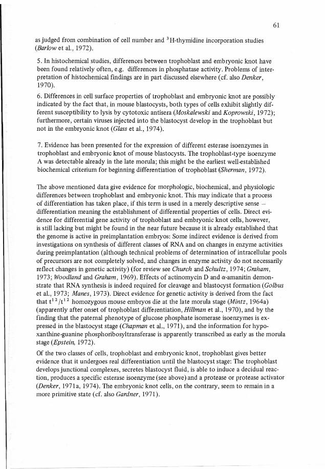
Very impressive are experiments in which all blastome res of a 3H-thymidine-labeled 8-16cell embryo were brought into an inside position by the way that this embryo was beingsurrounded by another 14 unlabeled embryos (Fig. 3) (Hillman et al., 1972). By thismeans, all cells of the labeled inside embryo could be forced, in some cases, to form partof the embryonie knot, even those which normally would have formed trophoblast.
Another way of labeling cells was used in experiments by Wilson et al. (1972) and Sternand Wilson (1972): silicone oll microdroplets were put into the cytoplasm. Peripheral cellsof late morulae or early blastocysts of the mouse were labeled this way and the embryosfused with either identical stages or eight-cell embryos. In the resulting chimeras, part ofthe labeled outside cells were found in the embryonie knot. This again demonstrates that
64
either the fate of outside (presumptive trophoblast) cells of the late morula/early blasto-cyst is not fixed yet, or that not all of these cells are presumptive trophoblast (cf. p. 72).
fF @ßlWC)'+ -m - ...
00'·0"0·'" .. ','
.+0000000'0' 00'......... .: ....-
co00..
'--()()'OJ
-Fig, 3. Fusion experiments: Demonstration of decisive role of inside or outside position of blastomeres(after Hillman et al., 1972). Mouse embryos. Labeling was done with 3H-thymidine.Above: Two labeled blastome res of 8-cell stage on outside of 8- to 16-cell unlabeled embryo. Positionof Iabeled cells was deterrnined in resulting blastocyst, In 40 experiments performed, 92% of all labeledcells were found in trophoblast.Below: Labeled 8- to 16-cell ernbryo is being completely surrounded by 14 unlabeled ernbryos of samestage (note that each symbol here represents one whole ernbryo). Result is a giant blastocyst able togive rise to a welJ-shaped embryo. In 4 out of 7 experiments, labeled celJswere found exclusively inembryonie knot
The marked ability of disaggregated mouse embryos (even blastocysts) to reaggregate andform regular blastocysts was also taken as an argument for developmentallability ofblastome res (Stern, 1972).
The type of experiments described above have been reviewed in detail by Herbert andGraham (1974).
Another line of evidence that theory A might be correct was derived from observations ofcoincidence; there was a marked increase of uridine incorporated into RNA in the stagewhen blastome res begin to acquire an inside position. But note that in the mouse, all majorclasses of RNA are synthesized from the four-cell stage on, and on the other hand, the prob-lem of interference of intracellular pools of precursors has not yet been solved (reviewed byGraham, 1973; Church and Schultz, 1974).
2. Theory B: Determination Depending on Polar Bilateral Organization of the Egg(Morula) (Fig. IB1, B2)
According to this theory, determination does not depend on the inside or outside positionof blastome res but on morphogenetic factors restricted to a localized area in the egg ormorula. In the most elaborate version of this theory (Fig. 1B2), these factors are presumedto be present in a certain area of cytoplasm of the unc1eaved egg; during c1eavage, segrega-

65
tion occurs, and those blastomeres which receive part of this material will be determinedto differentiate into a certain direction: e.g. if the factors are embryonie knot-deterrnining,the progeny of these blastomeres will develop into embryonie knot (part of them may, inaddition, also form trophoblast).
It is also possible to formulate a more general version of this theory which does not includepolar organization of the uncleaved egg but only postulates that, during cleavage, the em-bryo acquires polarity due to unknown processes (governed e.g., by locally acting exogenicfactors (Fig. 1B1).
In each case, according to this theory, the primary arrangement of presumptive trophoblastand embryonie knot cells in a cleavage stage embryo would exhibit polarity, depending onthe eccentric localization of the determining factors. This is in clear contrast to the radialsymmetry suggested by the inside-outside model (theory A, Fig. 1A).
Observations supporting theory B have been made in the course of experimentation andof morphologie and histochemical investigations in several species.
a) Experiments
One of the blastome res of the rabbit two-cell embryo or three of the blastomeres of thefour-cell stage were destroyed by pricking them with a needle, and the developmental po-tential of the surviving blastomere was followed (Seidel, 1952, 1956, 1960). The result,relevant to the problem discussed here, was that not all of these surviving blastomeresformed regular blastocysts: in a certain proportion of them (ab out 1:2 in case of experi-mentation in the two-cell stage) the embryonie knot was lacking and only trophoblast(and sometimes also entoderm) developed.
This experiment demonstrates limits of regulative capacities of the rabbit embryo. Refer-ring also to comparative aspects based on numerous data from lower animals, Seidel for-mulated the theory that, in the mammalian egg cytoplasm, a specialized area exists(Plasmatischer Faktorenbereich, plasmatic field of factors), which is organized like a for-mative center (Bildungszentrum). Whereas all blastomeres primarily have the potential toform trophoblast, only those of them which receive, during cleavage divisions, part of thecytoplasrnic field of factors will be able to differentiate, in addition, an embryonie knot(which means they have all the information available to form a whole blastocyst). The re-sult of the deletion experiment (blastocyst or trophoblastic vesicle) would depend onwhether the surviving blastomere by chance was one of those which do possess the factors,or one of those which do not (Fig. 4).
Comparable experiments in the mouse seemed to show the same trend, and at first the sameinterpretation has been given (Tarkowski, 1959, 1961). Later on, the results of studies ofthe developmental capacities of all blastomeres of four to eight cell embryos were givenan interpretation in favor of the inside-outside model (theory A) (Tarkowski and Wr6b-lewska, 1967) (see general discussion).
Labeling certain parts of cytoplasm by injecting silicone oil droplets revealed in the mouseegg, that there is no important spatial disturbance of the cytoplasmic pattern of the eggduring cleavage: the cortical region of the egg being converted to the outer cells of the

66
morula. (Nevertheless, fusion experiments done with these labeled embryos show that thesecortical regions also can be forced to become part of the embryonie knot) (Wilson et al.,1972; Stern and Wilson, 1972).
u.-
+
Fig. 4. Seidel's experiment in rabbit. One blastomere of the 2-cell stage is being destroyed (marked bylarge cross). The surviving blastomere will develop (after transfer of whole embryo into foster mother)into either a regular blastocyst with embryonie knot (above), or a trophoblastic vesicle without embryonieknot (below). Result is thought to reflect that the surviving blastomere either contained or lacked materialof a cytoplasmic field of factors (Bildungszentrum) present in a restricted area of egg cytoplasm whichprovides information for determination of embryonie knot. Formation of trophoblast, on the contrary,is assumed to be a general ability common to all blastomeres (with or without Bildungszentrum)
The assumption that the cytoplasmic field of factors determines embryonie knot ratherthan trophoblast is not necessarily an integral part of theory A. The available experimentalresults do not completely rule out other interpretations like: Both embryonie knot - de-termining as well as trophoblast - determining fields of factors might exist at oppositepoles of the egg. Or there might be on1y factors providing the information for determina-tion of trophoblast; these factors might be organized like a field, and the embryonie knot beformed at the opposite pole. Available information does not yet allow one to decide whichof these possibilities is the correct one. Because there is more evidence for the trophoblastthan for the embryonie knot to undergo real differentiation during these early stages (seep. 61), we like to reillustrate Seidel's experiment assuming that the cytoplasmic factors aretrophoblast-determining (Fig. 5). Embryos lacking trophoblast (i.e., pure embryonie knots)(Fig. 5 I) were not found by Seidel (1960, 1969), or rarely found by Tarkowski and Wrob-lewska (1967). Apart from other possibilities, this could still be explained assuming that thefield of factors is trophoblast-determining but extends over a rather large area so that evenblastomeres of the opposite pole would still be ab1e to form trophoblast (in addition toembryonie knot) (Fig. 6).
b) Morphologie and Histoehemieal Data
In a large series ofpapersDalcq andMulnard(Dalcq, 1951, 1954, 1955, 1962a, b, c, 1966;Mulnard, 1955, 1965 ;Mulnard and Dalcq, 1955) presented a number of data on histochern-
67
TIO~~-1t-o-0+m0~W~~~.~e>~\~('
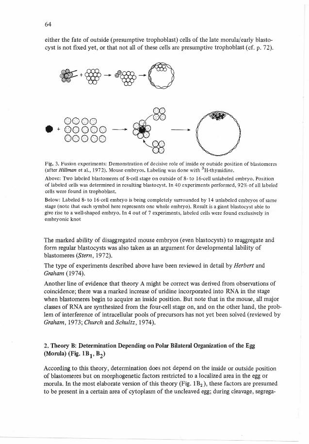
Fig. 5. Seidel's experirnent in rabbit (cf. Fig. 4) redrawn assuming that the cytoplasmic field of factors istrophoblast-determining. First cleavage furrow can lie in different planes and either restriet this fieldto one of two blastomeres (I, 11)or devide it (111).For descriptive purposes, the situation is oversim-plified in this diagram illustrating .mosaic-type reactions: Half-embryos which consist only of "tro-phoblast-factor cytoplasm" form only trophoblast (II), half-ernbryos without it form only embryonieknot (I), half-embryos with both types of cytoplasrn form both types of cells, i.e. a whole blastocyst(III)
Fig. 6. Same as in Fig. 5 I, but assuming that the area which the trophoblast-determining cytoplasmicfield of factors takes in the egg is larger. This could explain why pure embryonie knots (cf', Fig. 5 I)are never (or rarely) seen in the experiments. Another possible explanation for the same phenomenonis provided by theory described in Fig, 4 (legend)
ical differences between blastomeres, and correlated them with differences betweentrophoblast and embryonie knot seen in blastocysts. Most of this work was done in the rat,including a number of investigations in the mouse. The uncleaved egg was described to ex-hibit aplane of bilateral symmetry (most obvious in the rat) forming a certain angle withthe animal-vegetal axis, the cytoplasm of the so-called dorsal side being especially rich inRNA (basophilic region, cf. Jones-Seaton, 1950; see also De Geeter, 1954) and exhibitinga characteristic diffuse type of acid phosphatase reaction (Mulnard, 1955, 1965). In cleav-age stages, these histochemical characteristics were found to be restricted to certain blasto-meres that finally will form the embryonie knot after becoming enveloped by the presump-tive trophoblast cells (cf. e.g. Mulnard, 1966, Fig. 2). As a submicroscopic equivalent ofthe basophilic region, Krauskopf (1968) described in the rabbit egg an area rich in poly-ribosomes and poor in other organelles. There is no report on comparable observations byother electron microscopists.
68
The conclusions drawn from the above-mentioned investigations are not widely acceptedtoday (cf. Tarkowski and wroblewska, 1967; Herbert and Graham, 1974). Reinvestigationsof the phosphatase distribution using azo-dye methods instead of Gomori-type reactionsfailed to confirm differences of enzyme reaction between blastomeres in the rat (Rode etal., 1968) or mouse (Denker, unpublished), although in the hamster they did show themore intense reaction of the embryonie knot (Ishida, 1972).
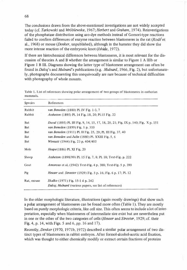
If there are histochemical differences between blastomeres, it is most relevant for the dis-cussion of theories A and B whether the arrangement is sirnilar to Figure 1 A I1Ib orFigure 1 B III. Diagrams showing the latter type of blastomere arrangement can often befound in Dalcq's and Mulnard's publications (e.g. Mulnard, 1966, Fig. 2), but unfortunate-ly, photographs documenting this unequivocally are rare because of technical difficultieswith photography of whole mounts.
Table 1. List of references showing polar arrangement of two groups of blastorneres in eutherianmammals.
Species References
RabbitRabbit
BatBatBatBatBat
Mole
Sheep
Goat
Pig
Rat, mouse
van Beneden (1880) PI. IV Fig. 1-3,7Assheton (1895) PI. 14 Fig. 18,20; PI.15 Fig. 22
Duval (1895) PI. III Fig. 9,14,15,17,18,20,21; Fig. IX p. 140; Fig. X p. 151van Beneden (1899) Fig. 1 p. 310van Beneden (1911) PI. II Fig. 25, 29; PI. III Fig. 37,40van Beneden and Julin (1880) PI. XXIII Fig. 5, 6Wimsatt (1944) Fig. 23 p. 404/405
Heape(1886) PI. XI Fig. 20
Assheton (1898/99) PI. 15 Fig. 7,8; PI. 18; Text-Fig. p. 222
Amoroso et al. (1942) Text-Fig. 4 p. 388; Text-Fig. 5 p. 390
Heuser and Streeter (1929) Fig. 5 p. 16; Fig. 6 p. 17; PI. 12
Skalko (1971) Fig. 15-1 d p. 242Dalcq; Mulnard (various paper s, see list of references)
In the older morphologie literature, illustrations (again mostly drawings) that show sucha polar arrangement of blastomeres can be found more often (Table 1). They are mostlybased on purely morphologie criteria, like cell size. This often seems to include a lot of inter-pretation, especially when blastomeres of intermediate size exist but are nevertheless putin one or the other of the two categories of cells (Heuser and Streeter , 1929; cf. theirFig. 4, p. 14, with Figs. 5 and 6, pp. 16 and 17).
Recently, Denker (1970, 1971b, 1972) described a similar polar arrangement of two dis-tinct types of blastomeres in rabbit embryos. After forrnol-alcohol-acetic acid fixation,which was thought to either chemically modify or extract certain fractions of proteins

\
a b
69
,!
c
9
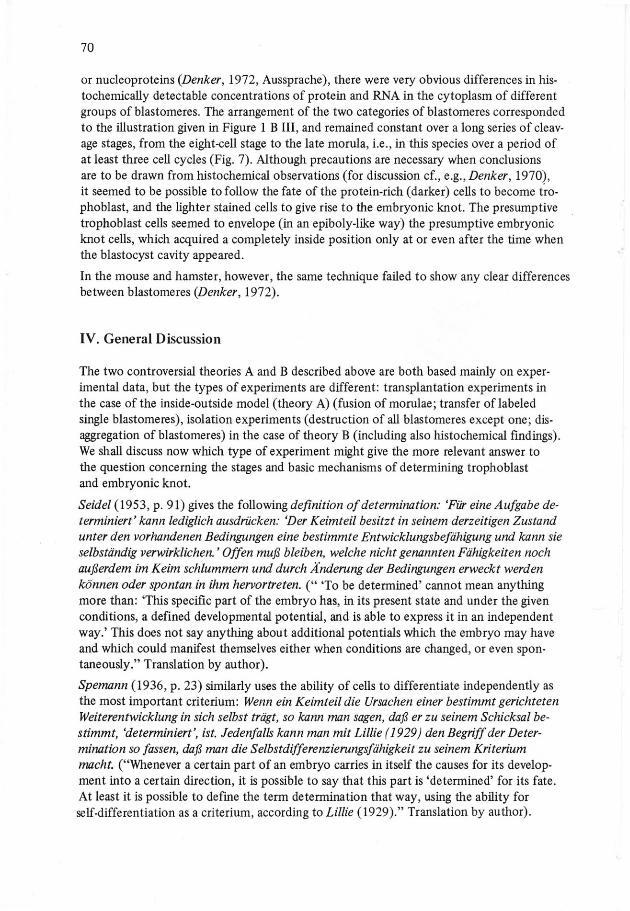
Fig, 7. Aspects of polar arrangernent of two groups of blastomeres in the rabbit. Embryos were fixedwith formol-alcohol-acetic acid, paraffin seetions were taken, proteins were stained with Hg-bromphenolblue. x 260. a-c: 54 h p.c., three sections from same embryo; d: 54 h p.c.; e, f: 63 h p.c., g: 76 h p.c.,blastocyst cavity just appearing (cleft); h, i: 80 h p.c., early blastocyst,Two categories of blastomeres can be distinguished by intensity of cytoplasmic staining: 1. Lighterstained, not polarized cells; in early blastocyst, light cells form embryonie knot. 2. Darker stained cellswhich often show very obvious maximum of stain uptake in parts of cytoplasm directed toward centerof egg. When cavitation begins, they can be identified as prospective trophoblast cells (g). Trophoblastof early blastocysts also stains more intensely than embryonie knot (h, i). Note that in c1eavagestagestrophoblast-type cells form a single-layered cap which surrounds other blastomeres only incompletely(polarity l), but area taken in different eggs is different (a-f)
70
or nucleoproteins (Denker, 1972, Aussprache), there were very obvious differences in his-tochemically detectable concentrations of protein and RNA in the cytoplasm of differentgroups of blastomeres. The arrangement of the two categories of blastomeres correspondedto the illustration given in Figure 1 B III, and remained constant over a long series of cleav-age stages, from the eight-cell stage to the late morula, i.e., in this species over aperiod ofat least three cell cycles (Fig. 7). Although precautions are necessary when conclusionsare to be drawn from histochemical observations (for discussion cf., e.g., Denker, 1970),it seemed to be possible to follow the fate of the protein-rich (darker) cells to become tro-phoblast, and the lighter stained cells to give rise to the embryonie knot. The presumptivetrophoblast cells seemed to envelope (in an epiboly-like way) the presumptive embryo nieknot cells, which acquired a completely inside position only at or even after the time whenthe blastocyst cavity appeared.
In the mouse and hamster, however, the same technique failed to show any clear differencesbetween blastomeres (Denker, 1972).
IV. General Discussion
The two controversial theories A and B described above are both based mainly on exper-imental data, but the types of experiments are different: transplantation experiments inthe case ofthe inside-outside model (theory A) (fusion ofmorulae; transfer oflabeledsingle blastomeres), isolation experiments (destruction of a11blastomeres except one; dis-aggregation of blastomeres) in the case of theory B (including also histochemical findings).We shall discuss now which type of experiment might give the more relevant answer tothe question concerning the stages and basic mechanisms of determining trophoblastand embryonie knot.
Seidel (1953, p. 91) gives the following definition of determination: 'Für eine Aufgabe de-terminiert' kann lediglich ausdrücken: 'Der Keimteil besitzt in seinem derzeitigen Zustandunter den vorhandenen Bedingungen eine bestimmte Entwicklungsbefähigung und kann sieselbständig verwirklichen. ' Offen muß bleiben, welche nicht genannten Fähigkeiten nochaußerdem im Keim schlummern und durch Änderung der Bedingungen erweckt werdenkönnen oder spontan in ihm hervortreten. (" 'To be determined' cannot mean anythingmore than: 'This specific part of the embryo has, in its present state and under the givenconditions, a defined developmental potential, and is able to express it in an independentway.' This does not say anything about additional potentials wh ich the embryo may haveand which could manifest themselves either when conditions are changed, or even spon-taneously." Translation by author).
Spemann (1936, p. 23) similarly uses the ability of cells to differentiate independently asthe most important criterium: Wenn ein Keimteil die Ursachen einer bestimmt gerichtetenWeiterentwicklung in sich selbst trägt, so kann man sagen, daß er zu seinem Schicksal be-stimmt, 'determiniert', ist. Jedenfalls kann man mit Lillie (1929) den Begriff der Deter-mination so fassen, daß man die Selbstdifferenzierungsfähigkeit zu seinem Kriteriummacht. ("Whenever a certain part of an embryo carries in itself the causes for its develop-ment into a certain direction, it is possible to say that this part is 'determined' for its fate.At least it is possible to define the term determination that way, using the ability forself-differentiation as a criterium, according to Lillie (1929)." Translation by author).
71
It is not necessary to discuss here the concept of "self-determination" which itself can becriticized for being, in its strict form, too narrow for most systems. The important pointis that the ability to differentiate after isolation, which can be proved experimentally, isbeing used as a criterium. If this definition of determination is accepted, it follows thatisolation experiments should give better information about the determined or nondetenninedstatus of a cell than transplantation experiments can do. In the latter type of experiments,the cells are brought under the influence of different parts of the embryo (or other tissues),and the original state of the transplant and the conditions are drastically changed. Spemann,who has done a lot of transplantation experiments, comments (1936, p. 31): "So wird Trans-plantation im neuen Gewebsverband nur dann sichere Auskunft geben können, wenn Selbst-differenzierung stattfindet, wenn also die Determination des Implantats genügend befestigtist, um sich auch gegen einen etwaigen Einfluß der Umgebung durchzusetzen. Der ersteEintritt der Determination wird sich dagegen nur bei völliger Isolierung erkennen lassen. "(Transplantation ... "will give a clear answer only in case of self-differentiation, i.e., whendetermination of the implant is stabile enough to dominate over possible influences of thesurrounding tissues. The very beginning of determination can only be recognized in caseof complete isolation." Translation by author).
According to this, the results of the described isolation experiments which favor theory Bwould seem more relevant for the discussion of mechanisms involved in determination oftrophoblast and embryonie knot. The transplantation experiments described on pp. 62 ff.,on the other hand, demonstrate the high regulative capacity of these early embryonie states.When, for example, all blastomeres of a morula, even those which normally would have de-veloped into trophoblast, can be forced to form part of the embryonie knot (p. 63, Fig. 3),this shows in an impressive way the flexibility of the system and that it does not exhibitfeatures of a mosaic. But is seems questionable if this type of experiment can uncoverwhether cells are already inclined (but not irreversibly switched yet) to form trophoblast,which isolation experiments possibly do reveal.
Often a different definition of cell determination is being used, e.g. by Herbert and Graham(1974): 'Tell determination is the process by which the developmental potential of a cellbecomes limited during embryogenesis." Transplantation experiments can certainly uncoverlimited developmental potentials. This is definitely the case when the limitation became ir-reversible. But, we feel that this is a secondary process following the establishment of aninclination of cells to develop into one or the other direction.
Isolation experiments are also certainly problematic because they do change the state andconditions of cells, both due to the isolation procedure (e.g. pronase and EDTA-treatment,Tarkowski and Wroblewska, 1967) and to in vitro culture conditions. If part of the embryois being destroyed but not removed (Seidel, 1960), it rnight influence the results of the ex-periment: when, in the amphibian embryo, one blastomere of a two-cell stage was destroyedbut not removed, the surviving blastomere produced only a half embryo (w. Roux); on thecontrary, when both blastomeres were completely separated, they both regulated and eachone formed a whole twin embryo (Spemann, 1936, p. 11 ff.). It is therefore interesting thatin Tarkowski's and Wroblewska's experiments (1967), the completely isolated blastomeresdid develop into different forms of vesicles (blastocysts, "false blastocysts," trophoblasticvesicles), and some of them even formed only uncavitated masses of cells. The authors felt,however, that these differences might be due to differences in culture conditions. They con-cluded that: 1. At least in some eggs, all blastomeres have the potential to form vesicular

72
structures (this view also being part of Seidel's theory, see p. 65). 2. Incidence of differen-tiation into real blastocysts decreases from one-quarter blastome res to one-eighth forms.Tarkowski and Wr6blewska feel that most probably this is due to the fact that with advanc-ing stage of development of the blastomeres at the time of their separation, the number ofcells attained by them at the time of cavitation decreases. Consequently, the probabilitythat cells become enveloped decreases. The authors imagine that up to the eight-cell stage,all blastomeres possess the ability to differentiate into trophoblastic direction, and develop-ment into embryonie knot cells is being triggered by an inside position (theory A).
Data presented by Moore et al. (1968) (rabbit) unfortunately do not contribute to the dis-cussion of these two theories because an account of purely trophoblastic structures is notincluded.
As a different kind of approach, morphologic and histochemic analyses give additional in-formation. By fixing the embryos it is intended to preserve certain characteristics of themomentary state of blastomeres without changing them by initiating regulatory processesas it probably happens in both transplantation and isolation experiments. The polar arrange-ment of different types of blastomeres as seen in the rabbit morula seems to form an argu-ment for theory B (see p. 68 0. This arrangement also provides important aspects for the in-terpretation of fusion experiments: if it is correct, as suggested by the histochemical findings,that only some but not all of the peripheral cells are determined to form trophoblast, thenthe fact that a proportion of labeled outside cells become included in the embryonie knotof the chimera (p. 63 f.) could reflect simply the yet undetermined state of some of theoutside cells. This fmding would then not contradict theory B anymore.
Theory B, in its elaborate form, implies that a specific area of the egg cytoplasm exerts a reg-ulatory effect on gene activity. Influences of the egg cytoplasm on nuclei are in fact knownfrom experiments in which egg nuclei were replaced by somatic cell nuclei (Gurdon, 1962;Gurdon and Woodland, 1969). There is some first evidence for the same phenomenon inthe mouse egg (Bernstein and Mukherjee, 1972). Localization of factors ofthis type inspecific areas of egg cytoplasm has been demonstrated in certain species (insect egg:Seidel, 1936).
A promising different approach is to study cell strains derived from preimplantation embryos,although there are certainly numerous differences between cultured cells and the originalcells of the embryo. Trophoblast-resembling cells developed in vitro when isolated blasto-meres of a stage as early as the four-cell stage were used (Edwards, 1964; Cole and Paul,1965; Cole et al., 1965, 1966). It is hoped that additional information will come from in-vestigations of teratomas derived from ectopically transplanted embryos or from unfertil-ized eggs in the ovary (Evans, 1972; Damjanov and Solter, 1974).
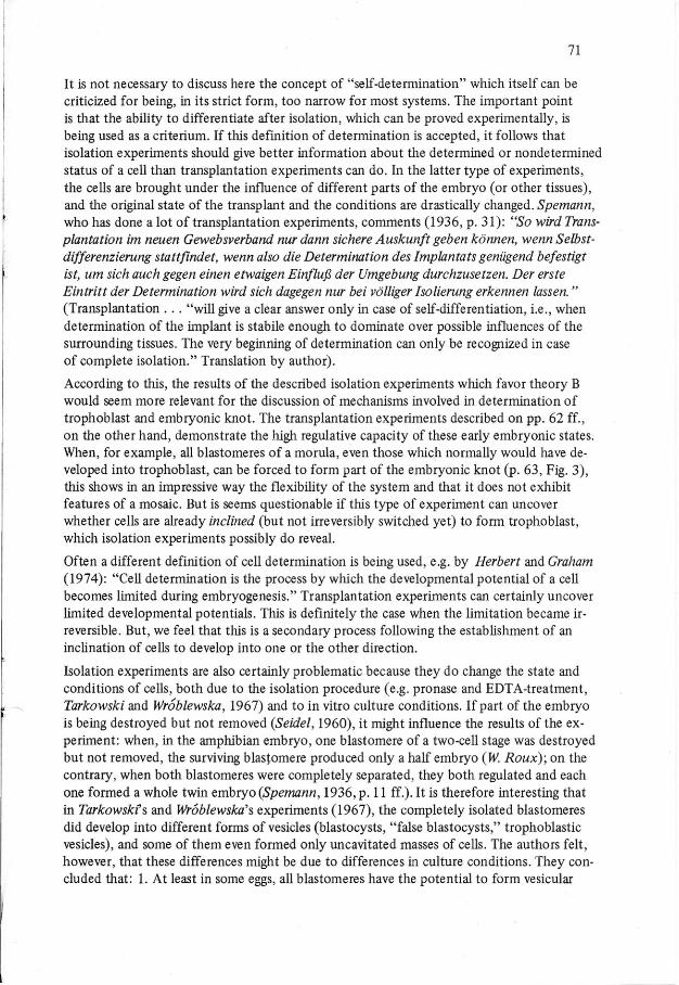
Finally, some comparative aspects shall be mentioned. In marsupials, cells are being deter-mined to become embryonie disc (formative cells) without ever having been enclosed byother cells (Fig. 8) (Hili, 1911; Hartman, 1919). The same seems to hold true for at leastone eutherian mammal: Hemicentetes (Tenrecoidea) (Goetz, 1937, 1939;Bluntschli, 1938)(Fig. 8). The inside-outside model (theory A) cannot be applied to these embryos becauseinside cells do not exist in these species before formation of the embryonie knot/ disc. Prob-ably the polarity of the blastocyst (embryonic disc, trophoblast) here results from a polar-ity of the uncleaved egg.
73
Opossum
I
E.M'c.
P.TR.
Fig. 8. Comparative aspeets. In opossum and in Hemieentetes, eells being determined to form embryo niedisc without having been in inside position. EM. C,' entodermal mother cell; Ent: entoderm; F,' forma-tive ceUs; P. TR.,' primitive trophoblast; T. C,' trophoblast cells. (For data on opossum and Hemicentetes,seeHill, 1911; Hartman, 1919; Goetz, 1937, 1939; Bluntschli, 1938) (from Wimsatt, 1975, by courtesyof author and of editor, Biology 01'Reproduction; slightly modified)
In conclusion, it appears weIl-established that the mammalian cleavage stage embryo pos-sesses vast regulative eapaeities as impressively demonstrated by transplantation (fusion)experiments. The inside or outside position of blastome res does influenee their fate and eanbeeome deeisive far their determination to form either trophoblast or embryonie knot. Onthe other hand, the egg does exhibit polarity, and blastomeres seem to be unequal, indepen-dent of their inside or outside position, as shown by their histochemical properties as weIlas their inclination to form only trophoblast or both trophoblast and embryonie knot. Itis probable that this type of "preformation" is, in the beginning, weak and changeable, andean easily eseape the experimentalist (cryptie preformation, Graham, 1971). The questionremains open whieh type of experiment might be the most suitable to reveal physiologiein vive meehanisms rather than in vitro regulations.
74
Acknowledgments. This review was written during the author's work at the ArbeitsgruppeProf. Dr. G.H.M. Gottschewski am Max-Planck-Institut für Immunbiologie, Freiburg (WestGermany).The author wishes to thank Mrs. M. Polack for preparing the diagrarns, and Dr. W.A. Wim·satt for the permission to use his diagram on comparative aspects (Fig. 8). Own experimentsincluded in this review were supported by the Deutsche Forschungsgemeinschaft (De181/1-3).
References
Amoroso, E.C., Griffiths, W.F.B., Hamilton, W.J.: The early development of the goat(Capra hirsus). J. Anat. (Lond.).1i, 377-406 (1942).
Assheton, R.: A re-investigation into the early stages of the development of the rabbit.Quart. 1. micr , Sei. 37, 113-164 (1895).
Assheton, R.: The segmentation of the ovum of the sheep, with observations on the hypo-thesis of a hypoblastic origin for the trophoblast. Quart. J. micr. Sei. ±J., 205-262(1898/99).
Barlow, P., Owen, D.A.J., Graham, c.: DNA synthesis in the preimplantation mouse ernbryo.J. Ernbryol. exp. Morph. 27, 431-445 (1972).
Beneden, E. van: Recherehes sur l'ernbryologie des rnamrniferes. La formation des feuilletschez Je Lapin. Arch. Bio1.J., 137-224 (1880).
Beneden, E. van: Recherehes sur les premiers stad es du developpement du Murin (Vesper-tilio murinus). Anat. Anz.l.§., 305-334 (1899).
Beneden, E. van:Recherehes sur I'embryologie des mamlniferes. De la segmentation, de laformation de la cavite blastodermique et de l'ernb ryon didermiq ue chez le Murin. Arch.Bio!. 26,1-63 (1911).
Benede;;:E. van, Julin, C.: Observations sur la maturation, la fecondation et la segmentationde l'oeuf chez les cheiropteres. Arch. Bio!. 1., 551-571 (1880).
Bernstein, R.M., Mukherjee, B.B.: Control of nuclear RNA synthesis in 2-cell and 4-cellmouse embryos. Nature (Lond.) 238, 457-459 (1972).
Bluntschli, H.: Le developpement primaire et l'implantation chez un Centetine (Hernicen-tetes). C.R. Ass. Anat. 44, 39-46 (1938).
Calarco, P.G., Brown, E.H.: An ultrastructural and cytological study of preimplantationdevelopment of the mouse. 1. exp. Zoo!.ll.L 253-284 (1969).
Chapman, V.M., Whitten, W.K., Ruddle, F.H.: Expression of paternal glucose phosphateisomerase-l (GPI-l) in preimplantation stages of mouse embryos. Develop. Bio!. 26,153-158 (1971).
Church, R.B., Schultz, G.A.: Differential gene activity in the pre- and postimplantationmammalian embryo. Curr. Top. Develop. Bio!...8., 179-202 (1974).
Cole, R.J., Edwards, R. G., Paul, J.: Cytodifferentiation in cell colonies and eell strainsderived from cleaving ova and blastocysts of the rabbit. Exp, Cell Res. 37, 501-504(1965).
Cole, R.J., Edwards, R. G., Paul, J.: Cytodifferentia tion and ernbryogenesis in cell coloniesand tissue cultures derived from ova and blastocysts of the rabbit. Develop. Biol.ll,385-407 (1966).
Cole, R.J., Paul, J.: Properries of cultured preimplantation mouse and rabbit embryos, andcell strains derived from them. In: Preimplantation Stages of Pregnancy (eds. G.H. W.Wolstenholme, M. O'Connori, pp, 82-122. London: Churchill 1965.

75
Dalcq, A.M.: New descriptive and experimental data concerning the mammalian egg ,principally of the rat (With a specification of a previous interpretation). Proc. kon. ned.Akad. Wet., Sero C,Ji, 351-363, 364-372,469-479 (1951).
Dalcq, A.M.: Nouvelles donnees structurales et cytochimiques sur l'oeuf des marnmiferes.Rev. gen, Sei. Pures. Appl...§l, 19-41 (1954).
Dalcq, A.M.: Processes of synthesis during early development of rodent's eggs and embryos.In: Studies on Fertility (ed. R.G. Harrison) 2113-122 (1955).
Dalcq, A.M.: Evolution de l'organisation morphogenetique dans l'oocyte chez le Rat et laSouris. Verh. Anat. Ges., Anat. Anz. Suppl. 109,373-382 (1962a).
Dalcq, A.M.: Localisations et evolution des phosphatases aux premiers stades du döveloppe-ment. Bull. Acad. roy, Med. Belg., VIIe Serie, 2,573-610 (1962b).
Dalcq, A.M.: Etudes cyto-enzymologiques sur les-;eufs vivants de Souris incubes cn pre-sence d' ATP et d'autres mononucleotides. Arch. Biol..1l, 405-444 (1962c).
Dalcq, A.M.: Detectio n des enzymes de dephosphorylation dans les oeufs de rat et de sourisfixes au formol et traites in toto. Arch. Biol. 77, 205-344 (1966).
De Geeter, L.: Etudes sur la structure de l'oeuf vierge et les premiers stades du developpe-ment chez le Cobaye et le Lapin. Arch. Biol..2l., 363-436 (1954).
Denker, H.-W.: Topochernie hochmolekularer Kohlenhydratsubstanzen in Frühentwicklungund Implantation des Kaninchens. II. Beiträge zu entwicklungsphysiologischen Frage-stellungen. Zool. Jahrb. Abt. Allg. Zool. Physiol. 75, 246-308 (1970).
Denker, H.-W.: Enzym-Topochemie von Frühentwicklung und Implantation des Kaninchens.III. Proteasen. Histochemie 25, 344-360 (1971 a).
Denker, H.-W.:ln discussion to~.G. Mulnard: Manipulation of cleaving mammalian ern-bryo ... Advanc. Biosei ...§., 255-277 (1971b).
Denker, H.-W.:Furchung beim Säugetier; Differenzierung von Trophoblast- und Embryonal-knotenzellen. Verh. Anat. Ges. 66, Anat. Anz. Supp!. 130,267-272 (1972).
Denker, H.-W.:Trophoblastic factors involved in lysis of the blastocyst coverings and inimplantation in the rabbit: observations on inversely orientated blastocysts. J. Embryo!.exp , Morph. 32,739-748 (1974).
Duval, M.: Etud-;-sur I'ernbryologie des Cheiropteres. J. Anat. Physiol..1L 93-160(1895).
Dvoiak, M.: Submicroscopic cytodifferentiation. Ergebn. Anat. Entwickl.-Gesch. 45/4(1971).
Edwards, R.G.: Cleavage of one- and two-celled rabbit eggs in vitro after removal of thezona pellucida. J. Reprod. Fertil.2 413-415 (1964).
Eps te in, C.J.: Expression of the mammalian X chromosome before and after fertilization.Science 175,1467-1468 (1972).
Evans, M.J.: The isolation and properties of a clonal tissue culture strain of pluripotentmouse teratoma cells. J. Embryol, exp. Morph.1ß., 163-176 (1972).
Gardner, R.L.: Manipulations on the blastocyst. Advanc. Biosei ...§.;279-301 (1971).Gardner, R.L.: An investigation of inner cell mass and trophoblast tissues following their
isolation from the mouse blastocyst. J. Ernbryol. exp. Morph. 28, 279-312 (1972a).Gardner, R.L.: Manipulation of development. In: Reproduction in Mammals, Book 2:
Embryonie and Fetal Development (eds. C.R. Austin, R. V. Shorty, pp. 110-133.Cambridge U niversity Press 1972b.
Gardner, R.L.: Differentiation of trophoblast in the early mammalian ernbryo. Res. Reprod.2"No. 5,1-2 (1973).
Gardner, R.L., Johnson, M.H.: Investigation of early mammalian development using inter-specific chimaeras between rat and mouse. Nature (New Biol.) 246, 86-89 (1973).
76
Glass, R.H., Calareo, P.G., Lin, T.P., Florence, J., Oh, J.O.: Development of the mouseblastocyst following injection with Newcastle disease virus. Biol, Reprod.lQ, 502-51 I(1974).
Goetz, R.H.: Studien zur Placentation der Centetiden. 11. Die Implantation und Frühent-wicklung von Hernicentetes semispinosus (Cuvier). Z. Anat. Entwick!.Gesch. 107274-318 (1937).
Goetz, R.H.: On the early development of the Tenrecoidea (Hemicentetes sernispinosus}.Bio-Morphosis I , 67-79 (1939).
Golbus, M.S., Calareo, P.G., Epstein, CJ.: The effects of inhibitors of RNA synthesis(o-arnanitin and actinomycin D) on preimplantation mouse embryogenesis. J. exp. Zool.186,207-216 (1973).
Graham, CF.: The design of the mouse blastocyst. Symp. Soc, e xp. Biol..li, 371-378 (1971).Graham, CF.: Nuc1eic acid metabolism during early mammalian development. In: The Reg-
ulation of Mammalian Reproduction (eds. S.J. Segal et al.), pp. 286-301. Springfie1d,Ill.: Charles C.Thomas 1973.
Gurdon, J.B.: The developmental capacity of nuc1ei taken from intestinal epithelium cellsof fee ding tadpo1es. J. Embryo l. exp. Morph.l.Q., 622-640 (1962).
Gurdon, J.B., Woodland, H.R.: The inf1uence of the cytop1asm on the nuc1eus during celldifferentiation, with special reference to RNA synthesis during amphibian cleavage.Proc. roy. Soc. B 173, 99-111 (1969).
Hartman, C G.: Studies on the developrnent of the opossum Dide1phys virginiana L.III. Description of new material on maturation, cleavage and entoderm formation.IV. The bilaminar b1astocyst. J. Morph.2l, 1-142 (1919).
Heape, W.: The development of the mole (Talpa europea), the ovarian ovum, and seg me n-tation of the ovum. Quart. 1.micr. Sci.~ 157-174 (1886).
Herbert, M.C, Graham, CF.: Cell determination and biochemica1 differentiation of theearly mamma1ian embryo. Curr. Top. Develop. Bio!.ji, 151-178 (1974).
Hesseldahl, H.: Ulstrastructure of ear1y cleavage stages and preimp1antation in the rabbit.Z. Anat. Entwickl. Gesch . .!]2, 139-155 (1971).
Heuser, CH., Streeter, G.L.: Ear1y stages in the developrnent of pig embryos, from theperiod of initial cleavage to the time of the appearance of limb-buds. Contr. Embryol.Carneg. Inst.1Q.., 1-29, (1929).
Hill, J.P.: The early developrnent of the marsupia1ia, with special reference to the nativecat (Dasyurus viverrinus). (Contributions to the embryo1ogy of the marsupialia, IV.).Quart. J. micr. Sei.1&., 1-134 (1911).
Hillman, N., Hillman, R., Wileman, G.: Ultrastructura1 studies of cleavage stage t12 /t12
mouse embryos. Amer. 1. Anat.l11L 311-340 (1970).Hillman, N., Sherman, M.l., Graham, C: The effect of spacia1 arrangement on cell deter-
mination during mouse deve1opment. 1. Embryol. exp. Morph. 28, 263-278 (1972).Ishida, K.: Enzymohistochemica1 studies of differentiated 8-16 cell eggs of the hamster.
Jap. 1. Anima1 Reprod.1..§., 105-109 (1972).Jones-Seaton, A.: Etude de l'organisation cytoplasmique de l'oeuf des rongeurs, princi-
pa1ement quant a la basophilie ribonucleique. Arch. Biol..Q ..L 291-444 (1950).Krauskopf, C.: Elektronenmikroskopische Untersuchungen über die Struktur der Oozyte
und des 2-Zellenstadiums beim Kaninchen. II. Blastomeren. Z. Zellforsch. 92, 296-312(1968). -
Manes, C: The participation of the embryo nie genome during early cleavage in the rabbit.Deve1op. Biol.1b 453-459 (1973).
Mintz, B.: Formation of genotypically mosaic mouse embryos. Amer. Zool.1.., 432 (1962).

77
Mintz, B.: Gene expression in the morula stage of mouse embryos, as observed duringdevelopment of t12 /t1 2 let hal mutants in vitro. J. exp, Zool. 157, 267 -271 (1964a).
Mintz, B.: Synthetic processes and early development in the mammalian egg. J. exp.Zool. ill, 85-100 (l964b).
Mintz, B.: Experimental genetic mosaicism in the mouse. In: Preimplantation Stages ofPregnancy (eds. G.E.W. Wolstenholme, M. O'Connor), pp, 194-216. London: Churchill1965.
Mintz; B.:Clonal basis of mammalian differentiation. Syrnp. Soc. exp. Biol.Q, 345-370(1971).
Moore, N. W., Adams, C.E., Rowson, L.E.A.: Developmental potential of single blasto-meres of the rabbit egg. J. Reprod. Fertil.J], 527-531 (1968).
Moskalewski, S., Koprowski, H.: Presence of egg antigen in immature oocytes and pre-implantation embryos. Nature (Lond.) 237,167-168 (1972).
Mulnard, J.: Contribution a la connaissanc-;ctes enzymes dans l'ontogenese. Les phospho-monoesterases acide et alkaline dans le developpement du Rat et de la Souris. Arch.Biol.M., 525-685 (1955).
Mulnard, J.: Aspects cytochimiques de la regulation in vitro de l'oeuf de souris apresdestruction d'un des blastomeres du stade 11. I. La phosphornonoesterase acide. Bull.Acad. roy. Med. Belg., Series 2,2, 31-67 (1965).
Mulnard, J.: Les mecanismes de la regulation aux premiers stades du developpernent desmammiferes. Bull. Soc. zool. Fr. 91, 253-277 (1966).
Mulnard, J.: Analyse microcinematographique du developpment de l'oeuf de souris dustade 11au blastocyste. Arch. Biol.1§., 107-138 (1967).
Mulnard, J., Dalcq, A.M.: Les polysaccharides dans le developpement de l'oeuf tubairedu Rat. C.R. Soc. Biol. (Paris) 149,836-839 (1955).
Rode, B., Damjanov, 1., Skreb, N.: Distribution of acid and alkaline phosphatase activityin early stages of rat embryos. Bull. Sei., Cons. Acad. RSF Yougoslavie, Sect. A, l1.,304 (1968).
Schlafke, S., Enders, A.e.: Cytological changes during cleavage and blastocyst formationin the rat. J. Anat. (Lond.) l.Q1, 13-32 (1967).
Seidel, F.: Entwicklungsphysiologie des Insektenkeimes. Verh. dtsch. zool. Ges., Freiburgp. 291-336 (1936).
Seidel, F.: Die Entwicklungspotenzen einer isolierten Blastomere des Zweizellenstadiumsim Säugetierei. Naturwissenschaften 39, 355-356 (1952).
Seidel, F.: Entwicklungsphysiologie der Tiere. I. Ei und Furchung. Sammlung GöschenBand 1162. Berlin: Walter de Gruyter & Co. 1953.
Seidel, F.: Nachweis eines Zentrums zur Bildung der Keimscheibe im Säugetierei. Natur-wissenschaften.1l., 306-307 (1956).
Seidel, F.: Die Entwicklungsfähigkeiten isolierter Furchungszellen aus dem Ei des Kanin-chens Oryctolagus cuniculus. Wilhelm Roux' Arch. Entwicklungsmech. Organismenill, 43-130 (1960).
Seidel, F.: Entwicklungspotenzen des frühen Säugetierkeimes. Arbeitsgemeinschaft fürForschung des Landes Nordrhein-Westfalen, Heft 193, pp. 1-91. Köln-Opladen: West-deutscher Verlag 1969.
Sherman, M.l.: Biochemistry of differentiation of mouse trophoblast: esterase. Exp. CellRes. 12, 449-459 (1972).
Skalko, R. G.: Methods for histologic and autoradiographic analysis of the early mouse ern-bryo. In: Methods in Mammalian Ernbryology (ed. J.e. Daniel, jr.). pp. 238-246. SanFrancisco: Freeman 1971.
Spemann, H.: Experimentelle Beiträge zu einer Theorie der Entwicklung. Berlin: Springer1936. Reprint Berlin-Heidelberg-New York: Springer 1968.

78
Stern, M.S.: Experimental studies on the organization of the preimplantation mouse embryo.H. Reaggregation of disaggregated embryos. J. Embryol. exp, Morph.1§., 255-261 (1972).
Stern, M.S.: Chimaeras obtained by aggregation of mouse eggswith rat eggs.Nature (Lond.)243,472-473 (1973).
Stern, M.S., Wilson, 1.B.: Experimental studies on the organization of the preimp1antationmouse ernbryo. I. Fusion of asynchronous1y cleaving eggs. J. Embryol. exp. Morph.1§., 247-254 (1972).
Tarkowski, A.K.: Experiments on the development of iso1ated blastomeres of mouse eggs.Nature (Lond.) 184,1286-1287 (1959).
Tarkowski, A.K.: Mouse chimaeras developed from fused eggs. Nature (Lond.) 190,857-860 (1961).
Tarkowski, A.K.: Embryonie and postnatal development of mouse chimeras. In: Preimplan-tation Stages of Pregnancy (eds. G.E. W. Wolstenholme, M. O'Connor), pp. 183-193.London: Churchilll965.
Tarkowski, A.K., Wr6blewska, J.: Development of blastomeres of mouse eggs isolated atthe 4- and 8-cell stage. J. Embryol. exp. Morph.~ 155-180 (1967).
Wilson, 1.B., Bolton, E., Cuttler, R.H.: Preimplantation differentiation in the mouse eggas revea1edby microinjection of vital markers. 1. Embryol. exp, Morph. 27, 467479(1972).
Wimsatt, W.A.: An analysis of implantation in the bat, Myotis lucifugus lucifugus. Amer.1. Anat..l±, 355-411 (1944).
Wimsatt, W.A.: Some comparative aspects of implantation. Biol, Reprod.ll, 1-40 (1975).Woodland, H.R., Graham, C.F.: RNA synthesis during early developrnent of the mouse.
Nature (Lond.) 221,327-332 (1969).Zeilmaker, G.H.: Fusion of rat and mouse morulae and formation of chimaeric blastocysts.
Nature (Lond.) 242, 115-116 (1973).
Note added in Proof
After this manuscript was finished, a number of publieations appeared giving additionalinformation and supporting either theory A or B respeetively. Avendano et al. (1975)found, in a seven-eell human embryo, two differently staining groups of blastomeres thearrangement of whieh resembles the polar grouping of presumptive trophoblast and embro-nie knot cells seen in the rabbit (see p. 70 and Fig. 1 B2). This may support theory B. Anexeellent review of studies of early eell determination and differentiation using the experi-mental teratoma model is given by Damjanov and Solter (1974). These and additional re-levant referenees are listed below.
Avendano, S., Croxatto, H.D., Pereda, J., Croxatto, H.B.: A seven-cell human egg recoveredfrom the oviduct. Fertil. Steril.1&., 1167-1172 (1975).
Damjanov, 1., Solter, D.: Experimental teratoma. Current Topics in Patho1ogy (eds. E.Grundmann, W.H. Kirsten).~ 69-130 (1974).
Ducibella, Th., Albertini, D.F., Anderson, E., Biggers, J.D.: The preimp1antation mammalianembryo. Characterization of intercellu1ar junetions and their appearance during deve1op-ment. Devel. Bio!. 45, 231-250 (1975).
Ducibella, Th., Anderson, E.: Cell shape and membrane changes in the eight-cell mouseembryo, Prerequisites for morphogenesis of the b1astocyst. Deve1op.Biol, 47, 45-58(1975). -

79
Ford, C.E., Evans, E.P., Gardner, R.L.: Marker chromosome analysis of two mousechimaeras. J. Embryo!. exp. Morph.21, 447-457 (1975).
Garner, w., McLaren, A.: Cell distribution in chimaerie mouse embryos before implan-tation. J. Embryol, exp. Morph. 12,495-503 (1974).
Gulyas, B.J.: Areexamination of cleavage patterns in eutherian mammalian eggs. Rotationof blastomere pairs during second cleavage in the rabbit. 1. exp. Zoo!. 1.2.2.. 235-248(1975).
Izquierdo, L., Marticorena, P.: Alkaline phosphatase in preimplantation mouse embryos.Exp. Cell Res ..2l, 399-402 (1975).
Izquierdo, L., Ortiz, M.E.: Differentiation in the mouse morulae. Wilh. Roux' Arch.177,67-74 (1975).
Panigel, M., Kraemer, D.C., Kalter, S.S., Smith, G.S., Heberling, R.L.: Ultrastructure ofcleavage stages and preimplantation embryos of the baboon. Anat. Embryo!. 147,45-62 (1975).
Rossant, J.: Investigation of the determinative state of the mouse inner eell mass. I. Aggre-gation of isolated inner eell masses with morulae. J. Embryol. Exp, Morph.ll, 979-990(1975 ).
Rossant, J.: Invesitgation of the determinative state of the mouse inner cell mass. II. Thefate of isolated inner eell masses transferred to the oviduct. 1. Ernbryol. exp , Morph.11,991-1001 (1975).
Sherman, M.I.: The role of eell-cell interaction during early mouse embryogenesis. In:The Early Development of Mammals (British Soeiety for Developrnental BiologySymposium 2), pp. 145-164. Cambridge University Press, Cambridge 1975.
Sherman, M.l.: Long term culture of cells derived from mouse blastocyts. Differentiation..l, 51-68 (1975).

Reprint from
Current Topics in PathologyErgebnisse der Pathologie
Edited by: E. Grundmann, W. H. KirstenVolume 62 Developmental Biology and PathologyEditors: A. Gropp, K. Benirschke
© Springer-Verlag Berlln Heidelberg 1976Printed in Germany. Not for Sale
Springer-VerlagBerlin Heidelberg New York