Embed Size (px)
Citation preview
www.wjpr.net Vol 7, Issue 19, 2018. 1026
Annamma et al. World Journal of Pharmaceutical Research
FORMULATION AND EVALUATION OF PHYTOSOMAL
NANOCARRIERS FOR ENHANCED CHRYSIN DELIVERY
Annamma Anthrayose* and Neema George
Department of Pharmaceutical Sciences, Centre for Professional and Advanced Studies
Kottayam, Kerala, India.
ABSTRACT
The present work was designed to improve solubility and
bioavailability of Chrysin by the development of Chrysin loaded
nanophytosomes by rotary evaporation method. The formulation of
phytosome evaluated for particle size ,zeta potential, SEM, FTIR,
DSC, entrapment efficiency, drug content, solubilty , in vitro drug
release and release kinetics. The particle size by DLS was found to be
in nano size range. The SEM, FTIR, DSC and solubility analysis
confirmed the formation of phytosome. Chrysin nanophytosome
showed better in vitro drug release when compared to Chrysin. The
kinetics of the drug release was found that it predominately follows
zero order and Higuchi kinetics with non fickian diffusion mechanism.
KEYWORDS: Chrysin, flavanoid, nanocarrier, nanophytosome, rotary evaporation.
1. INTRODUCTION
Flavonoids are among the biggest group of polyphenols, widely distributed in plant-based
foods. A plethora of evidence supports the health benefits and value of flavonoids can play in
the physiological function treatment and in the prevention of disease particularly in the
prevention of degenerative conditions including cancers, cardiovascular and
neurodegenerative diseases. Water solubility and gastric stability are the major limiting
factors for flavonoids to pass the biological membrane. Flavonoids when administered
through nano-sized delivery systems show much better stability and absorption profile.
Nanocarriers could be useful to enhance biovailability and bioefficacy of flavonoids because
they can increase solubilization potential, alter absorption pathways, and prevent the
metabolic degradation within the gastrointestinal tract. The technology of nanoscale
World Journal of Pharmaceutical Research SJIF Impact Factor 8.074
Volume 7, Issue 19, 1026-1041. Research Article ISSN 2277– 7105
Article Received on
29 September 2018,
Revised on 19 October 2018,
Accepted on 09 Nov. 2018,
DOI: 10.20959/wjpr201819-13752
*Corresponding Author
Annamma Anthrayose
Department of
Pharmaceutical Sciences,
Centre for Professional and
Advanced Studies
Kottayam, Kerala, India.
www.wjpr.net Vol 7, Issue 19, 2018. 1027
Annamma et al. World Journal of Pharmaceutical Research
modification could overcome obstacles in the development of functional dietary supplements
and medicines.[1,2]
The phytosome technique has emerged as one of the leading methods of improving
bioavailability of phyto-pharmaceuticals having poor competency of solubilising and
crossing the biological membranes.[3]
Phytosome is a patented technology of Indena where
plant polyphenolics are complexed with phospholipids to improve bioavailability.[4]
Phospholipid mainly phosphatidylcholine, are lipophilic substances and readily form complex
with polyphenolic compounds. Phosphatidylcholine is a major structural constituent of all
biological membranes. Phosphatidylcholine is a major component of soybean lecithin which
provides free choline in the blood for the manufacture of acetylcholine; regulates digestive,
cardiovascular and liver functions.[5]
Chrysin belongs to the flavonoids and has been used as traditional medicine from ancient.
Chrysin (5, 7-dihydroxyflavone) is a natural flavone present in many plant extracts, flowers
such as the blue passion flower (Passiflora caerulea), honey and propolis. Chrysin has
multiple biological activities, such as antitumor activity, anti-inflammatory,
antioxidant, anti-allergic, anti-aging, anti-hypertensive, anti-angiogenesis, antiviral,
anti-atherogenic, antibacterial, anti-diabetic, neuroprotective, hepatoprotective,
nephroprotective and positively effect on reproductive system. Although, chrysin has
multiple health benefits in humans, it has limited therapeutic use. The major constraint in the
use of chrysin is its poor aqueous solubility which results in low bioavailability.[6,7]
Therefore the present work was designed to improve the solubility and the bioavailability of
Chrysin by the development of Chrysin loaded nanophytosomes by rotary evaporation
method.
2. MATERIALS AND METHODS
2.1 Materials
Chrysin and Soyabean lecithin (Soya lecithin / Phosphatidyl Choline) was obtained from
Chemical House, Cochin (Make – TCI, Japan). All other reagents used were of analytical
grade.
www.wjpr.net Vol 7, Issue 19, 2018. 1028
Annamma et al. World Journal of Pharmaceutical Research
2.2 Method
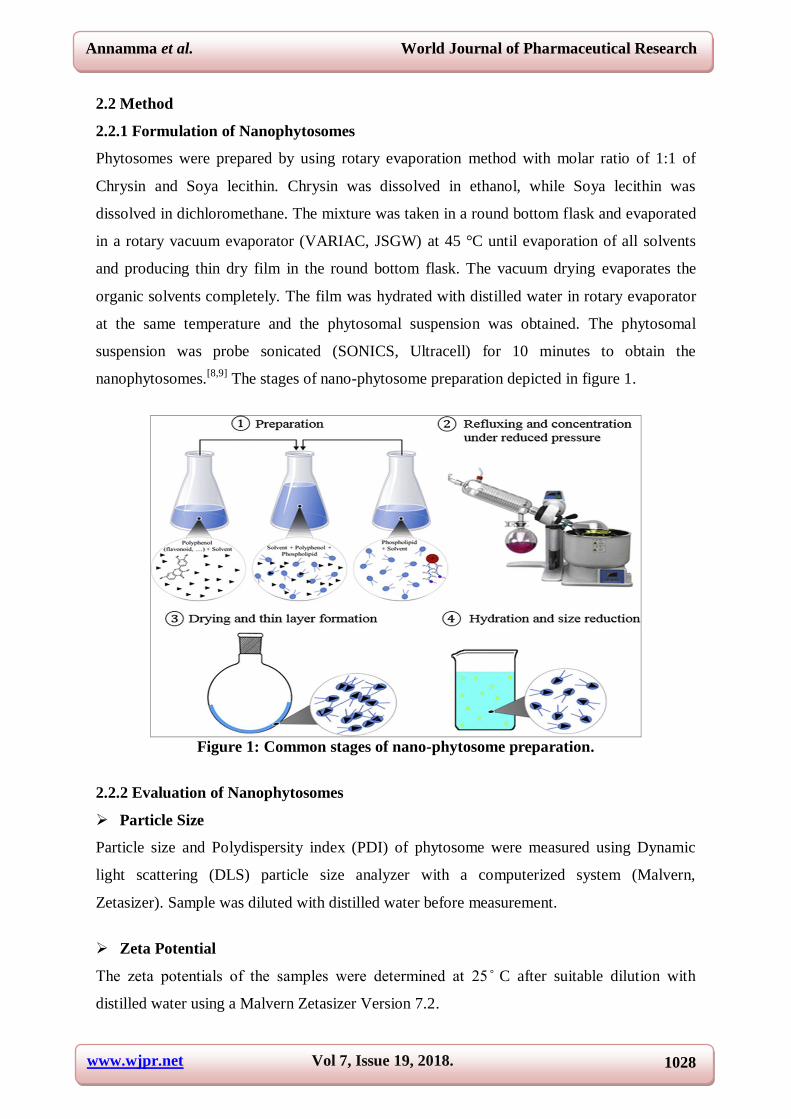
2.2.1 Formulation of Nanophytosomes
Phytosomes were prepared by using rotary evaporation method with molar ratio of 1:1 of
Chrysin and Soya lecithin. Chrysin was dissolved in ethanol, while Soya lecithin was
dissolved in dichloromethane. The mixture was taken in a round bottom flask and evaporated
in a rotary vacuum evaporator (VARIAC, JSGW) at 45 °C until evaporation of all solvents
and producing thin dry film in the round bottom flask. The vacuum drying evaporates the
organic solvents completely. The film was hydrated with distilled water in rotary evaporator
at the same temperature and the phytosomal suspension was obtained. The phytosomal
suspension was probe sonicated (SONICS, Ultracell) for 10 minutes to obtain the
nanophytosomes.[8,9]
The stages of nano-phytosome preparation depicted in figure 1.
Figure 1: Common stages of nano-phytosome preparation.
2.2.2 Evaluation of Nanophytosomes
Particle Size
Particle size and Polydispersity index (PDI) of phytosome were measured using Dynamic
light scattering (DLS) particle size analyzer with a computerized system (Malvern,
Zetasizer). Sample was diluted with distilled water before measurement.
Zeta Potential
he eta potentia of the amp e were determined at C after suitable dilution with
distilled water using a Malvern Zetasizer Version 7.2.

www.wjpr.net Vol 7, Issue 19, 2018. 1029
Annamma et al. World Journal of Pharmaceutical Research
Scanning Electron Microscopy (SEM)
Scanning electron microscopy (Model TESCAN VEGA 3 SBH) study was done to determine
the surface morphology, size and shape of prepared phytosomes formulation. Sample was
placed on an electron microscope brass stub and coated with gold in an ion sputter. Picture of
phytosomes were taken by random scanning of the stub.
FTIR
The FTIR spectra of Chrysin, physical mixture of Chrysin-Soya lecithin and Chrysin
nanophytosomes were taken using FTIR spectrophotometer (SPECTRUM 400). Infrared
spectra of the test samples were determined using the KBr disc technique. The FTIR
measurements were performed in the scanning range from 4000 to 400 cm-1.
Differential scanning calorimetry (DSC)
The DSC of Chrysin, and physical mixture of Chrysin–Soya lecithin and Chrysin
Nanophytosomes were analyzed in DSC analyzer (Q20 V24.10 Build 122). Each sample was
placed in an aluminum pan separately with heating and cooling rates of 10°C/min and
250°C/min, respectively. Measurements were performed over 50-300°C under nitrogen purge
at 50 ml/min.
Solubility
Solubility of chrysin and phytosome formulation was carried out in solvents like chloroform,
ethyl ether for eva uating whether flavonoid are incorporated into the phyto ome
structures. As flavonoid , in their pure forms, are insoluble in these solvents, if there
was any un- incorporated flavonoid to the phytosome structure it can be seen by
precipitation of flavonoid . hey convert to be soluble after incorporating into the
phytosomes. Hence, the phytosome generate a stable lipid compatible molecular
complex.[10]
Entrapment efficiency
The entrapment efficiency of phytosome was determined by centrifuging 2 mL of the
phytosome formulation at 1500 rpm for 30 min at room temperature. The supernatant was
taken carefully using pipette. Pure supernatant was then dissolved in ethanol to disrupt the
vesicles and appropriate dilution was made and measured using UV spectrophotometer
(SHIMADZU 1800) at 270 nm.[11]
The percentage of drug entrapped was determined using
the formula
www.wjpr.net Vol 7, Issue 19, 2018. 1030
Annamma et al. World Journal of Pharmaceutical Research
Entrapment efficiency % = Total drug added – un entrapped drug × 100
Total drug
Drug Content
Drug content of was determined by dissolving accurately weighed quantity of phytosome
dispersion in 10 ml ethanol. After suitable dilution absorbance was determined by UV
spectrophotometer (SHIMADZU 1800) at 270 nm and drug content was determined by using
the formula.[12]
Drug Content (%) = Actual drug content in Phytosomes × 100
Theoretical yield
In-vitro drug release study
• Preparation of egg membrane
From local department store egg was purchased. The egg yolk was separated carefully by
means of hole on the surface of the egg. After that the egg shell was immersed in HCl for 2
hours with constant stirring followed by the complete separation of egg membrane. The
membrane was washed with phosphate buffer pH 7.4 and further used for the experimental
work.
• Drug release through egg membrane
The invitro drug release studies were carried out in an open diffusion tube which was opened
at both the ends. The phytosome sample (2ml) was spread uniformly on the surface of egg
membrane and was fixed to the one end of tube such that the preparation occupies inner
circumference of the tube. The whole assembly was fixed in such a way that the lower end of
tube containing phytosome was just touched (1-2 mm deep) the surface of diffusion medium
i.e., 50ml pH 7.4 phosphate buffer contained in 100 ml beaker which was placed in water
bath and maintained at 37±2ºC. The egg membrane acts as a barrier between the phytosome
and pH 7.4 phosphate buffers (sink condition). A quantity of 2 ml samples were withdrawn
from receptor fluid at the time interval of 15min, 30min, 45min, 1, 2, 3, 4, 5, 6 hrs and 2 ml
phosphate buffer pH 7.4 was replaced at each time interval. The released drug was estimated
spectrophotometrically at 270 nm.
Release kinetics of in vitro drug release study
To understand the drug release kinetics and mechanism of drug release, the in vitro drug
www.wjpr.net Vol 7, Issue 19, 2018. 1031
Annamma et al. World Journal of Pharmaceutical Research
release study was fitted to mathematical equations of different kinetics model such zero order
(cumulative percentage of drug release versus time), first-order (log cumulative percentage of
drug remaining versus time), Higuchi (cumulative percentage of release versus square root of
time) and Korsmeyer- Peppas (log cumulative percentage of drug released versus log time)
equation models. The equation with the high regression coefficient (R2) for formulation will
be the best fit of release data.
3. RESULT AND DISCUSSION
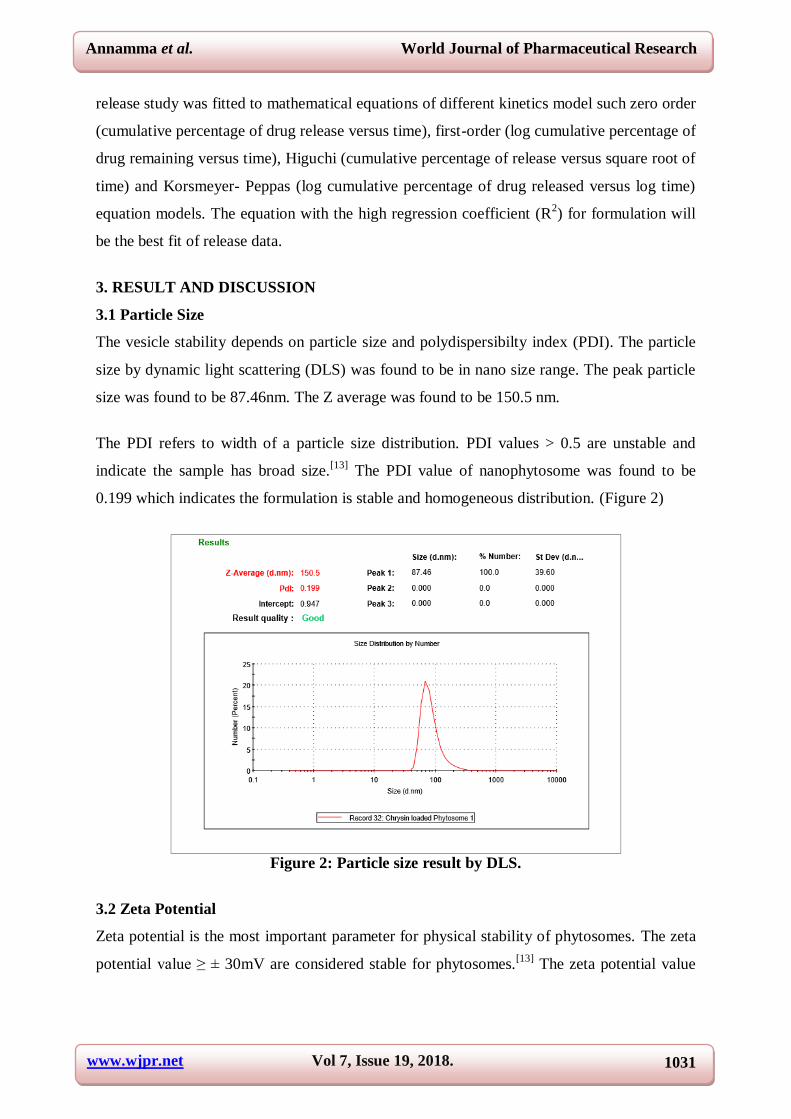
3.1 Particle Size
The vesicle stability depends on particle size and polydispersibilty index (PDI). The particle
size by dynamic light scattering (DLS) was found to be in nano size range. The peak particle
size was found to be 87.46nm. The Z average was found to be 150.5 nm.
The PDI refers to width of a particle size distribution. PDI values > 0.5 are unstable and
indicate the sample has broad size.[13]
The PDI value of nanophytosome was found to be
0.199 which indicates the formulation is stable and homogeneous distribution. (Figure 2)
Figure 2: Particle size result by DLS.
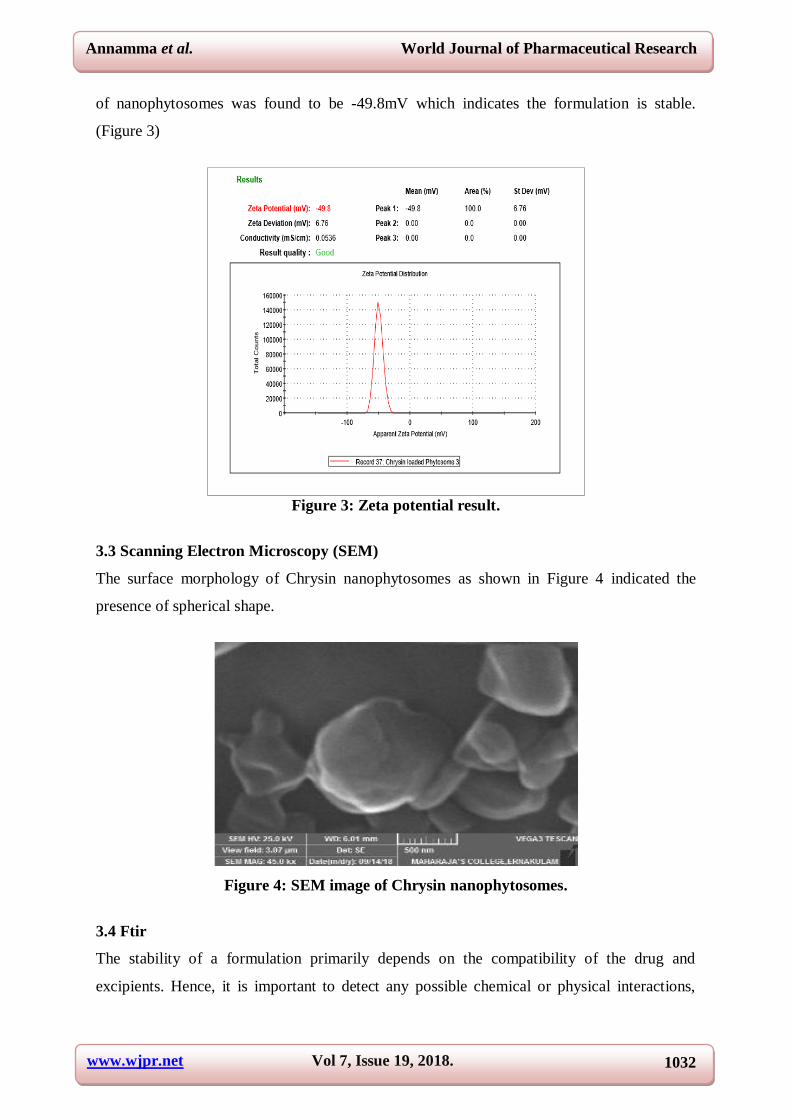
3.2 Zeta Potential
Zeta potential is the most important parameter for physical stability of phytosomes. The zeta
potential va ue ≥ ± 30mV are considered stable for phytosomes.[13]
The zeta potential value
www.wjpr.net Vol 7, Issue 19, 2018. 1032
Annamma et al. World Journal of Pharmaceutical Research
of nanophytosomes was found to be -49.8mV which indicates the formulation is stable.
(Figure 3)
Figure 3: Zeta potential result.
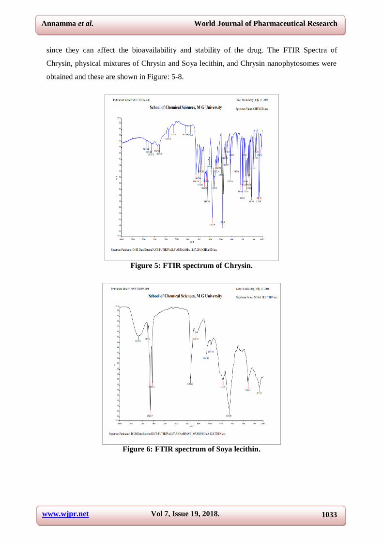
3.3 Scanning Electron Microscopy (SEM)
The surface morphology of Chrysin nanophytosomes as shown in Figure 4 indicated the
presence of spherical shape.
Figure 4: SEM image of Chrysin nanophytosomes.
3.4 Ftir
The stability of a formulation primarily depends on the compatibility of the drug and
excipients. Hence, it is important to detect any possible chemical or physical interactions,
www.wjpr.net Vol 7, Issue 19, 2018. 1033
Annamma et al. World Journal of Pharmaceutical Research
since they can affect the bioavailability and stability of the drug. The FTIR Spectra of
Chrysin, physical mixtures of Chrysin and Soya lecithin, and Chrysin nanophytosomes were
obtained and these are shown in Figure: 5-8.
Figure 5: FTIR spectrum of Chrysin.
Figure 6: FTIR spectrum of Soya lecithin.
www.wjpr.net Vol 7, Issue 19, 2018. 1034
Annamma et al. World Journal of Pharmaceutical Research
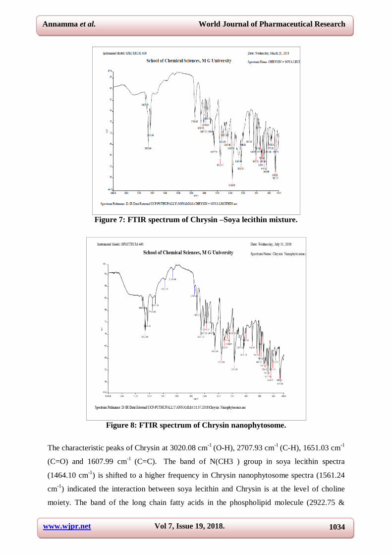
Figure 7: FTIR spectrum of Chrysin –Soya lecithin mixture.
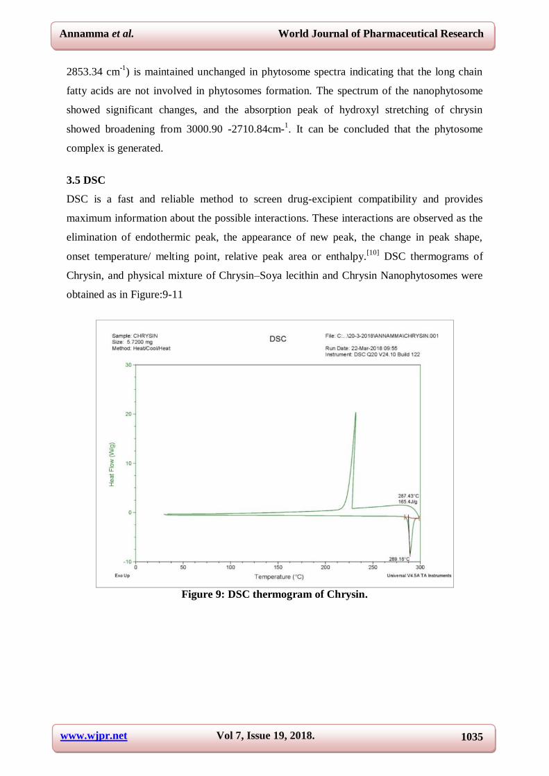
Figure 8: FTIR spectrum of Chrysin nanophytosome.
The characteristic peaks of Chrysin at 3020.08 cm-1
(O-H), 2707.93 cm-1
(C-H), 1651.03 cm-1
(C=O) and 1607.99 cm-1
(C=C).
The band of N(CH3 ) group in soya lecithin spectra
(1464.10 cm-1
) is shifted to a higher frequency in Chrysin nanophytosome spectra (1561.24
cm-1
) indicated the interaction between soya lecithin and Chrysin is at the level of choline
moiety. The band of the long chain fatty acids in the phospholipid molecule (2922.75 &
www.wjpr.net Vol 7, Issue 19, 2018. 1035
Annamma et al. World Journal of Pharmaceutical Research
2853.34 cm-1
) is maintained unchanged in phytosome spectra indicating that the long chain
fatty acids are not involved in phytosomes formation. The spectrum of the nanophytosome
showed significant changes, and the absorption peak of hydroxyl stretching of chrysin
showed broadening from 3000.90 -2710.84cm-1. It can be concluded that the phytosome
complex is generated.
3.5 DSC
DSC is a fast and reliable method to screen drug-excipient compatibility and provides
maximum information about the possible interactions. These interactions are observed as the
elimination of endothermic peak, the appearance of new peak, the change in peak shape,
onset temperature/ melting point, relative peak area or enthalpy.[10]
DSC thermograms of
Chrysin, and physical mixture of Chrysin–Soya lecithin and Chrysin Nanophytosomes were
obtained as in Figure:9-11
Figure 9: DSC thermogram of Chrysin.
www.wjpr.net Vol 7, Issue 19, 2018. 1036
Annamma et al. World Journal of Pharmaceutical Research
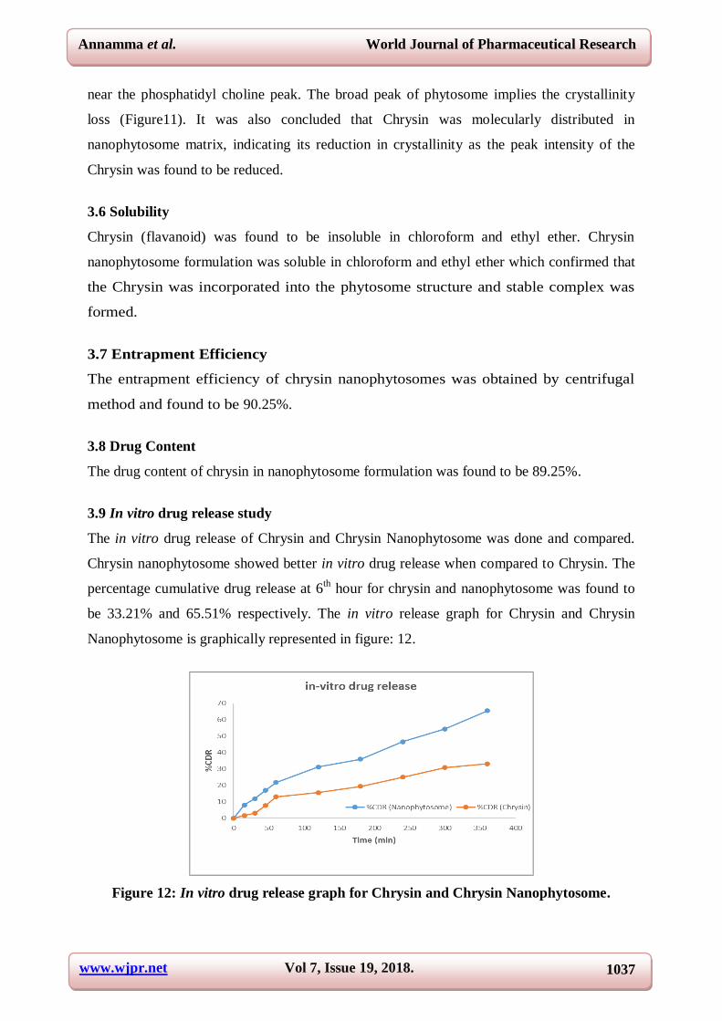
Figure 10: DSC thermogram of Chrysin-Soya lecithin mixture.
Figure 11: DSC thermogram of Chrysin nanophytosomes.
The endothermic peak of Chrysin was observed at 289.18ºC corresponding to its crystalline
nature (Figure: 9 ). In DSC thermogram of physical mixture of Chrysin- Soya lecithin, the
endothermal peaks of both are still detectable but shifted towards lower temperatures (Figure
10). On the other hand, the DSC thermogram of Chrysin nanophytosomes, a broad peak
appeared at 100.51ºC. This peak may be attributed to the formation of a new complex peak
www.wjpr.net Vol 7, Issue 19, 2018. 1037
Annamma et al. World Journal of Pharmaceutical Research
near the phosphatidyl choline peak. The broad peak of phytosome implies the crystallinity
loss (Figure11). It was also concluded that Chrysin was molecularly distributed in
nanophytosome matrix, indicating its reduction in crystallinity as the peak intensity of the
Chrysin was found to be reduced.
3.6 Solubility
Chrysin (flavanoid) was found to be insoluble in chloroform and ethyl ether. Chrysin
nanophytosome formulation was soluble in chloroform and ethyl ether which confirmed that
the Chrysin was incorporated into the phytosome structure and stable complex was
formed.
3.7 Entrapment Efficiency
The entrapment efficiency of chrysin nanophytosomes was obtained by centrifugal
method and found to be 90.25%.
3.8 Drug Content
The drug content of chrysin in nanophytosome formulation was found to be 89.25%.
3.9 In vitro drug release study
The in vitro drug release of Chrysin and Chrysin Nanophytosome was done and compared.
Chrysin nanophytosome showed better in vitro drug release when compared to Chrysin. The
percentage cumulative drug release at 6th hour for chrysin and nanophytosome was found to
be 33.21% and 65.51% respectively. The in vitro release graph for Chrysin and Chrysin
Nanophytosome is graphically represented in figure: 12.
Figure 12: In vitro drug release graph for Chrysin and Chrysin Nanophytosome.
www.wjpr.net Vol 7, Issue 19, 2018. 1038
Annamma et al. World Journal of Pharmaceutical Research
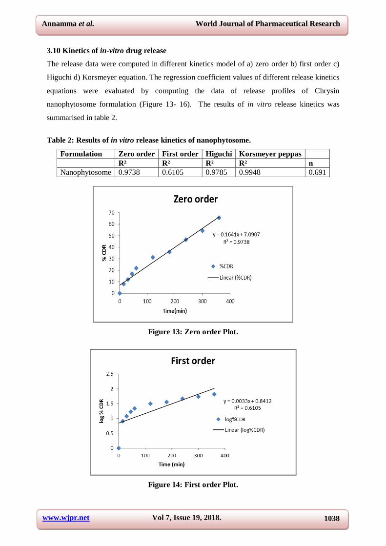
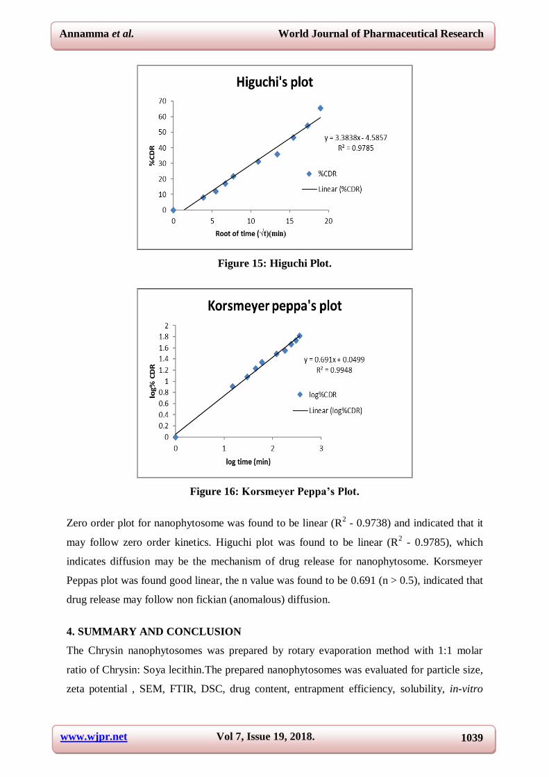
3.10 Kinetics of in-vitro drug release
The release data were computed in different kinetics model of a) zero order b) first order c)
Higuchi d) Korsmeyer equation. The regression coefficient values of different release kinetics
equations were evaluated by computing the data of release profiles of Chrysin
nanophytosome formulation (Figure 13- 16). The results of in vitro release kinetics was
summarised in table 2.
Table 2: Results of in vitro release kinetics of nanophytosome.
Formulation Zero order First order Higuchi Korsmeyer peppas
R² R² R² R² n
Nanophytosome 0.9738 0.6105 0.9785 0.9948 0.691
Figure 13: Zero order Plot.
Figure 14: First order Plot.
www.wjpr.net Vol 7, Issue 19, 2018. 1039
Annamma et al. World Journal of Pharmaceutical Research
Figure 15: Higuchi Plot.
Figure 16: Korsmeyer Peppa’s Plot.
Zero order plot for nanophytosome was found to be linear (R2 - 0.9738) and indicated that it
may follow zero order kinetics. Higuchi plot was found to be linear (R2 - 0.9785), which
indicates diffusion may be the mechanism of drug release for nanophytosome. Korsmeyer
Peppas plot was found good linear, the n value was found to be 0.691 (n > 0.5), indicated that
drug release may follow non fickian (anomalous) diffusion.
4. SUMMARY AND CONCLUSION
The Chrysin nanophytosomes was prepared by rotary evaporation method with 1:1 molar
ratio of Chrysin: Soya lecithin.The prepared nanophytosomes was evaluated for particle size,
zeta potential , SEM, FTIR, DSC, drug content, entrapment efficiency, solubility, in-vitro
www.wjpr.net Vol 7, Issue 19, 2018. 1040
Annamma et al. World Journal of Pharmaceutical Research
drug release and release kinetics. The particle size by DLS was found to be in nano size
range. The peak particle size was found to be 87.46nm. The Z average was found to be
150.5nm.The PDI of nanophytosome was found to be 0.199 which indicates the formulation
is stable and homogeneous distribution. The zeta potential was found to be -49.8 m V which
indicates the formulation is stable. The surface morphology of Chrysin nanophytosomes was
done by scanning electron microscopy which indicated the presence of spherical shape. The
percentage entrapment efficiency of Chrysin Nanophytosome formulation was found to be
90.25%. The drug content of Chrysin in nanophytosome formulation was found to be
89.25%. The in vitro drug release of Chrysin and Chrysin Nanophytosome was done and
compared. Chrysin nanophytosome showed better in vitro drug release when compared to
Chrysin. The percentage cumulative drug release at 6th hour for Chrysin and nanophytosome
was found to be 33.21% and 65.51% respectively. The kinetics of the drug release was found
that it predominately follows zero order and Higuchi kinetics with non fickian diffusion
mechanism.
REFERENCES
1. Bilia, A.R., Isacchi, B., Righeschi, C., Guccione, C. and Bergonzi, M.C. Flavonoids
Loaded in Nanocarriers: An Opportunity to Increase Oral Bioavailability and Bioefficacy.
Food and Nutrition Sciences, 2014; 5: 1212-1227.
2. Jung J. Emerging Utilization of Chrysin Using Nanoscale Modification. J Nanomater,
2016; 1-7.
3. Acharya NS, Parihar GV, Acharya SR. Phytosomes: Novel approach for delivering herbal
extract with improved bioavailability. Int J pharm scien, 2011; 2(1): 144-60.
4. Kadu AS, Apte M. Phyto ome : A Nove Approach to Enhance the bioavai abi ity of
phytoconstitutent. Asian J pharm, 2017; 11(2): 453-461.
5. Jing Li, Xuling W, Ting Z, Chunling W, Zhenjun H, Xiang L, et al. A review on
phospholipids and their main applications in drug delivery systems. Asian J pharm sci.,
2015; 10(2): 81-98.
6. Zeinali M, Rezaee SA, Hosseinzadeh H. An overview on immunoregulatory and anti-
inflammatory properties of chrysin and flavonoids substances. Biomed Pharmacother,
2017; 92: 998–1009.
7. Mani R, Natesan V. Chrysin: Sources, beneficial pharmacological activities, and
molecular mechanism of action. Phytochemistry, 2018; 145: 187–96.
www.wjpr.net Vol 7, Issue 19, 2018. 1041
Annamma et al. World Journal of Pharmaceutical Research
8. Rasaie S, Ghanbarzadeh S, Mohammadi M, Hamishehkar H. Nano Phytosomes of
Quercetin: A Promising Formulation for Fortification of Food Products with
Antioxidants. Pharm Sci., 2014; 20: 96–101.
9. Hooresfand Z, Ghanbarzadeh S, Hamishehkar H. Preparation and Characterization of
Rutin-loaded Nanophytosomes. Pharm Sci., 2015; 21(3): 145–51.
10. Ghanbarzadeh B, Babazadeh A, Hamishehkar H. Nano-phytosome as a potential food-
grade delivery system. Food Biosci, 2016; 15: 126–35.
11. Anwar E, Farhana N. Formulation and Evaluation of Phytosome-Loaded Maltodextrin-
Gum Arabic Microsphere System for Delivery of Camellia sinensis Extract. J Young
Pharm, 2018; 10(2): s56-s62.
12. Amudha S., Manna P K, Jeganathan N.S . Formulation and evaluation of capsules of
Syzygium cumini phytosomes. J Pharm Sci Innov, 2018; 7(3): 70–8.
13. Gnananath K, Nataraj KS, Rao BG. Phospholipid complex technique for superior
bioavailability of phytoconstituents. Adv Pharm Bull, 2017; 7(1): 35–42.