Embed Size (px)
Citation preview
Formulation of photoreactive drug substances:
The role of excipients and type of preparation
Milica Vukićević
Thesis submitted for the degree of Philosophiae Doctor
School of Pharmacy Faculty of Mathematics and Natural Sciences
University of Oslo 2015
© Milica Vuki evi , 2015 Series of dissertations submitted to the Faculty of Mathematics and Natural Sciences, University of Oslo No. 1640 ISSN 1501-7710 All rights reserved. No part of this publication may be reproduced or transmitted, in any form or by any means, without permission. Cover: Hanne Baadsgaard Utigard. Printed in Norway: AIT Oslo AS. Produced in co-operation with Akademika Publishing. The thesis is produced by Akademika Publishing merely in connection with the thesis defence. Kindly direct all inquiries regarding the thesis to the copyright holder or the unit which grants the doctorate.
iii
Acknowledgements This thesis presents the results of the work carried out at the School of Pharmacy, Faculty of
Mathematics and Natural Sciences, University of Oslo, in the period 2010-2014. A portion of
the experiments described in Paper I was performed at the Department of Electronics and
Telecommunications, Norwegian University of Science and Technology.
Foremost, I would like to thank my main supervisor, Professor Hanne Hjorth Tønnesen, for
introducing me to the field of drug photoreactivity and for making this work possible. Thank
you for your inspiration, guidance and for sharing the highs and lows over the course of my
research. I would also like to acknowledge Dr. Anne Bee Hegge for being the co-supervisor
of this project. Anne Bee, I appreciate all of the productive discussions we had and your
invaluable support in the lab. I would as well like to thank Professor Solveig Kristensen for
her help and advice.
I am grateful to my co-authors Professor Lise Randeberg, Professor Thomas Tybell and Jos
Boschker from the Department of Electronics and Telecommunications, Norwegian
University of Science and Technology for enabling me to visit their laboratories and helping
me to complete Paper I. I would like to thank Дp. Пpeдpaг Byлић for welcoming me in
Laboratory of Crystallography, Faculty of Mining and Geology, University of Belgrade and
performing part of the experiments within the Paper III. Thank you for being a helpful
colleague and a great friend! I am deeply grateful to Professor Jan Karlsen for invaluable
advice during the writing of this thesis, for your encouragement and for cheering me up along
the way. In addition, I thank Director Henrik Schultz for encouragement and help.
My appreciation also goes to Ivar Grove, Tove Larsen and Halvor Aandal for help with
problems of all kinds. I especially want to thank Bente Amalie Breiby, not only for her
priceless help in the laboratories, but also for making our daily work more joyful. You are a
true part of the team!
I would like to thank all of the past and present members of the PharmaLuxLab research
group and my colleagues from the School of Pharmacy for their support and friendship.
Afonso, Brianna, Helene, Julia, Kristine, Marianne, Mollie, Sanko, Sara, Victoria and Wai
iv
Lam; thank you for precious advice, scientific and technical help. Thank you for being there
for me through all of the frustrating moments, as well as all the fun we had together.
My dearest friends, Angela, Kateřina, Lilia and Teresa, I cannot express how wonderful you
have been and how appreciative I am of your support during this journey. Thanks for putting
up with my moods and my unconventional expressions of love! You helped me through many
hard times with your advice (both academic and personal) but most of all thank you for
always finding a way to make me laugh. Benjy and Jan, thank you for helping me with the
writing of the papers and this thesis. Not only that you provided thorough proofreading,
scientific and technical help, but you also cooked for me and took me out when I needed
cheering up. I am truly fortunate to have friends like you! I am deeply grateful to Marijana,
Mladen and Natalia for their kindness and care.
Finally I would like to thank my parents and my brother. You were the first teachers I ever
had and you continue to inspire and encourage through life. Without your endless love,
patience and faith in me, this work would have never been possible!
Contents Acknowledgements ................................................................................................................... iii
List of publications ..................................................................................................................... 1
Abbreviations ............................................................................................................................. 2
1. Aim of the project ............................................................................................................... 3
2. Definitions ........................................................................................................................... 4
3. Introduction ......................................................................................................................... 5
3.1 Photoreactivity of the drug substance ............................................................................. 7
3.1.1 Direct and indirect photoreactions ............................................................................... 8
3.1.2 Outcome of the photoreactivity of the drug substance ................................................ 9
3.2 Formulation of photoreactive drug substances .............................................................. 15
3.2.1 Influence of the formulation on the photostability .................................................... 15
3.2.2 Formulation of products intended for aPDT .............................................................. 19
3.2.3 Influence of excipients and type of preparation on protein-PDS interaction ............ 23
3.3 Model photoreactive drug substances ........................................................................... 24
3.3.1 Riboflavin as model of a photolabile drug substance ................................................ 24
3.3.2 Curcumin as the model of a photosensitizer for aPDT .............................................. 26
4. General experimental conditions ...................................................................................... 33
4.1 Materials ........................................................................................................................ 33
4.2 Preparation of samples .................................................................................................. 33
4.3 Methods ......................................................................................................................... 34
5. Results and discussion ...................................................................................................... 35
5.1 The physical state of the drug substance in the solid preparation ................................. 35
5.1.1 The physical state of the riboflavin as a bulk substance applied in tablets ............... 35
5.1.2 Formulation of curcumin as a solid dispersion .......................................................... 35
5.2 Photoreactivity of the drug substance ........................................................................... 40
5.2.1 Photoreactivity of riboflavin in the solid state ........................................................... 40
5.2.2 Photoreactivity of curcumin in solution .................................................................... 43
5.3 Physical stability of the curcumin in solution ............................................................... 45
5.3.1 Supersaturated solutions of curcumin prepared from the solid dispersions .............. 45
5.3.2 Precipitation inhibition of curcumin in a supersaturated solution ............................. 45
5.4 Preparations in biorelevant media: bacterial phototoxicity of curcumin and interaction with human serum albumin ...................................................................................................... 47
5.4.1 Phototoxicity of curcumin towards Gram-positive and Gram-negative bacteria ...... 48
5.4.2 Formulation dependent binding of curcumin to human serum albumin.................... 50
6. Conclusion ........................................................................................................................ 52
References ................................................................................................................................ 53
1
List of publications The present work is based on the following papers, which are referred to by their Roman
numerals:
Paper I
Vukicevic M, Randeberg LL, Boschker JE, Tybell T, Tønnesen HH (2014), Influence of
crystal modification on the photoinduced color change in riboflavin, Die Pharmazie 69, 117-
124
Paper II
Hegge AB,Vukicevic M, Bruzell E, Kristensen S, Tønnesen HH (2013), Solid dispersions for
preparation of phototoxic supersaturated solutions for antimicrobial photodynamic therapy
(aPDT) Studies on curcumin and curcuminoides L, Eur J Pharm Biopharm, 83, 1, 95-105
Paper III
Vukicevic M, Hegge AB, Vulic P, Tønnesen HH (2014), Poloxamer-based curcumin solid
dispersions for ex tempore preparation of supersaturated solutions intended for antimicrobial
photodynamic therapy Studies on curcumin and curcuminoids LIII, Pharm Dev Technol,
accepted for publication, available online, doi:10.3109/10837450.2014.930489
Paper IV
Vukicevic M, Tønnesen HH (2015), Interaction between curcumin and human serum albumin
in the presence of excipients and the effect of binding on curcumin photostability Studies on
curcumin and curcuminoides LV, Pharm Dev Technol, accepted for publication, available
online, doi:10.3109/10837450.2015.1016618
2
Abbreviations aPDT antimicrobial photodynamic therapy PDT photodynamic therapy
C. albicans Candida albicans PEG 400 polyethylene glycol 400
CD cyclodextrin PF127 Pluronic® F127
D-D reaction Dye-Dye reaction PF68 Pluronic® F68
DMSO dimethyl sulfoxide PI precipitation inhibitor
DSC diferentail scanning calorimetry PP123 Pluronic® P123
E. coli Escherichia coli PS photosensitizer
E. faecalis Enterococcus faecalis PSt photostability
ESIPT excited state intramolecular proton transfer PVP polyvinylpyrrolidone
Ex excipients RFbs riboflavin biosynthetic
FT-IR Fourier transform infrared spectroscopy RFdh riboflavin dihydrate
G- Gram-negative RFs riboflavin synthetic
G+ Gram-positive ROS reactive oxygen species
HA hyaluronic acid SD solid dispersion
HME hot melt extrusion SEM scanning electron microscopy
HPLC high-performance liquid chromatography SS solid state
HPMC hydroxypropyl methylcellulose S. aureus Staphylococcus aureus
HP-β-CD hydroxypropyl-β-cyclodextrin Tg glass transition temperature
HP-γ-CD hydroxypropyl-γ-cyclodextrin UV-Vis ultraviolet–visible
PDS photoreactive drug substance XRD X-ray diffractometry
3
1. Aim of the project
The overall aim of the project was to investigate how the preparation and formulation process
influence the photostability and phototoxicity of respectively a photolabile drug molecule in
the solid state and a photosensitizer.
The specific aims of the work were to:
investigate the influence of the solid state properties (crystal modification) on the
photoreactivity of the photolabile drug substance riboflavin (Paper I)
evaluate the effect of the type of preparation and excipients on the photodynamic
effect of the photosensitizer curcumin (Papers II and III)
study the effect of the excipients on the interaction of the photosensitizer curcumin
with a biomolecule (human serum albumin) and the photoreactivity of the
photosensitizer in biorelevant media (Paper IV).
4
2. Definitions
A drug substance is an active ingredient contained in a formulated preparation intended to
provide therapeutic effect directly or indirectly.
Pharmaceutical formulation is the process of combining different chemical substances,
including the drug substance (active ingredient) in an appropriate fashion, to produce a final
preparation.
A formulated preparation, typically a product of a drug substance and excipients (non-drug
substances), is made to administrate a drug substance to the body. This term is used
interchangeably with product, final product, and dosage form throughout this work.
A photosensitizer (PS) is a light-activated drug substance which, upon absorption of light,
induces a chemical or physical alteration of another chemical entity. Some photosensitizers
are employed therapeutically such as in photodynamic therapy of cancer (PDT) or in the
treatment of bacterial infections (aPDT).
The photostability of drug substances and products is understood not only as the degradation
caused by exposure to radiation, but also processes such as radical formation, energy transfer
and luminescence, within this work. The term is used interchangeable with photoreactivity
throughout this work.
5
3. Introduction
The general aim of pharmaceutical formulation work is to achieve safe, efficient, reproducible
and convenient administration of the drug substance to the body. Excipients are added in
order to e.g., solubilize, stabilize, suspend, thicken, preserve, emulsify, modify dissolution,
improve the compressibility or the flavor of the drug substance (1). Excipients, however, need
to be compatible with the drug substance for the final product to be safe, stable and efficient.
Most drug substances are white or slightly colored, i.e., they absorb radiation in the UV or
visible part of the electromagnetic spectrum (2). The photoreactivity of such drug substances
can be either beneficial or undesirable. In some situations, photoreactivity can be used in
photoactivated therapeutic systems. Conversely, many drugs are degraded due to exposure to
visible and UV radiation. We therefore need to control the photoreactivity of drug substances.
This creates additional complexity during the formulation work.
The type of preparation and the selected excipients can alter the photostability of the
photoreactive drug substance (PDS) and affect the efficiency and safety of the final product
(3-5). Photostability is recognized as an integral part of the stability studies during the drug
development; photostability testing of the new drug substances and products are addressed in
the ICH Q1B document of ICH Harmonised Tripartite Guideline (ICH stands for International
Conference on Harmonisation of Technical Requirements for Registration of Pharmaceuticals
for Human Use) (6).
Therapies based on light-activated substances have been established, such as photodynamic
therapy, PUVA therapy (psoralen based UVA therapy) or drug release via light-sensitive drug
delivery systems. To maximize the efficiency and safety of such substances or systems, novel
preparations tailored for (a) the type of condition that is treated, (b) the site of application and
(c) the therapy protocol, need to be developed.
The different aspects of the formulation of photoreactive drug substances that will be
addressed in this work are the photostability of the drug substance in both solid and liquid
form, preparations specifically designed to combat microbials (aPDT therapy) and
formulation dependent bacterial phototoxicity, and interaction with human serum albumin
(HSA). The structure of the thesis in relation to these issues is presented in Figure 1.
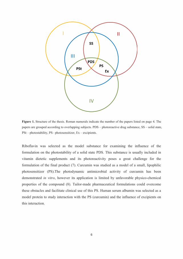
6
Figure 1. Structure of the thesis. Roman numerals indicate the number of the papers listed on page 4. The
papers are grouped according to overlapping subjects. PDS – photoreactive drug substance, SS – solid state,
PSt – photostability, PS– photosensitizer, Ex – excipients.
Riboflavin was selected as the model substance for examining the influence of the
formulation on the photostability of a solid state PDS. This substance is usually included in
vitamin dietetic supplements and its photoreactivity poses a great challenge for the
formulation of the final product (7). Curcumin was studied as a model of a small, lipophilic
photosensitizer (PS).The photodynamic antimicrobial activity of curcumin has been
demonstrated in vitro, however its application is limited by unfavorable physico-chemical
properties of the compound (8). Tailor-made pharmaceutical formulations could overcome
these obstacles and facilitate clinical use of this PS. Human serum albumin was selected as a
model protein to study interaction with the PS (curcumin) and the influence of excipients on
this interaction.
7
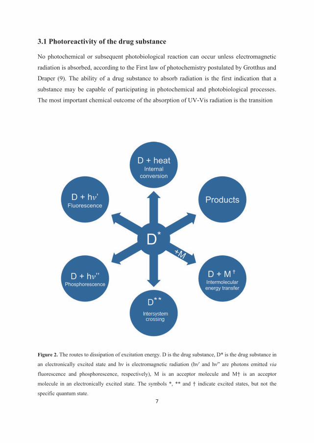
3.1 Photoreactivity of the drug substance
No photochemical or subsequent photobiological reaction can occur unless electromagnetic
radiation is absorbed, according to the First law of photochemistry postulated by Grotthus and
Draper (9). The ability of a drug substance to absorb radiation is the first indication that a
substance may be capable of participating in photochemical and photobiological processes.
The most important chemical outcome of the absorption of UV-Vis radiation is the transition
Figure 2. The routes to dissipation of excitation energy. D is the drug substance, D* is the drug substance in
an electronically excited state and hν is electromagnetic radiation (hν’ and hν’’ are photons emitted via
fluorescence and phosphorescence, respectively), M is an acceptor molecule and M† is an acceptor
molecule in an electronically excited state. The symbols *, ** and † indicate excited states, but not the
specific quantum state.
8
of an electron from an orbital in the ground state into a higher energy orbital resulting in an
electronically excited-state. The molecule in its excited state can exhibit a dramatically
different reactivity from the ground state, not only because it possesses excess energy but also
as a result of the new electronic arrangement (10). The energy associated with radiation in the
UV-Vis region is of the same magnitude as certain bond energies and thus electronic
excitation can cause photochemical degradation of the compound. Furthermore, the absorbed
energy can be transferred to other, non-absorptive molecules (e.g., oxygen) as in
photodynamic therapy. The resulting products are usually chemically distinct from the
thermal degradation products of the drug molecule. Excess energy can also be lost by several
other radiative or non-radiative routes (Fig. 2). As a result of intersystem crossing and
intermolecular energy transfer, new excited electronic states can be formed that can take part
in the processes shown in the diagram (Fig. 2).
3.1.1 Direct and indirect photoreactions
Direct photoreactions
Absorption of UV-Vis radiation may lead to chemical changes of the drug substance, i.e.,
reactions in which the reactant and products differ in chemical identity rather than in their
state of excitation. This process is referred to as a direct photoreaction.
The reactivity of the molecule in an excited state is governed by the excess energy it
possesses, the intrinsic reactivity of the electronic arrangement and the relative efficiencies of
the different competing pathways for loss of the excess energy (9, 11). Therefore, even if the
drug substance absorbs radiation strongly, a direct photoreaction is less likely to occur if it
fluoresces or efficiently transfers the energy to another molecule present in the preparation.
The excited state of the molecule can exhibit markedly altered chemistry compared to the
molecule in the ground state, as mentioned above. Bond lengths, bond orders and bond angles
in excited state molecules may be considerably different from their corresponding ground
states as a consequence of the redistribution of the electron density (10).
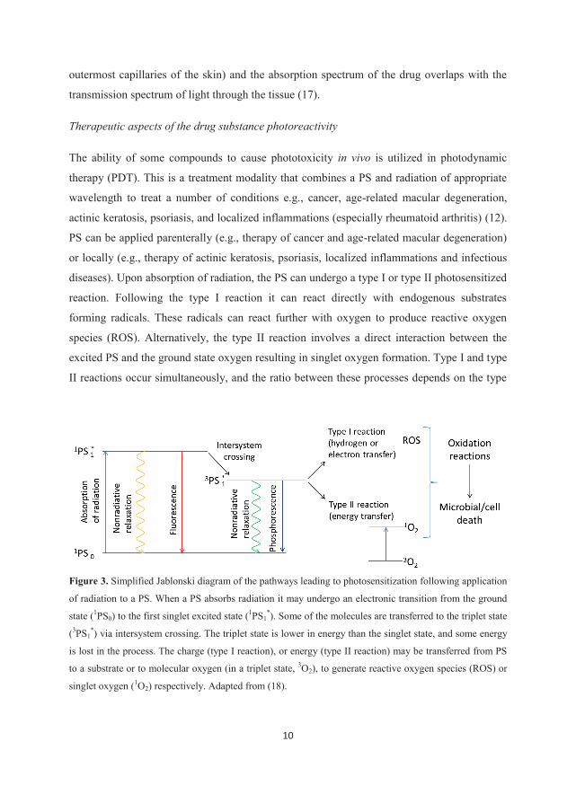
Indirect photoreactions (photosensitized reactions)
In the case of an indirect or sensitized photoreaction there is a transfer of excitation energy to
a molecule other than the compound which initially absorbs the radiation. The compound that
absorbs the radiation, the photosensitizer, is initially transformed from its ground state (singlet
9
state) into a relatively long-lived excited triplet state via a short-lived excited singlet state (Fig.
2, Intersystem crossing; Fig. 3). The excited triplet state can mediate a photosensitized
reaction due to the long lifetime and bi-radical nature with unpaired electron spins (11). In the
type I photosensitized reactions the sensitizer can transfer an electron, including simultaneous
transfer of a proton corresponding to the transfer of a hydrogen atom, to the molecules in its
vicinity resulting in a free radical reaction. Energy transfer from the excited triplet state of
sensitizer to the spin-matched oxygen in the ground state leads to the formation of excited
singlet oxygen. This process is regarded as type II photosensitized reaction (Fig. 2,
intermolecular energy transfer; Fig. 3) (12). During the energy transfer process the PS is
simultaneously brought back to its singlet ground state where it in principle can take part in
further sensitization cycles (13).
3.1.2 Outcome of the photoreactivity of the drug substance
Outcome of the photoreactivity on in vitro stability and adverse drug reactions in vivo
The photodecomposition of a large number of drug molecules in both the solution (14) and
the solid state (15) has been reported. Loss of the potency can occur as a result of in vitro
instability, leading to inactive products. Furthermore, PDS degradation may lead to adverse
drug reactions due to formation of degradation products during storage (2). However, the
inherent photoreactivity of the drug substance is not the only factor determining the storage
stability of the final product. The substance will only degrade if it comes in contact with
radiation of an appropriate wavelength. The overlap of the absorption spectrum of the drug
substance with the spectral output of the incident light is described as the overlap integral, and
it determines the rate of the photoreaction (11, 16). Basic understanding of photoreactivity of
the compound is required to provide information for handling, packaging, labeling and use of
the drug substance and the final product. In most of the cases suitable packaging provides
adequate protection for PDS. However, sometimes a modification of the preparation must be
considered.
The in vivo light-induced interactions of the drug substance with endogenous substrates can
lead to adverse photosensitivity effects. Phototoxicity, one type of photosensitivity effect, is
defined as an alteration of cell function by an interaction between the phototoxic compound
and nonionizing radiation. A phototoxic effect can only occur if the drug substance or the
phototoxic metabolite is distributed near the body surface (e.g., the eye, skin, hair or
10
outermost capillaries of the skin) and the absorption spectrum of the drug overlaps with the
transmission spectrum of light through the tissue (17).
Therapeutic aspects of the drug substance photoreactivity
The ability of some compounds to cause phototoxicity in vivo is utilized in photodynamic
therapy (PDT). This is a treatment modality that combines a PS and radiation of appropriate
wavelength to treat a number of conditions e.g., cancer, age-related macular degeneration,
actinic keratosis, psoriasis, and localized inflammations (especially rheumatoid arthritis) (12).
PS can be applied parenterally (e.g., therapy of cancer and age-related macular degeneration)
or locally (e.g., therapy of actinic keratosis, psoriasis, localized inflammations and infectious
diseases). Upon absorption of radiation, the PS can undergo a type I or type II photosensitized
reaction. Following the type I reaction it can react directly with endogenous substrates
forming radicals. These radicals can react further with oxygen to produce reactive oxygen
species (ROS). Alternatively, the type II reaction involves a direct interaction between the
excited PS and the ground state oxygen resulting in singlet oxygen formation. Type I and type
II reactions occur simultaneously, and the ratio between these processes depends on the type
Figure 3. Simplified Jablonski diagram of the pathways leading to photosensitization following application
of radiation to a PS. When a PS absorbs radiation it may undergo an electronic transition from the ground
state (1PS0) to the first singlet excited state (1PS1*). Some of the molecules are transferred to the triplet state
(3PS1*) via intersystem crossing. The triplet state is lower in energy than the singlet state, and some energy
is lost in the process. The charge (type I reaction), or energy (type II reaction) may be transferred from PS
to a substrate or to molecular oxygen (in a triplet state, 3O2), to generate reactive oxygen species (ROS) or
singlet oxygen (1O2) respectively. Adapted from (18).
11
of sensitizer used, the concentrations of substrate and oxygen, as well as the binding affinity
of the sensitizer for the substrate (12). The ROS and singlet state oxygen can kill microbial or
cancer cells by inflicting damage on biomolecules such as proteins, unsaturated lipids,
steroids and nucleic acids (Fig. 3).
Antimicrobial photodynamic therapy
PDT applied in the treatment of microbial infections is termed antimicrobial photodynamic
therapy (aPDT). The delivery of the radiation to living tissue is a localized process and
therefore aPDT is limited to localized as opposed to systemic infections (19). Furthermore,
due to the limited penetration depth of radiation through the tissue, aPDT is mostly applied at
areas of the body where radiation can be easily delivered (20). However, with the progress in
optical fibre technology even deep-seated infections could potentially be treated by aPDT (21).
The therapy is gaining increasing attention owing to the rise of microbial resistance to the
major families of antibiotic compounds (18). Many antibiotic-resistant microbial strains have
shown to be susceptible to aPDT due to the substantially different pathways of inactivation
compared to that of the antibiotic and chemotherapeutic agents. No selection of the
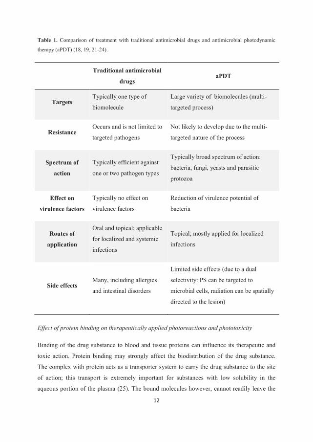
photoresistant species occurred even after multiple treatments (18, 19, 21, 22). A comparison
of some of the main advantages and disadvantages of the treatment with traditional
antimicrobial drugs and aPDT are presented in Table 1.
12
Table 1. Comparison of treatment with traditional antimicrobial drugs and antimicrobial photodynamic
therapy (aPDT) (18, 19, 21-24).
Traditional antimicrobial
drugs aPDT
Targets Typically one type of
biomolecule
Large variety of biomolecules (multi-
targeted process)
Resistance Occurs and is not limited to
targeted pathogens
Not likely to develop due to the multi-
targeted nature of the process
Spectrum of
action
Typically efficient against
one or two pathogen types
Typically broad spectrum of action:
bacteria, fungi, yeasts and parasitic
protozoa
Effect on
virulence factors
Typically no effect on
virulence factors
Reduction of virulence potential of
bacteria
Routes of
application
Oral and topical; applicable
for localized and systemic
infections
Topical; mostly applied for localized
infections
Side effects Many, including allergies
and intestinal disorders
Limited side effects (due to a dual
selectivity: PS can be targeted to
microbial cells, radiation can be spatially
directed to the lesion)
Effect of protein binding on therapeutically applied photoreactions and phototoxicity
Binding of the drug substance to blood and tissue proteins can influence its therapeutic and
toxic action. Protein binding may strongly affect the biodistribution of the drug substance.
The complex with protein acts as a transporter system to carry the drug substance to the site
of action; this transport is extremely important for substances with low solubility in the
aqueous portion of the plasma (25). The bound molecules however, cannot readily leave the
13
capillaries. Only the unbound molecules can be distributed to tissues and exhibit
pharmacological activity as well as toxic effects. Thus, depending on the drug substance and
the target, the high affinity for plasma proteins may be either beneficial or a drawback for
efficacy (26). Binding to plasma proteins also affects metabolism and elimination, since both
hepatic uptake and glomerular filtration are directly proportional to the free drug molecule
fraction present in the plasma (25, 26).
The factors that can affect binding of drug substance to plasma proteins are concurrent
administration of other drugs, excipients in the preparation, pathological conditions, age, and
sex (26).
Phototoxicity is dependent upon the concentration of the sensitizer and the overlap integral
between the sensitizer and the incident radiation at the site of action. Therefore, a prerequisite
for the phototoxic reaction to occur is that the drug substance with sensitizing properties is
distributed to the tissues that are exposed to radiation (17). Drug substances highly bound to
plasma proteins usually have low apparent volume of distribution (Vd) because they are
confined to the vascular space. Conversely, substances that are largely free in plasma are
generally available for distribution to tissues. However, some substances that are highly
bound to plasma proteins are bound with greater affinity to tissue proteins, resulting in a large
Vd value. The distribution of a drug substance is a function of both plasma protein binding
and tissue protein binding (26). Prolonged phototoxicity can be expected as the result of
delayed elimination of the drug substance with photosensitizing properties if the substance is
highly bound to plasma proteins.
The effect of the PS affinity for plasma proteins on the efficiency of photodynamic therapy
depends upon the route of administration and the target tissue. Following parenteral
administration PS needs to be delivered to the target tissue prior to exposure to radiation.
Pharmacokinetics and biodistribution of PS can vary greatly among the different types of
photosensitizers (27). These parameters can be influenced by the plasma protein binding as
discussed above. Moderate binding to the plasma proteins can promote distribution of PS in
the body whereas a high binding affinity may result in prolonged skin phototoxicity (28). The
interval between parenteral PS administration and radiation exposure needs to be adjusted
according to pharmacokinetic properties of PS to secure PDT efficacy (27).
14
When the PS reaches the target tissue it can be further influenced by the interaction with
plasma proteins. Binding to the plasma proteins have been correlated to both increased (29)
and decreased (30) uptake by the target cells. The PS-protein interaction often affects the
equilibrium between monomeric and aggregated species which is particularly important for
porphyrins and phthalocyanines. The interaction can promote monomerization or support self-
assembling (31). Binding-induced aggregation is not a desirable process since it leads to
decrease in photodynamic efficiency. Binding can alter the photophysical and photochemical
properties of PS. Generally, type I reactions are more likely to occur in the presence of
proteins and many PS are photodegraded much faster in medium rich in proteins or amino
acids (32). The absorption spectrum of PS (porphyrines) bound to proteins can significantly
deviate from the corresponding monomer in an aqueous solution. The spectral effects are due
to the altered microenvironment of PS – the polarity of the protein environment is lower than
that of water (31). The lifetime of the triplet state of PS bound to proteins recorded in the
absence of oxygen is much longer than the corresponding lifetime of the free molecules in
solution. However, production of singlet oxygen and ROS competes with other photophysical
and photochemical processes in the PS-protein complex (31). It has been suggested that
human serum albumin removes singlet oxygen from the sensitizing system. Sole binding of
the PS should not affect the generation of ROS under the condition that the PS does not
aggregate or is not bound close to protein constituents that quench the excited states
(electron/energy transfer processes) (31). The photostability of PS generally decreases in the
presence of proteins (33) and amino acids (34) compared to pure buffer.
The influence of physiological conditions on aPDT efficiency has been studied in vitro using
biological materials and ex vivo (19, 35). Proteins present in the medium typically protect
microorganisms towards aPDT (36-38). However, the actual impact of the protein rich media
on the aPDT effect was shown to be dependent both upon the type of PS and microorganism
(38, 39).
The following factors have been suggested as the cause of the observed effect of the proteins
on the photokilling of microorganisms (37):
proteins may absorb light and reduce the number of photons available to interact with
PS molecules thereby reducing the production of ROS
15
proteins may compete with the microorganisms for PS, decreasing the number of PS
molecules available to interact with target microorganisms
ROS have short lifetimes and, unless generated in close proximity of the cells would
be unlikely to produce any cytotoxic effect. Accordingly, proteins may protect
microorganisms from cytotoxic species generated in the supernatant. The culture
medium has also been reported to quench singlet oxygen and free radicals produced by
PS
PS bound to protein is therefore trapped and unavailable to interact with microorganisms
while the ROS produced by exposure to irradiation may more easily be quenched by proteins
complexed with PS. Moreover, direct reaction of plasma proteins with the photosensitizer
methylene blue was proposed as a reason for bleaching of the PS, low singlet oxygen yield,
and consequently the low photoinactivation of bacteria in plasma (40). On the other hand,
some of the products formed in photodynamic protein oxidation can sustain chain reactions
and therefore be highly detrimental to cells (28). This mechanism could explain the recently
reported improved aPDT effect of toluidine blue in serum and human serum albumin solution
compared to the buffer medium (41).
3.2 Formulation of photoreactive drug substances
Formulation of the PDS can be viewed from the two main points:
the influence of the formulation on the photostability of the PDS
the effect of the formulation on the efficiency of the light-activated drug substance
3.2.1 Influence of the formulation on the photostability
Most photochemical reactions are affected by the immediate environment of the reacting
molecule. Therefore, both the excipients and the type of preparation could influence the
photoreactivity of the drug substance.
Solid preparations
In the solid state (e.g., tablets, capsules, powder) the photochemical process takes place at the
surface of the dosage form. In most cases the interior of the preparation will be unaffected,
16
independent of exposure time (3). Therefore, photostability of PDS in a solid dosage form is
not only dependent on the photochemistry of the substance in the given environment, but also
on factors that will influence the depth of the light penetration, i.e., change in the absorption
and reflection at the surface (e.g., particle size, shape and surface properties, color, thickness
of powder bed, coating of the individual particles or dosage form) (42). Furthermore, if the
photoreaction of the active ingredient results in products that are strong absorbers, only a
limited overall reaction will occur (4, 15).
Crystal modification and lyophilization
The photodegradation rate of the substance as a dry powder, in addition to the above listed
factors, can be affected by the physical form of the substance (crystalline or amorphous form)
and the presence of impurities (3, 4). Common practice in drug development is to prepare
several different crystal modifications and amorphous forms and identify the one with the
most suitable properties, mainly related to bioavailability and stability. Photostability of the
different crystal forms have been shown to vary greatly (43, 44). The factors that can govern
photoreactivity of a substance in the solid state are specific inter- and intramolecular bonds
(characteristic for each crystalline form), differences in diffusability (crystalline vs.
amorphous form) and differences in water content (crystal water, adsorbed water) (3).
The factors that can influence the photostability of freeze dried products are the physical state
of the substance, porosity, surface properties and the presence of residue solvent. Although
both poor (45) and satisfactory (46) photostability of PDS in the freeze dried state have been
reported, no comparison to the non-lyophilized drug substance was provided and therefore it
is hard to determine actual influence of the lyophilization. Chongprasert et al. have shown
that different crystalline forms of the drug substance were obtained depending upon
lyophilization parameters which resulted in different photostability of the products (44).
Tablets (compressed dosage forms)
Aman and Thoma (4) used tablets containing the highly photosensitive drug substances
nifedipine or molsidomine to investigate the influence of the formulation and tableting
processes on the photostability of the products. Granulation can induce destabilization of the
PDS if the substance is soluble in the granulation fluid. A residue of the granulation fluid
present in tablets can cause partial dissolution of the substance and lead to increased
17
degradation (4). Even if the degradation is not increased, the dissolution and recrystallization
of the active ingredient can lead to altered photoreactivity. Therefore, the authors suggested
use of the direct compression method if the drug substance is soluble in the granulation fluid.
Furthermore, high porosity caused by low compression forces did not promote
photodegradation in the selected drug substances. Conversely, a relative increase in active
ingredient content in the surface regions caused by high compression forces, led to more drug
substance being exposed and degraded. The authors proposed that high compression forces
should be avoided (4).
The physical state of the drug substance can influence the photostability of the tablets as
discussed above. The production method of the bulk substance may therefore determine the
photostability of the final product. Furthermore, any change in the physical state of the drug
substance introduced by formulation can lead to altered photoreactivity of the final product.
The particle size of the bulk substance has been shown to have significant influence on the
photostability of the drug powders themselves but not of the tablets (4).
Formulation methods commonly employed to modify solubility and dissolution rate of active
ingredients can interfere with the overall photostability of the product. Micronization of raw
materials using milling techniques can cause a change in crystalline form or amorphization of
the substance, which may alter its photoreactivity (3). Dispersions of drug substance in a
carrier can have either a stabilizing or destabilizing effect depending upon the level of
dispersion of active ingredient (as molecules or particles), physical state of the compound,
transparency and sensitizing properties of the carrier, and mobility of the active ingredient in
the solid dispersion (3).
The excipients, due to dilution and other possible shielding effects, usually decrease the effect
of radiation. However, some excipients may act as photosensitizers and decrease
photostability (15). Conversely, addition of a quencher (substance that can react with any
photochemical intermediates ideally to produce harmless products) can stabilize PDS against
the radiation (9).
Cyclodextrins (CDs) are cone shaped oligosaccharides, used for increasing stability and water
solubility of drug substances by forming water-soluble complexes (47). Although there are
reports of increased photostability of the drug substances by CDs in the solid state, it is not
always clear whether the inclusion of the molecules in the CD cavity or simply dilution in a
18
physical mixture brings upon photostabilization (15). The photochemistry and photophysics
within the CD cavity can be quite different from those of the uncomplexed substances. The
interior of the cavity represents an isolated environment where the included substrates are
usually present as single molecules restricting the photochemistry to intramolecular events. In
some cases complexation with CDs can increase photodegradation. This would be likely if the
guest molecule was only partially included in the CD cavity with the photosensitive region of
the molecule exposed (15).
Photodegradation, one of the most obvious outcomes of the molecules’ photoreactivity, may
lead to the loss of potency or adverse effects caused by decomposition products. Interaction
with light may also result in the modification of physico-chemical properties, usually fading
or discoloration of solid state preparations. The change in appearance is not only of aesthetic
concern. Even if this is not correlated to decomposition and loss of activity it can lead to
reduced compliance and discarding of qualitatively sound products.
Liquid preparations
Photoreactivity of the substances in solution may largely differ from that observed in the solid
state and is generally more pronounced (4, 15). Secondary reactions of primary photoproducts
with the solvent (typically water) can result in the formation of species that are not possible in
the solid state (15). In samples containing the drug substance at a high concentration (i.e.,
high absorbance) the drug molecules in the inner volume of a sample will be protected from
irradiation due to absorption of most of the radiation by outer layers of the solution (close to
the sample surface). As a result of this process, termed inner filter effect, concentrated
solutions of PDS will apparently undergo less photodegradation than diluted samples of the
same compound. This may cause severe problems in formulation and application of parenteral
preparations which typically contain the drug substance in a low concentration (42).
Buffers, tonicity adjusters, preservatives, bulking agents and protectants commonly present in
parenteral preparations can influence photostability of the active ingredient by a number of
mechanisms (5). Buffers and tonicity adjusters can influence solvation of the molecular
ground or excited state leading to altered absorption of radiation or altered reactivity.
Additionally, some salts are able to complex the drug substance. Interaction between
primaquine and citrates is suspected to be the reason for photostabilization of the substance in
citrate buffer (5). The amino acid histidine is used as a bulking agent in lyophilization and can
19
serve as part of the buffer system and stabilizer. Histidine is a quencher of singlet oxygen and
a scavenger of hydroxyl radicals and can therefore contribute to the overall photostability of
PDS in solution (5). Methyl paraben, one of the commonly used antimicrobial preservatives,
has been shown to decrease the photodegradation of riboflavin phosphate in solution,
probably acting like a radical scavenger (48). Antioxidants can be included in the preparations
to protect the active ingredient or excipients from oxidation, if other means cannot be applied
(e.g., purging of the preparation by inert gas) (5). Metabisulfite is an antioxidant used in
aqueous parenteral preparations. Photochemical decomposition of epinephrine was found to
be accelerated by metabisulfite (5). The reaction has a complex pathway and includes the in
situ formation of a photosensitizer by thermal (dark) reaction in the epinephrine infusion
solutions (5).
Cosolvents and surfactants affect the photoreactivity of the active ingredient by changing the
polarity of the reaction medium or increasing the solubility of the drug substance and thus
changing the absorption properties of the compound or preparation (42).
Complexation with cyclodextrins and encapsulation in micelles are some of the common
approaches for solubilization of drug substances suffering from poor water solubility. The
steric constriction and microenvironment of molecules included in the cavity of CDs (e.g.,
polarity, specific interactions, presence of oxygen) will influence the excited state and
deactivation pathways (3). Photostabilization of active ingredients by complexation with CDs
has been reported in many cases. However, a destabilizing effect can also occur (3). Micellar
systems consist of agglomerates of amphiphilic macromolecules with a hydrophobic core and
a hydrophilic corona. Hydrophobic compounds can be solubilized in aqueous media by
incorporation in the micellar core. Micelles can alter the photoreactivity of the drug
substances due to changes in microenvironment (e.g., polarity, viscosity), molecular
orientation as well as charge and redox properties. Furthermore, upconcentration of reactant
within a small volume occurs within the micellar core. This can lead to stabilization or
destabilization of the drug molecules (3).
3.2.2 Formulation of products intended for aPDT
The ideal drug delivery system for aPDT should provide selective accumulation of the PS in
the target tissue, and more specifically in the target cells, with little or no uptake by non-target
cells. The carrier should not cause loss or alternation of PS activity, and it should preferably
20
be biodegradable and have little or no immunogenicity. Most PSs suffer from high
hydrophobicity and thus the delivery system should facilitate interaction with the aqueous
environment. Further, the PS should be delivered in a monomeric state as aggregation of the
molecules can decrease ROS production and thereby PDT effect (49). In oncological PDT the
PS or its precursor is administrated intravenously and accumulated in target tissues owing to
disordered metabolism and blood flow peculiar to neoplastic tissue. Targeting of the PS to
sites of infection cannot be achieved by the same mechanism and the PS must be applied
topically (50). The uptake by microbial cells occurs in a nonspecific manner (i.e., not
mediated by a photosensitizer-specific uptake mechanism). Selectivity of aPDT therefore
mainly relays on a more rapid uptake of PS by microbials than by the human cells (18). The
combination of a short incubation time, low PS concentration and a low irradiation dose is
desirable since it allows a selective killing of microbial pathogens under conditions in which
human cells (e.g., fibroblasts or keratinocytes) are spared (51). Delivery systems with a fast
release of the active ingredient immediately available for interaction with the pathogens are
required to allow for a short incubation time. Furthermore, the site of action may pose specific
demands on the formulation of the PS. Preparations aimed to be used in the oral cavity should
be able to withstand an aqueous environment, proteins including enzymes and mechanical
stress without degradation or removal. For the delivery to dry wounds a moist environment is
essential for the PS to reach the target. In some cases the temperature and pH may also affect
the release of the PS from the preparation (20). The protein content depends upon the site of
action (e.g., wounds vs oral cavity), the state of infection (chronic vs acute infection) and type
of microorganisms. Although proteins typically protect microorganisms towards aPDT, this
effect may be moderated by selection of appropriate excipients in the preparation as discussed
in section 3.2.3. Finally, stability of the preparation and the ease of application are important
for clinical use of PS. The preparation should not influence the penetration depth/intensity of
the radiation due to scattering. If this is the case the drug delivery system must be removed
prior to irradiation (50).
Supersaturated solutions
Only PS molecules which are bound to the cells are effective in promoting phototoxic effect
due to the short life time and high reactivity of ROS (52). Therefore, the delivery system for
antimicrobial PS should provide sufficient interaction between the microbial cell wall or

21
plasma membrane and the PS. Passive diffusion of the molecule can be assessed in the
context of Fick’s First low (53)
J = - D dc/dx (1)
where J is the flux of the molecule across a plane of unit area, D is the diffusion coefficient of
the molecule and dc/dx is the concentration gradient. The rate of transfer, for example uptake
of PS in the microbial membrane per unit area and time is proportional to the concentration
gradient at the membrane surface. For a poorly water soluble PS a low concentration at the
membrane surface can limit the uptake. However, the concentration of the substance does not
have to be limited by its equilibrium solubility. Solutions containing the drug substance in a
concentration exceeding its equilibrium solubility are known as supersaturated solutions.
Higuchi has first recognized the potential of supersaturation as a mean of enhancing the
transport of a drug substance across a biological membrane (54). Compared to conventional
solubilizing strategies, e.g., incorporation of the drug molecule into colloidal species or
complexing agents, a supersaturated solution possesses a higher free drug substance
concentration which may create an enhanced driving force for the uptake (55).
A higher energy form of the drug substance (compared to crystalline form) is required in
order to generate a supersaturated solution. Some of the approaches include cosolvent systems
and lipid-based formulation for the delivery of the substance in the solution or production of
high energy solid forms (e.g., amorphous forms, crystalline salts, co-crystals). Supersaturation
is a thermodynamically unstable state and therefore prone to precipitation. Supersaturated
solutions need to be kinetically stabilized to be useful, i.e., precipitation needs to be
temporarily inhibited.
Precipitation inhibition
Precipitation from supersaturated solutions consists of two steps - nucleation and crystal
growth (56). Although precipitation is a thermodynamically preferred process, the nucleation
step requires activation energy. As long as this energy barrier is not overcome, the metastable
state of supersaturation is maintained (57). Once the nucleation is initiated, nuclei can grow to
macroscopic crystals. The crystal growth follows two stages: the diffusion of molecules from
the supersaturated solution to the crystal interface and integration into the crystal lattice which
22
is accompanied with desolvation. Precipitation inhibitors (PI) are excipients that can interfere
with the nucleation and/or crystal growth and delay the precipitation from the supersaturated
solution. PI may act by one or more of the following mechanisms (57):
increasing the solubility and thus reducing the degree of supersaturation (decreasing
both nucleation and crystal growth)
increasing the viscosity (decreasing both nucleation and crystal growth)
increasing nucleus-liquid interfacial energy (decreasing nucleation)
changing the adsorption layer at the crystal-medium interface (e.g., by adsorbing onto
the crystal surface) and hindering crystal growth
changing the level of solvation at the crystal-liquid interface and affecting the
integration of molecules into the crystal lattice
The capacity of PI to inhibit precipitation of the drug substance depends on the properties of
the inhibitor, the drug substance and the medium and typically needs to be assessed for each
system.
Solid dispersion
A supersaturated state of the substance can be achieved by dissolving a solid dispersion (SD)
of the substance in aqueous medium. SDs aim to generate a supersaturated solution of poorly
soluble substances by increasing apparent solubility and/or dissolution properties (57). The
increase in dissolution rate and apparent solubility are achieved through reduction of the
particle size, improved wetting, reduced agglomeration, changes in the physical state of the
substance and possible dispersion at the molecular level (58). Change of the physical state i.e.,
conversion of the crystalline form to the amorphous form enhances the release of the drug
substance because no energy is required to break up the crystal lattice during the dissolution
process (59).
The carriers commonly used in the formulation of SDs are polyethylene glycol (PEG),
polyvinylpyrrolidone (PVP), polyviniyl alcohol, hydroxypropylmethylcellulose (HPMC),
hydroxypropylcellulose, carboxymethylethylcellulose , polyacrylates, polymethacrylates, urea,
sugars and their derivatives, emulsifiers and others (58, 60).
23
3.2.3 Influence of excipients and type of preparation on protein-PDS interaction
The binding affinity of a drug molecule to plasma protein (bovine serum albumin-BSA) has
previously been found both to increase or decrease in the presence of micelles, depending on
the model drug substance, when the substances were delivered from micellar form of the
surfactant (61). The same study showed that the micellar solution did not affect the integrity
of the binding site. In another study it was shown that increased protein concentration resulted
in decreased stability of polymeric micelles, although micellar stability could not be linked to
protein adsorption to micelles (62).
Published in vivo studies have indicated that formation of drug substance-CD complexes have
negligible effect on drug pharmacokinetics (63-65). Kurkov et al. (66) have studied
competitive binding of drug substances between CDs and HSA in vitro and concluded that the
molecules with high affinity for both HSA and CD are likely to be affected by parenterally
administered CD. Application of CDs was suggested as a mean for photostabilization (both in
vitro and in vivo) and the decrease of phototoxicity of drug substances due to the observed
altered photoreactivity of the PDS-CD complexes compared to uncomplexed molecules (67).
There is the clear evidence that CDs do suppress the photosensitizing power of some PDS
(68). However, Partyka et al. (69) have shown in an appropriate in vitro model that due to the
displacement of the drug substance from CD complexes by plasma proteins, considerably
weaker complexation with CDs can be expected in vivo. Reduction in phototoxicity cannot be
attributed solely to the ability of the CD to complex the drug molecules. The inhibition of
phototoxicity may be due to CD complexation of toxic photoproducts and trapping of radical
species formed during photolysis (69).
Delivery vehicle can affect both the distribution of the substance and the availability at the
site of action (i.e., bound to plasma proteins or to the delivery vehicle). The overall outcome
depends on the model substance, the excipients and the type of preparation (e.g., monomeric
surfactants vs micellar solution). Understanding the effect of the delivery system on the
protein-drug substance interaction, combined with information about the outcome of the
protein binding on the final effect (e.g., distribution in the body, target cell uptake,
photoreactivity of the molecule in the bound state etc.) can help to create a delivery system
with improved efficiency and decreased toxicity.
24
3.3 Model photoreactive drug substances
3.3.1 Riboflavin as model of a photolabile drug substance
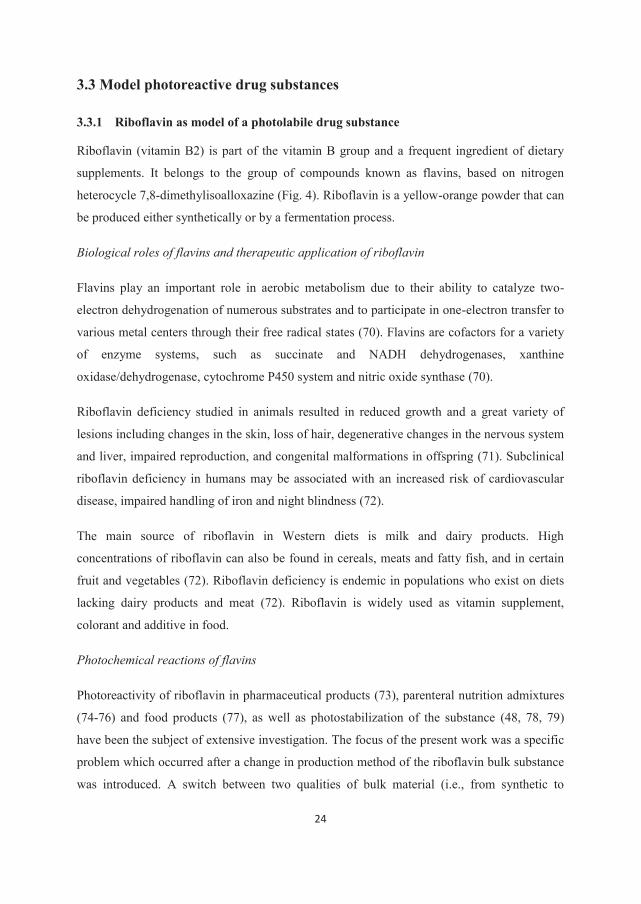
Riboflavin (vitamin B2) is part of the vitamin B group and a frequent ingredient of dietary
supplements. It belongs to the group of compounds known as flavins, based on nitrogen
heterocycle 7,8-dimethylisoalloxazine (Fig. 4). Riboflavin is a yellow-orange powder that can
be produced either synthetically or by a fermentation process.
Biological roles of flavins and therapeutic application of riboflavin
Flavins play an important role in aerobic metabolism due to their ability to catalyze two-
electron dehydrogenation of numerous substrates and to participate in one-electron transfer to
various metal centers through their free radical states (70). Flavins are cofactors for a variety
of enzyme systems, such as succinate and NADH dehydrogenases, xanthine
oxidase/dehydrogenase, cytochrome P450 system and nitric oxide synthase (70).
Riboflavin deficiency studied in animals resulted in reduced growth and a great variety of
lesions including changes in the skin, loss of hair, degenerative changes in the nervous system
and liver, impaired reproduction, and congenital malformations in offspring (71). Subclinical
riboflavin deficiency in humans may be associated with an increased risk of cardiovascular
disease, impaired handling of iron and night blindness (72).
The main source of riboflavin in Western diets is milk and dairy products. High
concentrations of riboflavin can also be found in cereals, meats and fatty fish, and in certain
fruit and vegetables (72). Riboflavin deficiency is endemic in populations who exist on diets
lacking dairy products and meat (72). Riboflavin is widely used as vitamin supplement,
colorant and additive in food.
Photochemical reactions of flavins
Photoreactivity of riboflavin in pharmaceutical products (73), parenteral nutrition admixtures
(74-76) and food products (77), as well as photostabilization of the substance (48, 78, 79)
have been the subject of extensive investigation. The focus of the present work was a specific
problem which occurred after a change in production method of the riboflavin bulk substance
was introduced. A switch between two qualities of bulk material (i.e., from synthetic to
biosynthetic riboflavin) induced a decrease in the photostability of riboflavin tablets. Severe
discoloration was observed after inadvertent exposure to light, although the tablets were
quantitatively sound (80). Investigations revealed that a particular photoreactivity of the
biosynthetic bulk material caused a decrease in photostability of the tablets (81). A thorough
understanding of the reaction mechanism was needed in order to reformulate and stabilize
tablets containing biosynthetic riboflavin.
Figure 4. Structure of riboflavin
The absorption spectrum of riboflavin in aqueous solutions consists of four structureless
peaks centered at 446, 375, 265 and 220 nm.
The three main types of photochemical reactions which flavins take part in are photoreduction,
photodealkylation and photoaddition (82). Some or all of these photoreactions may occur
concurrently, depending upon the structure of the flavin and the reaction conditions.
Photoreduction may occur as an intermolecular or intramolecular reaction. The overall
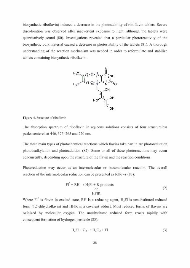
reaction of the intermolecular reduction can be presented as follows (83):
Fl* + RH H2Fl + R-products or
HFlR (2)
Where Fl* is flavin in excited state, RH is a reducing agent, H2Fl is unsubstituted reduced
form (1,5-dihydroflavin) and HFlR is a covalent adduct. Most reduced forms of flavins are
oxidized by molecular oxygen. The unsubstituted reduced form reacts rapidly with
consequent formation of hydrogen peroxide (83):
H2Fl + O2 H2O2 + Fl (3)
26
The mechanism of the reduction is quite complex and involves a two step reduction via a
semiquinone free radical intermediate (HFl•) (82, 83). Some of the suggested mechanisms of
reduction involve a dimeric triplet state, triplet-ground state reaction or triplet-triplet reaction:
2Fl Fl2 → 3Fl2 → Fl•+ + Fl•- (4a)
Where 3Fl2 is a dimeric triplet state of flavins, Fl•+ and Fl•- are flavin radical species. The
reaction between the ground (Fl) and triplet states (3Fl) is known as Dye-Dye reaction (D-D
reaction).
3Fl + Fl → Fl•+ + Fl•- (4b)
Semi-reduced flavin (flavosemiquinone radical) and fully reduced flavin (1,5-dihydroflavin or
flavohydroquinone) can be formed starting from flavin radical species:
Fl•- + H+ → HFl• (5a)
2HFl• → H2Fl + Fl (5b)
Hemmerich et al. (84) showed formation of flavosemiquinone radicals in solution in the
absence of the electron donors (reducing agents).
Intramolecular reduction involves dehydrogenation of the hydroxyalkyl side chain on the N-
10 position to yield a variety of ketonic and aldehydic products. Riboflavin intramolecular
photoreduction results in a complex mixture of 2’ and 4’ keto-derivatives together with
formylmerthyl flavin (83). It has been suggested that for efficient intramolecular hydrogen-
transfer to occur, the side chain should be co-planar with the main flavin ring-system (82).
Photodealkylation, a strictly intramolecular process, results in the formation of an alloxazine
and an alkene. Photoaddition can occur as an intermolecular or intramolecular reaction
(addition of cyanide, ammonia or water to benzenoid subnucleus of flavin, or addition of the
C-2’ hydroxyl group at the C-9 position, respectively) (82, 83).
3.3.2 Curcumin as the model of a photosensitizer for aPDT
Curcumin, a yellow pigment, is a constituent of turmeric or the rhizome of the plant Curcuma
longa L. Turmeric has been used in the traditional Chinese and Indian medicine for thousands
of years (85). Curcumin is an established photosensitizer in aPDT (45, 86-91). Commercially
available pure curcumin is a mixture of curcumin, demetoxycurcumin and
bisdemetoxycurcumin, in which curcumin represents the main constituent (~77%) (92).
27
Curcumin synthetized according to Pabon (93) was used in the present work in order to avoid
interference from the two other curcuminoids present in the commercially available product.
Physico-chemical properties of curcumin
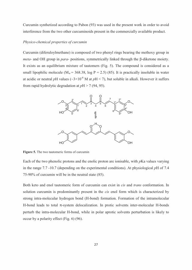
Curcumin (diferuloylmethane) is composed of two phenyl rings bearing the methoxy group in
meta- and OH group in para- positions, symmetrically linked through the β-diketone moiety.
It exists as an equilibrium mixture of tautomers (Fig. 5). The compound is considered as a
small lipophilic molecule (Mw = 368.38, log P = 2.5) (85). It is practically insoluble in water
at acidic or neutral pH values (~3×10⁻⁸ M at pH < 7), but soluble in alkali. However it suffers
from rapid hydrolytic degradation at pH > 7 (94, 95).
Figure 5. The two tautomeric forms of curcumin
Each of the two phenolic protons and the enolic proton are ionisable, with pKa values varying
in the range 7.7 -10.7 (depending on the experimental conditions). At physiological pH of 7.4
75-90% of curcumin will be in the neutral state (85).
Both keto and enol tautomeric form of curcumin can exist in cis and trans conformation. In
solution curcumin is predominantly present in the cis enol form which is characterized by
strong intra-molecular hydrogen bond (H-bond) formation. Formation of the intramolecular
H-bond leads to total π-system delocalization. In protic solvents inter-molecular H-bonds
perturb the intra-molecular H-bond, while in polar aprotic solvents perturbation is likely to
occur by a polarity effect (Fig. 6) (96).
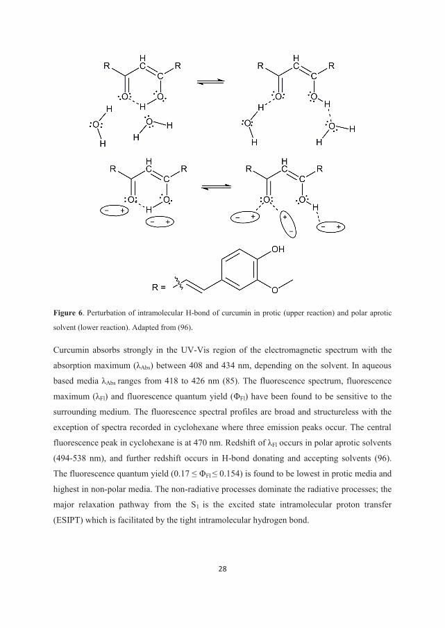
28
Figure 6. Perturbation of intramolecular H-bond of curcumin in protic (upper reaction) and polar aprotic
solvent (lower reaction). Adapted from (96).
Curcumin absorbs strongly in the UV-Vis region of the electromagnetic spectrum with the
absorption maximum (λAbs) between 408 and 434 nm, depending on the solvent. In aqueous
based media λAbs ranges from 418 to 426 nm (85). The fluorescence spectrum, fluorescence
maximum (λFl) and fluorescence quantum yield (ΦFl) have been found to be sensitive to the
surrounding medium. The fluorescence spectral profiles are broad and structureless with the
exception of spectra recorded in cyclohexane where three emission peaks occur. The central
fluorescence peak in cyclohexane is at 470 nm. Redshift of λFl occurs in polar aprotic solvents
(494-538 nm), and further redshift occurs in H-bond donating and accepting solvents (96).
The fluorescence quantum yield (0.17 ≤ ΦFl ≤ 0.154) is found to be lowest in protic media and
highest in non-polar media. The non-radiative processes dominate the radiative processes; the
major relaxation pathway from the S1 is the excited state intramolecular proton transfer
(ESIPT) which is facilitated by the tight intramolecular hydrogen bond.
29
Curcumin degrades upon exposure to UV-Vis radiation, both in solution and the solid state.
Several degradation products are formed including vanillin, vanillic acid, ferulic aldehyde,
ferulic acid, and 4-vinylguaiacol (97). Reported photodegradation quantum yields in methanol
and acetonitrile are 0.021 and 0.06, respectively (96). The rate of photodegradation depends
on the surrounding medium (98-100). Absorption and fluorescence maxima and fluorescence
quantum yield of curcumin are affected by the binding to plasma proteins (human and bovine
serum albumin) (85).
Curcumin as a photosensitizer with antimicrobial effect
The potential of curcumin as an antimicrobial and anticancer PS has been extensively studied
over the past 25 years (8, 86, 88, 89, 101-104). The phototoxicity of curcumin is found to be
oxygen dependent (101). The phototoxic effect is attributed to the production of ROS such as
singlet oxygen (1O2) and superoxide anion (O2•-). However, production of 1O2 was barely
detectable in protic solvents (96, 105) and could not be detected the biological model
involving Gram-negative (G-) bacteria (106). The role of singlet oxygen in curcumin
phototoxicity is therefore not yet clear. The superoxide anion was detected upon irradiation of
curcumin under both protic and non-protic conditions and may be involved in the
photodynamic action of the compound (105). Studies on the uptake of curcumin by bacteria
showed that the compound is adhered to G- bacteria Escherichia coli (E. coli). In Gram-
positive (G+) bacteria Enterococcus faecalis (E. faecalis) curcumin is either absorbed or
adsorbed to the bacterial wall (8, 106). A recent study showed that photoactivated curcumin
induced damage of the membrane integrity of the G+ Staphylococcus aureus (S. aureus),
leading to bacterial death (107).
Low concentrations of curcumin are sufficient to photoinactivate a broad range of pathogenic
species in vitro: G+ bacteria (8, 45, 107) including methicillin resistant S. aureus (88), G-
bacteria (45, 91) and fungi (87, 89). Microbial cells were found more susceptible to the
phototoxic effect of curcumin than the host cells (88, 89, 108) although significant reduction
of the host cells was reported (88, 89). In a recent investigation of oral candidiasis caused by
Candida albicans (C. albicans) in a murine model, curcumin mediated aPDT caused
significant reduction in C. albicans viability without harming the host tissue of mice. Finally,
studies on healthy humans showed that curcumin combined with blue light efficiently reduced
the concentration of salivary microorganisms up to 2 h post-treatment (109, 110). No major
30
adverse effects were reported (110). The main properties of curcumin are compared to the
properties of an optimal photosensitizer in Table 2.
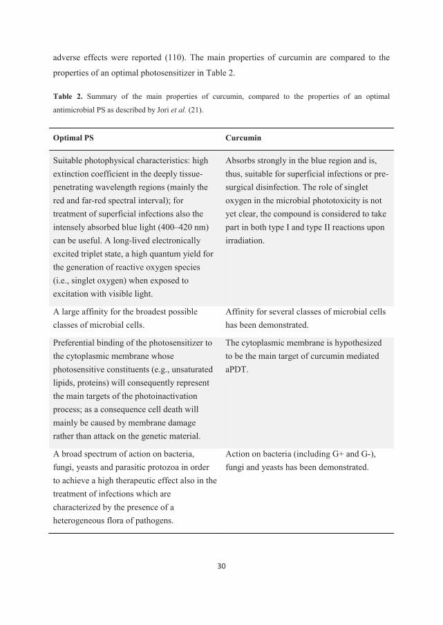
Table 2. Summary of the main properties of curcumin, compared to the properties of an optimal
antimicrobial PS as described by Jori et al. (21).
Optimal PS Curcumin
Suitable photophysical characteristics: high extinction coefficient in the deeply tissue-penetrating wavelength regions (mainly the red and far-red spectral interval); for treatment of superficial infections also the intensely absorbed blue light (400–420 nm) can be useful. A long-lived electronically excited triplet state, a high quantum yield for the generation of reactive oxygen species (i.e., singlet oxygen) when exposed to excitation with visible light.
Absorbs strongly in the blue region and is, thus, suitable for superficial infections or pre-surgical disinfection. The role of singlet oxygen in the microbial phototoxicity is not yet clear, the compound is considered to take part in both type I and type II reactions upon irradiation.
A large affinity for the broadest possible classes of microbial cells.
Affinity for several classes of microbial cells has been demonstrated.
Preferential binding of the photosensitizer to the cytoplasmic membrane whose photosensitive constituents (e.g., unsaturated lipids, proteins) will consequently represent the main targets of the photoinactivation process; as a consequence cell death will mainly be caused by membrane damage rather than attack on the genetic material.
The cytoplasmic membrane is hypothesized to be the main target of curcumin mediated aPDT.
A broad spectrum of action on bacteria, fungi, yeasts and parasitic protozoa in order to achieve a high therapeutic effect also in the treatment of infections which are characterized by the presence of a heterogeneous flora of pathogens.
Action on bacteria (including G+ and G-), fungi and yeasts has been demonstrated.
31
The research on curcumin as an antimicrobial PS is currently moving from in vitro to in vivo
studies. Although application of curcumin in aPDT is limited by low penetration depth of blue
light in tissue (111), the broad antimicrobial spectrum and efficacy of the compound (as
shown in vitro) and few side effects (reported in the limited number of in vivo studies) make it
an attractive candidate for treatment of, e.g., superficial wounds and oral infections that are
characterized by the presence of a heterogeneous flora of pathogens (21). Additional
advantages of curcumin are: cheap and easy production and purification (93), good
tolerability when administrated orally (85) and fast elimination in vivo after oral
administration (112).
Formulation of curcumin as solid dispersion for ex tempore preparation of supersaturated
solutions
Within this work the possibility of formulating curcumin as a solid dispersion for ex tempore
preparation of phototoxic supersaturated solutions has been explored. The high bacterial
phototoxicity of curcumin in the supersaturated state has already been shown in vitro (45, 86-
91). However, to be beneficial clinically curcumin needs to be in a preparation which is
convenient for storage and application. Therefore, readily dispersible SDs of curcumin able to
generate curcumin supersaturated solutions of desired and reproducible concentration were
developed. Precipitation inhibitors were included in the preparations in order to prolong the
supersaturated state. The overall stability of ex tempore prepared supersaturated solutions (i.e.,
rate of precipitation and hydrolytic and photolytic stability of curcumin in the solutions) was
estimated. Finally, phototoxicity against G+ and G- bacteria was tested (Papers II and III).
Solid dispersion is a beneficial formulation approach for increasing apparent solubility and
dissolution properties of curcumin. The high melting point of curcumin (183°C) reflects
strong crystal lattice energy and therefore a major improvement in apparent solubility can be
expected upon disruption of the crystalline structure (113). Indeed, the amorphous form of
curcumin possesses higher solubility compared to its crystalline counterpart (114). The
carriers selected for the SDs prepared within this work were aimed to meet two requirements:
to facilitate creation of supersaturated solutions quickly and easily upon dissolution of
SDs
to enable a prolonged metastable supersaturated state.
32
CDs and polymeric surfactants Pluronics® were selected for this purpose.
The common criticism towards ex tempore preparation of solutions aimed for aPDT is that the
procedure is not practical in a clinical setting when many people may need immediate
treatment. Ex tempore preparation can also introduce error in dose if performed incorrectly
(20). Suitable dosage forms consisting of premeasured SD of the compound with or without
the adequate amount of the dissolution medium may facilitate the use and secure a correct
dose. Another disadvantage of solution-based topical preparations is that they are hard to keep
at the site of action (20). This may be resolved by addition of a viscosity modifier or
mucoadhesive agent either to the dissolution medium or to the SD.
The application of a solid preparation, on the other hand, does not require any manipulation
before application and would be easier to keep at the target site. A disadvantage of such a
system in this context is the dependence upon some liquid already present at the target site to
facilitate dissolution. A solid preparation could also prevent the radiation from reaching the
target and might need to be removed prior to irradiation.
The general advantage of solid forms over creams, gels and solutions typically used in aPDT
is increased stability and thus longer shelf-life of the product. However, in the case of solid
dispersions an additional concern is the physical stability of the active ingredients within the
matrix. Due to the possibility of a crystallization of active ingredients resulting in markedly
changed bioavailability, the number of SD based products on the market is still very limited
(60). The prototypes of solid dispersions for the preparation of supersaturated solutions as
presented here, could be further modified according to the particular site of action and type of
infection.
33
4. General experimental conditions
A general description of materials, sample preparation and methods are given below. The
specific experimental conditions are described in the individual papers (I-IV).
4.1 Materials
Riboflavin (both biosynthetic and synthetic sample) were generously provided as a gift by
Weifa A/S (Oslo, Norway). Riboflavin dehydrate was produced by exposing a biosynthetic
riboflavin sample to elevated humidity (Paper I). Curcumin was synthetized according to
method described by Pabon (93).
Hydroxypropyl-β-cyclodextrin (HP-β-CD) and HPMC were used for making SDs in Paper II.
In Paper III Pluronics® F127, F68 and P123 (PF127, PF68 and PP123) were employed for
production of SDs; HPMC, polyethylene glycol 400 (PEG 400) and hyaluronic acid (HA)
were tested as the second PI. Within the Paper IV the effect of the following excipients on the
photostability and the protein binding of curcumin was studied: hydroxypropyl- -cyclodextrin
(HP- -CD), HP-β-CD, PEG 400 and PF127.
4.2 Preparation of samples
Within Paper II the solid dispersions were prepared in two steps. In the first step, co-
precipitates of curcumin and HP-β-CD were prepared by evaporation from methanol solutions
with varying CD/curcumin molar ratios (0.5, 0.9, 1.8 and 2.8). Depending on the molar ratio,
co-precipitates are referred to as 0.5, 0.9, 1.8 and 2.8. In the second step supersaturated
curcumin solutions obtained by dissolving selected co-precipitates were added HPMC and
freeze dried to yield the final product. Depending on the co-precipitate used for the production,
lyophilizates are referred to as 0.9, 1.8 and 2.8.
The solid dispersions made within Paper III were produced by the solvent evaporation method
from methanol or methanol-dichloromethane solutions. The second PI was added to the
dissolution medium to evaluate its potential to stabilize the supersaturated solution.
Curcumin was added from a stock solution in methanol to the solutions containing the
excipient of interest and/or human serum albumin within the Paper IV.
34
4.3 Methods
The following methods were applied to identify the crystalline form of riboflavin in Paper I:
X-ray diffractometry (XRD), Fourier-Transform Infrared Spectroscopy (FT-IR) and scanning
electron microscopy (SEM). The influence of the radiation exposure on the samples was
studied by visible (Vis)-, near Infrared (NIR)-, FT-IR and fluorescence reflectance
spectroscopy. Qualitative thin layer chromatography (TLC) and HPLC were employed for the
detection of amino acids and the degradation products of riboflavin.
The solid dispersions and the co-precipitates of curcumin in Paper II were studied by
differential scanning calorimetry (DSC), Carl-Fisher titration and SEM, to evaluate
crystallinity of curcumin within SDs, morphology and the moisture content of SDs,
respectively. The drug load was determined by HPLC. Solutions of solid dispersions were
characterized by UV–Vis spectrophotometry and HPLC to estimate their physical stability
and the hydrolytic stability of curcumin, respectively. Solutions of the selected SDs were
assessed for potential phototoxicity towards E. coli.
The solid dispersions produced within Paper III were examined by XRD to determine the
physical state of curcumin. The dissolved SDs with and without the second PI added to
dissolution medium, were studied by UV–Vis spectrophotometry to evaluate the stability of
the supersaturated curcumin solutions. HPLC was employed to study hydrolytic and
phototlytic stability of curcumin in the solutions. Supersaturated solutions of selected SDs
were tested for phototoxic potential towards the bacterium E. faecalis.
In Paper IV the effect of excipients on curcumin-HSA binding was studied using HSA
fluorescence quenching. Absorption and fluorescence emission spectra of curcumin in the
solutions containing HSA, excipients or both were recorded. HPLC was employed to study
the effect of excipients and HSA on curcumin photostability.
In Papers II, III and IV the samples were exposed to the radiation in the wavelength range
350–500 nm (emission maximum at 430 nm) for the photostability studies and bacterial
studies.
35
5. Results and discussion
5.1 The physical state of the drug substance in the solid preparation
The physical state of the drug substance can govern the physico-chemical properties of the
substance and the final product such as melting point, solubility, stability, density and
hardness (115). Some of these properties, such as solubility and stability, play a key role in
the drug formulation and the quality of the final product. As part of this project, the influence
of the physical state of PDS on the apparent solubility and dissolution rate, and photostability
in the solid state was studied.
5.1.1 The physical state of the riboflavin as a bulk substance applied in tablets
A change in the production method of the active ingredient induced a change in the
photostability of riboflavin tablets (section 3.3.1). The two different qualities of riboflavin
were examined by means of XRD and FT-IR. Both techniques indicated a difference in the
crystalline structure (Paper I). Synthetic riboflavin (RFs) and biosynthetic riboflavin (RFbs)
were identified as the previously described forms anhydrate I and monohydrate II respectively,
by means of XRD analyses (116). Furthermore, the previously described dihydrate II was
prepared by exposing RFbs to elevated humidity and identified by XRD. The FT-IR study
indicated the presence of a markedly different H-bonding pattern in the two forms of
riboflavin. The morphology of RFs and RFbs crystals observed by SEM and the color of the
powder correlated to previously reported morphology and color of anhydrate I and
monohydrate II crystals, respectively (117). A clear difference in the shape of the
fluorescence emission spectra of RFs and RFbs (in the solid state) could be observed. This
was ascribed to the structural differences between the two examined forms of riboflavin.
5.1.2 Formulation of curcumin as a solid dispersion
In vitro studies showed that curcumin can be efficient in photoinactivation of G+ bacteria (E.
faecalis) at concentrations as low as 5×10⁻⁷ M (combined with a radiation dose of 11 J/cm2)
and of G– bacteria (E. coli) at concentrations of 5×10⁻⁶ and 10-5 M (combined with a radiation
dose of 32 J/cm2 and 16 J/cm2, respectively) in an appropriate preparation (45). In a recent
animal study in a murine model of oral candidiasis the most promising results of
photoinactivation of C. albicans were achieved with the combination of a 8×10⁻⁵ M curcumin
solution and a 37.5 J/cm2 radiation dose (90). A concentration of curcumin much higher than
36
the saturation solubility is required for the successful application of the compound as PS in
aPDT in vivo. In Papers II and III the possibility of increasing the apparent solubility and
dissolution rate by formulating curcumin as a solid dispersion was explored.
Formulation of solid dispersions of curcumin in HP-β-CD and HPMC was a two step process
(Paper II, section 4.2). In the first step the co-precipitates of curcumin and HP-β-CD were
prepared and investigated by DSC. A loss of curcumin crystallinity was indicated in all of the
samples except for the co-precipitate with CD/curcumin ratio of 0.5. This product was not
studied further. Two glass transition temperatures (Tg) were observed for the remaining co-
precipitates. Neither could be superimposed on Tgs of the pure HP-β-CD or amorphous
curcumin, suggesting the occurrence of inter-molecular interactions between the ingredients.
The dissolution of the co-precipitates in water resulted in a supersaturated curcumin solution,
as shown by the decrease in curcumin concentration upon prolonged dissolution. The
concentration of dissolved curcumin depended on the CD/curcumin molar ratio of the co-
precipitates (Paper II). CD-curcumin interactions may dictate the physical state of the SD and
thus affect the release of the active ingredient. Once the supersaturated solution is formed, a
high concentration of HP-β-CD present in solutions of co-precipitates with a high
CD/curcumin ratio may offer more efficient crystallization inhibition than corresponding
solutions with a low CD/curcumin ratio (58).
In the second production step lyophilizates 1.8 and 2.8 were selected for further studies based
on the DSC investigations. These samples displayed uniform thermal behavior and batch to
batch reproducibility. No crystalline curcumin was present in the freeze dried product, as
indicated by the absence of the curcumin melting endotherm. Similar to co-precipitates, two
Tgs were observed. This may indicate a mixed system comprised of the amorphous drug
substance and the solid solution (118). However, the lyophilizates did not display the Tg of
the amorphous curcumin. Therefore, two glass transitions may indicate the existence of two
types of molecular interactions between curcumin and the carriers within the solid dispersions.
Scanning electron microscopy of the lyophilizate 2.8 revealed the porous structure
characteristic for freeze dried products.
Dispersion of curcumin within the polymeric carriers was made by evaporation method and
investigated for the presence of crystalline curcumin by XRD (Paper III). PF127 and PP123
were combined with Pluronic®/curcumin in 0.5:1 and 1:1 molar ratio, while PF68 and
37
curcumin were combined in 1:1 and 2:1 molar ratio. SDs based on PP123 could not be
studied by XRD for practical reasons. The remaining four SDs showed no presence of
crystalline curcumin.
Solid dispersions presented here were made by use of the solvent method which has many
advantages over the melting method but is associated with many ecological and economic
problems. Because of this the current method of choice for the manufacturing of solid
dispersions is hot melt extrusion (HME) (60). The low melting point of Pluronics® makes
them suitable carriers for SD production by HME, since the technique requires one or more
‘meltable’ excipients to be included in the carrier system (119, 120). Therefore HME should
be considered for scaling up the production of Pluronic® based SDs.
A combination of HP-β-CD and HPMC offered efficient crystallization inhibition of
curcumin during freeze drying and amorphous final products were obtained (Paper II). The
drug load of the final products was ranging from 0.5 to 1% w/w. In Paper III we show that no
crystalline curcumin precipitated during fast evaporation of the common solvent at the given
carrier/curcumin ratios, yielding solid dispersions with a drug load in the range 2.1 - 5.5%
w/w. Within the same work the crystallinity of curcumin in the solid dispersions with the
highest drug loads (i.e., 6.1 and 11.4% w/w, based on PP123) could not be determined by
XRD for practical reasons. However, good dissolution properties and stability of the
supersaturated solution formed upon dissolution of these SDs suggested the absence of a
crystalline compound, as will be discussed later. Thus, the selected formulation methods
combined with the selected carriers in the chosen ratios resulted in SDs with curcumin either
transformed to an amorphous form or dispersed on a molecular level (i.e., as a solid solution).
The physical state of the SDs depends on the physico-chemical properties of the carrier and
active ingredient, active ingredient-carrier interactions and the formulation method. When
prepared by the solvent method the physical state of SD depends on the temperature, the
evaporation rate and the type of solvent (58). Curcumin can be considered an intermediate
crystallizer according to the classification system of Van Eerdenbrugh et al. (113, 121).
Therefore, it was not possible to obtain the amorphous curcumin by evaporation (Paper II),
and curcumin-Pluronic® interactions were essential for the transformation of the physical state
of the compound (Paper III). Wegiel et al. (113) have recently studied the physical stability of
amorphous curcumin SDs and concluded that, for a compound like curcumin which forms
intramolecular H-bonds, formation of H-bonding with the carrier is hindered and thus it is
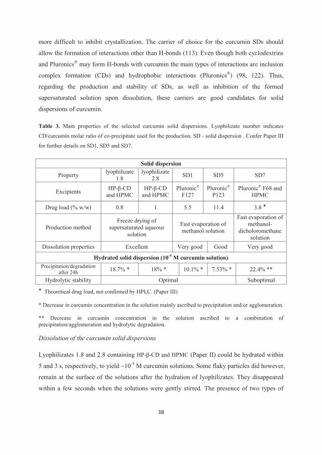
38
more difficult to inhibit crystallization. The carrier of choice for the curcumin SDs should
allow the formation of interactions other than H-bonds (113). Even though both cyclodextrins
and Pluronics® may form H-bonds with curcumin the main types of interactions are inclusion
complex formation (CDs) and hydrophobic interactions (Pluronics®) (98, 122). Thus,
regarding the production and stability of SDs, as well as inhibition of the formed
supersaturated solution upon dissolution, these carriers are good candidates for solid
dispersions of curcumin.
Table 3. Main properties of the selected curcumin solid dispersions. Lyophilizate number indicates
CD/curcumin molar ratio of co-precipitate used for the production. SD - solid dispersion . Confer Paper III
for further details on SD1, SD5 and SD7.
Solid dispersion
Property lyophilizate 1.8
lyophilizate 2.8 SD1 SD5 SD7
Excipients HP-β-CD and HPMC
HP-β-CD and HPMC
Pluronic® F127
Pluronic® P123
Pluronic® F68 and HPMC
Drug load (% w/w) 0.8 1 5.5 11.4 3.8
Production method Freeze drying of
supersaturated aqueous solution
Fast evaporation of methanol solution
Fast evaporation of methanol-
dicholoromethane solution
Dissolution properties Excellent Very good Good Very good
Hydrated solid dispersion (10-5 M curcumin solution) Precipitation/degradation
after 24h 18.7% * 18% * 10.1% * 7.53% * 22.4% **
Hydrolytic stability Optimal Suboptimal
Theoretical drug load, not confirmed by HPLC. (Paper III)
* Decrease in curcumin concentration in the solution mainly ascribed to precipitation and/or agglomeration.
** Decrease in curcumin concentration in the solution ascribed to a combination of precipitation/agglomeration and hydrolytic degradation.
Dissolution of the curcumin solid dispersions
Lyophilizates 1.8 and 2.8 containing HP-β-CD and HPMC (Paper II) could be hydrated within
5 and 3 s, respectively, to yield ~10-5 M curcumin solutions. Some flaky particles did however,
remain at the surface of the solutions after the hydration of lyophilizates. They disappeared
within a few seconds when the solutions were gently stirred. The presence of two types of
39
curcumin-carrier interactions within the SDs as suggested by the presence of two Tgs may
explain this behavior.
Dissolution of SDs containing PF127 and PF123 based SDs resulted in 10-5 M curcumin
solutions corresponding to the theoretical amount of the substance in the dissolved state. PF68
based SDs were as easily dissolved but did not yield in the theoretical curcumin concentration
(Table 3).
The good dissolution properties of these SDs could be explained by good dissolution
properties of the carriers, particle size reduction, change in the physical state of active
ingredient and possibly dispersion at the molecular level (58). When the drug substance is
dispersed at molecular level, the dissolution rate is determined by the dissolution rate of the
carrier (60). Freeze dried SDs additionally benefit from their porous structure and the
amorphous state of the carriers (i.e., HP-β-CD and HPMC) and display better dissolution
properties than Pluronic® based SDs.
A short incubation time prior to irradiation is favored in aPDT. Thus preparations with a fast
drug release are required. The lyophilizates hydrated almost instantaneously, resulting in a
clear solution. They were proposed for application on infected, moist wounds (Paper II). SDs
based on Pluronics® are suggested to be used after hydration in the form of a supersaturated
solution (Paper III). Pluronics® display a complex phase behavior. Depending on the
temperature, polymer concentration and the type of polymer (polyethylene
oxide/polypropylene oxide composition ratio), unimer, micellar, cubic (gel), hexagonal and
lamellar phases can be observed (123). The drug release from each of these states differs.
Depending upon viscosity of the preparation the light scattering/absorption could pose the
problem if the preparation is not removed prior to exposure to radiation. Neither the amount
of dissolution media (wound liquid/saliva) nor the temperature of the infected area can be
controlled. Supersaturated solutions containing polymer in a submicellar concentration and
prepared immediately before use, should be applied to control the release of curcumin from
Pluronics® containing preparations.
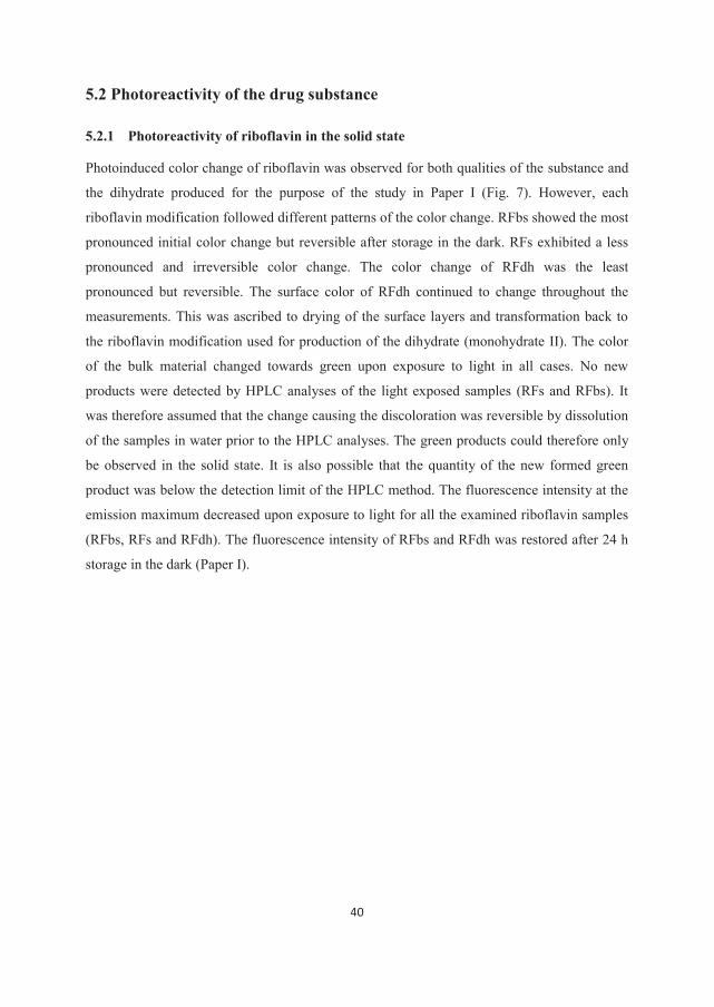
40
5.2 Photoreactivity of the drug substance
5.2.1 Photoreactivity of riboflavin in the solid state
Photoinduced color change of riboflavin was observed for both qualities of the substance and
the dihydrate produced for the purpose of the study in Paper I (Fig. 7). However, each
riboflavin modification followed different patterns of the color change. RFbs showed the most
pronounced initial color change but reversible after storage in the dark. RFs exhibited a less
pronounced and irreversible color change. The color change of RFdh was the least
pronounced but reversible. The surface color of RFdh continued to change throughout the
measurements. This was ascribed to drying of the surface layers and transformation back to
the riboflavin modification used for production of the dihydrate (monohydrate II). The color
of the bulk material changed towards green upon exposure to light in all cases. No new
products were detected by HPLC analyses of the light exposed samples (RFs and RFbs). It
was therefore assumed that the change causing the discoloration was reversible by dissolution
of the samples in water prior to the HPLC analyses. The green products could therefore only
be observed in the solid state. It is also possible that the quantity of the new formed green
product was below the detection limit of the HPLC method. The fluorescence intensity at the
emission maximum decreased upon exposure to light for all the examined riboflavin samples
(RFbs, RFs and RFdh). The fluorescence intensity of RFbs and RFdh was restored after 24 h
storage in the dark (Paper I).
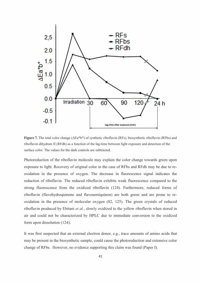
41
Figure 7. The total color change (ΔEa*b*) of synthetic riboflavin (RFs), biosynthetic riboflavin (RFbs) and
riboflavin dihydrate II (RFdh) as a function of the lag-time between light exposure and detection of the
surface color. The values for the dark controls are subtracted.
Photoreduction of the riboflavin molecule may explain the color change towards green upon
exposure to light. Recovery of original color in the case of RFbs and RFdh may be due to re-
oxidation in the presence of oxygen. The decrease in fluorescence signal indicates the
reduction of riboflavin. The reduced riboflavin exhibits weak fluorescence compared to the
strong fluorescence from the oxidized riboflavin (124). Furthermore, reduced forms of
riboflavin (flavohydroquinone and flavosemiquinon) are both green and are prone to re-
oxidation in the presence of molecular oxygen (82, 125). The green crystals of reduced
riboflavin produced by Ebitani et al., slowly oxidized to the yellow riboflavin when stored in
air and could not be characterized by HPLC due to immediate conversion to the oxidized
form upon dissolution (124).
It was first suspected that an external electron donor, e.g., trace amounts of amino acids that
may be present in the biosynthetic sample, could cause the photoreduction and extensive color
change of RFbs. However, no evidence supporting this claim was found (Paper I).
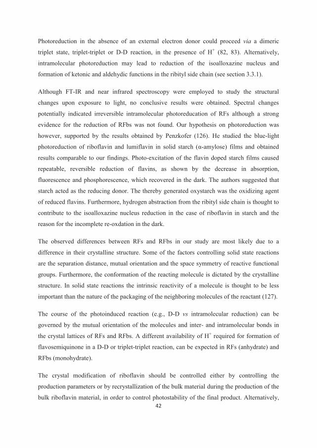
42
Photoreduction in the absence of an external electron donor could proceed via a dimeric
triplet state, triplet-triplet or D-D reaction, in the presence of H+ (82, 83). Alternatively,
intramolecular photoreduction may lead to reduction of the isoalloxazine nucleus and
formation of ketonic and aldehydic functions in the ribityl side chain (see section 3.3.1).
Although FT-IR and near infrared spectroscopy were employed to study the structural
changes upon exposure to light, no conclusive results were obtained. Spectral changes
potentially indicated irreversible intramolecular photoreducation of RFs although a strong
evidence for the reduction of RFbs was not found. Our hypothesis on photoreduction was
however, supported by the results obtained by Penzkofer (126). He studied the blue-light
photoreduction of riboflavin and lumiflavin in solid starch (α-amylose) films and obtained
results comparable to our findings. Photo-excitation of the flavin doped starch films caused
repeatable, reversible reduction of flavins, as shown by the decrease in absorption,
fluorescence and phosphorescence, which recovered in the dark. The authors suggested that
starch acted as the reducing donor. The thereby generated oxystarch was the oxidizing agent
of reduced flavins. Furthermore, hydrogen abstraction from the ribityl side chain is thought to
contribute to the isoalloxazine nucleus reduction in the case of riboflavin in starch and the
reason for the incomplete re-oxdation in the dark.
The observed differences between RFs and RFbs in our study are most likely due to a
difference in their crystalline structure. Some of the factors controlling solid state reactions
are the separation distance, mutual orientation and the space symmetry of reactive functional
groups. Furthermore, the conformation of the reacting molecule is dictated by the crystalline
structure. In solid state reactions the intrinsic reactivity of a molecule is thought to be less
important than the nature of the packaging of the neighboring molecules of the reactant (127).
The course of the photoinduced reaction (e.g., D-D vs intramolecular reduction) can be
governed by the mutual orientation of the molecules and inter- and intramolecular bonds in
the crystal lattices of RFs and RFbs. A different availability of H+ required for formation of
flavosemiquinone in a D-D or triplet-triplet reaction, can be expected in RFs (anhydrate) and
RFbs (monohydrate).
The crystal modification of riboflavin should be controlled either by controlling the
production parameters or by recrystallization of the bulk material during the production of the
bulk riboflavin material, in order to control photostability of the final product. Alternatively,
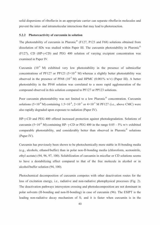
43
solid dispersions of riboflavin in an appropriate carrier can separate riboflavin molecules and
prevent the inter- and intramolecular interactions that may lead to photoreaction.
5.2.2 Photoreactivity of curcumin in solution
The photostability of curcumin in Pluronic® (F127, P123 and F68) solutions obtained from
dissolution of SDs was studied within Paper III. The curcumin photostability in Pluronic®
(F127), CD (HP- -CD) and PEG 400 solution of varying excipient concentration was
examined in Paper IV.
Curcumin (10-5 M) exhibited very low photostability in the presence of submicellar
concentrations of PF127 or PP123 (5×10⁻⁶ M) whereas a slightly better photostability was
observed in the presence of PF68 (10-5 M) and HPMC (0.001% w/v) (Paper III). A better
photostability in the PF68 solution was correlated to a more rapid agglomeration of the
compound observed in this solution compared to PF127 or PP123 solutions.
Poor curcumin photostability was not limited to a low Pluronic® concentration. Curcumin
solutions (5×10⁻⁶ M) containing 1.5×10⁻⁴, 2×10⁻³ or 4×10⁻³ M PF127 (i.e., above CMC) were
also rapidly degraded upon exposure to radiation (Paper IV).
HP- -CD and PEG 400 offered increased protection against photodegradation. Solutions of
curcumin (5×10⁻⁶ M) containing HP- -CD or PEG 400 in the range 0.05 - 5% w/v exhibited
comparable photostability, and considerably better than observed in Pluronic® solutions
(Paper IV).
Curcumin has previously been shown to be photochemically more stable in H-bonding media
(e.g., alcohols, ethanol/buffer) than in polar non-H-bonding media (chloroform, acetonitrile,
ethyl acetate) (94, 96, 97, 100). Solubilization of curcumin in micellar or CD solutions seems
to have a destabilizing effect compared to that of the free molecule in alcohol or in
alcohol/buffer solution (94, 100).
Photochemical decomposition of curcumin competes with other deactivation routes for the
loss of excitation energy, i.e., radiative and non-radiative photophysical processes (Fig. 2).
The deactivation pathways intersystem crossing and photodecomposition are not dominant in
polar solvents (H-bonding and non-H-bonding) in case of curcumin (96). The ESIPT is the
leading non-radiative decay mechanism of S1 and it is faster when curcumin is in the
44
intramolecular H-bonded cis enol form. In H-bonding solvents formation of H-bonds between
curcumin and solvent molecules results in the perturbation of intramolecular H-bonds and
thereby prevents direct ESIPT (Fig. 6). The fast-decaying intramolecular H-bonded enol
structure can be formed only after desolvation. However, alternative decay mechanism can be
the delivery of excitation energy to the solvent molecules, which is expected to be highly
enhanced when intermolecular H-bonds are formed. On the other hand, polar non-H-bonding
solvents can also efficiently perturb intramolecular H-bonds and inhibit deactivation through
ESIPT (Fig. 6), but their interactions with curcumin do not introduce new decay mechanisms.
More pronounced photodecomposition of curcumin in polar non-H-bonding than in H-
bonding environments is probably the consequence of the less efficient non-radiative
deactivation in the first case.
Very low curcumin photostability in the Pluronic® solutions could be explained by analogy
with polar non-H-bonding solvents. Ghosh et al. (129) suggested that the ESIPT mediated
decay channel of curcumin is inhibited in Tween® 20, Tween® 80, Pluronics® P123 and F127
micelles. Furthermore, the authors suggested that the decrease of ESIPT rate observed in
polymeric micelles compared to micelles of conventional surfactants was a result of the
interaction between curcumin and the lone pair oxygen of oxyethilene moiety (128, 129).
Addition of cosolvents (PEG 400 or ethanol, 20% v/v) to micellar solutions of Pluronics®
(P123, F127 or P85) was correlated to the transfer of curcumin from the core of the micelles
to the bulk solvent and resulted in slightly increased photostability of curcumin, as reported
by Singh et al. (98).
Increased photostability of curcumin in HP- -CD and PEG 400 solutions compared to
Pluronic® solutions could be rationalized by a more efficient S1 deactivation pathway through
energy transfer to solvent and/or solubilizer molecules. Fluorescence emission studies
suggested that curcumin may be in a H-bonding environment in these media (Paper IV).
However, other factors can influence the behavior of the excited state since the complexation
with HP- -CD may determine curcumin conformation and tautomeric form (130).
Although CDs and PEG offer better photostabilization of curcumin compared to Pluronics®,
other aspects of the formulation should also be considered such as phototoxicity of curcumin
and inhibition of precipitation. Furthermore, the demands regarding the drug photostability
depend upon the type of the preparation. Solid dispersions of curcumin as suggested to be
45
used in aPDT either in the solid state or after rehydration with an aqueous medium, could
easily be protected from light exposure during the storage period, both before and after
rehydration. Further, as a part of aPDT protocol, the application site could also be protected.
5.3 Physical stability of the curcumin in solution
Solid dispersions of curcumin were prepared within Papers II and III in order to increase the
apparent solubility and dissolution rate of curcumin as discussed above. Solid dispersions
typically yield supersaturated solutions of the active ingredient upon dissolution which are
thermodynamically unstable systems prone to precipitation (55, 58). Following administration
the drug substance must remain in solution to ensure a high and reproducible interaction with
the target such as the bacterial membrane in the case of aPDT. Thus, precipitation of
curcumin from hydrated SDs needs to be delayed for the relevant time period.
5.3.1 Supersaturated solutions of curcumin prepared from the solid dispersions
The decrease in curcumin concentration in the solutions of lyophilizates 1.8 and 2.8
(theoretical curcumin concentration of 1×10⁻⁵ and 2.5×10⁻⁵ M) upon storage (up to 168 h) and
the decrease in UV-Vis absorbance of the solutions during 24 h storage suggested the
agglomeration and precipitation of the substance since no indications of hydrolytic
degradation products were found by HPLC (Paper II). Furthermore, the shape of the
absorption spectrum of curcumin in dissolved lyophilizate indicated the presence of the free
curcumin molecule rather than the curcumin-HP-β-CD complex.
A decrease in curcumin concentration and UV-Vis absorbance upon storage (2, 8 and 24 h)
were also observed for dissolved Pluronic®-based SDs (Paper III).
Thus, both the HP-β-CD-HPMC based SDs prepared within Paper II and Pluronic® based SDs
prepared in Paper III can generate supersaturated solutions of curcumin.
5.3.2 Precipitation inhibition of curcumin in a supersaturated solution
Solutions of lyophilizates 1.8 and 2.8 equivalent to 10-5 M curcumin exhibited a decrease in
curcumin concentration ≥ 18% within 24 h (Paper II). Solutions of PF127 and PP123 based
SDs (10-5 M curcumin) showed a decrease in curcumin concentration ≥ 10% during the 24 h
storage whereas solutions of PF68 based SDs exhibited much more pronounced curcumin
precipitation, comparable to that of a supersaturated solution without PI. The capability of the
46
second PI (HPMC, PEG 400 and HA) to improve the physical stability of curcumin
supersaturation obtained from Pluronic® based SDs was evaluated. HPMC showed the best
results and SD based on PF68 was reformulated by addition of this excipient. However, this
SD was shown to be hydrolytically unstable in solution and was therefore not studied further
(Paper III).
In general, PF127 and PP123 based SDs showed better physical stability in the solution than
HP-β-CD-HPMC based SDs, despite a 5-10 times higher drug load in the former (Table 3).
Thus, PF127 and PP123 can be considered better precipitation inhibitors for curcumin than
the combination of HP-β-CD and HPMC. It is difficult however, to distinguish between the
contribution of each of the excipients included in the lyophilizates to the stability of the
supersaturated solutions generated from these lyophilizates.
HP-β-CD and HPMC have been proposed to interact with an active ingredient by establishing
H-bonds which increase the activation energy of the crystal nucleation. Furthermore, due to
the ability to form H-bonds these excipients can adsorb to the crystal surface and prevent the
crystal growth (57, 131, 132). Similarly, Pluronics® are suggested to adsorb to the crystal
surface and inhibit crystal nucleation and growth (133-135). However, a hydrophobic reaction
between Pluronics® and the active ingredient is expected to be the main driving force behind
the precipitation inhibition mechanism (134). HP-β-CD and Pluronics® can improve the
solvation of the drug substance in solution and thereby increase the activation energy for
desolvation during the crystal growth (55, 133, 136). Finally, HP-β-CD and Pluronics® can
solubilize the drug substance and decrease the degree of supersaturation (137, 138). Curcumin
is however, not expected to be in the solubilized state in the examined SD solutions and the
delay of precipitation is not likely due to solubilization. Dissolved lyophilizates probably
contain curcumin in a free form rather than complexed with HP-β-CD, as indicated by the
UV-Vis spectra. Pluronic® based SD solutions contain submicellar concentrations of
Pluronics® and therefore curcumin is most likely in the molecular state rather than
encapsulated in micelles.
The mechanisms suggested to explain precipitation inhibition by HP-β-CD and HPMC on the
one side and Pluronics® on the other, largely overlap. However, the main difference seems to
be the type of interactions that these excipients form with the substance i.e., H-bonds (HP-β-
CD and HPMC) or hydrophobic interaction (Pluronics®). It has been suggested that the
47
excipients mainly forming H-bonds with curcumin are not optimal carriers to inhibit
crystallization of amorphous curcumin in the solid dispersions due to the strong
intramolecular H-bonds in curcumin (113). Curcumin forms intramolecular H-bonds in the
solution as well as in the solid state. Therefore Pluronics® F127 and P123 may inhibit
precipitation of dissolved curcumin more efficiently than the combination of HP-β-CD and
HPMC because the former mainly form hydrophobic bonds with curcumin. PF127 and PP123
were found to be more efficient in stabilizing supersaturation than PF68, likely due to the
larger size of hydrophobic polypropylene oxide block of the two former copolymers (Paper
III). This emphasizes the importance of the hydrophobic interactions between the excipients
and curcumin for the physical stability of a supersaturated solution.
The various supersaturated solutions formed from the SDs may not have the same individual
requirements for the physical stability. SDs based on the Pluronics® need to be rehydrated
prior to application (as discussed in the section 5.1.2) and thus the curcumin solutions need to
be stable both during the short term storage and the in vivo incubation time. Application of the
SDs based on combined HP-β-CD-HPMC in the solid state requires only the stabilization of
the curcumin supersaturation during the in vivo incubation time.
5.4 Preparations in biorelevant media: bacterial phototoxicity of curcumin
and interaction with human serum albumin
Sensitivity of bacteria to photodynamic inactivation by curcumin in vitro was shown to be
dependent upon the formulation. A similar effect could be expected under physiological
conditions (8, 45, 139). It is therefore essential to optimize the formulation in regard to
interaction with bacterial cells prior to in vivo studies. Dai et al. (19) pointed out that for
aPDT to be clinically useful a combination of PS and light needs to be able to kill microbial
cells in the environment found in actual infections. A number of in vitro studies showed the
reduction of the aPDT effect in protein rich media (e.g., serum, saliva, albumin suspensions).
The decrease in PS molecules available for interaction with bacteria due to competitive
binding to the proteins present in the media was suggested as one of the causes of the
described effect (37). It is therefore important to investigate how the formulation will affect
the protein-curcumin binding.
48
5.4.1 Phototoxicity of curcumin towards Gram-positive and Gram-negative bacteria
Encouraging in vitro aPDT results and the recently reported in vivo study emphasize the
potential of curcumin as a PS (88-90, 108). The required concentration of curcumin in these
studies was achieved by use of DMSO (10%) or ethanol as a cosolvent. DMSO is
immediately being absorbed through human tissues and it can carry dissolved substances with
it across the membranes. This is undesirable in the case of aPDT where local action is the
main aim. Furthermore, high concentration of DMSO can have a damaging effect on the
tissue and use of this solvent as an enhancer produces a foul odour on the breath (140).
Therefore alternative formulation approaches should be found for the application of curcumin
in a clinical setting. A number of formulations with curcumin solubilized by use of
cyclodextrins, micelles and polymers were developed to avoid use of DMSO or other organic
solvents (8, 102, 141). A high concentration of curcumin was achieved by use of CDs or
micelles but the PDT effect was decreased (even completely lost in the case of 5% HP-β-CD
and -CD) compared to formulations with organic cosolvents (5% DMSO or 1% ethanol) (8,
102). This clearly showed that a high concentration of solubilized curcumin is not sufficient
for the phototoxic effect. To be efficient curcumin needs to be in the free molecular form
rather than complexed or encapsulated. Application of supersaturation rather than inclusion
complexation enhances the free curcumin concentration and facilitates interaction with the
microbial outer wall (55, 91).
Supersaturated curcumin solutions cannot be stored for longer periods due to rapid
precipitation. Even phototoxicity, shown to be high in vitro, might decrease in vivo if the
longer incubation times are required (141). Solid dispersions made within Papers II and III
were aimed to generate supersaturated solutions upon hydration without the use of organic
cosolvents and to maintain physical stability of supersaturation and hydrolytic stability of
curcumin for the sufficient time periods. Phototoxicity of selected hydrated solid dispersions
was studied against G– bacteria E. coli (Paper II) and G+ bacteria E. faecalis (Paper III). A
phototoxic effect higher than or comparable to the one previously obtained with a 5% DMSO
preparation proved that the stabilizing effect by the excipients did not reduce the curcumin
efficiency (Table 4).
Further development of the supersaturation concept gave improved results. Wikene et al. (45)
have described solid dispersions based on a M-β-CD-HPMC or M-β-CD-HPMC-HA
49
combination produced by lyphilization. Solutions of these SDs efficiently eradicated E. coli
and E. faecalis at a concentrations 2.5×10⁻⁶ and 5×10⁻⁷ M respectively, and the results
corresponded to a reference supersaturated curcumin solution containing 1% ethanol (45).
Winter et al. described a PVP based solid formulation of curcumin and examined
phototoxicity against S. aureus and E. coli upon dissolution in aqueous media (139). The
minimal concentration required for successful inactivation of S. aureus was 5×10⁻⁶ M
(≥99.99% reduction of viable bacteria), and thus could be compared to the results previously
obtained with a 10% DMSO curcumin solution as demonstrated by Ribeiro et al (88).
However, this preparation was less efficient in photoinactivation of E. coli than 5% DMSO
solutions reported by Haukvik et al. (8). The curcumin-PVP solution was unstable as shown
by the authors, and the adequate concentration may not be maintained long enough for the
preparation to interact with the bacteria. This may explain the lower activity of the PVP
preparation towards E. coli than the reference DMSO containing solution.
Work presented here shows that the supersaturation approach is a realistic alternative to the
DMSO preparations typically applied in aPDT with curcumin.
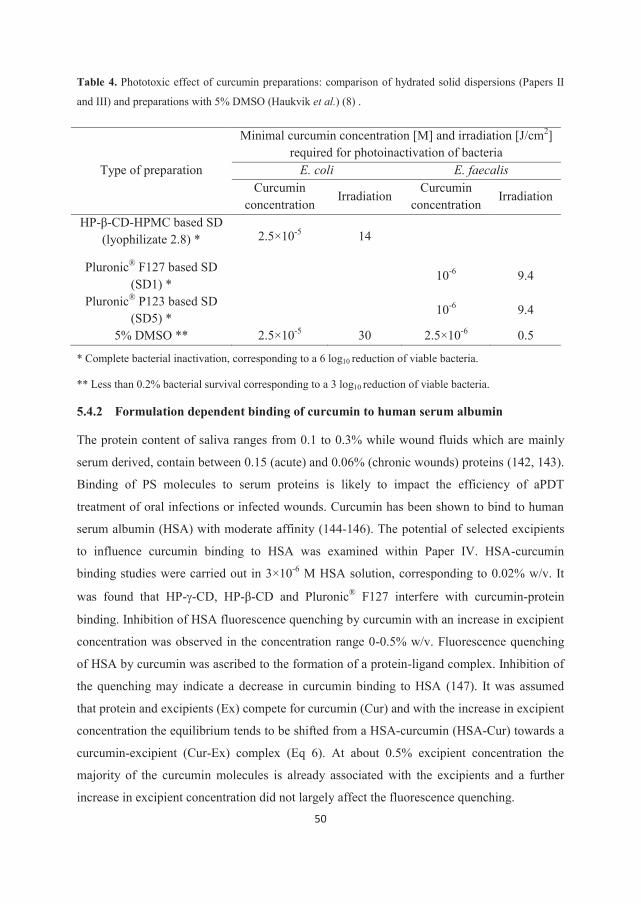
50
Table 4. Phototoxic effect of curcumin preparations: comparison of hydrated solid dispersions (Papers II
and III) and preparations with 5% DMSO (Haukvik et al.) (8) .
Type of preparation
Minimal curcumin concentration [M] and irradiation [J/cm2] required for photoinactivation of bacteria
E. coli E. faecalis Curcumin
concentration Irradiation
Curcumin concentration
Irradiation
HP-β-CD-HPMC based SD (lyophilizate 2.8) * 2.5×10-5 14
Pluronic® F127 based SD (SD1) *
10-6 9.4
Pluronic® P123 based SD (SD5) *
10-6 9.4
5% DMSO ** 2.5×10⁻⁵ 30 2.5×10⁻⁶ 0.5
* Complete bacterial inactivation, corresponding to a 6 log10 reduction of viable bacteria.
** Less than 0.2% bacterial survival corresponding to a 3 log10 reduction of viable bacteria.
5.4.2 Formulation dependent binding of curcumin to human serum albumin
The protein content of saliva ranges from 0.1 to 0.3% while wound fluids which are mainly
serum derived, contain between 0.15 (acute) and 0.06% (chronic wounds) proteins (142, 143).
Binding of PS molecules to serum proteins is likely to impact the efficiency of aPDT
treatment of oral infections or infected wounds. Curcumin has been shown to bind to human
serum albumin (HSA) with moderate affinity (144-146). The potential of selected excipients
to influence curcumin binding to HSA was examined within Paper IV. HSA-curcumin
binding studies were carried out in 3×10⁻⁶ M HSA solution, corresponding to 0.02% w/v. It
was found that HP- -CD, HP-β-CD and Pluronic® F127 interfere with curcumin-protein
binding. Inhibition of HSA fluorescence quenching by curcumin with an increase in excipient
concentration was observed in the concentration range 0-0.5% w/v. Fluorescence quenching
of HSA by curcumin was ascribed to the formation of a protein-ligand complex. Inhibition of
the quenching may indicate a decrease in curcumin binding to HSA (147). It was assumed
that protein and excipients (Ex) compete for curcumin (Cur) and with the increase in excipient
concentration the equilibrium tends to be shifted from a HSA-curcumin (HSA-Cur) towards a
curcumin-excipient (Cur-Ex) complex (Eq 6). At about 0.5% excipient concentration the
majority of the curcumin molecules is already associated with the excipients and a further
increase in excipient concentration did not largely affect the fluorescence quenching.
51
HSA-Cur + Ex HSA + Cur + Ex HSA + Cur-Ex (6)
This model did not take into account the possible interaction between HSA and excipients.
Relying on literature data and the results obtained within the present study it was assumed
that excipient-protein interactions are not considerably influencing HSA-curcumin binding.
However, it is reasonable to expect excipient-protein interactions, particularly at high
excipient concentrations.
The study of curcumin fluorescence spectra showed that an increase from low (0.05% CDs,
0.19% PF127) to high (5% CDs, 5.04% PF127) excipient concentration at constant protein
concentration resulted in evident change in the curcumin microenvironment. The change in
excipient concentration indicated a shift from the complex with HSA towards the complex
with the excipients and thus confirmed the findings of the fluorescence quenching study.
PEG 400 also influenced the HSA fluorescence quenching by curcumin but in the different
manner than PF127 and CDs. Constant inhibition of the HSA fluorescence quenching was
observed over the whole investigated PEG 400 concentration range (0-5%). The study of
curcumin fluorescence spectra showed that even at the highest examined PEG 400
concentration curcumin was still associated with HSA. It has been suggested that the decrease
in fluorescence quenching is caused by a PEG 400-HSA interaction.
A considerable impact of proteins on curcumin aPDT effect might be expected if the excipient
concentration at the application site is low (e.g., supersaturated solutions) or excipients have
low affinity for PS. Formulations leading to high excipient concentration upon application
(e.g., curcumin solubilized by complexation with CDs or micellar solutions) with high affinity
for PS may be less affected by the presence of proteins.
52
6. Conclusion Any modification of the production of bulk material or the final product that may
introduce a change in the physical state of the PDS can alter the photostability of PDS
and the final product in the solid state. Therefore, the physical state of the PDS should
be assessed after a modification of the production method accompanied by a
photostability study of the bulk material/final product according to the ICH Guidelines
(6). The type and concentration of excipients can strongly influence the photostability
of the PDS in solution. The overall effect is difficult to predict and each combination
must be investigated separately.
A supersaturated solution increases the thermodynamic activity of the photosensitizer
curcumin and enhances the delivery to the target bacterial cells thereby enhancing the
efficiency of aPDT. Furtheremore, solid dispersions which can create the state of
supersaturaton provide a fast delivery of the PS. This is one of the requirements for the
drug delivery systems aimed for aPDT.
Supersaturated solutions need to be physically stabilized for the time period sufficient
for both in vivo interaction and the short in vitro storage period. Although both the
HP-β-CD-HPMC combination and Pluronics® F127 and P123 provided satisfactory
physical and hydrolytic stability of curcumin in the solution, Pluronics® were shown
to be better precipitation inhibitors, emphasizing the importance of hydrophobic
interactions between curcumin and the PI.
The type and concentration of excipients affect curcumin binding to plasma protein
(HSA). Cyclodextrins and PF-127 seem to compete with HSA for curcumin, whereas
PEG 400 appears to affect binding differently. The complexation of curcumin with
plasma proteins can in turn influence aPDT efficiency through e.g., changed uptake of
PS by the target cells or modified photoreactivity of the PS.
53
References
1. Aulton ME. Pharmaceutics: the science of the dosage form design. Second edition
London, UK: Churchill Livingstone; 2002.
2. Tønnesen HH. Introduction: photostability testing of drugs and drug formulations -
why and how? In Tønnesen HH, editor. Photostability of drugs and drug formulations. Second
edition Boca Raton, USA: CRC Press LLC; 2004. p. 1-9.
3. Tønnesen HH. Formulation approaches for improving solubility and its impact on drug
photostability. In Tønnesen HH, editor. Photostability of drugs and drug formulations. Second
edition Boca Raton, USA: CRC Press LLC; 2004. p. 351-73.
4. Aman W, Thoma K. The influence of formulation and manufacturing process on the
photostability of tablets. Int J Pharm. 2002;243(1-2):33-41.
5. Kristensen S. Photostability of parenteral products. In Tønnesen HH, editor.
Photostability of drugs and drug formulations. Second edition Boca Raton, USA: CRC Press
LLC; 2004. p. 303-31.
6. ICH Q1B Harmonised tripartite guideline on stability testing: photostability testing of
new drug substances and products. Fed Reg. 1997; 62, 27115-22.
7. Ahmad I, Vaid FHM. Photochemistry of flavins in aqueous and organic solvents.
Compr Ser Photochem Photobiol Sci. 2006;6:13-40.
8. Haukvik T, Bruzell E, Kristensen S, Tønnesen HH. Photokilling of bacteria by
curcumin in different aqueous preparations. Studies on curcumin and curcuminoids XXXVII.
Pharmazie. 2009;64(10):666-73.
9. Allen JM, Allen SK. Basic principles of drug photostability testing. In Piechocki JT
and Thoma K, editors. Pharmaceutical photostability and stabilization technology. New York,
USA: Informa Healthcare; 2007. p. 79-87.
10. Evans RC, Douglas P, Burrow HD, editors. Applied Photochemistry. New York, USA:
Springer; 2013.
54
11. Moore DE. Photophysical and photochemical aspects of drug stability. In Tønnesen
HH, editor. Photostability of drugs and drug formulations. Second edition Boca Raton, USA:
CRC Press LLC; 2004. p. 9-41.
12. Dolmans DEJGJ, Fukumura D, Jain RK. Timeline: Photodynamic therapy for cancer.
Nat Rev Cancer. 2003;3(5):380-7.
13. Garland MJ, Cassidy CM, Woolfson D, Donnelly RF. Designing photosensitizers for
photodynamic therapy: strategies, challenges and promising developments. Future Med Chem.
2009;1(4):667-91.
14. Greenhill JV, McLelland MA. Photodecomposition of drugs. Prog Med Chem.
1990;27:51-121.
15. Glass BD, Novak C, Brown ME. The thermal and photostability of solid
pharmaceuticals. J Therm Anal Calorim. 2004;77(3):1013-36.
16. Moore DE. Standardization of kinetic studies of photodegradation reactions. In
Tønnesen HH, editor. Photostability of drugs and drug formulations. Second edition Boca
Raton, USA: CRC Press LLC; 2004. p. 41-67.
17. Tønnesen HH, Kristensen S. In vitro screening of the photoreactivity of antimalarials:
a test case. In Tønnesen HH, editor. Photostability of drugs and drug formulations. Second
edition Boca Raton, USA: CRC Press LLC; 2004. p. 213-35.
18. Craig RA, McCoy CP, Gorman SP, Jones DS. Photosensitisers - the progression from
photodynamic therapy to anti-infective surfaces. Expert Opin Drug Delivery. 2015;12(1):85-
101.
19. Dai T, Huang Y-Y, Hamblin MR. Photodynamic therapy for localized infections -
state of the art Photodiagnosis Photodyn Ther. 2009;6(3-4):170-88.
20. Cassidy CM, Tunney MM, McCarron PA, Donnelly RF. Drug delivery strategies for
photodynamic antimicrobial chemotherapy: from benchtop to clinical practice. J Photochem
Photobiol, B. 2009;95(2):71-80
21. Jori G, Camerin M, Soncin M, Guidolin L, Coppellotti O. Antimicrobial
photodynamic therapy: basic principles. In Hamblin MR, Jori G, editors. Photodynamic
55
inactivation of microbial pathogens: medical and environmental Applications. London, UK:
RSC Publishing; 2011. p. 1-19.
22. Jori G. Photodynamic therapy of microbial infections: state of the art and perspectives.
J Environ Pathol Toxicol Oncol. 2006;25(1-2):505-19.
23. Hegge AB. Formulation of a hydrophobic photosensitizer for topical photodynamic
therapy. Oslo, Norway: University of Oslo; 2010.
24. Winckler KD. Special section: Focus on anti-microbial photodynamic therapy (PDT).
J Photochem Photobiol, B. 2007;86(1):43-4.
25. Vallner JJ. Binding of drugs by albumin and plasma protein. J Pharm Sci.
1977;66(4):447-65.
26. Fanali G, di Masi A, Trezza V, Marino M, Fasano M, Ascenzi P. Human serum
albumin: from bench to bedside. Mol Aspects Med. 2012;33(3):209-90.
27. Castano AP, Demidova TN, Hamblin MR. Mechanisms in photodynamic therapy: part
three - photosensitizer pharmacokinetics, biodistribution, tumor localization and modes of
tumor destruction. Photodiagn Photodyn Ther. 2005;2(2):91-106.
28. Bose B, Dube A. Interaction of Chlorin p6 with bovine serum albumin and
photodynamic oxidation of protein. J Photochem Photobiol, B. 2006;85(1):49-55.
29. Pandey RK, Constantine S, Tsuchida T, Zheng G, Medforth CJ, Aoudia M, et al.
Synthesis, photophysical properties, in vivo photosensitizing efficacy, and human serum
albumin binding properties of some novel bacteriochlorins. J Med Chem. 1997;40(17):2770-9.
30. Cunderlikova B, Sikurova L, Moan J. pH, serum proteins and ionic strength influence
the uptake of merocyanine 540 by WiDr cells and its interaction with membrane structures.
Bioelectrochemistry. 2003;59(1-2):1-10.
31. Lang K, Mosinger J, Wagnerova DM. Photophysical properties of porphyrinoid
sensitizers non-covalently bound to host molecules; models for photodynamic therapy. Coord
Chem Rev. 2004;248(3-4):321-50.
56
32. Moan J, Juzenas P. Biological effects of combinations of drugs and light. In Tønnesen
HH, editor. Photostability of drugs and drug formulations. Second edition Boca Raton, USA:
CRC Press LLC; 2004. p. 189-213.
33. Moan J, Rimington C, Malik Z. Photoinduced degradation and modification of
Photofrin II in cells in vitro. Photochem Photobiol. 1988;47(3):363-7.
34. Krieg M, Whitten DG. Self-sensitized photooxidation of protoporphyrin IX and
related free-base porphyrins in natural and model membrane systems. Evidence for novel
photooxidation pathways involving amino acids. J Am Chem Soc. 1984;106(8):2477-9.
35. Hamblin MR, Hasan T. Photodynamic therapy: a new antimicrobial approach to
infectious disease? Photochem Photobiol Sci. 2004;3(5):436-50.
36. Nitzan Y, Balzam-Sudakevitz A, Ashkenazi H. Eradication of Acinetobacter
baumannii by photosensitized agents in vitro. J Photochem Photobiol B. 1998;42(3):211-8.
37. Komerik N, Wilson M. Factors influencing the susceptibility of Gram-negative
bacteria to toluidine blue O-mediated lethal photosensitization. J Appl Microbiol.
2002;92(4):618-23.
38. Lambrechts SAG, Aalders MCG, Verbraak FD, Lagerberg JWM, Dankert JB,
Schuitmaker JJ. Effect of albumin on the photodynamic inactivation of microorganisms by a
cationic porphyrin. J Photochem Photobiol, B. 2005;79(1):51-7.
39. Wilson M, Mia N. Effect of environmental factors on the lethal photosensitization of
Candida albicans in vitro. Laser Med Sci. 1994;9(2):105-9.
40. Chen J, Cesario TC, Rentzepis PM. Rationale and mechanism for the low
photoinactivation rate of bacteria in plasma. Proc Natl Acad Sci U S A. 2014;111(1):33-8.
41. Schiffner U, Cachovan G, Bastian J, Sculean A, Eick S. In vitro activity of
photoactivated disinfection using a diode laser in infected root canals. Acta Odontol Scand.
2014;72(8):673-80.
42. Tønnesen HH. Formulation and stability testing of photolabile drugs. Int J Pharm.
2001;225(1-2):1-14.
57
43. Nyqvist H, Wadsten T. Preformulation of solid dosage forms: light stability testing of
polymorphs as a part of a preformulation program. Acta Pharm Technol. 1986;32(3):130-2.
44. Chongprasert S, Griesser UJ, Bottorff AT, Williams NA, Byrn SR, Nail SL. Effects of
freeze-dry processing conditions on the crystallization of pentamidine isethionate. J Pharm
Sci. 1998;87(9):1155-60.
45. Wikene KO, Hegge AB, Bruzell E, Tønnesen HH. Formulation and characterization of
lyophilized curcumin solid dispersions for antimicrobial photodynamic therapy (aPDT):
studies on curcumin and curcuminoids LII. Drug Dev Ind Pharm. 2014:1-9.
46. Bouma M, Nuijen B, Sava G, Perbellini A, Flaibani A, van Steenbergen MJ, et al.
Pharmaceutical development of a parenteral lyophilized formulation of the antimetastatic
ruthenium complex NAMI-A. Int J Pharm. 2002;248(1-2):247-59.
47. Loftsson T, Jarho P, Masson M, Jaervinen T. Cyclodextrins in drug delivery. Expert
Opin Drug Delivery. 2005;2(2):335-51.
48. Asker AF, Habib MJ. Effect of certain stabilizers on photobleaching of riboflavin
solutions. Drug Dev Ind Pharm. 1990;16(1):149-56.
49. Konan YN, Gurny R, Allemann E. State of the art in the delivery of photosensitizers
for photodynamic therapy. J Photochem Photobiol, B. 2002;66(2):89-106.
50. Donnelly RF, Cassidy CM, Tunney MM. Photosensitizer delivery for photodynamic
antimicrobial chemotherapy. In Hamblin MR, Jori G, editors. Photodynamic inactivation of
microbial pathogens: medical and environmental applications. London, UK: RSC Publishing;
201. P. 187-216.
51. Jori G, Fabris C, Soncin M, Ferro S, Coppellotti O, Dei D, et al. Photodynamic
therapy in the treatment of microbial infections: basic principles and perspective applications.
Lasers Surg Med. 2006;38(5):468-81.
52. Jori G, Coppellotti O. Inactivation of pathogenic microorganisms by photodynamic
techniques: mechanistic aspects and perspective applications. Anti-Infect Agents Med Chem.
2007;6(2):119-31.
58
53. Felton LA. Physicochemical principles of pharmacy, 4th Edition, Florence AT and
Attwood D. Drug Dev Ind Pharm. 2007;33(2):209.
54. Higuchi T. Physical chemical analysis of percutaneous absorption process from
creams and ointments. J Soc Cosmet Chem. 1960;11:85-97.
55. Brouwers J, Brewster ME, Augustijns P. Supersaturating drug delivery systems: the
answer to solubility-limited oral bioavailability? J Pharm Sci. 2009;98(8):2549-72.
56. Rodriguez-Hornedo N, Murphy D. Significance of controlling crystallization
mechanisms and kinetics in pharmaceutical systems. J Pharm Sci. 1999;88(7):651-60.
57. Brewster ME, Vandecruys R, Peeters J, Neeskens P, Verreck G, Loftsson T.
Comparative interaction of 2-hydroxypropyl-β-cyclodextrin and sulfobutylether-β-
cyclodextrin with itraconazole: phase-solubility behavior and stabilization of supersaturated
drug solutions. Eur J Pharm Sci. 2008;34(2-3):94-103.
58. Janssens S, Van den Mooter G. Review: physical chemistry of solid dispersions. J
Pharm Pharmacol. 2009;61(12):1571-86.
59. Vasconcelos T, Sarmento B, Costa P. Solid dispersions as strategy to improve oral
bioavailability of poor water soluble drugs. Drug Discovery Today. 2007;12(23&24):1068-75.
60. Leuner C, Dressman J. Improving drug solubility for oral delivery using solid
dispersions. Eur J Pharm Biopharm. 2000;50(1):47-60.
61. Choudhary S, Kishore N. Drug-protein interactions in micellar media: thermodynamic
aspects. J Colloid Interface Sci. 2014;413:118-26.
62. Lu J, Owen SC, Shoichet MS. Stability of self-assembled polymeric micelles in serum.
Macromolecules (Washington, DC, U S). 2011;44(15):6002-8.
63. Buggins TR, Dickinson PA, Taylor G. The effects of pharmaceutical excipients on
drug disposition. Adv Drug Delivery Rev. 2007;59(15):1482-503.
64. Egan TD, Kern SE, Johnson KB, Pace NL. The pharmacokinetics and
pharmacodynamics of propofol in a modified cyclodextrin formulation (Captisol) versus
propofol in a lipid formulation (Diprivan): an electroencephalographic and hemodynamic
study in a porcine model. Anesth Analg. 2003;97(1):72-9.
59
65. Piel G, Evrard B, Van Hees T, Delattre L. Comparison of the i.v. pharmacokinetics in
sheep of miconazole-cyclodextrin solutions and a micellar solution. Int J Pharm.
1999;180(1):41-5.
66. Kurkov SV, Loftsson T, Messner M, Madden D. Parenteral delivery of HPβCD:
effects on drug-HSA binding. AAPS Pharm Sci Tech. 2010;11(3):1152-8.
67. Uekama K. Cyclodextrins in drug delivery systems. Adv Drug Delivery Rev.
1999;36(1):1-2.
68. Monti S, Sortino S. Photoprocesses of photosensitizing drugs within cyclodextrin
cavities. Chem Soc Rev. 2002;31(6):371.
69. Partyka M, Au BH, Evans CH. Cyclodextrins as phototoxicity inhibitors in drug
formulations: studies on model systems involving naproxen and β-cyclodextrin. J Photochem
Photobiol, A. 2001;140(1):67-74.
70. Massey V. The chemical and biological versatility of riboflavin. Biochem Soc Trans.
2000;28(4):283-96.
71. Goldsmith G. Riboflavin Deficiency. In Rivlin R, editor. Riboflavin. New York, US:
Springer; 1975. P. 221-302.
72. Powers HJ. Riboflavin (vitamin B-2) and health. Am J Clin Nutr. 2003;77(6):1352-60.
73. DeRitter E. Vitamins in pharmaceutical formulations. J Pharm Sci. 1982;71(10):1073-
96.
74. Allwood MC, Kearney MCJ. Compatibility and stability of additives in parenteral
nutrition admixtures. Nutrition. 1998;14(9):697-706.
75. Chen MF, Boyce HWJr, Triplett L. Stability of the B vitamins in mixed parenteral
nutrition solution. JPEN, J Parenter Enteral Nutr. 1983;7(5):462-4.
76. Smith JL, Canham JE, Wells PA. Effect of phototherapy light, sodium bisulfite, and
pH on vitamin stability in total parenteral nutrition admixtures. JPEN, J Parenter Enteral Nutr.
1988;12(4):394-402.
60
77. Min DB, Boff JM. Chemistry and reaction of singlet oxygen in foods. Compr Rev
Food Sci Food Saf. 2002;1(1):58-72.
78. Habib MJ, Asker AF. Photostabilization of riboflavin by incorporation into liposomes.
J Parenter Sci Technol. 1991;45(3):124-7.
79. Loukas YL, Jayasekera P, Gregoriadis G. Novel liposome-based multicomponent
systems for the protection of photolabile agents. Int J Pharm. 1995;117(1):85-94.
80. Sue-Chu M, Kristensen S, Tønnesen HH. Photoinduced color changes in two different
qualities of riboflavin in the solid state and in various tablet formulations. Photoreactivity of
biologically active compounds. XX. Pharmazie. 2009;64(7):428-35.
81. Sue-Chu M, Kristensen S, Tønnesen HH. Influence of lag-time between light exposure
and color evaluation of riboflavin in the solid state. Pharmazie. 2008;63(7):545-6.
82. Heelis PF. The photophysical and photochemical properties of flavins (isoalloxazines).
Chem Soc Rev. 1982;11(1):15-39.
83. Müller F. Chemistry and biochemistry of flavoenzymes, vol. 1–3. Boca Raton, USA:
CRC Press; 1992.
84. Hemmerich P, Knappe W-R, Kramer HEA, Traber R. Distinction of 2e- and 1e-
reduction modes of the flavin chromophore as studied by flash photolysis. Eur J Biochem.
1980;104(2):511-20.
85. Priyadarsini KI. Photophysics, photochemistry and photobiology of curcumin: studies
from organic solutions, bio-mimetics and living cells. J Photochem Photobiol, C.
2009;10(2):81-95.
86. Haukvik T, Bruzell E, Kristensen S, Tønnesen HH. Photokilling of bacteria by
curcumin in selected polyethylene glycol 400 (PEG 400) preparations: studies on curcumin
and curcuminoids, XLI. Pharmazie. 2010;65(8):600-6.
87. Dovigo LN, Pavarina AC, Carmello JC, Machado AL, Brunetti IL, Bagnato VS.
Susceptibility of clinical isolates of Candida to photodynamic effects of curcumin. Lasers
Surg Med. 2011;43(9):927-34.
61
88. Ribeiro APD, Pavarina AC, Dovigo LN, Brunetti IL, Bagnato VS, Vergani CE, et al.
Phototoxic effect of curcumin on methicillin-resistant Staphylococcus aureus and L929
fibroblasts. Lasers Med Sci. 2013;28(2):391-8.
89. Dovigo LN, Pavarina AC, Ribeiro APD, Brunetti IL, Costa CAdS, Jacomassi DP, et al.
Investigation of the photodynamic effects of curcumin against Candida albicans. Photochem
Photobiol. 2011;87(4):895-903.
90. Dovigo LN, Carmello JC, Costa CAdS, Vergani CE, Brunetti IL, Bagnato VS, et al.
Curcumin-mediated photodynamic inactivation of Candida albicans in a murine model of
oral candidiasis. Med Mycol. 2013;51(3):243-51.
91. Hegge AB, Nielsen TT, Larsen KL, Bruzell E, Tønnesen HH. Impact of curcumin
supersaturation in antibacterial photodynamic therapy - effect of cyclodextrin type and
amount: studies on curcumin and curcuminoides XLV. J Pharm Sci. 2012;101(4):1524-37.
92. Goel A, Kunnumakkara AB, Aggarwal BB. Curcumin as "curcumin": from kitchen to
clinic. Biochem Pharmacol. 2008;75(4):787-809.
93. Pabon HJJ. Synthesis of curcumin and related compounds. Recl Trav Chim Pays-Bas.
1964;83(4):379-86.
94. Tønnesen HH, Masson M, Loftsson T. Studies of curcumin and curcuminoids. XXVII.
Cyclodextrin complexation: solubility, chemical and photochemical stability. Int J Pharm.
2002;244(1-2):127-35.
95. Tønnesen HH, Karlsen J. Alkaline degradation of curcumin: studies on curcumin and
curcuminoids V. Z Lebensm-Unters Forsch. 1985;180(2):132-4.
96. Nardo L, Paderno R, Andreoni A, Masson M, Haukvik T, Tønnesen HH. Role of H-
bond formation in the photoreactivity of curcumin. Spectroscopy. 2008;22(2,3):187-98.
97. Tønnesen HH, Karlsen J, van Henegouwen GB. Photochemical stability of curcumin:
studies on curcumin and curcuminoids VIII. Z Lebensm Unters Forsch. 1986;183(2):116-22.
98. Singh R, Kristensen S, Tønnesen HH. Influence of vehicle properties and excipients
on hydrolytic and photochemical stability of curcumin in preparations containing pluronics:
studies of curcumin and curcuminoids XLVIII. Pharmazie. 2013;68(3):160-9.
62
99. Tomren MA, Masson M, Loftsson T, Tønnesen HH. Symmetric and asymmetric
curcuminoids: stability, activity and complexation with cyclodextrins: studies on curcumin
and curcuminoids XXXI. Int J Pharm. 2007;338(1-2):27-34.
100. Tønnesen HH. Solubility, chemical and photochemical stability of curcumin in
surfactant solutions: studies of curcumin and curcuminoids, XXVIII. Pharmazie.
2002;57(12):820-4.
101. Dahl TA, McGowan WM, Shand MA, Srinivasan VS. Photokilling of bacteria by the
natural dye curcumin. Arch Microbiol. 1989;151(2):183-5.
102. Singh R, Tønnesen HH, Kristensen S, Berg K. The influence of Pluronics on dark
cytotoxicity, photocytotoxicity, localization and uptake of curcumin in cancer cells: studies on
curcumin and curcuminoids XLIX. Photochem Photobiol Sci. 2013;12(3):559-75.
103. Tøennesen HH, de Vries H, Karlsen J, Beijersbergen van Henegouwen G.
Investigation of the photobiological activity of curcumin using bacterial indicator systems:
studies on curcumin and curcuminoids IX. J Pharm Sci. 1987;76(5):371-3.
104. Park K, Lee J-H. Photosensitizer effect of curcumin on UVB-irradiated HaCaT cells
through activation of caspase pathways. Oncol Rep. 2007;17(3):537-40.
105. Chignell CF, Bilski P, Reszka KJ, Motten AG, Sik RH, Dahl TA. Spectral and
photochemical properties of curcumin. Photochem Photobiol. 1994;59(3):295-302.
106. Haukvik T. Curcumin as a photosensitizer in antimicrobial photodynamic therapy:
influence of formulation and molecular structure modification. Oslo, Norway: University of
Oslo; 2012.
107. Jiang Y, Leung AW, Hua H, Rao X, Xu C. Photodynamic action of LED-activated
curcumin against Staphylococcus aureus involving intracellular ROS increase and membrane
damage. Int J Photoenergy. 2014:637601/1-/8.
108. Bulit F, Grad I, Manoil D, Filieri A, Simon S, Wataha JC, et al. Antimicrobial activity
and cytotoxicity of 3 photosensitizers activated with blue light. J Endod. 2014;40(3):427-31.
63
109. Leite DPV, Paolillo FR, Parmesano TN, Fontana CR, Bagnato VS. Effects of
photodynamic therapy with blue light and curcumin as mouth rinse for oral disinfection: a
randomized controlled trial. Photomed Laser Surg. 2014;32(11):627-32.
110. Araujo NC, Fontana CR, Gerbi MEM, Bagnato VS. Overall-mouth disinfection by
photodynamic therapy using curcumin. Photomed Laser Surg. 2012;30(2):96-101.
111. Singh R. Investigation and development of aqueous curcumin preparations for
photodynamic therapy by use of nanovehicles. Oslo, Norway: Univeristy of Oslo; 2013.
112. Strimpakos AS, Sharma RA. Curcumin: preventive and therapeutic properties in
laboratory studies and clinical trials. Antioxid Redox Signaling. 2008;10(3):511-46.
113. Wegiel LA, Zhao Y, Mauer LJ, Edgar KJ, Taylor LS. Curcumin amorphous solid
dispersions: the influence of intra and intermolecular bonding on physical stability. Pharm
Dev Technol. 2014;19(8):976-86.
114. Sanphui P, Goud NR, Khandavilli UBR, Bhanoth S, Nangia A. New polymorphs of
curcumin. Chem Commun. 2011;47(17):5013-5.
115. Hilfiker R, editor. Polymorphism: In the pharmaceutical industry. Weinheim,
Germany: Wiley-VCH Verlag; 2006.
116. Gloor A, inventor; DSM IP Assets B. V., Neth., assignee. Process for the purification
of riboflavin. Patent WO2005014594A1. 2005.
117. Mohnicke M. On the control of riboflavin (vitamin B2) crystal structures. Aachen,
Germany: Shaker Verlag; 2007.
118. van Drooge DJ, Hinrichs WLJ, Visser MR, Frijlink HW. Characterization of the
molecular distribution of drugs in glassy solid dispersions at the nano-meter scale, using
differential scanning calorimetry and gravimetric water vapour sorption techniques. Int J
Pharm. 2006;310(1-2):220-9.
119. Repka MA, Battu SK, Upadhye SB, Thumma S, Crowley MM, Zhang F, et al.
Pharmaceutical applications of hot-melt extrusion: Part II. Drug Dev Ind Pharm.
2007;33(10):1043-57.
64
120. Crowley MM, Zhang F, Repka MA, Thumma S, Upadhye SB, Battu SK, et al.
Pharmaceutical applications of hot-melt extrusion: part I. Drug Dev Ind Pharm.
2007;33(9):909-26.
121. Van Eerdenbrugh B, Baird JA, Taylor LS. Crystallization tendency of active
pharmaceutical ingredients following rapid solvent evaporation-classification and comparison
with crystallization tendency from undercooled melts. J Pharm Sci. 2010;99(9):3826-38.
122. Hegge AB, Masson M, Kristensen S, Tøennesen HH. Investigation of curcumin-
cyclodextrin inclusion complexation in aqueous solutions containing various alcoholic co-
solvents and alginates using an UV-VIS titration method: studies of curcumin and
curcuminoides XXXV. Pharmazie. 2009;64(6):382-9.
123. Alexandridis P, Alan Hatton T. Poly(ethylene oxide)-poly(propylene oxide)-
poly(ethylene oxide) block copolymer surfactants in aqueous solutions and at interfaces:
thermodynamics, structure, dynamics, and modeling. Colloids Surf, A. 1995;96(1/2):1-46.
124. Ebitani M, Kashiwagi H, Inoue M, Nishibe K. Preparation, stability, and compositions
of the crystals of reduced riboflavins. Bull Chem Soc Jpn. 1991;64(4):1093-6.
125. Mueller F, Hemmerich P, Ehrenberg A, Palmer G, Massey V. Chemical and electronic
structure of the neutral flavin radical as revealed by electron spin resonance spectroscopy of
chemically and isotopically substituted derivatives. Eur J Biochem. 1970;14(1):185-96.
126. Penzkofer A. Reduction-oxidation photocycle dynamics of flavins in starch films. Int J
Mol Sci. 2012;13:9157-83.
127. Ramamurthy V, Venkatesan K. Photochemical reactions of organic crystals. Chem
Rev. 1987;87(2):433-81.
128. Mandal S, Banerjee C, Ghosh S, Kuchlyan J, Sarkar N. Modulation of the
photophysical properties of curcumin in nonionic surfactant (Tween® 20) forming micelles
and niosomes: a comparative study of different microenvironments. J Phys Chem B.
2013;117(23):6957-68.
129. Ghosh S, Kuchlyan J, Banik D, Kundu N, Roy A, Banerjee C, et al. Organic additive,
5-methylsalicylic acid induces spontaneous structural transformation of aqueous Pluronic
65
triblock copolymer solution: a spectroscopic investigation of interaction of curcumin with
Pluronic micellar and vesicular aggregates. J Phys Chem B. 2014.
130. Baglole KN, Boland PG, Wagner BD. Fluorescence enhancement of curcumin upon
inclusion into parent and modified cyclodextrins. J Photochem Photobiol, A.
2005;173(3):230-7.
131. Raghavan SL, Trividic A, Davis AF, Hadgraft J. Crystallization of hydrocortisone
acetate: influence of polymers. Int J Pharm. 2001;212(2):213-21.
132. Gao P, Akrami A, Alvarez F, Hu J, Li L, Ma C, et al. Characterization and
optimization of AMG 517 supersaturatable self-emulsifying drug delivery system (S-SEDDS)
for improved oral absorption. J Pharm Sci. 2009;98(2):516-28.
133. Overhoff KA, McConville JT, Yang W, Johnston KP, Peters JI, Williams RO, III.
Effect of stabilizer on the maximum degree and extent of supersaturation and oral absorption
of tacrolimus made by ultra-rapid freezing. Pharm Res. 2008;25(1):167-75.
134. Dai W-G, Dong LC, Li S, Deng Z. Combination of Pluronic/vitamin E TPGS as a
potential inhibitor of drug precipitation. Int J Pharm. 2008;355(1-2):31-7.
135. Mackellar AJ, Buckton G, Newton JM, Orr CA. The controlled crystallization of a
model powder: 2. Investigation into the mechanism of action of poloxamers in changing
crystal properties. Int J Pharm. 1994;112(1):79-85.
136. Samanta S, Roccatano D. Interaction of curcumin with PEO-PPO-PEO block
copolymers: a molecular dynamics study. J Phys Chem B. 2013;117(11):3250-7.
137. Kurkov SV, Loftsson T. Cyclodextrins. Int J Pharm. 2013;453(1):167-80.
138. Kabanov AV, Alakhov VY. Pluronic block copolymers in drug delivery: from
micellar nanocontainers to biological response modifiers. Crit Rev Ther Drug Carrier Syst.
2002;19(1):1-72.
139. Winter S, Tortik N, Kubin A, Krammer B, Plaetzer K. Back to the roots:
photodynamic inactivation of bacteria based on water-soluble curcumin bound to
polyvinylpyrrolidone as a photosensitizer. Photochem Photobiol Sci. 2013;12(10):1795-802.
66
140. Williams AC, Barry BW. Penetration enhancers. Adv Drug Delivery Rev.
2004;56(5):603-18.
141. Hegge AB, Bruzell E, Kristensen S, Tønnesen HH. Photoinactivation of
Staphylococcus epidermidis biofilms and suspensions by the hydrophobic photosensitizer
curcumin - Effect of selected nanocarrier. Eur J Pharm Sci. 2012;47(1):65-74.
142. Bonilla CA. Human mixed saliva protein concentration. J Dent Res. 1972;51(2)(Pt.
2):664.
143. Moseley R, Hilton JR, Waddington RJ, Harding KG, Stephens P, Thomas DW.
Comparison of oxidative stress biomarker profiles between acute and chronic wound
environments. Wound Repair Regen. 2004;12(4):419-29.
144. Zsila F, Bikadi Z, Simonyi M. Unique, pH-dependent biphasic band shape of the
visible circular dichroism of curcumin-serum albumin complex. Biochem Biophys Res
Commun. 2003;301(3):776-82.
145. Mandeville J-S, Froehlich E, Tajmir-Riahi HA. Study of curcumin and genistein
interactions with human serum albumin. J Pharm Biomed Anal. 2009;49(2):468-74.
146. Reddy ACP, Sudharshan E, Rao AGA, Lokesh BR. Interaction of curcumin with
human serum albumin-a spectroscopic study. Lipids. 1999;34(10):1025-9.
147. Mohammadi F, Bordbar A-K, Divsalar A, Mohammadi K, Saboury AA. Analysis of
binding interaction of curcumin and diacetylcurcumin with human and bovine serum albumin
using fluorescence and circular dichroism spectroscopy. Protein J. 2009;28(3-4):189-96.