Embed Size (px)
Citation preview
http://www.enm-kes.org
Received: 31 March 2011, Accepted: 1 June 2011Corresponding author: Won Bae KimDepartment of Endocrinology and Metabolism, Asan Medical Center, University of Ulsan College of Medicine, 388-1 Pungnap 2-dong, Songpa-gu, Seoul 138-736, KoreaTel: +82-2-3010-3913, Fax: +82-2-3010-6962, E-mail: [email protected]
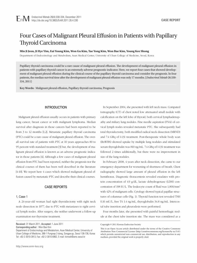
Four Cases of Malignant Pleural Effusion in Patients with Papillary Thyroid CarcinomaMin Ji Jeon, Ji Hye Yim, Eui Young Kim, Won Gu Kim, Tae Yong Kim, Won Bae Kim, Young Kee ShongDepartment of Endocrinology and Metabolism, Asan Medical Center, University of Ulsan College of Medicine, Seoul, Korea
Papillary thyroid carcinoma could be a rare cause of malignant pleural effusion. The development of malignant pleural effusion in patients with papillary thyroid cancer is an extremely adverse prognostic indicator. Here, we report four cases that showed develop-ment of malignant pleural effusion during the clinical course of the papillary thyroid carcinoma and consider the prognosis. In four patients, the median survival time after the development of malignant pleural effusion was only 17 months. (Endocrinol Metab 26:330-334, 2011)
Key Words: Malignant pleural effusion, Papillary thyroid carcinoma, Prognosis
Endocrinol Metab 26(4):330-334, December 2011http://dx.doi.org/10.3803/EnM.2011.26.4.330 CASE REPORT
INTRODUCTION
Malignant pleural effusion usually occurs in patients with primary
lung cancer, breast cancer or with malignant lymphoma. Median
survival after diagnosis in those cancers had been reported to be
from 3 to 12 months [1,2]. Metastatic papillary thyroid carcinoma
(PTC) could be a rare cause of malignant pleural effusion. The over-
all survival rate of patients with PTC at 10 years approaches 80 to
95 percent with standard treatment [3] but, the development of ma-
lignant pleural effusion is known as an adverse prognostic indica-
tor in those patients [4]. Although a few cases of malignant pleural
effusion from PTC had been reported, neither the prognosis nor the
clinical courses of them has been well described in the literature
[4-10]. We report here 4 cases which showed malignant pleural ef-
fusion caused by metastatic PTC and describe their clinical courses.
CASE REPORTS
1. Case 1
A 24-year-old woman had right thyroidectomy with right neck
node dissection in 1977, due to PTC with metastases to right cervi-
cal lymph nodes. After surgery, she neither underwent a follow-up
examination nor thyroxine treatment.
In September 2004, she presented with left neck mass. Computed
tomography (CT) of chest noted low attenuated small nodule with
calcification on the left lobe of thyroid, both cervical lymphadenop-
athy and miliary lung nodules. Fine needle aspiration (FNA) of cer-
vical lymph nodes revealed metastatic PTC. She subsequently had
total thyroidectomy, both modified radical neck dissection (MRND)
and 7.4 GBq of I-131 treatment. Post-therapeutic whole body scan
(RxWBS) showed uptake by multiple lung nodules and stimulated
serum thyroglobulin was 810 ng/mL. 7.4 GBq of I-131 treatment was
followed 2 times additionally, but there were no improvement in
size of the lung nodules.
In February 2008, 4 years after neck dissection, she came to our
emergency department for worsening of shortness of breath. Chest
radiography showed large amount of pleural effusion in the left
hemithorax. Diagnostic thoracentesis revealed exudates with pro-
tein concentration of 4.9 g/dL, lactate dehydrogenase (LDH) con-
centration of 198 IU/L. The leukocyte count of fluid was 1,800/mm3
with 42% of malignant cells. Cytology showed typical papillae struc-
tures of columnar cells (Fig. 1). Thyroid function test revealed TSH
0.16 mU/L, free T4 1.4 ng/mL, thyroglobulin 34.8 ng/mL. Intercos-
tal tube insertion and pleurodesis were performed.
Four months later, she presented with painful hemorrhagic nod-
ule at the chest tube insertion site. The mass was considered as a
This is an Open Access article distributed under the terms of the Creative Commons Attribution Non-Commercial License (http://creativecommons.org/licenses/by-nc/3.0/) which permits unrestricted non-commercial use, distribution, and reproduction in any medium, provided the original work is properly cited.
Copyright © 2011 Korean Endocrine Society
Malignant Pleural Effusion from PTC
http://dx.doi.org/10.3803/EnM.2011.26.4.330
331
http://www.enm-kes.org
metastatic nodule. Radio-frequency ablation and 7.4 GBq of I-131
treatment were followed, but she died because of the uncontrolled
pleural effusion at September 2008, only 6 months after develop-
ment of malignant pleural effusion.
2. Case 2
A 64-year-old woman had total thyroidectomy and right MRND
due to right neck mass confirmed as PTC in 1999. Surgical and path-
ological findings showed 3 cm mass in right thyroid which invaded
perithyroidal soft tissue with right cervical lymph nodes metastasis.
5.6 GBq of I-131 treatment was followed with uptake in thyroid bed.
In August 2000, 11 years after initial diagnosis, stimulated serum
thyroglobulin was 33.7 ng/mL but, I-131 diagnostic whole body scan
(DxWBS) showed no abnormal uptake. Tc-99m MIBI SPECT was
revealed hypermetabolic lesions at left supraclavicular area. She
underwent FNA of lymph nodes and it turned out to be metastatic
PTC. Left MRND was performed in December 2000.
Three years later, she presented with left neck mass again. Neck
ultrasonography (USG) and whole body fluorodeoxyglucose-posi-
tron emission tomography scan showed multiple cervical lymph-
adenopathy and lung nodules. FNA of enlarged cervical lymph node
noted metastatic PTC. She had radiation therapy of cervical area
(23 fractions, 4,600 cGy) and thorax area (10 fractions, 1,800 cGy).
No evidence of residual disease was noted in neck but, serum thy-
roglobulin level was still high (3.0-4.7 ng/mL, TSH 1.6 mU/L).
In July 2008, 4 years after radiation therapy, she sought medical
attention for progressive dyspnea. Chest radiography revealed large
amount of right pleural effusion. Serum thyroglobulin was 10.0 ng/
mL (TSH 1.7 mU/L). Pleural fluid was exudates (Protein 5.2 g/dL,
LDH 191 IU/L) and the leukocyte count of fluid was 300/mm3 in-
cluding 37% of malignant cells. But, cytology of fluid could not di-
agnose the origin of cancer cells (Fig. 2). To evaluate the cause of
pleural effusion, pleural fluid thyroglobulin was checked. It was
115 ng/mL and high enough to diagnose pleural effusion as malig-
nant effusion from thyroid. Chest tube insertion was performed
and pleurodesis with doxycycline 800 mg was followed.
Four months later, chest radiography showed increased amount
of right pleural effusion. 5.6 GBq of I-131 treatment was performed
but, there was no uptake. She died in August 2009, 1 year after di-
agnosis of malignant pleural effusion.
3. Case 3
A 68-year-old man had routine health checkup at May 2002 and
neck USG detected 3.5 cm thyroid nodule. FNA of thyroid nodule
showed typical papillary structure. He had total thyroidectomy and
pathology revealed 5.5 cm thyroid mass that metastasize to the para-
tracheal lymph nodes. 5.6 GBq of I-131 treatment was followed.
RxWBS noted multifocal increased uptakes of iodine in abdomi-
nal cavity, right chest wall, thus CT of chest and abdomen were
performed. Multiple variable sized nodules at basal lungs with 1.3
cm nodule at left adrenal gland were detected and considered as
metastatic PTC. 7.4 GBq I-131 therapy was done twice consecutively
but, RxWBS showed increased number and size of nodules. After
two more 7.4 GBq I-131 treatments, RxWBS revealed no definite
uptakes. However, chest radiography couldn’t show improvement.
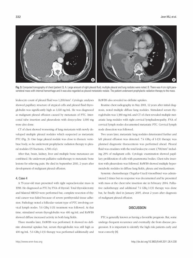
In December 2008, 6 years after initial diagnosis, chest radiogra-
phy detected right pleural effusion. It was hemorrhagic pleural fluid
with protein concentration 4.8 g/dL, LDH 455 IU/L and the total
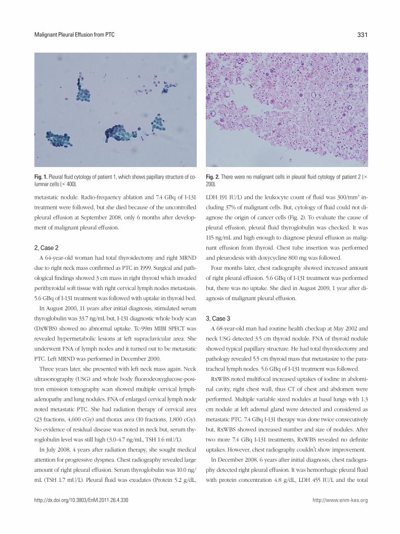
Fig. 1. Pleural fluid cytology of patient 1, which shows papillary structure of co-lumnar cells (× 400).
Fig. 2. There were no malignant cells in pleural fluid cytology of patient 2 (× 200).
Jeon MJ, et al.
http://dx.doi.org/10.3803/EnM.2011.26.4.330
332
http://www.enm-kes.org
leukocyte count of pleural fluid was 1,200/mm3. Cytologic analyses
showed papillary structure of atypical cells and pleural fluid thyro-
globulin was significantly high as 1,610 ng/mL. He was diagnosed
as malignant pleural effusion caused by metastasis of PTC. Inter-
costal tube insertion and pleurodesis with doxycycline 1,000 mg
were also done.
CT of chest showed worsening of lung metastasis with newly de-
veloped multiple pleural nodules which suspected as metastatic
PTC (Fig. 3). One large pleural nodule was close to thoracic verte-
brae body, so he underwent prophylactic radiation therapy to pleu-
ral nodules (15 fractions, 4,500 cGy).
After that, brain, kidney, liver and multiple bone metastases are
combined. He underwent palliative radiotherapy to metastatic bone
lesions for relieving pain. He died in September 2010, 2 years after
development of malignant pleural effusion.
4. Case 4
A 55-year-old man presented with right supraclavicular mass in
1998. He diagnosed as PTC by FNA of thyroid. Total thyroidectomy
and bilateral MRND were performed but, complete resection of thy-
roid cancer was failed because of severe perithyroidal tissue adhe-
sion. Pathology noted a follicular variant type of PTC involving cer-
vical lymph nodes. 5.6 GBq I-131 treatment was followed. At that
time, stimulated serum thyroglobulin was 406 ng/mL and RxWBS
showed diffuse increased activity in both lung fields.
Three months later, DxWBS was performed. It showed no defi-
nite abnormal uptakes but, serum thyroglobulin was still high as
400 ng/mL. 5.6 GBq I-131 therapy was performed additionally and
RxWBS also revealed no definite uptakes.
Routine chest radiography in May 2001, 12 years after initial diag-
nosis, noted multiple diffuse lung nodules. Stimulated serum thy-
roglobulin was 1,380 mg/mL and CT of chest revealed multiple met-
astatic lung nodules with right cervical lymphadenopathy. FNA of
cervical lymph nodes documented metastatic PTC. Cervical lymph
node dissection was followed.
Two years later, metastatic lung nodules deteriorated further and
left pleural effusion was detected. 7.4 GBq of I-131 therapy was
planned diagnostic thoracentesis was performed ahead. Pleural
fluid was exudates with the total leukocyte count 1,700/mm3 includ-
ing 29% of malignant cells. Cytologic examination showed papil-
lary proliferation of cells with psammoma bodies. Chest tube inser-
tion with pleurodesis was followed. RxWBS showed multiple hyper-
metabolic nodules in diffuse lung fields, pleura and mediastinum.
Systemic chemotherapy (Tegafur-Uracil/vinorelbine) was admin-
istered 3 times but no response was documented and he presented
with mass at the chest tube insertion site in February 2004. Pallia-
tive radiotherapy and additional 7.4 GBq I-131 therapy was done
but, he finally died in January 2005, about 2 years after diagnosis
of malignant pleural effusion.
DISCUSSION
PTC is generally known as having a favorable prognosis. But, some
undergo frequent recurrence and eventually die from disease pro-
gression. It is important to identify the high risk patients early and
treat correctly [8].
Fig. 3. Computed tomography of chest (patient 3). A. Large amount of right pleural fluid, multiple pleural and lung nodules were noted. B. There was 4 cm right para-vertebral mass with internal hemorrhage and it was also regarded as pleural metastatic nodule. The patient underwent prophylactic radiation therapy to the mass.
A B
Malignant Pleural Effusion from PTC
http://dx.doi.org/10.3803/EnM.2011.26.4.330
333
http://www.enm-kes.org
In this report, 3 patients (patient 2, 3, and 4 in Table 1) were over
50 years and had cervical lymphadenopathy, extrathyroidal invasion
at the time of initial surgery. Moreover, 2 patients among them (pa-
tient 3 and 4 in Table 1) had distant metastatic lesions at the time of
initial diagnosis. They were considered as high risk group and had
aggressive treatment with total thyroidectomy and radioiodine ab-
lation therapy. But, the disease showed recurrence and progression.
In patient 1 (Table 1), she was young at the time of diagnosis and
disease was stable over 20 years. The metastatic progression ap-
peared late but, disease progression after the appearance of pleural
effusion was extremely rapid.
In retrospective report of Vassilopoulous-sellin and Sneige [8], only
10 patients (0.6%) had malignant pleural effusion during the course
of PTC among 1,772 patients. Regardless of the timing of the devel-
opment of pleural effusion, the disease exhibited aggressive bio-
logic behavior with rapid deterioration and its appearance carries
an adverse prognostic significance. The median overall survival time
was 27 months after the diagnosis of thyroid cancer and 11 months
after the development of pleural effusion [8]. At this report, the me-
dian overall survival time after the development of malignant pleu-
ral effusion of 4 patients was also only 17 months (Table 2).
In reported cases so far, malignant pleural effusion resulting from
metastasis of PTC had been diagnosed when cytologic examina-
tion show papillary structures of epithelial cells in pleural fluid with
or without psammoma bodies [9]. In patient 3 (Table 1), cytology
noted atypical papillary clusters and pleural fluid thyroglobulin was
also high as 1,610 ng/mL (serum thyroglobulin 171 ng/mL). In pa-
tient 2 (Table 1), cytologic examination of pleural fluid could not
identify the primary cancer foci due to degeneration. We checked
thyroglobulin level in pleural fluid. It was 115 ng/mL which was sig-
nificantly higher compared with serum thyroglobulin level (10.0 ng/
mL). We could diagnose the pleural effusion due to metastatic dif-
ferentiated thyroid carcinoma based on pleural fluid thyroglobulin
level. It would be the first reported case that pleural fluid of unknown
cause diagnosed as malignant pleural effusion due to differentiated
thyroid carcinoma by using pleural fluid thyroglobulin level.
During the course of the disease and before the diagnosis of pleu-
ral effusion, radiologically apparent lung metastasis were found in
all cases in this report as well as the report of Vassilopoulous-sellin
and Sneige [8]. However, Jung et al. [6] reported a patient presented
with malignant pleural effusion and pleural nodules without lung
metastasis and Jeong et al. [5] reported malignant pleural effusion
associated with breast metastasis.
Radioiodine therapy, local radiotherapy and systemic chemother-
apy could be applied for the treatment of malignant pleural effusion
from PTC but, nothing was proved to be effective to control malig-
nancy itself [8]. Intercostal tube drainage with intrapleural instilla-
tion of sclerosant was preferred to control symptom, prevent recur-
Table 1. Patients characteristics, extent of disease
Patient No. Age*/Sex Clinical disease extent at initial diagnosis Characteristics of pleural effusion
1 24/F Cervical LNs Lung
WBC 1,800/mm3 (malignant cell 42%) Cytology: metastatic PTC
2 64/F Cervical LNsExtrathyroidal tissueLung
WBC 300/mm3 (malignant cell 37%) Cytology: no evidence of malignancyFluid Tg 115 ng/mLSerum Tg 10 ng/mL
3 68/M Cervical LNsExtrathyroidal tissueLung, adrenal gland bone, kidney, liver brain
WBC 1,200/mm3 (malignant cell 0%) Cytology: metastatic PTCFluid Tg 1,610 ng/mLSerum Tg 171 ng/mL
4 55/M Cervical LNsExtrathyroidal tissue Lung
WBC 1,700/mm3 (malignant cell 29%) Cytology: metastatic PTC, psammoma body
*Age at diagnosis. LNs, lymphnodes; PTC, papillary thyroid carcinoma; WBC, white blood cell.
Table 2. Clinical course of patients
Patient No.Survival time
after the diagnosis of PTC (yr)
Survival time after the development of
pleural effusion (mo)
1 31 6 2 10 13 3 8 21 4 7 21 Median survival time 9 17
PTC, papillary thyroid carcinoma.
Jeon MJ, et al.
http://dx.doi.org/10.3803/EnM.2011.26.4.330
334
http://www.enm-kes.org
rence of effusion and recurrent aspiration [2]. In this review, all 4
patients were treated with chest tube insertion and pleurodesis.
However, patient 1 and 4 was suffered from metastatic nodule at
chest tube insertion site and pleural effusion was recurred in sev-
eral months in patient 1 and 2.
In conclusion, patients with PTC generally have an excellent prog-
nosis but, high risk patients could undergo frequent recurrence and
finally die of disease progression. The development of malignant
pleural effusion from PTC means poor prognosis and patients with
malignant pleural effusion may survive only for several months.
SUMMARY
We report 4 cases that showed development of malignant pleural
effusion during the clinical course of the PTC. Pleural fluid thyro-
globulin is a useful diagnostic marker when cytologic examination
could not identify the origin of malignant pleural effusion. There
were no effective ways to control malignant pleural effusion and
the median survival time after the development of malignant pleu-
ral effusion of 4 patients was only 17 months.
REFERENCES
1.HausheerFH,YarbroJW:Diagnosisandtreatmentofmalignantpleuraleffusion.CancerMetastasisRev6:23-40,1987
2.RobertsME,NevilleE,BerrisfordRG,AntunesG,AliNJ;BTSPleuralDiseaseGuidelineGroup:Managementofamalignantpleuraleffusion:BritishThoracicSocietyPleuralDiseaseGuideline2010.Thorax65:ii32-ii40,2010
3.SchlumbergerMJ:Papillaryandfollicularthyroidcarcinoma.NEnglJMed338:297-306,1998
4.KimJY,ParkDW,NaJO,HwangBY,KimDL,ShinDH,KimSG,ChoiKM,BaikSH,ChoiDS,ChoSJ,KimNH:Acaseofmalignantpleuralef-fusionwithpleuralmetastasisinapatientwithpapillarythyroidcarcino-ma.JKoreanSocEndocrinol17:269-274,2002
5.JeongJ,ShinSY,SonMK,LeeYJ,KimSH,KieJH,ChoiYJ,HongYK,HahnCH,LeeSM,KimCJ:Acaseofpleuralmetastasisfrompapillarythyroidcarcinoma.TubercRespirDis63:188-193,2007
6.JungKH,SeoJA,LeeJH,JoWM,KimJH,ShinC:Acaseofpapillarythyroidcancerpresentingaspleuraleffusion.TubercRespirDis64:314-317,2008
7.SiddarajuN,ViswanathanVK,SakaVK,BasuD,ShanmughamC:Fineneedleaspirationoffollicularvariantofpapillarythyroidcarcinomapre-sentingwithpleuraleffusion:acasereport.ActaCytol51:911-915,2007
8.Vassilopoulou-SellinR,SneigeN:Pleuraleffusioninpatientswithdiffer-entiatedpapillarythyroidcancer.SouthMedJ87:1111-1116,1994
9.VernonAN,SheelerLR,BiscottiCV,StollerJK:Pleuraleffusionresultingfrommetastaticpapillarycarcinomaofthethyroid.Chest101:1448-1450,1992
10.CuervoPinnaMA,MagroLedesmaD,ArrebolaGarcíaJD:Metastaticpleu-raleffusionsecondarytopapillarycarcinomaofthethyroid.ArchBronco-neumol34:566-567,1998

![Management of Malignant Pleural Effusion...Asymptomatic patients with either a malignant or a paramalignant effusion need not be treated initially [9]. Malignant pleural effusion will](https://img.pdfslide.net/doc/110x75/5f8bc67cd3c5026bc44819fe/management-of-malignant-pleural-effusion-asymptomatic-patients-with-either-a.jpg)