Embed Size (px)
Citation preview
159
Journal on New Biological Reports 3(3): 159 – 166 (2014) ISSN 2319 – 1104 (Online)
Four interesting Hyphomycetes from Himachal Pradesh
I. B. Prasher and R.K. Verma*
Mycology & Plant Pathology Laboratory, Department of Botany, Panjab University, Chandigarh 160014
(Received on: 23 July, 2014; accepted on: 31 August, 2014)
ABSTRACT
Dictyosporium heptasporum (Garov.) Damon, Torula ellisii Yadav & Lal, Torula herbarum (Pers.) Link and
Ceratosporium fuscescens Schwein (anamorphic fungi- hyphomycetes), collected from Himachal Pradesh
(North-Western Himalayas), are being described and illustrated. Dictyosporium heptasporum and Torula ellisii
are being recorded for the first time from Himalayas.
Key words: Anamorphic fungi, Hyphomycetes, Himalayas, India.
INTRODUCTION
This communication is in continuation with our
earlier reports on new records of anamorphic fungi
from Himachal Pradesh, North-Western Hmalayas
(Prasher & Verma 2012a, b). During the surveys of
saprobic conidial fungi occurring on dead wood,
branches, bark, twigs and leaves four interesting
hyphomycetes viz. D. heptasporum, T. ellisii. T.
herbarum and C. fuscescens were collected. These
are described and illustrated. Dictyosporium
heptasporum and Torula ellisii are new records for
Himalayas (Bilgrami et al. 1991 and Jamaluddin et
al. 2004).
MATERIALS AND METHOD
Decaying culms, leaves, twigs, bark and dead wood
have been collected into separate ziplock plastic
bags and brought to the laboratory. The specimens
have been mounted on glass slides either in 4%
KOH or Lactophenol Kirk et al. (2008). The
specimens were studied microscopically under
Matrix stereo trinocular microscope (VL-Z60) and
transmission microscope (VRS-2f) for macroscopic
and microscopic characters. All the measurements
have been taken with the help of Pro MED
software. The specimens have been deposited in
herbarium of Department of Botany, Panjab
University (PAN). The map was constructed with
_________________________________________
Corresponding author:
DIVA-GIS 7.5.0 software (Hijmans et al. 2011) by
using the geographical co-ordinates recorded at the
site from where the samples collected to depict the
distribution of species.
RESULTS
Taxonomic description
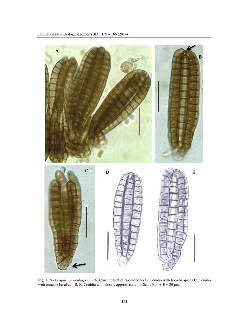
Dictyosporium heptasporum (Garov.)
Damon, Lloydia 15: 118 (1952) Fig. 2
=Cattanea heptaspora Garov., Rc. Ist. Lomb.,
Milano, ser. 2 8: 125 (1875)
=Speira heptaspora (Garov.) Lindau, Rabenh.
Krypt.-Fl., Edn 2 (Leipzig) 1.9: 201 (1908)
Colonies on the natural substratum conspicuous,
black, scattered in the form of compact
sporodochia. Sporodochia 132–416 µm in
diameter. Conidiophores hyaline, thin walled,
short. Conidia 62-75×19-27µm,
cylindrical in
lateral view, clavate, cheiroid in venteral view,
smooth walled, consisting of a subhyaline, cubical
truncate basal cell 4-6.5µm in diameter, on which
5-8 discrete mostly 7discrete vertical rows of cells
are inserted in different plane. These rows of cells
are tightly appressed together as a cylinder and
separate only under pressure. Each row somewhat
recurved or distinctly hooked apices 14-18
eusepate, slightly constricted at septa, unbranched.
Width of the separated arm is 6-7.5µm. Conidia
devoid of appendages.
160
I. B. Prasher and R.K. Verma
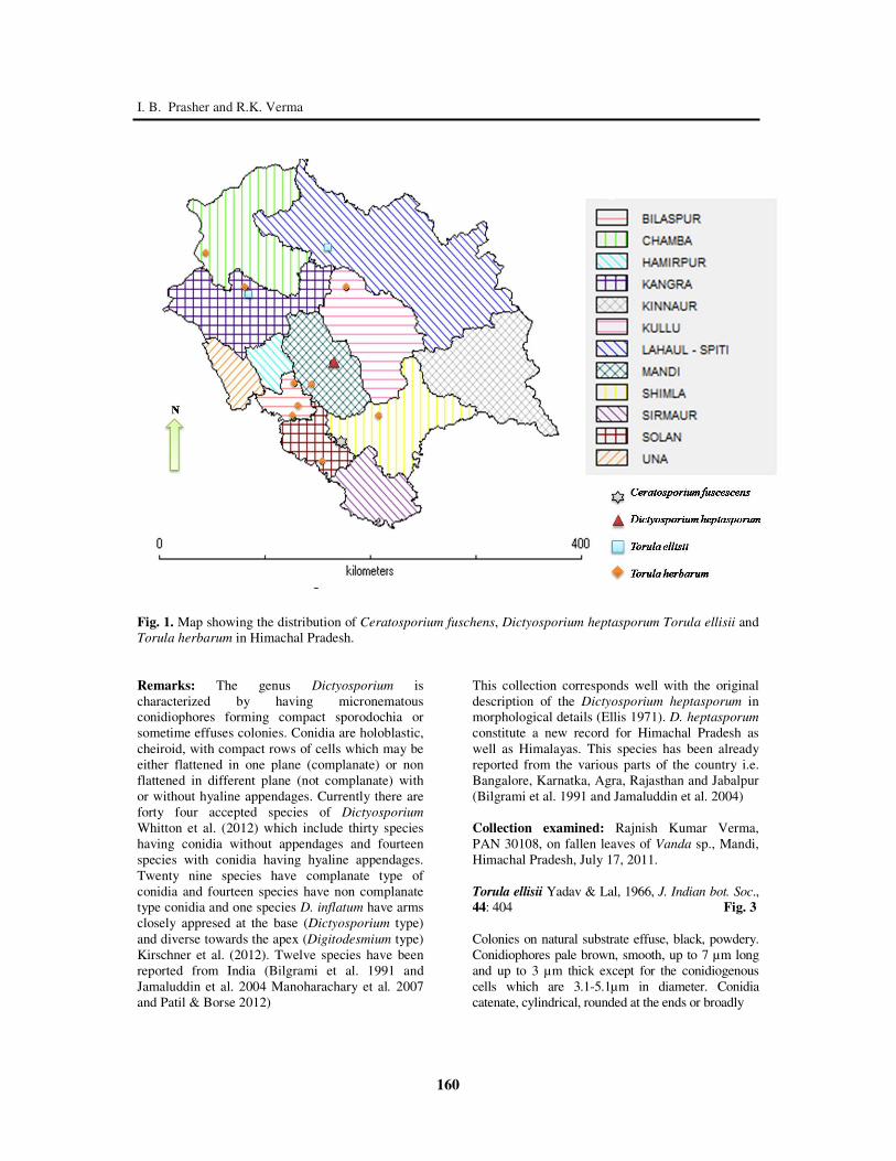
Fig. 1. Map showing the distribution of Ceratosporium fuschens, Dictyosporium heptasporum Torula ellisii and
Torula herbarum in Himachal Pradesh.
Remarks: The genus Dictyosporium is
characterized by having micronematous
conidiophores forming compact sporodochia or
sometime effuses colonies. Conidia are holoblastic,
cheiroid, with compact rows of cells which may be
either flattened in one plane (complanate) or non
flattened in different plane (not complanate) with
or without hyaline appendages. Currently there are
forty four accepted species of Dictyosporium
Whitton et al. (2012) which include thirty species
having conidia without appendages and fourteen
species with conidia having hyaline appendages.
Twenty nine species have complanate type of
conidia and fourteen species have non complanate
type conidia and one species D. inflatum have arms
closely appresed at the base (Dictyosporium type)
and diverse towards the apex (Digitodesmium type)
Kirschner et al. (2012). Twelve species have been
reported from India (Bilgrami et al. 1991 and
Jamaluddin et al. 2004 Manoharachary et al. 2007
and Patil & Borse 2012)
This collection corresponds well with the original
description of the Dictyosporium heptasporum in
morphological details (Ellis 1971). D. heptasporum
constitute a new record for Himachal Pradesh as
well as Himalayas. This species has been already
reported from the various parts of the country i.e.
Bangalore, Karnatka, Agra, Rajasthan and Jabalpur
(Bilgrami et al. 1991 and Jamaluddin et al. 2004)
Collection examined: Rajnish Kumar Verma,
PAN 30108, on fallen leaves of Vanda sp., Mandi,
Himachal Pradesh, July 17, 2011.
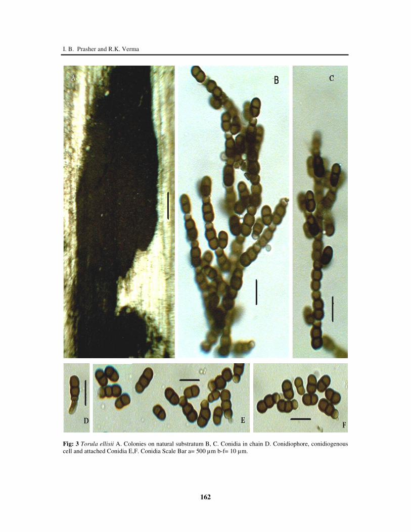
Torula ellisii Yadav & Lal, 1966, J. Indian bot. Soc.,
44: 404 Fig. 3
Colonies on natural substrate effuse, black, powdery.
Conidiophores pale brown, smooth, up to 7 µm long
and up to 3 µm thick except for the conidiogenous
cells which are 3.1-5.1µm in diameter. Conidia
catenate, cylindrical, rounded at the ends or broadly
161
Journal on New Biological Reports 3(3): 159 – 166 (2014)
Fig. 2. Dictyosporium heptasporum A. Crush mount of Sporodochia B. Conidia with hooked apices C. Conidia
with truncate basal cell D, E. Conidia with closely apprressed arms. Scale Bar A-E = 20 µm.
162
I. B. Prasher and R.K. Verma
Fig: 3 Torula ellisii A. Colonies on natural substratum B, C. Conidia in chain D. Conidiophore, conidiogenous
cell and attached Conidia E,F. Conidia Scale Bar a= 500 µm b-f= 10 µm.
163
Journal on New Biological Reports 3(3): 159 – 166 (2014) ellipsoidal, mid dark brown or reddish brown, smooth,
slightly constricted at septa, almost all 1 septate rarely
2 septate and 7.11-10.34 × 4.2-6.31 µm.
Remarks: The genus Torula is characterized by
micronematous or semi–macronematous
unbranched or regularly branched straight or
flexuous conidiophores. Conidiogenous cells
polyblastic or some time monoblastic integrated
and terminal determinate, usually spherical, which
bear conidia simple or branched chains arising
from the upper half of the conidiogenous cell,
cylindrical with rounded ends, ellipsoidal or
subspherical, brown or olivaceous brown, smooth,
verruculose or echinated with 0, 1 or several
transverse septa, strongly constricted at septa.
Terminal cell of the conidia is a conidiogenous cell
(Ellis 1971).
The above collection resembles the T.
ellisii in morphological details (Ellis 1976). This is
new record for Himachal Pradesh as well as
Himalayas, though the species has been already
reported from Mehboob Nagar and Andhra Pradesh
(Bilgrami et al. 1991 and Jamaluddin et al. 2004).
Collection examined: Rajnish Kumar Verma, PAN
30358 on dead and decaying angiospermic twig
Dharamshala, fallen twigs PAN 32602 Keylong,
Himachal Pradesh.
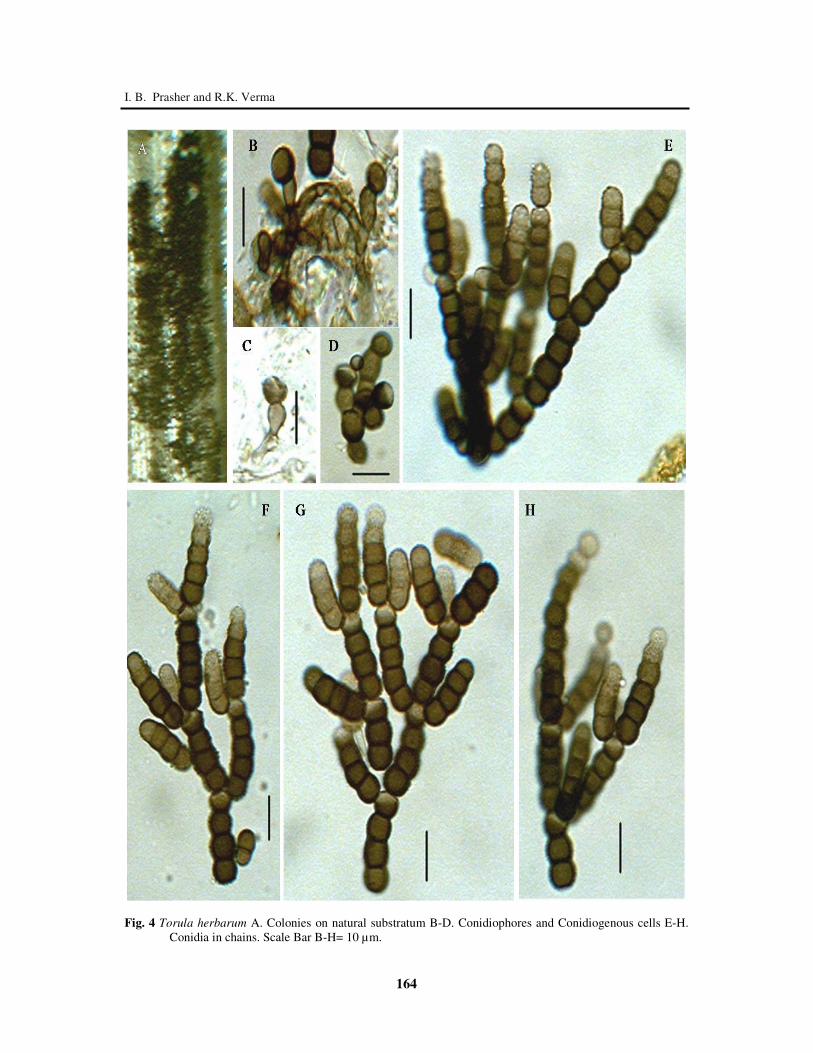
Torula herbarum (Pers.) Link, Mag. Gesell. naturf.
Freunde, Berlin 3(1-2): 19 (1809) Fig. 4
= Monilia herbarum Pers., Syn. meth.
fung. (Göttingen) 2: 693 (1801)
= Torula herbarum (Pers.) Link, Mag. Gesell.
naturf. Freunde, Berlin 3(1-2): 19 (1809) f.
herbarum
= Torula herbarum f. quaternella Sacc., Annls
mycol. 11(6): 556 (1913)
= Torula monilis Pers., Ann. Bot. (Usteri) 15: 25
(1794)
Colonies on natural substratum very variable in size,
sometimes only a few mm diameter, at others
completely encircling stem and extending along them
for several centimeters, black, velvety. Setae absent.
Conidiogenous cells 6.1-7.5 µm in diameter. Conidia
catenate, straight or slightly curved, more or less
cylindrical, rounded at the ends, dark brown,
smoother, 1-3- (mostly 2) septate and 9.4-21.2 × 5.4-
7.2 µm.
Remarks: The above collection resembles the type
species in morphological details (Ellis 1971). The
species have been earlier reported from the Solan
(Bilgrami et al. 1991 and Jamaluddin et al. 2004)
but the species has been first time reported from the
Bilaspur, Chamba, Kangra, Kullu, Mandi and
Shimla districts of Himachal Pradesh, which
indicates it is widely distributed in Himachal
Pradesh (Bilgrami et al. 1991 and Jamaluddin et al.
2004).
Collection examined: Rajnish Kumar Verma PAN
30357, on dead and decaying leaves of
Pterospermum sp. Bilaspur 19 Nov. 2012, PAN
32601 on fallen twigs Dharamshala, PAN 32558 on
fallen twigs Swarghat 17 Nov. 2012, PAN 32605 on
fallen twigs Narkanda, 2 Oct. 2013, PAN 32604 on
Urtica dioica, Manali, 3 Oct. 2012, PAN 32640 on
fallen dead twigs Jolplakhin, 22 July 2012, PAN
32650 on fallen twigs Sunder Nagar, PAN 32651 on
fallen twigs Dalhausi 18 June 2014, PAN 32652 on
fallen twigs Kasauli 10 Nov. 2011.
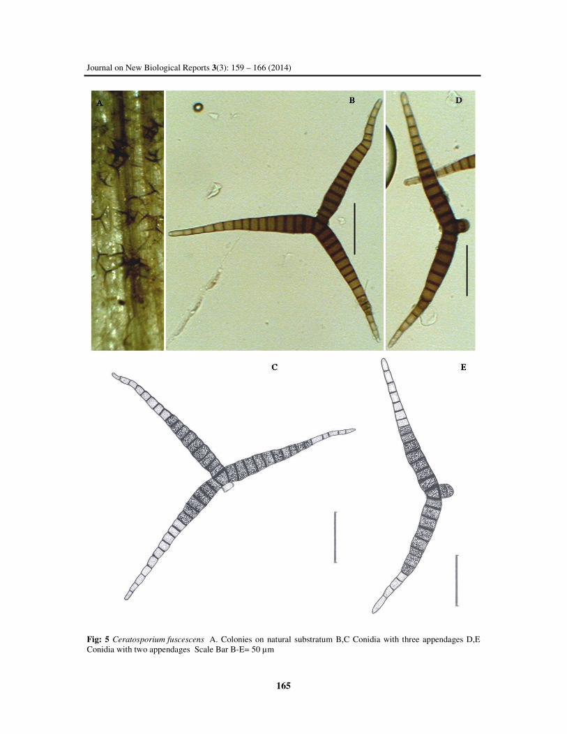
Ceratosporium fuscescens Schw., 1832, Trans.
Ann. Phil. Soc., N.S., 4: 300. Fig.5
Colonies on natural substratum effuse, dark brown,
mycelium superficial and immersed, composed of
septate, pale brown to brown, smooth-walled
hyphae. Conidia solitary, basal cell single, 8-12µm
thick at its broadest part, with 2-3 divergent arms
arising from the apex, Arms divergent from the
base, distal ends of the arms straight or flexuous,
smooth, brown, pale brown toward the apex, arms
12-18 septate, not constricted at septa, up to 192µm
long (most of unequal length), 11-19µm thick
below, tapering to 5-8 µm.
Remarks: The genus Ceratosporium was
established by Schweinitz (1832) with type species
C. fuscescens. The detailed account of taxonomy
and conidial development was given by Hughes
(1951). Hughes (1964) drew attention to the
occurrence of secondary conidial fructification. The
genus is characterized by inconspicuous flexuous
brown conidiophores and integrated intercalary
monoblastic determinate denticulate conidiogenous
cells which produce solitary conidia with pyriform
central cell and divergent pleuriseptate branches
(Ellis 1971). There are eleven accepted species of
Ceratosporium (Ma et al. 2014). Only three species
viz. C. fuscescens Schwein., C. productum Petch
and C. indicum V.G. Rao & D. Rao reported from
India (Bilgrami et al. 1991 and Jamaluddin et al.
2004).
Conidia in our collection are shorter (up to
192 µm vs. 230 µm), narrower at the base (11-19
µm vs. 14-22 µm). Despite these minor differences,
we believe they are basically the same species.
Ceratosporium fuscescens have been already
reported from the Solan but first time reported from
Shimla district of Himachal Pradesh (Bilgrami et
al. 1991 and Jamaluddin et al. 2004)
164
I. B. Prasher and R.K. Verma
Fig. 4 Torula herbarum A. Colonies on natural substratum B-D. Conidiophores and Conidiogenous cells E-H.
Conidia in chains. Scale Bar B-H= 10 µm.
165
Journal on New Biological Reports 3(3): 159 – 166 (2014)
Fig: 5 Ceratosporium fuscescens A. Colonies on natural substratum B,C Conidia with three appendages D,E
Conidia with two appendages Scale Bar B-E= 50 µm
166
I. B. Prasher and R.K. Verma
Collection examined: Rajnish Kumar Verma,
PAN 30110, on fallen twigs, Shogi (Shimla), Nov.
10, 2011.
ACKNOWLEDGEMENT
The authors are thankful to Ministry of
Environment and Forests, Government of India for
the financial assistance (vide letter no. 14/26/2008-
ERS/RE dt. 06.06.2010), UGC (SAP, DRS III) and
Chairperson Department of Botany Panjab
University Chandigarh for providing infrastructural
and laboratory facilities.
REFERENCES
Bilgrami KS, Jamaluddin and Rizwi MA. 1991.
Fungi of India List and References. Today
and tomorrow’s Printers & Publishers, New
Delhi, India.
Ellis MB. 1971. Dematiaceous Hyphomycetes.
Coomonwealth Mycological Instititute,
Kew, UK
Ellis MB. 1976. More Dematiaceous
Hyphomycetes. Coomonwealth Mycological
Instititute, Kew, UK.
Hijmanas RJ, Guarino L and Rojas E. 2011. DIVA-
GIS, Version 7.5. A geographical
information system for the analysis of
Biodiversity data. Downloaded from
http://www.diva.org.
Hughes SJ. 1951. Studies on micro-fungi. VI.
Ceratosporium, Hirudinaria, and
Hippocrepidium. Mycol Pap 39: 1–25.
Hughes SJ. 1964. New Zealand fungi. 1.
Ceratosporium Schw. New Zealand J Bot 2:
305–309.
Jamaluddin, MG Goswami and Ojha BM.
2004.Fungi of India 1989-2001. Scientific
Publishers, Jodhpur, India.
Kirk PM, Cannon PF, Minter DW and Stalpers JA.
2008. Dictionary of the Fungi. 10th
edn.
CAB International, Wallingford, UK
Kirschner R, Pang KL and Gerath Jones EB. 2012.
Two cheirosporous hyphomycetes reassessed
based on morphological and molecular
examination. Mycological progress; doi:
10.1007/s11557–012–0812–3.
Ma J, Zhang XG and Castañeda-Ruíz RF. 2014.
Ceratosporium hainanense and
Solicorynespora obovoidea spp. Nov., and a
first record of Bactrodesmiastrum obscurum
from southern china. Mycotaxon 127 135-
143.
Manoharachary C, Kunwar IK and Rao NK. 2007.
Two new species of Dictyosporium from
India. Indian Phytopath 60: 341–344
Patil SY and Borse BD. 2012. Dematiaceous
Hyphomycetes from North Maharashtra.
Internat Multidisciplinary Res J 2(3):36-38
Prasher IB and Verma RK. 2012 a. Periconia
species new to North- Western Himalayas. J
New Biol Rep 1(1): 1-2
Prasher IB, Verma RK. 2012 b. Two hyphomycetes
new To Himalayas. Pl Sc Feed 2(8): 122-
124.
Schweinitz LD von. 1832. Synopsis fungorum in
America boreali media degentium. Trans.
Amer. Philos. Soc. 4(2): 141–316.
Whitton SR, McKenzie EHC, Hyde KD. 2012.
Fungi Associated with Pandanaceae,
Springer.