Embed Size (px)
Citation preview
1
Foxa2 (HNF3ββββ) controls multiple genes implicated in metabolism-secretion coupling of
glucose-induced insulin release
Haiyan Wang‡, Benoit R. Gauthier, Kerstin A. Hagenfeldt, Mariella Iezzi, and Claes B.
Wollheim
Division of Clinical Biochemistry, Department of Internal Medicine University Medical Center,
CH-1211 Geneva 4, Switzerland
Running Title: Foxa2 regulates islet glucose-sensing
‡To whom correspondence should be addressed
E-mail: [email protected]
Haiyan Wang, Ph.D
Division de Biochimie Clinique et de Diabétologie Expérimentale
Départment de Médecine Interne
Centre Médical Universitaire
CH-1211 Geneva 4
Switzerland
Tel.: 41 22 702 5570
Fax: 41 22 702 5543
Copyright 2002 by The American Society for Biochemistry and Molecular Biology, Inc.
JBC Papers in Press. Published on March 1, 2002 as Manuscript M111037200 by guest on February 13, 2019
http://ww
w.jbc.org/
Dow
nloaded from
2
The transcription factor Foxa2 is implicated in blood glucose homeostasis. Conditional
expression of Foxa2 or its dominant-negative mutant DN-Foxa2 in INS-1 cells reveals that
Foxa2 regulates the expression of genes important for glucose sensing in pancreatic ββββ-cells.
Overexpression of Foxa2 results in blunted glucose-stimulated insulin secretion, whereas
induction of DN-Foxa2 causes a left-shift of glucose-induced insulin release. The mRNA
levels of GLUT2 and glucokinase are drastically decreased after induction of Foxa2. In
contrast, loss of Foxa2 function leads to up-regulation of hexokinase (HK) I and II, and
glucokinase (HK-IV) mRNA expression. The glucokinase and the low Km hexokinase
activities as well as glycolysis are increased proportionally. In addition, induction of DN-
Foxa2 also reduces the mRNA expression of ββββ-cell KATP channel subunits Sur1 and Kir6.2
by 70%. Furthermore, in contrast to previous reports, Induction of Foxa2 causes
pronounced decreases in the HNF4αααα and HNF1α α α α mRNA levels. Foxa2 fails to regulate the
expression of Pdx1 transcripts. The expression of insulin and IAPP is markedly suppressed
after induction of Foxa2, while the glucagon mRNA levels are significantly increased.
Conversely, Foxa2 is required for glucagon expression in these INS-1-derived cells. These
results suggest that Foxa2 is a vital transcription factor evolved to control the expression of
genes essential for maintaining ββββ-cell glucose-sensing and glucose homeostasis.
by guest on February 13, 2019http://w
ww
.jbc.org/D
ownloaded from
3
The forkhead/winged-helix Foxa family of transcription factors, encoded by three genes Foxa1
(Hnf3α), Foxa2 (Hnf3β), and Foxa3 (Hnf3γ), regulate hepatic and/or pancreatic gene expression
(1-9). Foxa1 and Foxa3 are required for maintaining glucose homeostasis by activation,
respectively, of pancreatic glucagon and hepatic gluconeogenic enzymes (2,3,5). Targeted
disruption of Foxa2 resulted in embryonic lethality with defective development of the foregut
endoderm, from which the liver and pancreas arise (10). Foxa2, which is expressed in islets, has
been suggested as the upstream transactivator of Hnf4α, Hnf1α, Pdx1, and Hnf1β in the
transcriptional hierarchy (1,9,11). Mutations in the genes encoding these pancreatic transcription
factors are linked to four monogenic forms of MODY (maturity-onset diabetes of the young):
MODY1/HNF1α, MODY3/HNF4α, MODY4/IPF1(PDX1), and MODY5/HNF1β (12,13).
However, the search for the association of FOXA2 mutations with MODY patients has not been
successful (14,15). Most recently, Sund et al. (16) have suggested that FOXA2 rather might be a
candidate gene for familial hyperinsulinism. Pancreatic β-cell-specific deletion of Foxa2 resulted
in postnatal death due to severe hyperinsulinemic hypoglycemia and the down-regulation of
ATP-sensitive K+ (KATP) channel subunits Sur1 and Kir6.2 have been demonstrated in these
mutant mice (16).
In order to assess whether Foxa2 indeed controls the expression of the transcription factors
associated with MODY, we have established INS-1-derived stable cell lines which allow
conditional expression of the wild type Foxa2 or its dominant-negative mutant DN-Foxa2 under
tight control of the reverse tetracycline-dependent transactivator (17). DN-Foxa2 is a Myc-tagged
truncated Foxa2 mutant protein which possesses the intact DNA-binding domain but lacks the
transactivation domain (7). DN-Foxa2 exerts its dominant-negative function by competing with
the endogenous Foxa2 for cognate DNA-binding (7). The impact of altered Foxa2 function on
glucose metabolism and insulin secretion was assessed in these stable clones. The gene-
expression profile before and after induction of the Foxa2 or DN-Foxa2 was quantified.
EXPERIMENTAL PROCEDURES
Establishment of Stable Cell Lines-Rat insulinoma INS-1 cell line-derived stable clones were
cultured in RPMI 1640 in 11.2 mM glucose (18), unless otherwise indicated. The first-step stable
clone INSrαβ, which expresses the reverse tetracycline-dependent transactivator, was described
by guest on February 13, 2019http://w
ww
.jbc.org/D
ownloaded from
4
previously (17,18). Plasmids used in the secondary stable transfection were constructed by
subcloning the cDNAs encoding the mouse Foxa2 (kindly supplied by Prof. G.Schütz) and its
dominant-negative mutant (DN-Foxa2) into the expression vector PUHD10-3 (a kind gift from
Prof. H. Bujard). DN-Foxa2 (truncated mutation lacking the transactivation domain but
containing the intact DNA-binding domain) (7) was PCR-amplified from Foxa2 cDNA using the
following primers, 5'gcaggatccgtaatggtgctcgggcttcaggtg3' and
5'gcaggatccggcgccatggcgggcatgagcggctca3'. The PCR fragment was subcloned into modified
pcDNA3.1myc (Invitrogene, The Netherlands) and sequenced. The stable transfection and the
clone selection and screening procedures were described previously (17).
Immunoblot and Immunofluorescence-Immunoblotting procedures were performed as described
previously using enhanced chemiluminescence (Pierce, Rockford, Illinois) for detection (18). The
dilutions for antibodies against Foxa2 C-terminus (Santa Cruz Biotech., Heidelberg, Germany)
and Myc-tag (19) were 1:5,000 and 1:10, respectively. Nuclear extracts were isolated from the
cells cultured with or without 500 ng/ml doxycycline for 24 h.
For immunofluorescence, cells grown on polyornithine treated glass coverslips were treated for
24 h with or without 500 ng/ml doxycycline. The cells were then washed, fixed in 4%
paraformaldehyde, permeabilized with 0.1% Triton X-100 in phosphate-buffered saline
containing 1% BSA (PBS-BSA). The preparation was then blocked with PBS-BSA before
incubating with the first antibodies, anti-Foxa2 (1:500 dilution) and mouse monoclonal anti-Myc-
tag, (1:2 dilution), followed by the second antibody labeling.
Nuclear Extract Preparation-Nuclear extracts from INS-1 cells grown in culture medium with or
without 500 ng/ml doxycycline for 24 h were prepared according to Schreiber et al. (20).
Measurements of Insulin Secretion and Cellular Insulin content-Cells in 12-well plates were
cultured in 11.2 mM glucose medium with or without 500 ng/ml doxycycline for 19 h, followed
by an additional 5 h equilibration in 2.5 mM glucose medium. Insulin secretion was measured
over a period of 30 min, in Krebs-Ringer-Bicarbonate-HEPES buffer (KRBH, 140 mM NaCl, 3.6
mM KCl, 0.5 mM NaH2PO4, 0.5 mM MgSO4, 1.5 mM CaCl2, 2 mM NaHCO3, 10 mM HEPES,
0.1% BSA) containing indicated concentrations of glucose. Insulin content was determined after
extraction with acid ethanol following the procedures of Wang et al. (21). Insulin was detected by
radioimmunoassay using rat insulin as standard (21).
by guest on February 13, 2019http://w
ww
.jbc.org/D
ownloaded from
5
Assay of Glucokinase and High-affinity Hexokinase Activities-Cytosolic proteins were extracted,
according to Wang and Iynedjian (17), from cells cultured in 11.2 mM glucose medium in the
presence or absence of 500 ng/ml doxycycline for 24 h. Total hexokinase activity was measured
at 30°C by a glucose-6-phosphate dehydrogenase-coupled assay in a fluorometer (Lambda Bio20,
Perkin Elmer) estimation of NADH production (17). Glucokinase activity and high-affinity
hexokinase activity were calculated, respectively, as the differences in NADH produced at 100
mM, 0.5, and 0 mM glucose and expressed in nmol/min (=mU) per mg of protein.
Measurement of Glucose Utilization-Cells in 24-well dishes were cultured in 2.5 mM glucose
medium with or without 500 ng/ml doxycycline for 24 h. The rate of glycolysis was estimated
from the production of [3H] water from D-[5-3H]glucose according to Wang and Iynedjian (17).
Total RNA Isolation and Northern Blotting-Cells in 10-cm diameter dishes were cultured in 2.5
mM glucose medium with or without 500 ng/ml doxycycline for 16 h, followed by an additional
8 h in culture medium with 2.5, 6, 12 and 24 mM glucose. Total RNA was extracted and blotted
to nylon membranes as described previously (17). The membrane was prehybridized and then
hybridized to 32P-labeled random primer cDNA probes according to Wang and Iynedijian (17).
To ensure equal RNA loading and even transfer, all membranes were stripped and re-hybridized
with a "house-keeping gene" probe cyclophilin. cDNA fragments used as probes for Foxa2,
Hnf1α, Hnf4α, glucokinase, hexokinase I, Glut2, L-pyruvate kinase, insulin, Sur1, Kir6.2 and
Pdx1 mRNA detection were digested from corresponding plasmids. cDNA probes for rat islet
amyloid polypeptide (IAPP), glucagon, Nkx6.1, Nkx2.2, Isl-1, β2/NeuroD, aldolase B, adenine
nucleotide translocator 1 and 2 (ANT1, ANT2), mitochondrial uncoupling protein 2 (UCP2),
mitochondrial glutamate dehydrogenase (GDH), citrate synthase, glyceraldehydes-3 phosphate
dehydrogenase (GAPDH), hexokinase II and glucagon-like peptide-1 receptor (GLP-1R), were
prepared by RT-PCR and confirmed by sequencing.
Statistics-Results are expressed as mean + SEM and statistical analyses were performed by
Student’s t-test for unpaired data.
RESULTS
Foxa2 and DN-Foxa2 Were Induced in An All-Or-None Manner-We have established over 10
clones positively expressing Foxa2 and DN-Foxa2, respectively, using the parental INS-rαβ
by guest on February 13, 2019http://w
ww
.jbc.org/D
ownloaded from
6
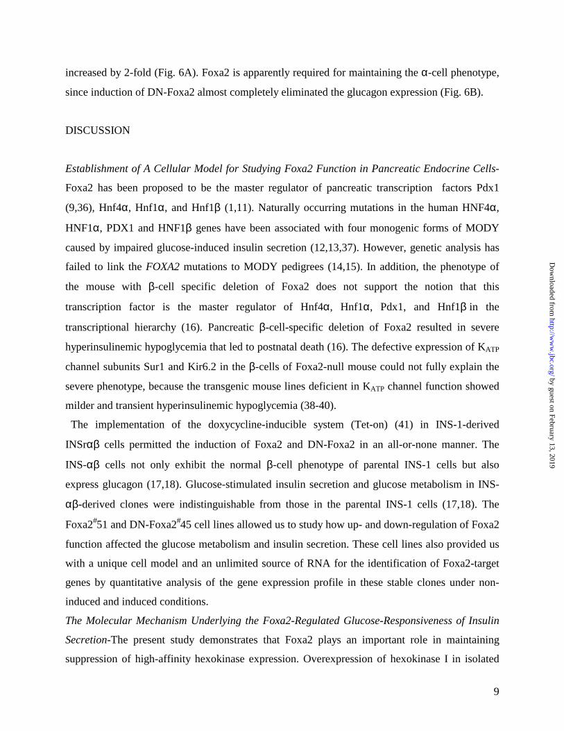
(INS-r3) cells (17,18). The clones designated as Foxa2#51 and DN-Foxa2#45, which displayed
the highest inducible protein levels without leakage under non-induced state, were selected for
the present study. As illustrated in Fig. 1A and D, the INS-1 derived cells express endogenous
Foxa2 in the nucleus. Foxa2 protein was overexpressed in all of the cells treated with 500 ng/ml
doxycycline for 24 h. As predicted, the antibody against the carboxy terminus of Foxa2 did not
detect DN-Foxa2 with the C-terminal deletion (7) (Fig. 1B). As shown in the Western blotting
(Fig. 1B) and immunostaining (Fig. 1D) with a monoclonal anti-Myc antibody, this Myc-tagged
DN-Foxa2 protein was induced in a doxycycline-dependent and an all-or-none manner. Induction
of DN-Foxa2 did not interrupt the endogenous Foxa2 expression (Fig. 1B) and the induced DN-
Foxa2 protein was localized in the nucleus of DN-Foxa2#45 cells (Fig. 1B and D). We also
performed an electrophoretic mobility shift assay (EMSA) (data not shown) using the Foxa2-
binding site containing glucagon G2 element as a probe (22). Induction of Foxa2 led to a 10-fold
increase in the signal density of Foxa2 binding, whereas induction of DN-Foxa2 almost
completely abolished the binding activity of endogenous Foxa2 (data not shown).
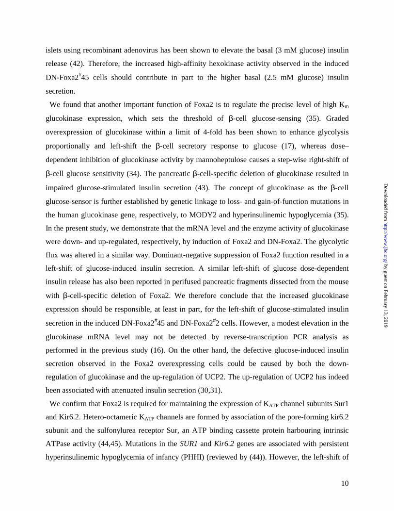
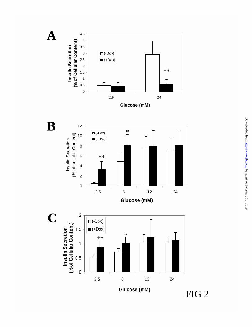
Foxa2 Regulates the Glucose-Responsiveness of Insulin Secretion-As demonstrated in Fig. 2A,
overexpression of Foxa2 almost completely blunted glucose-stimulated insulin release. The
cellular insulin content was reduced by 46.3 + 5.1% (p<0.001) after induction of Foxa2 for 24 h
(see also Fig. 6 for the decrease in insulin mRNA levels). Secretion data were therefore
normalized for cellular insulin content. In contrast, induction of DN-Foxa2 resulted in a left-shift
of the dose-response curve of glucose-stimulated insulin release (Fig. 2B) without altering insulin
content. To verify the clonal variability, we randomly chose another clone DN-Foxa2#2 and
studied the effects of DN-Foxa2 induction on glucose-stimulated insulin secretion. As shown in
Fig. 2C, induction of DN-Foxa2 in this clone also led to a typical left-shift of glucose-dependent
insulin release, suggesting a common phenomenon rather than a clonal peculiarity. Next, we
examined the gene expression patterns in these cell lines to elucidate the mechanisms underlying
the changes in insulin secretion.
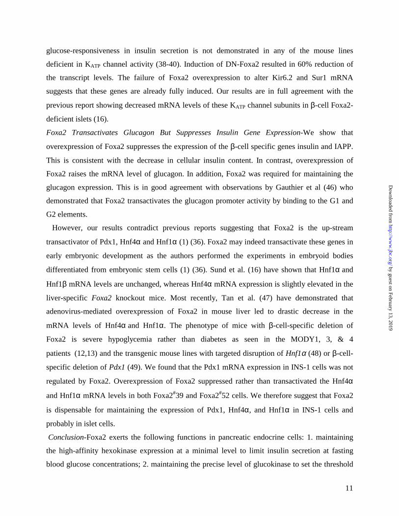
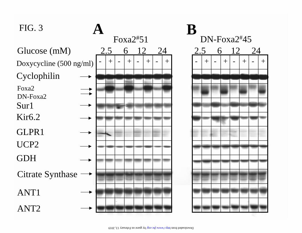
Foxa2 is Required for Maintaining the Expression of KATP Channel Subunits Sur1 and Kir6.2-
Northern blot analysis of the gene expression pattern in Foxa2#51 and DN-Foxa2#45 cells
cultured in indicated concentrations of glucose and treated with or without 500 ng/ml
doxycycline for 24 h is described in Fig. 3. Consistent with the immunoblotting (Fig. 1B), DN-
by guest on February 13, 2019http://w
ww
.jbc.org/D
ownloaded from
7
Foxa2 mRNA was induced in an all-or-none manner and such induction did not alter endogenous
Foxa2 mRNA expression (Fig. 3B). The mRNA levels of the KATP channel subunits Sur1 and
Kir6.2 were reduced by 60% and 70%, respectively, after dominant-negative suppression of
Foxa2 function (Fig. 3B). However, overexpression of Foxa2 alone was not sufficient to promote
the expression of Sur1 and Kir6.2 (Fig. 3A). The mRNA levels of mitochondrial glutamate
dehydrogenase (GDH), citrate synthase, and adenine nucleotide translocator 1 and 2 (ANT1 and
ANT2) were not modulated by Foxa2 (Fig. 3A and B). On the other hand, overexpression of
Foxa2 caused up-regulation of UCP2 (Fig. 3A), whereas induction of DN-Foxa2 did not affect
the expression of UCP2 mRNA (Fig. 3B). Furthermore, overexpression of Foxa2 resulted in
down-regulation of glucagon-like peptide-1 receptor (GLP-1R) (Fig. 3A).
Persistent hyperinsulinemic hypoglycemia of infancy (PHHI) has been linked to mutations in
the genes encoding Sur1, Kir6.2, glucokinase, and GDH (23-28). Increased glucose-dependent
insulin release was also observed in the UCP2-deleted mouse (29), whereas decreased insulin
secretion was reported after overexpression of UCP2 in islets (30) and INS-1 cells (31). We could
rule out the possible involvement of GDH and UCP2 in the enhanced glucose-stimulated insulin
secretion observed in β-cells deficient in Foxa2 function, since their expression was not altered
by induction of DN-Foxa2.
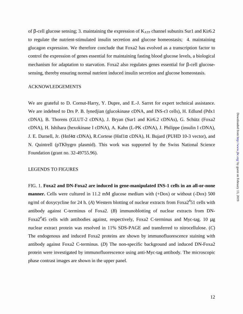
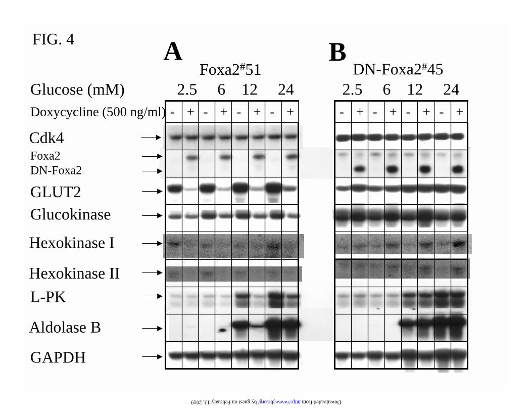
Foxa2 Targets Genes Essential for β-Cell Glucose-Sensing-The rodent pancreatic β-cell
expresses high levels of the glucose-transporter Glut2, which allows rapid equilibration of
glucose across the plasma membrane (32,33). This is associated with extremely low-levels of
high-affinity hexokinase isoforms (hexokinase I, II and III) to optimize glucose-sensing in the
physiological blood glucose range. A β-cell-specific promoter in the glucokinase (hexokinase IV)
gene maintains a precise expression level of this rate-limiting enzyme for glucose metabolism,
which determines the glucose-sensing in pancreatic β-cells (reviewed in (33-35)). Alterations of
glucokinase activity by gene manipulation or pharmacological inhibition, or by naturally
occurring genetic mutations, have been demonstrated to change the physiological threshold of β-
cell glucose-sensing (reviewed in (33-35)).
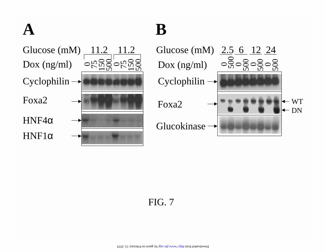
As shown in Fig. 4, overexpression of Foxa2 in Foxa2#51 cells reduced the glucokinase mRNA
level by 60%, whereas induction of DN-Foxa2 in DN-Foxa2#45 raised the glucokinase
expression by 2-fold. The increased glucokinase expression after induction of DN-Foxa2 was
also demonstrated in another clone, DN-Foxa2#2 (Fig. 7). The INS-1-derived clones expressed
by guest on February 13, 2019http://w
ww
.jbc.org/D
ownloaded from
8
hexokinase I and II (but not III) mRNAs at barely-detectable levels and induction of Foxa2 and
DN-Foxa2 resulted in, respectively, down- and up-regulation of these mRNA levels (Fig. 4).
Overexpression of Foxa2 also caused a 90% reduction of Glut2 mRNA expression, while
induction of DN-Foxa2 left-shifted the glucose dose-dependent increase in Glut2 transcript level
(Fig. 4). The suppressive effects of Foxa2 on glucose-sensing were also reflected by the blunted
glucose-responsiveness of L-pyruvate kinase (L-PK) and aldolase B mRNA expression (Fig. 4).
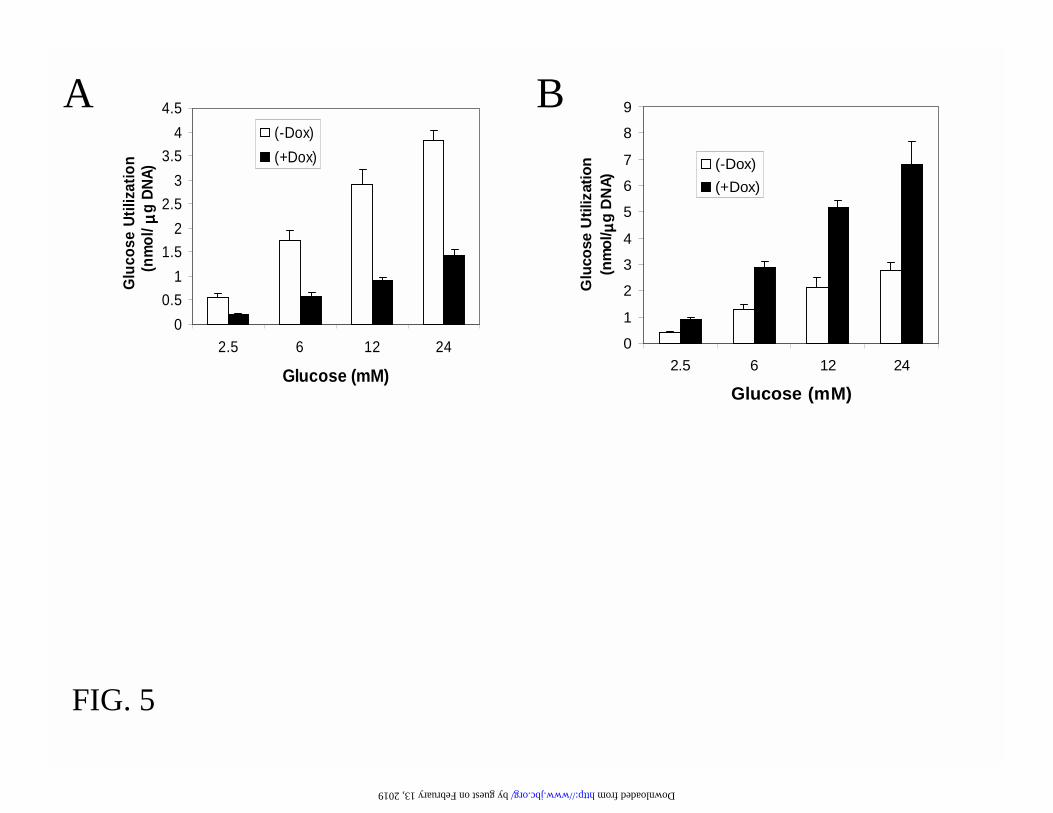
To confirm the Northern blot analysis, we also measured the activities of glucokinase and high-
affinity hexokinase. As seen in Table I, the glucokinase activity was reduced by 60% following
overexpression of Foxa2 and was increased 2.5-fold by dominant-negative suppression of Foxa2
function. Similarly, the high-affinity hexokinase activity was down-regulated by 50% and up-
regulated by 3-fold, respectively, by induction of Foxa2 and DN-Foxa2. Thus, Foxa2 is essential
for the transcriptional regulation of enzymes controlling the β-cell glucose phosphorylation. This
conclusion was corroborated by the measurements of glycolytic flux, which was decreased by
60% after overexpression of Foxa2 and increased by 2-fold after induction of DN-Foxa2 (Fig. 5).
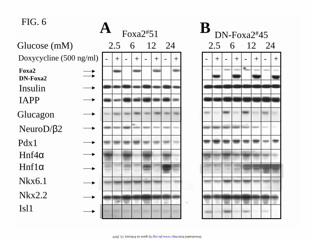
Foxa2 Promotes Glucagon Level and Suppresses β-cell Gene Expression-Foxa2 has been
previously suggested as a master transactivator of the pancreatic transcription factors, Hnf4α,
Hnf1α, Hnf1β and Pdx1, in the transcriptional hierarchy (1,9,11). The results we obtained were
unexpected and in disagreement with previous reports (1,9). We found that Pdx1 expression was
not significantly affected by induction of Foxa2 or DN-Foxa2 (Fig. 6). Isl-1 is the only pancreatic
transcription factor, the expression of which requires Foxa2 function. (Fig. 6B). Overexpression
of Foxa2 suppressed rather than enhanced the expression of Hnf4α and Hnf1α mRNAs (Fig.
6A). To verify whether this is due to a clonal variability or a paradoxical effect of high level
overexpression, we also studied the effect of graded overexpression of Foxa2 on mRNA levels of
Hnf4α and Hnf1α in another randomly selected clone, Foxa2#39 (Fig. 7). Titrated overexpression
of Foxa2 by 3.5-, 10-, and 20-fold at 75, 150, and 500 ng/ml of doxycycline all caused significant
inhibition of Hnf4α and Hnf1α expression (Fig 7).
The Foxa2#51 and DN-Foxa2#45 clones were derived from a parental INS-rαβ cell line that
expresses not only insulin but also detectable levels of glucagon (18). These clones enable us to
assess the function of Foxa2 in the regulation of both insulin and glucagon expression. The
mRNA levels of β-cell-specific genes, insulin and IAPP, were reduced by 50% and 60%,
respectively, after overexpression of Foxa2 for 24 h, whereas the glucagon expression was
by guest on February 13, 2019http://w
ww
.jbc.org/D
ownloaded from
9
increased by 2-fold (Fig. 6A). Foxa2 is apparently required for maintaining the α-cell phenotype,
since induction of DN-Foxa2 almost completely eliminated the glucagon expression (Fig. 6B).
DISCUSSION
Establishment of A Cellular Model for Studying Foxa2 Function in Pancreatic Endocrine Cells-
Foxa2 has been proposed to be the master regulator of pancreatic transcription factors Pdx1
(9,36), Hnf4α, Hnf1α, and Hnf1β (1,11). Naturally occurring mutations in the human HNF4α,
HNF1α, PDX1 and HNF1β genes have been associated with four monogenic forms of MODY
caused by impaired glucose-induced insulin secretion (12,13,37). However, genetic analysis has
failed to link the FOXA2 mutations to MODY pedigrees (14,15). In addition, the phenotype of
the mouse with β-cell specific deletion of Foxa2 does not support the notion that this
transcription factor is the master regulator of Hnf4α, Hnf1α, Pdx1, and Hnf1β in the
transcriptional hierarchy (16). Pancreatic β-cell-specific deletion of Foxa2 resulted in severe
hyperinsulinemic hypoglycemia that led to postnatal death (16). The defective expression of KATP
channel subunits Sur1 and Kir6.2 in the β-cells of Foxa2-null mouse could not fully explain the
severe phenotype, because the transgenic mouse lines deficient in KATP channel function showed
milder and transient hyperinsulinemic hypoglycemia (38-40).
The implementation of the doxycycline-inducible system (Tet-on) (41) in INS-1-derived
INSrαβ cells permitted the induction of Foxa2 and DN-Foxa2 in an all-or-none manner. The
INS-αβ cells not only exhibit the normal β-cell phenotype of parental INS-1 cells but also
express glucagon (17,18). Glucose-stimulated insulin secretion and glucose metabolism in INS-
αβ-derived clones were indistinguishable from those in the parental INS-1 cells (17,18). The
Foxa2#51 and DN-Foxa2#45 cell lines allowed us to study how up- and down-regulation of Foxa2
function affected the glucose metabolism and insulin secretion. These cell lines also provided us
with a unique cell model and an unlimited source of RNA for the identification of Foxa2-target
genes by quantitative analysis of the gene expression profile in these stable clones under non-
induced and induced conditions.
The Molecular Mechanism Underlying the Foxa2-Regulated Glucose-Responsiveness of Insulin
Secretion-The present study demonstrates that Foxa2 plays an important role in maintaining
suppression of high-affinity hexokinase expression. Overexpression of hexokinase I in isolated
by guest on February 13, 2019http://w
ww
.jbc.org/D
ownloaded from
10
islets using recombinant adenovirus has been shown to elevate the basal (3 mM glucose) insulin
release (42). Therefore, the increased high-affinity hexokinase activity observed in the induced
DN-Foxa2#45 cells should contribute in part to the higher basal (2.5 mM glucose) insulin
secretion.
We found that another important function of Foxa2 is to regulate the precise level of high Km
glucokinase expression, which sets the threshold of β-cell glucose-sensing (35). Graded
overexpression of glucokinase within a limit of 4-fold has been shown to enhance glycolysis
proportionally and left-shift the β-cell secretory response to glucose (17), whereas dose–
dependent inhibition of glucokinase activity by mannoheptulose causes a step-wise right-shift of
β-cell glucose sensitivity (34). The pancreatic β-cell-specific deletion of glucokinase resulted in
impaired glucose-stimulated insulin secretion (43). The concept of glucokinase as the β-cell
glucose-sensor is further established by genetic linkage to loss- and gain-of-function mutations in
the human glucokinase gene, respectively, to MODY2 and hyperinsulinemic hypoglycemia (35).
In the present study, we demonstrate that the mRNA level and the enzyme activity of glucokinase
were down- and up-regulated, respectively, by induction of Foxa2 and DN-Foxa2. The glycolytic
flux was altered in a similar way. Dominant-negative suppression of Foxa2 function resulted in a
left-shift of glucose-induced insulin secretion. A similar left-shift of glucose dose-dependent
insulin release has also been reported in perifused pancreatic fragments dissected from the mouse
with β-cell-specific deletion of Foxa2. We therefore conclude that the increased glucokinase
expression should be responsible, at least in part, for the left-shift of glucose-stimulated insulin
secretion in the induced DN-Foxa2#45 and DN-Foxa2#2 cells. However, a modest elevation in the
glucokinase mRNA level may not be detected by reverse-transcription PCR analysis as
performed in the previous study (16). On the other hand, the defective glucose-induced insulin
secretion observed in the Foxa2 overexpressing cells could be caused by both the down-
regulation of glucokinase and the up-regulation of UCP2. The up-regulation of UCP2 has indeed
been associated with attenuated insulin secretion (30,31).
We confirm that Foxa2 is required for maintaining the expression of KATP channel subunits Sur1
and Kir6.2. Hetero-octameric KATP channels are formed by association of the pore-forming kir6.2
subunit and the sulfonylurea receptor Sur, an ATP binding cassette protein harbouring intrinsic
ATPase activity (44,45). Mutations in the SUR1 and Kir6.2 genes are associated with persistent
hyperinsulinemic hypoglycemia of infancy (PHHI) (reviewed by (44)). However, the left-shift of
by guest on February 13, 2019http://w
ww
.jbc.org/D
ownloaded from
11
glucose-responsiveness in insulin secretion is not demonstrated in any of the mouse lines
deficient in KATP channel activity (38-40). Induction of DN-Foxa2 resulted in 60% reduction of
the transcript levels. The failure of Foxa2 overexpression to alter Kir6.2 and Sur1 mRNA
suggests that these genes are already fully induced. Our results are in full agreement with the
previous report showing decreased mRNA levels of these KATP channel subunits in β-cell Foxa2-
deficient islets (16).
Foxa2 Transactivates Glucagon But Suppresses Insulin Gene Expression-We show that
overexpression of Foxa2 suppresses the expression of the β-cell specific genes insulin and IAPP.
This is consistent with the decrease in cellular insulin content. In contrast, overexpression of
Foxa2 raises the mRNA level of glucagon. In addition, Foxa2 was required for maintaining the
glucagon expression. This is in good agreement with observations by Gauthier et al (46) who
demonstrated that Foxa2 transactivates the glucagon promoter activity by binding to the G1 and
G2 elements.
However, our results contradict previous reports suggesting that Foxa2 is the up-stream
transactivator of Pdx1, Hnf4α and Hnf1α (1) (36). Foxa2 may indeed transactivate these genes in
early embryonic development as the authors performed the experiments in embryoid bodies
differentiated from embryonic stem cells (1) (36). Sund et al. (16) have shown that Hnf1α and
Hnf1β mRNA levels are unchanged, whereas Hnf4α mRNA expression is slightly elevated in the
liver-specific Foxa2 knockout mice. Most recently, Tan et al. (47) have demonstrated that
adenovirus-mediated overexpression of Foxa2 in mouse liver led to drastic decrease in the
mRNA levels of Hnf4α and Hnf1α. The phenotype of mice with β-cell-specific deletion of
Foxa2 is severe hypoglycemia rather than diabetes as seen in the MODY1, 3, & 4
patients (12,13) and the transgenic mouse lines with targeted disruption of Hnf1α (48) or β-cell-
specific deletion of Pdx1 (49). We found that the Pdx1 mRNA expression in INS-1 cells was not
regulated by Foxa2. Overexpression of Foxa2 suppressed rather than transactivated the Hnf4α
and Hnf1α mRNA levels in both Foxa2#39 and Foxa2#52 cells. We therefore suggest that Foxa2
is dispensable for maintaining the expression of Pdx1, Hnf4α, and Hnf1α in INS-1 cells and
probably in islet cells.
Conclusion-Foxa2 exerts the following functions in pancreatic endocrine cells: 1. maintaining
the high-affinity hexokinase expression at a minimal level to limit insulin secretion at fasting
blood glucose concentrations; 2. maintaining the precise level of glucokinase to set the threshold
by guest on February 13, 2019http://w
ww
.jbc.org/D
ownloaded from
12
of β-cell glucose sensing; 3. maintaining the expression of KATP channel subunits Sur1 and Kir6.2
to regulate the nutrient-stimulated insulin secretion and glucose homeostasis; 4. maintaining
glucagon expression. We therefore conclude that Foxa2 has evolved as a transcription factor to
control the expression of genes essential for maintaining fasting blood glucose levels, a biological
mechanism for adaptation to starvation. Foxa2 also regulates genes essential for β-cell glucose-
sensing, thereby ensuring normal nutrient induced insulin secretion and glucose homeostasis.
ACKNOWLEDGEMENTS
We are grateful to D. Cornut-Harry, Y. Dupre, and E.-J. Sarret for expert technical assistance.
We are indebted to Drs P. B. Iynedjian (glucokinase cDNA, and INS-r3 cells), H. Edlund (Pdx1
cDNA), B. Thorens (GLUT-2 cDNA), J. Bryan (Sur1 and Kir6.2 cDNAs), G. Schütz (Foxa2
cDNA), H. Ishihara (hexokinase I cDNA), A. Kahn (L-PK cDNA), J. Philippe (insulin I cDNA),
J. E. Darnell, Jr. (Hnf4α cDNA), R.Cortese (Hnf1α cDNA), H. Bujard (PUHD 10-3 vector), and
N. Quintrell (pTKhygro plasmid). This work was supported by the Swiss National Science
Foundation (grant no. 32-49755.96).
LEGENDS TO FIGURES
FIG. 1. Foxa2 and DN-Foxa2 are induced in gene-manipulated INS-1 cells in an all-or-none
manner. Cells were cultured in 11.2 mM glucose medium with (+Dox) or without (-Dox) 500
ng/ml of doxycycline for 24 h. (A) Western blotting of nuclear extracts from Foxa2#51 cells with
antibody against C-terminus of Foxa2. (B) immunoblotting of nuclear extracts from DN-
Foxa2#45 cells with antibodies against, respectively, Foxa2 C-terminus and Myc-tag. 10 µg
nuclear extract protein was resolved in 11% SDS-PAGE and transferred to nitrocellulose. (C)
The endogenous and induced Foxa2 proteins are shown by immunofluorescence staining with
antibody against Foxa2 C-terminus. (D) The non-specific background and induced DN-Foxa2
protein were investigated by immunofluorescence using anti-Myc-tag antibody. The microscopic
phase contrast images are shown in the upper panel.
by guest on February 13, 2019http://w
ww
.jbc.org/D
ownloaded from
13
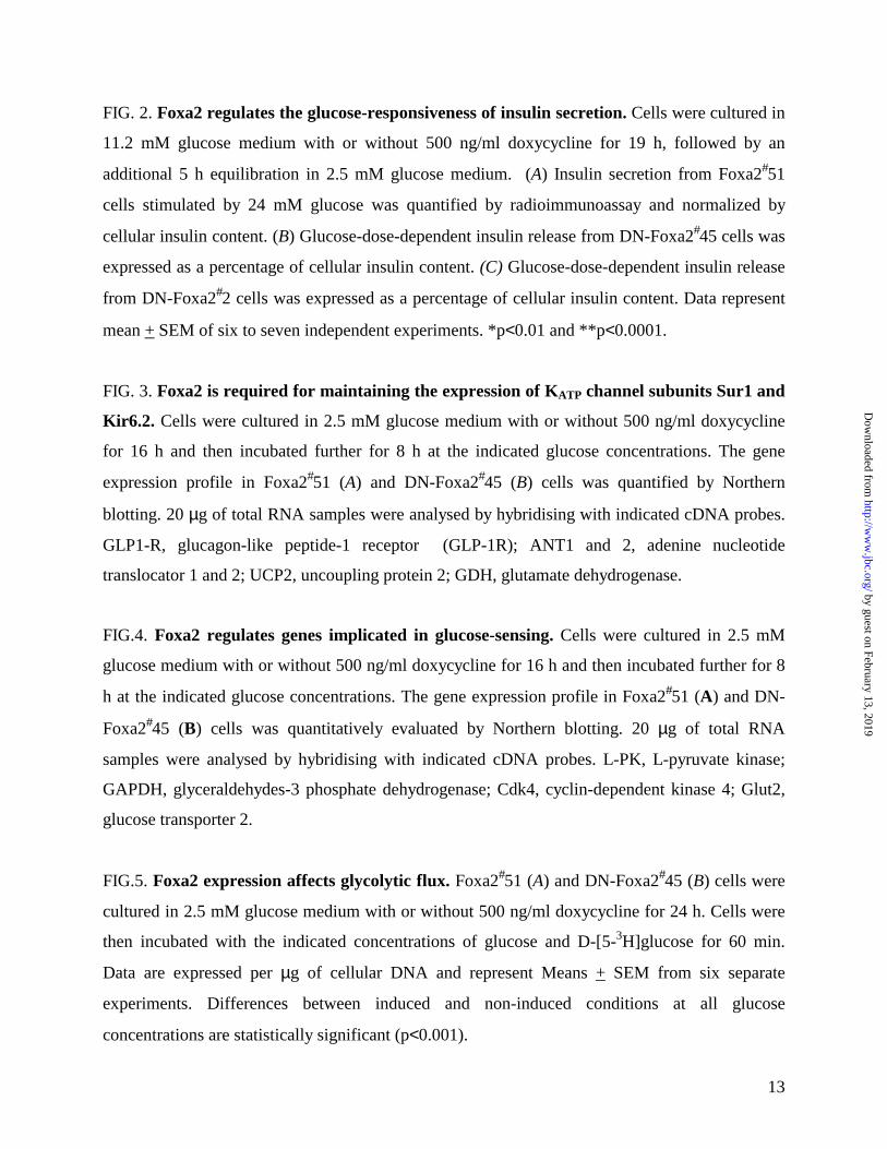
FIG. 2. Foxa2 regulates the glucose-responsiveness of insulin secretion. Cells were cultured in
11.2 mM glucose medium with or without 500 ng/ml doxycycline for 19 h, followed by an
additional 5 h equilibration in 2.5 mM glucose medium. (A) Insulin secretion from Foxa2#51
cells stimulated by 24 mM glucose was quantified by radioimmunoassay and normalized by
cellular insulin content. (B) Glucose-dose-dependent insulin release from DN-Foxa2#45 cells was
expressed as a percentage of cellular insulin content. (C) Glucose-dose-dependent insulin release
from DN-Foxa2#2 cells was expressed as a percentage of cellular insulin content. Data represent
mean + SEM of six to seven independent experiments. *p<0.01 and **p<0.0001.
FIG. 3. Foxa2 is required for maintaining the expression of KATP channel subunits Sur1 and
Kir6.2. Cells were cultured in 2.5 mM glucose medium with or without 500 ng/ml doxycycline
for 16 h and then incubated further for 8 h at the indicated glucose concentrations. The gene
expression profile in Foxa2#51 (A) and DN-Foxa2#45 (B) cells was quantified by Northern
blotting. 20 µg of total RNA samples were analysed by hybridising with indicated cDNA probes.
GLP1-R, glucagon-like peptide-1 receptor (GLP-1R); ANT1 and 2, adenine nucleotide
translocator 1 and 2; UCP2, uncoupling protein 2; GDH, glutamate dehydrogenase.
FIG.4. Foxa2 regulates genes implicated in glucose-sensing. Cells were cultured in 2.5 mM
glucose medium with or without 500 ng/ml doxycycline for 16 h and then incubated further for 8
h at the indicated glucose concentrations. The gene expression profile in Foxa2#51 (A) and DN-
Foxa2#45 (B) cells was quantitatively evaluated by Northern blotting. 20 µg of total RNA
samples were analysed by hybridising with indicated cDNA probes. L-PK, L-pyruvate kinase;
GAPDH, glyceraldehydes-3 phosphate dehydrogenase; Cdk4, cyclin-dependent kinase 4; Glut2,
glucose transporter 2.
FIG.5. Foxa2 expression affects glycolytic flux. Foxa2#51 (A) and DN-Foxa2#45 (B) cells were
cultured in 2.5 mM glucose medium with or without 500 ng/ml doxycycline for 24 h. Cells were
then incubated with the indicated concentrations of glucose and D-[5-3H]glucose for 60 min.
Data are expressed per µg of cellular DNA and represent Means + SEM from six separate
experiments. Differences between induced and non-induced conditions at all glucose
concentrations are statistically significant (p<0.001).
by guest on February 13, 2019http://w
ww
.jbc.org/D
ownloaded from
14
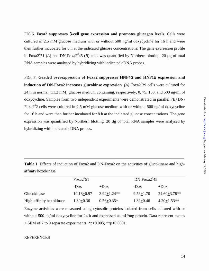
FIG.6. Foxa2 suppresses ββββ-cell gene expression and promotes glucagon levels. Cells were
cultured in 2.5 mM glucose medium with or without 500 ng/ml doxycycline for 16 h and were
then further incubated for 8 h at the indicated glucose concentrations. The gene expression profile
in Foxa2#51 (A) and DN-Foxa2#45 (B) cells was quantified by Northern blotting. 20 µg of total
RNA samples were analysed by hybridizing with indicated cDNA probes.
FIG. 7. Graded overexpression of Foxa2 suppresses HNF4αααα and HNF1αααα expression and
induction of DN-Foxa2 increases glucokinse expression. (A) Foxa2#39 cells were cultured for
24 h in normal (11.2 mM) glucose medium containing, respectively, 0, 75, 150, and 500 ng/ml of
doxycycline. Samples from two independent experiments were demonstrated in parallel. (B) DN-
Foxa2#2 cells were cultured in 2.5 mM glucose medium with or without 500 ng/ml doxycycline
for 16 h and were then further incubated for 8 h at the indicated glucose concentrations. The gene
expression was quantified by Northern blotting. 20 µg of total RNA samples were analysed by
hybridizing with indicated cDNA probes.
______________________________________________________________________________
Table I Effects of induction of Foxa2 and DN-Foxa2 on the activities of glucokinase and high-
affinity hexokinase
Foxa2#51 DN-Foxa2#45
-Dox +Dox -Dox +Dox
Glucokinase 10.18+0.97 3.94+1.24** 9.53+1.70 24.60+3.78**
High-affinity hexokinase 1.30+0.36 0.56+0.35* 1.32+0.46 4.20+1.53**
Enzyme activities were measured using cytosolic proteins isolated from cells cultured with or
without 500 ng/ml doxycycline for 24 h and expressed as mU/mg protein. Data represent means
+ SEM of 7 to 9 separate experiments. *p<0.005, **p<0.0001.
REFERENCES
by guest on February 13, 2019http://w
ww
.jbc.org/D
ownloaded from
15
1. Duncan, S., Navas, M., Dufort, D., Rossant, J., and Stoffel, M. (1998) Sience 281, 692-
695
2. Kaestner, K. H., Hiemisch, H., and Schutz, G. (1998) Mol Cell Biol 18(7), 4245-51.
3. Kaestner, K., Katz, J., Liu, Y., Drucker, D., and Schutz, G. (1999) Genes Dev 13, 495-504
4. Rausa, F. M., Tan, Y., Zhou, H., Yoo, K. W., Stolz, D. B., Watkins, S. C., Franks, R. R.,
Unterman, T. G., and Costa, R. H. (2000) Mol Cell Biol 20(21), 8264-82.
5. Shih, D., Navas, M., Kuwajima, S., Duncan, S., and Stoffel, M. (1999) Proc Natl Acad
Sci U S A 96, 10152-10157
6. Sund, N., Ang, S., Sackett, S., Shen, W., Daigle, N., Magnuson, M., and Kaestner, K.
(2000) Mol Cell Biol 20, 5175
5183
7. Vallet, V., Antoine, B., Chafey, P., Vandewalle, A., and Kahn, A. (1995) Mol Cell Biol
15, 5463-5460
8. Wang, J. C., Stafford, J. M., Scott, D. K., Sutherland, C., and Granner, D. K. (2000) J Biol
Chem 275(19), 14717-21.
9. Wu, K. L., Gannon, M., Peshavaria, M., Offield, M. F., Henderson, E., Ray, M., Marks,
A., Gamer, L. W., Wright, C. V., and Stein, R. (1997) Mol Cell Biol 17(10), 6002-13.
10. Weinstein, D. C., Ruiz i Altaba, A., Chen, W. S., Hoodless, P., Prezioso, V. R., Jessell, T.
M., and Darnell, J. E., Jr. (1994) Cell 78(4), 575-88.
11. Kaestner, K. (2000) Trends Endocrinol Metab 11, 281-285
12. Hattersley, A. T. (1998) Diabet Med 15(1), 15-24.
13. Ryffel, G. U. (2001) J Mol Endocrinol 27(1), 11-29.
14. Abderrahmani, A., Chevre, J. C., Otabe, S., Chikri, M., Hani, E. H., Vaxillaire, M.,
Hinokio, Y., Horikawa, Y., Bell, G. I., and Froguel, P. (2000) Diabetes 49(2), 306-8.
15. Hinokio, Y., Horikawa, Y., Furuta, H., Cox, N. J., Iwasaki, N., Honda, M., Ogata, M.,
Iwamoto, Y., and Bell, G. I. (2000) Diabetes 49(2), 302-5.
16. Sund, N. J., Vatamaniuk, M. Z., Casey, M., Ang, S. L., Magnuson, M. A., Stoffers, D. A.,
Matschinsky, F. M., and Kaestner, K. H. (2001) Genes Dev 15(13), 1706-15.
17. Wang, H., and Iynedjian, P. B. (1997) Proc Natl Acad Sci U S A 94(9), 4372-7.
18. Wang, H., Maechler, P., Ritz-Laser, B., Hagenfeldt, K. A., Ishihara, H., Philippe, J., and
Wollheim, C. B. (2001) J Biol Chem 276(27), 25279-86.
by guest on February 13, 2019http://w
ww
.jbc.org/D
ownloaded from
16
19. Wang, H., Maechler, P., Antinozzi, P. A., Hagenfeldt, K. A., and Wollheim, C. B. (2000)
J Biol Chem 275(46), 35953-9.
20. Schreiber, E., Matthias, P., Muller, M., and Schaffner, W. (1988) EMBO J. 7, 4221
4229
21. Wang, H., Maechler, P., Hagenfeldt, K. A., and Wollheim, C. B. (1998) Embo J 17(22),
6701-13.
22. Philippe, J. (1995) Mol Endocrinol 9, 368
374
23. Glaser, B., Kesavan, P., Heyman, M., Davis, E., Cuesta, A., Buchs, A., Stanley, C. A.,
Thornton, P. S., Permutt, M. A., Matschinsky, F. M., and Herold, K. C. (1998) N Engl J
Med 338(4), 226-30.
24. Meissner, T., Beinbrech, B., and Mayatepek, E. (1999) Hum Mutat 13(5), 351-61
25. Nestorowicz, A., Wilson, B. A., Schoor, K. P., Inoue, H., Glaser, B., Landau, H., Stanley,
C. A., Thornton, P. S., Clement, J. P. t., Bryan, J., Aguilar-Bryan, L., and Permutt, M. A.
(1996) Hum Mol Genet 5(11), 1813-22.
26. Stanley, C. A., Fang, J., Kutyna, K., Hsu, B. Y., Ming, J. E., Glaser, B., and Poncz, M.
(2000) Diabetes 49(4), 667-73.
27. Thomas, P. M., Cote, G. J., Wohllk, N., Haddad, B., Mathew, P. M., Rabl, W., Aguilar-
Bryan, L., Gagel, R. F., and Bryan, J. (1995) Science 268(5209), 426-9.
28. Kane, C., Shepherd, R. M., Squires, P. E., Johnson, P. R., James, R. F., Milla, P. J.,
Aynsley-Green, A., Lindley, K. J., and Dunne, M. J. (1996) Nat Med 2(12), 1344-7.
29. Zhang, C. Y., Baffy, G., Perret, P., Krauss, S., Peroni, O., Grujic, D., Hagen, T., Vidal-
Puig, A. J., Boss, O., Kim, Y. B., Zheng, X. X., Wheeler, M. B., Shulman, G. I., Chan, C.
B., and Lowell, B. B. (2001) Cell 105(6), 745-55.
30. Chan, C. B., De Leo, D., Joseph, J. W., McQuaid, T. S., Ha, X. F., Xu, F., Tsushima, R.
G., Pennefather, P. S., Salapatek, A. M., and Wheeler, M. B. (2001) Diabetes 50(6), 1302-
10.
31. Lameloise, N., Muzzin, P., Prentki, M., and Assimacopoulos-Jeannet, F. (2001) Diabetes
50(4), 803-9.
32. Thorens, B., Sarkar, H. K., Kaback, H. R., and Lodish, H. F. (1988) Cell 55(2), 281-90.
by guest on February 13, 2019http://w
ww
.jbc.org/D
ownloaded from
17
33. Schuit, F. C., Huypens, P., Heimberg, H., and Pipeleers, D. G. (2001) Diabetes 50(1), 1-
11.
34. Matschinsky, F. M. (1996) Diabetes 45(2), 223-41.
35. Matschinsky, F. M., Glaser, B., and Magnuson, M. A. (1998) Diabetes 47(3), 307-15.
36. Gerrish, K., Gannon, M., Shih, D., Henderson, E., Stoffel, M., Wright, C. V., and Stein,
R. (2000) J Biol Chem 275(5), 3485-92.
37. Clocquet, A. R., Egan, J. M., Stoffers, D. A., Muller, D. C., Wideman, L., Chin, G. A.,
Clarke, W. L., Hanks, J. B., Habener, J. F., and Elahi, D. (2000) Diabetes 49(11), 1856-
64.
38. Miki, T., Tashiro, F., Iwanaga, T., Nagashima, K., Yoshitomi, H., Aihara, H., Nitta, Y.,
Gonoi, T., Inagaki, N., Miyazaki, J., and Seino, S. (1997) Proc Natl Acad Sci U S A
94(22), 11969-73.
39. Miki, T., Nagashima, K., Tashiro, F., Kotake, K., Yoshitomi, H., Tamamoto, A., Gonoi,
T., Iwanaga, T., Miyazaki, J., and Seino, S. (1998) Proc Natl Acad Sci U S A 95(18),
10402-6.
40. Seghers, V., Nakazaki, M., DeMayo, F., Aguilar-Bryan, L., and Bryan, J. (2000) J Biol
Chem 275(13), 9270-7.
41. Gossen, M., Freundlieb, S., Bender, G., Muller, G., Hillen, W., and Bujard, H. (1995)
Science 268(5218), 1766-9.
42. Becker, T. C., BeltrandelRio, H., Noel, R. J., Johnson, J. H., and Newgard, C. B. (1994) J
Biol Chem 269(33), 21234-8.
43. Postic, C., Shiota, M., Niswender, K. D., Jetton, T. L., Chen, Y., Moates, J. M., Shelton,
K. D., Lindner, J., Cherrington, A. D., and Magnuson, M. A. (1999) J Biol Chem 274(1),
305-15.
44. Seino, S. (1999) Annu Rev Physiol 61, 337-62
45. Zingman, L. V., Alekseev, A. E., Bienengraeber, M., Hodgson, D., Karger, A. B., Dzeja,
P. P., and Terzic, A. (2001) Neuron 31(2), 233-45.
46. Gauthier, B. R., Schwitzgebel, V. M., Zaiko, M., Mamin, A., Ritz-Laser, B., and Philippe,
J. (2002) Mol Endocrinol 16(1), 170-83.
47. Tan, Y., Hughes, D., Wang, X., and Costa, R. H. (2002) Hepatology 35(1), 30-9.
t&artType=abs&id=ajhep0350030&target=
by guest on February 13, 2019http://w
ww
.jbc.org/D
ownloaded from

18
48. Pontoglio, M., Sreenan, S., Roe, M., Pugh, W., Ostrega, D., Doyen, A., Pick, A. J.,
Baldwin, A., Velho, G., Froguel, P., Levisetti, M., Bonner-Weir, S., Bell, G. I., Yaniv, M.,
and Polonsky, K. S. (1998) J Clin Invest 101(10), 2215-22.
49. Ahlgren, U., Jonsson, J., Jonsson, L., Simu, K., and Edlund, H. (1998) Genes Dev 12(12),
1763-8.
by guest on February 13, 2019http://w
ww
.jbc.org/D
ownloaded from
Foxa2DN-Foxa2
11697.466
45
29
Doxycycline - - + + - - + + - - + + Anti-Foxa2 Anti-myc
A Foxa2#51 B DN-Foxa2#45
205 kDa
Anti-Foxa2
FIG. 1
by guest on February 13, 2019 http://www.jbc.org/ Downloaded from
C Foxa2#51-Dox +Dox
FIG. 1
by guest on February 13, 2019 http://www.jbc.org/ Downloaded from
D DN-Foxa2#45-Dox +Dox
FIG. 1
by guest on February 13, 2019 http://www.jbc.org/ Downloaded from
0
0.5
1
1.5
2
2.5 6 12 24
Glucose (mM)
Insu
lin S
ecre
tion
(% o
f Cel
lula
r Con
tent
) (-Dox)(+Dox)
0
0.5
1
1.5
2
2.5
3
3.5
4
4.5
2.5 24
Glucose (mM)
Insu
lin S
ecre
tion
(% o
f Cel
lula
r Con
tent
)
(-Dox)(+Dox)
**
0
2
4
6
8
10
12
2.5 6 12 24
Glucose (mM)
Insu
lin S
ecre
tion
(% o
f cel
lula
r Con
tent
) (-Dox)
(+Dox)
**
*
** *
A
B
C
FIG 2
by guest on February 13, 2019http://w
ww
.jbc.org/D
ownloaded from
ANT2
ANT1
Citrate Synthase
Foxa2DN-Foxa2
Doxycycline (500 ng/ml)Glucose (mM) 2.5 6 12 24 2.5 6 12 24
AFoxa2#51
BDN-Foxa2#45
Cyclophilin
Sur1Kir6.2GLPR1UCP2GDH
+-+-+-+-+-+-+-+-
FIG. 3
by guest on February 13, 2019 http://www.jbc.org/ Downloaded from
GAPDH
GLUT2
Aldolase B
L-PK
Glucokinase
Foxa2DN-Foxa2
Doxycycline (500 ng/ml)Glucose (mM) 2.5 6 12 24 2.5 6 12 24
AFoxa2#51
BDN-Foxa2#45
Hexokinase I
Hexokinase II
Cdk4
+-+-+-+-+-+-+-+-
FIG. 4
by guest on February 13, 2019 http://www.jbc.org/ Downloaded from
00.5
11.5
22.5
33.5
44.5
2.5 6 12 24
Glucose (mM)
Glu
cose
Util
izat
ion
(nm
ol/ µ µµµ
g DN
A)(-Dox)(+Dox)
0
1
2
3
4
5
6
7
8
9
2.5 6 12 24
Glucose (mM)
Glu
cose
Util
izat
ion
(nm
ol/ µ µµµ
g D
NA)
(-Dox)(+Dox)
A B
FIG. 5
by guest on February 13, 2019 http://www.jbc.org/ Downloaded from
Foxa2DN-Foxa2
InsulinIAPPGlucagonNeuroD/β2Pdx1Hnf4αHnf1αNkx6.1Nkx2.2
Doxycycline (500 ng/ml)Glucose (mM) 2.5 6 12 24 2.5 6 12 24
A Foxa2#51 DN-Foxa2#45B
Isl1
+-+-+-+- +-+-+-+-
FIG. 6
by guest on February 13, 2019 http://www.jbc.org/ Downloaded from
Cyclophilin Cyclophilin
WTDN
Foxa2
HNF4αHNF1α
Foxa2
Glucokinase
A B
Dox (ng/ml) 0 75 150
500 0 75 150
500
Glucose (mM) 11.2 11.2Dox (ng/ml) 0 50
00 500 0 500 0 500
Glucose (mM) 2.5 6 12 24
FIG. 7
by guest on February 13, 2019 http://www.jbc.org/ Downloaded from
WollheimHaiyan Wang, Benoit R. Gauthier, Kerstin A. Hagenfeldt, Mariella Iezzi and Claes B.
coupling of glucose-induced insulin releaseFoxa2 (HNF3beta) controls multiple genes implicated in metabolism-secretion
published online March 1, 2002J. Biol. Chem.
10.1074/jbc.M111037200Access the most updated version of this article at doi:
Alerts:
When a correction for this article is posted•
When this article is cited•
to choose from all of JBC's e-mail alertsClick here
by guest on February 13, 2019http://w
ww
.jbc.org/D
ownloaded from


![[Domingues, Iezzi] Álgebra Moderna 4 ed..pdf](https://img.pdfslide.net/doc/110x75/577c7d371a28abe0549dd31a/domingues-iezzi-algebra-moderna-4-edpdf.jpg)


![[Exercícios] Revisão IME ITA - Iezzi](https://img.pdfslide.net/doc/110x75/55721347497959fc0b91fb87/exercicios-revisao-ime-ita-iezzi.jpg)