Embed Size (px)
Citation preview
Song et al. BMC Cancer (2016) 16:530 DOI 10.1186/s12885-016-2419-6
RESEARCH ARTICLE Open Access
Foxp3 overexpression in tumor cellspredicts poor survival in oral squamous cellcarcinoma
Jing-Jing Song1†, Si-Jia Zhao2†, Juan Fang1, Da Ma1, Xiang-Qi Liu1, Xiao-Bing Chen1, Yun Wang1, Bin Cheng1*and Zhi Wang1*
Abstract
Background: Forkhead Box P3 (Foxp3) is a regulatory T cells marker, and its expression correlates with prognosis ina number of malignancies. The aim of this study is to determine the relationship of Foxp3 expression withclinicopathological parameters and prognosis in oral squamous cell carcinoma (OSCC).
Methods: Foxp3 expression was examined using immunohistochemistry (IHC) in paraffin-embedded tissue samplesfrom 273 OSCC patients. Statistical analysis was performed to evaluate the associations between Foxp3 expression,the clinicopathologic characteristics and prognostic factors in OSCC.
Results: Foxp3 protein expression was significantly associated with lymph node metastasis (P <0.01). Bothunivariate and multivariate analyses revealed that Foxp3 was an independent factor for both 5 years overall survival(OS) and relapse-free survival (RFS) (both P <0.01). Patients with Foxp3 overexpression had shorter OS and RFS.
Conclusions: Our results determined that elevated Foxp3 protein expression was a predictive factor of outcome inOSCC and could act as a promising therapeutic target.
Keywords: Oral squamous cell carcinoma (OSCC), Foxp3, Five years overall survival, Relapse-Free Survival(RFS)
BackgroundConsisting 90 % of oral malignancies, oral squamous cellcarcinoma (OSCC) is one of the leading causes ofcancer-related death worldwide due to poor prognosis.The annual estimated occurrence of lip and oral cavitymalignancies is around 400,000 cases [1]. Despite arapidly growing number of treatment options, the 5-yearsurvival rate of OSCC still remains under 60 %. Apartfrom classification of differentiation and TNM stagethere are as yet hardly any predictive factors for progno-sis in patients with OSCC. In this situation it is ofutmost importance to identify potential molecularmarkers and targets which provide the necessary condi-tion of accurate prediction of 5 years overall survival(OS) and relapse-free survival (RFS) [2].
* Correspondence: [email protected]; [email protected]†Equal contributors1Guanghua School of Stomatology, Guangdong Provincial Key Laboratory ofOral Diseases, Sun Yat-sen University, No. 56, Lingyuan West Road,Guangzhou 510055, ChinaFull list of author information is available at the end of the article
© 2016 The Author(s). Open Access This articInternational License (http://creativecommonsreproduction in any medium, provided you gthe Creative Commons license, and indicate if(http://creativecommons.org/publicdomain/ze
Forkhead Box P3 (Foxp3) has played a key role in theimmunosuppressive functions in Tregs and a markedmolecule of Tregs as well [3]. The presence of Foxp3expression in cancer cells has been proved to be associ-ated with tumor biological behaviors regulation [4]. Aseries of studies have illustrated that the mutation or lossof Foxp3 might lead to a high rate of tumor formation,such as developing in mammary [5] or prostatic [6]epithelial tissues, which indicated Foxp3 as an oncosup-pressive factor. Moreover, high tumor Foxp3 levels intumor cells are also found to be related to better outcomeof cancer patients of gastric cancer [7] and Her2-positivebreast carcinoma [8]. In contrast, only two retrospectivestudies in patients with tongue squamous cell carcinoma(TSCC) [9] and orohypopharynx squamous cell carcinoma(OHSCC) [10] have demonstrated accordant results thathigh Foxp3 expression was associated with poor overallsurvival. Until now, no further studies have been con-ducted to examine the prognostic significance of Foxp3expression in OSCC. In the present study, we investigated
le is distributed under the terms of the Creative Commons Attribution 4.0.org/licenses/by/4.0/), which permits unrestricted use, distribution, andive appropriate credit to the original author(s) and the source, provide a link tochanges were made. The Creative Commons Public Domain Dedication waiverro/1.0/) applies to the data made available in this article, unless otherwise stated.

Song et al. BMC Cancer (2016) 16:530 Page 2 of 7
the tumor cell Foxp3 expression in a large 273 case OSCCcohort and determined the correlation of Foxp3 withclinicopathologic factors, relapse, and prognostic signifi-cance of OSCC.
MethodsStudy populationPatients included in the study were 188 males and 85 fe-males with OSCC treated at the Department of Oral-Maxillofacial Surgery, Stomatology Hospital Affiliated toSun Yat-sen University between 2002 and 2009. Deter-mination of TNM classification is on basis of the Unionfor International Cancer Control 2002 standard(UICC2002). The end point of this study is that of 5 yearoverall survival (OS) and relapse-free survival (RFS).Pathological examination was performed by two inde-pendent pathologist according to the 2005 revisedWorld Health Organization classification of tumors.
Tissue microarrays and IHC testing of patient samplesTissue microarrays (TMAs) were constructed for 273patients cohorts using three 2 mm tissue cores col-lected from areas of OSCC epithelium on each tissueblock. Sections were cut into 4 μm thickness, deparaf-finized, rehydrated and treated with a peroxidaseblock. Foxp3 staining procedures was based on manu-facturers protocol. Heat-based antigen retrieval wascarried out by microwave treatment in 10 mM citratebuffer (pH 6.0). The sections were incubated with mousemonoclonal anti-human Foxp3 (clone AO1042a) diluted
Fig. 1 Expression and scoring of Foxp3 in oral squamous cell carcinoma (Ocores indicating the low, medium and high cytoplasmic expression in tum
1:200 (Abgent, USA) at 4 °C overnight, followed by incu-bation with ready to use EnVision HRP-IgG secondaryantibody (DAKO, Denmark) for 30 min. Staining wasdeveloped using 3, 3′-diaminobenzidine (DAB) as achromogen substrate. The nuclei were counterstainedwith Mayer’s hematoxylin.Predominantly cytoplasmic staining was observed in
tumor cells. The immunoreactive score (IRS) was calcu-lated as intensity of the staining reaction multiplied bythe percentage of positive cells. Based on the IRS values,Foxp3 were scored as low, medium and high [10].Representative micrographs indicating that medium andhigh levels of Foxp3 were found only in malignant tis-sues, while healthy epithelial tissues displayed only lowlevels of Foxp3. Consequently according to the HLA &Cancer Component of the 12th IHW [11], the resultswere confirmed by two experienced pathologists whowere blinded to the clinicopathologic data of the pa-tients. The whole sections were repeatly operated in thecondition that tissue microarray staining was not typicalrepresented. Negative controls which were treated with-out primary antibodies were implemented during eachexperimentation.
Statistical analysisThe OS rate was the primary endpoint of this study, andthe secondary endpoint was the relapse-free survival (RFS)of the OSCC patients. OS was defined as the durationfrom the date of each patient’s hospitalization to the dateof death from any cause or to the censoring of the patient
SCC) tissue. Representative micrographs from tissue microarray (TMA)or nest

Table 1 Correlation between the Foxp3 expression and clinicopathological characteristics of OSCC
Clinical pathological feature N Expression of Foxp3
Stain value = 1 Stain value = 2 Stain value = 3 P Value
N = 99 N = 106 N = 68
Age 0.65
<60 133 51 (38.3 %) 49 (36.9 %) 33 (24.8 %)
≥60 140 48 (34.3 %) 57 (40.7 %) 35 (25.0 %)
Sex 0.29
Male 188 63 (33.5 %) 74 (39.4 %) 51 (27.1 %)
Female 85 36 (42.4 %) 32 (37.6 %) 17 (20.0 %)
Smoking 0.84
Yes 132 47 (35.6 %) 50 (37.9 %) 35 (26.5 %)
No 141 52 (36.9 %) 56 (39.7 %) 33 (23.4 %)
Drinking 0.47
Yes 131 45 (34.4 %) 49 (37.4 %) 37 (28.2 %)
No 142 54 (38.0 %) 57 (40.1 %) 31 (21.9 %)
Differentiation 0.30
High 200 73 (36.5 %) 82 (41.0 %) 45 (22.5 %)
Medium 64 23 (35.9 %) 22 (34.4 %) 19 (29.7 %)
Low 9 3 (33.3 %) 2 (22.2 %) 4 (44.5 %)
Tumor site 0.23
Gingiva 52 16 (30.8 %) 19 (36.5 %) 17 (32.7 %)
Tongue 131 50 (38.1 %) 47 (35.9 %) 34 (26.0 %)
Buccal 61 26 (42.6 %) 24 (39.4 %) 11 (18.0 %)
Othersa 29 7 (24.1 %) 16 (55.2 %) 6 (20.7 %)
T stage 0.63
T1-T2 186 68 (36.5 %) 74 (39.8 %) 44 (23.7 %)
T3-T4 87 31 (35.6 %) 32 (36.8 %) 24 (27.6 %)
N stage 0.000***
N0 183 73 (39.9 %) 82 (44.8 %) 28 (15.3 %)
N1-N3 90 26 (28.9 %) 24 (26.7 %) 40 (44.4 %)
Clinical Stage 0.09
I-II 142 55 (38.8 %) 59 (41.5 %) 28 (19.7 %)
III-IV 131 44 (33.6 %) 47 (35.9 %) 40 (30.5 %)
Stain value = 1, low level of Foxp3 expression; Stain value = 2, medium level of Foxp3 expression; Stain value = 3, high levels of Foxp3 expressionaOthers included include hard palate, oropharynx and lipsP value was determined using the Linear-by-Linear Association test. *,P<0.05;**, P<0.01;***, P<0.001
Song et al. BMC Cancer (2016) 16:530 Page 3 of 7
at the date of the last follow-up. RFS was defined as thetime from hospitalization to local, regional, distant failure,or other second primary cancer. All statistical analyseswere conducted using SPSS 22.0 statistical software. Therelationships between Foxp3 expression and the clinico-pathological characteristics were using Linear-by-LinearAssociation test (Age, Differentiation, T stage, N stage,and Clinical stage), and Pearson’s chi-square test (Gender,Smoking, Drinking, and Tumor site). Patient survival wasevaluated using the Kaplan-Meier method and comparedusing Log-Rank test. Univariate and multivariate Cox
regression analyses were performed to analyze the survivaldata. Multiple comparison (Wilcoxon Rank Sum test) wasperformed to analyze the mean OSCC survival time ofFoxp3 staining value. P-value <0.05 was considered statis-tically significant.
ResultsAssociation of Foxp3 with lymph node metastasis andother clinicopathological variablesIn a cohort of 273 patients with OSCC, Foxp3 expressionwas determined to be low in 99 (36.3 %), medium in 106
Table 2 Univariate Analyses of Selected Characteristics with 5 years Overall Survival (OS) and Relapse-free Survival (RFS) amongPatients with Oral Squamous Cell Carcinoma (N = 273)
Characteristics 5-years OS rate (95 % CI) P Value 5-year RFS rate (95 % CI) P Value
T stage
T1-T2 0.415 (0.256–0.574) 0.212 0.652 (0.540–0.764) 0.034*
T3-T4 0.252 (−0.101–0.605) 0.713 (0.448–0.978)
N stage
N0 0.470 (0.290–0.650) 0.000*** 0.671 (0.548–0.794) 0.584
N1-N3 0.252 (−0.101–0.605) 0.733 (0.623–0.843)
Clinical stage
I-II 0.437 (0.249–0.625) 0.012* 0.627 (0.502–0.752) 0.043*
III-IV 0.303 (0.052–0.554) 0.727 (0.527–0.927)
Foxp3 staining value
1 0.474 (0.268–0.680) 0.000*** 0.701 (0.562–0.840) 0.003**
2 0.611 (0.499–0.723) 0.795 (0.711–0.879)
3 0.162 (0.074–0.250) 0.330 (0.133–0.793)
Abbreviations: 95 % CI 95 % confidence intervalP value was determined using the Log-rank test. *,P<0.05;**, P<0.01;***, P<0.001
Song et al. BMC Cancer (2016) 16:530 Page 4 of 7
(38.8 %) and high in 68 (24.9 %) patients (Fig. 1). The me-dian age of patients at diagnosis was 60 (range 24–85) andwas followed up for an average of 3.58 years. Our analysesshowed that the level of Foxp3 in OSCC was significantlycorrelated with lymph node metastasis (P <0.001), but wasnot associated with age, gender, smoking, drinking,grade of differentiation, tumor site, T stage, Clinicalstage (P >0.05) (Table 1). Notably, the correlation ofFoxp3 with prominent lymph node metastasis positivitysuggested a potential role of Foxp3 in increased inva-sion and metastasis of OSCC.
Table 3 Multivariate COX Regression analysis on factors for OSCC su
Characteristics 5-years OS rate
HR (95 % CI) P V
T Stage
T1 or T2 0.547 (0.331–0.904) 0.0
T3 or T4 Reference
N Stage
N0 0.238 (0.143–0.398) 0.0
N1-N3 Reference
Clinical Stage
I-II 2.503 (1.348–4.649) 0.0
III-IV Reference
Foxp3 Staining Value
1 0.197 (0.123–0.315) 0.0
2 0.271 (0.174–0.422) 0.0
3 Reference
Abbreviations: OS overall survival, RFS, relapse free survival, HR hazard ratio, 95 % CIP value was determined using Cox proportional-hazards model. *,P<0.05;**, P<0.01;
Relationship between Foxp3 expression and prognosis inOSCC patientsTo determine whether Foxp3 expression might be a prog-nostic predictor in OSCC, we examined Foxp3 expressionlevels and the clinical follow-up information in all 273patients of OSCC by Kaplan-Meier analysis and Log-Rank test. 126 patients died during the follow-upperiod, whereas 147 patients were still alive at the endof follow-up. The crude and adjusted relative risks ofall-cause mortality in these 273 patients were assessedby univariate and multivariate analyses. As univariate
rvival
5-year RFS rate
alue HR (95 % CI) P Value
19* 1.188 (0.483–2.923) 0.707
Reference
00*** 0.614 (0.280–1.347) 0.223
Reference
04** 2.249 (0.865–5.852) 0.097
Reference
00*** 0.451 (0.239–0.849) 0.014*
00*** 0.371 (0.192–0.714) 0.003**
Reference
95 % confidence interval***, P<0.001
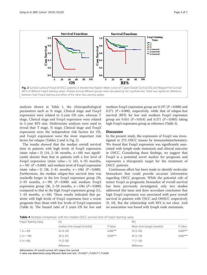
Fig. 2 Survival curves of Foxp3 of OSCC patients. It showed that Kaplan–Meier curves of 5 years Overall Survival (OS) and Relapse-Free Survival(RFS) of different Foxp3 staining values. PValues among different groups were calculated by the Log-Rank test. There was significant differencebetween high Foxp3 staining and either of the other two staining grades
Song et al. BMC Cancer (2016) 16:530 Page 5 of 7
analyses shown in Table 1, the clinicopathologicalparameters such as N stage, Clinical stage and Foxp3expression were related to 5-year OS rate, whereas Tstage, Clinical stage and Foxp3 expression were relatedto 5-year RFS rate. Multivariate analyses were used toreveal that T stage, N stage, Clinical stage and Foxp3expression were the independent risk factors for OS,and Foxp3 expression were the most important riskfactor for relapse (Tables 2 and 3; Fig. 2).The results showed that the median overall survival
time in patients with high levels of Foxp3 expression(stain value = 3) (14, 2–56 months, n = 68) was signifi-cantly shorter than that in patients with a low level ofFoxp3 expression (stain value = 1) (43, 4–93 months,n = 99) (P = 0.000) and medium level of Foxp3 expression(stain value = 2) (38, 2–81 months, n = 106) (P = 0.000).Furthermore, the median relapse-free survival time wasmarkedly longer in the low Foxp3 expression group (39,2–93 months, n = 99) (P = 0.000) and medium Foxp3expression group (36, 2–59 months, n = 106) (P = 0.000)compared to that in the high Foxp3 expression group (11,1–54 months, n = 68). These results indicated that pa-tients with high levels of Foxp3 expression have a worseprognosis than those with low levels of Foxp3 expression(Table 4). The hazard ratio of 5 years OS for low and
Table 4 Multiple comparison with the median OSCC survival time o
Foxp3 Staining Value OS
median time (range) (months)
1 (n = 99) 43 (4–93)
2 (n = 106) 38 (2–81)
3 (n = 68) 14 (2–56)
Reference
Abbreviations: OS overall survival, RFS relapse free survivalP value was determined using Wilcoxon Rank Sum test. *,P<0.05;**, P<0.01;***, P<0.
medium Foxp3 expression group are 0.197 (P = 0.000) and0.271 (P = 0.000), respectively, while that of relapse-freesurvival (RFS) for low and medium Foxp3 expressiongroup are 0.451 (P = 0.014) and 0.371 (P = 0.003) takinghigh Foxp3 expression group as reference (Table 3).
DiscussionIn the present study, the expression of Foxp3 was inves-tigated in 273 OSCC tissues by immunohistochemistry.We found that Foxp3 expression was significantly asso-ciated with lymph node metastasis and clinical outcomein OSCC. Considering these findings, we suggest thatFoxp3 is a potential novel marker for prognosis andrepresents a therapeutic target for the treatment ofOSCC patients.Continuous effort has been made to identify molecular
biomarkers that could provide accurate informationregarding OSCC prognosis. While the potential role oftumor Foxp3 as prognostic biomarker of overall survivalhas been previously investigated, only two studiesaddressed this issue and draw accordant conclusion thathigh Foxp3 expression was associated with poor overallsurvival in patients with TSCC and OHSCC respectively[9, 10]. But the relationship with RFS is not clear. Andno association was found with lymph node metastasis.
f Foxp3 staining value
RFS
P Value Mean time (range) (months) P Value
0.000*** 39 (2–93) 0.000***
0.000*** 36 (2–59) 0.000***
11 (1–54)
Reference
001
Song et al. BMC Cancer (2016) 16:530 Page 6 of 7
Takenaka et al. observed that tumor cytoplasm Foxp3expression was associated with worse relapse-free sur-vival in breast cancer [12]. In small cell lung cancer,relapse- free survival in patients with Foxp3-positivetumor was better with earlier follow-up [13]. WhetherFoxp3-positive cancer cells are relevant to recurrence iscontroversial. For all the head and neck squamous cellcarcinoma(HNSCC) types, the relationship of Foxp3expression with RFS is not clear. This is the first studyto describe the association of Foxp3 expression withboth five years overall survival (OS) and relapse-free sur-vival (RFS) in OSCC patients. It helps to define the pos-sible link between the biological function of Foxp3 andthe progression of OSCC.Moreover, we provided evidence that Foxp3
expressed by OSCC cells might play a role in the me-tastasis of OSCC. We found that OSCC patients withhigh tumor Foxp3 expression had significantly shortersurvival time and more lymphnodes involvement thanother groups. Although no association was found withlymph node metastasis in the previous TSCC andOHSCC cohort studies, there is still considerableamount of evidence available which indicates Foxp3expression in breast cancer [14], gastric cancer [6],non-small cell lung cancer [15] and esophageal squa-mous cell carcinoma [16] is correlated with tumormetastasis. The molecular mechanism of Foxp3 aboutmetastasis remained unclear. One study demonstratedthat Foxp3 inhibited breast cancer cell adhesion, inva-sion and metastasis, while study on the molecularmechanism revealed that Foxp3 inhibited breast can-cer metastasis by down-regulating CD44 expressiondirectly [17]. Further studies are required to elucidateits mechanism in OSCC.
ConclusionsIn summary, this study demonstrated that Foxp3 expressedin OSCC cells were significantly correlated with worseprognosis of both overall survival and relapse-free survivalin OSCC. Ultimately, these findings provided evidence thatFoxp3 expressed by OSCC cells might play a role in theinvasion of OSCC, and tumoral Foxp3 might be suit-able as a prognostic marker in OSCC. More studies areneeded to further explore how tumor cells regulatingthe metastasis behavior during this interaction, espe-cially in light of the current anticancer efforts to inter-fere with Foxp3 expression.
AbbreviationsFoxp3, Forkhead Box P3; HNSCC, head and neck squamous cell carcinoma;IHC, immunohistochemistry; IRS, immunoreactive score; OHSCC,orohypopharynxsquamous cell carcinoma; OS, overall survival; OSCC, oralsquamous cell carcinoma; RFS, relapse-free survival; TMAs, tissue microarrays;TSCC, tongue squamous cell carcinoma
AcknowledgementsWe sincerely thank Dr Vivian Lui (HongKong University) for thoughtfulediting of this manuscript. We sincerely thank the patients for theirparticipation in this study.
FundingThis project was supported by grants from National Natural Science Foundationsof China (No. 81272954, 81472524). The funders had no role in study design, datacollection and analysis, decision to publish, or preparation of the manuscript.
Availability of data and materialsData supporting the findings are included within the manuscript.
Authors’ contributionsZW and BC conceived of the study, and participated in its design andcoordination and helped to draft the manuscript. JJS and SJZ carried out the IHCexperiments and drafted the manuscript. JF and XQL performed the statisticalanalysis. DM, XBC and YW participated in the pathological evaluation and helpedto draft the manuscript. All authors have read and approved the final manuscript.
Competing interestsThe authors declare that they have no competing interest.
Consent for publicationNot applicable.
Ethics approval and consent to participateEthical approval was obtained from the Scientific and Ethical Committee ofSun Yat-sen University, China(ERC-2013-11). Written informed consent wasobtained from all patients. The data were analyzed anonymously.
EndnotesNot applicable.
Author details1Guanghua School of Stomatology, Guangdong Provincial Key Laboratory ofOral Diseases, Sun Yat-sen University, No. 56, Lingyuan West Road,Guangzhou 510055, China. 2Chinese Academy of Medical Sciences & PekingUnion Medical College, Beijing, China.
Received: 10 December 2015 Accepted: 27 June 2016
References1. Fitzmaurice C, Dicker D, et al. The Global Burden of Cancer 2013. JAMA
Oncol. 2015;1:505–27.2. Siegel R, Ma J, Zou Z, Jemal A. Cancer statistics. CA Cancer J Clin. 2014;64(1):9–29.3. Burzyn D, Benoist C, Mathis D. Regulatory T cells in nonlymphoid tissues.
Nat Immunol. 2013;14(10):1007–13.4. Triulzi T, Tagliabue E, Balsari A, Casalini P. FOXP3 expression in tumor cells
and implications for cancer progression. J Cell Physiol. 2013;228(1):30–5.5. Zuo T, Wang L, Morrison C, Chang X, Zhang H, Li W, Liu Y, Wang Y, Liu X, Chan
MW, Liu JQ, Love R, Liu CG, Godfrey V, Shen R, Huang TH, Yang T, Park BK, WangCY, Zheng P, Liu Y. FOXP3 is an X-linked breast cancer suppressor gene and animportant repressor of the HER-2/ErbB2 oncogene. Cell. 2007;129(7):1275–86.
6. Wang L, Liu R, Li W, Chen C, Katoh H, Chen GY, McNally B, Lin L, Zhou P, Zuo T,Cooney KA, Liu Y, Zheng P. Somatic single hits inactivate the X-linked tumorsuppressor FOXP3 in the prostate. Cancer Cell. 2009;16(4):336–46.
7. Ma GF, Miao Q, Liu YM, Gao H, Lian JJ, Wang YN, Zeng XQ, Luo TC, Ma LL,Shen ZB, Sun YH, Chen SY. High Foxp3 expression in tumour cells predictsbetter survival in gastric cancer and its role in tumour microenvironment. BrJ Cancer. 2014;110(6):1552–60.
8. Ladoire S, Arnould L, Mignot G, Coudert B, Rebe C, Chalmin F, Vincent J,Bruchard M, Chauffert B, Martin F, Fumoleau P, Ghiringhelli F. Presence ofFoxp3 expression intumor cells predicts better survival in HER2-overexpressing breast cancer patients treatedwith neoadjuvantchemotherapy. Breast Cancer Res Treat. 2011;125:65–72.
9. Liang YJ, Liu HC, Su YX, Zhang TH, Chu M, Liang LZ, Liao GQ. Foxp3expressed by tongue squamous cell carcinoma cells correlates withclinicopathologic features and overall survival in tongue squamous cellcarcinoma patients. Oral Oncol. 2011;47(7):566–70.
Song et al. BMC Cancer (2016) 16:530 Page 7 of 7
10. Weller P, Bankfalvi A, Gu X, Dominas N, Lehnerdt GF, Zeidler R, LangS, Brandau S, Dumitru CA. The role of tumour Foxp3 as prognosticmarker in different subtypes of head and neck cancer. Eur J Cancer.2014;50(7):1291–300.
11. Cabrera T, Salinero J, Fernandez MA, Garrido A, Esquivias J, Garrido F. Highfrequency of altered HLA class I phenotypes in laryngeal carcinomas. HumImmunol. 2000;61(5):499–506.
12. Takenaka M, Seki N, Toh U, Hattori S, Kawahara A, Yamaguchi T, Koura K,Takahashi R, Otsuka H, Takahashi H, Iwakuma N, Nakagawa S, Fujii T, SasadaT, Yamaguchi R, Yano H, Shirouzu K, Kage M.. FOXP3 expression in tumorcells and tumor-infiltrating lymphocytes is associated with breast cancerprognosis. Mol Clin Oncol. 2013;1(4):625–32.
13. Tao H, Mimura Y, Aoe K, Kobayashi S, Yamamoto H, Matsuda E, Okabe K,Matsumoto T, Sugi K, Ueoka H. Prognostic potential of FOXP3 expression innon-small cell lung cancer cells combined with tumor-infiltrating regulatoryT cells. Lung Cancer. 2012;75(1):95–101.
14. Merlo A, Casalini P, Carcangiu ML, Malventano C, Triulzi T, Mènard S,Tagliabue E, Balsari A. FOXP3 expression and overall survival in breastcancer. J Clin Oncol. 2009;27(11):1746–52.
15. Dimitrakopoulos FI, Papadaki H, Antonacopoulou AG, Kottorou A, Gotsis AD,Scopa C, Kalofonos HP, Mouzaki A. Association of FOXP3 expression withnon-small cell lung cancer. Anticancer Res. 2011;31(5):1677–83.
16. Xue L, Lu HQ, He J, Zhao XW, Zhong L, Zhang ZZ, Xu ZF. Expression ofFOXP3 in esophageal squamous cell carcinoma relating to the clinical data.Dis Esophagus. 2010;23(4):340–6.
17. Zhang C, Xu Y, Hao Q, Wang S, Li H, Li J, Gao Y, Li M, Li W, Xue X1, Wu S,Zhang Y, Zhang W. FOXP3 suppresses breast cancer metastasis throughdownregulation of CD44. Int J Cancer. 2015;137(6):1279–90. 4.
• We accept pre-submission inquiries
• Our selector tool helps you to find the most relevant journal
• We provide round the clock customer support
• Convenient online submission
• Thorough peer review
• Inclusion in PubMed and all major indexing services
• Maximum visibility for your research
Submit your manuscript atwww.biomedcentral.com/submit
Submit your next manuscript to BioMed Central and we will help you at every step:


![Circulating and Tumor-Infiltrating Foxp3 Regulatory T Cell ... · traditional Th1, Th2 helper T cell subsets, Foxp3+ reg-ulatory T cell (Tregs) and IL-17-producing Th17 cells[9]](https://img.pdfslide.net/doc/110x75/5e4b79c0f61ac961cb5bf5de/circulating-and-tumor-infiltrating-foxp3-regulatory-t-cell-traditional-th1.jpg)