Embed Size (px)
Citation preview
Case Report Article
Free gingival graft to increase gingival dimensions in Down syndrome patients: clinical case report
Carla Hariene Montanha1
Maria Estela Plens1 Henrique Scheffer Ferreira1 Luiz Augusto Ribas1
Vinicius Augusto Tramontina1
João Armando Brancher1, 2
Andréa Paula Fregoneze1
Corresponding author: Andréa Paula FregonezeAvenida Manoel Ribas, n. 750, apto 303 – MercêsCEP 80510-020 – Curitiba – PR – BrasilE-mail: [email protected]
1 Department of Dentistry, Pontifical Catholic University of Paraná – Curitiba – PR – Brazil.2 Department of Dentistry, Positivo University – Curitiba – PR – Brazil.
Received for publication: November 7, 2013. Accepted for publication: September 17, 2014.
Keywords: Down syndrome; free gingival graft; periodontal disease.
Abstract
Introduction: Down Syndrome (DS) patients have increased susceptibility to the development of periodontal diseases by the occurrence of several factors, such as inadequate hygiene, mouth breathing, dental morphology, leukocyte reduction and increased inflammatory mediators. Objective: This study aimed to review the literature on the main aspects of DS and present a clinical case of a DS patient treated with basic periodontal therapy and free gingival graft surgery. Case report: DS patient, leucoderma, aged 26 years showed gingival recession and little amount of keratinized tissue in the area of teeth #31 and #41. After surgery, there was an increase in the keratinized tissue band. Conclusion: The free gingival graft surgery performed in DS patients was effective, as the increase of keratinized tissue band occurred.
ISSN: Electronic version: 1984-5685RSBO. 2015 Jan-Mar;12(1):103-8

104 – RSBO. 2015 Jan-Mar;12(1):103-8
Montanha et al. – Free gingival graft to increase gingival dimensions in Down syndrome patients: clinical case report
Introduction
Down's syndrome (DS) or trisomy 21 is a chromosomal disorder resulting from the presence of an extra chromosome 21, in all cells or part of them. It is the most common birth defect and the most recognized form of mental retardation, appearing in about one in every 700 live births. Although the syndrome has been described many years before, it was named by John Langdon Down in 1866 [8].
Dreux et al. [5], Biselli et al. [2], and Valencia et al. [12] explained that three types of chromosome 21 abnormalities can be described: a) free trisomy 21, b) mosaicism, and c) translocation. In about 95% of DS cases, the anomaly occurs because of an extra total chromosome 21 in all cells. In 2% of cases of DS, mosaicism is observed, meaning the anomaly is located in only some cells of the body, while others have normal chromosomes. Translocation involves the long arm of acrocentric chromosomes. This defect can be transmitted over several generations, resulting in chromosomally abnormal products with complete aneuploidy.
The risk of syndrome recurrence in cases of free trisomy mosaicism and usually it is not repeated in siblings of DS people, while the translocation can be recurrent [2, 5, 12].
Among the main risk factors for DS, stand out advanced maternal age, increasing exponentially after 35 years of age; previous child with DS or other chromosomal abnormality; parietal translocation and parents with chromosomal disorders [3].
Girirajan [6] cited some clinical features of DS, among which we highlight the severe mental retardation, facial profile characteristics, short stature, delayed speech development, chronic ear infections with consequent loss of hearing and hypotonia. The characteristics of the facial profile consist of epicanthus, flattened nasal plan, palpebral fissures and protruding tongue. DS patients may also exhibit congenital heart disease between 40 and 50% of the cases an increased risk for developing Alzheimer's disease, acute megakaryocytic leukemia, Hirschsprung's disease and duodenal atresia.
The most frequent general DS problem is mental retardation associated with a high incidence of congenital heart disease and abnormalities of the gastrointestinal tract. In addition to these changes, hypothyroidism, celiac disease, type I diabetes mellitus, transient myeloproliferative disease occur frequently in these syndromic people [10].
Many oral manifestations inherent to DS were described by Amano et al. [1] and Davidovich et al. [4], e.g., macroglossia, cleft lip and tongue, maxillary
atresia, malocclusion, high palate, hypersalivation, microdontia, agenesis, conoid teeth, eruption retardation, low incidence of caries, mouth breathing and open bite. Surprised with the low prevalence of caries despite the presence of risk factors such as cariogenic diet consumption, mouth breathing, difficulty in oral hygiene (resulting from mental retardation), the authors associated this condition with the high average values of pH and buffer capacity checked in sialochemical analysis.
The severity of periodontal disease in individuals with DS is associated with different local factors of the oral cavity and also to the problems associated with systemic disease gene itself. DS patients have an inability to maintain proper oral hygiene. Other conditions contribute to colonization of periodontal pathogens such as mouth breathing, tooth morphology and acute necrotizing ulcerative gingivitis. There is evidence that immunity is impaired due to the reduction of neutrophils, T lymphocytes and increasing the production of inflammatory mediators and proteolytic enzymes. All this can help increase the prevalence and aggravate severity of periodontal disease in DS people [8].
The mucogengival deformities, such as changes in the dimensions of the mucosa an in the morphological characteristics often have an impact on patients in terms of function and aesthetics. Although clinical evidence shows that no attached gingiva minimum value is required to maintain gingival health, there are clinical situations, such as thin or absent gingiva which make it difficult to maintain the hygiene at that location, causing plaque accumulation, inflammation, and consequently the loss ligament insertion [11].
The trauma resulting from inadequate or excessive brushing, gingival inflammatory reactions, bone dehiscence, poor positioning of the teeth, and orthodontic movement are considered predisposing factors for gingival recession. The gingival recession is classified as Class I when it extends below the mucogingival junction, and class II when it reaches these areas [9]. In such cases, the free gingival graft (FGG) is a surgical technique introduced to address the lack of keratinized tissue, leading to increase in the size of this tissue [11].
Because of the lack of studies on the treatment of periodontal morphological changes in DS patients, this study aimed to present a case in which FGG was performed to increase the size of that tissue. Such intervention was carried out by multidisciplinary treatment of the Specialization Course in Periodontics and Discipline for Special Patients of the Pontifical Catholic University of Paraná (PUC-PR).
105 – RSBO. 2015 Jan-Mar;12(1):103-8
Montanha et al. – Free gingival graft to increase gingival dimensions in Down syndrome patients: clinical case report
Clinical case
A male, leucoderm DS patient aged 26 years was referred to treatment. During the anamnesis, the mother reported that she discovered the pregnancy at 44 years of age and that this went uneventfully. The delivery was normal, with no complications, the baby cried at birth and sucked in the first 24 hours. Its development was satisfactory. He was breastfed until six months. At eight months, his first tooth erupted and crawled at nine months. At two years of age, he started to stand up alone and talk. He started to wear glasses at 16 years of age.
The survey of current medical history revealed that the only systemic problem is a tremor in his eyes. Accordingly, the patient has been doing eye exercises for more than three years and uses Endura®, which is an ocular lubricant. As regards the behavioral aspect, the patient was very open,
since related pleasantly with all professionals. The mother also told the team that he was irritated only when some people fought or talking loudly near him.
He was referred to the Discipline of Dentistry for Patients with Special Needs of PUC-PR by the Association of Parents and Friends of Exceptional People (Apae), to receive dental treatment. The patient had received previous dental treatment at this same Association.
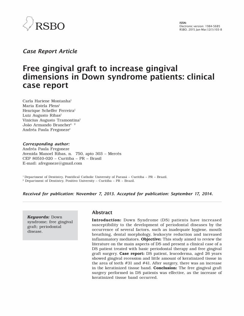
During intraoral clinical examination, we verified plaque accumulation and gingivitis. The gingiva on the labial surface of the mandibular incisors showed little amount of keratinized mucosa and recession. The analysis of occlusion showed a convex profile, Class III malocclusion, left posterior cross bite and crowding in the anterior region. The initial clinical condition of each tooth is described in figure 1.
Figure 1 – Patient’s initial odontogram
Prior to dental procedures, interproximal and panoramic radiographs were taken. Panoramic radiograph showed the agenesis of the maxillary and mandibular third molars and the presence of included maxillary canines. Then, the basic periodontal therapy was carried out, which included root scaling, planning and polishing. Oral hygiene instructions were transmitted to the mother and the patient so that both continue motivated to maintain oral health. The next step was the application of fluoride varnish.
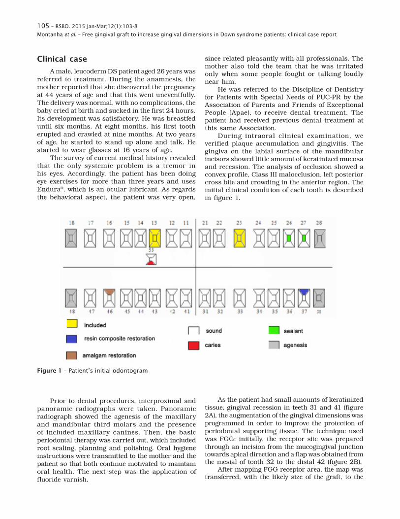
As the patient had small amounts of keratinized tissue, gingival recession in teeth 31 and 41 (figure 2A), the augmentation of the gingival dimensions was programmed in order to improve the protection of periodontal supporting tissue. The technique used was FGG: initially, the receptor site was prepared through an incision from the mucogingival junction towards apical direction and a flap was obtained from the mesial of tooth 32 to the distal 42 (figure 2B).
After mapping FGG receptor area, the map was transferred, with the likely size of the graft, to the
106 – RSBO. 2015 Jan-Mar;12(1):103-8
Montanha et al. – Free gingival graft to increase gingival dimensions in Down syndrome patients: clinical case report
donor area between right maxillary premolars and first molar, on the palate. With the aid of a 15C blade, a superficial incision was made around the aluminum map to demarcate the graft area to be removed. After this initial incision and removal of the map, the depth incisions were performed at a distance of 2 to 3 mm for the removal of epithelialized free graft.
After removal of tissue from FGG donor site (figure 2C), local hemostasis was promoted with bismuth subgallate and then the wound was protected with surgical cement (figure 2D). FGG removed from the donor area was sutured to the receptor site with simple and suspensory suture, and then the area was protected with surgical cement (figures 2E, F and G).
Elapsed one week after surgery, the sutures were removed and the presence of a reddish bleeding
tissue with characteristics of vitality of the graft was observed. It was also found desquamation of graft epithelial tissue, which is considered normal at this healing step. Ibuprofen 600 mg, 3 times daily for 4 days was prescribed to reduce swelling and to prevent inflammation.
A f ter t wo mont hs, t here was a g reat incorporation of the graft to the receptor site. After 6 months, 1 year and 2,5 years, there has been significant growth in the keratinized mucosa band, thus achieving the increase of gingival tissue dimensions (figures 2H, I, and J). The patient continues to be followed-up to receive oral health maintenance and treatment of included upper canines at the Clinics of the Discipline of Dentistry for Patients with Special Needs of PUC-PR.
A B
C D
E F
G H
I J
Figure 2 – Sequence of FGG surgical procedure. A. Initial clinical aspect with gingival recession of teeth #31 and #41; B. preparation of receptor site; C. tissue removal of the donor site; D. protection of donor site with surgical cement; E. FGG aspect removed from the donor site; F. tissue sutured on receptor site; G. protection with surgical cement on receptor site; H. 6 month follow-up; I. 1 year follow-up; J. 2,5 year follow-up
107 – RSBO. 2015 Jan-Mar;12(1):103-8
Montanha et al. – Free gingival graft to increase gingival dimensions in Down syndrome patients: clinical case report
Discussion
Among the main DS risk factors there is advanced maternal age, significantly increasing after 35 years of age [3]. This statement meets the clinical case study, as in anamnesis, the mother reported that the pregnancy occurred at 44 years of age. Corroborating the descriptions of Girirajan [6], the following features were noted in the patient: epicanthus, palpebral fissures, flat nasal plan. On the other hand, hypotonia, chronic ear infections and hearing loss have not been verified.
Similarly, the delay in speech development, quoted by the same author, differs from the information obtained in the anamnesis, since the mother said the baby started talking at 2 years old. The severe mental retardation [6] has not been observed because the patient had amazing ability to paint. With regard to the systemic conditions, none was found. The only problem was that the patient had the tremor in his eyes.
Among inherent DS oral manifestations [1, 4] maxillary atresia, malocclusion, fissured tongue, low incidence of caries, mouth breathing and agenesis were present. In contrast, cleft lips, high palate, hypersalivation, microdontia and conoid teeth were not observed.
Morgan [8] said that the severity of periodontal disease in persons with DS is associated with several factors, such as failure to maintain proper oral hygiene, mouth breathing and tooth morphology. Despite these conditions confirmed in the present study patient, periodontal disease was not severe.
Therefore, in order to solve the biofi lm accumulation and gingivitis, initially the mother and the patient were instructed on the proper technique for oral hygiene. Then root scaling, planning, and polishing were executed.
FGG was justified by the fact that the patient has Class I gingival recession [9] on teeth #31 and #41, and little amount of keratinized tissue on the same region [11]. The team was concerned on the continuity of insertion ligament loss in these teeth, which is a consequence of the presence of mucogingival [11]. FGG surgery was satisfactory, because both the recession and the alteration of mucosa dimensions were solved. The surgery goal was to improve the function, because the patient was asymptomatic and did not present esthetic demands.
Conclusion
FGG surgery performed on DS patient was effective, because the keratinized mucosa band increase, thus achieving the increase of gingival dimensions.
References
1. Amano A, Murakami J, Akiyama S, Morisaki I. Etiologic factors of early-onset periodontal diseasein Down syndrome. Japanese Dental Science Review. 2008 Jul;44:118-27.
2. Biselli J, Bertollo EG, Ruiz M, Bertelli EP. Cytogenetic profile of Down syndrome cases seen by a general genetics outpatient service in Brazil. Down Syndrome Research and Pratice. 2009 Feb;12(3):4.
3. Davidson MA. Primary care for children and adolescents with Down syndrome. Pediatric Clinics of North America Syndrome. 2008 Oct;55:1099-111.
4. Davidovich E, Aframian DJ, Shapira J, Peretz B. A comparison of the sialochemistry, oral pH, and oral health status of Down syndrome children to healthy children. International Journal Of Paediactric Dentistry. 2010;20:235-41.
5. Dreux S, Olivier C, Dupont JM, Leporrier N, Group S, Oury JF et al. Maternal serum screening in cases of mosaic and translocation Down syndrome. Prenat Diagn. 2008 Jul;28:699-703.
6. Girirajan S. Parental-age effects in Down syndrome. Journal of Genetics. 2009 Apr;88:9-14.
7. Mégarbané A, Ravel A, Mircher C, Sturtz F, Grattau Y, Rethoré MO et al. The 50th anniversary of the discovery of trisomy 21: the past, present, and future of research and treatment of Down syndrome. Genet Med. 2009 Sep;11(9):611-6.
8. Morgan J. Why is periodontal disease more prevalent and more severe in people with Down syndrome? Spec Care Dentist. 2007;27(5):196-201.
9. Müller HP, Eger T, Schorb A. Alteration of gingival dimensions in a complicated case of gingival recession. Int J. Periodont Rest Dent. 1998;18(4):345-53.

108 – RSBO. 2015 Jan-Mar;12(1):103-8
Montanha et al. – Free gingival graft to increase gingival dimensions in Down syndrome patients: clinical case report
1 0 . R a m G , C h i n e n J . I n f e c t i o n s a n d immunodeficiency in Down syndrome. Clinical and Experimental Immunology. 2011 Jan;164:9-16.
11. Silva CO, Ribeiro EDP, Sallum AW, Tatakis DN. Free gingival grafts: graft shrinkage and donor-site healing in smokers and non-smokers. J Periodontol. 2010 May;81(5):692-701.
12. Valencia LG, Angles MMR, Hernández AM, Gonzáles MRB. Down’s syndrome associated with a balanced Robertsonian translocation 13;14 maternally transmitted in the product of a twin diamniotic pregnancy. Bol Med Hosp Infant Mex. 2011;68(3):206-10.