Embed Size (px)
Citation preview

Enhanced Inactivation of Salmonella and Pseudomonas Biofilms onStainless Steel by Use of T-128, a Fresh-Produce Washing Aid, inChlorinated Wash Solutions
Cangliang Shen,a Yaguang Luo,b Xiangwu Nou,b Gary Bauchan,c Bin Zhou,a Qin Wang,a and Patricia Millnerb
Department of Nutrition and Food Science, University of Maryland, College Park, Maryland, USAa; U.S. Department of Agriculture, Agricultural Research Service,Environmental Microbial and Food Safety Laboratory, Beltsville, Maryland, USAb; and U.S. Department of Agriculture, Agricultural Research Service, Electron and ConfocalMicroscopy Unit, Beltsville, Maryland, USAc
The effect of the washing aid T-128 (generally recognized as safe [GRAS] formulation, composed mainly of phosphoric acid andpropylene glycol) on inactivation of Salmonella and Pseudomonas populations in biofilms on stainless steel was evaluated underconditions of increasing organic matter loads in chlorinated wash solutions dominated by hypochlorous acid. Biofilms wereformed statically on stainless steel coupons suspended in 2% lettuce extract after inoculation with Salmonella enterica serovarThompson or Newport or with Pseudomonas fluorescens. Coupons with biofilms were washed in chlorine solutions (0, 0.5, 1, 2,5, 10, or 20 mg/liter at pH 6.5, 5.0 and 2.9), with or without T-128, and with increasing loads of organic matter (0, 0.25, 0.5, 0.75,or 1.0% lettuce extract). Cell populations on coupons were dispersed using intermittent, pulsed ultrasonication and vortexingand enumerated by colony counts on XLT-4 or Pseudomonas agars. Cell responses to fluorescent viability staining of biofilmtreatment washing solutions were examined using confocal laser scanning microscopy. Results showed that 0.1% T-128 (withoutchlorine) reduced P. fluorescens biofilm populations by 2.5 log10 units but did not reduce Salmonella populations. For both Sal-monella and Pseudomonas, the sanitizing effect of free chlorine (1.0 to 5.0 mg/liter) was enhanced (P < 0.05) when it was com-bined with T-128. Application of T-128 decreased the free chlorine depletion rate caused by increasing organic matter in washwaters and significantly (P < 0.05) augmented inactivation of bacteria in biofilms compared to treatments without T-128. Imageanalysis of surfaces stained with SYTO and propidium iodide corroborate the cultural assay results showing that T-128 can aidin reducing pathogen viability in biofilms and thus can aid in sanitizing stainless steel contact surfaces during processing offresh-cut produce.
Biofilm formation by food-borne pathogens or spoilage micro-organisms on food processing equipment and contact sur-
faces is a major concern in fresh-cut produce safety (1, 3). Salmo-nella spp. biofilms on plastic, cement, glass, and stainless steelsurfaces (4, 11, 13) can be persistent sources of contamination inthe fresh-food processing environment. Spoilage bacteria, such asPseudomonas fluorescens, form biofilms that provide a protectivematrix for surviving pathogenic cells and may cross-contaminatevarious stages of food production operations (16).
Typical cleaning methods, e.g.., physical scrubbing followed bysanitizer treatment, that are used to remove bacteria from foodprocessing equipment and contact surfaces (8) do not always in-activate bacteria in biofilms as these are more resistant to chemicalsanitizers than planktonic cells. For example, 200 mg/liter freechlorine (FC) did not inactivate Escherichia coli O157:H7 in bio-films (24), and FC at 10 mg/liter for 10 min resulted in less than a10-fold reduction in Salmonella biofilm-associated cells (13).Studies of the limitations of chlorine on viability of biofilm pop-ulations suggest that high concentrations of FC are effective inremoving food spoilage bacterial biofilms on certain surfaces. Freechlorine at 200 mg/liter reduced Bacillus cereus and Pseudomonasbiofilm populations more than 4 and 6 log CFU per stainless steelcoupon, respectively (14), and FC at 100 mg/liter for 2 min re-duced P. fluorescens biofilm populations by approximately 3 logCFU per stainless steel coupon (5).
Sodium hypochlorite solution at pH 6.5 is currently the mostcommon sanitizer used in the fresh-cut produce industry. At pH5.0 to 6.5, the free chlorine in solution is dominated by hypochlo-
rous acid (HOCl), the most effective disinfectant of the forms ofchlorine including hypochlorite ion, OCl�, and gaseous chlorine,which off-gases from solutions in substantial amounts at pH �4.0(26). In addition to the capacity of hypochlorous acid to reducethe viable microbial bioburden and to prevent cross-contamina-tion, it is also used as a sanitizer during equipment cleaning. Itsoverall minimal impact on nutritional and esthetic qualities of theproduce, its well-known capability to inactivate pathogens in sus-pensions, and its low cost contribute to its common usage (19).Maintaining free chlorine in washing solutions with fresh-cutproduce is challenging because fresh-cut products release copiousamounts of juice into the wash water. The combined high solidsand plant sap loads in the wash solution exert a high demand forthe oxidation potential of FC (i.e., free available chlorine [FAC],comprising primarily HOCl, with minor amounts of Cl2 andOCl�), and with very high processing rates and intermittent ad-dition of sanitizer, FC concentrations rapidly decline to less than0.5 mg/liter (9, 19). Replenishing chlorine periodically, either me-chanically or manually, is common practice in fresh-produce pro-cessing; however, repeated additions of sodium hypochlorite to
Received 3 April 2012 Accepted 25 June 2012
Published ahead of print 29 June 2012
Address correspondence to Patricia Millner, [email protected].
Copyright © 2012, American Society for Microbiology. All Rights Reserved.
doi:10.1128/AEM.01094-12
October 2012 Volume 78 Number 19 Applied and Environmental Microbiology p. 6789–6798 aem.asm.org 6789
Dow
nloa
ded
from
http
s://j
ourn
als.
asm
.org
/jour
nal/a
em o
n 26
Jan
uary
202
2 by
80.
51.1
69.5
5.
high-organic-load wash water can cause formation of toxic chlo-rine by-products, accumulation of chloramines, and generation ofchlorine off-gas in the processing environment (23).
Recently, a formulation (T-128; New Leaf Food Safety Solu-tions, LLC, Salinas, CA) containing generally recognized as safe(GRAS) chemicals was reported to stabilize free chlorine concen-trations in wash solutions receiving high organic loads (K. E. Lem-ons, U.S. patent application 20090192231). The main compo-nents of T-128 are phosphoric acid and propylene glycol; thus, thepH of pure T-128 is �1.0 (Lemons, U.S. patent application20090192231). By pulsed addition of T-128 into sodium hypo-chlorite solution, controlled acidic solutions at pH 6.5 and 5.0 canbe achieved. Our recent studies show that using T-128 in fresh-cutlettuce processing reduced pathogen survival in wash solutionscontaining high organic loads and prevented cross-contamina-tion when inoculated and uninoculated lettuce were washed to-gether (23). We hypothesized that T-128, developed as a novelfresh-cut washing aid, may also enhance the effects of chlorinesanitizer in reducing pathogen survival in biofilms. Thus, the ob-jectives of this study were to (i) determine the effect of T-128 onthe efficacy of FC in wash water in reducing viability of bacteria inbiofilms and (ii) evaluate the effect of FC in T-128-treated washwater on the survival of pure or cocultured biofilms of Salmonellaand Pseudomonas in solutions of different organic loads.
MATERIALS AND METHODSBacterial strains and culture preparation. Salmonella enterica serovarThompson strain RM1987 (cilantro outbreak isolate; referred to as S.Thompson), S. enterica serovar Newport strain F3307 (mango isolate;referred to as S. Newport), and Pseudomonas fluorescens (ATCC 17400)were used in this study. Strains were maintained in brain heart infusionbroth with 20% glycerol and stored at �80°C. Prior to experiments, thestock cultures were streaked on XLT-4 agar (BD, Sparks, MD) (Salmonellastrains) or Pseudomonas agar F (Fluka/Sigma-Aldrich, St. Louis, MO) (P.fluorescens) and incubated at 37°C (Salmonella strains) or 28°C (P. fluore-scens) for 24 to 48 h.
Media and material preparation. Lettuce juice extract (LJE) was pre-pared from cored, whole fresh iceberg lettuce that was processed in acommercial household juice maker (model BJE200XL Juice Fountain;Breville, Shanghai, China). The extracted juice was filtered through a dou-ble layer of cheesecloth and centrifuged (4,629 � g for 10 min at 4°C) twiceto remove coarse particles. Supernatants were membrane filter sterilized(0.22-�m pore size) and then diluted in sterile distilled water to make 2%LJE, pH 6.3. Stainless steel coupons (type 302; 0.9-mm thickness, 50 by 20mm; 21.26 cm2) were cleaned by being soaked in methanol for 30 min toremove oil/grease and were rinsed in distilled water prior to being soakedin an alkaline detergent for 1 h at 60°C; they were then ultrasonicated for5 min to remove attached debris. Coupons were rinsed in distilled waterand separated between paper tissues before sterilization by autoclaving at121°C for 15 min.
Biofilm formation. A single colony of each Salmonella strain onXLT-4 agar or P. fluorescens on Pseudomonas agar F was cultured in 25 mlof 1/4 strength Bacto M9 minimal salt broth (0.4% glucose, pH 7.0; DifcoLaboratories, Detroit, MI) and incubated at 22°C for 24 h. Cells werewashed by centrifugation at 4,629 � g for 10 min at 4°C three times with25 ml of phosphate-buffered saline (PBS; pH 7.4). Cell pellets were resus-pended and diluted in PBS to 5 to 6 log10 CFU/ml. Pure cultures or co-culture cell suspensions (0.1 ml) of Salmonella strains and P. fluorescenswere inoculated into 30 ml of LJE in a 50-ml centrifuge tube containing asterile stainless steel coupon which was fully submerged in the solution toallow biofilm formation on the coupon surface. Tubes were incubatedstatically at 22°C for 24 h to allow formation of biofilms.
Preparation of chlorine solutions and T-128 treatments. The chlo-rine wash solution (CWS) was prepared by diluting 6% NaOCl (Clorox,Oakland, CA) into cold distilled water (5°C). The washing aid T-128 (NewLeaf Food Safety Solutions, LLC, Salinas, CA) was directly added to theCWS to reach pH 5.0, which corresponds to approximately 0.075% (re-ferred to as pH 5.0 with T-128) or pH 2.9, corresponding to approxi-mately 0.1% (referred to as pH 2.9 with T-128). When T-128 was notused, pH was adjusted to 6.5, 5.0, or 2.9 using citric acid. The FC concen-tration of each treatment was measured with a commercial test systemincluding a DPD (N,N-diethyl-p-phenylenediamine) powder dispenserand chlorine photometer (CP-15; HF Scientific Inc., Ft. Myers, FL).
Water quality characteristics of treatments, including pH, turbidity,total dissolved solids (TDS), and chemical oxygen demand (COD), weredetermined using a digital pH meter (Oakton Instruments, Vernon Hills,IL), a turbidity meter (Aquafast, Thermo Orion Research Inc., BeverlyCity, MA), a conductivity meter (model 135A; Orion Research Inc., Bev-erly City, MA), and a reactor digestion method (COD2 Mercury-freeCOD reagent; Hach Co., Loveland, CO) (12, 18), respectively.
The different organic loads in solutions were prepared by adding freshiceberg LJE to chlorine solutions, with or without T-128, to the desiredconcentration; solutions were mixed for 30 s prior to determining theresidual FC concentrations and used immediately. All chlorine solutionswere freshly prepared and used within 2 min.
The possible interference by manganese and monochloramine withthe DPD free-chlorine test with wash waters also was evaluated to ensurethat free chlorine was not overestimated by any of the unknown compo-nents in the wash solutions. Manganese and chloramine concentrations indistilled water, 0.5 or 1.0% LJE, and CWS (FC of 1 to 10 mg/liter), with orwithout T-128 (pH 5.0), were tested according to the indophenol method(10). For manganese tests, samples (25 ml; pH adjusted to 6 to 7) wereeither pretreated or not by mixing with three 5-�l drops of KI (30 g/liter)and NaAsO2 (5 g/liter); measurements were made with a chlorine pho-tometer (CP-15; HF Scientific Inc., Ft. Myers, FL) after the addition ofpowdered DPD reagent. Manganese content was calculated by subtractingthe pretreated concentration from the unpretreated one. For the chlora-mine test, samples were mixed with Monochlor-F pillows (Hach, Love-land, CO), followed by the addition of one drop of free ammonia reagentsolution, and the optical density 655 nm (OD655) was recorded in a UV-1601 spectrophotometer (Shimaozu Scientific Instruments, Inc., Colum-bia, MD). Chloramine-T standard solutions (0.5 to 10 mg/liter, reagentgrade; Fisher Scientific, Fair Lawn, NJ) were tested likewise and measuredto generate a standard curve. The chloramine content of samples wasdetermined from the standard curve regression line.
Treatment of biofilms. Coupons with biofilms were removed fromthe LJE broth and rinsed with 10 ml of PBS to remove unattached cells.Two experiments were conducted in this study. In the first experiment,coupons were immersed for 2 min in 30 ml of sterile distilled water (con-trol) and 0.2, 0.5, 1.0, 2.0, 5.0, 10.0, and 20.0 mg/liter FC in CWS, with orwithout T-128 at pH 2.9 or 5.0. In the second experiment, coupons wereplaced into 10 to 12 mg/liter of CWS containing 0, 0.25, 0.5, 0.75, or 1.0%LJE, with or without T-128 at pH 2.9 or 5.0. The pH adjustment with citricacid was included to determine the effect of pH on bacterial populationsurvival due to the pH achieved with T-128. During the 2-min exposure toCWS, the test tubes containing the coupons were agitated on an orbitalshaker (VWR International, LLC; West Chester, PA) at 118 rpm. Aftertreatment, each coupon was transferred to a 50-ml centrifuge tube con-taining 30 ml of Dey-Engley (DE) neutralizing both (pH 7.4) (BBL/Difco,Detroit, MI); thereafter tubes were ultrasonicated for 2 min (eight 15-spulses with 10-s static intervals) and vortexed at 3,000 rpm for 2 min todisperse biofilm cells from the coupons. Cell suspensions were diluted inPBS and spiral plated using a spiral plater (WASP2; Microbiology Inter-national, Frederick, MD) on XLT-4 agar (Salmonella) or Pseudomonas Fagar (P. fluorescens). Petri dishes were incubated at 28°C for 48 h (P.fluorescens) or at 37°C for 24 h (Salmonella) before automated colonycounting (ProtoCol; Microbiology International, Frederick, MD).
Shen et al.
6790 aem.asm.org Applied and Environmental Microbiology
Dow
nloa
ded
from
http
s://j
ourn
als.
asm
.org
/jour
nal/a
em o
n 26
Jan
uary
202
2 by
80.
51.1
69.5
5.

Crystal violet staining of biofilms on stainless steel. Cocultured S.Thompson and P. fluorescens formed biofilms on stainless steel coupons asdescribed above. Coupons were then statically immersed or shaken (118rpm) for 2 min in 30 ml of sterile distilled water and 3.0 mg/liter FC inCWS, pH 5.0, adjusted with citric acid or T-128. After treatment, eachcoupon was transferred to 30 ml of PBS containing dechlorinating agent(Hach, Loveland, CO); the tubes were either kept stationary or ultrasoni-cated and vortexed (2 min). Each coupon was stained with 10% crystalviolet for 15 min, gently rinsed with sterile distilled water, and examinedand photographed using an Olympus SZX12 microscope (Melville, NY)and Nikon charge-coupled device (CCD) camera (DS-2 M series) withACT-2U, version 1.52, software (Nikon Instruments Inc., Melville, NY).Both wash water (mixed with dechlorination agent) and cell suspensionsolutions were spiral plated onto tryptic soy agar (BBL/Difco, Detroit, MI)and then incubated at room temperature for 72 h before colonies werecounted.
CLSM. Samples for confocal laser scanning microscopy (CLSM) wereprepared in 50-mm glass-bottom culture dishes (part number P50G-1.5-14-F; MatTek Co., Ashland, MA) and on stainless steel coupons (type 302;0.9-mm thickness, 17 by 17 mm) by inoculation of 5 ml of sterile 2% LJEwith, respectively, 0.1 ml of cocultured S. Thompson and S. Newport orcocultured S. Thompson and P. fluorescens incubated at 25°C for 24 h. The24-h culture liquid was then removed gently by pipetting and replenishedwith fresh LJE; then the samples were reinoculated with the same amountof the bacterial strains and incubated at 25°C for another 24 h. For glass-bottom culture dishes, the culture liquid was removed, and 5 ml of 2mg/liter FC in CWS (pH 5.0) solution, adjusted with citric acid or T-128,was added to the culture dishes and left for 2 min under static conditions.Stainless steel coupons with biofilms were shaken (118 rpm) for 2 min in5 ml of sterile distilled water and 2.0 mg/liter FC in CWS, adjusted withcitric acid or T-128 to pH 5.0. Finally, the culture dishes or coupons wererinsed gently with PBS containing dechlorinating reagent.
The biofilms on the glass portion of the petri dishes or on stainless steelcoupons were stained using a Live/Dead BacLight Bacterial Viability Kit(Invitrogen, Eugene, OR) according to the manufacturer’s recommenda-tions. The BacLight Bacterial Viability Kit contains both SYTO9 stain andpropidium iodide; when used alone, SYTO9 stain labels both live anddead bacteria. No interference by T-128-adjusted solutions was found inpretests conducted on biofilms stained with the Live/Dead kit and ob-served with a fluorescence microscope (Nikon Instruments, Inc., Melville,NY). In contrast, propidium iodide penetrates only bacteria with dam-aged membranes, reducing SYTO9 fluorescence when both dyes are pres-ent. Thus, live bacteria with intact membranes fluoresce green, while deadbacteria with damaged membranes fluoresce red. A Zeiss 710 confocallaser scanning microscopy (CLSM) system with a Zeiss Axio Observerinverted microscope equipped with a 100� (1.4 numerical aperture[NA]) oil immersion Plan-Apochromat objective was used to observe anddigitally capture images of the stained bacteria in the biofilms. A 488-nmargon laser with a pinhole of 32 �m passing through an MBS 488/561/633beam splitter filter with limits set between 490 and 560 nm was used fordetection of green fluorescence for live cells stained with SYTO9. A561-nm diode-pumped solid-state laser with a pinhole of 32 �m passingthrough a Main Beam Splitter (MBS) 488/561/633 beam splitter filter withlimits set between 651 and 720 nm was used for detection of red fluores-cence, indicative of dead cells, in which propidium iodide overstains theSYTO9. Dual lasers were used simultaneously to observe the differentiallystained bacteria. At least 20 fields were examined to ascertain the consis-tency of the staining reaction for each different treatment, and imageswere captured of fields with large numbers of cells aligned within a clearlyresolved focal plane. Zeiss Zen 2009 software (Carl Zeiss Microscopy,Germany) was used to capture the images, and Axiophot, version 4.6 (CarlZeiss Microscopy, Germany), and Photoshop, version 7.0 (Adobe Sys-tems, San Jose, CA), were utilized to design the figures.
Data analysis. Each test series was repeated twice, and in each repli-cate, three samples were analyzed per treatment. Microbiological data
(survivors or reductions, converted to log10 CFU/coupon) were analyzedusing the “mixed procedure” of the SAS program, with independent vari-ables including the chlorine or LJE concentrations, pH of wash solutions(adjusted by citric acid or T-128), and the interaction between them.Means and standard deviations for microbiological data were calculated,and mean significant differences (� � 0.05) among interactions wereseparated with the least significant difference procedure.
RESULTSWater quality and FC concentration in CWS. CWS pH was notaffected by increasing levels of LJE, as evidenced by the less than0.1-pH unit fluctuation (data not shown). As expected, turbidity,TDS, and COD of CWS increased proportionally with addition ofLJE (Table 1), which agreed with our previous results (23).
The FC concentration decreased significantly as increasingconcentrations of LJE were added to chlorine wash solutions, re-gardless of T-128 application (Fig. 1). CWS with T-128 (pH 2.9 or5.0) resulted in higher residual FC levels than the wash solutions ofthe same pH level adjusted by citric acid. In the presence of 1.0%LJE, the FC concentration dropped to 0.26 and 0.48 mg/liter forpH 5.0 and 2.9 for CWS adjusted by citric acid. In contrast, in thepresence of the same level of LJE, FC levels decreased to 0.71 and1.08 mg/liter in pH 5.0 and 2.9 in chlorine solutions containingT-128.
The manganese and monochloramine concentrations in washwater samples, including distilled water, 0.5 and 1.0% LJE, andCWS with FC (10 mg/liter) mixed with 1.0% LJE, with or without0.1% T-128, ranged from 0.0 to 0.02 mg/liter for manganese andfrom 0.001 to 0.004 mg/liter for monochloramine. Thus, the tracelevels of manganese and monochloramine in the CWS of thisstudy did not interfere with the free chlorine DPD test results orthereby lead to an overestimate of the FC in CWS.
Effect of chlorine water with/without T-128 on Salmonella orPseudomonas biofilm cells. After overnight incubation at 22°Cfor 24 h, S. Thompson, S. Newport, or P. fluorescens biofilm pop-ulations reached 7.6, 7.7, and 8.0 log10 CFU/coupon, respectively.Compared to chlorine water without T-128 (pH 2.9 or 5.0 ad-justed with citric acid), chlorine water with T-128 (at both pH 2.9and 5.0) significantly (P � 0.05) enhanced the inactivation ofbacteria in biofilms (Fig. 2). Notably, treatment with T-128 atneither pH 2.9 nor 5.0 T-128 without chlorine (0 at x axis) pro-duced no reduction in Salmonella biofilm populations (Fig. 2).
For Salmonella cells, chlorine water (FC of 0.5 to 5.0 mg/liter,pH 6.5) reduced biofilm cell populations by 1.6 to 4.6 (S. Thomp-son) and 0.8 to 4.4 (S. Newport) log10 CFU/coupon compared to
TABLE 1 Water quality characteristics of chlorine wash watercontaining increasing amounts of organic matter as lettuce juice extracta
LJE concn(%)b
Turbidity(NTU)c
TDS(mg/liter)
COD(mg/liter)
0 0.02 � 0.01 38.67 � 1.15 192.00 � 10.000.25 4.59 � 0.26 75.33 � 0.58 323.67 � 24.280.50 7.07 � 0.15 94.67 � 0.58 392.83 � 20.820.75 10.60 � 0.96 113.67 � 1.53 486.17 � 5.201.0 15.27 � 1.22 124.67 � 1.53 540.33 � 34.49a Means and standard deviations of water quality parameters measured as described inMaterials and Methods.b Volume of lettuce juice relative to the volume of chlorine wash water, expressed as apercentage.c NTU, nephelometric turbidity units.
T-128 Enhances Biofilm Inactivation
October 2012 Volume 78 Number 19 aem.asm.org 6791
Dow
nloa
ded
from
http
s://j
ourn
als.
asm
.org
/jour
nal/a
em o
n 26
Jan
uary
202
2 by
80.
51.1
69.5
5.

the populations of the untreated coupons (Fig. 2A and B). Patho-gen population reductions in biofilms were not different in CWSsadjusted with citric acid to pH 6.5 and pH 2.9 (Fig. 2). In CWS atpH 2.9 adjusted by T-128, 1 and 2 mg/liter FAC significantly (P �0.05) reduced viability of biofilm populations by an additional 1.6to 1.7 log10 CFU/coupon (S. Thompson) (Fig. 2A) and 1.7 to 2.8log10 CFU/coupon (S. Newport) (Fig. 2B) compared to popula-tions in pH 2.9 chlorine solutions without T-128. At 5 mg/liter,CWS adjusted to pH 2.9 with T-128 reduced viability of pathogensin biofilms to below the detection limit, whereas without T-128(pH 2.9), pathogens survived in biofilms at 3.0 log10 CFU/coupon(Fig. 2A and B). A similar trend in enhancing the sanitizing effecton biofilm populations was observed in chlorine solutions ad-justed to pH 5.0 by T-128, compared to the same pH level adjustedby citric acid (Fig. 2). In S. Thompson and S. Newport biofilmcells, treatments (0.5 to 2.0 mg/liter FAC, pH 5.0) containingT-128 resulted in additional (P � 0.05) reductions of 0.7 to 1.5log10 CFU/coupon compared to those of the citric acid-adjustedpH 5.0 (Fig. 2).
For P. fluorescens, 5 mg/liter of CWS (pH 6.5) reduced biofilmpopulations by 2.9 log10 CFU/coupon (Fig. 2C). CWS (FAC of 0 to5 mg/liter) at pH 2.9 (adjusted by citric acid) reduced the P. fluo-rescens biofilm cell population by 3.2 to 3.7 log10 CFU/coupon(Fig. 2C), and these reductions were greater (P � 0.05) than thosewith pH 6.5 or 5.0 CWS at the same chlorine concentrations,indicating that P. fluorescens cells are sensitive to the low pH. The
pH 2.9, nonchlorine, T-128 treatment reduced P. fluorescens pop-ulations by 2.6 log10 CFU/coupon (Fig. 2C), whereas the pH 5.0,nonchlorine, T-128 treatment did not show similar reductions(Fig. 2C). CWS at pH 2.9 or 5.0 with T-128 and with 2 to 10mg/liter FAC showed an additional (P � 0.05) approximately 1.0log10 CFU/coupon reduction of the biofilm population comparedto CWS at the same citric acid-adjusted pH.
Effect of T-128 and organic loads on CWS efficacy againstpure-culture Salmonella or Pseudomonas biofilms. The sanitiz-ing effect of CWS on bacterial biofilms decreased with increasingconcentrations of LJE in wash solutions, regardless of T-128 ap-plication. Generally, in comparison with the pH 6.5 CWS, adjust-ment of CWS to pH 2.9 with T-128 significantly (P � 0.05)
FIG 1 Effect of T-128 on chlorine retention in wash solutions containingvarious amounts of organic materials. CA, citric acid.
FIG 2 Effects of free chlorine (0 to 20 mg/liter) and T-128 (0.075% at pH 5.0and 0.1% at pH 2.9), including T-128 without chlorine, in wash solution on thesurvival of S. Thompson (A), S. Newport (B), and P. fluorescens (C) in biofilmsformed on stainless steel surfaces. The detection limit (dashed line) is 1.3 log10
CFU/coupon. The unwashed-biofilm populations of S. Thompson or S. New-port and P. fluorescens were 7.5 and 8.0 log10 CFU/coupon, respectively.
Shen et al.
6792 aem.asm.org Applied and Environmental Microbiology
Dow
nloa
ded
from
http
s://j
ourn
als.
asm
.org
/jour
nal/a
em o
n 26
Jan
uary
202
2 by
80.
51.1
69.5
5.
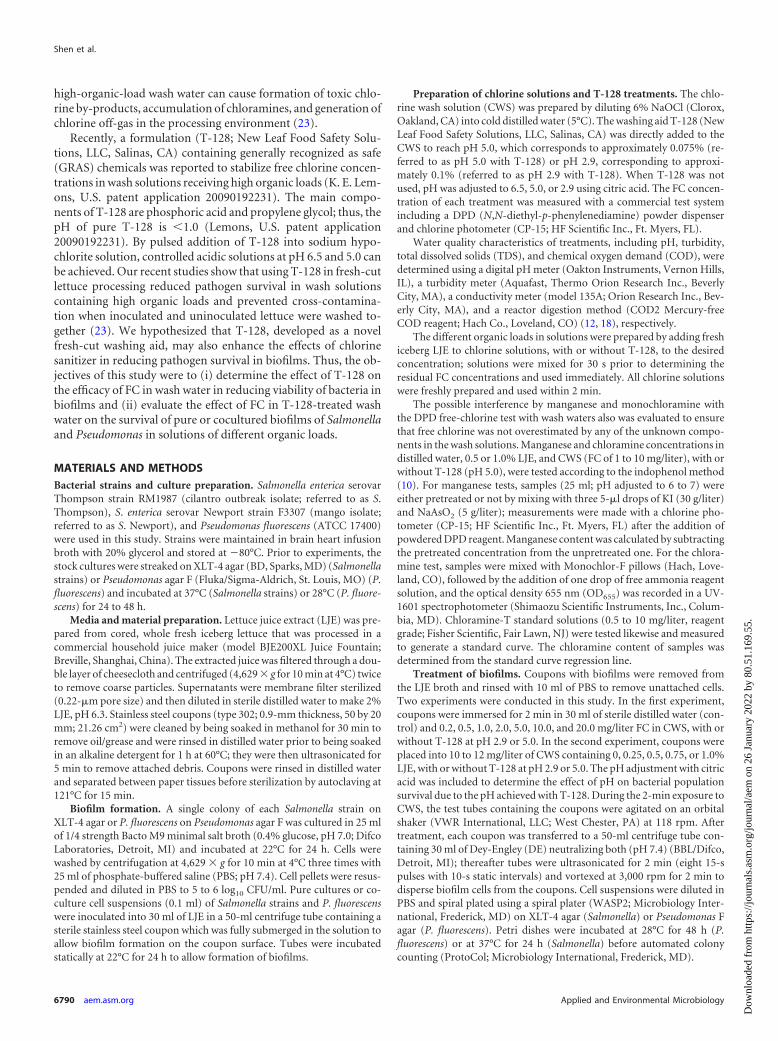
enhanced the killing of bacterial biofilms when different concen-trations of LJE were added as a source of increasing organic loads.For instance, adjustment of chlorine solutions containing 0.75%LJE to pH 2.9 with T-128 resulted in an additional (P � 0.05) 2.3log10 CFU/coupon reduction of S. Thompson compared to thecorresponding solution without T-128 (Fig. 3A). Similarly, CWSadjusted to pH 2.9 with T-128 and 1.0% LJE resulted in an addi-tional (P � 0.05) 2.1 to 2.6 log10 CFU/coupon reduction of Pseu-domonas compared to CWS at pH 6.5 or 2.9 adjusted by citric acidinstead of with T-128 (Fig. 3C).
In CWS adjusted to pH 5.0 with T-128, the number of surviv-ing Salmonella bacteria in biofilms was approximately 0.5 to 1.5
log10 CFU/coupon less than in similar solutions at the same pHwithout T-128 (Fig. 4A and B). For P. fluorescens biofilms, anadditional reduction of 0.6 to 1.4 log10 CFU/coupon was observedin wash solutions containing T-128 compared to the citric acid-adjusted pH when 0.75% LJE was added to wash solutions (Fig.4C). However, no reduction of P. fluorescens populations was ob-served in pH 5.0 washing solutions containing 1.0% LJE com-pared to populations in unwashed coupons (Fig. 4C).
Effect of T-128 and organic loads on CWS efficacy againstcocultured Salmonella and Pseudomonas biofilms. In cocul-tures, after incubation at 22°C for 24 h, S. Thompson biofilm
FIG 3 Recovery of S. Thompson (A), S. Newport (B), and P. fluorescens (C)biofilms formed on stainless steel coupons exposed to 10 to 12 mg/liter initialfree-chlorine solutions at pH 2.9, with or without T-128, in the presence ofdifferent amounts of lettuce juice. The detection limit (dashed line) is 1.3 log10
CFU/coupon. The unwashed-biofilm populations of S. Thompson or S. New-port and P. fluorescens were 7.5 and 8.0 log10 CFU/coupon, respectively.
FIG 4 Recovery of S. Thompson (A), S. Newport (B), and P. fluorescens (C)biofilm populations formed at pH 5.0, with or without T-128, in the presenceof different amounts of lettuce juice. The detection limit (dashed line) is 1.3log10 CFU/coupon. The unwashed-biofilm populations of S. Thompson, S.Newport, and P. fluorescens were 7.5, 7.2, and 8.0 log10 CFU/coupon, respec-tively.
T-128 Enhances Biofilm Inactivation
October 2012 Volume 78 Number 19 aem.asm.org 6793
Dow
nloa
ded
from
http
s://j
ourn
als.
asm
.org
/jour
nal/a
em o
n 26
Jan
uary
202
2 by
80.
51.1
69.5
5.

populations (6.5 log10 CFU/coupon) were 1.0 log10 CFU/couponless than those of the pure-culture biofilms (7.5 log10 CFU/cou-pon) (Fig. 3 to 5). Coculture P. fluorescens populations were sim-ilar to those of the pure culture.
Compared to the pure cultures, there was a distinct differencein the responses of cocultured biofilm cells of S. Thompson and P.fluorescens to CWS at different pH levels. In the pH 6.5 or 5.0CWS, pure-culture and coculture P. fluorescens biofilm cells weresimilar in their sensitivities to CWS, whereas S. Thompson cocul-ture biofilms were less susceptible to CWS than the pure culture(Fig. 3 to 5). For example, no significant reductions of cocul-tured S. Thompson populations were found in wash solutionswith 0.75% LJE (Fig. 5), while the reduction of its respectivepure-culture cells ranged from 1.1 to 2.1 log CFU/couponamong all treatments (Fig. 4A). In contrast, in the pH 2.9 CWS,cocultured cells of both bacterial species were more sensitive tothe chlorine treatment than their respective pure-culture cells(Fig. 3 to 5).
Coculture biofilm studies showed, in general, that CWS ad-justed to pH 2.9 or 5.0 with T-128 again showed higher (P � 0.05)
inactivation on bacterial biofilms than CWS of the same pH ad-justed with citric acid. In the pH 2.9 CWS with T-128, no survivalof S. Thompson or P. fluorescens in biofilm was observed evenwhen 0.50% LJE was added to the CWS (Fig. 5). In the high-organic-load (0.75 to 1.0% LJE) wash solutions, bacterial survivalin biofilm was less (P � 0.05) in pH 2.9 CWS adjusted by T-128than in the citric acid-adjusted CWS (Fig. 5A). At pH 5.0, CWSwith T-128 reduced survival (P � 0.05) of S. Thompson and P.fluorescens until 0.50% and 1.0% LJE was added to the wash solu-tions, respectively (Fig. 5B).
Crystal violet staining of biofilm cells on stainless steel. Asstained with 10% crystal violet, the purple matrix shown in Fig.6AA indicates that cocultured bacterial cells of S. Thompson andP. fluorescens formed biofilm on stainless steel after 24 h of culti-vation in LJE. Purple-stained bacterial biofilms were present onstainless steel coupons that were stationary or shaken in distilledor chlorinated water, regardless of T-128 presence (Fig. 6B, D, F,and H). The washed coupons that were ultrasonicated and vor-texed showed reduced purple staining, as demonstrated in Fig. 6C,E, G, and I, indicating that this treatment actually removed someof the biofilm matrix. The viability of biofilm cells associated withthese various treatments was verified by ultrasonication and vor-texing and then plating cells recovered from a parallel set of stain-less steel coupons not stained with crystal violet. Results in Table 2show that ultrasonication and vortexing caused approximately a1.2 to 1.3 log10 CFU/coupon increase in bacterial cell recovery insuspensions compared to those from stationary coupons. Thenumber of surviving bacteria in distilled-water solutions was asgreat as 5.3 log10 CFU/ml regardless of stationary or shaking treat-ment. Again, it is observed that T-128-adjusted CWS led to a 1.0-to 1.4-log10 reduction of bacterial survival in wash water and cellsuspensions compared to samples without T-128.
Imaging viability of biofilm cells with CLSM. Biofilm cellsand their response to sanitizers were evaluated using specializedglass-bottom petri dishes and stainless steel coupons to facilitateimaging. The clear matrix of green cells in Fig. 7A and B and 8Aand B shows that Salmonella and Pseudomonas biofilms wereformed on the glass-bottom dishes and stainless steel couponsafter 48 h of cultivation with LJE. The two-channel laser confocalimages of the biofilm cells in Fig. 7A, B, and C or Fig. 8A, B, C, andD correspond to the colocalization scatter graphs in Fig. 7D, E,and F or Fig. 8E, F, G, and H, respectively. The graphs illustrate therelative prevalence of live and dead cells in each correspondingframe based on the distribution and intensity of pixels detected,which are displayed as a color spectrum using an absolute inten-sity frequency scale across the x- and y-axis space. This array of theintensity distribution corresponds to detection of live (x axis) anddead (y axis) cells as well as the biofilm matrix. The SYTO9/pro-pidium iodide stain produced a range of intermediate fluorescentcolors (yellow, orange, rust, and brown) between green and red inthe T-128 treatment, indicating loss of membrane integrity (25).As recommended (22), all green cells were considered live,whereas the other colors are considered to represent dead cells. Afew red (dead) cells appear in the 2 mg/liter FC in CWS-treatedsamples on glass-bottom dishes (Fig. 7B) and stainless steel cou-pons (Fig. 8C), but a significantly greater number of orange-red-stained cells was present in samples treated with 2 mg/liter FC inCWS with T-128 at pH 5.0 (Fig. 7C and 8D).
FIG 5 Recovery of cocultures of S. Thompson and P. fluorescens biofilm pop-ulations formed on stainless steel coupons exposed to free chlorine solutions atpH 2.9 (A) or 5.0 (B), with or without T-128, in the presence of differentamounts of lettuce juice. The detection limit (dashed line) is 1.3 log10 CFU/coupon. Unwashed-biofilm populations of Salmonella Thompson and P. fluo-rescens were 6.5 and 8.2 log10 CFU/coupon, respectively.
Shen et al.
6794 aem.asm.org Applied and Environmental Microbiology
Dow
nloa
ded
from
http
s://j
ourn
als.
asm
.org
/jour
nal/a
em o
n 26
Jan
uary
202
2 by
80.
51.1
69.5
5.

DISCUSSION
In the commercial fresh produce industry, the degradation of san-itizing concentrations of free chlorine results from accumulationof organic materials in wash water as freshly cut produce is intro-duced into processing wash solutions. The typical approach tomaintaining sanitizer concentration involves periodic replenish-ment of chlorine. However, repeated addition of chlorine into
high-organic-load wash solutions will increase chlorine off-gasand, as some have suggested, possibly lead to formation of chlo-rine by-products, such as trihalomethanes and haloacetic acids(19). Furthermore, bacterial cells can accumulate on the surface,niches of washing machines, or tanks; biofilm formation can en-sue, and this can be exceptionally resistant to physical and chem-ical removal.
To test the effect of T-128 on FC in different organic-load washwaters, LJE (0.25 to 1.0%) was added to 10 to 12 mg/liter of CWSin the presence or absence of T-128. The increases in turbidity,COD, and TDS in wash waters were mainly attributed to release oforganic material from LJE into wash solutions (19). CWS in whichpH was adjusted with T-128 resulted in higher residual FC thansolutions with citric acid controls. This indicates that T-128 de-creased the free chlorine depletion rate when high organic loadsexisted in wash waters. These results are in agreement with ourprevious studies (23) showing that 0.1% T-128 significantly de-creased the free chlorine depletion rate in the presence of soil withorganic matter. The mechanism by which T-128 tends to stabilizefree chlorine in high-organic-load solutions is still unclear.
FIG 6 Crystal violet-stained microscopic images of cocultures of S. Thompson and P. fluorescens biofilms on stainless steel coupons exposed to chlorine washsolutions (CWS) at pH 5.0, with and without T-128. Treatments were as follows: control biofilm, untreated (A); distilled water, static treatment, without (B) orwith (C) ultrasonication (U) and vortexing (V); distilled water, shaken treatment, without (D) or with (E) ultrasonication and vortexing; CWS (pH 5.0, citricacid), shaken treatment, without (F) or with (G) ultrasonication and vortexing; CWS (pH 5.0, T-128), shaken treatment, without (H) or with (I) ultrasonicationand vortexing.
TABLE 2 Recovery of bacterial biofilm cells in wash waters andsuspension solutions with or without ultrasonication and vortexing
Treatment(s)a
Bacterial recovery (log10 CFU/coupon)
Beforeultrasonicationand vortexing
Afterultrasonicationand vortexing
In washingliquid
DW, static 4.21 � 0.05 5.42 � 0.35 5.28 � 0.12DW, shaken 4.29 � 0.12 5.52 � 0.10 5.33 � 0.24HOCl, shaken 3.14 � 0.14 4.51 � 0.12 1.33 � 0.16HOCl � T-128, shaken 2.03 � 0.25 3.44 � 0.21 0.40 � 0.05a DW, distilled water.
T-128 Enhances Biofilm Inactivation
October 2012 Volume 78 Number 19 aem.asm.org 6795
Dow
nloa
ded
from
http
s://j
ourn
als.
asm
.org
/jour
nal/a
em o
n 26
Jan
uary
202
2 by
80.
51.1
69.5
5.

The effect of T-128 on CWS inactivation of bacterial biofilmpopulations was determined using coupons inoculated with purecultures of Salmonella or P. fluorescens followed by direct platingand colony enumeration on appropriate media. Our direct-plat-ing results, which show significant reductions compared to thecontrol treatments, are consistent with a similar trend reported forSalmonella detection from lettuce and in CWS by enrichment af-ter 1-min exposures to 0 to 1% LJE and FC of 0.5 to 16 mg/liter inCWS (23). This suggests that our direct-plating approach did notsubstantially underestimate the survival of Salmonella.
Our results suggest that there is a synergistic effect betweenT-128 and FC in CWS relative to reduction of Salmonella biofilmson stainless steel coupons. This effect does not rely solely on lowpH because a similar enhancing effect was not observed in pH 2.9CWS adjusted with citric acid instead of T-128; and the enhancedreductions by T-128 in CWS on biofilm populations were alsoobserved at pH 5.0, which is the target pH currently used by manyfresh-cut produce processors that have implemented T-128 use.The mechanism for the synergistic effect between T-128 and CWS
in the inactivation of Salmonella biofilms remains to be deter-mined. However, T-128 alone, without chlorine, did not show anyreductions in Salmonella biofilm cell counts. This result agreeswith a previous study (23), in which T-128 alone was found tohave only weak bactericidal activity against Escherichia coliO157:H7 on lettuce.
Although P. fluorescens is not a food-borne pathogen, it cancontribute to formation of biofilms by food-borne pathogens oncontact surfaces. In this study, P. fluorescens biofilm cells were lesssensitive to CWS than the Salmonella biofilm cells; however, theywere sensitive to low-pH wash solutions. The low pH of washwater achieved by adding 0.1% T-128 was due to the phosphoricacid, one of its major components (Lemons, U.S. patent applica-tion 20090192231). Furthermore, the synergistic effect betweenT-128 and CWS against bacterial biofilms also occurred with P.fluorescens. Thus, T-128, as a washing aid, enhances the sanitizerefficacy of chlorine solutions against P. fluorescens biofilm popu-lations, and this effect is likely due in part to low pH and thesurfactant action of polyethylene glycol.
FIG 7 CLSM images of cocultured S. Thompson and S. Newport biofilms on glass coverslips exposed to chlorine wash solutions (CWS) at pH 5.0, with andwithout T-128. (A) Control biofilm, untreated. Green cells are live. (B) CWS (pH 5.0, citric acid) treatment of biofilm. Red cells are dead. (C) CWS containing0.075% T-128 at pH 5.0. Orange, yellow, and red cells are membrane damaged and considered dead. Colocalization intensity graphs of green fluorescent, live (xaxis) versus red, dead (y axis) cells in the untreated control biofilm from panel A (D) and in the CWS-treated biofilm from panel B (E) are shown. (F)Colocalization intensity graph of green fluorescent, live (x axis) versus red-orange, dead and dying (y axis) cells in the biofilm treated with CWS and T-128 shownin panel C. For all colocalization graphs, the regions designated 1, 2, and 3 correspond to regions where 50% or more of the pixels detected were green fluorescent(live cells), red fluorescent (dead cells), or intermediate colors (membrane-damaged cells), respectively.
Shen et al.
6796 aem.asm.org Applied and Environmental Microbiology
Dow
nloa
ded
from
http
s://j
ourn
als.
asm
.org
/jour
nal/a
em o
n 26
Jan
uary
202
2 by
80.
51.1
69.5
5.

Evaluation of the effect of T-128 in CWS on biofilm inactiva-tion in solutions of different organic loads was conducted usingbiofilms grown on stainless steel coupons with pure cultures of S.Thompson, S. Newport, or P. fluorescens treated with 10 to 12mg/liter of FC in CWS with or without T-128. The enhanced re-duction of bacterial biofilm cells observed in CWS adjusted to pH2.9 with T-128 was partly due to its ability to maintain higherresidual chlorine levels than citric acid.
Crystal violet staining as a method to verify the formation ofbacterial biofilms on certain surfaces has been well documented(2, 7, 20). The purple matrix retained on stainless steel couponsthat had been washed in distilled water and in CWS solutions, withor without T-128, shows that the surfactant component in T-128did not simply dislodge the bacterial biofilm cells from the stain-less steel during washing. The purple-stained matrix was presenton coupons following ultrasonication and vortexing, indicatingthat biofilms were still present on coupons and available for fur-ther microbial analysis. The bacterial recovery results (Table 2)correspond to results from other experiments in this study andverify that T-128 assists CWS in reducing viability of bacterialbiofilm cells in wash solutions and on stainless steel coupons.
The morphology and structure of bacterial biofilms can be ef-fectively analyzed by CLSM combined with fluorescent stainingtechniques (15). The differential, live-dead staining of a slightlygreater number of red cells present in the control than in the 2mg/liter CWS-treated samples showed that 2 mg/liter FC killedsome cells; however, most cells were still viable and thus fluo-resced green. Viability was corroborated in the cultural assays. Incontrast, most cells in samples treated with chlorine and T-128stained yellow, orange, red, or rust, indicating loss of membraneintegrity; these cells were deemed dead accordingly (21). The im-ages of T-128 chlorine treatment corroborate the cultural assaydata that show significantly reduced viability of Salmonella bio-film cells exposed to T-128 CWS.
In commercial fresh produce processing facilities, biofilms aremost likely formed by a consortium of microorganisms. Thus,coculture of Salmonella and Pseudomonas was used in this biofilmstudy in addition to pure cultures of these bacteria. Resultsshowed that in cocultures the P. fluorescens populations were sim-ilar to those of the pure culture, indicating the competitive advan-tage of Pseudomonas in coculture with Salmonella even when ini-tial inoculum concentrations were equivalent. This result was
FIG 8 CLSM images of cocultured S. Thompson and P. fluorescens biofilms on stainless steel coupons exposed to chlorine wash solutions (CWS) at pH 5.0, withand without T-128. (A) Control biofilm, untreated. Green cells are live. (B) Distilled-water treatment of biofilm. Green cells are live, and red cells are dead. (C)CWS (pH 5.0, citric acid) treatment of biofilm. Red cells are dead. (D) CWS containing 0.075% T-128 at pH 5.0. Orange, yellow, and red cells are membranedamaged and considered dead. Colocalization intensity graphs of green fluorescent, live (x axis) versus red, dead (y axis) cells are shown for the untreated controlbiofilm from panel A (E) the distilled-water-treated biofilm from panel B (F), and the CWS-treated biofilm from panel C (G). (H) Colocalization intensity graphof green fluorescent, live(x axis) versus red-orange, dead and dying (y axis) cells in the biofilm treated with CWS and T-128 shown in panel D. For allcolocalization graphs, the regions designated 1, 2, and 3 correspond to regions where 50% or more of the pixels detected were green fluorescent (live cells), redfluorescent (dead cells), or intermediate colors (membrane-damaged cells), respectively.
T-128 Enhances Biofilm Inactivation
October 2012 Volume 78 Number 19 aem.asm.org 6797
Dow
nloa
ded
from
http
s://j
ourn
als.
asm
.org
/jour
nal/a
em o
n 26
Jan
uary
202
2 by
80.
51.1
69.5
5.
similar to that reported by Fatemi and Frank (6) and Lourenço etal. (17), in which Pseudomonas was the dominant species in thetotal coculture biofilm of Listeria monocytogenes and Pseudomonassp. At pH 5.0, cells of S. Thompson in coculture biofilms weremore resistant to CWS than those in pure-culture biofilms. Thisfinding agrees with other reports (6, 17, 22), which state thatpathogenic bacteria were protected by Pseudomonas sp. whenmixed-culture biofilms were exposed to the sanitizers. However,at pH 2.9, the opposite was observed. This can be explained by thesensitivity of P. fluorescens cells in the biofilm matrix to low pH.When S. Thompson lost the protection of the P. fluorescens bio-film matrix, it became more sensitive to the sanitizer.
The present study has demonstrated that washing in CWS doesnot eliminate viable Salmonella or P. fluorescens from biofilmsformed on stainless steel. In contrast, T-128 at pH 2.9 or pH 5.0with CWS enhanced Salmonella and P. fluorescens population re-ductions in biofilms on stainless steel. Thus, use of T-128 in fresh-cut produce processing environments shows promise as a saniti-zation enhancer on stainless steel and possibly also other types ofsurfaces commonly present on food-processing equipment andfor increased inactivation of pathogenic or spoilage bacteria inbiofilms.
ACKNOWLEDGMENTS
We are grateful for the support provided by grant number 2009-74 fromthe Center for Produce Safety, University of California, Davis, Davis, CA,for this research and for the supply of T-128 provided by New Leaf FoodSafety Solutions, LLC, Salinas, CA.
Mention of trade names or commercial products in this article is solelyfor the purpose of providing specific information and does not implyrecommendation or endorsement by the U.S. Department of Agriculture.
REFERENCES1. Beuchat LR. 2002. Ecological factors influencing survival and growth of
human pathogens on raw fruits and vegetables. Microbes Infect. 4:413–423.
2. Burton E, Yakandawala N, LoVetri K. 2007. A microplate spectrofluo-rometric assay for bacterial biofilms. J. Ind. Microbiol. Biotechnol. 34:1– 4.
3. Deibel V, Schoeni J. 2002. Biofilms: forming a defense strategy for thefood plant. Food Saf. Mag. 8:49 –50.
4. Dhir VK, Todd CER. 1995. Susceptibility of suspended and surface-attached Salmonella enteritidis to biocides and elevated temperatures.Appl. Environ. Microbiol. 61:1731–1738.
5. Dotsi B, Guzel-Seydim Z, Greene AK. 2005. Effectiveness of ozone, heat,and chlorine for destroying common food spoilage bacteria in syntheticmedia and biofilms. Int. J. Dairy Technol. 58:19 –24.
6. Fatemi P, Frank JF. 1999. Inactivation of Listeria monocytogenes/Pseudomonas biofilms by peracid sanitizers. J. Food Prot. 62:761–765.
7. Favre-Bonté S, Köhler T, Delden CV. 2003. Biofilm formation by Pseu-domonas aeruginosa: role of the C4-HSL cell-to-cell signal and inhibitionby azithromycin. J. Antimicrob. Chemother. 52:598 – 604.
8. Frank JF. 2000. Control of biofilm in the food and beverage industry, p205–224. In Walker J, Surman S, Jass J (ed), Industrial biofouling. JohnWiley & Sons, Ltd., Chichester, United Kingdom.
9. Gil MI, Selma MV, Lopez-Galvez F, Allende A. 2009. Fresh-cut producessanitation and wash water disinfection: problems and solutions. Int. J.Food Microbiol. 134:37– 45.
10. Greenberg AB, Ciesceri LS, Eaton AD (ed). 1992. Standard methods forthe examination of water and wastewater, 18th ed. American PublicHealth Association, Washington, DC.
11. Helke DM, Wong ACL. 1993. Survival and growth characteristics ofListeria monocytogenes and Salmonella typhimurium on stainless steel andbuna-N rubber. J. Food Prot. 57:963–968.
12. Jirka AM, Carter MJ. 1975. Micro-semi-automated analysis of surfaceand wastewaters for chemical oxygen demand. Anal. Chem. 47:1397–1402.
13. Joseph B, Otta SK, Karunasagar I, Karunasagar I. 2001. Biofilm forma-tion by Salmonella spp. on food contact surfaces and their sensitivity tosanitizers. Int. J. Food Microbiol. 64:367–372.
14. Kreske AC, Ryu J-H, Pettigrew CA, Beuchat LR. 2006. Lethality ofchlorine, chlorine dioxide, and a commercial produce sanitizer to Bacilluscereus and Pseudomonas in a liquid detergent, on stainless steel, and inbiofilm. J. Food Prot. 69:2621–2634.
15. Lawrence JR, Neu TR. 1999. Confocal laser scanning microscopy foranalysis of microbial biofilms. Methods Enzymol. 310:131–144.
16. Lindsay D, Brözel VS, Mostert JF, von Holy A. 2002. Differential efficacyof chlorine dioxide against single species and binary biofilms of a dairy-associated Bacillus cereus and a Pseudomonas fluorescens isolate. J. Appl.Microbiol. 92:352–361.
17. Lourenço A, Machado H, Brito L. 2011. Biofilms of Listeria monocyto-genes produced at 12°C either in pure culture or in co-culture with Pseu-domonas aeruginosa showed reduced susceptibility to sanitizers. J. FoodSci. 76:M143–M148.
18. Luo Y. 2007. Fresh-cut produce wash water reuse affects water quality andpackaged product quality and microbial growth in romaine lettuce. Hort-Science 42:1413–1419.
19. Luo Y, et al. 2011. Determination of free chlorine concentrations neededto prevent Escherichia coli O157:H7 cross-contamination during fresh-cutproduce wash. J. Food Prot. 74:352–358.
20. Merritt JH, Kadouri DE, O’Toole GA. 2005. Growing and analyzingstatic biofilms. Curr. Protoc. Microbiol. Chapter 1, unit 1B.1. doi:10.1002/9780471729259.mc01b01s00.
21. Molecular Probes, Inc. 1994. Live/Dead BacLight viability kit instructionmanual with appendix. Molecular Probes, Inc., Eugene, OR.
22. Norwood DE, Gilmour A. 2000. The growth and resistance to sodiumhypochlorite of Listeria monocytogenes in steady-state multispecies bio-film. J. Appl. Microbiol. 88:512–520.
23. Nou X, et al. 2011. Chlorine stabilizer T-128 enhances efficacy of chlorineagainst cross-contamination by E. coli O157:H7 and Salmonella in fresh-cut lettuce processing. J. Food Sci. 76:M218 –M224.
24. Ryu J-H, Beuchat LR. 2005. Biofilm formation by Escherichia coliO157:H7 on stainless steel: effect of exopolysaccharide and curli produc-tion on its resistance to chlorine. Appl. Environ. Microbiol. 71:247–254.
25. Terzieva S, et al. 1996. Comparison of methods for detection and enu-meration of airborne microorganisms collected by liquid impingement.Appl. Environ. Microbiol. 62:2264 –2272.
26. White GC. 2010. White’s handbook of chlorination and alternative dis-infectants, 5th ed. John Wiley & Sons, Inc., Hoboken, NJ.
Shen et al.
6798 aem.asm.org Applied and Environmental Microbiology
Dow
nloa
ded
from
http
s://j
ourn
als.
asm
.org
/jour
nal/a
em o
n 26
Jan
uary
202
2 by
80.
51.1
69.5
5.