Embed Size (px)
Citation preview

From cell surface adhesion to From cell surface adhesion to final architecture of final architecture of XylellaXylella
fastidiosafastidiosa biofilms: investigation biofilms: investigation with advanced microscopywith advanced microscopy
Mônica Mônica Mônica Mônica A.CottaA.CottaA.CottaA.CottaUniversidadeUniversidade EstadualEstadual de Campinas (UNICAMPde Campinas (UNICAMP))
InstitutoInstituto de de FísicaFísica GlebGleb WataghinWataghin, Campinas, Campinas--SP, BrazilSP, Brazil
InP AuInPInPInAs
Materials ScienceMaterials Science
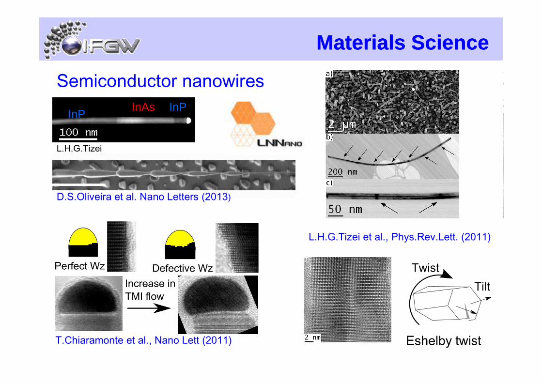
Semiconductor nanowires
L.H.G.Tizei
D.S.Oliveira et al. Nano Letters (2013)
T.Chiaramonte et al., Nano Lett (2011)
L.H.G.Tizei et al., Phys.Rev.Lett. (2011)
D.S.Oliveira et al. Nano Letters (2013)
Eshelby twist

Scanning Probe MicroscopyScanning Probe Microscopy
A.C.Narvaez et al.,
The image cannot be displayed. Your computer may not have enough memory to open the image, or the image may have been corrupted. Restart your computer, and then open the file again. If the red x still appears, you may have to delete the image and then insert it again.
P.Sahoo et al., J.Phys.Chem.C (2011)
Electrical modesElectrical modesElectrical modesElectrical modesCCCC----AFMAFMAFMAFMKPFMKPFMKPFMKPFM
A.C.Narvaez et al., Nanotechnol. (2009)
J.Phys.Chem.C (2011)
BDA C
++The image cannot be displayed. Your computer may not have enough memory to open the image, or the image may have been corrupted. Restart your computer, and then open the file again. If the red x still appears, you may have to delete the image and then insert it again.
Dibucaine (DBC)
G.S.Lorite et al., Biophysical Chemistry (2009)

XylellaXylella fastidiosafastidiosa
IFGW/LNB:IFGW/LNB:IFGW/LNB:IFGW/LNB:Prof. Mônica A.CottaAlberto L.D.Moreau (PhD) Gabriela S.Lorite (M.Sc)Eng. João H. Clerici
IAC/CCIAC/CCIAC/CCIAC/CC::::Dr.Alessandra A.de SouzaCarol Rodrigues
Materials Science & MicroscopyMany years ago…. (~2008)
Eng. João H. Clerici
Univ. Ulm, Germany:Univ. Ulm, Germany:Univ. Ulm, Germany:Univ. Ulm, Germany:Prof. Boris MizaikoffDr. Christine Kranz
Dr. Richard Janissen (PD)

IFGW/LNB:IFGW/LNB:IFGW/LNB:IFGW/LNB:Prof. Mônica A.CottaDr. Richard Janissen (now TU Delft)Dr. Prasana SahooAlberto L.D.Moreau (now ITFSP)Gabriela S.Lorite (now Univ.Oulu)Duber M.MurilloEng. João H. Clerici
IAC/CCIAC/CCIAC/CCIAC/CC::::Dr.Alessandra A.de SouzaCarol RodriguesDr.Juarez P.TomazBarbara Niza
XylellaXylella fastidiosafastidiosa -- nownow
Prof. Hernandes F. de CarvalhoProf. Carlos L.Cesar
Profa. Marcia L.A.TemperiniMarcelo Nobrega
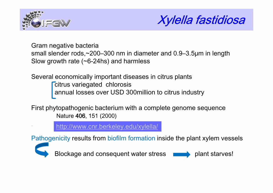
XylellaXylellaXylellaXylella fastidiosafastidiosafastidiosafastidiosa
Gram negative bacteriasmall slender rods,~200–300 nm in diameter and 0.9–3.5μm in lengthSlow growth rate (~6-24hs) and harmless
Several economically important diseases in citrus plantscitrus variegated chlorosisannual losses over USD 300million to citrus industry
First phytopathogenic bacterium with a complete genome sequenceNature 406406406406, 151 (2000)
Pathogenicity results from biofilm formation inside the plant xylem vessels
Blockage and consequent water stress plant starves!
http://www.cnr.berkeley.edu/xylella/

Bacterial Bacterial BiofilmsBiofilms
Biofilm Formation in 5 steps: Biofilm Formation in 5 steps: Biofilm Formation in 5 steps: Biofilm Formation in 5 steps:
(1)(1)(1)(1) Pioneer bacterial cells reversible attachment to surface Pioneer bacterial cells reversible attachment to surface Pioneer bacterial cells reversible attachment to surface Pioneer bacterial cells reversible attachment to surface
(2)(2)(2)(2) Irreversible attachement (influence of EPS Irreversible attachement (influence of EPS Irreversible attachement (influence of EPS Irreversible attachement (influence of EPS ---- extracellular polymeric substanceextracellular polymeric substanceextracellular polymeric substanceextracellular polymeric substance))))
(3) (3) (3) (3) CellCellCellCell----Cell adhesion and proliferation Cell adhesion and proliferation Cell adhesion and proliferation Cell adhesion and proliferation
(4) (4) (4) (4) Mature biofilmMature biofilmMature biofilmMature biofilm
K. Sauer, Genome Biology 4444, 219 (2003)
(5) Detachment (5) Detachment (5) Detachment (5) Detachment
SPM SPM SPM SPM Large sizesBiological sample reproducibility issues
Fractal analysisFractal analysisFractal analysisFractal analysisJAP (2009)JAP (2009)JAP (2009)JAP (2009)

N.Killiny et al, Appl.Environ.Microbiol. 75757575, 521 (2009)
Adhesiveness and life cycleAdhesiveness and life cycle
S. Chatterjee et al, Ann.Rev.Phytopathol. 46464646, 243(2008)
K.L.Newman et al., PNAS 101101101101, 1737 (2004)

Reversible adhesion:Reversible adhesion:Reversible adhesion:Reversible adhesion:Electrostatic natureElectrostatic natureElectrostatic natureElectrostatic nature
Initial adhesion stagesInitial adhesion stages
K. Sauer, Genome Biology 4444, 219 (2003)
Irreversible adhesion: Irreversible adhesion: Irreversible adhesion: Irreversible adhesion: Chemical natureChemical natureChemical natureChemical nature

SPM & IR SPM & IR –– Glass surfacesGlass surfaces
Live Bacteria grown inside AFM liquid cell at 28C
ATRATRATRATR---- IRIRIRIR
Conditioning film due to Conditioning film due to Conditioning film due to Conditioning film due to culture medium:culture medium:culture medium:culture medium:changes in changes in changes in changes in hydrophobicityhydrophobicityhydrophobicityhydrophobicity
G.S.Lorite et al., Journal of Colloids and Interface Science 359 (2011) 289–295
Dry samples (cells around BF, 7 days)

XylellaXylella fastidiosafastidiosa BiofilmsBiofilms
1800 1600 1400 1200 10000.00
0.04
0.08
0.12
0.16
0.20
1578.6
995.2
1043.2
1077.2
1111.91300.9
1408.9
1460.0
1549.8
1620.1
Abs
orpt
ion
Inte
nsity
(u.
a.)
Experiment A Experiment B PW conditioning film
1656.8
0 500 1000 1500 2000 2500 3000 3500
0.00
0.01
0.02
0.03
0.04
0.05
Hei
ght P
eaks
Abs
ropt
ion
Time (min)
Experiment B(b)
ATRATRATRATR---- IRIRIRIR
polysaccharide groups (C–O–P, P=Osymmetric stretch, C–C and C–O stretch)
polyphosphate groups
Wavenumber (cm -1)
G.S.LoriteG.S.LoriteG.S.LoriteG.S.Lorite et al.et al.et al.et al.Colloid and Surfaces B: Colloid and Surfaces B: Colloid and Surfaces B: Colloid and Surfaces B: BiointerfacesBiointerfacesBiointerfacesBiointerfaces (2013)(2013)(2013)(2013)
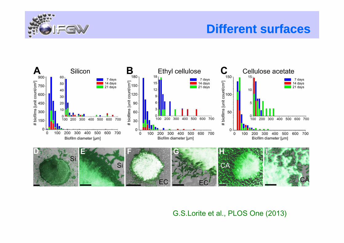
Different surfacesDifferent surfaces
G.S.Lorite et al., PLOS One (2013)

Different surfacesDifferent surfaces
- What happens after incubation in culture media (PW)?
- How homogeneous are these surfaces (roughness and electrically)?
celullose (EC, CA): zeta potential ~ -10 to -50 mV

SPM analysisSPM analysisSi EC CA
Surface potentialAfter PW incubation
G.S.Lorite et al., PLOS One (2013)

Force spectroscopyForce spectroscopy
Tip functionalization XadA1 adhesinePBS buffer
control
Surface
Protein
offset
control
AC
AC

Force spectroscopyForce spectroscopy
G.S.Lorite et al., PLOS One (2013)

Force spectroscopyForce spectroscopy
G.S.Lorite et al., PLOS One (2013)
Specific binding to CA surfaces less likely than for Si and ECSpecific binding to CA surfaces less likely than for Si and ECSpecific binding to CA surfaces less likely than for Si and ECSpecific binding to CA surfaces less likely than for Si and EC
SP + force spectroscopy: different shapes of biofilmsSP + force spectroscopy: different shapes of biofilmsSP + force spectroscopy: different shapes of biofilmsSP + force spectroscopy: different shapes of biofilms

Intriguing features
Bacterial Poles
Textures
G.Lorite et al., J.Coll.Inter.Sci. (2011)
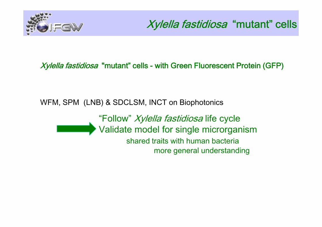
XylellaXylellaXylellaXylella fastidiosafastidiosafastidiosafastidiosa "mutant” cells "mutant” cells "mutant” cells "mutant” cells ---- with Green Fluorescent Protein (GFP)with Green Fluorescent Protein (GFP)with Green Fluorescent Protein (GFP)with Green Fluorescent Protein (GFP)
WFM, SPM (LNB) & SDCLSM, INCT on Biophotonics
XylellaXylellaXylellaXylella fastidiosafastidiosafastidiosafastidiosa “mutant” cells“mutant” cells“mutant” cells“mutant” cells
“Follow” Xylella fastidiosa life cycle“Follow” Xylella fastidiosa life cycleValidate model for single microrganism
shared traits with human bacteriamore general understanding

ConclusionsConclusionsQuantitativeQuantitativeQuantitativeQuantitative measurements of important biological parameters:
- Roughness
- Hidrophobicity
- Electrostatic character
- Surface modification
hydrophilic surfaces
- Surface modification
for different materials on which Xyllela fastidiosa can growBiology: complete life cycle evolution of Xylella fastidiosa
Biofilm structure that maximizes nutrient flow and adhesion to different surfaces
issues on mechanical stabilityadaptation to host/vector transmission mechanism
Important:Important:Important:Important: physicists, biologists, chemists working in same environment!!physicists, biologists, chemists working in same environment!!physicists, biologists, chemists working in same environment!!physicists, biologists, chemists working in same environment!!

Financial Support
Thank you!!Thank you!!Thank you!!Thank you!!