Embed Size (px)
Citation preview

Brain (1998),121,1013–1052
R E V I E W A R T I C L E
From sensation to cognitionM.-Marsel Mesulam
The Cognitive Neurology and Alzheimer’s Disease Center, Correspondence to: M. Mesulam, Cognitive Neurology andDepartments of Neurology and Psychiatry and Behavioral Alzheimer’s Disease Center, Northwestern UniversitySciences, Northwestern University Medical School, Medical School, 320 East Superior Street, 11–450,Chicago, USA Chicago, IL 60611, USA. E-mail: [email protected]
SummarySensory information undergoes extensive associativeelaboration and attentional modulation as it becomesincorporated into the texture of cognition. This processoccurs along a core synaptic hierarchy which includesthe primary sensory, upstream unimodal, downstreamunimodal, heteromodal, paralimbic and limbic zones ofthe cerebral cortex. Connections from one zone to anotherare reciprocal and allow higher synaptic levels to exerta feedback (top-down) influence upon earlier levels ofprocessing. Each cortical area provides a nexus for theconvergence of afferents and divergence of efferents. Theresultant synaptic organization supports parallel as wellas serial processing, and allows each sensory event toinitiate multiple cognitive and behavioural outcomes.Upstream sectors of unimodal association areas encodebasic features of sensation such as colour, motion, formand pitch. More complex contents of sensory experiencesuch as objects, faces, word-forms, spatial locations andsound sequences become encoded within downstreamsectors of unimodal areas by groups of coarsely tunedneurons. The highest synaptic levels of sensory-fugalprocessing are occupied by heteromodal, paralimbic andlimbic cortices, collectively known as transmodal areas.The unique role of these areas is to bind multiple unimodaland other transmodal areas into distributed but integratedmultimodal representations. Transmodal areas in themidtemporal cortex, Wernicke’s area, the hippocampal–entorhinal complex and the posterior parietal cortexprovide critical gateways for transforming perception intorecognition, word-forms into meaning, scenes and eventsinto experiences, and spatial locations into targets forexploration. All cognitive processes arise from analogousassociative transformations of similar sets of sensoryinputs. The differences in the resultant cognitive operationare determined by the anatomical and physiologicalproperties of the transmodal node that acts as the criticalgateway for the dominant transformation. Interconnectedsets of transmodal nodes provide anatomical and
© Oxford University Press 1998
computational epicentres for large-scale neurocognitivenetworks. In keeping with the principles of selectivelydistributed processing, each epicentre of a large-scalenetwork displays a relative specialization for a specificbehavioural component of its principal neurospycho-logical domain. The destruction of transmodal epicentrescauses global impairments such as multimodal anomia,neglect and amnesia, whereas their selective disconnectionfrom relevant unimodal areas elicits modality-specificimpairments such as prosopagnosia, pure word blindnessand category-specific anomias. The human brain containsat least five anatomically distinct networks. The networkfor spatial awareness is based on transmodal epicentresin the posterior parietal cortex and the frontal eye fields;the language network on epicentres in Wernicke’s andBroca’s areas; the explicit memory/emotion network onepicentres in the hippocampal–entorhinal complex andthe amygdala; the face-object recognition network onepicentres in the midtemporal and temporopolar cortices;and the working memory-executive function network onepicentres in the lateral prefrontal cortex and perhapsthe posterior parietal cortex. Individual sensorymodalities give rise to streams of processing directed totransmodal nodes belonging to each of these networks.The fidelity of sensory channels is actively protectedthrough approximately four synaptic levels of sensory-fugal processing. The modality-specific cortices at thesefour synaptic levels encode the most veridicalrepresentations of experience. Attentional, motivationaland emotional modulations, including those related toworking memory, novelty-seeking and mental imagery,become increasingly more pronounced within downstreamcomponents of unimodal areas, where they help to createa highly edited subjective version of the world. Theprefrontal cortex plays a critical role in these attentionaland emotional modulations and allows neural responsesto reflect the significance rather than the surfaceproperties of sensory events. Additional modulatory
1014 M.-M. Mesulam
influences are exerted by the cholinergic and mono-aminergic pathways of the ascending reticular activatingsystem. Working memory, one of the most prominentmanifestations of prefrontal cortex activity, prolongs theneural impact of environmental and mental events in away that enriches the texture of consciousness. Thesynaptic architecture of large-scale networks and themanifestations of working memory, novelty-seekingbehaviours and mental imagery collectively help to loosenthe rigid stimulus–response bonds that dominate thebehaviour of lower animal species. This phylogenetictrend has helped to shape the unique properties of human
Keywords: cerebral cortex; language; memory; consciousness; attention; neural networks
Abbreviations: AIT 5 anterior inferotemporal cortex; ARAS5 ascending reticular activating system; BA5 Brodmannarea; LIP5 lateral intraparietal sulcus; MST5 medial superior temporal area; PIT5 posterior inferotemporal cortex
IntroductionA major task of the CNS is to configure the way in whichsensory information becomes linked to adaptive responsesand meaningful experiences. Small brains cope with thischallenge by shifting the detection of significant events toperipheral organs. In the frog, for example, the inner ear isselectively tuned to spectral and temporal properties ofspecies-specific mating calls (Capranica, 1978) and theretinotectal projection contains ‘bug perceiver’ fibres thatrespond best when a dark object, smaller than a receptivefield, enters that field, stops, and moves about intermittently(Lettvin et al., 1959). Although this type of automatic patterndetection maximizes the limited processing capacity of theCNS, it also restricts the range of events that can be identifiedand the flexibility with which they become interpreted.
Inflexible bonds between sensation and action lead toinstinctual and automatic behaviours that are resistant tochange, even when faced by negative consequences. Forexample, frogs whose optic nerve has been cut and allowedto fully regenerate after a 180° rotation of the eye willrepeatedly snap at mud and moss on the ground whenpresented with a fly above the head (Sperry, 1965); a turkeyhen, whose protective maternal instincts dictate an attack onany moving object that fails to utter the characteristic peepof her chicks, will peck her own newly hatched progeny todeath if she is made deaf (Schleidt and Schleidt, 1960); aherring gull whose eggs have been displaced to an adjacentand clearly visible site will proceed to incubate the emptiedoriginal nest and ignore the clutch of eggs lying right nextto her (Tinbergen, 1951); and rats with crossed sensorynerves in the hind limbs, one of which is inflamed, will hopon three legs to protect the healthy rather than the sore foot(Sperry, 1965).
Advanced mammals with an intact CNS are less vulnerableto the emergence of such inflexible patterns. With theexception of some autonomic, brainstem and spinal reflexes,the behaviour of primates displays a much greater latitude
consciousness and to induce the emergence of second-order (symbolic) representations related to language.Through the advent of language and the resultant abilityto communicate abstract concepts, the critical pacemakerfor human cognitive development has shifted from theextremely slow process of structural brain evolution tothe much more rapid one of distributed computationswhere each individual intelligence can becomeincorporated into an interactive lattice that promotes thetransgenerational transfer and accumulation of know-ledge.
in translating sensation into action, so that identical sensoryevents can potentially trigger one of many different reactions,depending on the peculiarities of the prevailing context. Astimulus that deserves to be approached in one setting mayneed to be avoided in another; highly desirable consummatoryacts may need to be postponed in the presence of danger;the same glass may appear half-full or half-empty dependingon mood; and apetite madeleinecan trigger a spectrum ofreactions ranging from brief salivation to a torrent of wordsthat can hardly be contained by seven volumes of compactprose. This loosening of stereotyped stimulus–responselinkages endows the organism with the biological freedomto choose one of many potentially available responses. Theresultant coarse mapping of responses onto circumstancescreates a setting where the rules of competitive selection canoperate to promote rapid change and adaptation.
Streams of sensory processing in amphibians, reptiles andbirds are kept on a short leash: the synaptic interval betweenthe onset of a sensory event and its closure in the formof multimodal convergence, perceptual recognition,motivational valuation and action is brief, usually of theorder of one or two synapses (Ingle, 1975; Northcutt, 1978;Gorlick et al., 1984; Gaillard, 1990). The CNS of advancedmammals displays major modifications in this plan oforganization. The fidelity of sensory encoding is enhancedby reserving vast areas of the cerebral neocortex for modality-specific processing. The opportunities for subsequentintegration are enhanced by the development of extensivemultimodal association areas. And an intrinsic bias emergesto pursue novelty and flexibility rather than sameness andstereotypy. The emergence of behavioural flexibility inmammals can be attributed to an expansion of the synapticbridge that links sensation to action and recognition. Anexcessive length in such a synaptic bridge could have led tounacceptable delays in reacting to the environment, whereastoo many lanes could have undermined the capacity for
From sensation to cognition 1015
integration. As will be shown below, primate evolutionreflects a compromise between such extremes: the synapticchain that links sensation to action has been lengthened, butnot by too many synapses; and the bridge between the twohas been broadened by the introduction of numerous parallellines of communication, but with ample opportunities forintegrative interaction among individual channels.
The neural systems that bridge the gap between sensationand action provide the substrates for ‘intermediary’ or‘integrative’ processing. The behavioural outcome ofintermediary processing is known as ‘cognition’, and includesthe diverse manifestations of memory, emotion, attention,language, thought and consciousness. The synaptic volumededicated to intermediary processing shows a marked increasein phylogeny and occupies the great majority of the cerebralcortex in advanced primates and cetaceans. Theseintermediary areas of the brain enable identical stimuli totrigger different responses depending on the situationalcontext, past experience, present needs and contemplatedconsequences.
The neurons that support intermediary processing arelocated predominantly within the ‘association’ and ‘limbic’areas of the cerebral cortex. The importance of the cerebralcortex to behaviour varies from species to species. In thefish, frog and pigeon, decortication produces little change insensation or locomotion (Ferrier, 1876; Northcutt, 1978).Extensive neocortical lesions in hamsters, cats and monkeyscause considerably lesser and more reversible functionaldeficits than analogous lesions in humans (Lashley, 1952;MacLean, 1982). Furthermore, cortical lesions may be farmore effective than tectal lesions in causing deficits suchas hemispatial neglect in monkeys, whereas the converserelationship is seen in cats (Mesulamet al., 1977). Theseobservations point to the existence of a trend towards aprogressive corticalization of function, and suggest thatcognition, defined as the behavioural outcome of intermediaryprocessing in the cerebral cortex, becomes established as anobligatory rather than a facultative correlate of informationprocessing in the course of evolution.
The synaptic arrangement of neural pathways involved inintermediary processing provides biological constraints thatshape the nature of cognition and comportment. The goal ofthis review is to sketch some of the principles that guide theorganization of these pathways. Several limitations willbecome apparent in the course of the following account.First, much of the discussion will be limited to the cerebralcortex and will omit the very important contributions ofsubcortical structures such as the thalamus, striatum,claustrum and brainstem. Second, only visual and auditorystreams of processing will be discussed in any detail. Third,although domains such as perception, language, memory,attention and emotion will be addressed, this will be done ina very limited fashion, to provide highly selective examplesof common principles that link sensation to cognition. Fourth,although several phylogenetic trends will be mentioned,including numerous references to the CNS in the frog, only
the monkey and human brains will be discussed in anyserious detail. Fifth, the subject matter will lead to a discussionof consciousness, but only in the form of a brief incursioninto realms of immense philosophical complexity.
Behavioural neuroanatomy of the cerebralcortexFunctional zonesThe human cerebral cortex contains ~20 billion neurons(Pakkenberg and Gundersen, 1997). The absence of clearanatomical demarcations has encouraged the development ofnumerous independent approaches to the subdivision of thecerebral cortex. The resultant maps can be divided into twogroups: those based primarily on structural (architectonic)features, and those based primarily on functional affiliations.Proponents of one school have constructed a wide variety ofcortical maps, ranging in complexity from the map ofExner (1881), which boasted hundreds of sharply delineatedsubdivisions, to the more modest and also more widelyaccepted ones of Brodmann (1909), the Vogts (Vogt andVogt, 1919), von Economo (Economo and Koskinas, 1925)and Flechsig (1920). The second school is more difficult toidentify since few of its proponents have produced systematicsurveys of the entire brain. Members of this second schoolinclude theoreticians of brain function such as Campbell(1905), Broca (1878), Abbie (1942), Filimonoff (1947),Yakovlev (1959) and Sanides (1970). The thinking of thisschool has led to the subdivision of the cerebral cortex intothe five major functional subtypes that will be describedbelow: primary sensory–motor, unimodal association,heteromodal association, paralimbic and limbic (Mesulam,1985b). The principal factual base for this parcellationis derived from anatomical, physiological and behaviouralexperiments in macaque monkeys. The homologies to thehuman brain have been inferred from comparative cyto-architectonics, electrophysiological recordings, functionalimaging, and the behavioural effects of focal lesions. Unlessstated otherwise, the descriptions in this section can beassumed to apply to the brains of both species.
The primary sensory and motor cortices are easilydelineated on cytoarchitectonic and functional grounds. Theprimary visual cortex [also known as V1, striate cortex,calcarine cortex or Brodmann area (BA) 17] covers the banksof the calcarine fissure; the primary auditory cortex (alsoknown as A1, or BA 41–42) covers Heschl’s gyrus on thefloor of the Sylvian cistern; the primary somatosensory cortex(also known as S1 or BA 3b) covers the anterior flank of thepostcentral gyrus; the primary gustatory cortex is probablylocated in the fronto-insular junction in BA 43; and theprimary motor cortex (also known as M1) includes BA 4and probably also a posterior rim of BA 6 in the precentralgyrus. The primary sensory areas provide an obligatory portalfor the entry of sensory information into the cortical circuitry,whereas the primary motor cortex provides a major gateway
1016 M.-M. Mesulam
for relaying complex motor programmes into bulbar andspinal motoneurons. The primary sensory areas (wheredensely packed small neurons give rise to a distinctivekoniocortical architecture) and the primary motor cortex(where large pyramidal neurons give rise to a distinctivemacropyramidal architecture) represent the most highlydifferentiated and specialized subdivisions of the cerebralcortex (Mesulam, 1985b).
Sensory-fugal processing starts with the transfer ofinformation from primary sensory areas to unimodalassociation areas. These areas fulfil three criteria: (i) theirmajor source of sensory-fugal projections is located inprimary sensory areas and other unimodal association areasin that modality; (ii) the constituent neurons respond tostimulation predominantly, if not exclusively, in that particularsensory modality; and (iii) lesions lead to behavioural deficitsconfined to tasks under the control of that particular sensorymodality. Unimodal areas can be divided into upstream anddownstream components: upstream areas are only one synapseaway from the relevant primary sensory area whereasdownstream areas are at a distance of two or more synapticunits from the corresponding primary area.
The unimodal visual association cortex can be divided intoan upstream peri-striate component which includes areasBA 18–19, and a downstream temporal component whichincludes the inferotemporal regions (BA 21–20) in themonkey, and the fusiform, inferior temporal and perhapsparts of the middle temporal gyri in humans.1 The unimodalauditory association cortex covers the superior temporal gyrus(BA 22) and perhaps also parts of the middle temporal gyrus(BA 21) in the human (Creutzfeldtet al., 1989). Theconnectivity of the monkey brain would suggest that theposterior parts of the superior temporal cortex (BA 22)display the properties of upstream auditory association cortexwhereas the more anterior parts of this gyrus and the dorsalbanks of the superior temporal sulcus may fit the designationof downstream auditory association cortex (Pandya andYeterian, 1985). In the monkey brain, rostral BA 5 representsthe upstream component of somatosensory unimodalassociation cortex, whereas caudal BA 5 and BA 7b mayrepresent its downstream components (Pandya and Yeterian,1985). In the human, unimodal somatosensory associationcortex may include BA 5, parts of BA 7 and perhaps BA 2.The subdivision of unimodal auditory and somatosensoryassociation cortices into upstream and downstream areasremains to be elucidated in the human brain. Premotor regions(anterior BA 6 and caudal BA 8) fulfil the role of motorassociation areas because they provide the principal corticalinput into the primary motor cortex.
The next stage of sensory-fugal processing occurs in
1 Approximately half of middle temporal gyrus (MTG) neurons respond tospeech (Creutzfeldtet al., 1989; Vincentet al., 1997). However, some tasksbased on visually presented faces, words and objects can also activate theMTG (Vandenbergheet al., 1996; Gorno Tempiniet al., 1997; Schultzet al.,1997). At this time it is not clear whether all of the MTG should be classifiedas heteromodal cortex or whether it should be divided into sections ofunimodal auditory, heteromodal, and unimodal visual cortex.
heteromodal association areas, which are characterized bythree criteria: (i) they receive convergent inputs fromunimodal areas in more than one modality; (ii) unit recordingsshow that constituent neurons respond to stimulation in morethan one sensory modality or that neurons responding to onemodality are interspersed with those that respond to another;(iii) lesions always yield multimodal behavioural deficits.The monkey brain has heteromodal areas in the prefrontalcortex (BA 9, 10, 45, 46, anterior BA 8, anterior BA 11–12), in the inferior parietal lobule (parts of BA 7), in thebanks of the superior temporal sulcus (junction of BA 22with BA 21), and in the parahippocampal region (Pandyaand Yeterian, 1985). In the human brain the analogous realmsof the heteromodal cortex are located in the prefrontal cortex,the posterior parietal cortex (posterior BA 7, BA 39–40),parts of the lateral temporal cortex (probably correspondingto parts of BA 37 and BA 21) and portions of theparahippocampal gyrus (parts of BA 35–36). The unimodaland heteromodal areas are characterized by a six-layeredhomotypical architecture. The columnarization and laminardifferentiation of neurons is more conspicuous in unimodalthan heteromodal areas. If primary sensory and motor areasconstitute the most highly specialized and differentiated partsof the cortex, unimodal and heteromodal areas occupy thetwo subsequent levels of differentiation.
A further stage of sensory-fugal processing occurs in agroup of areas designated paralimbic. These areas provide azone of gradual cytoarchitectonic transition between thehomotypical isocortex and the more primitive allocortexof core limbic structures. The primate brain contains fiveparalimbic regions: the caudal orbitofrontal cortex (caudalBA 11–12, BA 13), the insula (BA 14–16), the temporalpole (BA 38), the parahippocampal gyrus (BA 27, 28 andparts of BA 35), and the retrosplenial-cingulate-parolfactorycomplex (BA 23–26 and BA 29–33). These paralimbicformations can be divided into two groups. The temporopolar-insular-orbitofrontal regions merge into each other andconstitute the olfactocentric subdivision of the paralimbicbelt because they provide a transition between the olfactoryallocortex and the homotypical cortex. The amygdala is themajor core limbic structure associated with this set ofparalimbic regions. The parahippocampal and posteriorcingulate regions constitute the hippocampocentricsubdivision of the paralimbic zone because they provide atransition between the hippocampal formation (including itsinduseal rudiment) and the homotypical cortex. These twosubdivisions collectively form a gapless paralimbic ringwhich encircles the medial and basal components of thecerebral hemispheres (Mesulam and Mufson, 1985).
The last cortical stage in the sensory-fugal stream ofinformation processing occurs within five core limbicformations: the hippocampal complex, the amygdaloidcomplex, the prepiriform olfactory cortex, the septal areaand the substantia innominata. These regions are characterizedby two properties: they display a primitive allocorticalarchitecture (palaeocortical in the case of olfactory areas,
From sensation to cognition 1017
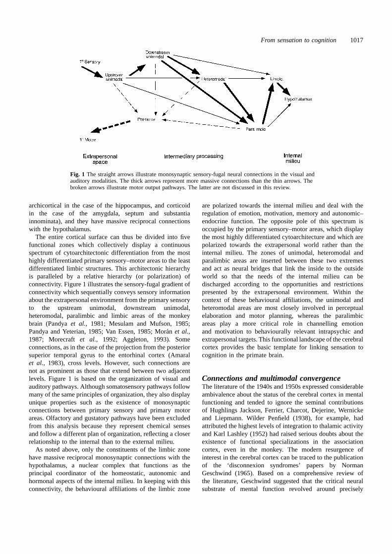
Fig. 1 The straight arrows illustrate monosynaptic sensory-fugal neural connections in the visual andauditory modalities. The thick arrows represent more massive connections than the thin arrows. Thebroken arrows illustrate motor output pathways. The latter are not discussed in this review.
archicortical in the case of the hippocampus, and corticoidin the case of the amygdala, septum and substantiainnominata), and they have massive reciprocal connectionswith the hypothalamus.
The entire cortical surface can thus be divided into fivefunctional zones which collectively display a continuousspectrum of cytoarchitectonic differentiation from the mosthighly differentiated primary sensory–motor areas to the leastdifferentiated limbic structures. This architectonic hierarchyis paralleled by a relative hierarchy (or polarization) ofconnectivity. Figure 1 illustrates the sensory-fugal gradient ofconnectivity which sequentially conveys sensory informationabout the extrapersonal environment from the primary sensoryto the upstream unimodal, downstream unimodal,heteromodal, paralimbic and limbic areas of the monkeybrain (Pandyaet al., 1981; Mesulam and Mufson, 1985;Pandya and Yeterian, 1985; Van Essen, 1985; Mora´n et al.,1987; Morecraft et al., 1992; Aggleton, 1993). Someconnections, as in the case of the projection from the posteriorsuperior temporal gyrus to the entorhinal cortex (Amaralet al., 1983), cross levels. However, such connections arenot as prominent as those that extend between two adjacentlevels. Figure 1 is based on the organization of visual andauditory pathways. Although somatosensory pathways followmany of the same principles of organization, they also displayunique properties such as the existence of monosynapticconnections between primary sensory and primary motorareas. Olfactory and gustatory pathways have been excludedfrom this analysis because they represent chemical sensesand follow a different plan of organization, reflecting a closerrelationship to the internal than to the external milieu.
As noted above, only the constituents of the limbic zonehave massive reciprocal monosynaptic connections with thehypothalamus, a nuclear complex that functions as theprincipal coordinator of the homeostatic, autonomic andhormonal aspects of the internal milieu. In keeping with thisconnectivity, the behavioural affiliations of the limbic zone
are polarized towards the internal milieu and deal with theregulation of emotion, motivation, memory and autonomic–endocrine function. The opposite pole of this spectrum isoccupied by the primary sensory–motor areas, which displaythe most highly differentiated cytoarchitecture and which arepolarized towards the extrapersonal world rather than theinternal milieu. The zones of unimodal, heteromodal andparalimbic areas are inserted between these two extremesand act as neural bridges that link the inside to the outsideworld so that the needs of the internal milieu can bedischarged according to the opportunities and restrictionspresented by the extrapersonal environment. Within thecontext of these behavioural affiliations, the unimodal andheteromodal areas are most closely involved in perceptualelaboration and motor planning, whereas the paralimbicareas play a more critical role in channelling emotionand motivation to behaviourally relevant intrapsychic andextrapersonal targets. This functional landscape of the cerebralcortex provides the basic template for linking sensation tocognition in the primate brain.
Connections and multimodal convergenceThe literature of the 1940s and 1950s expressed considerableambivalence about the status of the cerebral cortex in mentalfunctioning and tended to ignore the seminal contributionsof Hughlings Jackson, Ferrier, Charcot, Dejerine, Wernickeand Liepmann. Wilder Penfield (1938), for example, hadattributed the highest levels of integration to thalamic activityand Karl Lashley (1952) had raised serious doubts about theexistence of functional specializations in the associationcortex, even in the monkey. The modern resurgence ofinterest in the cerebral cortex can be traced to the publicationof the ‘disconnexion syndromes’ papers by NormanGeschwind (1965). Based on a comprehensive review ofthe literature, Geschwind suggested that the critical neuralsubstrate of mental function revolved around precisely
1018 M.-M. Mesulam
organized cortico-cortical pathways that interconnectbehaviourally specialized regions of the cerebral cortex. Thisinference was particularly interesting, since very little wasknown then about the details of cortical connectivity. Atabout the same time, new methods based on the selectivesilver impregnation of degenerated axons were becomingavailable and enabled a detailed investigation of cortico-cortical connectivity in monkeys. The basic organization ofthese connections was described in two classic papers, oneby Pandya and Kuypers (1969) and the other by Jonesand Powell (1970). These papers outlined an orderly andhierarchical connectivity, mostly consistent with Geschwind’saccount, for linking sensory cortices to primary, secondaryand sometimes even tertiary modality-specific associationareas, which in turn sent convergent projections to multimodalsensory association zones. Such multimodal sensoryconvergence areas were identified in the posterior parietal,lateral prefrontal and temporal cortices of the rhesus monkey.
The field of neuroscience had been primed to anticipatesuch a sequential organization through the work of Hubeland Wiesel (1965), who had demonstrated a hierarchy ofsimple, complex and hypercomplex neurons in the primaryvisual cortex, each successive level encoding a morecomposite aspect of visual information. The discoveries ofPandya and Kuypers and of Jones and Powell seemed to beextending this serial and convergent organization from therealms of the primary sensory cortex and sensation to thoseof the association cortex and cognition. A great deal ofemphasis was placed on the pivotal role of multimodalconvergence as the final and supreme site of integration forall aspects of mental function, including the storage ofmemories, the formation of concepts and the acquisition oflanguage (Geschwind, 1965; Pandya and Kuypers, 1969;Jones and Powell, 1970; Van Hoesenet al., 1972; Pandyaand Seltzer, 1982; Mesulam, 1985b).
While the importance of serial processing and multimodalconvergence to cognitive function was widely accepted, somepotentially serious computational limitations of such anarrangement were also acknowledged (Rumelhart andMcClelland, 1986; Goldman-Rakic, 1988; Mesulam, 1990).The surfacing of these concerns coincided with thedevelopment of far more powerful neuroanatomical methods,based on the intra-axonal transport of horseradish peroxidaseand tritiated amino acids, which started to show that thesensory-fugal flow of information was more complicatedthan previously surmised. The classic description of visuo-fugal pathways, for example, had assumed that informationfrom V1 (BA 17) is transferred successively and serially toa first-order visual association area, V2 (BA 18), then to asecond-order visual association area corresponding to BA 19,and finally to a third-order association area in theinferotemporal cortex. The newer methods revealed asomewhat different picture. The entire extent of V1 didindeed project to V2 (BA 18) in a topographically well-ordered fashion. However, V1 and V2 then gave rise tomultiple parallel pathways that projected to numerous discrete
peristriate visual association areas, located mostly withinBA 19 and designated V3, V4, V5 (MT), VP, V6 (PO) andso on (Felleman and Van Essen, 1991). The further occipito-fugal flow of visual information took the form of twodivergent multisynaptic pathways. One was directed dorsallytowards the parietal cortex and was specialized for encodingthe spatial attributes of visual information, and the secondwas directed ventrally towards downstream visual associationareas of the temporal lobe and was specialized in theidentification of faces and objects (Ungerleider and Mishkin,1982; Mesulam, 1994a). This expanded view of visuo-fugalpathways will provide a starting point for exploring a newform of connectivity that links sensation to cognition.
From sensation to perceptionThe representation of visual experienceFigure 2a contains a schematized summary of corticalconnectivity in the visual system of the monkey brain and isbased on the review by Felleman and Van Essen (1991).Virtually all of these connections are reciprocal. They arerepresented on a template of concentric circles where eachcircle is separated from the next by at least one unit ofsynaptic distance. V1 occupies the first synaptic level. Theascending synaptic levels in Fig. 1 follow a ‘downstream,’‘feed-forward,’ ‘sensory-fugal’ or ‘bottom-up’ direction withrespect to the visual modality, whereas descending levelscan be described as following an ‘upstream,’ ‘feed-back,’‘sensory-petal’ or ‘top-down’ direction.
Primary visual cortex (V1) is the exclusive corticalrecipient of projections from the magnocellular andparvocellular layers of the lateral geniculate nucleus andprovides a precise retinotopic mapping of the visual fields.In addition to retinotopic location, its neurons are sensitiveto orientation, movement, binocular disparity, length, spatialfrequency, wavelength and luminance. Aggregations ofneurons that are preferentially sensitive to colour, stereopsis,orientation and movement form a multidimensional mosaicof columns, layers and cytochrome oxidase-reactive (or-negative) modules (Van Essen, 1985). Areas V2, V3, V4and V5 (MT) are monosynaptically connected with V1 andtherefore constitute ‘upstream’ visual association areas atthe second synaptic level, whereas MST (medial superiortemporal area), LIP (lateral intraparietal sulcus), the posteriorand anterior inferotemporal cortex (PIT and AIT), temporalarea TF (BA 20) and the caudal inferior parietal lobule(BA 7a) constitute some of the ‘downstream’ visualassociation areas at the third and fourth synaptic levels.2 Thecortical nodes at the second and perhaps also third synapticlevels fulfil the criteria for unimodal visual association areasas described above. When taken as a whole, area TF andarea 7a display features of heteromodal cortex but appear to
2 Parts of V4 receive monosynaptic input from V1, but this may not be seenin every case (Felleman and Van Essen, 1991). Area V4 may therefore haveparts that should be designated upstream visual association cortex and partsthat more closely fit the definition of a downstream visual association cortex.

From sensation to cognition 1019
Fig. 2 Each concentric ring represents a different synaptic level. Any two consecutive levels are separated by at least one unit ofsynaptic distance. Level 1 is occupied by the primary sensory cortex. Small empty circles represent macroscopic cortical areas or‘nodes’, one to several centimetres in diameter. Nodes at the same synaptic level are reciprocally interconnected by the black arcs of theconcentric rings. Coloured lines represent reciprocal monosynaptic connections from one synaptic level to another. (a) Visual pathwaysas demonstrated by experimental neuroanatomical methods in the macaque brain. (b) The inferred organization of the homologous visualpathways in the human brain. (c) Visual (green) and auditory (blue) pathways in the human brain. (d) Visual (green), auditory (blue) andtransmodal (red) pathways in the human brain. Inb, c andd, the anatomical details of individual pathways are inferred fromexperimental work in the monkey. The anatomical identity of many of the nodes is not specified because their exact anatomical locationis not critical. This review is guided by the hypothesis that these types of anatomical interconnections and functionally specialized nodesexist in the human brain even though their exact location has not yet been determined. The terms ‘dorsal’ and ‘ventral’ ina andb referto the separation of visuo-fugal pathways, especially at the fourth synaptic level, into dorsal and ventral streams of processing. The gapsin the circles at the first four levels indicate the absence of monosynaptic connections between modality-specific components of auditoryand visual pathways. Abbreviations: A15 primary auditory cortex; AIT5 anterior inferotemporal cortex; f5 area specialized for faceencoding; L5 hippocampal–entorhinal or amygdaloid components of the limbic system; LIP5 lateral intraparietal cortex; MST5medial superior temporal cortex; P5 heteromodal posterior parietal cortex; Pf5 lateral prefrontal cortex; s5 area specialized forencoding spatial location; PIT5 posterior inferotemporal cortex; T5 heteromodal lateral temporal cortex; TF5 part of medialinferotemporal cortex; v5 area specialized for identifying individual voice patterns; V15 primary visual cortex; V2, V3, V4, V55additional visual areas; W5 Wernicke’s area; wr5 area specialized for encoding word-forms; 7a(Opt)5 part of dorsal parieto-occipitalcortex.
1020 M.-M. Mesulam
have subregions (such as Opt in the inferior parietal lobuleand the caudal part of area TF) that may be engagedpredominantly in the processing of visual information(Mesulamet al., 1977; Pandya and Yeterian, 1985; Andersenet al., 1990). The nodes at the fourth synaptic level in Fig. 2arepresent these relatively modality-specific subregions ofareas 7a and TF.
The primary dimension of visual mapping is retinotopicand is achieved by finely tuned neurons which provide anexquisitely ordered spatial representation of the visual fieldsin V1. Other dimensions of visual experience, such as colourand motion, are mapped in V1 and V2 by coarsely tunedneurons, and become more fully encoded at further synapticstages such as V4 and V5. Nodes at upstream synaptic levelstend to contain neuronal groups specialized for encodingrelatively elementary attributes of visual experience, whereasnodes at more downstream levels are organized into neuronalgroups specialized for encoding more composite features.The gradual increase of response latency, visual field sizeand response complexity in the progression from V1 to V2,V4, PIT and AIT confirms the existence of a synaptichierarchy in the organization of visuo-fugal pathways.Although a visual event activates nodes at higher levels ofthis hierarchy with increasing latencies (11 ms between V1and V2 and 9 ms between V1 and V5, but 40 ms from V1to PIT–AIT), all areas eventually become concurrently activein the course of visual processing (Raiguelet al., 1989; Dinseand Kruger, 1994). It seems as if each node is continuallypassing on information to the others rather than fulfilling itspart of the processing and then transmitting a completedproduct to the next station (Tove`e, 1994).
The specialization of V4 for colour perception and of V5for movement perception have been documented extensivelyin the monkey (Van Essen, 1985). In humans, studies basedon functional imaging have shown that the posterior parts ofthe lingual and, to a lesser extent, fusiform gyri (ventralBA 19) display selective activation in response to colourstimulation (Luecket al., 1989; Chaoet al., 1997). This areaconstitutes a human homologue of the colour-sensitive V4region of the monkey since its unilateral destruction leads toa contralateral loss of colour perception (hemi-achromatopsia)without equivalent impairments of acuity, movementperception or object identification (Damasio, 1985; Mesulam,1994a). Other functional activation studies have shown thata lateral occipitotemporal area at the confluence of BA 19and 37 shows selective activation in response to visual motion(Watsonet al., 1993). This region appears to represent thehuman homologue of area V5 in the monkey. Its destructioncauses a state known as akinetopsia where the patient cannotperceive visual motion, although acuity and colour perceptionmay be relatively preserved (Zeki, 1991; Zihlet al., 1991;Mesulam, 1994a). The clinical dissociation of achromatopsiafrom akinetopsia proves that the V1 projections to V4 andV5 are organized largely in parallel rather than in series. Thepresence of such parallel pathways would be expected to
increase processing efficiency by allowing the simultaneousanalysis of individual attributes associated with a visual event.
The elementary sensory features encoded at the first twosynaptic levels are used by more downstream areas alongthe ventral visuofugal pathway for the discrimination ofform and complex patterns. In the monkey, for example, aposterolateral inferotemporal region (area TEO or PIT at thejunction of lateral BA 19 with BA 20–21) plays a criticalrole in form and pattern discrimination (Yaginumaet al.,1982). A homologous area in the human brain includes partsof the fusiform gyrus (BA 19 and 37), just anterior to V4,and probably extends into the adjacent lingual and inferioroccipital gyri (Halgrenet al., 1997; Kanwisheret al., 1997b).This area appears to be involved in the construction of shapefrom simpler visual features since it becomes activated bytasks that require attention to both simple and complex shapesand does not give differential responses to upright versusinverted faces, real versus nonsense objects, or novel versusfamiliar stimuli (Corbettaet al., 1991; Haxbyet al., 1994,1997b; Martin et al., 1996; Clarket al., 1997; Kanwisheret al., 1997b). In comparison with the brain of the macaque,the downstream components of the ventral visuo-fugalpathway in the human appear to have been transposedventromedially, probably in response to the expansion of thelateral temporal and posterior parietal cortices.
If the identification of visual events and objects had to bebased on a sequential compilation of the colour, form andmotion data encoded at the first three synaptic levels,perception would probably take an inordinately long timeand might not allow the rapid recognition of frequentlyencountered and behaviourally significant patterns. Thispotential limitation is overcome at the fourth synaptic level,where neuronal groups selectively tuned to specific visualcategories promote the rapid identification of entities suchas faces, objects and words. In the monkey, the anteriorinferotemporal area (AIT or anterior BA 20–21) containsneuronal ensembles specialized for face and objectidentification (Grosset al., 1972; Desimone, 1991). In thehuman brain, functional imaging studies, electrophysiologicalevoked responses and the location of lesions in patients withthe syndrome of prosopagnosia indicate that the homologousareas specialized for face and object identification are locatedpredominantly within the mid-portion of the fusiform gyrus(BA 37 and BA 20) (Damasio, 1985; Sergentet al., 1992;Allison et al., 1994; Puceet al., 1996).
The ‘face’ area in the human brain (‘f’ in Fig. 2b) is morestrongly activated by faces than by other objects (Kanwisheret al., 1997a). It is also more strongly activated by uprightand intact faces than by inverted or scrambled ones, but doesnot show a differential response to familiar versus novelfaces (Gorno Tempiniet al., 1997; Haxbyet al., 1997b).This area therefore appears to encode faces at a categoricalor generic level, prior to the stage of individual recognition.The fourth synaptic level of the human brain containsadditional regions specialized for the identification of othercommon objects such as chairs and houses (Ishaiet al.,
From sensation to cognition 1021
1997). An area specialized for the encoding of word-formsand word-like letter-strings (‘wr’ in Fig. 2b) has also beenidentified in this region, at a location perhaps slightly morelateral to that of the fusiform face area (Nobreet al., 1994;Puce et al., 1996). A second potential visual word-formidentification area may be located in a more lateral occipito-temporal region, at the confluence of BA 19 with BA 37(Petersenet al., 1988). Considering the extremely recentemergence of written language in human phylogeny and itsrelatively late acquisition in ontogeny, the organization ofthe word-form area is almost certainly not genetically orepigenetically programmed. A more likely possibility isthat it represents an experiential modification of neuronalsubgroups within populations tuned to the encoding of facesand objects. The visual word-form areas could thus mediatea sort of processing where written words are handled asobjects rather than as symbols.
The fourth synaptic level also contains components of thedorsal visuofugal pathways (‘s’ in Fig. 2b), where relativelymore elementary retinotopic and visuomotor informationleads to the selective identification of extrapersonal targets.Neurons in parts of area 7a of the macaque, for example,can compute the allocentric coordinates of extrapersonalevents by combining retinotopic location with informationabout eye position (Zipser and Andersen, 1988). Some ofthese neurons display tuning for locations in head-centredcoordinate space and show response enhancements to spatialpositions containing events that will become targets of visualor manual grasp (Mesulam, 1981; Andersenet al., 1985).Functional imaging experiments based on tasks of spatiallocalization indicate that the analogous region in the humanbrain may be located in the dorsal occipito-parietal region,at the junction of BA 19 with BA 7 (Haxbyet al., 1994).
Representation of individual faces and objectsNeuronal ensembles within downstream visual associationareas provide representations of objects and faces through aprocess of group encoding. The tuning is broad and coarse:one neuron may be activated by several faces and the sameface may excite several neurons (Rolls, 1987). The faceneurons in the inferotemporal cortex are selectively tuned tocategory-specific canonical features such as intereye distance,style of hair, expression, and direction of gaze (Yamaneet al.,1988). Neurons tuned to similar object features form verticalcolumns measuring ~0.4 mm in diameter. Several adjacentcolumns responsive to similar effective features may belinked to form larger ‘patches’ or modules measuring severalmillimetres (Harries and Perrett, 1991; Tanaka, 1996). Groupsof such patches may form interconnected but distributedensembles collectively tuned to the set of canonical featuresthat define an object class. The tangential inter-patchconnections that are necessary for establishing such anorganization could be stabilized during the period of corticaldevelopment and subsequently strengthened by experience
through the mediation of temporally correlated multifocalactivity (Lowel and Singer, 1992).
Although neurons in a given column respond preferentiallyto similar canonical features of an object or face, optimaltuning properties vary among constituent cells so thatcolumnar activation may encode generic properties whereasthe activities of individual cells within the column may helpto encode distinguishing (subordinate) features of individualexemplars (Fujitaet al., 1992). In response to an object, asmall subset of neurons in a given column can fire maximallyand set constraints to guide and restrict the interpretation ofless active neurons within the same ensemble (Geisler andAlbrecht, 1995). Identification can thus start by matching thecoarse (or generic) features and then focusing on finer(subordinate) detail. A visual entity may be represented bya small number of modules, each broadly selective for somereference object or face, which collectively measure thesimilarity of the target stimulus to the reference entities(Edelman, 1998). This type of encoding, also known assecond-order isomorphism, is thought to be computationallymore parsimonious than representations based on a moredirect isomorphic matching of the target shape (Edelman,1998).
The face-responsive ensembles display considerableplasticity so that some neurons alter their firing rate to agiven face when it becomes more familiar or when a newone is added to the set (Rollset al., 1989), suggesting thatthe identity of an individual face is not encoded by fixedrates of firing but by the relative firing frequencies (andperhaps interneuronal correlation patterns) across the entireensemble. The processing parsimony offered by thisorganization is substantial (Erickson, 1982). A very largenumber of faces can be encoded by a small number ofneurons, recognition can be graded (rather than being all-or-none) and based on partial information, the same informationcan be probed through multiple associations, generalizationsbased on a few common features (or analysis based ondifferences) can be achieved rapidly, the progression fromcategorical to subordinate identification can proceedsmoothly, and damage or refractory states in a subset ofneurons within the ensemble can lead to a graceful, partialdegradation of function. These neurons can achieve the rapiddetection of behaviourally relevant, recurrent and compositevisual events, obviating the need for a cumbersomecompilation of the more elementary sensory features. Thegeneral principles that guide the visual identification of facesprobably also apply to the encoding of other classes ofobjects, words and spatial targets.
In the frog’s brain, the equivalent process of objectidentification begins in the retina and is largely completedone synapse later in the tectum. In the primate brain, atleast four synaptic levels of cerebral cortex are devoted toidentifying a bug and transforming its location into a targetfor action. The disadvantages of the increased synapticdistance are partially offset by the insertion of parallelpathways (AIT, for example, can receive inputs either through
1022 M.-M. Mesulam
PIT or directly from V4) and the extensive myelination ofthe axons. The advantages of this more flexible arrangementare extensive: we can identify the individual species of bug,comment on its function, remember the experience for futurereference, let it inspire poetry,3 and, if necessary, inhibit thetendency for immediate predatory action in order to plan astrategy for approach, avoidance or attack.
The anatomical areas that play crucial roles in theidentification of colour, movement, faces, words, objects andspatial targets display relative rather than absolutespecializations. For example, V4 is specialized for colourperception but also participates in spatial attention, theidentification of salience and the encoding of form (Moranand Desimone, 1985; Schiller, 1995; Connoret al., 1996).In turn, the processing of colour information may involvenot only V4 but also a part of the lateral peristriate cortex(Corbetta et al., 1990). Furthermore, neuronal ensemblesselectively tuned to canonical features of faces participate,although to a lesser extent, in encoding other visual entities(Rolls, 1987). It is quite likely that several ensembles, eachcomposed of neurons optimally tuned to a different category,form an interdigitating or partially overlapping mosaic andthat the predominant type of ensemble varies from onelocation to another.4 This organization has been designated‘selectively distributed processing’ (Mesulam, 1994c; Seecket al., 1995) to set it apart from other models based onequipotentially distributed processing (Lashley, 1929),parallel distributed processing (Rumelhart and McClelland,1986) and modular processing (Fodor, 1983).
Although the flow of visual information shown in Fig. 2bobeys a core hierarchy, it also contains many nodes ofconvergence and divergence embedded within multipleparallel pathways. This arrangement offers the well-knowncomputational advantages of parallel processing. It also helpsto sort the undifferentiated visual information that impingeson the eye into attributes that become available for newcombinatorial permutations. This process may be likened tothe prismatic diffraction of white light into primary colourswhich, in turn, become available for creating a large numberof secondary and tertiary colours not present in the originalspectrum.
Auditory experienceThe primary dimension of auditory mapping is tonotopic.The superior temporal plane of the monkey contains threetonotopic maps, one each in areas A1, R and CM, raisingthe possibility that the primate cerebral cortex containsmultiple auditory areas just as it contains multiple visualareas (Rauscheckeret al., 1997). Two of these areas, A1 and
3 Just so much honour, when thou yield’st to me,/Will waste, as this flea’sdeath took life from thee. John Donne,The Flea.
4 The ‘specialization’ of an anatomical region for a certain task cantheoretically be assessed by calculating the sum of changes (in metabolism,relative frequency of firing or cross-correlation of activity) across the entireregion of interest when responding to one versus another class of stimuli.
R, receive the most extensive input from the primary thalamicauditory relay nucleus in the ventral medial geniculate. Theycan be activated directly by pure-tone responses whereasarea CM depends on inputs from A1 and R for its pure-toneresponses and is more responsive to complex broad-bandstimuli and high-frequency sounds used for soundlocalization. These three areas may occupy cortical nodes atthe first two synaptic levels of auditory pathways.
The complex cytoarchitecture and connectivity of thesuperior temporal gyrus in the monkey brain suggest thatmultiple association areas with hierarchies and intercon-nections analogous to those described in the visual systemare also likely to exist in the auditory modality. Pitch andtone discrimination are accomplished at the level of A1 andclosely related upstream auditory association areas of theposterior superior temporal gyrus, whereas the identificationof more complex auditory sequences, the discrimination ofspecies-specific calls and the detection of sound motion andlocalization engage downstream auditory association areasin the superior temporal gyrus (Colomboet al., 1996).
As in the case of visual pathways, the auditory pathwaysof the monkey brain may be divided into dorsal and ventralprocessing streams. Area Tpt of the posterior superiortemporal gyrus, for example, may play a prominent role inthe dorsal audio-fugal stream of processing, and may bespecialized for detecting the spatial localization of soundsources, whereas the more anterior and ventral parts of thesuperior temporal gyrus may be specialized for identifyingcomplex auditory sequences and species-specific vocaliza-tions (Leinonenet al., 1980; Heffner and Heffner, 1984;Colombo et al., 1996). The role of the superior temporalgyrus in the auditory identification of species-specific callsappears analogous to the role of inferotemporal cortex inface identification, both processes serving crucial functionsin social communication.
A similar organization may exist in the human brain.Neurons in A1 are tuned to pure tones and pitch, whereasthose of the mid to anterior parts of the human superiortemporal gyrus are relatively unresponsive to pure tonesand non-linguistic noises but respond to specific phoneticparameters of spoken language (Petersenet al., 1988;Demonet et al., 1992; Zatorreet al., 1994). The superiortemporal gyrus neurons are broadly tuned to the segmentationand sequencing of phonemes as well as to their coherencewithin polysyllabic and compound words (Creutzfeldtet al.,1989). They encode speech at a presemantic level, since theyrespond to spoken real words as readily as to distortedbackward speech (Creutzfeldtet al., 1989). They may thusbe analogous to the visual word-form neurons of the fusiformand occipitotemporal areas where word-forms are processedas perceptual patterns rather than symbols. The middletemporal gyrus in the human brain (BA 21) appears tocontain a further downstream auditory association area, sinceapproximately half of its neurons give highly selectiveresponses, mostly in the form of suppression, to
From sensation to cognition 1023
understandable speech but not to distorted speech (Creutzfeldtet al., 1989).
These observations suggest that the flow of auditoryinformation in the human brain may follow a template similarto the one in the visual modality (Fig. 2c). Although theevidence is not as easily interpreted as in the case of visualpathways, it appears that upstream auditory areas tend toencode more elementary features such as frequency and pitch,whereas downstream areas may contain neuronal groups thatencode more composite features related to the identificationof words (‘wr’ in Fig. 2c), the localization of sound sources(‘s’ in Fig. 2c), the categorization of object-specific sounds,and perhaps also the characterization of individual voicepatterns (area ‘v’ in Fig. 2c).
Sensory fidelity and memory in unimodal areasOne of the most important principles in the organization ofthe primate cerebral cortex is the absence of interconnectionslinking unimodal areas that serve different sensory modalities.In the monkey, for example, the auditory association cortexin BA 22 has no direct connections with the visual associationcortex in BA 19, BA 20 or BA 21 (Pandya and Yeterian,1985). Neurons in visually responsive areas such as BA 19–21 do not respond to auditory stimuli, and neurons in auditorycortices such as BA 22 do not respond to visual stimuli. Thisis particularly interesting since many of these unimodalassociation areas receive monosynaptic feedback projectionsfrom heteromodal cortices which are responsive to bothauditory and visual stimuli. The sensory-petal (or feedback)projections from heteromodal cortices therefore appear todisplay a highly selective arrangement that actively protectsthe fidelity of sensory tuning during the first four synapticlevels of sensory-fugal processing.
Virtually every neuron and synapse in the CNS canpotentially alter its excitability or efficacy in a manner thatallows the storage of information for periods of time rangingfrom a few milliseconds to a lifetime. The molecular andcellular bases of such learning include a broad spectrum ofphenomena such as reversible alterations in the conductanceof ionic channels, long-term potentiation (or depression),changes of synaptic efficacy, and the expression of geneswhich alter the structure and number of synapses (Baileyet al., 1996; Bear, 1996). Durable learning effects, however,would be least desirable among neurons of the first synapticlevel, where the accurate registration of new inputsnecessitates a rapid return to a narrowly tuned baseline,whereas they would be highly useful at more downstreamlevels, where synaptic plasticity, induced by life experiences,could play a critical role in the adaptive modification ofresponse patterns.5
5 Breuer expressed this succinctly when he said that the mirror of a telescopecannot at the same time be a photographic plate (Breuer and Freud, 1895).Although experience-induced changes have been reported in primary sensoryareas in the adult brain of phylogenetically more advanced species (Creutz-feldt and Heggelund, 1975; Wiesel, 1981; Cruikshank and Weinberger,1996), they tend to be less common than in association or limbic areas, andrequire more drastic circumstances such as prolonged exposure to anomalousvisual input, nerve section and limb amputation.
In keeping with these expectations, prominent learningeffects have been identified in downstream unimodalassociation areas of the cerebral cortex. In the monkey, forexample, when patternα is paired with patternβ in a paired-associates task acquired prior to the recording period, an AITneuron responsive toβ but not toα will increase its firingin anticipation of β towards the end of the delay periodfollowing stimulation byα, showing that it has learned thearbitrary association between the two stimuli (Nayaet al.,1996). Neurons in AIT also display a familiarity response tofaces encountered as long as 24 h ago, indicating that theinitial exposure had been stored in long-term memory (Fahyet al., 1993). These observations have led to the suggestionthat the downstream visual association cortices in the temporallobe act as a ‘memory storehouse’ for object vision (Mishkin,1982; Nayaet al., 1996). Analogous long-term memoryencoding probably exists in the downstream componentsof unimodal auditory association cortices, although thesephenomena have not been investigated as extensively as thosein the visual modality. Modifications of synaptic efficacy, suchas those necessary for long-term potentiation and depression,have been obtained in the human middle and inferior temporalgyri and could mediate similar long-term encoding (Chenet al., 1996).
The foregoing comments indicate that the neural nodes inFig. 2c can both identify and record visual and auditoryevents. Furthermore, evanescent cross-modal coherence ofthe visual and auditory features encoded by the nodes inFig. 2c could arise through the temporal synchrony of thetwo sensory channels during the actual unfolding of an event.Modality-specific cortices may thus initially appear to provideall the necessary ingredients for the stable registration ofexperience. A brain that contained only those componentsshown in Fig. 2c, however, would face serious challenges ifan associative synthesis or retrospective reconstruction of therelevant experience became necessary. It would be impossible,for example, to encode the relationships between the visualand auditory components of the experience, since the two setsof unimodal cortices have no interconnections. Experience forsuch a brain would therefore tend to be incoherent acrossmultiple channels of sensory processing. The unimodalassociation areas in Fig. 2c would be sufficient for decidingif the sensory features of two words or two faces are identicalor not, and even for distinguishing one object category fromanother, but could not lead from word to meaning, fromphysiognomy to recognition, or from sensory events tocoherent experiences. Such transformations of sensation intocognition necessitate the participation of a different class ofcortical areas that can be classified as transmodal.
From perception to recognition: transmodalgatewaysTransmodal areas and bindingTransmodal areas include all heteromodal, paralimbic andlimbic areas and occupy the fifth and sixth synaptic levels
1024 M.-M. Mesulam
in Fig. 2d. Their only common feature is the absence ofspecificity for any single modality of sensory input. Theyreceive afferents predominantly from the more downstreamparts of unimodal areas and from other transmodal regions(Fig. 1). These connections are reciprocal and enabletransmodal areas to provide a site for multimodal convergenceand also to exert a ‘top-down’, ‘sensory-petal’ or ‘feedback’influence upon unimodal areas.
Everyday experiences unfold in multiple modalities. Theestablishment of a durable record of experience, and itsassociative incorporation into the existing base of knowledge,necessitate multimodal integration. The desirability of suchintegration (or binding) had been articulated and its presencepostulated on multiple occasions. The most widelyacknowledged version was introduced more than threecenturies ago by Descartes, who proposed a convergence ofsensory information within the pineal gland, where theimmaterial mind could observe the representation ofexperience provided by the material brain. Although Cartesiandualism has attracted severe and often justified criticism(Dennett and Kinsbourne, 1992), the convergence that itpostulated continues to be as compelling now as it was then.Its organization, however, appears to entail more than aspatial confluence within a single theatre where, in Dennettand Kinsbourne’s words, ‘it all comes together’ for thebenefit of its immaterial spectator. As will be shown in thefollowing discussion, the brain favours a distributed type ofCartesian convergence where there are multiple theatres andwhere the actor and spectator are one and the same.
The neuroanatomy of the 1960s and 1970s identifiedseveral areas for multimodal convergence, raising thepossibility that these might be the sites where amultidimensional synthesis of knowledge, memory andexperience could be taking place. The logistic andepistemological arguments against such a purely conver-gent organization of knowledge have been enumerated onmultiple occasions (Goldman-Rakic, 1988; Mesulam, 1990;McClelland, 1994). Two of these objections are most relevantto this review: (i) if knowledge ofα was encoded inconvergent form by a small number of neurons within atransmodal area, the brain would have to resolve thecumbersome problem of conveyingα-related information inall relevant modalities to the one highly specific addresswhere this convergent synthesis is located; (ii) the modality-specific attributes ofα would succumb to cross-modalcontamination during the process of convergence and thesensorial fidelity of the experience would be lost. This secondcircumstance can be likened to the mixing of yellow andblue to obtain green, a process which precludes the subsequentextraction of the original hues from the resultant mixture.
Both objections can be addressed by assuming that therole of transmodal nodes is not only to support convergentmultimodal synthesis but also, predominantly, to createdirectories (or address codes, maps, look-up tables) forbinding distributed modality-specific fragments into coherentexperiences, memories and thoughts. This alternative process
can be likened to obtaining green by superimposing a blueand a yellow lens, which can then be separated from eachother to yield back the original uncontaminated colours.Transmodal areas allow multidimensional integration throughtwo interactive processes: (i) the establishment, by localneuronal groups, of convergent cross-modal associationsrelated to a target event; and (ii) the formation of a directorypointing to the distributed sources of the related information.Transmodal areas can thus enable the binding of modality-specific information into multimodal representations that havedistributed as well as convergent components.
Transmodal areas are not necessarily centres whereconvergent knowledge resides, but critical gateways (orhubs, sluices, nexuses) for accessing the relevant distributedinformation (Mesulam, 1994c). Paradoxically, they alsoprovide ‘neural bottlenecks’ in the sense that they constituteregions of maximum vulnerability for lesion-induced deficitsin the pertinent cognitive domain. Transmodal areas indifferent parts of the brain share similar principles oforganization, each in relation to a specific cognitive domain.Examples that will be examined in this review include thepivotal role of midtemporal and temporopolar cortices in faceand object recognition, Wernicke’s area in lexical retrieval,the hippocampal–entorhinal cortex in explicit memory, theprefrontal cortex in working memory, the amygdala inemotion, and the dorsal parietal cortex in spatial awareness.
Face recognition and associative agnosiasDownstream visual association cortices are essential for thecategorical encoding of faces and objects. By itself, thisinformation would provide an isolated percept devoid ofmeaning or context. The ability of this modality-specificperceptual information to activate the relevant multimodalassociations that lead to recognition requires the mediationof transmodal cortical areas. In the monkey, for example,unimodal AIT neurons are sensitive to the sensory propertiesof face stimuli whereas more downstream transmodal neuronsof the superior temporal sulcus are also tuned to morecomplex aspects of faces such as their expression andfamiliarity (Young and Yamane, 1992). In humans, theperceptual identification of unfamiliar faces activatesunimodal visual association areas in the fusiform region,whereas the recognition of familiar faces also activatestransmodal nodes, including those in the lateral midtemporalcortex (Gorno Tempiniet al., 1997). Other transmodal areas,such as those in the temporopolar cortex, also appear to playan important role in face recognition, since their involvementin neurological lesions frequently impairs the ability torecognize famous faces (Tranelet al., 1997). Transmodalareas in the midtemporal and temporopolar cortex(represented by area ‘T’ in Fig. 2d) may therefore act asgateways for binding the additional associations (such as thename, voice, expression, posture, and private recollections)that collectively lead to the recognition of familiar faces.
Some neurological lesions lead to a specific face

From sensation to cognition 1025
recognition deficit known as associative prosopagnosias.6
This syndrome is most commonly caused by bilateral lesionsin the mid to anterior parts of the lingual and fusiform gyri.Bilateral lesions in this part of the brain are not infrequentsince the posterior cerebral arteries which supply the relevantregions arise from a common basilar trunk. In the scheme ofFig. 2d, associative prosopagnosia can potentially arise fromone of three different types of interruption in the connectionsbetween area ‘f’ and transmodal node ‘T’: (i) the visual inputinto region ‘f’ of the fusiform face area may be interruptedby a more upstream lesion; (ii) area ‘f’ may be damageddirectly; (iii) the output from area ‘f’ to the inferotemporalheteromodal cortex (‘T’ in Fig. 2d) may be interrupted by amore downstream lesion. In all three instances perception(as tested by the ability to tell if two faces have an identicalshape) can be relatively intact, presumably because moreupstream areas of the occipito-temporal cortex remain intact.Although a prosopagnosic patient cannot recognize familiarfaces by visual inspection, recognition becomes possiblewhen information in a non-visual modality, for example thevoice pattern characteristic of that person, becomes available.This auditory information can presumably access transmodalarea ‘T’ through area ‘v’ of the unimodal auditory cortex inFig. 2d, leading to the activation of the other distributedassociations that lead to recognition. Furthermore, a face thatis not consciously recognized can occasionally still elicit aphysiological emotional response (Bauer, 1984; Tranel andDamasio, 1985), presumably because the damage is locateddownstream to ‘f’ and interrupts its connections to area ‘T’but not to limbic areas such as those represented by area ‘L’in Fig. 2d.
The most conspicuous manifestation of prosopagnosia isthe impaired recognition of familiar faces, including thepatient’s own face. However, patients with prosopagnosiacan also display impaired subordinate-level recognition ofadditional object categories (Damasio, 1985). Such patientsmay have no difficulty in the generic recognition and namingof object classes (for example, they may recognize and namea car as a car or a face as a face) but may not be able todetermine the make of a particular car, recognize a favouritepet, or identify a personal object from among other examplesof the same category. This additional feature of prosopagnosiaraises the possibility that area ‘f’ may also participate in theidentification of objects other than faces or, alternatively, thatmost lesion sites may involve immediately adjacent regionsthat encode additional object categories.
If prosopagnosia represents an impairment of subordinate-level recognition, associative visual object agnosia representsan impairment that extends to the level of categoricalrecognition. The patient with this syndrome can neithername a familiar object nor describe its nature. While aprosopagnosic patient can tell that a face is a face and apencil is a pencil, a patient with object agnosia is unable to
6 This syndrome should be differentiated fromapperceptiveprosopagnosia,where the face recognition deficit results from faulty visuospatial per-ceptual synthesis.
perform this task but retains the ability to determine if twoobjects are perceptually identical or not. In analogy toprosopagnosia, object agnosia appears to represent adisconnection between visual association areas involved inthe categorical encoding of visual entities and transmodalnodes analogous to ‘T’ in Fig. 2d.
The question may be asked why all visual agnosias arenot of this type. If the fusiform and inferior occipital areasspecialized for face and object identification are organizedas described above, how can a prosopagnosic patient withdamage to these areas continue to name and identify objectsand faces at a categorical level? Conceivably, the encodingof proprietary features that lead to subordinate classificationmay require more information than the encoding of genericfeatures. The identification of unique exemplars would thusnecessitate the participation of a larger group of neurons orfurther downstream processing. Object agnosia may thereforerepresent the outcome of a more extensive or more upstreamlesion than that associated with prosopagnosia. Althoughclinical reports show that the lesions in object agnosia appearvery similar to those in prosopagnosia, minor differences inlesion size or location could easily escape detection in casestudies based on cerebrovascular accidents.
A second potential distinction between prosopagnosia andobject agnosia may be based on the memory systems thatsupport the recognition of generic versus proprietaryinformation. The generic recognition of familiar objects ispart of semantic knowledge, whereas the recognition offamiliar faces and objects is more closely related toautobiographical experience. Although prosopagnosia andobject agnosia are usually seen after bilateral lesions, thesesyndromes do occasionally arise after unilateral lesions, inwhich case prosopagnosia tends to result from lesions in theright hemisphere and object agnosia from lesions in the left(Farah and Feinberg, 1997). This dissociation is interestingsince the right hemisphere appears to have a greater role in theactivation of autobiographical memories (Finket al., 1996).
Associative agnosias have also been identified in theauditory modality. Patients with a condition known as auditoryobject agnosia fail to associate the ringing of a telephone orthe siren of an ambulance with the corresponding entity, eventhough more elementary auditory perceptual abilities remainrelatively preserved. This syndrome may reflect adisconnection of unimodal auditory areas specialized forencoding auditory properties of familiar objects fromtransmodal nodes (such as ‘T’) that coordinate theirmultimodal recognition. The lesions that give rise to auditoryagnosia typically involve the auditory association cortex, butthe more detailed anatomical correlates of this relatively raresyndrome remain to be elucidated (Spreenet al., 1965).
Associative agnosias arise when unimodal areas specializedfor categorical perceptual encoding fail to access transmodalgateways that lead to explicit multimodal recognition andconceptual knowledge. The transmodal areas involved in thisprocess are not centres for convergent conceptual knowledge,but optimal conduits for accessing the relevant distributed
1026 M.-M. Mesulam
associations. These syndromes highlight the importance ofsensory-fugal pathways to the process of recognition andoffer a neuroanatomical basis for the distinction betweenperception and recognition. Other cognitive domains, suchas explicit memory, language, spatial awareness and emotion,display analogous principles of organization but revolvearound transmodal gateways located in different parts ofthe brain.
Arbitrary associations of sensory experience:explicit memory and languageExplicit/declarative/episodic memoryThe encoding of space, colour, movement and form displaysconsiderable species-specific invariance and is relativelyunaffected by peculiarities of individual experience. Muchof mental content, however, is based on idiosyncraticassociations which endow percepts and events with personalsignificance. The recording and explicit recall of thesearbitrary relationships require the establishment of long-termmemories. The long-term storage of individual experiencebecomes increasingly more important in nervous systemswhere the linkage of sensation to cognition is complex, andwhere experience can alter the contingencies signalled by asensory event. It would seem superfluous to evolve a personalsystem of explicit memory if future contingencies are identicalto those of the past, and if the significance of sensory eventsdoes not vary from one time to another, or from one individualto another. In the frog, where a certain retinal patterninvariably signals a bug to be snapped at, regardless ofcontext or experience, the encoding of individual encounterswould appear relatively unimportant. In fact, even the mostrudimentary form of conditioning has been quite difficult toestablish in frogs (Russek, 1969).
The situation is drastically different in the primate brain.Each component of the unimodal and transmodal cortexappears to participate in learning arbitrary associations in itsown area of specialization. The inferotemporal cortex encodesnew information in memory tasks related to faces andcomplex visual patterns (Sobotka and Ringo, 1993; Nayaet al., 1996; Owenet al., 1996; Squire and Zola, 1996),the midtemporal cortex in memory tasks related to words(Ojemannet al., 1988), the dorsal parietal cortex in memorytasks related to spatial relationships (Roland and Friberg,1985; Owenet al., 1996), and the posterior parietal andprefrontal cortices in memory tasks involving multimodalassociations (Kapuret al., 1996; Haxbyet al., 1997a).
In accessing established knowledge, such as the name ofa colour, the meaning of a word or the identity of a familiarface, recall is based on rich and stable associations that havebeen consolidated for many years. In order to encode andaccess new facts and experiences, however, fragile andinitially sparse linkages have to be established, nurtured andinserted into the matrix of existing knowledge. This kind oflearning, also known as explicit, declarative or episodic
memory,7 is critically dependent on a special type of bindingsubserved by transmodal nodes within the limbic system,8
especially within its hippocampal and entorhinal components.The first case report describing the onset of severe amnesia
in a patient with bilateral hippocampal damage was publishedin 1900 by Bechterew. Since then, a very large number ofclinical observations have shown that limbic lesions,especially those that involve the hippocampal–entorhinalcomplex, completely abolish the conscious recall of new andrecent events while allowing relatively more effective accessto remote memories and semantic knowledge (Scoville andMilner, 1957; Signoret, 1985; Mesulam, 1988; Nadel andMoscovitch, 1997). Lesions outside the limbic system do notlead to similar deficits.
The amnestic state caused by limbic lesions is characterizedby dissociation between the explicit/declarative/episodicrecording of new experience, which is severely impaired,and the implicit learning of motor tasks and perceptualassociations, which is relatively preserved. An amnesticpatient, for example, may develop new motor skills, improveperformance in priming and stem completion tasks, and learnto avoid situations that have recently been associated withaversive consequences, even when he or she has no consciousmemories of the relevant experiences (Clapare`de, 1911;Milner et al., 1968; Schacter, 1995). In addition to theimpairment of new learning (anterograde amnesia), thesepatients also display a retrograde amnesia for events thatoccurred before the onset of the limbic lesion, and a gradualshrinkage of the time period encompassed by this retrogradeamnesia in the course of recovery (Benson and Geschwind,1967). The shrinkage of the memory impairment duringrecovery suggests that some of the memories lost to retrogradeamnesia had not been obliterated, but had become impossibleto retrieve.
These observations, especially those related to theshrinkage of the retrograde amnesia, suggest that the limbicsystem is unlikely to be a central storage site for memories.According to a model of explicit memory which is graduallyattracting considerable support, facts and events are initiallyrecorded at multiple sites with an anatomical distribution thatreflects the modality- and category-specific aspects of therelevant information. This information is relayed, throughreciprocal multisynaptic pathways, to transmodal nodeswithin the limbic system. These transmodal nodes appear to
7 Explicit memory refers to voluntary and conscious recall that can bereported overtly;episodic memoryrefers to the explicit recall of personalexperiences, including their temporal and spatial contexts and the feeling ofhaving been there;semantic memoryrefers to the explicit recall of generaland invariable facts related to the world around us;declarative memoryisa collective term referring to episodic and semantic memories and isusually used as a synonym for explicit memory;implicit memoryrefers tocircumstances where exposure to a task or stimulus influences futureperformance even when the subject has no conscious awareness of theexperience related to the prior exposure.
8 The interconnected components of the limbic system include the corticalareas in the paralimbic and limbic zones, the limbic nuclei of the thalamus(such as the midline, anterior and magnocellular dorsomedial nuclei), andthe hypothalamus (Mesulam, 1985b).
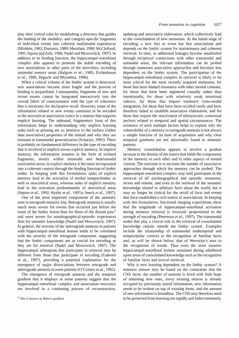
From sensation to cognition 1027
play their critical roles by establishing a directory that guidesthe binding of the modality- and category-specific fragmentsof individual events into coherent multimodal experiences(Mishkin, 1982; Damasio, 1989; Mesulam, 1990; McClelland,1995; Squire and Zola, 1996; Nadel and Moscovitch, 1997). Inaddition to its binding function, the hippocampal–entorhinalcomplex also appears to promote the stable encoding ofnew associations in other parts of the neocortex, includingunimodal sensory areas (Halgrenet al., 1985; Eichenbaumet al., 1996; Higuchi and Miyashita, 1996).
When a critical volume of the limbic system is destroyed,new associations become more fragile and the process ofbinding is jeopardized. Consequently, fragments of new andrecent events cannot be integrated interactively into theoverall fabric of consciousness with the type of coherencethat is necessary for declarative recall. However, some of theinformation related to new events continues to be encodedin the neocortical association cortex in a manner that supportsimplicit learning. The unbound, fragmentary form of thisinformation helps in understanding why implicit learningtasks such as priming are so sensitive to the surface (ratherthan associative) properties of the stimuli and why they areresistant to transmodal generalization (Schacter, 1995). Thereis probably no fundamental difference in the type of encodingthat is involved in implicit versus explicit memory. In implicitmemory, the information remains in the form of isolatedfragments, mostly within unimodal and heteromodalassociation areas; in explicit memory it becomes incorporatedinto a coherent context through the binding function of limbicnodes. In keeping with this formulation, tasks of explicitmemory lead to the activation of medial temporolimbic aswell as neocortical areas, whereas tasks of implicit memorylead to the activation predominantly of neocortical areas(Squireet al., 1992; Haxbyet al., 1997a; Seecket al., 1997).
One of the most important components of the amnesticstate is retrograde memory loss. Retrograde amnesia is usuallymuch more severe for events that occurred just before theonset of the limbic lesion than for those of the distant past,9
and more severe for autobiographical-episodic experiencesthan for semantic knowledge (Nadel and Moscovitch, 1997).In general, the severity of the anterograde amnesia in patientswith hippocampal–entorhinal lesions tends to be correlatedwith the severity of the retrograde component, suggestingthat the limbic components are as crucial for encoding asthey are for retrieval (Nadel and Moscovitch, 1997). Thehippocampal subregions that participate in retrieval may bedifferent from those that participate in encoding (Gabrieliet al., 1997), providing a potential explanation for theemergence of major dissociations between retrograde andanterograde amnesia in some patients (O’Connoret al., 1992).
The emergence of retrograde amnesia and the temporalgradient that it displays in some patients suggest that thehippocampal–entorhinal complex and association neocortexare involved in a continuing process of reconstruction,
9 This is known as Ribot’s gradient.
updating and associative elaboration, which collectively leadto the consolidation of new memories. At the initial stage ofencoding, a new fact or event has few associations anddepends on the limbic system for maintenance and coherentretrieval. In time, as additional linkages become establishedthrough reciprocal connections with other transmodal andunimodal areas, the relevant information can be probedthrough numerous associative approaches and becomes lessdependent on the limbic system. The participation of thehippocampal–entorhinal complex in retrieval is likely to bemost critical for the most recently acquired memories, forthose that have limited resonance with other mental contents,for those that have been registered casually rather thanintentionally, for those with relatively weak emotionalvalence, for those that require extensive cross-modalintegration, for those that have been recalled rarely and havetherefore failed to establish associative elaboration, and forthose that require the reactivation of idiosyncratic contextualanchors related to temporal and spatial circumstances. Theexistence of such multiple factors helps to explain why thevulnerability of a memory to retrograde amnesia is not alwaysa simple function of its time of acquisition and why cleartemporal gradients are not universally found in amnesticpatients.
Memory consolidation appears to involve a gradualincrease in the density of the matrix that binds the componentsof the memory to each other and to other aspects of mentalcontent. The outcome is to increase the number of associativeapproaches through which the memory can be probed. Thehippocampal–entorhinal complex may well participate in theretrieval of all autobiographical and episodic memories,recent and remote, and even in the retrieval of the semanticknowledge related to arbitrary facts about the world, but itmay no longer be critical for the recall of facts and eventsthat have established a rich matrix of associations. In keepingwith this formulation, functional imaging experiments showthat the magnitude of hippocampal–entorhinal activationduring memory retrieval is inversely proportional to thestrength of encoding (Peterssonet al., 1997). The transmodalnodes that play a critical role in the retrieval of consolidatedknowledge remain outside the limbic system. Examplesinclude the relationship of transmodal midtemporal andtemporopolar cortices to the recognition of familiar facesand, as will be shown below, that of Wernicke’s area tothe recognition of words. Thus even the most massivehippocampal–entorhinal lesions sustained during adulthoodspare areas of consolidated knowledge such as the recognitionof familiar faces and lexical retrieval.
Why is new learning dependent on the limbic system? Atentative answer may be based on the constraints that theCNS faces: the number of neurons is fixed with little hopeof obtaining new ones, every existing neuron is alreadyoccupied by previously stored information, new informationneeds to be written on top of existing items, and the amountof new information is boundless. The CNS may therefore needto be protected from learning too rapidly and indiscriminately,
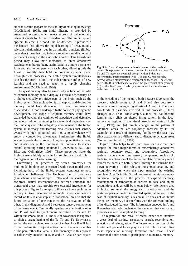
1028 M.-M. Mesulam
since this could jeopardize the stability of existing knowledge(McClelland, 1995). An initial filtering is provided byattentional systems which select subsets of behaviourallyrelevant events for further consideration. The limbic systemappears to erect a second line of defence. It provides amechanism that allows the rapid learning of behaviourallyrelevant relationships, but in an initially transient (limbic-dependent) form that may induce a relatively small amount ofpermanent change in the association cortex. This transitionalperiod may allow new memories to enter associativereadjustments before being assimilated in a more permanentform and also to compete with each other, allowing only thefittest to solidify their hold on precious synaptic space.Through these processes, the limbic system simultaneouslysatisfies the need to limit the indiscriminate influx of newlearning and the need to adapt to a rapidly changingenvironment (McClelland, 1994).
The question may also be asked why a function as vitalas explicit memory should display a critical dependency ona phylogenetically primitive part of the brain such as thelimbic system. One explanation is that explicit and declarativememory could have developed to recall contingenciesassociated with food and danger. In the course of phylogeneticdevelopment, the scope of explicit memory could haveexpanded beyond the confines of appetitive and defensivebehaviours while maintaining its anatomical dependency onthe limbic system. The obligatory involvement of the limbicsystem in memory and learning also ensures that sensoryevents with high emotional and motivational valence willenjoy a competitive advantage. Furthermore, the limbicsystem is particularly prone to long-term potentiation effects,and is also one of the few areas that continue to displayaxonal sprouting during adulthood (Benowitzet al., 1989;Bliss and Collinridge, 1993). These properties make thelimbic system highly suitable for serving a critical role inthe organization of new learning.
Unravelling the processes by which directories formultimodal binding are constructed within transmodal areas,including those of the limbic system, continues to poseformidable challenges. The Hebbian rule of covariance(Cruikshank and Weinberger, 1996) and the existence ofreciprocal neural interconnections between unimodal andtransmodal areas may provide two essential ingredients forthis process. Figure 3 attempts to illustrate how synchronousactivity in two unconnected unimodal areas can leave arecord of this relationship in a transmodal area so that thefuture activation of one can elicit the reactivation of theother. In this diagram, A and B represent sensory componentsof the same event. Temporally synchronous activation of Aand B will lead to synchronous activation in Ta and Tbwithin transmodal node Tr. The rule of covariance is expectedto elicit a strengthening of the Ta–Tb and Tb–Ta synapsesso that the next isolated excitation of either A or B will leadto the preferential conjoint activation of the other memberof the pair, rather than area C. The ‘memory’ in this processis collectively encoded in A, B and Tr. Area Tr participates
Fig. 3 A, B and C represent unimodal areas of the cerebralcortex. Tr represents a transmodal node of the cerebral cortex. Ta,Tb and Tc represent neuronal groups within T that arepreferentially interconnected with A, B and C, respectively.Arrows denote monosynaptic reciprocal connections. The circuitA–Ta–Tb–B is emboldened to show the preferential strengthening(1) of the Ta–Tb and Tb–Ta synapses upon the simultaneousactivation of A and B.
in the encoding of the memory both because it contains thedirectory which points to A and B and also because itcontains some convergent synthesis of A and B. There aretwo kinds of plasticity involved in this process: (i) localchanges in A or B—for example, a face that has becomefamiliar may elicit an altered firing pattern in the face-responsive regions of the visual association cortex (Rollset al., 1989); and (ii) remote changes in the pattern ofadditional areas that are conjointly accessed by Tr—forexample, as a result of increasing familiarity the face mayelicit activation in a different set of distant association areas(Gorno Tempiniet al., 1997).
Figure 3 also helps to illustrate how such a circuit cansupport the three major forms of remembering: associativeretrieval, voluntary recall and recognition. Associativeretrieval occurs when one sensory component, such as A,leads to the activation of the entire template; voluntary recallreflects the access to both A and B through the intrinsic top-down activation of the relevant transmodal area Tr, andrecognition occurs when the input matches the existingtemplate. Area Tr in Fig. 3 could represent the hippocampal–entorhinal complex in the process of explicit memory;midtemporal or temporopolar cortices in face and objectrecognition; and, as will be shown below, Wernicke’s areain lexical retrieval, the amygdala in motivation, and theposterior parietal cortex in directed spatial attention. In thecase of explicit memory, a lesion in Tr does not obliteratethe entire ‘memory’, but interferes with the coherent bindingof its distributed features. The information encoded in A andB remains relatively unchanged in a manner that can sustainprocesses related to implicit learning.
The registration and recall of recent experience involvesa great deal of sorting, associative search, recombination,selection and reintegration. The heteromodal cortices of thefrontal and parietal lobes play a critical role in controllingthese aspects of memory formation and recall. Thesetransmodal nodes seem to participate in numerous memory-
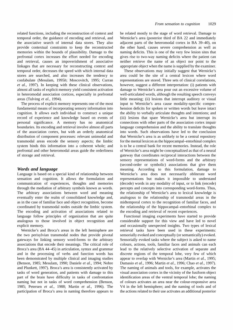
From sensation to cognition 1029
related functions, including the reconstruction of context andtemporal order, the guidance of encoding and retrieval, andthe associative search of internal data stores. They alsoprovide contextual constraints to keep the reconstructedmemories within the bounds of plausibility. Damage to theprefrontal cortex increases the effort needed for encodingand retrieval, causes an impoverishment of associativelinkages that are necessary for reconstructing context andtemporal order, decreases the speed with which internal datastores are searched, and also increases the tendency toconfabulate (Mesulam, 1995b; Moscovitch, 1995; Curranet al., 1997). In keeping with these clinical observations,almost all tasks of explicit memory yield consistent activationin heteromodal association cortices, especially in prefrontalareas (Tulvinget al., 1994).
The process of explicit memory represents one of the mostfundamental means of incorporating sensory information intocognition. It allows each individual to construct a uniquerecord of experience and knowledge based on events ofpersonal significance. A memory has no anatomicalboundaries. Its encoding and retrieval involve almost all partsof the association cortex, but with an orderly anatomicaldistribution of component processes: relevant unimodal andtransmodal areas encode the sensory aspects; the limbicsystem binds this information into a coherent whole; andprefrontal and other heteromodal areas guide the orderlinessof storage and retrieval.
Words and languageLanguage is based on a special kind of relationship betweensensation and cognition. It allows the formulation andcommunication of experiences, thoughts and memoriesthrough the mediation of arbitrary symbols known as words.The arbitrary associations between word and meaningeventually enter the realm of consolidated knowledge and,as in the case of familiar face and object recognition, becomecoordinated by transmodal nodes outside the limbic system.The encoding and activation of associations related tolanguage follow principles of organization that are quiteanalogous to those involved in object recognition andexplicit memory.
Wernicke’s and Broca’s areas in the left hemisphere arethe two perisylvian transmodal nodes that provide pivotalgateways for linking sensory word-forms to the arbitraryassociations that encode their meanings. The critical role ofBroca’s area (BA 44–45) in articulation, syntax and grammarand in the processing of verbs and function words hasbeen demonstrated by multiple clinical and imaging studies(Benson, 1985; Mesulam, 1990; Danieleet al., 1994; Nobreand Plunkett, 1997). Broca’s area is consistently activated bytasks of word generation, and patients with damage to thispart of the brain have difficulty in tasks of confrontationnaming but not in tasks of word comprehension (Benson,1985; Petersenet al., 1988; Martin et al., 1996). Theparticipation of Broca’s area in naming therefore appears to
be related mostly to the stage of word retrieval. Damage toWernicke’s area (posterior third of BA 22 and immediatelyadjacent parts of the heteromodal cortex in BA 39–40), onthe other hand, causes severe comprehension as well asnaming deficits. This is one of the very few lesion sites thatgives rise to two-way naming deficits where the patient canneither retrieve the name of an object nor point to theappropriate object when the name is supplied by the examiner.
These observations may initially suggest that Wernicke’sarea could be the site of a central lexicon where wordrepresentations are stored. Three sets of clinical correlations,however, suggest a different interpretation: (i) patients withdamage to Wernicke’s area pour out an excessive volume ofwell-articulated words, although the resulting speech conveyslittle meaning; (ii) lesions that interrupt auditory or visualinput to Wernicke’s area cause modality-specific compre-hension deficits for spoken or written words but leave intactthe ability to verbally articulate thoughts and intentions; and(iii) lesions that spare Wernicke’s area but interrupt itsconnections with other parts of the association cortex impairlanguage comprehension and the ability to translate thoughtsinto words. Such observations have led to the conclusionthat Wernicke’s area is as unlikely to be a central repositoryfor the mental lexicon as the hippocampal–entorhinal complexis to be a central bank for recent memories. Instead, the roleof Wernicke’s area might be conceptualized as that of a neuralgateway that coordinates reciprocal interactions between thesensory representations of word-forms and the arbitrary(second-order or symbolic) associations that give themmeaning. According to this formulation, damage toWernicke’s area does not necessarily obliterate wordrepresentations but makes it impossible to understand(decode) words in any modality of input, or to link (encode)percepts and concepts into corresponding word-forms. Thus,the relationship of Wernicke’s area to lexical knowledge isanalogous to the relationship of transmodal areas in themidtemporal cortex to the recognition of familiar faces, andthe relationship of the hippocampal–entorhinal complex tothe encoding and retrieval of recent experiences.
Functional imaging experiments have started to provideconsiderable support for this view and have led to noveland occasionally unexpected insights. Two types of lexicalretrieval tasks have been used in these experiments:sensorially evoked and conceptually (or semantically) evoked.Sensorially evoked tasks where the subject is asked to namecolours, actions, tools, familiar faces and animals can eachlead to the relatively selective activation of separate anddiscrete regions of the temporal lobe, very few of whichappear to overlap with Wernicke’s area (Martinet al., 1995;Damasioet al., 1996; Martinet al., 1996; Chaoet al., 1997).The naming of animals and tools, for example, activates thevisual association cortex in the vicinity of the fusiform objectidentification areas of the ventral temporal lobe; the namingof colours activates an area near the colour-responsive areaV4 in the left hemisphere; and the naming of tools and ofthe actions related to their use activates an additional posterior

1030 M.-M. Mesulam
midtemporal area slightly anterior to the movement-responsive area V5 and a premotor area (also activated byimagined hand movements), both in the left hemisphere(Martin et al., 1995, 1996). Conceptually evoked lexicalretrieval experiments have led to analogous results. Forexample, the generation of a colour word, characteristic ofan object shown to the subject in the form of a black-and-white drawing, leads to activations near the colour area V4(Martin et al., 1995). It appears, therefore, that lexicalretrieval is a highly distributed function and that the namingof visual entities and features is mediated by areas adjacentto those that support the perceptual encoding of thecorresponding entities and features. This relationship holdswhen the lexical labelling is triggered by a sensory event inthe outside world and also when it is directed to the conceptualknowledge of objects as retrieved from semantic memory.Lexical knowledge related to objects is thus anchored to theperceptual mapping of the corresponding experience.
These functional imaging experiments might have led tothe conclusion that the naming of visual entities is entirelycontrolled by a set of category-specific lexical retrieval siteslocated distinctly outside of Wernicke’s area. In fact, lesionsand electrical stimulations in these areas are known to yieldnaming deficits, some of which can be category-specific(Ojemann, 1983; Benson, 1985; Damasioet al., 1996).However, the clinical experience mentioned above also showsthat the most severe and generalized naming deficits occuras a consequence of damage to Wernicke’s area and adjacentperisylvian regions. The category-specific lexical retrievalareas located outside Wernicke’s area therefore appear to benecessary but not sufficient to support naming. These areascould provide category-specific prelexical or implicitrepresentations that need to be transformed into explicitoutputs (in the form of spoken, written or signed words)through the intervention of Wernicke’s area and othercomponents of the language network.
Figure 4 outlines a neural organization that is consistentwith these observations. According to this figure, sensoriallyevoked verbal naming entails multiple steps: (i) the visualinput triggers the processes of sensory and perceptualencoding (in the regions indicated by ‘Per’ in Fig. 4); (ii) anearby ‘intermediary’ lexical labelling area is activated(shown by ‘Lex’ in Fig. 4); (iii) projections from ‘Lex’ reachWernicke’s area, which acts as a transmodal gateway foractivating a corresponding auditory (phonological) word-form so that the word can be articulated through the mediationof Broca’s area and other premotor regions. Conceptuallyevoked lexical labelling takes a similar course except thatarea ‘Lex’ becomes activated by an internally generatedrepresentation evoked by semantic knowledge, probablythrough the intervention of transmodal gateways that mediateobject recognition, such as area ‘T’ in Fig. 2D.10
The reverse process of word comprehension related tovisual entities and features has not been investigated in asmuch detail by functional imaging. However, a similarorganization may exist such that words are first encoded as
perceptual entities in the word-form areas of auditory or visualassociation cortex; this representation is relayed throughWernicke’s area and induces optimal resonance incorresponding intermediary lexical areas (‘Lex’ in Fig. 4),leading to the activation of the associations that define themeaning of the word. Wernicke’s area would thus appear toplay a central role in lexical retrieval and comprehension byacting as a transmodal gateway which links the perceptualaspects of a word form to the distributed associations thatdefine its meaning. The organization outlined in Fig. 4 helpsto explain why naming deficits arise after lesions in so manydifferent parts of the temporal lobe, why some of theselesions can give rise to category-specific anomias and alsowhy lesions in Wernicke’s area give rise to global namingdeficts.
The intermediary lexical areas in Fig. 4 (‘Lex’) containpart of the distributed knowledge pertaining to colours,actions, faces and objects. The location of the regions thatprovide access to the additional conceptual attributes ofthese visual features and entities seems to follow a similarorganization. Thus, conceptual knowledge related to animalsis most likely to become impaired by lesions that involvethe fusiform region, whereas conceptual knowledge relatedto tools is more likely to become impaired by more laterallysituated lesions that involve the posterior midtemporal cortex,in the vicinity of V5 (Tranel et al., 1997). Despite thissimilarity of anatomical organization, the areas that arecritical for lexical access are probably not identical to thosethat are critical for accessing additional aspects of conceptualknowledge. Thus, anomic patients may have no difficultydescribing the use and nature of objects they cannot name,probably because access to the transmodal gateways thatmediate object recognition remains intact. This dissociationof lexical from conceptual knowledge provides the anatomicalbasis for the separation of thought from language.
Although its spontaneous development only in humansendows language with a sense of uniqueness, its neurologicalfoundations are quite analogous to those of other cognitivedomains (Nobre and Plunkett, 1997). Word-forms (verbaland written), for example, are likely to be encoded withinunimodal areas according to the principles that also guidethe encoding of faces and objects. Lexical labelling,furthermore, can be conceptualized as a component of objectrecognition in the sense that a name is as much an attributeof an object as its colour, location or past associations. Wordcomprehension is also an object recognition task where theperceptual features first lead to the categorical identificationof a word as a word, then to a subordinate level identificationof the individual word, and finally to the establishment of
10 This organization can also provide a neurological substrate for cognitively-based models of speech production. According to Morton (1969), forexample, the mental lexicon is a collection of logogens, each sensitive toits own conceptual or perceptual information. All logogens are simultan-eously active but only some reach threshold in response to a given set ofinputs and then induce a phonological code which eventually results in wordoutput. The ‘intermediary’ lexical areas in Fig. 4 could provide potentialanatomical substrates for logogens.
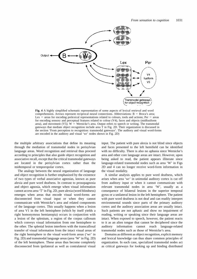
From sensation to cognition 1031
Fig. 4 A highly simplified schematic representation of some aspects of lexical retrieval and wordcomprehension. Arrows represent reciprocal neural connections. Abbreviations: B5 Broca’s area;Lex 5 areas for encoding prelexical representations related to colours, tools and actions; Per5 areasfor encoding sensory and perceptual features related to colour (V4), faces and objects (midfusiformarea), and movement (V5); W5 Wernicke’s area. Output refers to speech or writing. The transmodalgateways that mediate object recognition include area T in Fig. 2D. Their organization is discussed inthe section ‘From perception to recognition: transmodal gateways’. The auditory and visual word-formsare encoded in the auditory and visual ‘wr’ nodes shown in Fig. 2D)
the multiple arbitrary associations that define its meaningthrough the mediation of transmodal nodes in perisylvianlanguage areas. Word recognition and retrieval thus proceedaccording to principles that also guide object recognition andassociative recall, except that the critical transmodal gatewaysare located in the perisylvian cortex rather than themidtemporal or temporopolar cortex.
The analogy between the neural organization of languageand object recognition is further emphasized by the existenceof two types of verbal associative agnosias, known as purealexia and pure word deafness. In contrast to prosopagnosiaand object agnosia, which emerge when visual informationcannot access area ‘T’ in Fig. 2D, pure alexia (word blindness)emerges when areas that encode visual word-forms aredisconnected from visual input or when they cannotcommunicate with Wernicke’s area and related componentsof the language cortex. This usually happens when a lesionof area V1 in the left hemisphere (which by itself yields aright homonymous hemianopia) occurs in conjunction witha lesion of the splenium, a region of the corpus callosumwhich conveys visual information from one hemisphere tothe other. The splenial lesion interferes with the transcallosaltransfer of visual information from the intact visual areas ofthe right hemisphere to the visual word-form areas (‘wr’ inFig. 2D) and transmodal language areas (area ‘W’ in Fig. 2D)of the left hemisphere. These areas thus become completelydisconnected from ipsilateral as well as contralateral visual
input. The patient with pure alexia is not blind since objectsand faces presented to the left hemifield can be identifiedwith no difficulty. There is also no aphasia since Wernicke’sarea and other core language areas are intact. However, uponbeing asked to read, the patient appears illiterate sincelanguage-related transmodal nodes such as area ‘W’ in Figs2D and 4 can no longer receive word-form information inthe visual modality.
A similar analysis applies to pure word deafness, whicharises when area ‘wr’ in unimodal auditory cortex is cut offfrom auditory input or when it cannot communicate withrelevant transmodal nodes in area ‘W’, usually as aconsequence of bilateral lesions in the superior temporalgyrus or a unilateral lesion in the left hemisphere. The patientwith pure word deafness is not deaf and can readily interpretenvironmental sounds since parts of the primary auditorycortex and the auditory association areas are usually intact.Such patients are not aphasic and show no impairment ofreading, writing or speaking since their language areas areintact. When exposed to speech, however, the patient reactsto it as an alien tongue that cannot be deciphered since theauditory information cannot reach language-relatedtransmodal nodes such as those of Wernicke’s area.
Domains as different as object recognition, explicit memoryand lexical knowledge can thus share common principles oforganization. In each case, specialized transmodal nodes actas critical gateways for looking up and binding distributed
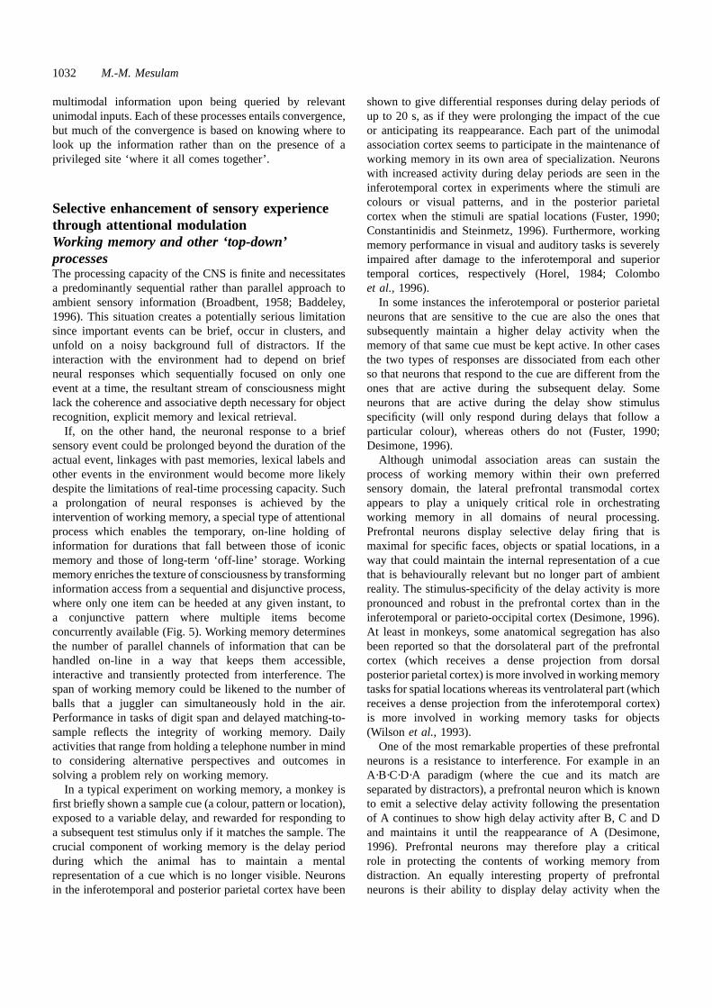
1032 M.-M. Mesulam
multimodal information upon being queried by relevantunimodal inputs. Each of these processes entails convergence,but much of the convergence is based on knowing where tolook up the information rather than on the presence of aprivileged site ‘where it all comes together’.
Selective enhancement of sensory experiencethrough attentional modulationWorking memory and other ‘top-down’processesThe processing capacity of the CNS is finite and necessitatesa predominantly sequential rather than parallel approach toambient sensory information (Broadbent, 1958; Baddeley,1996). This situation creates a potentially serious limitationsince important events can be brief, occur in clusters, andunfold on a noisy background full of distractors. If theinteraction with the environment had to depend on briefneural responses which sequentially focused on only oneevent at a time, the resultant stream of consciousness mightlack the coherence and associative depth necessary for objectrecognition, explicit memory and lexical retrieval.
If, on the other hand, the neuronal response to a briefsensory event could be prolonged beyond the duration of theactual event, linkages with past memories, lexical labels andother events in the environment would become more likelydespite the limitations of real-time processing capacity. Sucha prolongation of neural responses is achieved by theintervention of working memory, a special type of attentionalprocess which enables the temporary, on-line holding ofinformation for durations that fall between those of iconicmemory and those of long-term ‘off-line’ storage. Workingmemory enriches the texture of consciousness by transforminginformation access from a sequential and disjunctive process,where only one item can be heeded at any given instant, toa conjunctive pattern where multiple items becomeconcurrently available (Fig. 5). Working memory determinesthe number of parallel channels of information that can behandled on-line in a way that keeps them accessible,interactive and transiently protected from interference. Thespan of working memory could be likened to the number ofballs that a juggler can simultaneously hold in the air.Performance in tasks of digit span and delayed matching-to-sample reflects the integrity of working memory. Dailyactivities that range from holding a telephone number in mindto considering alternative perspectives and outcomes insolving a problem rely on working memory.
In a typical experiment on working memory, a monkey isfirst briefly shown a sample cue (a colour, pattern or location),exposed to a variable delay, and rewarded for responding toa subsequent test stimulus only if it matches the sample. Thecrucial component of working memory is the delay periodduring which the animal has to maintain a mentalrepresentation of a cue which is no longer visible. Neuronsin the inferotemporal and posterior parietal cortex have been
shown to give differential responses during delay periods ofup to 20 s, as if they were prolonging the impact of the cueor anticipating its reappearance. Each part of the unimodalassociation cortex seems to participate in the maintenance ofworking memory in its own area of specialization. Neuronswith increased activity during delay periods are seen in theinferotemporal cortex in experiments where the stimuli arecolours or visual patterns, and in the posterior parietalcortex when the stimuli are spatial locations (Fuster, 1990;Constantinidis and Steinmetz, 1996). Furthermore, workingmemory performance in visual and auditory tasks is severelyimpaired after damage to the inferotemporal and superiortemporal cortices, respectively (Horel, 1984; Colomboet al., 1996).
In some instances the inferotemporal or posterior parietalneurons that are sensitive to the cue are also the ones thatsubsequently maintain a higher delay activity when thememory of that same cue must be kept active. In other casesthe two types of responses are dissociated from each otherso that neurons that respond to the cue are different from theones that are active during the subsequent delay. Someneurons that are active during the delay show stimulusspecificity (will only respond during delays that follow aparticular colour), whereas others do not (Fuster, 1990;Desimone, 1996).
Although unimodal association areas can sustain theprocess of working memory within their own preferredsensory domain, the lateral prefrontal transmodal cortexappears to play a uniquely critical role in orchestratingworking memory in all domains of neural processing.Prefrontal neurons display selective delay firing that ismaximal for specific faces, objects or spatial locations, in away that could maintain the internal representation of a cuethat is behaviourally relevant but no longer part of ambientreality. The stimulus-specificity of the delay activity is morepronounced and robust in the prefrontal cortex than in theinferotemporal or parieto-occipital cortex (Desimone, 1996).At least in monkeys, some anatomical segregation has alsobeen reported so that the dorsolateral part of the prefrontalcortex (which receives a dense projection from dorsalposterior parietal cortex) is more involved in working memorytasks for spatial locations whereas its ventrolateral part (whichreceives a dense projection from the inferotemporal cortex)is more involved in working memory tasks for objects(Wilson et al., 1993).
One of the most remarkable properties of these prefrontalneurons is a resistance to interference. For example in anA·B·C·D·A paradigm (where the cue and its match areseparated by distractors), a prefrontal neuron which is knownto emit a selective delay activity following the presentationof A continues to show high delay activity after B, C and Dand maintains it until the reappearance of A (Desimone,1996). Prefrontal neurons may therefore play a criticalrole in protecting the contents of working memory fromdistraction. An equally interesting property of prefrontalneurons is their ability to display delay activity when the
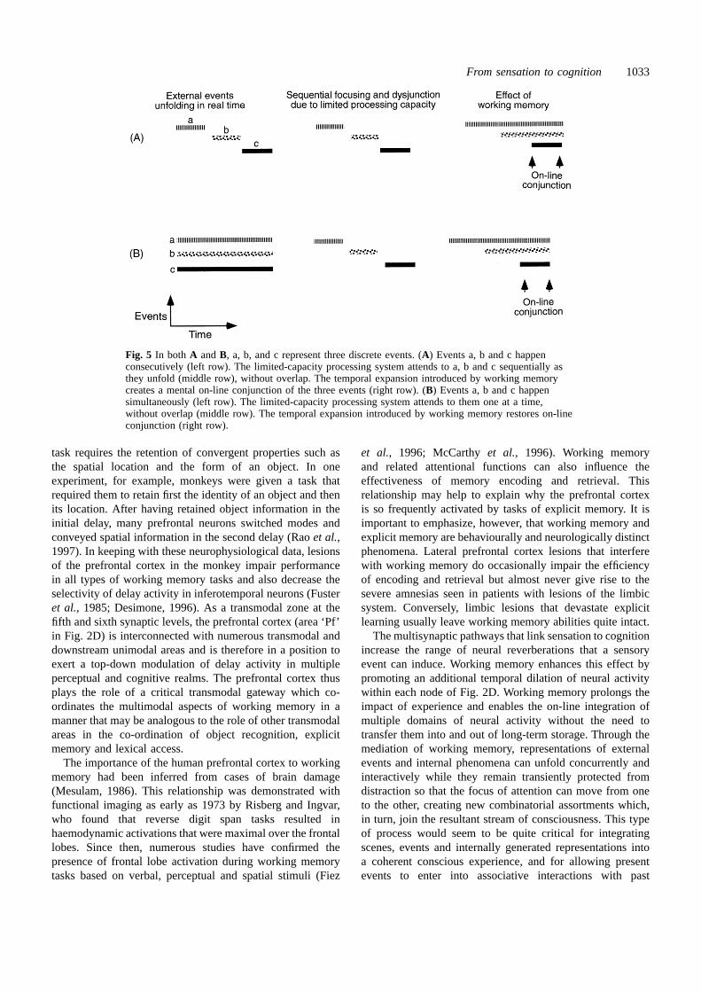
From sensation to cognition 1033
Fig. 5 In both A andB, a, b, and c represent three discrete events. (A) Events a, b and c happenconsecutively (left row). The limited-capacity processing system attends to a, b and c sequentially asthey unfold (middle row), without overlap. The temporal expansion introduced by working memorycreates a mental on-line conjunction of the three events (right row). (B) Events a, b and c happensimultaneously (left row). The limited-capacity processing system attends to them one at a time,without overlap (middle row). The temporal expansion introduced by working memory restores on-lineconjunction (right row).
task requires the retention of convergent properties such asthe spatial location and the form of an object. In oneexperiment, for example, monkeys were given a task thatrequired them to retain first the identity of an object and thenits location. After having retained object information in theinitial delay, many prefrontal neurons switched modes andconveyed spatial information in the second delay (Raoet al.,1997). In keeping with these neurophysiological data, lesionsof the prefrontal cortex in the monkey impair performancein all types of working memory tasks and also decrease theselectivity of delay activity in inferotemporal neurons (Fusteret al., 1985; Desimone, 1996). As a transmodal zone at thefifth and sixth synaptic levels, the prefrontal cortex (area ‘Pf’in Fig. 2D) is interconnected with numerous transmodal anddownstream unimodal areas and is therefore in a position toexert a top-down modulation of delay activity in multipleperceptual and cognitive realms. The prefrontal cortex thusplays the role of a critical transmodal gateway which co-ordinates the multimodal aspects of working memory in amanner that may be analogous to the role of other transmodalareas in the co-ordination of object recognition, explicitmemory and lexical access.
The importance of the human prefrontal cortex to workingmemory had been inferred from cases of brain damage(Mesulam, 1986). This relationship was demonstrated withfunctional imaging as early as 1973 by Risberg and Ingvar,who found that reverse digit span tasks resulted inhaemodynamic activations that were maximal over the frontallobes. Since then, numerous studies have confirmed thepresence of frontal lobe activation during working memorytasks based on verbal, perceptual and spatial stimuli (Fiez
et al., 1996; McCarthyet al., 1996). Working memoryand related attentional functions can also influence theeffectiveness of memory encoding and retrieval. Thisrelationship may help to explain why the prefrontal cortexis so frequently activated by tasks of explicit memory. It isimportant to emphasize, however, that working memory andexplicit memory are behaviourally and neurologically distinctphenomena. Lateral prefrontal cortex lesions that interferewith working memory do occasionally impair the efficiencyof encoding and retrieval but almost never give rise to thesevere amnesias seen in patients with lesions of the limbicsystem. Conversely, limbic lesions that devastate explicitlearning usually leave working memory abilities quite intact.
The multisynaptic pathways that link sensation to cognitionincrease the range of neural reverberations that a sensoryevent can induce. Working memory enhances this effect bypromoting an additional temporal dilation of neural activitywithin each node of Fig. 2D. Working memory prolongs theimpact of experience and enables the on-line integration ofmultiple domains of neural activity without the need totransfer them into and out of long-term storage. Through themediation of working memory, representations of externalevents and internal phenomena can unfold concurrently andinteractively while they remain transiently protected fromdistraction so that the focus of attention can move from oneto the other, creating new combinatorial assortments which,in turn, join the resultant stream of consciousness. This typeof process would seem to be quite critical for integratingscenes, events and internally generated representations intoa coherent conscious experience, and for allowing presentevents to enter into associative interactions with past
1034 M.-M. Mesulam
memories, the present context and contemplated goals.Despite this potentially crucial role of working memory inmodulating the texture of consciousness, patients with severefrontal lobe lesions and severe impairments of workingmemory appear to maintain all external manifestations ofconsciousness, although at a possibly reduced level ofcomplexity. There is, therefore, relatively little support for amodern sort of Cartesianism which would substitute thefrontal lobe for the pineal gland as the final seat that sensationmust reach before it can hope to gain access to consciousness(Crick and Koch, 1995).
The influence of the prefrontal cortex upon the delayactivity of unimodal neurons during working memory tasksprovides an example of top-down processing. As has beenmentioned above, top-down processing is a generic term forall sensory-petal pathways, especially those that emanatefrom transmodal areas. The top-down processing of theprefrontal cortex in working memory is analogous to the top-down processing of the hippocampal–entorhinal cortex inexplicit memory and of Wernicke’s area in lexical access.Other top-down processes that contribute to the attentionalmodulation of sensory experiences include mental imagery,novelty-seeking behaviours and the influence of emotion andmotivation upon neural activity. The prefrontal cortex playsan important integrative role in each of these attentionalprocesses.
Mental imageryMental imagery refers to the activation of sensoryrepresentations that are not part of ambient reality. It couldbe conceptualized as a special type of attentional processtargeted to internal representations. The neural substrates formental imagery appear to include the same areas that wouldhave supported the corresponding acts of perception if theimagined scene were actually unfolding in the external world.Functional imaging experiments, for example, have shownthat the visualization of spatial relationships leads to theactivation of the dorsal occipitoparietal cortex, whereas thevisualization of objects leads to the activation of ventraloccipito-temporal visual areas (Roland and Gulya´s, 1994;Sakai and Miyashita, 1994). There may even be someretinotopic organization since visualizing larger objects(which presumably subtend a greater visual angle inthe mind’s eye) leads to more extensive activation in the partof V1 that maps the more peripheral aspects of the visualfields (Kosslynet al., 1993).
Activation during mental imagery has been shown inalmost all of the primary, upstream and downstream visualareas. Primary and upstream visual areas such as V1–V5may be activated predominantly by imagery that emphasizesinternal sensory detail and mental rotation, whereasinferotemporal visual association areas may be activatedpredominantly by imagery tasks that reactivate storedmemories (Roland and Friberg, 1985; Kosslynet al., 1994;Sakai and Miyashita, 1994). Even in imagery tasks that are
optimally effective, however, the activation of V1 appears tobe less robust and less consistent than activation in the othervisual cortices (Sakai and Miyashita, 1994).
Virtually any sphere of sensory processing could potentiallybecome the target of mental imagery. Mental rotations andother reconfigurations of visual percepts could subserve non-verbal thinking, whereas the mental activation of auditoryword-forms and related associations could support innerspeech and verbal thinking. Functional imaging suggests thatthe lateral prefrontal cortex plays a particularly importantrole in the top-down processes related to mental imagery(Kosslyn et al., 1994), a role that is consistent with thecritical influence of this area upon interrelated attentionalprocesses such as working memory. There is no convincingevidence yet, however, that prefrontal lesions impair mentalimagery.
Mental imagery provides one of at least three settingswhere the activation of sensory areas can transcend theconstraints imposed by external reality. The two otherexamples include working memory, where the representationof a sensory experience is prolonged beyond the duration ofthe stimulus, and anticipatory paired-associate responses,where sensory representations become activated inexpectation of an event which is not yet present in reality.
Novelty-seekingSome sensory events elicit a greater neural response becauseof objective properties such as excessive brightness, loudnessor noxiousness. Others do so on subjective grounds, becausethey have become relevant to current needs, because theyhave acquired special associations in the course of pastexperience, or because they are novel. The preferentialenhancement of responses to novel stimuli is a luxury thatonly advanced brains can afford. In primitive animals theCNS contains numerous neural circuits, exemplified by the‘bug detectors’ in the frog, for identifying familiar patternson the basis of partial evidence and for linking them toautomatic responses. The frog may not be able to distinguishone bug from another or a wind-swept leaf from a bug, butthe processing efficiency offered by this arrangement appearsto offset the attendant limitation in the resolution of reality.
The bias for perceiving sameness and emitting automaticresponses represents a ‘default mode’ of brain function andleads to behaviours that can also be designated as instinctual,stereotyped or stimulus-bound. One consequence of thisorganization is to enhance efficiency and reliability, butanother is to promote premature closure, perseveration andresponse rigidity, phenomena frequently encountered in thebehaviours of less evolved species, of infants and of brain-injured humans. The CNS of more advanced species hascompensated for these limitations by developing specializedneural circuits for the rapid detection of unfamiliar eventsand for inhibiting automatic response tendencies that havebecome contextually inappropriate.
Play and novelty-seeking behaviours, for example,

From sensation to cognition 1035
represent an antitheses to the pursuit of sameness. Suchbehaviours are inconspicuous in amphibians and reptiles,emerge in birds, and reach their most exuberant expression inadvanced mammals, especially primates. In humans, sensorydeprivation and monotony induce restlessness and even vividhallucinations (Heron, 1957). Monkeys will work hard in asetting where the only reward is a peek through a window,and human subjects who are given a choice between familiarand simple versus novel and complex patterns, willconsistently spend more time viewing the latter (Butler, 1953;Berlyne, 1960). The tendency for seeking novelty mayeven have survival value: in a prospective study of 2153community-dwelling elderly subjects, those with a higherbaseline level of ‘curiosity’ had a better chance to be aliveand well 5 years later (Swan and Carmelli, 1996).
Nearly all transmodal and downstream unimodal nodes inFig. 2D respond more actively to novel stimuli in theirspecific domains of specialization (Brownet al., 1987; Wilsonand Rolls, 1993; Bernset al., 1997). However, the frontallobe plays a particularly critical role in this realm of neuralfunction. The P300 response elicited by novel or deviantstimuli, for example, is critically dependent on the integrityof the prefrontal cortex; an N2–P3 response which is maximalover the prefrontal cortex appears to determine the attentionalresources that will be allocated to novel events; and theregion of the frontal eye-fields belongs to a distributednetwork for exploring the extrapersonal space and seekingmotivationally relevant targets (Mesulam, 1981; Knight,1984; Daffneret al., 1998). Patients with damage to variouscomponents of the prefrontal cortex display placid disinterestin novel aspects of the environment, inability to inhibitpreviously rewarded responses when reinforcementcontingencies change, and vulnerability to perseveration,impulsivity and stimulus-bound behaviours (Leimkuhler andMesulam, 1985; Mesulam, 1986). The prefrontal cortex thusseems to play an essential role in enhancing the impact ofnovelty and inhibiting habitual reactions that have becomeinappropriate. These multiple attentional functions of theprefrontal cortex and its role in various forms of responseselection have led to the suggestion that it fulfils an ‘executive’function in the linkage of sensation to cognition andbehaviour.
The valuation of sensationEmotion, mood and motivation modulate the neural impactof sensory events in a manner that reflects the subjectivevalue of these events to the individual. These partiallyoverlapping processes are closely related to the functions ofthe hypothalamus and other components of the limbic system,especially the amygdala and the paralimbic areas with whichit is interconnected. The relationship of the amygdala toemotion was emphasized by Downer (1962), who showed thatnaturalistic sensory events triggered appropriate emotionalresponses in the monkey only if the sensory information hadaccess to an intact amygdala. The amygdala also plays an
important role in altering the impact of previously neutralstimuli that become linked to emotionally relevant events.For example, initially neutral auditory stimuli start to elicitamygdaloid activation only after they become associatedwith fear (LeDoux et al., 1983). In humans, amygdaloidstimulations evoke emotionally charged memories whereasamygdaloid lesions cause states of hypoemotionality andinterfere with the acquisition of conditioned emotionalresponses (Glooret al., 1982; Aggleton, 1993; Becharaet al.,1995). In one patient, neurological damage involving thevisuo-amygdaloid pathways led to the cancellation of asubscription to Playboy, apparently because the visualevocation of erotic feelings became blunted (Bauer, 1982).
The amygdala is reciprocally interconnected with unimodalsensory association cortices in the superior and inferiortemporal gyri (Aggleton, 1993). Through these connections,the amygdala can selectively modulate sensory responses ina manner that reflects their intrinsic or acquired emotionalrelevance. In analogy to the organization of the othertransmodal nodes in the cerebral cortex, the amygdala(represented by node ‘L’ in Fig. 2D) appears to act as aneural gateway for binding the sensory representations ofprimary and secondary reinforcers with each other and withthe neural correlates of emotional and motivational valence.
The amygdala has a dual role in relation to attention andmemory. Its role in attention is to selectively enhance theprocessing resources allocated to ambient events with highemotional and hedonic valence. Its role in memory is toencode the implicit and explicit emotional correlates ofexperience. The latter aspect of amygdaloid function can beconceptualized as complementary to that of the hippocampalformation, but with a selective emphasis on the associationof sensory experiences to emotional valence (Cahillet al.,1995). The roles of the hippocampus and of the amygdala inmemory can be dissociated from each other. Hippocampallesions interfere with the explicit recall of specific events butnot with the autonomic responses they elicit on the basis oftheir emotional significance, whereas amygdaloid lesionsleave explicit recall intact but abolish the associatedautonomic responses (Becharaet al., 1995).
The frontal lobe also participates in the modulation ofneural responses to events that have become motivationallysignificant. Identical sensory stimuli, for example, can elicitdramatically different responses from lateral prefrontalneurons when their relationships to reward-producingresponses are altered (Sakagami and Niki, 1994).Furthermore, an arbitrary sensory cue elicits greater responsesfrom dorsolateral prefrontal neurons when it signals theimpending delivery of a preferred food item, such as a pieceof apple or cabbage, than when it signals a less favouritefood such as a raisin (Watanabe, 1996). Their pronouncedanatomical connectivity with the amygdala suggests that theparalimbic sectors of the orbitofrontal area might play aparticularly prominent role in the emotional modulation ofexperience (Morecraftet al., 1992). In fact, damage toorbitofrontal cortex can cause severe disruptions in the
1036 M.-M. Mesulam
linkage of experience to the appropriate emotional state andcan lead to extensive impairments of judgement, insight andcomportment. However, a blunting in the emotional andmotivational colouring of sensory experience is also seen inpatients with lesions in the more dorsolateral parts of theprefrontal cortex. These observations suggest that thedorsolateral and orbitofrontal components of the prefrontalcortex and the amygdala collectively modulate the neuralimpact of experience by encoding the acquired significanceof sensory events rather than their surface properties.
The modulatory effects of emotion and motivationinfluence the attentional resources allocated to a sensorystimulus. Attention is difficult to define but easy to detect. Itusually refers to an enhancement in the selectivity, intensityand duration of neuronal responses to events that have becomeemotionally and motivationally relevant. The attentionalcomponent of a neural response can be conceptualized as avector with a magnitude and a direction. The direction ofthe vector is determined by the specialization of the neuronsthat express the attentional effect. Thus, inferotemporalneurons show attentional enhancement to behaviourallyrelevant visual patterns, posterior parietal neurons to spatialtargets, pyriform neurons to odorants, and Wernicke’s areato words (Bushnellet al., 1981; Nobre and McCarthy, 1994;Desimone, 1996; Eichenbaumet al., 1996). The magnitudeof the attentional vector in each of these cortical areas is undertwo types of influences: bottom-up influences emanating fromthe ascending reticular activating system (ARAS), and top-down influences emanating from the limbic and prefrontalcortices. The ARAS modulates the intensity of attentionaccording to the general level of arousal and wakefulness,whereas the limbic and frontal cortices provide modulationsbased on factors such as novelty, emotion, motivation,intention and past experience. One manifestation of the top-down influence of prefrontal cortex upon the attentional vectoris known as working memory and has been reviewed above.
Attention, emotion and motivation introduce a value systemto sensory processing. These value-based modulations allowthe CNS to sculpt sensory experience into a subjectivelandscape. The effects of attention can be detected at everysynaptic level of Fig. 2D. For example, some neurons in theprimary auditory cortex respond to a sound only when theanimal appears to attend to its source (Hubelet al., 1959).Numerous experiments in primates and humans have alsoshown, however, that attentional responses are more commonand prominent at higher synaptic levels, especially in theheteromodal cortex and downstream components of theunimodal cortices (Mesulam, 1985a; Sakai and Miyashita,1994; Maunsell, 1995). Motivational modulation seems todisplay a similar pattern. In the monkey, for example, hungermodulates the neuronal response to palatable liquids almostexclusively at the final stage of the multisynaptic tastepathways in the orbitofrontal cortex (Rolls, 1987).
These observations suggest that early sensory processing,especially at the first two synaptic levels of the template inFig. 2D, is relatively protected from value-based modulations.
This arrangement allows internal states to influence sensory-fugal processing only after the essential features of sensoryevents have been encoded. In fact, there is little adaptivesense in altering responses to shapes, motion or colour,whereas it makes a lot of sense to modulate responses tospecific faces, objects or locations. The organization ofsensory-petal (top-down) connections is consistent with thispolarization, since transmodal cortices have very fewmonosynaptic projections to primary sensory areas and sendfewer projections to upstream than to downstreamcomponents of unimodal areas. In the frog, the situationmay be quite different. Attention can dramatically alter theresponse properties of visually responsive tectal neurons atthe first central synapse of visuo-fugal pathways, and caninduce an obligatory change in the nature of all the visualinput that the brain receives (Lettvinet al., 1961). Significanceand appearance are therefore much more tightly yoked toeach other in the frog than in the primate.
Modulatory pathways of the reticular activatingsystemThe initial accounts of the ARAS had focused on its thalamo-cortical components. More recent work has shown that theARAS also contains several sets of projections that arisefrom relatively small regions of the basal forebrain andbrainstem and that reach the cerebral cortex without a synapsein the thalamus. These modulatory pathways mediate thestate-dependent regulation of neural responses in accordancewith changes of arousal, mood and circadian phase (Mesulam,1995a). The cholinergic pathways that extend from thenucleus basalis of the basal forebrain (the Ch1–Ch4 cellgroups) to the cerebral cortex are the most massive of allmodulatory pathways. Others include histaminergicprojections from the hypothalamus, dopaminergic projectionsfrom the substantia nigra–ventral tegmental area, serotonergicprojections from the raphe nuclei, and noradrenergicprojections from the nucleus locus coeruleus. Each of thesepathways has a slightly different pattern of corticaldistribution and physiological effect. For example,serotonergic pathways are directed predominantly to corticalinterneurons whereas the cholinergic pathways are directedpredominantly to projection neurons (Smiley and Goldman-Rakic, 1996; Smileyet al., 1997).
Cortical cholinergic innervation reaches all cortical regionsbut is most intense within limbic areas (Mesulam, 1996).The major effect of acetylcholine upon neurons of the cerebralcortex is mediated through the m1 subtype of muscarinicreceptors and causes a prolonged reduction of potassiumconductance so as to make cortical neurons more receptiveto sensory stimulation (Satoet al., 1987; McCormick, 1990).Cholinergic neurons of the nucleus basalis are particularlysensitive to novel and motivationally relevant stimuli (Wilsonand Rolls, 1990). Furthermore, the novelty-related P300response in the human cerebral cortex can be abolished by

From sensation to cognition 1037
cholinergic antagonists (Hammondet al., 1987). In additionto playing a role in novelty-seeking behaviours, cholinergicprojections may also modulate attentional processes relatedto working memory. For example, locally infused cholinergicblockers interfere with the accuracy of saccadic eyemovements only when they are guided by spatial workingmemory but not when they are guided by ambient visualtargets (Diaset al., 1996).
In addition to these short-term effects, acetylcholine maypromote the establishment of long-term plasticity and synapticefficacy during the acquisition of new associations (Huertaand Lisman, 1993; Cruikshank and Weinberger, 1996; Woolf,1996; Baskervilleet al., 1997). Although the basal forebraincholinergic nuclei project to all cortical areas, they receivecortical input only from the limbic system. Thus, onlybehaviourally significant events which activate the limbicsystem can effectively induce a rapid change of activity inthe nucleus basalis (Mesulam, 1996). Such limbic activationof the nucleus basalis and the resultant release of acetylcholinein cortical areas would enhance the response of unimodaland heteromodal areas to the target sensory event, andselectively increase the responsivity of the limbic system tothe resultant input. Through these processes, cholinergicactivation can influence the preferential registration andbinding of significant events within the context of the circuitryshown in Fig. 3. Cortical cholinergic projections can thusselectively enhance the immediate and long-term impact ofsensory events that are novel and motivationally relevant.
The dopaminergic cells of the substantia nigra–ventraltegmental area are also selectively sensitive to motivationallyrelevant stimuli and to cues that signal their existence(Schultz, 1997). These neurons appear to encode deviationsbetween the prediction and occurrence of reward and maytherefore convey teaching signals for learning appetitivebehaviours (Schultz, 1997). In keeping with these tuningproperties, the dopaminergic projections from the ventraltegmental area to the cerebral cortex and nucleus accumbensappear to play an important role in mediating the neuralprocesses related to substance addiction (Pichet al., 1997). Asin the case of cholinergic projections, ascending dopaminergicinputs can also enhance the working memory capacity ofneurons in the frontal lobe (Goldman-Rakic, 1996). Thenucleus locus coeruleus provides another CNS site whereneurons can encode the behavioural significance rather thansurface appearance of sensory stimuli (Aston-Joneset al.,1997). The noradrenergic projection from the nucleus locuscoeruleus to the cerebral cortex increases the signal-to-noiseratio of sensory responses in the cerebral cortex and thereforeenhances the sharpness of neural tuning to significant sensoryevents (Footeet al., 1983; Levinet al., 1988). Throughβ-adrenergic synapses in the amygdala, ascending noradrenergicpathways are also thought to mediate the preferential storageand retrieval of emotionally relevant memories (McGaughet al., 1993).
These modulatory projections may also induce pathologicalbiases in the encoding of sensory experience, such as those
seen in patients with mania, depression, paranoia and chronicanxiety. Preliminary evidence suggests that the neuralresponse to sensory events may be altered in patients withsuch conditions. For example, when compared with controlsubjects, patients with generalized anxiety disorder showgreater metabolic activation of the temporal and frontal cortexduring the passive viewing of neutral stimuli (Wuet al., 1991).Indirect evidence based on the pharmacological treatment ofdepression and anxiety with noradrenergic and serotonergicagents and of paranoia with dopamine blockers suggests thatthe modulatory pathways of the cerebral cortex may playimportant roles in setting such fixed attitudinal biases in theemotional valuation of sensory experience.
As in the case of cholinergic pathways, the nuclei thatgive rise to these modulatory projections innervate all partsof the cerebral cortex but receive feedback (top-down)projections from a much more limited set of cortical areas,predominantly from those that belong to the limbic andparalimbic zones of the cerebral cortex (Mesulam, 1987).Additional inputs to these nuclei may also arise in thehypothalamus and other subcortical structures. Thisasymmetry of cortico-fugal versus cortico-petal connectionsallows the neurons of the basal forebrain, substantia nigra–ventral tegmental area, brainstem raphe and nucleus locuscoeruleus to act as transmitter-specific relays for rapidlymodulating the linkage of sensation to cognition throughoutthe cerebral cortex, and in a manner that is particularlyresponsive to the activation state of the limbic system. It isunlikely that there will be a one-to-one relationship betweenany single pathway in the ARAS and individual componentsof cognition or behaviour. In general, however, the activationof these modulatory pathways provides a mechanism foraugmenting the neural responses to novel and motivationallyrelevant events, facilitating their storage in memory,enhancing their access to on-line processing resources,sharpening the attentional focusing they elicit, and increasingtheir impact on consciousness.
The organization of neurocognitive networksDistributed large-scale networks and theircortical epicentres: a model based on theorganization of spatial awareness and directedspatial attentionTransmodal nodes in the midtemporal cortex, Wernicke’sarea and the hippocampal–entorhinal complex transformunimodal sensory input into coherent multimodal assembliesnecessary for face and object recognition, naming, andexplicit memory. These transmodal areas provide the neuralepicentres of large-scale distributed networks (Mesulam,1990). The organization of such networks has been exploredin relation to the process of directed spatial attention.
Observations on patients with focal brain damage, relevantanimal experiments and more recent work based on functionalimaging show that the distribution of attention within the
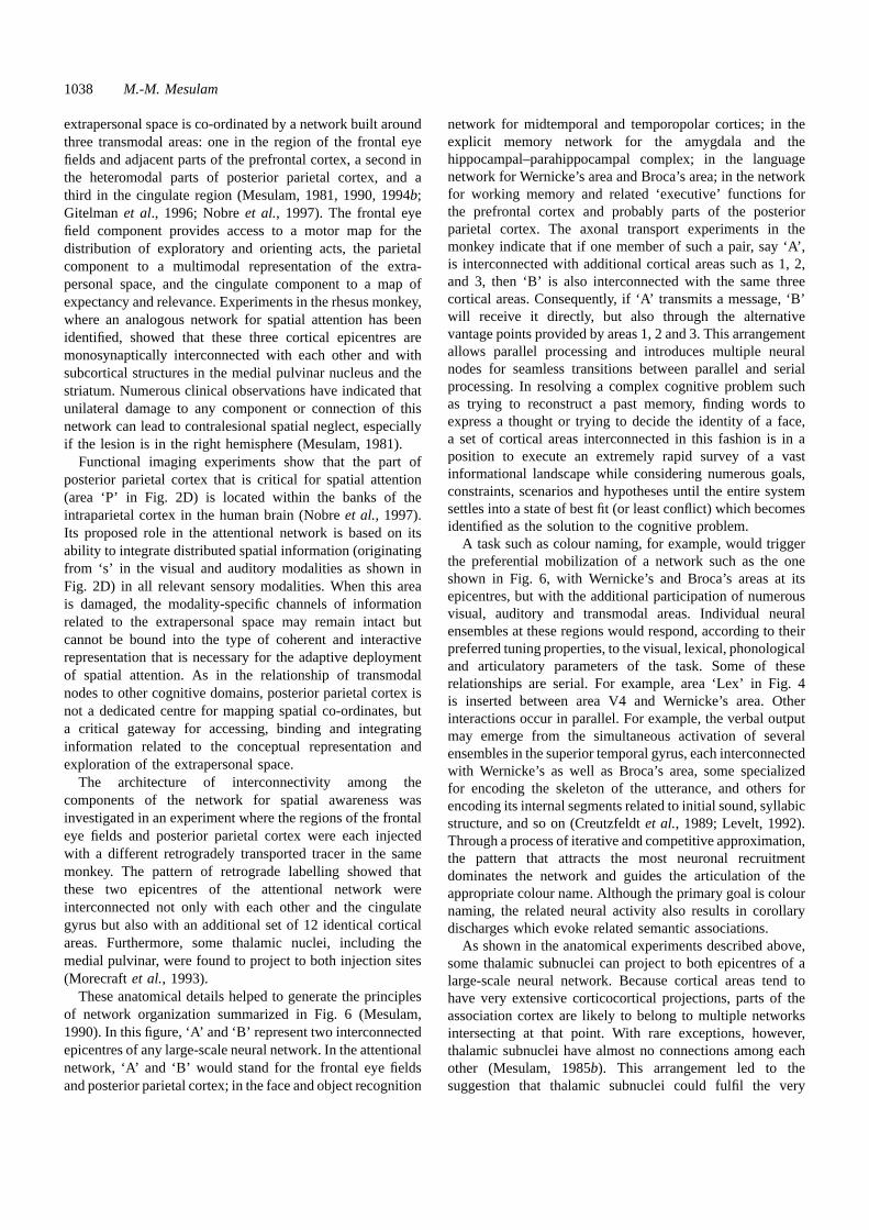
1038 M.-M. Mesulam
extrapersonal space is co-ordinated by a network built aroundthree transmodal areas: one in the region of the frontal eyefields and adjacent parts of the prefrontal cortex, a second inthe heteromodal parts of posterior parietal cortex, and athird in the cingulate region (Mesulam, 1981, 1990, 1994b;Gitelmanet al., 1996; Nobreet al., 1997). The frontal eyefield component provides access to a motor map for thedistribution of exploratory and orienting acts, the parietalcomponent to a multimodal representation of the extra-personal space, and the cingulate component to a map ofexpectancy and relevance. Experiments in the rhesus monkey,where an analogous network for spatial attention has beenidentified, showed that these three cortical epicentres aremonosynaptically interconnected with each other and withsubcortical structures in the medial pulvinar nucleus and thestriatum. Numerous clinical observations have indicated thatunilateral damage to any component or connection of thisnetwork can lead to contralesional spatial neglect, especiallyif the lesion is in the right hemisphere (Mesulam, 1981).
Functional imaging experiments show that the part ofposterior parietal cortex that is critical for spatial attention(area ‘P’ in Fig. 2D) is located within the banks of theintraparietal cortex in the human brain (Nobreet al., 1997).Its proposed role in the attentional network is based on itsability to integrate distributed spatial information (originatingfrom ‘s’ in the visual and auditory modalities as shown inFig. 2D) in all relevant sensory modalities. When this areais damaged, the modality-specific channels of informationrelated to the extrapersonal space may remain intact butcannot be bound into the type of coherent and interactiverepresentation that is necessary for the adaptive deploymentof spatial attention. As in the relationship of transmodalnodes to other cognitive domains, posterior parietal cortex isnot a dedicated centre for mapping spatial co-ordinates, buta critical gateway for accessing, binding and integratinginformation related to the conceptual representation andexploration of the extrapersonal space.
The architecture of interconnectivity among thecomponents of the network for spatial awareness wasinvestigated in an experiment where the regions of the frontaleye fields and posterior parietal cortex were each injectedwith a different retrogradely transported tracer in the samemonkey. The pattern of retrograde labelling showed thatthese two epicentres of the attentional network wereinterconnected not only with each other and the cingulategyrus but also with an additional set of 12 identical corticalareas. Furthermore, some thalamic nuclei, including themedial pulvinar, were found to project to both injection sites(Morecraftet al., 1993).
These anatomical details helped to generate the principlesof network organization summarized in Fig. 6 (Mesulam,1990). In this figure, ‘A’ and ‘B’ represent two interconnectedepicentres of any large-scale neural network. In the attentionalnetwork, ‘A’ and ‘B’ would stand for the frontal eye fieldsand posterior parietal cortex; in the face and object recognition
network for midtemporal and temporopolar cortices; in theexplicit memory network for the amygdala and thehippocampal–parahippocampal complex; in the languagenetwork for Wernicke’s area and Broca’s area; in the networkfor working memory and related ‘executive’ functions forthe prefrontal cortex and probably parts of the posteriorparietal cortex. The axonal transport experiments in themonkey indicate that if one member of such a pair, say ‘A’,is interconnected with additional cortical areas such as 1, 2,and 3, then ‘B’ is also interconnected with the same threecortical areas. Consequently, if ‘A’ transmits a message, ‘B’will receive it directly, but also through the alternativevantage points provided by areas 1, 2 and 3. This arrangementallows parallel processing and introduces multiple neuralnodes for seamless transitions between parallel and serialprocessing. In resolving a complex cognitive problem suchas trying to reconstruct a past memory, finding words toexpress a thought or trying to decide the identity of a face,a set of cortical areas interconnected in this fashion is in aposition to execute an extremely rapid survey of a vastinformational landscape while considering numerous goals,constraints, scenarios and hypotheses until the entire systemsettles into a state of best fit (or least conflict) which becomesidentified as the solution to the cognitive problem.
A task such as colour naming, for example, would triggerthe preferential mobilization of a network such as the oneshown in Fig. 6, with Wernicke’s and Broca’s areas at itsepicentres, but with the additional participation of numerousvisual, auditory and transmodal areas. Individual neuralensembles at these regions would respond, according to theirpreferred tuning properties, to the visual, lexical, phonologicaland articulatory parameters of the task. Some of theserelationships are serial. For example, area ‘Lex’ in Fig. 4is inserted between area V4 and Wernicke’s area. Otherinteractions occur in parallel. For example, the verbal outputmay emerge from the simultaneous activation of severalensembles in the superior temporal gyrus, each interconnectedwith Wernicke’s as well as Broca’s area, some specializedfor encoding the skeleton of the utterance, and others forencoding its internal segments related to initial sound, syllabicstructure, and so on (Creutzfeldtet al., 1989; Levelt, 1992).Through a process of iterative and competitive approximation,the pattern that attracts the most neuronal recruitmentdominates the network and guides the articulation of theappropriate colour name. Although the primary goal is colournaming, the related neural activity also results in corollarydischarges which evoke related semantic associations.
As shown in the anatomical experiments described above,some thalamic subnuclei can project to both epicentres of alarge-scale neural network. Because cortical areas tend tohave very extensive corticocortical projections, parts of theassociation cortex are likely to belong to multiple networksintersecting at that point. With rare exceptions, however,thalamic subnuclei have almost no connections among eachother (Mesulam, 1985b). This arrangement led to thesuggestion that thalamic subnuclei could fulfil the very
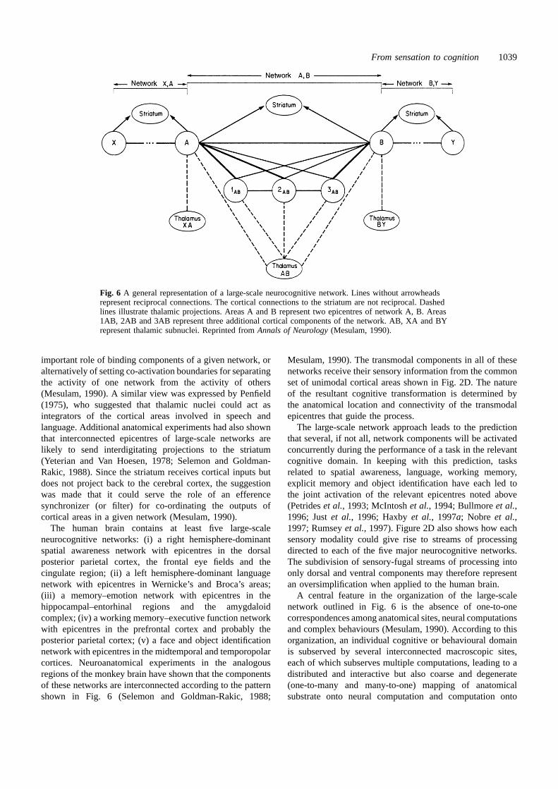
From sensation to cognition 1039
Fig. 6 A general representation of a large-scale neurocognitive network. Lines without arrowheadsrepresent reciprocal connections. The cortical connections to the striatum are not reciprocal. Dashedlines illustrate thalamic projections. Areas A and B represent two epicentres of network A, B. Areas1AB, 2AB and 3AB represent three additional cortical components of the network. AB, XA and BYrepresent thalamic subnuclei. Reprinted fromAnnals of Neurology(Mesulam, 1990).
important role of binding components of a given network, oralternatively of setting co-activation boundaries for separatingthe activity of one network from the activity of others(Mesulam, 1990). A similar view was expressed by Penfield(1975), who suggested that thalamic nuclei could act asintegrators of the cortical areas involved in speech andlanguage. Additional anatomical experiments had also shownthat interconnected epicentres of large-scale networks arelikely to send interdigitating projections to the striatum(Yeterian and Van Hoesen, 1978; Selemon and Goldman-Rakic, 1988). Since the striatum receives cortical inputs butdoes not project back to the cerebral cortex, the suggestionwas made that it could serve the role of an efferencesynchronizer (or filter) for co-ordinating the outputs ofcortical areas in a given network (Mesulam, 1990).
The human brain contains at least five large-scaleneurocognitive networks: (i) a right hemisphere-dominantspatial awareness network with epicentres in the dorsalposterior parietal cortex, the frontal eye fields and thecingulate region; (ii) a left hemisphere-dominant languagenetwork with epicentres in Wernicke’s and Broca’s areas;(iii) a memory–emotion network with epicentres in thehippocampal–entorhinal regions and the amygdaloidcomplex; (iv) a working memory–executive function networkwith epicentres in the prefrontal cortex and probably theposterior parietal cortex; (v) a face and object identificationnetwork with epicentres in the midtemporal and temporopolarcortices. Neuroanatomical experiments in the analogousregions of the monkey brain have shown that the componentsof these networks are interconnected according to the patternshown in Fig. 6 (Selemon and Goldman-Rakic, 1988;
Mesulam, 1990). The transmodal components in all of thesenetworks receive their sensory information from the commonset of unimodal cortical areas shown in Fig. 2D. The natureof the resultant cognitive transformation is determined bythe anatomical location and connectivity of the transmodalepicentres that guide the process.
The large-scale network approach leads to the predictionthat several, if not all, network components will be activatedconcurrently during the performance of a task in the relevantcognitive domain. In keeping with this prediction, tasksrelated to spatial awareness, language, working memory,explicit memory and object identification have each led tothe joint activation of the relevant epicentres noted above(Petrideset al., 1993; McIntoshet al., 1994; Bullmoreet al.,1996; Justet al., 1996; Haxbyet al., 1997a; Nobre et al.,1997; Rumseyet al., 1997). Figure 2D also shows how eachsensory modality could give rise to streams of processingdirected to each of the five major neurocognitive networks.The subdivision of sensory-fugal streams of processing intoonly dorsal and ventral components may therefore representan oversimplification when applied to the human brain.
A central feature in the organization of the large-scalenetwork outlined in Fig. 6 is the absence of one-to-onecorrespondences among anatomical sites, neural computationsand complex behaviours (Mesulam, 1990). According to thisorganization, an individual cognitive or behavioural domainis subserved by several interconnected macroscopic sites,each of which subserves multiple computations, leading to adistributed and interactive but also coarse and degenerate(one-to-many and many-to-one) mapping of anatomicalsubstrate onto neural computation and computation onto
1040 M.-M. Mesulam
behaviour. Since many sites within the association cortex arelikely to serve several intersecting networks, an individuallesion, even when confined to a single cytoarchitectonicfield, may impair multiple domains of cognitive function.Conversely, some lesions (or electrical stimulations) mayremain behaviourally silent under certain conditions becausealternative parallel channels may become available.Furthermore, the same cognitive function may be impairedby lesions in many different parts of the brain, as long asthese regions remain within the confines of the relevantnetwork. For the more practical purposes of neurobehaviouralassessment, this model predicts that no cognitive task canever provide a specific test for detecting damage within asingle region of association cortex, and that the clinicianneed not look for multiple lesions just because the patientshows more than one cognitive deficit.
Selectively distributed processing and shifts ofnetwork affiliationsAlthough all cortical nodes of a given network (for exampleA, B, 1AB, 2AB and 3AB in Fig. 6) contribute to the co-ordination of the same cognitive function, they are notinterchangeable. Each node displays specialization for aspecific behavioural component of the relevant domain. Thisspecialization is relative rather than absolute. For example,Wernicke’s area occupies the phonological/lexical/semanticpole of the network but also participates in articulationand syntax, whereas Broca’s area occupies the articulatory/syntactic pole of the network but also participates inphonological discrimination and lexical generation (Mesulam,1990). In the case of spatial localization, the frontal eyefields occupy the motor/exploratory pole of the relevantnetwork but also participate in the compilation of a perceptualrepresentation, whereas the posterior parietal cortex occupiesthe sensory/representational pole but also participates in theprogramming of exploratory movements (Mesulam, 1981).In the limbic network, the hippocampal complex is mostclosely related to explicit memory but also plays a role inemotional modulation whereas the amygdaloid complex ismost closely related to emotional modulation but alsoparticipates in the encoding of emotionally salient memories(Cahill et al., 1995; Mongeauet al., 1997).
This functional organization fits the designation ofselectively distributed processing (Mesulam, 1994c; Seecket al., 1995). The plausibility of selectively distributedprocessing was probed experimentally in subjects undergoingdepth electrode recordings as they were being investigatedfor the surgical treatment of epilepsy. The subjects wereshown a large number of faces and common objects andwere asked to engage in one of four tasks: detection offamiliarity, perceptual matching, perceptual categorization,and working memory. Recording sites were located in thehippocampus, amygdala, inferotemporal cortex and lateralprefrontal cortex. A recording site was noted to be engaged
by a given task if the averaged evoked potentials to twocontrasting stimuli (e.g. familiar vs unfamiliar faces) differedbeyond the 1 standard error range for each (Seecket al., 1995).
The results showed that all four recording sites wereengaged by several of these tasks, but each with a distinctprofile. The hippocampus was the one area that did not seemto show a definite preference for one task over another,probably because none of the tasks required long-term explicitmemory. The probability of activation in the amygdala washighest when the goal of the task was the detection offamiliarity; in the inferotemporal cortex, when the goal wasperceptual categorization or perceptual matching; and inprefrontal cortex, when the goal was to hold informationin working memory. These results are consistent with anorganization based on selectively distributed processing. Theyalso support the contention that there are no object or face‘centres’ in the brain, and that the anatomical distributionof stimulus-induced activation is a probabilistic functiondetermined by the goal of the task rather than the nature ofthe stimulus.
According to the principles of selectively distributedprocessing, many cortical nodes are likely to participate inthe function of more than one network. Conceivably, top-down connections from transmodal areas could differentiallyrecruit such a cortical node into the service of one networkor another. For example, Friston showed that a region of theinferotemporal cortex was able to yield differential activationto faces versus other objects only when there was a highlevel of activation in a region of the medial posterior parietalcortex (Friston, 1997). In another experiment, the same setof subjects were given two tasks, one of face identificationand the other of spatial attention (McIntoshet al., 1994). Aregion that corresponds to the parietal component of theattentional network (BA 7), a region of prefrontal cortex(BA 46) related to the frontal component of the same network,and a part of inferotemporal cortex (BA 21) were activatedby both tasks. However, the activation of BA 7 in the taskof object identification was much more strongly correlatedwith the activation of BA 21 than with that of BA 46,whereas the reverse relationship was observed during theattentional task, where the activation of BA 7 was morestrongly correlated with that of BA 46 than BA 21. Theseresults suggest that individual cortical areas can dynamicallyshift affiliation from one network to another depending onthe overall goal of the task.
The mechanisms underlying these shifts are poorlyunderstood. Changes in behavioural context can alter theway in which the activity of a neuron is temporally correlatedwith the activity of another in the same area of the cerebralcortex (Vaadiaet al., 1995). These effects could allow arapid association and dissociation of local clusters into distinctfunctional subgroups, each favouring a different networklinkage. Such dynamic network reconfigurations occur in thestomatogastric ganglion of decapod crustaceans, where theyappear to be controlled by monoaminergic inputs (Harris-Warrick and Marder, 1991). Furthermore, in the lamprey the

From sensation to cognition 1041
same group of neurons mediates swimming, burrowing andcrawling, and can be reoriented from one task-specificnetwork to the other by serotonin through the modulation ofa calcium-activated potassium current (Ayerset al., 1983).Conceivably, network realignments in the primate brain couldalso be influenced by the modulatory monoaminergic andcholinergic pathways of the cerebral cortex.
Overview and speculations on the evolution ofhuman consciousnessIn the frog, the distance from sensation to cognition is short.Only a very limited amount of sensory information can enterthe CNS, its fidelity is lost within a synapse or two due toattentional modulation and convergence, and there is analmost immediate closure of processing in the form ofobligatory skeletal or autonomic responses. The majordifference between humans and frogs is not based on brainsize, but on the synaptic distance that separates an initialsensory response from recognition, interpretation and action.The organization of the synaptic template shown in Fig. 2Dallows each sensory event to trigger an almost boundlessvariety of cognitive and behavioural outcomes while at thesame time maintaining accuracy and reliability. In such asystem adaptability is based on diversity, competition andchoice rather than on fail-safe, stereotyped mechanisms.
The sensory-fugal streams of processing that transformsensation into cognition start at the first synaptic level wheresensation is initially mapped by finely tuned neurons into aprimary dimension, retinotopic for vision and tonotopic foraudition. The second synaptic level participates in morecomplex integrations and also in the extraction of attributessuch as colour and motion. The third and fourth synapticlevels play critical roles in the perceptual and categoricalidentification of patterns and entities such as faces, objects,words, and extrapersonal targets by ensembles of coarselytuned neurons organized according to the principles ofselectively distributed processing.
The fidelity of modality-specific encoding is activelyprotected through the first four synaptic levels. Motivational,emotional, and attentional modulations based on the internalstate and experience of the individual are relatively weak atthe first two synaptic levels but become increasingly moreinfluential at higher synaptic levels, where they help to createa subjectively sculpted version of the world. There is ahierarchical organization of the synaptic levels in Fig. 2D,but the hierarchy becomes less and less strict at moredownstream stages of processing. Individual nodes at eachlevel have parallel connections with nodes at adjacent levelsand the vast majority of all connections are reciprocal,creating multiple opportunities for feed-forward and feed-back interactions. Each node in this template acts as a nexusfor receiving convergent afferents and sending divergentefferents. This arrangement facilitates rapid feature extraction,differential analysis, integrative synthesis and associativeelaboration.
The transfer of information from the first to the fourthsynaptic level does not follow the model of an assembly linewhere each level, having accomplished a finite job, terminatesits engagement as it delivers a product to the next higherlevel for further processing. Instead, several levels remainactive as the pertinent information is conveyed from onenode to the other, so that the identification of a face or wordreflects the combined outcome of concurrent activity at manyof the relevant synaptic levels. There is also no initial triage,so that any visual input can potentially activate most nodesin Fig. 2B. However, an input with canonical features thatelicit more robust activation in the face area of the temporallobe has a greater chance of being processed as a face,whereas another, with canonical features that elicit greateractivation in the word-form areas, has a greater chanceof being processed as a word. The selectively distributedprocessing model also indicates that faces activate not onlythe face area but also, although to a lesser extent, other visualassociation nodes, so that the final modality-specific neuralrepresentation of a face can be considered as a plane thathas its highest peak over the face area but which also haslesser peaks and valleys over numerous other nodes in Fig. 2B.
The fifth and sixth synaptic levels contain transmodalgateways serving two interactive purposes: (i) they haveneurons that provide a site for multimodal convergence atthe level of group encoding; and (ii) they bind transmodaland unimodal areas into coherent multimodal groupings.Transmodal nodes are critical for transforming perceptioninto recognition, words into meaning, scenes and events intoexperiences, and spatial locations into targets for exploration.Each transmodal zone acts as one of several epicentres for alarge-scale distributed network, and each epicentre displaysa relative specialization for specific behavioural componentsof the domain subserved by that network. Each transmodalzone can also potentially belong to several intersectingnetworks and can dynamically shift allegiance from one tothe other depending on the goal of the task. Networkcomponents are interconnected with an architecture thatenables parallel processing and that also allows rapidtransitions from parallel to serial processing.
Since the nature, purpose and consequences ofenvironmental events are relatively unpredictable, a newevent is likely to activate all networks, at least initially. Thesteps of perceptual identification, deployment of spatialattention, naming, association with past experiences,assessment of present context, planning of options, inhibitionof automatic responses and prediction of consequencesproceed simultaneously and interactively. The process canbe characterized as a rapid and iterative succession ofmental scripts, scenarios and hypotheses that are triggeredby interactions between environmental events and internalrepresentations. The most relevant ensembles and networksgradually dominate the landscape of neural activity as theybecome more and more resonant with the goals and constraintsof the prevailing context. The solution to the cognitiveproblem or task is defined as the settling of the entire system

1042 M.-M. Mesulam
into a state of best fit. This is not the final product in ahierarchical assembly line, but a complex surface with manypeaks and valleys, spread over much of the cerebral cortex.11
There is, therefore, both localization (phrenology) andequipotentiality, but the localization is distributed and theequipotentiality displays regional selectivity.
Facts and events that need to be encoded in memory arecomposed of modality-specific sensory components. Theinitial encoding of these components occurs within unimodalareas and provides the immediate substrates of perceptualinformation that can be tapped by implicit memory. Theconstruction of a consciously accessible and explicitlyidentifiable memory necessitates the transformation of theseisolated fragments into a coherent multimodal representation.The multimodal binding that subserves this process is co-ordinated by the hippocampal–entorhinal complex and relatedlimbic structures. Since behaviourally relevant events aremore likely to elicit limbic activation, this arrangementpromotes the preferential learning of behaviourally relevantexperiences. An initial entry into explicit memory does notguarantee longevity. Newly encoded memories remain moreheavily dependent on the integrity of limbic connections formonths to years as they gradually become consolidatedthrough extensive associative linkages. Consolidation is alife-long bidirectional process: it is enhanced by associativelinkages and may decrease if the memory remains dormantfor a long period of time. During the early period ofconsolidation, the new associations remain vulnerable as theycompete for limited synaptic space. Those that survivebecome part of the ever-changing matrix of knowledge andstart to influence the way in which new learning becomesincorporated. Once memories are consolidated, the bindingof their constituents becomes more dependent on transmodalareas outside the limbic system and they become lessvulnerable to limbic lesions. The hippocampus and relatedlimbic structures could thus be conceptualized as maintaininga nursery for memories in gestation. One purpose of thisprolonged limbic tutelage may be to protect the cerebralcortex from the disruptive influence of too many erraticchanges, many of which may later turn out to have beenunimportant (McClelland, 1995).
The template in Fig. 2D illustrates the multiple pathsthat link sensory information to perception, recognition andrecollection. Since informational channels are noisy andentropy tends to increase as information is transferred fromone node to another, private perceptions and recollections atthe higher synaptic levels are unlikely to contain faithfulreplicas of the real-life events they represent. The influenceof emotion, motivation and selective attention introduceadditional subjective biases. Each individual consciousnessmay thus be embedded within the illusory reality of areconstructed present and the personal mythology of anapproximated past. It is important to realize, however, that
11 Functional imaging emphasizes the peaks and therefore gives the impres-sion of a sharper localization.
the biological purpose of perceptual and mnemonic abilitiesis not necessarily to enhance representational fidelity butrather to optimize the adaptive value of the behaviours thatare guided by the relevant percepts and recollections.
From a strictly behavioural point of view, the existence ofconsciousness might be inferred when a living organismresponds to environmental events in an adaptive way thatis not entirely automatic. According to such a definition,consciousness is a property shared by a multitude of species.There are, however, differences of detail. The path fromsensation to cognition in the frog is straight and narrow,leading to an equally modest texture of consciousness. In thehuman brain, the multiple paths inserted between the firstand sixth synaptic levels of Fig. 2D and the additionalinfluence of working memory introduce a vast spatial andtemporal expansion of the neural landscape that linkssensation to cognition. Working memory, in particular, greatlyexpands the horizon of consciousness by lengthening thetemporal influence of internally or externally generated eventsand by increasing the number of processing channels thatcan be accommodated simultaneously. The impact of workingmemory on the quality of consciousness may resemble theimpact of resonance, reverberation and dynamic range on thequality of the auditory experience in a concert hall (Beranek,1996). Human consciousness is not a special facultyoccupying a specific site of the brain but an integrativemanifestation of many CNS systems, including all corticalnetworks reviewed above (Delacour, 1997).
Not all sensory events that activate nodes in Fig. 2D arenecessarily accessible to consciousness. Some facts andevents can be encoded in ways that covertly influencesubsequent behaviour even when the subject appears to haveno conscious awareness of the relevant information. Asalready mentioned, such dissociations between the implicitand explicit encoding of information have been demonstratedin amnestic patients. In other settings, patients who appearto be blind by traditional testing may nonetheless reachaccurately towards a visual target within their blind field(Weiskrantz et al., 1974); those who cannot consciouslyidentify a familiar face may still emit a galvanic skin responseas a covert sign of recognition (Bauer, 1984; Tranel andDamasio, 1985); and others, who cannot recognize words orfaces they have been asked to memorize, display primingeffects in stem completion and recognition paradigms(Warrington and Weiskrantz, 1974; Moscovitch, 1982;Schacter, 1995; Seecket al., 1997). The conscious awarenessof some sensory experiences may be delayed by up to 500ms beyond the time of the initial cortical response, duringwhich the incoming impulses appear to acquire ‘neuronaladequacy’ (Libet, 1966, 1982). It appears, therefore, thatthe accessibility of an event to explicit consciousness andintrospective commentary is the by-product of a special typeof cortical activity and not an automatic consequence ofsensory input. Access to the level of explicit consciousnesswould appear to be most likely for components of experience
From sensation to cognition 1043
that can elicit coherent binding and multimodal associativeelaboration through the intercession of transmodal nodes.
According to the organization of the template in Fig. 2D,the same event can potentially elicit numerous alternativerepresentations and the same goal can be reached throughequally numerous means. The consequences of thisorganization permeate all aspects of behaviour and underliethe uniquely human aptitude for discovering multiplesolutions to similar problems. All conspecific frogs tend tofancy the same type of bug and display a similar snappingstyle. Humans, however, have conjured up an almost infinitenumber of solutions to satisfy the need for clothing, shelter,food and communication. Although the basic verbal messagestransmitted from one individual to another have not variedmuch from one social group to another or from one epochto the next, they have nonetheless become expressed inthousands of different languages. If there ever was a pre-Babel epoch, it must have happened before humans emergedas a species or before the template in Fig. 2D assumed itspresent form.
The template in Fig. 2D allows the emergence of alarge number of alternative trajectories as sensation becomestransformed into cognition. In the course of this process,obligatory one-to-one linkages between stimulus and responseare transcended, creating a setting that encourages mentalrelativism. The realization that identity is not violated evenas the same object acquires different sensory properties whenviewed from different points in space (because of parallax)or time (because of aging), that an act may be condoned inone context but not in another, and that individual variationsin perspective may lead to alternative interpretations of thesame event are some examples of this mental relativism. Animportant component of mental relativism is the capacity toassume another person’s perspective, an ability which isprobably unique to humans,12 and which emerges relativelylate in cognitive development (Selman, 1971).
The neural substrate of mental relativism would appear tobe based on the ability to establish multiple representationsof the same fact, and to realize that such representations (forexample, the reflection of the self in the mirror) constitutealternative manifestations of the same basic phenomenon.This type of neural computation is not automatic. A maleturtle, for example, will fight its own reflection in the mirrorfrom dawn to dusk; only specially trained monkeys give anysign of rudimentary self-recognition in front of a mirror; andmany demented patients will react to their reflections as ifthey were intruders (Ajuriaguerraet al., 1963; Harless, 1979;Gallup et al., 1980; Hauseret al., 1995).
A tolerance for multiple alternative representations mayprovide the critical ingredient that sets the special flavour ofhuman consciousness. It is reasonable to assume that animalshave a relatively simple sort of consciousness, the content
12 Preprogrammed behaviours such as the broken wing display of a plovertrying to lure predators away from her offspring do not fit the descriptionof voluntary shifts of perspective, although they do depend, in an automaticsort of way, on assumptions concerning the point of view of the predator.
of which is closely determined by the here-and-now ofimmediate needs and sensations. A more complex form ofconsciousness would be expected to emerge if some criticalmass of neurons, freed from the household chores of sensationand action, could afford to form alternative and annotatedrepresentations of ambient events.13 One consequence of thisprocess could be the emergence of an observing self whobecomes differentiated from the sensory flux and who cantherefore intentionally comment (introspect) on experience.14
Such a capacity for introspection and intentionality may havegenerated first the sense of a ‘commenting self’ separatefrom the experiencing body, then the belief that others alsohave commenting selves, and, ultimately, that these othercommenting selves believe that others also havecommenting selves.
These additional dimensions of human consciousness arelikely to have created the driving force for the developmentof symbolic communications, including language. Thought,defined as the cerebral activity that intervenes betweensensation and action, is likely to arise in all experiencingorganisms of sufficient CNS complexity, and may thereforebe said to exist in many animal species. Animals can alsocommunicate, but in a concrete and reflexive fashion wherea specific set of emotive states or external events triggers aspecific call or gesture. The development of a communicationsystem based on consensually sanctioned arbitrary labelssuch as words, however, would appear to require not onlythe ability to transcend concrete stimulus–response linkages,but also the presence of an intentional, observing self andthe belief in the existence of other self-conscious individualswho will be able to decipher the message. Language, in thissequence of events, becomes a consequence of thought, notits cause. As Vygotsky (1962) has said, a thought may becompared to a cloud shedding a shower of words.
The development of language is likely to have shaped thecourse of cognitive evolution by enabling the emergence ofa uniquely human phenomenon known as civilization. Frogsmay have consciousness, brilliant apes may display occasionalbursts of symbolic communication, but only humans buildcivilizations. The impact of this distinction becomesparticularly significant within an evolutionary context.Humans and their closest cousins, apes, parted ways ~5million years ago. During the subsequent 4 million yearsevolutionary pressures led to expansion of cranial volume,the assumption of an erect posture, and the descent of thelarynx into a position that enabled a richer vocal output(Donald, 1991). In contrast to this relatively brisk pace of
13 The role of prefrontal cortex in this development needs to be givenconsiderable prominence. It is probably the only part of the brain that makesno essential contribution to routine sensory, skeletomotor or autonomicfunction. It is also the part of the brain that shows the greatest expansionin the primate line of evolution. The prefrontal cortex could be conceptualizedas a luxury dividend that phylogeny offered primates and that catalysed theability to transcend a stimulus-bound existence.
14 A most poignant example is in Genesis. ‘Then God said ‘‘Let there belight’’; and there was light.And God saw that the light was good...’
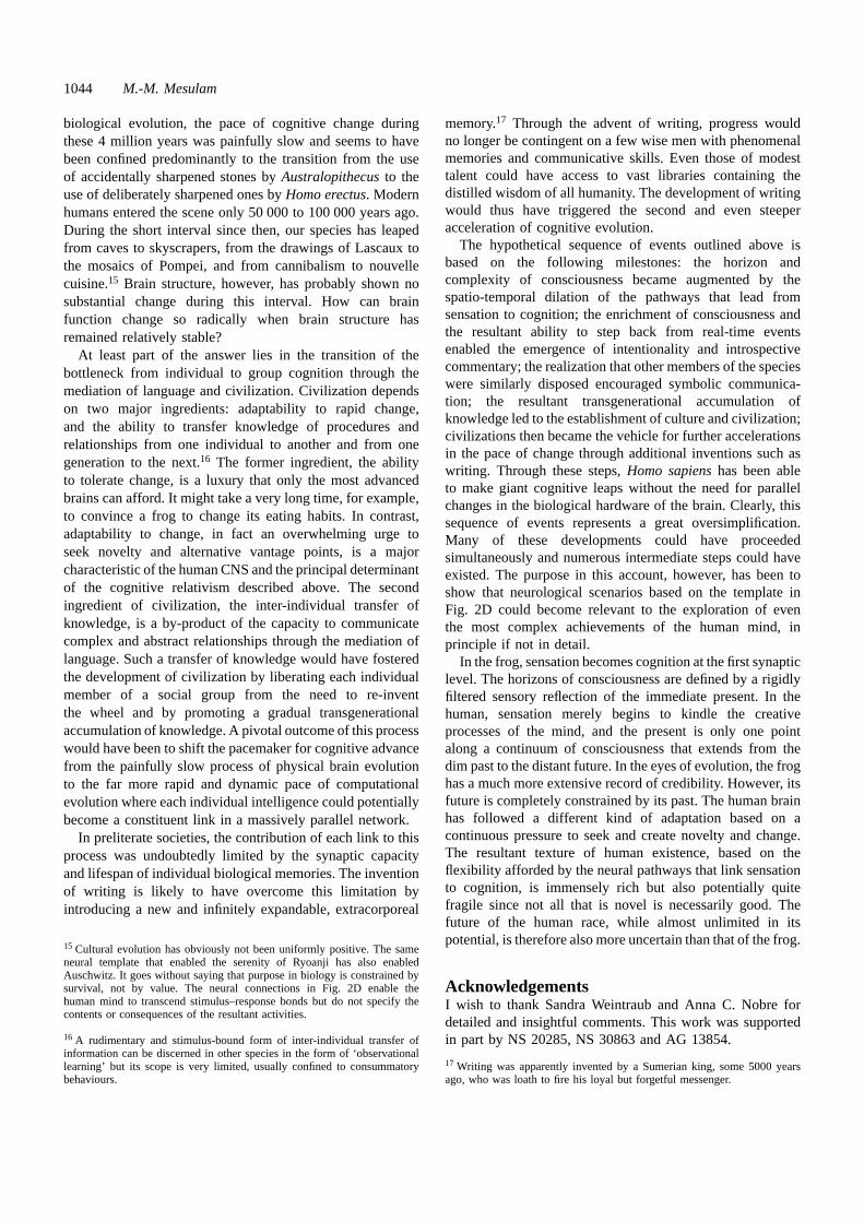
1044 M.-M. Mesulam
biological evolution, the pace of cognitive change duringthese 4 million years was painfully slow and seems to havebeen confined predominantly to the transition from the useof accidentally sharpened stones byAustralopithecusto theuse of deliberately sharpened ones byHomo erectus. Modernhumans entered the scene only 50 000 to 100 000 years ago.During the short interval since then, our species has leapedfrom caves to skyscrapers, from the drawings of Lascaux tothe mosaics of Pompei, and from cannibalism to nouvellecuisine.15 Brain structure, however, has probably shown nosubstantial change during this interval. How can brainfunction change so radically when brain structure hasremained relatively stable?
At least part of the answer lies in the transition of thebottleneck from individual to group cognition through themediation of language and civilization. Civilization dependson two major ingredients: adaptability to rapid change,and the ability to transfer knowledge of procedures andrelationships from one individual to another and from onegeneration to the next.16 The former ingredient, the abilityto tolerate change, is a luxury that only the most advancedbrains can afford. It might take a very long time, for example,to convince a frog to change its eating habits. In contrast,adaptability to change, in fact an overwhelming urge toseek novelty and alternative vantage points, is a majorcharacteristic of the human CNS and the principal determinantof the cognitive relativism described above. The secondingredient of civilization, the inter-individual transfer ofknowledge, is a by-product of the capacity to communicatecomplex and abstract relationships through the mediation oflanguage. Such a transfer of knowledge would have fosteredthe development of civilization by liberating each individualmember of a social group from the need to re-inventthe wheel and by promoting a gradual transgenerationalaccumulation of knowledge. A pivotal outcome of this processwould have been to shift the pacemaker for cognitive advancefrom the painfully slow process of physical brain evolutionto the far more rapid and dynamic pace of computationalevolution where each individual intelligence could potentiallybecome a constituent link in a massively parallel network.
In preliterate societies, the contribution of each link to thisprocess was undoubtedly limited by the synaptic capacityand lifespan of individual biological memories. The inventionof writing is likely to have overcome this limitation byintroducing a new and infinitely expandable, extracorporeal
15 Cultural evolution has obviously not been uniformly positive. The sameneural template that enabled the serenity of Ryoanji has also enabledAuschwitz. It goes without saying that purpose in biology is constrained bysurvival, not by value. The neural connections in Fig. 2D enable thehuman mind to transcend stimulus–response bonds but do not specify thecontents or consequences of the resultant activities.
16 A rudimentary and stimulus-bound form of inter-individual transfer ofinformation can be discerned in other species in the form of ‘observationallearning’ but its scope is very limited, usually confined to consummatorybehaviours.
memory.17 Through the advent of writing, progress wouldno longer be contingent on a few wise men with phenomenalmemories and communicative skills. Even those of modesttalent could have access to vast libraries containing thedistilled wisdom of all humanity. The development of writingwould thus have triggered the second and even steeperacceleration of cognitive evolution.
The hypothetical sequence of events outlined above isbased on the following milestones: the horizon andcomplexity of consciousness became augmented by thespatio-temporal dilation of the pathways that lead fromsensation to cognition; the enrichment of consciousness andthe resultant ability to step back from real-time eventsenabled the emergence of intentionality and introspectivecommentary; the realization that other members of the specieswere similarly disposed encouraged symbolic communica-tion; the resultant transgenerational accumulation ofknowledge led to the establishment of culture and civilization;civilizations then became the vehicle for further accelerationsin the pace of change through additional inventions such aswriting. Through these steps,Homo sapienshas been ableto make giant cognitive leaps without the need for parallelchanges in the biological hardware of the brain. Clearly, thissequence of events represents a great oversimplification.Many of these developments could have proceededsimultaneously and numerous intermediate steps could haveexisted. The purpose in this account, however, has been toshow that neurological scenarios based on the template inFig. 2D could become relevant to the exploration of eventhe most complex achievements of the human mind, inprinciple if not in detail.
In the frog, sensation becomes cognition at the first synapticlevel. The horizons of consciousness are defined by a rigidlyfiltered sensory reflection of the immediate present. In thehuman, sensation merely begins to kindle the creativeprocesses of the mind, and the present is only one pointalong a continuum of consciousness that extends from thedim past to the distant future. In the eyes of evolution, the froghas a much more extensive record of credibility. However, itsfuture is completely constrained by its past. The human brainhas followed a different kind of adaptation based on acontinuous pressure to seek and create novelty and change.The resultant texture of human existence, based on theflexibility afforded by the neural pathways that link sensationto cognition, is immensely rich but also potentially quitefragile since not all that is novel is necessarily good. Thefuture of the human race, while almost unlimited in itspotential, is therefore also more uncertain than that of the frog.
AcknowledgementsI wish to thank Sandra Weintraub and Anna C. Nobre fordetailed and insightful comments. This work was supportedin part by NS 20285, NS 30863 and AG 13854.
17 Writing was apparently invented by a Sumerian king, some 5000 yearsago, who was loath to fire his loyal but forgetful messenger.
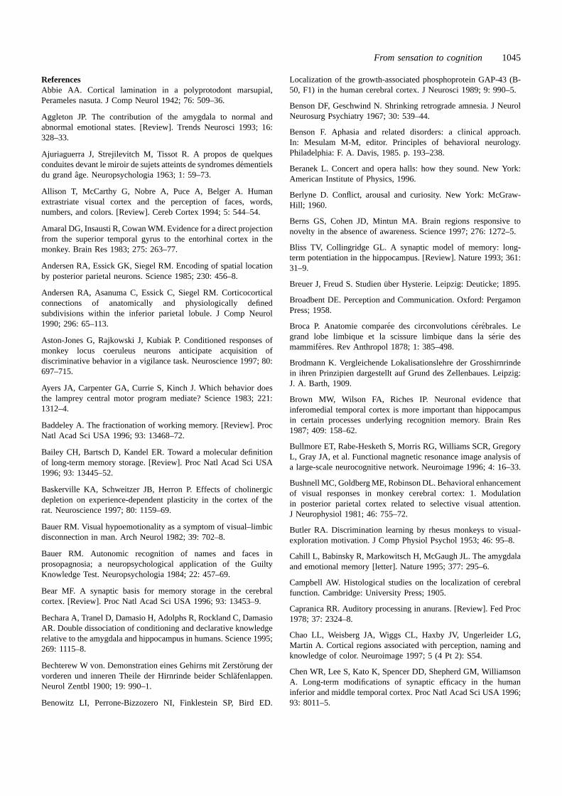
From sensation to cognition 1045
ReferencesAbbie AA. Cortical lamination in a polyprotodont marsupial,Perameles nasuta. J Comp Neurol 1942; 76: 509–36.
Aggleton JP. The contribution of the amygdala to normal andabnormal emotional states. [Review]. Trends Neurosci 1993; 16:328–33.
Ajuriaguerra J, Strejilevitch M, Tissot R. A propos de quelquesconduites devant le miroir de sujets atteints de syndromes de´mentielsdu grand aˆge. Neuropsychologia 1963; 1: 59–73.
Allison T, McCarthy G, Nobre A, Puce A, Belger A. Humanextrastriate visual cortex and the perception of faces, words,numbers, and colors. [Review]. Cereb Cortex 1994; 5: 544–54.
Amaral DG, Insausti R, Cowan WM. Evidence for a direct projectionfrom the superior temporal gyrus to the entorhinal cortex in themonkey. Brain Res 1983; 275: 263–77.
Andersen RA, Essick GK, Siegel RM. Encoding of spatial locationby posterior parietal neurons. Science 1985; 230: 456–8.
Andersen RA, Asanuma C, Essick C, Siegel RM. Corticocorticalconnections of anatomically and physiologically definedsubdivisions within the inferior parietal lobule. J Comp Neurol1990; 296: 65–113.
Aston-Jones G, Rajkowski J, Kubiak P. Conditioned responses ofmonkey locus coeruleus neurons anticipate acquisition ofdiscriminative behavior in a vigilance task. Neuroscience 1997; 80:697–715.
Ayers JA, Carpenter GA, Currie S, Kinch J. Which behavior doesthe lamprey central motor program mediate? Science 1983; 221:1312–4.
Baddeley A. The fractionation of working memory. [Review]. ProcNatl Acad Sci USA 1996; 93: 13468–72.
Bailey CH, Bartsch D, Kandel ER. Toward a molecular definitionof long-term memory storage. [Review]. Proc Natl Acad Sci USA1996; 93: 13445–52.
Baskerville KA, Schweitzer JB, Herron P. Effects of cholinergicdepletion on experience-dependent plasticity in the cortex of therat. Neuroscience 1997; 80: 1159–69.
Bauer RM. Visual hypoemotionality as a symptom of visual–limbicdisconnection in man. Arch Neurol 1982; 39: 702–8.
Bauer RM. Autonomic recognition of names and faces inprosopagnosia; a neuropsychological application of the GuiltyKnowledge Test. Neuropsychologia 1984; 22: 457–69.
Bear MF. A synaptic basis for memory storage in the cerebralcortex. [Review]. Proc Natl Acad Sci USA 1996; 93: 13453–9.
Bechara A, Tranel D, Damasio H, Adolphs R, Rockland C, DamasioAR. Double dissociation of conditioning and declarative knowledgerelative to the amygdala and hippocampus in humans. Science 1995;269: 1115–8.
Bechterew W von. Demonstration eines Gehirns mit Zersto¨rung dervorderen und inneren Theile der Hirnrinde beider Schla¨fenlappen.Neurol Zentbl 1900; 19: 990–1.
Benowitz LI, Perrone-Bizzozero NI, Finklestein SP, Bird ED.
Localization of the growth-associated phosphoprotein GAP-43 (B-50, F1) in the human cerebral cortex. J Neurosci 1989; 9: 990–5.
Benson DF, Geschwind N. Shrinking retrograde amnesia. J NeurolNeurosurg Psychiatry 1967; 30: 539–44.
Benson F. Aphasia and related disorders: a clinical approach.In: Mesulam M-M, editor. Principles of behavioral neurology.Philadelphia: F. A. Davis, 1985. p. 193–238.
Beranek L. Concert and opera halls: how they sound. New York:American Institute of Physics, 1996.
Berlyne D. Conflict, arousal and curiosity. New York: McGraw-Hill; 1960.
Berns GS, Cohen JD, Mintun MA. Brain regions responsive tonovelty in the absence of awareness. Science 1997; 276: 1272–5.
Bliss TV, Collingridge GL. A synaptic model of memory: long-term potentiation in the hippocampus. [Review]. Nature 1993; 361:31–9.
Breuer J, Freud S. Studien u¨ber Hysterie. Leipzig: Deuticke; 1895.
Broadbent DE. Perception and Communication. Oxford: PergamonPress; 1958.
Broca P. Anatomie compare´e des circonvolutions ce´rebrales. Legrand lobe limbique et la scissure limbique dans la se´rie desmammiferes. Rev Anthropol 1878; 1: 385–498.
Brodmann K. Vergleichende Lokalisationslehre der Grosshirnrindein ihren Prinzipien dargestellt auf Grund des Zellenbaues. Leipzig:J. A. Barth, 1909.
Brown MW, Wilson FA, Riches IP. Neuronal evidence thatinferomedial temporal cortex is more important than hippocampusin certain processes underlying recognition memory. Brain Res1987; 409: 158–62.
Bullmore ET, Rabe-Hesketh S, Morris RG, Williams SCR, GregoryL, Gray JA, et al. Functional magnetic resonance image analysis ofa large-scale neurocognitive network. Neuroimage 1996; 4: 16–33.
Bushnell MC, Goldberg ME, Robinson DL. Behavioral enhancementof visual responses in monkey cerebral cortex: 1. Modulationin posterior parietal cortex related to selective visual attention.J Neurophysiol 1981; 46: 755–72.
Butler RA. Discrimination learning by rhesus monkeys to visual-exploration motivation. J Comp Physiol Psychol 1953; 46: 95–8.
Cahill L, Babinsky R, Markowitsch H, McGaugh JL. The amygdalaand emotional memory [letter]. Nature 1995; 377: 295–6.
Campbell AW. Histological studies on the localization of cerebralfunction. Cambridge: University Press; 1905.
Capranica RR. Auditory processing in anurans. [Review]. Fed Proc1978; 37: 2324–8.
Chao LL, Weisberg JA, Wiggs CL, Haxby JV, Ungerleider LG,Martin A. Cortical regions associated with perception, naming andknowledge of color. Neuroimage 1997; 5 (4 Pt 2): S54.
Chen WR, Lee S, Kato K, Spencer DD, Shepherd GM, WilliamsonA. Long-term modifications of synaptic efficacy in the humaninferior and middle temporal cortex. Proc Natl Acad Sci USA 1996;93: 8011–5.

1046 M.-M. Mesulam
Claparede M. Recognition et moiite´. Archs Psychol (Gene`ve) 1911;11: 79–90.
Clark VP, Maisog JM, Haxby JV. FMRI studies of face memoryusing random stimulus sequences. Neuroimage (4 Pt 2) 1997; 5: S50.
Colombo M, Rodman HR, Gross CG. The effects of superiortemporal cortex lesions on the processing and retention of auditoryinformation in monkeys (Cebus apella). J Neurosci 1996; 16:4501–17.
Connor CE, Gallant JL, Preddie DC, Van Essen DC. Responses inarea V4 depend on the spatial relationship between stimulus andattention. J Neurophysiol 1996; 75: 1306–8.
Constantinidis C, Steinmetz MA. Neuronal activity in posteriorparietal area 7a during the delay periods of a spatial memory task.J Neurophysiol 1996; 76: 1352–5.
Corbetta M, Miezin FM, Dobmeyer S, Shulman GL, Petersen SE.Attentional modulation of neural processing of shape, color andvelocity in humans. Science 1990; 248: 1556–9.
Corbetta M, Miezin FM, Dobmeyer S, Shulman GL, Petersen SE.Selective and divided attention during visual discriminations ofshape, color, and speed: functional anatomy by positron emissiontomography. J Neurosci 1991; 11: 2383–402.
Creutzfeldt O, Ojemann G, Lettich E. Neuronal activity in thehuman lateral temporal lobe. I. Responses to speech. Exp Brain Res1989; 77: 451–75.
Creutzfeldt OD, Heggelund P. Neural plasticity in visual cortex ofadult cats after exposure to visual patterns. Science 1975; 188:1025–7.
Crick F, Koch C. Are we aware of neural activity in primary visualcortex? Nature 1995; 375: 121–3.
Cruikshank SJ, Weinberger NM. Evidence for the Hebbianhypothesis in experience-dependent physiological plasticity ofneocortex: a critical review. [Review]. Brain Res Rev 1996; 22:191–228.
Curran T, Schacter DL, Norman KA, Galluccio L. False recognitionafter a right frontal lobe infarction: memory for general and specificinformation. Neuropsychologia 1997; 35: 1035–49.
Daffner KR, Mesulam M-M, Scinto LFM, Cohen LG, Kennedy BF,West WC, et al. Control of novelty-seeking behavior by the frontallobes: an event-related potential study. Neuroreport 1998; 9: 787–91.
Damasio AR. Disorders of complex visual processing: agnosias,achromatopsia, Balint’s syndrome, and related difficulties oforientation and construction. In: Mesulam M-M, editor. Principlesof behavioral neurology. Philadelphia: F. A. Davis; 1985: 259–88.
Damasio AR. The brain binds entities and events by multiregionalactivation from convergence zones. Neurol Comp 1989; 1: 123–32.
Damasio H, Grabowski TJ, Tranel D, Hichwa RD, Damasio AR.A neural basis for lexical retrieval. Nature 1996; 380: 499–505.
Daniele A, Giustolisi L, Silveri MC, Colosimo C, Gainotti G.Evidence for a possible neuroanatomical basis for lexical processingof nouns and verbs. Neuropsychologia 1994; 32: 1325–41.
Delacour J. Neurobiology of consciousness: an overview. [Review].Behav Brain Res 1997; 85: 127–41.
Demonet J-F, Chollet F, Ramsay S, Cardebat D, Nespoulous J-L,Wise R, et al. The anatomy of phonological and semantic processingin normal subjects. Brain 1992; 115: 1753–68.
Dennett DC, Kinsbourne M. Time and the observer: the where andwhen of consciousness in the brain. Behav Brain Sci 1992; 15:183–247.
Desimone R. Face-selective cells in the temporal cortex of monkeys.J Cog Neurosci 1991; 3: 1–8.
Desimone R. Neural mechanisms for visual memory and their rolein attention. [Review]. Proc Natl Acad Sci USA 1996; 93: 13494–9.
Dias EC, Compaan DM, Mesulam M-M, Segraves MA. Selectivedisruption of memory-guided saccades with injection of a cholinergicantagonist in the frontal eye field of monkey [abstract]. Soc NeurosciAbstr 1996; 22: 418.
Dinse HR, Kruger K. The timing of processing along the visualpathway in the cat. Neuroreport 1994; 5: 893–7.
Donald M. Origins of the modern mind. Three stages in the evolutionof culture and cognition. Cambridge (MA): Harvard UniversityPress; 1991.
Downer CL de C. Interhemispheric integration in the visual system.In: Mountcastle VB, editor. Interhemispheric relations and cerebraldominance. Baltimore: Johns Hopkins University Press; 1962. p.87–100.
Economo C von, Koskinas GN. Die Cytoarchitektonik der Hirnrindedes erwachsenen Menschen. Wien: Springer; 1925.
Edelman S. Representation is representation of similarities. BehavBrain Sci. In press 1998.
Eichenbaum H, Schoenbaum G, Young B, Bunsey M. Functionalorganization of the hippocampal memory system. [Review]. ProcNatl Acad Sci USA 1996; 93: 13500–7.
Erickson RP. The across-fiber pattern theory: an organizing principlefor molar neural function. Contrib Sens Physiol 1982; 6: 79–110.
Exner S. Untersuchungen u¨ber die Localisation der Funktionen inder Grosshirnrinde des Menschen. Wien: W. Braumuller; 1881.
Fahy FL, Riches IP, Brown MW. Neuronal activity related to visualrecognition memory: long-term memory and the encoding of recencyand familiarity information in the primate anterior and medialinferior temporal and rhinal cortex. Exp Brain Res 1993; 96: 457–72.
Farah MJ, Feinberg TE. Visual object agnosia. In: Feinberg TE,Farah MJ, editor. Behavioral neurology and neuropsychology. NewYork: McGraw-Hill; 1997. p. 239–44.
Felleman DJ, Van Essen DC. Distributed hierarchical processing inthe primate cerebral cortex. [Review]. Cereb Cortex 1991; 1: 1–47.
Ferrier D. The functions of the brain. London: Smith, Elder; 1876.
Fiez JA, Raife EA, Balota DA, Schwarz JP, Raichle ME, PetersenSE. A positron emission tomography study of the short-termmaintenance of verbal information. J Neurosci 1996; 16: 808–22.
Filimonoff IN. A rational subdivision of the cerebral cortex. ArchNeurol Psychiatry 1947; 58: 296–311.
Fink GR, Markowitsch HJ, Reinkemeier M, Bruckbauer T, KesslerJ, Heiss W-D. Cerebral representation of one’s own past: neural
From sensation to cognition 1047
networks involved in autobiographical memory. J Neurosci 1996;16: 4275–82.
Flechsig PE. Anatomie des menschlichen Gehirns und Ru¨ckenmarks.Leipzig: Verlag von Georg Thieme; 1920.
Fodor JA. The modularity of mind: an essay on faculty psychology.Cambridge (MA): MIT Press; 1983.
Foote SL, Bloom FE, Aston-Jones G. Nucleus locus coeruleus: newevidence of anatomical and physiological specificity. [Review].Physiol Rev 1983; 63: 844–914.
Friston KJ. Imaging cognitive anatomy. Trends Cogn Sci 1997; 1:21–7.
Fujita I, Tanaka K, Ito M, Cheng K. Columns for visual featuresof objects in monkey inferotemporal cortex. Nature 1992; 360:343–6.
Fuster JM. Inferotemporal units in selective visual attention andshort-term memory. J Neurophysiol 1990; 64: 681–97.
Fuster JM, Bauer RH, Jervey JP. Functional interactions betweeninferotemporal and prefrontal cortex in a cognitive task. Brain Res1985; 330: 299–307.
Gabrieli JDE, Brewer JB, Desmond JE, Glover GH. Separate neuralbases of two fundamental memory processes in the human medialtemporal lobe. Science 1997; 276: 264–5.
Gaillard F. Visual units in the central nervous system of the frog.[Review]. Comp Biochem Physiol A 1990; 96: 357–71.
Gallup GG Jr, Wallnau LB, Suarez SD. Failure to find self-recognition in mother-infant and infant-infant rhesus monkey pairs.Folia Primatol (Basel) 1980; 33: 210–9.
Geisler WS, Albrecht DG. Bayesian analysis of identificationperformance in monkey visual cortex: nonlinear mechanisms andstimulus certainty. Vision Res 1995; 35: 2723–30.
Geschwind N. Disconnexion syndromes in animals and man. I.[Review]. Brain 1965; 88: 237–94.
Gitelman DR, Alpent NM, Kosslyn SM, Daffner K, Scinto L,Thompson W, et al. Functional imaging of human right hemisphericactivation for exploratory movements. Ann Neurol 1996; 39: 174–9.
Gloor P, Olivier A, Quesney LF, Andermann F, Horowitz S. Therole of the limbic system in experiential phenomena of temporallobe epilepsy. Ann Neurol 1982; 12: 129–44.
Goldman-Rakic PS. Topography of cognition: parallel distributednetworks in primate association cortex. [Review]. Annu RevNeurosci 1988; 11: 137–56.
Goldman-Rakic PS. Regional and cellular fractionation of workingmemory. [Review]. Proc Natl Acad Sci USA 1996; 93: 13473–80.
Gorlick DL, Constantine-Paton M, Kelley DB. A14C-2-deoxyglucose autoradiographic investigation of sensory inputs tothe optic tectum of Rana pipiens. J Comp Physiol A 1984; 154:617–24.
Gorno Tempini ML, Price C, Vandenberghe R, Josephs O, KapurN, Cappa SF, et al. Functional anatomy of a common semanticsystem for names and faces. Neuroimage 1997; 5 (4 Pt 2): S560.
Gross CG, Rocha-Miranda CE, Bender DB. Visual properties of
neurons in inferotemporal cortex of the macaque. J Neurophysiol1972; 35: 96–111.
Halgren E, Dale AM, Sereno MI, Tootell RBH, Marinkovic K,Rosen BR. Location of fMRI responses to faces anterior toretinotopic cortex. Neuroimage 1997; 5 (4 Pt 2): S150.
Halgren E, Wilson CL, Stapleton JM. Human medial temporal-lobestimulation disrupts both formation and retrieval of recent memories.Brain Cogn 1985; 4: 287–95.
Hammond EJ, Meador KJ, Aung-Din R, Wilder BJ. Cholinergicmodulation of human P3 event-related potentials. Neurology 1987;37: 346–50.
Harless M. Social behavior. In: Harless M, Morlock H, editors.Turtles. Perspective and research. New York: John Wiley; 1979:475–92.
Harries MH, Perrett DI. Visual processing of faces in temporalcortex: physiological evidence for a modular organization andpossible anatomical correlates. J Cogn Neurosci 1991; 3: 9–24.
Harris-Warrick RM, Marder E. Modulation of neural networks forbehavior. [Review]. Annu Rev Neurosci 1991; 14: 39–57.
Hauser MD, Kralik J, Botto-Mahan C, Garrett M, Oser J. Self-recognition in primates: phylogeny and the salience of species-typical features. Proc Natl Acad Sci USA 1995; 92: 10811–4.
Haxby JV, Clark VP, Courtney M. Distributed hierarchical neuralsystems for visual memory in human cortex. In: Hyman BT,Duyckaerts C, Christen Y, editors. Connections, cognition andAlzheimer’s disease. Berlin: Springer Verlag; 1997a. p. 167–80.
Haxby JV, Martin A, Clark VP, Hoffman EA, Schouten JL,Ungerleider LG. The processing of faces, inverted faces and otherobjects in the ventral object vision pathway. Neuroimage 1997b; 5(4 Pt 2): S4.
Haxby JV, Horwitz B, Ungerleider LG, Maisong JM, Pietrini P,Grady CL. The functional organization of human extrastriate cortex:a PET–rCBF study of selective attention to faces and locations.J Neurosci 1994; 14: 6336–53.
Heffner HE, Heffner RS. Temporal lobe lesions and perception ofspecies-specific vocalizations by macaques. Science 1984; 226:75–6.
Heron W. The pathology of boredom. Scient Am 1957; 196: 52–6.
Higuchi S-I, Miyashita Y. Formation of mnemonic neuronalresponses to visual paired associates in inferotemporal cortex isimpaired by perirhinal and entorhinal lesions. Proc Natl Acad SciUSA 1996; 93: 739–43.
Horel JA. Cold lesions in inferotemporal cortex produce reversibledeficits in learning and retention of visual discriminations. PhysiolPsychol 1984; 12: 259–70.
Hubel DH, Henson CO, Rupert A, Galambos R. ‘Attention’ unitsin the auditory cortex. Science 1959; 129: 1279–80.
Hubel DH, Wiesel TN. Receptive fields and functional architecturein two non-striate visual areas (18 and 19) of the cat. J Neurophysiol1965; 28: 229–89.
Huerta PT, Lisman JE. Heightened synaptic plasticity of
1048 M.-M. Mesulam
hippocampal CA1 neurons during a cholinergically induced rhythmicstate. Nature 1993; 364: 723–5.
Ingle D. Sensorimotor function of the midbrain tectum. II. Classesof visually guided behavior. Neurosci Res Program Bull 1975; 13:180–5.
Ishai A, Ungerleider LG, Martin A, Maisog JM, Haxby JV. fMRIreveals differential activation in the ventral object vision pathwayduring the perception of faces, houses, and chairs. Neuroimage1997; 5 (4 Pt 2): S149.
Jones EG, Powell TP. An anatomical study of converging sensorypathways within the cerebral cortex of the monkey. Brain 1970; 93:793–820.
Just MA, Carpenter PA, Keller TA, Eddy WF, Thulborn KR. Brainactivation modulated by sentence comprehension. Science 1996;274: 114–6.
Kanwisher N, McDermott J, Chun MM. The fusiform face area: amodule in human extrastriate cortex specialized for face perception.J Neurosci 1997a; 17: 4302–11.
Kanwisher N, Woods RP, Iacobini M, Mazziotta JC. A locus inhuman extrastriate cortex for visual shape analysis. J Cogn Neurosci1997b; 9: 133–42.
Kapur S, Tulving E, Cabeza R, McIntosh AR, Houle S, Craik FI.The neural correlates of intentional learning of verbal materials: aPET study in humans. Brain Res Cogn Brain Res 1996; 4: 243–9.
Knight RT. Decreased response to novel stimuli after prefrontallesions in man. Electroencephalogr Clin Neurophysiol 1984; 59:9–20.
Kosslyn SM, Alpert NM, Thompson WL, Maljkovic V, Weise SB,Chabris CF, et al. Visual mental imagery activates topographicallyorganized visual cortex: PET investigations. J Cogn Neurosci 1993;5: 263–87.
Kosslyn SM, Alpert NM, Thompson WM, Chabris CF, Rauch SL,Anderson AK. Identifying objects seen from different viewpoints.A PET investigation. Brain 1994; 117: 1055–71.
Lashley KS. Brain mechanisms and intelligence: a quantitativestudy of injuries to the brain. Chicago: University of ChicagoPress; 1929.
Lashley KS. Functional interpretation of anatomic patterns. ResPub Assoc Res Nerv Ment Dis 1952; 30: 529–47.
LeDoux JE, Thompson ME, Iadecola C, Tucker LW, Reis DJ. Localcerebral blood flow increases during auditory and emotionalprocessing in the conscious rat. Science 1983; 221: 576–8.
Leimkuhler ME, Mesulam MM. Reversible go-no go deficits in acase of frontal lobe tumor. Ann Neurol 1985; 18: 617–9.
Leinonen L, Hyvarinen J, Sovijarvi AR. Functional properties ofneurons in the temporo-parietal association cortex of awake monkey.Exp Brain Res 1980; 39: 203–15.
Lettvin JY, Maturana HR, McCulloch WS, Pitts WH. What thefrog’s eye tells the frog’s brain. Proc Inst Radio Engin 1959; 47:1940–51.
Lettvin JY, Maturana HR, Pitts WH, McCulloch WS. Two remarks
on the visual system of the frog. In: Rosenblith WV, editor. Sensorycommunication. Cambridge (MA): MIT Press; 1961. p. 757–76.
Levelt WJ. Accessing words in speech production: stages, processand representations. [Review]. Cognition 1992; 42: 1–22.
Levin BE, Craik RL, Hand PJ. The role of norepinephrine in adultrat somatosensory (SmI) cortical metabolism and plasticity. BrainRes 1988; 443: 261–71.
Libet B. Brain stimulation and the threshold of conscious experience.In: Eccles JC, editor. Brain and conscious experience. New York:Springer Verlag; 1966. p. 165–81.
Libet B. Brain stimulation in the study of neuronal functions forconscious sensory experiences. Hum Neurobiol 1982; 1: 235–42.
Lowel S, Singer W. Selection of intrinsic horizontal connections inthe visual cortex by correlated neuronal activity. Science 1992; 255:209–12.
Lueck CJ, Zeki S, Friston KJ, Deiber M-P, Cope P, CunninghamVJ, et al. The colour centre in the cerebral cortex of man. Nature1989; 340: 386–9.
MacLean PD. The co-evolution of the brain and the family.Anthroquest 1982; 24: 1, 14–5.
Martin A, Haxby JV, Lalonde FM, Wiggs CL, Ungerleider LG.Discrete cortical regions associated with knowledge of color andknowledge of action. Science 1995; 270: 102–5.
Martin A, Wiggs CL, Ungerleider LG, Haxby JV. Neural correlatesof category-specific knowledge. Nature 1996; 379: 649–52.
Maunsell JH. The brain’s visual world: representation of visualtargets in cerebral cortex. [Review]. Science 1995; 270: 764–9.
McCarthy G, Puce A, Constable RT, Krystal JH, Gore JC, Goldman-Rakic P. Activation of human prefrontal cortex during spatial andnonspatial working memory tasks measured by functional MRI.Cereb Cortex 1996; 6: 600–11.
McClelland JL. The organization of memory. A parallel distributedprocessing perspective. Rev Neurol (Paris) 1994; 150: 570–9.
McClelland JL. Constructive memory and memory distortions: aparallel-distributed processing approach. In: Schacter DL, Coyle JT,Fischbach GD, Mesulam M-M, Sullivan LE, editors. Memorydistortion. Cambridge (MA): Harvard University Press; 1995. p.69–90.
McCormick DA. Cellular mechanisms of cholinergic control ofneocortical and thalamic neuronal excitability. In: Steriade M,Biesold D, editors. Brain cholinergic systems. Oxford: OxfordUniversity Press; 1990: 236–64.
McGaugh JL, Introini-Collison IB, Cahill LF, Castellano C, DalmazC, Parent MB, et al. Neuromodulatory systems and memory storage:role of the amygdala. [Review]. Behav Brain Res 1993; 58: 81–90.
McIntosh AR, Grady CL, Ungerleider LG, Haxby JV, Rapoport SI,Horwitz B. Network analysis of cortical visual pathways mappedwith PET. J Neurosci 1994; 14: 655–66.
Mesulam M-M. A cortical network for directed attention andunilateral neglect. [Review]. Ann Neurol 1981; 10: 309–25.
Mesulam M-M. Attention, confusional states and neglect. In:
From sensation to cognition 1049
Mesulam M-M, editor. Principles of behavioral neurology.Philadelphia: F. A. Davis; 1985a. p. 125–68.
Mesulam M-M. Patterns in behavioral neuroanatomy. Associationareas, the limbic system, and hemispheric specialization. In:Mesulam M-M, editor. Principles of behavioral neurology.Philadelphia: F. A. Davis; 1985b. p. 1–70.
Mesulam M-M. Frontal cortex and behavior. Ann Neurol 1986; 19:320–5.
Mesulam M-M. Asymmetry of neural feedback in the organizationof behavioral states. Science 1987; 237: 537–8.
Mesulam M-M. Neural substrates of behavior: the effects of brainlesions upon mental state. In: Nicholi AM Jr, editor. The newHarvard guide to psychiatry. Cambridge (MA): Harvard UniversityPress; 1988. p. 91–128.
Mesulam M-M. Large-scale neurocognitive networks and distributedprocessing for attention, language, and memory. [Review]. AnnNeurol 1990; 28: 597–613.
Mesulam M-M. Higher visual functions of the cerebral cortex andtheir disruption in clinical practice. In: Albert DM, Jakobiec FA,editors. Principles and practice of ophthalmology. Philadelphia:Saunders; 1994a. p. 2640–2653.
Mesulam M-M. The multiplicity of neglect phenomena.Neuropsychol Rehabil 1994b; 4: 173–6.
Mesulam M-M. Neurocognitive networks and selectively distributedprocessing. Rev Neurol (Paris) 1994c; 150: 564–9.
Mesulam M-M. Cholinergic pathways and the ascending reticularactivating system of the human brain. Ann N Y Acad Sci 1995a;757: 169–79.
Mesulam M-M. Notes on the cerebral topography of memory andmemory distortion: a neurologist’s perspective. In: Schacter DL,Coyle JT, Fischbach GD, Mesulam M-M, Sullivan LE, editors.Memory distortion. Cambridge (MA): Harvard University Press;1995b: 379–85.
Mesulam M-M. The systems-level organization of cholinergicinnervation in the human cerebral cortex and its alterations inAlzheimer’s disease. [Review]. Prog Brain Res 1996; 109: 285–98.
Mesulam M-M, Mufson EJ. The insula of Reil in man and monkey.In: Peters A, Jones EG, editors. Cerebral cortex, Vol. 4. New York:Plenum Press; 1985. p. 179–226.
Mesulam MM, Van Hoesen GW, Pandya DN, Geschwind N. Limbicand sensory connections of the inferior parietal lobule (area PG) inthe rhesus monkey: a study with a new method for horseradishperoxidase histochemistry. Brain Res 1977; 136: 393–414.
Milner B, Corkin S, Teuber HL. Further analysis of the hippocampalamnesic syndrome: 14-year follow-up study of H.M.Neuropsychologia 1968; 6: 215–34.
Mishkin M. A memory system in the monkey. Phil Trans R SocLond B 1982; 298: 85–95.
Mongeau R, Blier P, de Montigny C. The serotonergic andnoradrenergic systems of the hippocampus: their interactions andthe effects of antidepressant treatments. [Review]. Brain Res BrainRes Rev 1997; 23: 145–95.
Moran J, Desimone R. Selective attention gates visual processingin the extrastriate cortex. Science 1985; 229: 782–4.
Moran MA, Mufson EJ, Mesulam MM. Neural inputs into thetemporopolar cortex of the rhesus monkey. J Comp Neurol 1987;256: 88–103.
Morecraft RJ, Geula C, Mesulam MM. Cytoarchitecture and neuralafferents of orbitofrontal cortex in the brain of the monkey. J CompNeurol 1992; 323: 341–58.
Morecraft RJ, Geula C, Mesulam M-M. Architecture of connectivitywithin a cingulo-fronto-parietal neurocognitive network for directedattention. Arch Neurol 1993; 50: 279–84.
Morton J. Interaction of information in word recognition. PsycholRev 1969; 76: 165–78.
Moscovitch M. Multiple dissociations of function in amnesia. In:Cermak LS, editor. Human memory and amnesia. Hillsdale (NJ):Lawrence Erlbaum; 1982. p. 337–70.
Moscovitch M. Confabulation. In: Schacter DL, Coyle JT, FischbachGD, Mesulam M-M, Sullivan LE, editors. Memory distortion.Cambridge (MA): Harvard University Press; 1995. p. 226–51.
Nadel L, Moscovitch M. Memory consolidation, retrograde amnesiaand the hippocampal complex. [Review]. Curr Opin Neurobiol1997; 7: 217–27.
Naya Y, Sakai K, Miyashita Y. Activity of primate inferotemporalneurons related to sought target in pair-association task. Proc NatlAcad Sci USA 1996; 93: 2664–9.
Nobre AC, McCarthy G. Language-related ERPs: scalp distributionsand modulation by word type and semantic priming. J Cogn Neurosci1994; 6: 233–55.
Nobre AC, Plunkett K. The neural system of language: structureand development. [Review]. Curr Opin Neurobiol 1997; 7: 262–8.
Nobre AC, Allison T, McCarthy G. Word recognition in the humaninferior temporal lobe. Nature 1994; 372: 260–3.
Nobre AC, Sebestyen GN, Gitelman DR, Mesulam M-M,Frackowiak RS, Frith CD. Functional localization of the system forvisuospatial attention using positron emission tomography. Brain1997; 120: 515–33.
Northcutt RG. Forebrain and midbrain organization in lizards andits phylogenetic significance. In: Greenberg N, MacLean PD, editors.Behavior and neurology of lizards. Rockville (MD): NationalInstitute of Mental Health; 1978. p. 11–64.
O’Connor M, Butters N, Miliotis P, Eslinger P, Cermak LS. Thedissociation of anterograde and retrograde amnesia in a patient withherpes encephalitis. J Clin Exp Neuropsychol 1992; 14: 159–78.
Ojemann GA. Brain organization for language from the perspectiveof electrical stimulation mapping. Behav Brain Sci 1983; 6: 189–230.
Ojemann GA, Creutzfeldt O, Lettich E, Haglund MM. Neuronalactivity in human lateral temporal cortex related to short-term verbalmemory, naming and reading. Brain 1988; 111: 1383–403.
Owen AM, Milner B, Petrides M, Evans AC. Memory for objectfeatures versus memory for object location: a positron-emissiontomography study of encoding and retrieval processes. Proc NatlAcad Sci USA 1996; 93: 9212–7.
1050 M.-M. Mesulam
Pakkenberg B, Gundersen HJG. Neocortical neuron number inhumans: effect of sex and age. J Comp Neurol 1997; 384: 312–20.
Pandya DN, Kuypers HG. Cortico-cortical connections in the rhesusmonkey. Brain Res 1969; 13: 13–36.
Pandya DN, Seltzer B. Association areas of the cerebral cortex.Trends Neurosci 1982; 5: 386–90.
Pandya DN, Yeterian EH. Architecture and connections of corticalassociation areas. In: Peters A, Jones EG, editors. Cerebral cortex.New York: Plenum Press; 1985. p. 3–61.
Pandya DN, Van Hoesen GW, Mesulam MM. Efferent connectionsof the cingulate gyrus in the rhesus monkey. Exp Brain Res 1981;42: 319–30.
Penfield W. The cerebral cortex in man. Arch Neurol Psychiatry1938; 40: 417–42.
Penfield W. The mystery of the mind. Princeton, New Jersey:Princeton University Press; 1975.
Petersen SE, Fox PT, Posner MI, Mintun M, Raichle ME. Positronemission tomographic studies of the cortical anatomy of single-word processing. Nature 1988; 331: 585–9.
Petersson KM, Elfgren C, Ingvar M. A dynamic role of the medialtemporal lobe during retrieval of declarative memory in man.Neuroimage 1997; 6: 1–11.
Petrides M, Alivisatos B, Meyer E, Evans AC. Functional activationof the human frontal cortex during the performance of verbalworking memory tasks. Proc Natl Acad Sci USA 1993; 90: 878–82.
Pich EM, Pagliusi SR, Tessari M, Talabot-Ayer D, Hooft vanHuijsduijnen R, Chiamulera C. Common neural substrates for theaddictive properties of nicotine and cocaine. Science 1997; 275:83–6.
Puce A, Allison T, Asgari M, Gore JC, McCarthy G. Differentialsensitivity of human visual cortex to faces, letterstrings, and textures:a functional magnetic resonance imaging study. J Neurosci 1996;16: 5205–15.
Raiguel SE, Lagae L, Gulyas B, Orban GA. Response latencies ofvisual cells in macaque areas V1, V2 and V5. Brain Res 1989; 493:155–9.
Rao SC, Rainer G, Miller EK. Integration of what and where in theprimate prefrontal cortex. Science 1997; 276: 821–4.
Rauschecker JP, Tian B, Pons T, Mishkin M. Serial and parallelprocessing in rhesus monkey auditory cortex. J Comp Neurol 1997;382: 89–103.
Risberg J, Ingvar DH. Patterns of activation in the grey matter ofthe dominant hemisphere during memorizing and reasoning. A studyof regional cerebral blood flow changes during psychological testingin a group of neurologically normal patients. Brain 1973; 96: 737–56.
Roland PE, Friberg L. Localization of cortical areas activated bythinking. J Neurophysiol 1985; 53: 1219–43.
Roland PE, Gulya´s B. Visual imagery and visual representation [seecomments]. Trends Neurosci 1994; 17: 281–7. Comment in: TrendsNeurosci 1994; 17: 287–94, Comment in: Trends Neurosci 1994;17: 514–6.
Rolls ET. Information representation, processing, and storage in thebrain: analysis at the single neuron level. In: Changeux J-P, KonishiM, editors. The neural and molecular bases of learning. Chichester:John Wiley; 1987. p. 503–39.
Rolls ET, Baylis CG, Hasselmo ME, Nalwa V. The effect of learningon the face selective responses of neurons in the cortex in thesuperior temporal sulcus of the monkey. Exp Brain Res 1989; 76:153–64.
Rumelhart DE, McClelland JL. Parallel distributed processing.Cambridge (MA): MIT Press; 1986.
Rumsey JM, Horwitz B, Donohue BC, Nace K, Maisog JM,Andreason P. Phonological and orthographic components of wordrecognition A PET–rCBF study. Brain 1997; 120: 739–59.
Russek M. Conditional avoidance in frogs and salamanders. BolEstud Med Biol 1969; 26: 121–8.
Sakagami M, Niki H. Encoding of behavioral significance of visualstimuli by primate prefrontal neurons: relation to relevant taskconditions. Exp Brain Res 1994; 97: 423–36.
Sakai K, Miyashita Y. Visual imagery: an interaction betweenmemory retrieval and focal attention [Review]. Trends Neurosci1994; 17: 287–9.
Sanides F. Functional architecture of motor and sensory cortices inprimates in the light of a new concept of neocortex evolution. In:Noback CR, Montagna W, editors. The primate brain. New York:Appleton-Century-Crofts; 1970. p. 137–208.
Sato H, Hata Y, Hagihara K, Tsumoto T. Effects of cholinergicdepletion on neuron activities in the cat visual cortex. J Neurophysiol1987; 58: 781–94.
Schacter DL. Implicit memory: a new frontier for cognitiveneuroscience. In: Gazzaniga MS, editor. The cognitiveneurosciences. Cambridge (MA): MIT Press; 1995. p. 815–24.
Schiller P. Effect of lesions in visual cortical area V4 on therecognition of transformed objects. Nature 1995; 376: 342–4.
Schleidt WM, Schleidt M, Magg M. Sto¨rung der Mutter-Kind-Beziehung bei Truthu¨hnern durch Geho¨rverlust. Behaviour 1960;16: 254–60.
Schultz RT, Gauthier I, Fulbright R, Anderson AW, Lacadie C,Skudlarski P, et al. Are face identity and emotion processedautomatically? Neuroimage 1997; 5 (4 Pt 2): S148.
Schultz W. Dopamine neurons and their role in reward mechanisms.[Review]. Curr Opin Neurobiol 1997; 7: 191–7.
Scoville WB, Milner B. Loss of recent memory after bilateralhippocampal lesions. J Neurol Neurosurg Psychiatry 1957; 20:11–21.
Seeck M, Schomer D, Mainwaring N, Ives J, Dubuissson D,Blume H, et al. Selectively distributed processing of visual objectrecognition in the temporal and frontal lobes of the human brain.Ann Neurol 1995; 37: 538–45.
Seeck M, Mainwaring N, Cosgrove R, Blume H, Dubuisson D,Mesulam M-M, Schomer DL. Neurophysiologic correlates ofimplicit face memory in intracranial visual evoked potentials.Neurology 1997; 49: 1312–16.
From sensation to cognition 1051
Selemon LD, Goldman-Rakic PS. Common cortical and subcorticaltargets of the dorsolateral prefrontal and posterior parietal corticesin the rhesus monkey: evidence for a distributed neural networksubserving spatially guided behavior. J Neurosci 1988; 8: 4049–68.
Selman RL. The relation of role taking to the development of moraljudgment in children. Child Dev 1971; 42: 79–91.
Sergent J, Ohta S, MacDonald B. Functional neuroanatomy of faceand object processing: a positron emission tomography study. Brain1992; 115: 15–36.
Signoret J-L. Memory and amnesias. In: Mesulam M-M, editor.Principles of behavioral neurology. Philadelphia: F. A. Davis; 1985.p. 169–92.
Smiley JF, Goldman-Rakic PS. Serotonergic axons in monkeyprefrontal cerebral cortex synapse predominantly on interneuronsas demonstrated by serial section electron microscopy. J CompNeurol 1996; 367: 431–43.
Smiley JF, Morrell F, Mesulam M-M. Cholinergic synapses inhuman cerebral cortex: an ultrastructural study in serial sections.Exp Neurol 1997; 144: 361–8.
Sobotka S, Ringo JL. Investigation of long term recognition andassociation memory in unit responses from inferotemporal cortex.Exp Brain Res 1993; 96: 28–38.
Sperry RW. Physiological plasticity and brain circuit theory. In:Harlow HF, Woolsey CN, editor. Biological and biochemical basesof behavior. Madison: University of Wisconsin Press; 1965. p.401–24.
Spreen O, Benton AL, Fincham RW. Auditory agnosia withoutaphasia. Arch Neurol 1965; 13: 84–92.
Squire LR, Zola SM. Structure and function of declarative andnondeclarative memory systems. [Review]. Proc Natl Acad SciUSA 1996; 93: 13515–22.
Squire LR, Ojemann JG, Miezin FM, Petersen SE, Videen TO,Raichle ME. Activation of the hippocampus in normal humans: afunctional anatomical study of memory. Proc Natl Acad Sci USA1992; 89: 1837–41.
Swan GE, Carmelli D. Curiosity and mortality in aging adults: a 5-year follow-up of the Western Collaborative Group Study. PsycholAging 1996; 11: 449–53.
Tanaka K. Inferotemporal cortex and object vision. [Review]. AnnuRev Neurosci 1996; 19: 109–39.
Tinbergen N. The Study of Instinct. Oxford: Clarendon Press; 1951.
Tovee MJ. Neuronal processing. How fast is the speed of thought?Curr Biol 1994; 4: 1125–7.
Tranel D, Damasio AR. Autonomic recognition of familiar facesby prosopagnosics: evidence for knowledge without awareness[abstract]. Neurology 1985; 35 Suppl 1: 119–20.
Tranel D, Damasio H, Damasio AP. A neural basis for the retrievalof conceptual knowledge. Neuropsychologia 1997; 35: 1319–27.
Tulving E, Kapur S, Craik FI, Moskovitch M, Houle S. Hemisphericencoding/retrieval asymmetry in episodic memory: positronemission tomography findings. [Review]. Proc Natl Acad Sci USA1994; 91: 2016–20.
Ungerleider LG, Mishkin M. Two cortical visual systems. In: IngleDJ, Mansfield RJW, Goodale MD, editors. The analysis of visualbehavior. Cambridge (MA): MIT Press; 1982. p. 549–86.
Vaadia E, Haalman I, Abeles M, Bergman H, Prut Y, Slovin H,et al. Dynamics of neuronal interactions in monkey cortex in relationto behavioural events. Nature 1995; 373: 515–8.
Van Essen DC. Functional organization of primate visual cortex.In: Peters A, Jones EG, editors. Cerebral cortex, Vol. 3. New York:Plenum Press; 1985. p. 259–329.
Van Hoesen GW, Pandya DN, Butters N. Cortical afferents to theentorhinal cortex of the rhesus monkey. Science 1972; 175: 1471–3.
Vandenberghe R, Price C, Wise R, Josephs O, Frackowiak RS.Functional anatomy of a common semantic system for words andpictures. Nature 1996; 383: 254–6.
Vincent DJ, Dubno JR, Horwitz AR, George MS. An auditory fMRIstudy comparing passive and active listening to speech. Neuroimage1997; 5 (4 Pt 2): S189.
Vogt C, Vogt O. Allgemeinere Ergebnisse unserer Hirnforschung.J Psychol Neurol 1919; 25: 279–461.
Vygotsky LS. Thought and language. Cambridge (MA): MITPress; 1962.
Warrington EK, Weiskrantz L. The effect of prior learning onsubsequent retention in amnesic patients. Neuropsychologia 1974;12: 419–28.
Watanabe M. Reward expectancy in primate prefrontal neurons.Nature 1996; 382: 629–32.
Watson JD, Myers R, Frackowiak RS, Hajnal JV, Woods RP,Mazziotta JC, et al. Area V5 of the human brain: evidence from acombined study using positron emission tomography and magneticresonance imaging. Cereb Cortex 1993; 3: 79–94.
Weiskrantz L, Warrington EK, Sanders MD, Marshall J. Visualcapacity in the hemianopic field following a restricted occipitalablation. Brain 1974; 97: 709–28.
Wiesel TN. The postnatal development of the visual cortex and theinfluence of the environment. Stockholm: Nobel Foundation; 1981.
Wilson FA, Rolls ET. Neuronal responses related to novelty andfamiliarity of visual stimuli in the substantia innominata, diagonalband of Broca and periventricular region of the primate basalforebrain. Exp Brain Res 1990; 80: 104–20.
Wilson FA, Rolls ET. The effects of stimulus novelty and familiarityon neuronal activity in the amygdala of monkeys performingrecognition memory tasks. Exp Brain Res 1993; 93: 367–82.
Wilson FA, Scalaidhe SP, Goldman-Rakic PS. Dissociation of objectand spatial processing domains in primate prefrontal cortex. Science1993; 260: 1955–8.
Woolf NJ. Global and serial neurons form a hierarchically arrangedinterface proposed to underlie memory and cognition. [Review].Neuroscience 1996; 74: 625–51.
Wu JC, Buchsbaum MS, Hershey TG, Hazlett E, Sicotte N, JohnsonJC. PET in generalized anxiety disorder. Biol Psychiatry 1991; 29:1181–99.
Yaginuma S, Niihara T, Iwai E. Further evidence on elevated
1052 M.-M. Mesulam
discrimination limens for reduced patterns in monkeys withinferotemporal lesions. Neuropsychologia 1982; 20: 21–32.
Yakovlev PI. Pathoarchitectonic studies of cerebral malformations.III. Arrhinencephalies (Holotelencephalies). J Neuropathol ExpNeurol 1959; 18: 22–55.
Yamane S, Kaji S, Kawano K. What facial features activate faceneurons in the inferotemporal cortex of the monkey? Exp BrainRes 1988; 73: 209–14.
Yeterian EH, Van Hoesen GW. Cortico-striate projections in therhesus monkey: the organization of certain cortico-caudateconnections. Brain Res 1978; 139: 43–63.
Young MP, Yamane S. Sparse population coding of faces in theinferotemporal cortex. Science 1992; 256: 1327–31.
Zatorre RJ, Evans AC, Meyer E. Neural mechanisms underlyingmelodic perception and memory for pitch. J Neurosci 1994; 14:1908–19.
Zeki S. Cerebral akinetopsia (visual motion blindness). A review.[Review]. Brain 1991; 114: 811–24.
Zihl J, von Cramon D, Mai N, Schmid C. Disturbance of movementvision after bilateral posterior brain damage. Further evidence andfollow up observations. Brain 1991; 114: 2235–52.
Zipser D, Andersen RA. A back-propagation programmed networkthat simulates response properties of a subset of posterior parietalneurons. Nature 1988; 331: 679–84.
Received October 28, 1997. Accepted January 12 1997.