Embed Size (px)
Citation preview
Molecular Biology of the CellVol. 16, 2028–2038, April 2005
From Sorting Endosomes to Exocytosis: Association ofRab4 and Rab11 GTPases with the Fc Receptor, FcRn,during Recycling□V
E. Sally Ward,* Cruz Martinez,* Carlos Vaccaro,* Jinchun Zhou,* Qing Tang,* andRaimund J. Ober*†
*Center for Immunology, University of Texas Southwestern Medical Center, Dallas, TX 75390-8576; and†Department of Electrical Engineering, University of Texas at Dallas, Richardson, TX 75080
Submitted August 24, 2004; Revised January 4, 2005; Accepted January 20, 2005Monitoring Editor: Suzanne Pfeffer
A longstanding question in cell biology is how is the routing of intracellular organelles within cells regulated? Althoughdata support the involvement of Rab4 and Rab11 GTPases in the recycling pathway, the function of Rab11 in particularis uncertain. Here we have analyzed the association of these two Rab GTPases with the Fc receptor, FcRn, duringintracellular trafficking. This Fc receptor is both functionally and structurally distinct from the classical Fc� receptors andtransports immunoglobulin G (IgG) within cells. FcRn is therefore a recycling receptor that sorts bound IgG fromunbound IgG in sorting endosomes. In the current study we have used dual color total internal reflection fluorescencemicroscopy (TIRFM) and wide-field imaging of live cells to analyze the events in human endothelial cells that areinvolved in the trafficking of FcRn positive (FcRn�) recycling compartments from sorting endosomes to exocytic sites atthe plasma membrane. Our data are consistent with the following model for this pathway: FcRn leaves sorting endosomesin Rab4�Rab11� or Rab11� compartments. For Rab4�Rab11� compartments, Rab4 depletion occurs by segregation of thetwo Rab proteins into discrete domains that can separate. The Rab11�FcRn� vesicle or tubule subsequently fuses with theplasma membrane in an exocytic event. In contrast to Rab11, Rab4 is not involved in exocytosis.
INTRODUCTION
Trafficking of proteins and other cellular contents in theendocytic and exocytic pathways has been extensively in-vestigated by analyzing the routes taken by different cargomolecules. However, the molecular processes that regulateintracellular trafficking are poorly understood. Rab proteins,which are small Ras-like GTPases, are known to play regu-latory functions in both endocytic and exocytic pathways(Somsel and Wandinger-Ness, 2000; Miaczynska and Zerial,2002). These proteins can exist as membrane-bound or cyto-solic proteins and are regulated by GTP-GDP exchange cy-cles. Rab GTPases, together with associated proteins such assoluble NSF attachment protein receptors (SNAREs) that areusually transmembrane proteins, regulate the merging ofdifferent organellar membranes (Jahn et al., 2003). AlthoughRabs also play a role in vesicle budding (Somsel and Wand-inger-Ness, 2000; Miaczynska and Zerial, 2002), less isknown about these processes. In addition, knowledge as tohow different Rabs are involved in an intracellular pathwaysuch as recycling from endosomes to the plasma membraneis limited. This latter pathway is the focus of the currentstudy.
We have chosen to use the MHC class I–related receptor,FcRn, as a model for a recycling receptor (Ghetie and Ward,2000). This receptor is structurally and functionally distinctfrom the classical Fc� receptors (Ghetie and Ward, 2000;Ravetch and Bolland, 2001). FcRn is expressed in a diversearray of cell types and plays a pivotal role in transportingIgG within (via recycling) and across cells (via transcytosis;Medesan et al., 1997; Ellinger et al., 1999; Praetor et al., 1999;Dickinson et al., 1999; McCarthy et al., 2000; Firan et al., 2001;Antohe et al., 2001; Claypool et al., 2002; Kobayashi et al.,2002; Spiekermann et al., 2002). Recent studies using endo-thelial cells have demonstrated that after uptake of IgGs intocells by fluid phase pinocytosis, IgG molecules that bind toFcRn at the permissive pH (�6.0) for FcRn-IgG interactionsin sorting endosomes are recycled away from the lysosomalroute (Ober et al., 2004b). These IgGs are consequently sal-vaged from degradation and exocytosed at the cell surfacevia a process in which FcRn is directly involved (Ober et al.,2004a). Conversely, IgGs that do not bind to FcRn enter thelysosomal route (Ober et al., 2004b). FcRn is therefore aprotective receptor, and as a result this Fc receptor acts as anIgG homeostat by regulating levels of antibodies throughoutthe body (Ghetie et al., 1996; Ghetie and Ward, 2000).
Despite models for the intracellular pathways taken by FcRn(Ghetie and Ward, 2000; Rojas and Apodaca, 2002), there is noknowledge concerning the intracellular effectors that mightregulate its trafficking. Here we have investigated the relation-ship of two Rab proteins, Rab4 and Rab11, to FcRn traffickingin human endothelial cells. Earlier studies in different cell typeshave indicated that Rab4 and Rab11 are involved in at leastsome steps of the recycling of cargo from the early endosomeback to the plasma membrane (van der Sluijs et al., 1992; Daro
This article was published online ahead of print in MBC in Press(http://www.molbiolcell.org/cgi/doi/10.1091/mbc.E04–08–0735)on February 2, 2005.□V The online version of this article contains supplemental materialat MBC Online (http://www.molbiolcell.org).
Address correspondence to: E. Sally Ward ([email protected]).
2028 © 2005 by The American Society for Cell Biology http://www.molbiolcell.org/content/suppl/2005/02/02/E04-08-0735.DC1.htmlSupplemental Material can be found at:
et al., 1996; Ullrich et al., 1996; Green et al., 1997; Calhoun et al.,1998; Ren et al., 1998; Casanova et al., 1999; Duman et al., 1999;Sheff et al., 1999; McCaffrey et al., 2001). Consistent with theinvolvement of Rab4 in recycling, a recent study has shownthat this GTPase can regulate the formation of recycling vesi-cles from endosomes (Pagano et al., 2004). Although Rab4 andRab11 have been proposed to be involved in the fast and slowrecycling pathways, respectively (van der Sluijs et al., 1992;Sheff et al., 1999; Sonnichsen et al., 2000), the role of Rab11 is lessclear and its distribution appears to vary in different cell types(Ullrich et al., 1996; Green et al., 1997; Casanova et al., 1999;Brown et al., 2000; Sonnichsen et al., 2000; van IJzendoorn et al.,2003). Furthermore, a subset of Rabs (or their mutated variants)have been shown to regulate recycling rates (van der Sluijs etal., 1992; Ullrich et al., 1996; Ren et al., 1998; Casanova et al.,1999; Duman et al., 1999; Wilcke et al., 2000; McCaffrey et al.,2001; Khvotchev et al., 2003), but which if any, of the Rabs aredirectly associated with exocytic events is not known. Our goalin the current study is therefore to provide insight into theinterrelationships between Rab proteins and endosomal sort-ing, recycling, and exocytosis, with a focus on the recyclingreceptor, FcRn. In turn, these studies relate to how FcRn func-tions as a transporter of IgGs within cells.
We have combined the use of dual color epifluorescencemicroscopy and total internal reflection fluorescence micros-copy (TIRFM; Steyer and Almers, 2001) to investigate FcRntrafficking in live endothelial cells. This has allowed us togain insight into how Rab4 and Rab11 might relate to theintracellular routing of FcRn and how these proteins corre-late with distinct steps of the recycling pathway. In addition,the use of TIRFM has allowed us to directly visualize eventsat the plasma membrane such as exocytosis (Toomre et al.,2000; Schmoranzer et al., 2000; Ober et al., 2004a). We observethat although both Rab4 and Rab11 can be associated withFcRn as it leaves sorting endosomes, only Rab11 diffusesinto the membrane during exocytic fusion events. We alsoprovide data to support a mechanism by whichRab4�Rab11� compartments can be depleted of Rab4 beforemembrane fusion. Taken together, our data provide newinsight into the processes that are involved in endosomal toplasma membrane trafficking of recycling receptors.
MATERIALS AND METHODS
Plasmid ConstructsExpression constructs for human FcRn �-chain with a C-terminal fusion ofenhanced GFP (in pEGFP-N1) and human �2-microglobulin have been de-scribed previously (Ober et al., 2004b). The human FcRn �-chain gene wasrecloned from pEGFP-N1 into pECFP-N1 or pEYFP-N1 (Clontech, Palo Alto,CA) as an EcoRI fragment using standard methods. Rab4-GFP, Rab4-YFP,Rab5-YFP, Rab11-GFP, and Rab11-YFP were generously provided by Dr.Marino Zerial (Max-Planck Institute of Molecular Cell Biology and Genetics,Dresden, Germany). The constructs encode GFP or YFP fused to the N-terminiof Rab proteins as in (Sonnichsen et al., 2000). Rab4-CFP was generated byrecloning the Rab4 gene as a KpnI-BamHI fragment into pECFP-C1 (Clontech).
Antibodies and ReagentsAnti-EEA1 antibody was obtained from BD Biosciences (Palo Alto, CA). Alexa568–labeled anti-mouse IgG (highly cross adsorbed) and Alexa 546– or Alexa647–labeled transferrin were obtained from Molecular Probes (Eugene, OR).
Cells and TransfectionsThe human endothelial cell line HMEC-1.CDC (Pruckler et al., 1993; a der-mally derived microvasculature cell line) was generously provided by Fran-cisco Candal at the CDC (Atlanta, GA). These cells were maintained in phenolred–free HAM’S F-12K medium (Biosource International, Camarillo, CA)before use in transfections. HMEC-1 cells were transiently transfected withexpression constructs (1–2 �g of FcRn and human �2-microglobulin con-structs and 200 ng of Rab constructs) using Nucleofector technology (AmaxaBiosystems, Cologne, Germany) as described (Ober et al., 2004b). Immediatelyafter transfection, cells were plated in phenol red–free HAM’S F-12K medium
on coverslips (for microscopy) or wells of 24-well plates (for flow cytometry).Cells were used in experiments at 19–27 h posttransfection.
For experiments in which exocytosis of transferrin was analyzed usingTIRFM, transfected cells were pulsed with 20 �g/ml Alexa 546–labeledtransferrin (Molecular Probes) in phenol red-free HAM’S F-12K medium for30 min at 37°C in a 5% CO2 incubator, washed with prewarmed medium, andimaged as described in Ober et al. (2004b). In a subset of experiments usingRab11-GFP–transfected cells, 1 �g/ml Alexa 546–labeled transferrin waspresent in the medium throughout the imaging period.
Flow Cytometric AnalysesTransfected HMEC-1 cells in 24-well plates were pulsed with 10 �g/ml Alexa647–labeled transferrin in phenol red–free HAM’S F-12K medium for 60 minat 37°C in a 5% CO2 incubator, washed, and then chased in medium contain-ing 1 mg/ml unlabeled holotransferrin for varying times up to 30 min. Aftereach chase period, cells were washed with ice-cold phosphate-buffered saline(PBS) and removed from the wells by trypsinization. Cells were then washedwith medium to remove trypsin and analyzed by flow cytometry on aFACScaliber (Becton Dickinson, Franklin Lakes, NJ). Data were processedusing WinMDI version 2.8 (copyright of Joseph Trotter).
Immunofluorescence Studies of Fixed CellsTransfected HMEC-1 cells were fixed using 3.4% paraformaldehyde, washedwith PBS, and mounted in Prolong (Molecular Probes). For analysis of EEA1distribution, FcRn-GFP–transfected HMEC-1 cells were fixed, permeabilized,and stained with anti-EEA1 antibody (BD Biosciences, San Jose, CA) asdescribed previously (Ober et al., 2004b).
Live Cell ImagingA Zeiss Axiovert 100TV inverted microscope (Zeiss, Thornwood, NY) wasused for imaging with a 100� 1.65 NA Olympus objective (Olympus,Melville, NY) and a 1.6� optovar lens for additional magnification. Forexcitation a custom laser excitation system was used consisting of four laserlines from three lasers: 488 nm/514 nm (Laser Physics, West Jordan, UT); 543nm (Research Electro-Optics, Boulder, CO) and 442 nm (Omnichrome/MellesGriot, Carlsbad, CA). This excitation system was used in two configurations.The 442- and 514-nm laser lines were used for the study of CFP and YFPlabeled proteins, respectively. The 488- and 543-nm lines were used to imageGFP and Alexa 546–labeled proteins, respectively. Images were acquiredsimultaneously with a dual intensified camera emission system consisting ofan I-PentaMAX camera (Roper Scientific, Trenton, NJ) and a Sitcam C2400–08camera (Hamamatsu, Bridgewater, NJ). Experiments were carried out both indual color wide-field and total internal reflection mode.
The dual color acquisitions were carried out with a frame rate of either 10 or5 frames per second and corresponding exposure times of 100 or 200 ms,respectively for the I-PentaMAX camera and 33 ms for the Sitcam camera.Acquired images were processed, registered, and overlaid in our custom writtenMatlab based software package MIATool (www4.utsouthwestern.edu/wardlab/miatool). Movies were exported in Quicktime format.
RESULTS
Both Rab4 and Rab11 Are Present in the SortingEndosomes of HMEC-1 CellsIn earlier studies we have used HMEC-1 cells, derived fromdermal microvasculature, to analyze the FcRn-mediatedtransport of IgGs within cells (Ober et al., 2004a, 2004b).These analyses show that IgGs that bind to FcRn in EEA1�
early endosomes are recycled away from the lysosomalpathway (Ober et al., 2004b) and are exocytosed at the cellsurface in a process involving FcRn (Ober et al., 2004a). FcRnis therefore a salvage receptor that is sorted with transferrinreceptors into the recycling pathway in early (sorting) en-dosomes (Ober et al., 2004b). Here we have analyzed theinvolvement of Rab4 and Rab11 in the intracellular traffick-ing pathway of FcRn, with a focus on the steps from endo-somal sorting to exocytosis at the plasma membrane. Thishas been carried out by imaging HMEC-1 cells after trans-fection with different Rab4, Rab11, and FcRn fluorescentprotein constructs.
Immunofluorescence analyses of HMEC-1 cells cotrans-fected with human FcRn-CFP and either Rab4-, Rab5-, orRab11-YFP were carried out to assess the distribution ofthese Rab proteins. For comparative purposes, Rab5,which recruits EEA1 to early endosomes (Simonsen et al.,
Roles of Rab4 and Rab11 in Recycling
Vol. 16, April 2005 2029
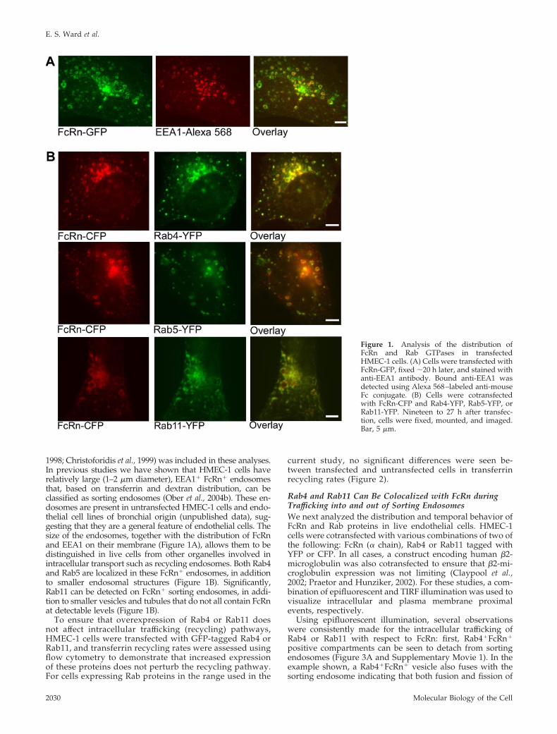
1998; Christoforidis et al., 1999) was included in these analyses.In previous studies we have shown that HMEC-1 cells haverelatively large (1–2 �m diameter), EEA1� FcRn� endosomesthat, based on transferrin and dextran distribution, can beclassified as sorting endosomes (Ober et al., 2004b). These en-dosomes are present in untransfected HMEC-1 cells and endo-thelial cell lines of bronchial origin (unpublished data), sug-gesting that they are a general feature of endothelial cells. Thesize of the endosomes, together with the distribution of FcRnand EEA1 on their membrane (Figure 1A), allows them to bedistinguished in live cells from other organelles involved inintracellular transport such as recycling endosomes. Both Rab4and Rab5 are localized in these FcRn� endosomes, in additionto smaller endosomal structures (Figure 1B). Significantly,Rab11 can be detected on FcRn� sorting endosomes, in addi-tion to smaller vesicles and tubules that do not all contain FcRnat detectable levels (Figure 1B).
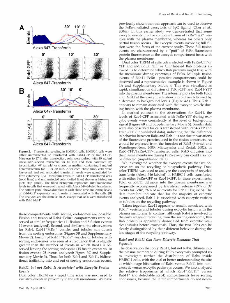
To ensure that overexpression of Rab4 or Rab11 doesnot affect intracellular trafficking (recycling) pathways,HMEC-1 cells were transfected with GFP-tagged Rab4 orRab11, and transferrin recycling rates were assessed usingflow cytometry to demonstrate that increased expressionof these proteins does not perturb the recycling pathway.For cells expressing Rab proteins in the range used in the
current study, no significant differences were seen be-tween transfected and untransfected cells in transferrinrecycling rates (Figure 2).
Rab4 and Rab11 Can Be Colocalized with FcRn duringTrafficking into and out of Sorting EndosomesWe next analyzed the distribution and temporal behavior ofFcRn and Rab proteins in live endothelial cells. HMEC-1cells were cotransfected with various combinations of two ofthe following: FcRn (� chain), Rab4 or Rab11 tagged withYFP or CFP. In all cases, a construct encoding human �2-microglobulin was also cotransfected to ensure that �2-mi-croglobulin expression was not limiting (Claypool et al.,2002; Praetor and Hunziker, 2002). For these studies, a com-bination of epifluorescent and TIRF illumination was used tovisualize intracellular and plasma membrane proximalevents, respectively.
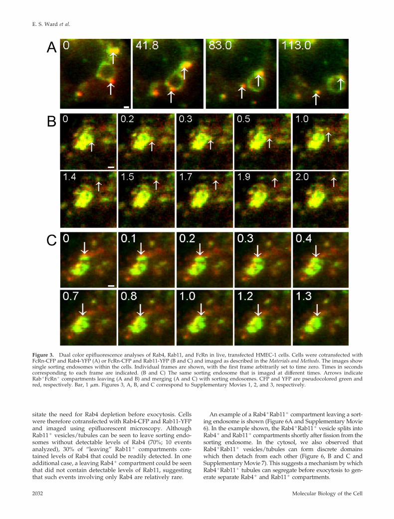
Using epifluorescent illumination, several observationswere consistently made for the intracellular trafficking ofRab4 or Rab11 with respect to FcRn: first, Rab4�FcRn�
positive compartments can be seen to detach from sortingendosomes (Figure 3A and Supplementary Movie 1). In theexample shown, a Rab4�FcRn� vesicle also fuses with thesorting endosome indicating that both fusion and fission of
Figure 1. Analysis of the distribution ofFcRn and Rab GTPases in transfectedHMEC-1 cells. (A) Cells were transfected withFcRn-GFP, fixed �20 h later, and stained withanti-EEA1 antibody. Bound anti-EEA1 wasdetected using Alexa 568–labeled anti-mouseFc conjugate. (B) Cells were cotransfectedwith FcRn-CFP and Rab4-YFP, Rab5-YFP, orRab11-YFP. Nineteen to 27 h after transfec-tion, cells were fixed, mounted, and imaged.Bar, 5 �m.
E. S. Ward et al.
Molecular Biology of the Cell2030
these compartments with sorting endosomes are possible.Fission and fusion of Rab4�FcRn� compartments were ob-served at similar frequencies (11 fission and 8 fusion events;19 events analyzed). Second, and similar to the observationsfor Rab4, Rab11�FcRn� vesicles and tubules can detachfrom the sorting endosomes (Figure 3B and SupplementaryMovie 2). Fusion of Rab11�FcRn� vesicles or tubules withsorting endosomes was seen at a frequency that is slightlygreater than the number of events in which Rab11 is ob-served leaving the sorting endosome (15 fusion events vs. 10fission events; 25 events analyzed; Figure 3C and Supple-mentary Movie 3). Thus, for both Rab4 and Rab11, bidirec-tional trafficking into and out of sorting endosomes occurs.
Rab11, but not Rab4, Is Associated with Exocytic FusionEventsDual color TIRFM on a rapid time scale was next used tovisualize events in proximity to the cell membrane. We have
previously shown that this approach can be used to observethe FcRn-mediated exocytosis of IgG ligand (Ober et al.,2004a). In this earlier study we demonstrated that someexocytic events involve complete fusion of FcRn�IgG� ves-icles with the plasma membrane, whereas for others onlypartial fusion occurs. The exocytic events involving full fu-sion were the focus of the current study. These full fusionevents are characterized by a “puff” of FcRn-fluorescentprotein fluorescence as the exocytic compartment fuses withthe plasma membrane.
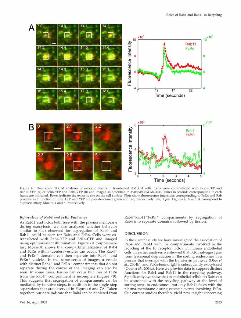
Dual color TIRFM of cells cotransfected with FcRn-CFP orYFP and individual YFP or CFP labeled Rab proteins al-lowed us to determine which Rab proteins might fuse withthe membrane during exocytosis of FcRn. Multiple fusionevents of Rab11�FcRn� positive compartments could beobserved and a representative example is shown in Figure4A and Supplementary Movie 4. This was visualized asrapid, simultaneous diffusion of FcRn-CFP and Rab11-YFPinto the plasma membrane. The intensity plots for both FcRnand Rab11 at the exocytic site show a rapid rise followed bya decrease to background levels (Figure 4A). Thus, Rab11appears to remain associated with the exocytic vesicle dur-ing fusion with the plasma membrane.
In marked contrast to the observations for Rab11, thelevels of Rab4-CFP associated with FcRn-YFP during exo-cytic events were consistently at the level of backgroundsignal (Figure 4B and Supplementary Movie 5). Similar datawere also observed for cells transfected with Rab4-YFP andFcRn-CFP (unpublished data), indicating that the differencein behavior between Rab4 and Rab11 is not due to variationsof the fluorescent proteins used in the fusion constructs. Aswould be expected from the function of Rab5 (Somsel andWandinger-Ness, 2000; Miaczynska and Zerial, 2002), inRab5-YFP/FcRn-CFP–transfected cells, Rab5 diffusion intothe plasma membrane during FcRn exocytosis could also notbe detected (unpublished data).
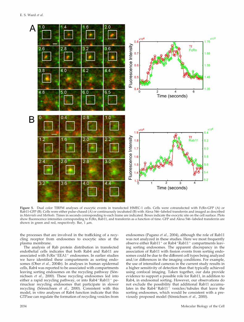
We investigated whether the exocytic events that we ob-serve are on the recycling or biosynthetic pathway. Dualcolor TIRFM was used to analyze the exocytosis of recycledtransferrin (Alexa 546 labeled) in HMEC-1 cells transfectedwith either FcRn-GFP or Rab11-GFP. In these experiments,FcRn or Rab11 diffusion into the plasma membrane wasfrequently accompanied by transferrin release (89% of 37events for FcRn, 76% of 42 events for Rab11; Figure 5). Thedata therefore indicate that for the majority of exocyticevents analyzed, Rab11 is associated with exocytic vesiclesor tubules on the recycling pathway.
Taken together, Rab11 appears to remain associated withFcRn� vesicles and tubules during exocytic fusion with theplasma membrane. In contrast, although Rab4 is involved inthe early stages of recycling from the sorting endosome, thisRab protein is apparently dissociated from exocytic vesi-cles/tubules before exocytosis. Thus, the two Rabs can beclearly distinguished by their different behavior during thelate stages of the recycling pathway.
Rab4 and Rab11 Can Form Discrete Domains ThatSeparateThe observation that only Rab11, but not Rab4, diffuses intothe plasma membrane during FcRn exocytosis prompted usto investigate further the distribution of Rabs insideHMEC-1 cells, with the goal of better understanding the siteat which stage bifurcation of Rab4 versus Rab11 into non-exocytic versus exocytic pathways occurs. We also analyzedthe relative frequencies at which Rab4�Rab11� versusRab11� (no detectable Rab4) compartments leave sortingendosomes, because the latter compartments do not neces-
Figure 2. Transferrin recycling in HMEC-1 cells. HMEC-1 cells wereeither untransfected or transfected with Rab4-GFP or Rab11-GFP.Nineteen to 27 h after transfection, cells were pulsed with 10 �g/mlAlexa 647–labeled transferrin for 60 min and then harvested bytrypsinization (0� sample) or chased in medium containing 1 mg/mlholotransferrrin for 10 or 30 min. After each chase time, cells wereharvested, and cell associated transferrin levels were quantitated byflow cytometry. (A) Transferrin levels in Rab4-GFP–transfected cells(solid lines) and untransfected cells (dotted lines) shown as histogramplots (top panel). The filled histogram represents autofluorescencelevels in cells that were not treated with Alexa 647–labeled transferrin.The bottom panel shows dot plots at each chase time, indicating levelsof Rab4-GFP expression and transferrin associated with the cells. (B)The analyses are the same as in A, except that cells were transfectedwith Rab11-GFP.
Roles of Rab4 and Rab11 in Recycling
Vol. 16, April 2005 2031
sitate the need for Rab4 depletion before exocytosis. Cellswere therefore cotransfected with Rab4-CFP and Rab11-YFPand imaged using epifluorescent microscopy. AlthoughRab11� vesicles/tubules can be seen to leave sorting endo-somes without detectable levels of Rab4 (70%; 10 eventsanalyzed), 30% of “leaving” Rab11� compartments con-tained levels of Rab4 that could be readily detected. In oneadditional case, a leaving Rab4� compartment could be seenthat did not contain detectable levels of Rab11, suggestingthat such events involving only Rab4 are relatively rare.
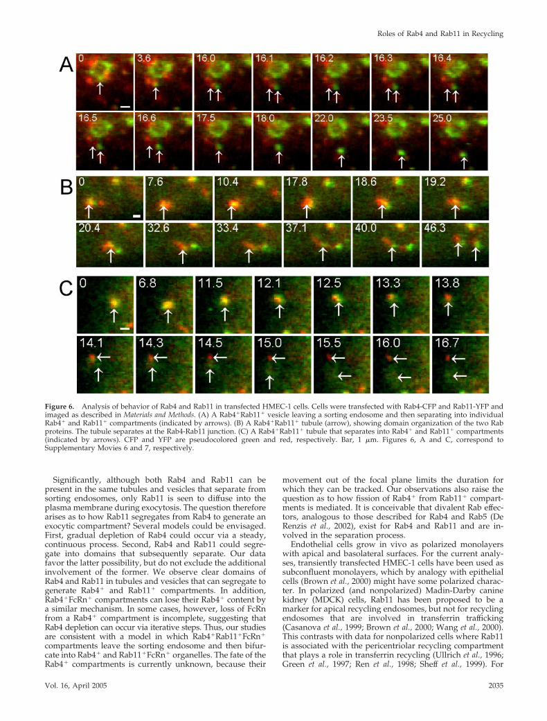
An example of a Rab4�Rab11� compartment leaving a sort-ing endosome is shown (Figure 6A and Supplementary Movie6). In the example shown, the Rab4�Rab11� vesicle splits intoRab4� and Rab11� compartments shortly after fission from thesorting endosome. In the cytosol, we also observed thatRab4�Rab11� vesicles/tubules can form discrete domainswhich then detach from each other (Figure 6, B and C andSupplementary Movie 7). This suggests a mechanism by whichRab4�Rab11� tubules can segregate before exocytosis to gen-erate separate Rab4� and Rab11� compartments.
Figure 3. Dual color epifluorescence analyses of Rab4, Rab11, and FcRn in live, transfected HMEC-1 cells. Cells were cotransfected withFcRn-CFP and Rab4-YFP (A) or FcRn-CFP and Rab11-YFP (B and C) and imaged as described in the Materials and Methods. The images showsingle sorting endosomes within the cells. Individual frames are shown, with the first frame arbitrarily set to time zero. Times in secondscorresponding to each frame are indicated. (B and C) The same sorting endosome that is imaged at different times. Arrows indicateRab�FcRn� compartments leaving (A and B) and merging (A and C) with sorting endosomes. CFP and YFP are pseudocolored green andred, respectively. Bar, 1 �m. Figures 3, A, B, and C correspond to Supplementary Movies 1, 2, and 3, respectively.
E. S. Ward et al.
Molecular Biology of the Cell2032
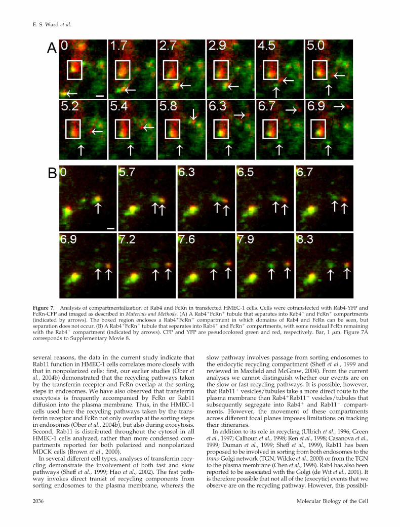
Bifurcation of Rab4 and FcRn PathwaysAs Rab11 and FcRn both fuse with the plasma membraneduring exocytosis, we also analyzed whether behaviorsimilar to that observed for segregation of Rab4 andRab11 could be seen for Rab4 and FcRn. Cells were co-transfected with Rab4-YFP and FcRn-CFP and imagedusing epifluorescent illumination. Figure 7A (Supplemen-tary Movie 8) shows that compartmentalization of Rab4and FcRn within tubules/vesicles can occur. The Rab4�
and FcRn� domains can then separate into Rab4� andFcRn� vesicles. In this same series of images, a vesiclewith distinct Rab4� and FcRn� compartments that do notseparate during the course of the imaging can also beseen. In some cases, fission can occur but loss of FcRnfrom the Rab4� compartment is incomplete (Figure 7B).This suggests that segregation of compartments can bemediated by iterative steps, in addition to the single-stepseparations that are observed in Figures 6 and 7A. Takentogether, our data indicate that Rab4 can be depleted from
Rab4�Rab11�FcRn� compartments by segregation ofRab4 into separate domains followed by fission.
DISCUSSION
In the current study we have investigated the association ofRab4 and Rab11 with the compartments involved in therecycling of the Fc receptor, FcRn, in human endothelialcells. In earlier analyses we showed that FcRn salvages IgGsfrom lysosomal degradation in the sorting endosomes in aprocess that overlaps with the transferrin pathway (Ober etal., 2004b), and FcRn-bound IgG is subsequently exocytosed(Ober et al., 2004a). Here we provide data to support distinctfunctions for Rab4 and Rab11 in the recycling pathway.Significantly, we show that in endothelial cells both Rabs canbe associated with the recycling pathway at the level ofsorting steps in endosomes, but only Rab11 fuses with theplasma membrane during exocytic events involving FcRn.Our current studies therefore yield new insight concerning
Figure 4. Dual color TIRFM analyses of exocytic events in transfected HMEC-1 cells. Cells were cotransfected with FcRn-CFP andRab11-YFP (A) or FcRn-YFP and Rab4-CFP (B) and imaged as described in Materials and Methods. Times in seconds corresponding to eachframe are indicated. Boxes indicate the exocytic site on the cell surface. Plots show fluorescence intensities corresponding to FcRn and Rabproteins as a function of time. CFP and YFP are pseudocolored green and red, respectively. Bar, 1 �m. Figures 4, A and B, correspond toSupplementary Movies 4 and 5, respectively.
Roles of Rab4 and Rab11 in Recycling
Vol. 16, April 2005 2033
the processes that are involved in the trafficking of a recy-cling receptor from endosomes to exocytic sites at theplasma membrane.
The analysis of Rab protein distribution in transfectedendothelial cells indicates that both Rab4 and Rab11 areassociated with FcRn�EEA1� endosomes. In earlier studieswe have identified these compartments as sorting endo-somes (Ober et al., 2004b). In analyses in human epidermalcells, Rab4 was reported to be associated with compartmentsleaving sorting endosomes on the recycling pathway (Son-nichsen et al., 2000). These recycling endosomes led intoeither a rapid recycling pathway, or into Rab4�Rab11� pe-rinuclear recycling endosomes that participate in slowerrecycling (Sonnichsen et al., 2000). Consistent with thismodel, in vitro analyses of Rab4 function indicate that thisGTPase can regulate the formation of recycling vesicles from
endosomes (Pagano et al., 2004), although the role of Rab11was not analyzed in these studies. Here we most frequentlyobserve either Rab11� or Rab4�Rab11� compartments leav-ing sorting endosomes. The apparent discrepancy in theassociation of Rab11 with fission events from sorting endo-somes could be due to the different cell types being analyzedand/or differences in the imaging conditions. For example,the use of intensified cameras in the current study results ina higher sensitivity of detection than that typically achievedusing confocal imaging. Taken together, our data provideevidence to support a possible role for Rab11, in addition toRab4, in endosomal sorting. However, our observations donot exclude the possibility that additional Rab11 accumu-lates in the Rab4�Rab11� vesicles/tubules that leave thesorting endosomes, which would be consistent with a pre-viously proposed model (Sonnichsen et al., 2000).
Figure 5. Dual color TIRFM analyses of exocytic events in transfected HMEC-1 cells. Cells were cotransfected with FcRn-GFP (A) orRab11-GFP (B). Cells were either pulse-chased (A) or continuously incubated (B) with Alexa 546–labeled transferrin and imaged as describedin Materials and Methods. Times in seconds corresponding to each frame are indicated. Boxes indicate the exocytic site on the cell surface. Plotsshow fluorescence intensities corresponding to FcRn, Rab11, and transferrin as a function of time. GFP and Alexa 546–labeled transferrin areshown in green and red, respectively. Bar, 1 �m.
E. S. Ward et al.
Molecular Biology of the Cell2034
Significantly, although both Rab4 and Rab11 can bepresent in the same tubules and vesicles that separate fromsorting endosomes, only Rab11 is seen to diffuse into theplasma membrane during exocytosis. The question thereforearises as to how Rab11 segregates from Rab4 to generate anexocytic compartment? Several models could be envisaged.First, gradual depletion of Rab4 could occur via a steady,continuous process. Second, Rab4 and Rab11 could segre-gate into domains that subsequently separate. Our datafavor the latter possibility, but do not exclude the additionalinvolvement of the former. We observe clear domains ofRab4 and Rab11 in tubules and vesicles that can segregate togenerate Rab4� and Rab11� compartments. In addition,Rab4�FcRn� compartments can lose their Rab4� content bya similar mechanism. In some cases, however, loss of FcRnfrom a Rab4� compartment is incomplete, suggesting thatRab4 depletion can occur via iterative steps. Thus, our studiesare consistent with a model in which Rab4�Rab11�FcRn�
compartments leave the sorting endosome and then bifur-cate into Rab4� and Rab11�FcRn� organelles. The fate of theRab4� compartments is currently unknown, because their
movement out of the focal plane limits the duration forwhich they can be tracked. Our observations also raise thequestion as to how fission of Rab4� from Rab11� compart-ments is mediated. It is conceivable that divalent Rab effec-tors, analogous to those described for Rab4 and Rab5 (DeRenzis et al., 2002), exist for Rab4 and Rab11 and are in-volved in the separation process.
Endothelial cells grow in vivo as polarized monolayerswith apical and basolateral surfaces. For the current analy-ses, transiently transfected HMEC-1 cells have been used assubconfluent monolayers, which by analogy with epithelialcells (Brown et al., 2000) might have some polarized charac-ter. In polarized (and nonpolarized) Madin-Darby caninekidney (MDCK) cells, Rab11 has been proposed to be amarker for apical recycling endosomes, but not for recyclingendosomes that are involved in transferrin trafficking(Casanova et al., 1999; Brown et al., 2000; Wang et al., 2000).This contrasts with data for nonpolarized cells where Rab11is associated with the pericentriolar recycling compartmentthat plays a role in transferrin recycling (Ullrich et al., 1996;Green et al., 1997; Ren et al., 1998; Sheff et al., 1999). For
Figure 6. Analysis of behavior of Rab4 and Rab11 in transfected HMEC-1 cells. Cells were transfected with Rab4-CFP and Rab11-YFP andimaged as described in Materials and Methods. (A) A Rab4�Rab11� vesicle leaving a sorting endosome and then separating into individualRab4� and Rab11� compartments (indicated by arrows). (B) A Rab4�Rab11� tubule (arrow), showing domain organization of the two Rabproteins. The tubule separates at the Rab4-Rab11 junction. (C) A Rab4�Rab11� tubule that separates into Rab4� and Rab11� compartments(indicated by arrows). CFP and YFP are pseudocolored green and red, respectively. Bar, 1 �m. Figures 6, A and C, correspond toSupplementary Movies 6 and 7, respectively.
Roles of Rab4 and Rab11 in Recycling
Vol. 16, April 2005 2035
several reasons, the data in the current study indicate thatRab11 function in HMEC-1 cells correlates more closely withthat in nonpolarized cells: first, our earlier studies (Ober etal., 2004b) demonstrated that the recycling pathways takenby the transferrin receptor and FcRn overlap at the sortingsteps in endosomes. We have also observed that transferrinexocytosis is frequently accompanied by FcRn or Rab11diffusion into the plasma membrane. Thus, in the HMEC-1cells used here the recycling pathways taken by the trans-ferrin receptor and FcRn not only overlap at the sorting stepsin endosomes (Ober et al., 2004b), but also during exocytosis.Second, Rab11 is distributed throughout the cytosol in allHMEC-1 cells analyzed, rather than more condensed com-partments reported for both polarized and nonpolarizedMDCK cells (Brown et al., 2000).
In several different cell types, analyses of transferrin recy-cling demonstrate the involvement of both fast and slowpathways (Sheff et al., 1999; Hao et al., 2002). The fast path-way invokes direct transit of recycling components fromsorting endosomes to the plasma membrane, whereas the
slow pathway involves passage from sorting endosomes tothe endocytic recycling compartment (Sheff et al., 1999 andreviewed in Maxfield and McGraw, 2004). From the currentanalyses we cannot distinguish whether our events are onthe slow or fast recycling pathways. It is possible, however,that Rab11� vesicles/tubules take a more direct route to theplasma membrane than Rab4�Rab11� vesicles/tubules thatsubsequently segregate into Rab4� and Rab11� compart-ments. However, the movement of these compartmentsacross different focal planes imposes limitations on trackingtheir itineraries.
In addition to its role in recycling (Ullrich et al., 1996; Greenet al., 1997; Calhoun et al., 1998; Ren et al., 1998; Casanova et al.,1999; Duman et al., 1999; Sheff et al., 1999), Rab11 has beenproposed to be involved in sorting from both endosomes to thetrans-Golgi network (TGN; Wilcke et al., 2000) or from the TGNto the plasma membrane (Chen et al., 1998). Rab4 has also beenreported to be associated with the Golgi (de Wit et al., 2001). Itis therefore possible that not all of the (exocytic) events that weobserve are on the recycling pathway. However, this possibil-
Figure 7. Analysis of compartmentalization of Rab4 and FcRn in transfected HMEC-1 cells. Cells were cotransfected with Rab4-YFP andFcRn-CFP and imaged as described in Materials and Methods. (A) A Rab4�FcRn� tubule that separates into Rab4� and FcRn� compartments(indicated by arrows). The boxed region encloses a Rab4�FcRn� compartment in which domains of Rab4 and FcRn can be seen, butseparation does not occur. (B) A Rab4�FcRn� tubule that separates into Rab4� and FcRn� compartments, with some residual FcRn remainingwith the Rab4� compartment (indicated by arrows). CFP and YFP are pseudocolored green and red, respectively. Bar, 1 �m. Figure 7Acorresponds to Supplementary Movie 8.
E. S. Ward et al.
Molecular Biology of the Cell2036
ity is made unlikely by our analyses of transferrin exocytosiswhere we show that the majority of exocytic events seen forFcRn and Rab11 also involve transferrin release. Our data alsoraise the question as to how Rab11 is recaptured from the cellsurface after the exocytic events. Several possible routes couldbe envisaged: for example, plasma membrane–bound Rab11could be endocytosed or bind to GDP dissociation inhibitors(GDIs) that play a role in maintaining cytosolic pools of Rabs(Pfeffer and Aivazian, 2004).
In summary, our data are consistent with the followingpathways for FcRn recycling from the sorting endosome:FcRn separates from the sorting endosome in associationwith Rab4 and Rab11 or with Rab11 only. Subsequent toseparation from Rab4�Rab11� compartments, Rab4 is lo-calized to separate domains in the recycling vesicle/tu-bule that dissociate to leave Rab11�FcRn� compartments.Rab11�FcRn� compartments subsequently undergo fullfusion with the plasma membrane to result in diffusion ofRab11 and FcRn out from the center of the exocytic site.These analyses therefore provide insight into the associ-ation of Rab GTPases with FcRn during the trafficking ofthis receptor from sorting endosomes to exocytic releaseat the plasma membrane.
ACKNOWLEDGMENTS
We are indebted to Palmer Long for expert assistance with microscopysoftware and preparation of figures. We thank Dr. Marino Zerial and Fran-cisco Candal for providing Rab expression constructs and HMEC-1 cells,respectively. This study was supported by grants from the National Institutesof Health (RO1 AI 39167, RO1 AI 50747, and R21 53748).
REFERENCES
Antohe, F., Radulescu, L., Gafencu, A., Ghetie, V., and Simionescu, M. (2001).Expression of functionally active FcRn and the differentiated bidirectionaltransport of IgG in human placental endothelial cells. Hum. Immunol. 62,93–105.
Brown, P. S., Wang, E., Aroeti, B., Chapin, S. J., Mostov, K. E., and Dunn,K. W. (2000). Definition of distinct compartments in polarized Madin-Darbycanine kidney (MDCK) cells for membrane-volume sorting, polarized sortingand apical recycling. Traffic 1, 124–140.
Calhoun, B. C., Lapierre, L. A., Chew, C. S., and Goldenring, J. R. (1998).Rab11a redistributes to apical secretory canaliculus during stimulation ofgastric parietal cells. Am. J. Physiol. 275, C163–C170.
Casanova, J. E., Wang, X., Kumar, R., Bhartur, S. G., Navarre, J., Woodrum,J. E., Altschuler, Y., Ray, G. S., and Goldenring, J. R. (1999). Association ofRab25 and Rab11a with the apical recycling system of polarized Madin-Darbycanine kidney cells. Mol. Biol. Cell 10, 47–61.
Chen, W., Feng, Y., Chen, D., and Wandinger-Ness, A. (1998). Rab11 isrequired for trans-Golgi network-to-plasma membrane transport and a pref-erential target for GDP dissociation inhibitor. Mol. Biol. Cell 9, 3241–3257.
Christoforidis, S., McBride, H. M., Burgoyne, R. D., and Zerial, M. (1999). TheRab5 effector EEA1 is a core component of endosome docking. Nature 397,621–625.
Claypool, S. M., Dickinson, B. L., Yoshida, M., Lencer, W. I., and Blumberg,R. S. (2002). Functional reconstitution of human FcRn in Madin-Darby caninekidney cells requires co-expressed human beta 2-microglobulin. J. Biol. Chem.277, 28038–28050.
Daro, E., van der Sluijs, S. P., Galli, T., and Mellman, I. (1996). Rab4 andcellubrevin define different early endosome populations on the pathway oftransferrin receptor recycling. Proc. Natl. Acad. Sci. USA 93, 9559–9564.
De Renzis, S., Sonnichsen, B., and Zerial, M. (2002). Divalent Rab effectorsregulate the sub-compartmental organization and sorting of early endosomes.Nat. Cell Biol. 4, 124–133.
de Wit, H., Lichtenstein, Y., Kelly, R. B., Geuze, H. J., Klumperman, J., and vander Sluijs, P. (2001). Rab4 regulates formation of synaptic-like microvesiclesfrom early endosomes in PC12 cells. Mol. Biol. Cell 12, 3703–3715.
Dickinson, B. L., Badizadegan, K., Wu, Z., Ahouse, J. C., Zhu, X., Simister,N. E., Blumberg, R. S., and Lencer, W. I. (1999). Bidirectional FcRn-dependent
IgG transport in a polarized human intestinal epithelial cell line. J. Clin.Invest. 104, 903–911.
Duman, J. G., Tyagarajan, K., Kolsi, M. S., Moore, H. P., and Forte, J. G. (1999).Expression of rab11a N124I in gastric parietal cells inhibits stimulatory re-cruitment of the H�-K�-ATPase. Am. J. Physiol 277, C361–C372.
Ellinger, I., Schwab, M., Stefanescu, A., Hunziker, W., and Fuchs, R. (1999).IgG transport across trophoblast-derived BeWo cells: a model system to studyIgG transport in the placenta. Eur. J. Immunol. 29, 733–744.
Firan, M., Bawdon, R., Radu, C., Ober, R. J., Eaken, D., Antohe, F., Ghetie, V.,and Ward, E. S. (2001). The MHC class I related receptor, FcRn, plays anessential role in the maternofetal transfer of gammaglobulin in humans. Int.Immunol. 13, 993–1002.
Ghetie, V., Hubbard, J. G., Kim, J. K., Tsen, M. F., Lee, Y., and Ward, E. S.(1996). Abnormally short serum half-lives of IgG in beta 2-microglobulin-deficient mice. Eur. J. Immunol. 26, 690–696.
Ghetie, V., and Ward, E. S. (2000). Multiple roles for the major histocompat-ibility complex class I-related receptor FcRn. Annu. Rev. Immunol. 18, 739–766.
Green, E. G., Ramm, E., Riley, N. M., Spiro, D. J., Goldenring, J. R., andWessling-Resnick, M. (1997). Rab11 is associated with transferrin-containingrecycling compartments in K562 cells. Biochem. Biophys. Res. Commun. 239,612–616.
Hao, M., Lin, S. X., Karylowski, O. J., Wustner, D., McGraw, T. E., andMaxfield, F. R. (2002). Vesicular and non-vesicular sterol transport in livingcells. The endocytic recycling compartment is a major sterol storage organelle.J. Biol. Chem. 277, 609–617.
Jahn, R., Lang, T., and Sudhof, T. C. (2003). Membrane fusion. Cell 112,519–533.
Khvotchev, M. V., Ren, M., Takamori, S., Jahn, R., and Sudhof, T. C. (2003).Divergent functions of neuronal Rab11b in Ca2�-regulated versus constitutiveexocytosis. J. Neurosci. 23, 10531–10539.
Kobayashi, N., Suzuki, Y., Tsuge, T., Okumura, K., Ra, C., and Tomino, Y.(2002). FcRn-mediated transcytosis of IgG in human renal proximal tubularepithelial cells. Am. J. Physiol. Renal Physiol. 282, F358–F365.
Maxfield, F. R., and McGraw, T. E. (2004). Endocytic recycling. Nat. Rev. Mol.Cell Biol. 5, 121–132.
McCaffrey, M. W., Bielli, A., Cantalupo, G., Mora, S., Roberti, V., Santillo, M.,Drummond, F., and Bucci, C. (2001). Rab4 affects both recycling and degra-dative endosomal trafficking. FEBS Lett. 495, 21–30.
McCarthy, K. M., Yoong, Y., and Simister, N. E. (2000). Bidirectional transcy-tosis of IgG by the rat neonatal Fc receptor expressed in a rat kidney cell line:a system to study protein transport across epithelia. J. Cell Sci. 113, 1277–1285.
Medesan, C., Matesoi, D., Radu, C., Ghetie, V., and Ward, E. S. (1997).Delineation of the amino acid residues involved in transcytosis and catabo-lism of mouse IgG1. J. Immunol. 158, 2211–2217.
Miaczynska, M., and Zerial, M. (2002). Mosaic organization of the endocyticpathway. Exp. Cell Res. 272, 8–14.
Ober, R. J., Martinez, C., Lai, X., Zhou, J., and Ward, E. S. (2004a). Exocytosisof IgG as mediated by the receptor, FcRn: an analysis at the single-moleculelevel. Proc. Natl. Acad. Sci. USA 101, 11076–11081.
Ober, R. J., Martinez, C., Vaccaro, C., Zhou, J., and Ward, E. S. (2004b).Visualizing the site and dynamics of IgG salvage by the MHC class I-relatedreceptor, FcRn. J. Immunol. 172, 2021–2029.
Pagano, A., Crottet, P., Prescianotto-Baschong, C., and Spiess, M. (2004). Invitro formation of recycling vesicles from endosomes requires adaptor pro-tein-1/clathrin and is regulated by rab4 and the connector rabaptin-5. Mol.Biol. Cell 15, 4990–5000.
Pfeffer, S., and Aivazian, D. (2004). Targeting Rab GTPases to distinct mem-brane compartments. Nat. Rev. Mol. Cell. Biol. 5, 886–896.
Praetor, A., Ellinger, I., and Hunziker, W. (1999). Intracellular traffic of theMHC class I-like IgG Fc receptor, FcRn, expressed in epithelial MDCK cells.J. Cell Sci. 112, 2291–2299.
Praetor, A., and Hunziker, W. (2002). beta(2)-Microglobulin is important forcell surface expression and pH-dependent IgG binding of human FcRn. J. CellSci. 115, 2389–2397.
Pruckler, J. M., Lawley, T. J., and Ades, E. W. (1993). Use of a humanmicrovascular endothelial cell line as a model system to evaluate cholesteroluptake. Pathobiology 61, 283–287.
Ravetch, J. V., and Bolland, S. (2001). IgG Fc receptors. Annu. Rev. Immunol.19, 275–290.
Roles of Rab4 and Rab11 in Recycling
Vol. 16, April 2005 2037
Ren, M., Xu, G., Zeng, J., Lemos-Chiarandini, C., Adesnik, M., and Sabatini,D. D. (1998). Hydrolysis of GTP on rab11 is required for the direct delivery oftransferrin from the pericentriolar recycling compartment to the cell surfacebut not from sorting endosomes. Proc. Natl. Acad. Sci. USA 95, 6187–6192.
Rojas, R., and Apodaca, G. (2002). Ig transport across polarized epithelialcells. Nat. Rev. Mol. Cell Biol. 3, 944–955.
Schmoranzer, J., Goulian, M., Axelrod, D., and Simon, S. M. (2000). Imagingconstitutive exocytosis with total internal reflection fluorescence microscopy.J. Cell Biol. 149, 23–32.
Sheff, D. R., Daro, E. A., Hull, M., and Mellman, I. (1999). The receptorrecycling pathway contains two distinct populations of early endosomes withdifferent sorting functions. J. Cell Biol. 145, 123–139.
Simonsen, A., Lippe, R., Christoforidis, S., Gaullier, J. M., Brech, A., Cal-laghan, J., Toh, B. H., Murphy, C., Zerial, M., and Stenmark, H. (1998). EEA1links PI(3)K function to Rab5 regulation of endosome fusion. Nature 394,494–498.
Somsel, R. J., and Wandinger-Ness, A. (2000). Rab GTPases coordinate endo-cytosis. J. Cell Sci. 113(Pt 2), 183–192.
Sonnichsen, B., De Renzis, S., Nielsen, E., Rietdorf, J., and Zerial, M. (2000).Distinct membrane domains on endosomes in the recycling pathway visual-ized by multicolor imaging of Rab4, Rab5, and Rab11. J. Cell Biol. 149,901–914.
Spiekermann, G. M., Finn, P. W., Ward, E. S., Dumont, J., Dickinson, B. L.,Blumberg, R. S., and Lencer, W. I. (2002). Receptor-mediated IgG transport
across mucosal barriers in adult life: functional expression of FcRn in themammalian lung. J. Exp. Med. 196, 303–310.
Steyer, J. A., and Almers, W. (2001). A real-time view of life within 100 nm ofthe plasma membrane. Nat. Rev. Mol. Cell Biol. 2, 268–275.
Toomre, D., Steyer, J. A., Keller, P., Almers, W., and Simons, K. (2000). Fusionof constitutive membrane traffic with the cell surface observed by evanescentwave microscopy. J. Cell Biol. 149, 33–40.
Ullrich, O., Reinsch, S., Urbe, S., Zerial, M., and Parton, R. G. (1996). Rab11regulates recycling through the pericentriolar recycling endosome. J. Cell Biol.135, 913–924.
van der Sluijs, S. P., Hull, M., Webster, P., Male, P., Goud, B., and Mellman,I. (1992). The small GTP-binding protein rab4 controls an early sorting eventon the endocytic pathway. Cell 70, 729–740.
van IJzendoorn, S. C., Mostov, K. E., and Hoekstra, D. (2003). Role of rabproteins in epithelial membrane traffic. Int. Rev. Cytol. 232, 59–88.
Wang, X., Kumar, R., Navarre, J., Casanova, J. E., and Goldenring, J. R. (2000).Regulation of vesicle trafficking in Madin-Darby canine kidney cells byRab11a and Rab25. J. Biol. Chem. 275, 29138–29146.
Wilcke, M., Johannes, L., Galli, T., Mayau, V., Goud, B., and Salamero, J.(2000). Rab11 regulates the compartmentalization of early endosomes re-quired for efficient transport from early endosomes to the trans-Golgi net-work. J. Cell Biol. 151, 1207–1220.
E. S. Ward et al.
Molecular Biology of the Cell2038