Embed Size (px)
Citation preview

Touro Scholar Touro Scholar
NYMC Faculty Publications Faculty
1-1-2018
From the Cover: 7,8-Dihydroxyflavone Rescues Lead-Induced From the Cover: 7,8-Dihydroxyflavone Rescues Lead-Induced
Impairment of Vesicular Release: A Novel Therapeutic Approach Impairment of Vesicular Release: A Novel Therapeutic Approach
for Lead Intoxicated Children for Lead Intoxicated Children
Xiao-Lei Zhang New York Medical College
Jennifer L. McGlothan
Omid Miry
Kirstie H. Stansfield
Meredith K. Loth
See next page for additional authors
Follow this and additional works at: https://touroscholar.touro.edu/nymc_fac_pubs
Part of the Medicine and Health Sciences Commons
Recommended Citation Recommended Citation Zhang, X., McGlothan, J., Miry, O., Stansfield, K., Loth, M., Stanton, P., & Guilarte, T. (2018). From the Cover: 7,8-Dihydroxyflavone Rescues Lead-Induced Impairment of Vesicular Release: A Novel Therapeutic Approach for Lead Intoxicated Children. Toxicological Sciences, 161 (1), 186-195. https://doi.org/10.1093/toxsci/kfx210
This Article is brought to you for free and open access by the Faculty at Touro Scholar. It has been accepted for inclusion in NYMC Faculty Publications by an authorized administrator of Touro Scholar. For more information, please contact [email protected].

Authors Authors Xiao-Lei Zhang, Jennifer L. McGlothan, Omid Miry, Kirstie H. Stansfield, Meredith K. Loth, Patric K. Stanton, and Tomas R. Guilarte
This article is available at Touro Scholar: https://touroscholar.touro.edu/nymc_fac_pubs/1356
1
7,8-Dihydroxyflavone Rescues Lead-Induced Impairment of Vesicular Release: 1 A Novel Therapeutic Approach for Lead Intoxicated Children 2
3 Xiao-lei Zhang1, Jennifer L. McGlothan2, Omid Miry1, Kirstie H. Stansfield3, Meredith K. 4
Loth2,3, Patric K. Stanton1, Tomás R. Guilarte2* 5 6
1Department of Cell Biology & Anatomy 7 New York Medical College 8
Valhalla, NY 9 2Department of Environmental & Occupational Health 10
Robert Stempel College of Public Health & Social Work 11 Florida International University 12
Miami, FL 13 3Department of Environmental Health Sciences 14
Mailman School of Public Health 15 Columbia University 16
New York, NY 17 18 *Correspondence to: 19 Tomás R. Guilarte, Ph.D. 20 Dean, Robert Stempel College of Public Health & Social Work 21 Dept. Environmental & Occupational Health; Cognitive Neuroscience & Imaging 22 Florida International University 23 Miami, FL 33199 24 Phone: 305-348-5344 25 E-mail: [email protected] 26
2
ABSTRACT 27 Lead (Pb2+) exposure during brain development inhibits neurotransmitter release 28 resulting in impaired synapse formation, synaptic plasticity and learning. In primary 29 hippocampal neurons in culture and hippocampal slices, Pb2+ exposure inhibits vesicular 30 release and reduces the number of fast-releasing sites, an effect associated with Pb2+ 31 inhibition of NMDA receptor-mediated trans-synaptic BDNF signaling. We hypothesized 32 that TrkB receptor activation, the cognate receptor for BDNF, would rescue Pb2+-induced 33 impairments of vesicular release. Rats were chronically exposed to Pb2+ prenatally and 34 postnatally until 50 days of age. This Pb2+ exposure paradigm enhanced paired-pulse 35 facilitation representative of reduced vesicular release probability. Reductions in Pb2+-36 induced release probability were also measured by both mean-variance analysis and 37 direct two-photon imaging of vesicular release from hippocampal slices. We also found a 38 Pb2+ impairment of calcium influx in presynaptic terminals. Intraperitoneal injections of 39 the TrkB agonist 7,8-dihydroxyflavone (5 mg/kg) for 14-15 days in Pb2+ rats starting at 40 postnatal day 35, reversed all Pb2+-induced impairments of presynaptic transmitter 41 release at Schaffer collateral-CA1 synapses. These data indicate that in vivo 42 pharmacological activation of TrkB receptors can reverse long-term effects of chronic 43 Pb2+ exposure on presynaptic terminals, pointing to TrkB receptor activation by small 44 molecules as a promising therapeutic intervention in Pb2+-intoxicated children. 45 46 KEYWORDS 47 7,8-dihydroxyflavone; vesicular release; lead neurotoxicity 48 49 50 51
3
Childhood lead (Pb2+) intoxication is a significant public health problem in the United 52 States and globally1,2. Recent episodes of Pb2+ exposure in children in communities like 53 Flint, Michigan demonstrate the pervasive nature of the problem3. The National 54 Resources Defense Council (NRDC) reports that millions of Americans get drinking 55 water from water systems that have Pb2+ violations and the problem could be much 56 larger, because systems known to have violations do not show up in government 57 databases that track such problems. Despite nearly a century of knowledge on the 58 detrimental effects of Pb2+ in children’s development, the widespread presence of this 59 poison in the global environment continues to affect children in the most vulnerable and 60 economically disadvantaged segments of the population. 61 62 Studies have consistently demonstrated that one of the most prominent effects of Pb2+ in 63 children is decreased capacity to learn, with devastating effects on cognitive and 64 intellectual development4-8, and in-school performance9,10. Early life Pb2+ intoxication 65 diminishes intellectual capacity of children with an immeasurable cost to society. Human 66 studies have shown that Pb2+ exposure in early life is associated with longitudinal 67 declines in cognitive function11, loss of brain volume12,13, and emergence of mental 68 disorders such as major depression and schizophrenia14,15. 69 70 Our laboratory has provided the first working model by which Pb2+ exposure during the 71 period of synaptogenesis can affect synapse development and function, that accounts 72 for both presynaptic and postsynaptic effects of Pb2+ on the synapse16-18. Using a Pb2+ 73 exposure paradigm during the period of synaptogenesis in primary hippocampal 74 neurons, we found that Pb2+ inhibition of postsynaptic NMDA receptors (NMDAR) 75 impairs CREB-dependent transcription of activity-regulated genes such as brain-derived 76 neurotrophic factor (BDNF), and alters the function of its cognate receptor TrkB and 77
4
downstream signaling and alters vesicle movement16-18. These studies also showed that 78 Pb2+-induced impairment of BDNF trans-synaptic retrograde signaling decreases the 79 presynaptic vesicular proteins synaptophysin and synaptobrevin and inhibits vesicular 80 release16. The addition of exogenous BDNF to Pb2+-exposed hippocampal neurons 81 normalized synaptophysin and synaptobrevin levels and reversed the impairment in 82 vesicular release, providing the first evidence of the beneficial effects of BDNF on Pb2+-83 induced synaptic dysfunction16. Consistent with these observations, electrophysiological 84 and two-photon imaging studies at Schaffer collateral-CA1 synapses in ex vivo 85 hippocampal slices from rats chronically exposed to Pb2+ during development, revealed 86 a marked inhibition of hippocampal Schaffer-collateral-CA1 synaptic transmission by 87 inhibiting vesicular release19. 88 89 In the present study, we determined whether BDNF activation of its cognate receptor, 90 TrkB, could rescue the Pb2+-induced deficits in vesicular release observed in Pb2+ 91 exposed animals in vivo. We used 7,8-dihydroxyflavone (7,8-DHF), a small, CNS 92 permeant molecule from the flavonoid family that is a BDNF mimetic and activates TrkB 93 receptors20. 7,8-DHF exhibits promising therapeutic efficacy in animal models of 94 neurodegenerative diseases20-22. Based on our previous in vitro studies demonstrating a 95 beneficial effect of BDNF on Pb2+-induced inhibition of vesicular release, we 96 hypothesized that 7,8-DHF could be useful to assess in our in vivo Pb2+ exposure 97 paradigm. Here we demonstrate that 7,8-DHF can rescue the inhibition of hippocampal 98 Schaffer-collateral-CA1 vesicular release resulting from developmental Pb2+ exposure. 99 100 101 102 103
5
RESULTS 104 Blood Pb2+ levels and body weight of rats: 105 The Pb2+ exposure paradigm did not produce any overt toxicity based on body weight 106 gain. Body weights at postnatal day 50 (PN50) rats were: 264.4 ± 11.9 g (n=10) for 107 control animals plus or minus 7,8-DHF and 231.9 ± 10.0 g (n=14) for Pb2+-exposed 108 animals plus or minus 7,8-DHF (p>0.05). Further, blood Pb2+ levels of littermates to 109 animals used in this study at PN50 were: 0.6 ± 0.1 μg/dL (n=67) for control animals and 110 22.2 ± 0.9 μg/dL (n=47) for Pb2+-exposed animals. This exposure level is 111 environmentally relevant and previous studies using this animal model have shown 112 deficits in synaptic plasticity23, decreased adult neurogenesis24, and impairments of 113 spatial learning and contextual fear conditioning23,25,26. 114 115 7,8-DHF reverses the increase in paired-pulse facilitation at Schaffer collateral-116 CA1 synapses produced by Pb2+ exposure: Neuronal short-term presynaptic plasticity 117 is often assessed by delivering paired-pulse stimulation, that is, two stimuli to the same 118 synaptic pathway in close succession27,28. One form of paired-pulse modulation, paired-119 pulse facilitation (PPF), is typically attributed to an increase of release probability (Pr) 120 during the second stimulus, arising from prior accumulation of residual Ca2+ near active 121 zones and/or lingering effects of Ca2+ on a Ca2+ sensor28,29. This residual Ca2+, when 122 present at terminals that fail to release on the first stimulus, will cause them to release 123 and increase response amplitude from the second stimulus. Therefore, if initial Pr is 124 reduced, as by manipulations such as reducing extracellular [Ca2+], the magnitude of 125 PPF (the ratio of second to first response amplitude) should increase28,29. 126 127
6
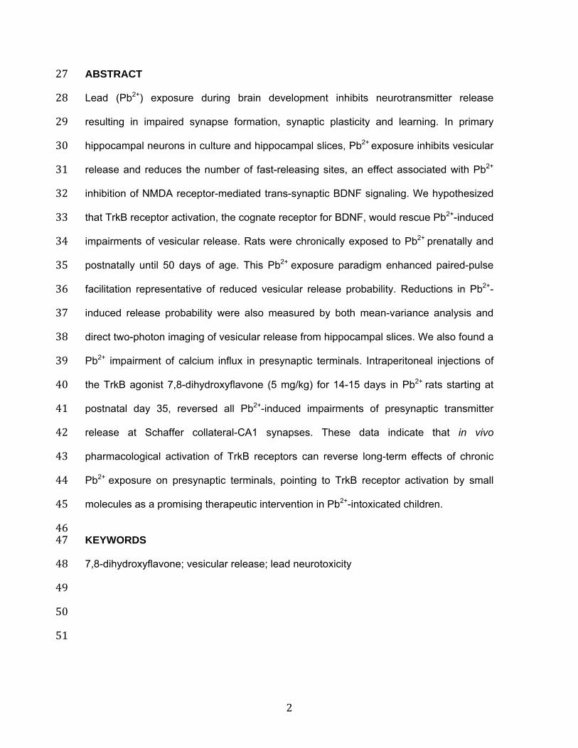
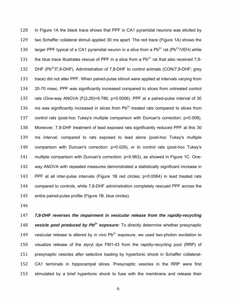
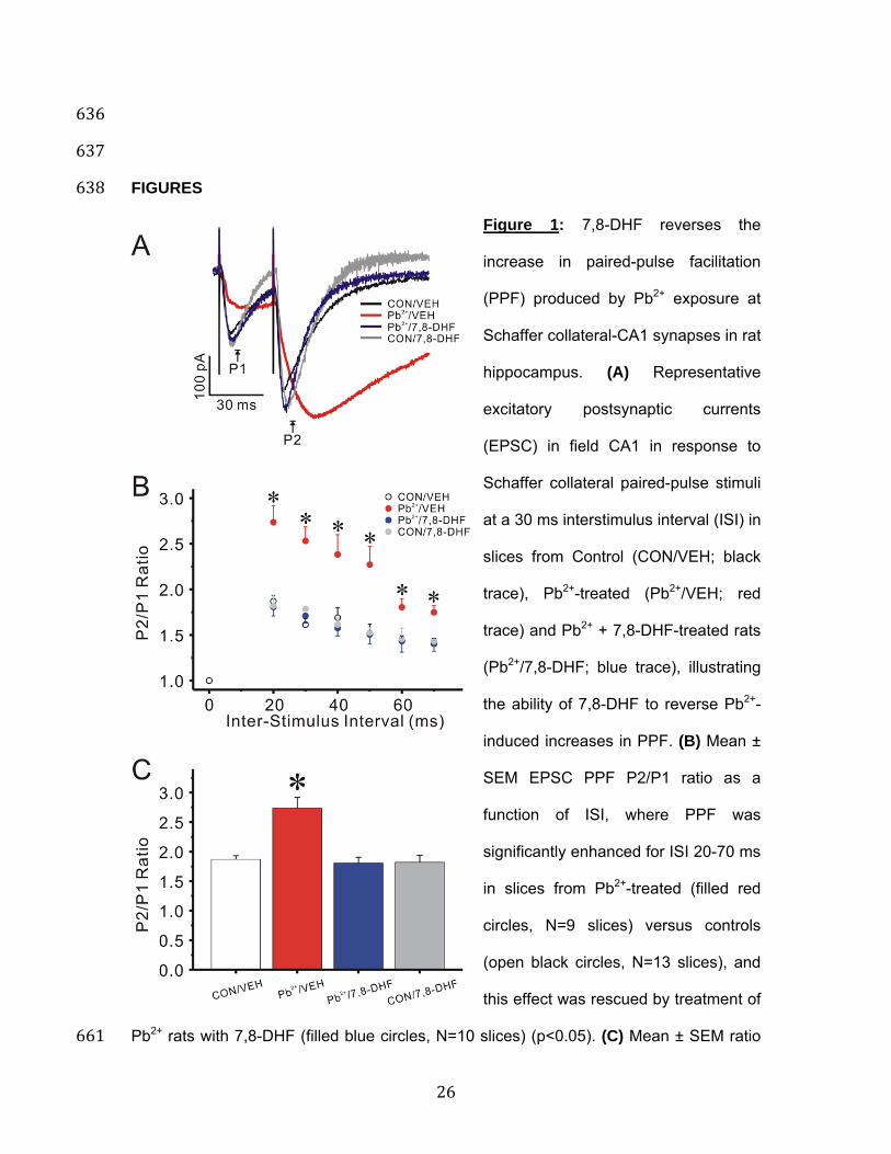
In Figure 1A the black trace shows that PPF in CA1 pyramidal neurons was elicited by 128 two Schaffer collateral stimuli applied 30 ms apart. The red trace (Figure 1A) shows the 129 larger PPF typical of a CA1 pyramidal neuron in a slice from a Pb2+ rat (Pb2+/VEH) while 130 the blue trace illustrates rescue of PPF in a slice from a Pb2+ rat that also received 7,8-131 DHF (Pb2+/7,8-DHF). Administration of 7,8-DHF to control animals (CON/7,8-DHF; grey 132 trace) did not alter PPF. When paired-pulse stimuli were applied at intervals varying from 133 20-70 msec, PPF was significantly increased compared to slices from untreated control 134 rats (One-way ANOVA (F(2,29)=9.786, p=0.0006). PPF at a paired-pulse interval of 30 135 ms was significantly increased in slices from Pb2+-treated rats compared to slices from 136 control rats (post-hoc Tukey's multiple comparison with Duncan's correction: p=0.008). 137 Moreover, 7,8-DHF treatment of lead exposed rats significantly reduced PPF at this 30 138 ms interval, compared to rats exposed to lead alone (post-hoc Tukey's multiple 139 comparison with Duncan's correction: p=0.029), or to control rats (post-hoc Tukey's 140 multiple comparison with Duncan's correction: p=0.963), as showed in Figure 1C. One-141 way ANOVA with repeated measures demonstrated a statistically significant increase in 142 PPF at all inter-pulse intervals (Figure 1B red circles; p=0.0064) in lead treated rats 143 compared to controls, while 7,8-DHF administration completely rescued PPF across the 144 entire paired-pulse profile (Figure 1B, blue circles). 145 146 7,8-DHF reverses the impairment in vesicular release from the rapidly-recycling 147 vesicle pool produced by Pb2+ exposure: To directly determine whether presynaptic 148 vesicular release is altered by in vivo Pb2+ exposure, we used two-photon excitation to 149 visualize release of the styryl dye FM1-43 from the rapidly-recycling pool (RRP) of 150 presynaptic vesicles after selective loading by hypertonic shock in Schaffer collateral-151 CA1 terminals in hippocampal slices. Presynaptic vesicles in the RRP were first 152 stimulated by a brief hypertonic shock to fuse with the membrane and release their 153
7
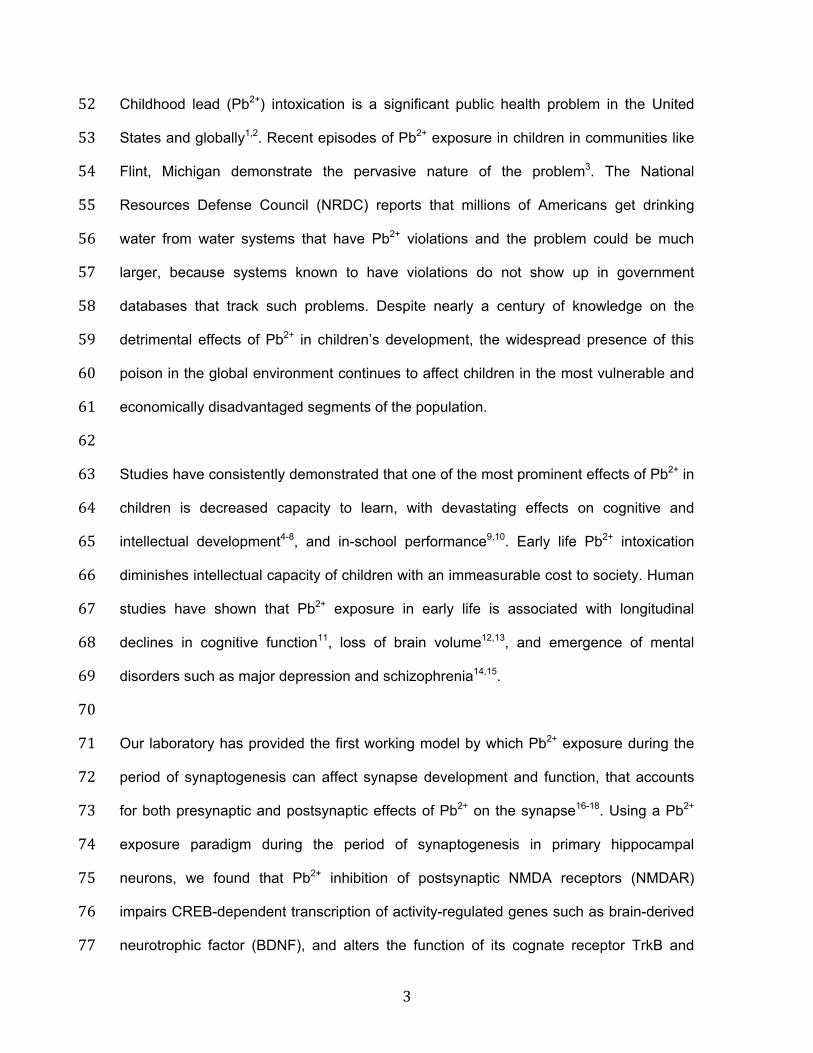
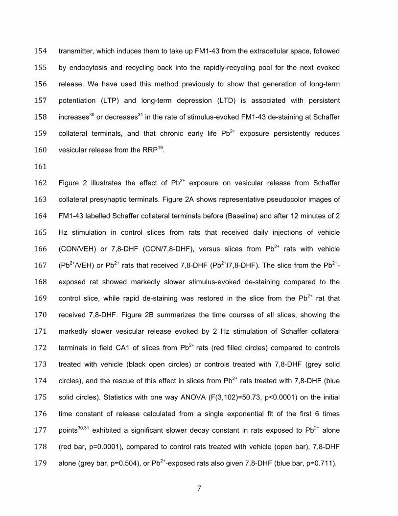
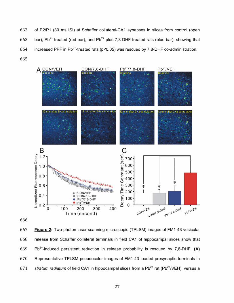
transmitter, which induces them to take up FM1-43 from the extracellular space, followed 154 by endocytosis and recycling back into the rapidly-recycling pool for the next evoked 155 release. We have used this method previously to show that generation of long-term 156 potentiation (LTP) and long-term depression (LTD) is associated with persistent 157 increases30 or decreases31 in the rate of stimulus-evoked FM1-43 de-staining at Schaffer 158 collateral terminals, and that chronic early life Pb2+ exposure persistently reduces 159 vesicular release from the RRP19. 160 161 Figure 2 illustrates the effect of Pb2+ exposure on vesicular release from Schaffer 162 collateral presynaptic terminals. Figure 2A shows representative pseudocolor images of 163 FM1-43 labelled Schaffer collateral terminals before (Baseline) and after 12 minutes of 2 164 Hz stimulation in control slices from rats that received daily injections of vehicle 165 (CON/VEH) or 7,8-DHF (CON/7,8-DHF), versus slices from Pb2+ rats with vehicle 166 (Pb2+/VEH) or Pb2+ rats that received 7,8-DHF (Pb2+/7,8-DHF). The slice from the Pb2+-167 exposed rat showed markedly slower stimulus-evoked de-staining compared to the 168 control slice, while rapid de-staining was restored in the slice from the Pb2+ rat that 169 received 7,8-DHF. Figure 2B summarizes the time courses of all slices, showing the 170 markedly slower vesicular release evoked by 2 Hz stimulation of Schaffer collateral 171 terminals in field CA1 of slices from Pb2+ rats (red filled circles) compared to controls 172 treated with vehicle (black open circles) or controls treated with 7,8-DHF (grey solid 173 circles), and the rescue of this effect in slices from Pb2+ rats treated with 7,8-DHF (blue 174 solid circles). Statistics with one way ANOVA (F(3,102)=50.73, p<0.0001) on the initial 175 time constant of release calculated from a single exponential fit of the first 6 times 176 points30,31 exhibited a significant slower decay constant in rats exposed to Pb2+ alone 177 (red bar, p=0.0001), compared to control rats treated with vehicle (open bar), 7,8-DHF 178 alone (grey bar, p=0.504), or Pb2+-exposed rats also given 7,8-DHF (blue bar, p=0.711). 179
8
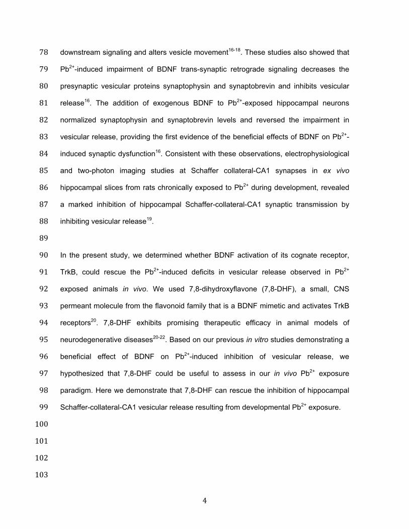
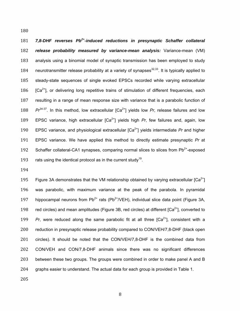
180 7,8-DHF reverses Pb2+-induced reductions in presynaptic Schaffer collateral 181 release probability measured by variance-mean analysis: Variance-mean (VM) 182 analysis using a binomial model of synaptic transmission has been employed to study 183 neurotransmitter release probability at a variety of synapses32,33. It is typically applied to 184 steady-state sequences of single evoked EPSCs recorded while varying extracellular 185 [Ca2+], or delivering long repetitive trains of stimulation of different frequencies, each 186 resulting in a range of mean response size with variance that is a parabolic function of 187 Pr34-37. In this method, low extracellular [Ca2+] yields low Pr, release failures and low 188 EPSC variance, high extracellular [Ca2+] yields high Pr, few failures and, again, low 189 EPSC variance, and physiological extracellular [Ca2+] yields intermediate Pr and higher 190 EPSC variance. We have applied this method to directly estimate presynaptic Pr at 191 Schaffer collateral-CA1 synapses, comparing normal slices to slices from Pb2+-exposed 192 rats using the identical protocol as in the current study19. 193 194 Figure 3A demonstrates that the VM relationship obtained by varying extracellular [Ca2+] 195 was parabolic, with maximum variance at the peak of the parabola. In pyramidal 196 hippocampal neurons from Pb2+ rats (Pb2+/VEH), individual slice data point (Figure 3A, 197 red circles) and mean amplitudes (Figure 3B, red circles) at different [Ca2+], converted to 198 Pr, were reduced along the same parabolic fit at all three [Ca2+], consistent with a 199 reduction in presynaptic release probability compared to CON/VEH/7,8-DHF (black open 200 circles). It should be noted that the CON/VEH/7,8-DHF is the combined data from 201 CON/VEH and CON/7,8-DHF animals since there was no significant differences 202 between these two groups. The groups were combined in order to make panel A and B 203 graphs easier to understand. The actual data for each group is provided in Table 1. 204 205
9
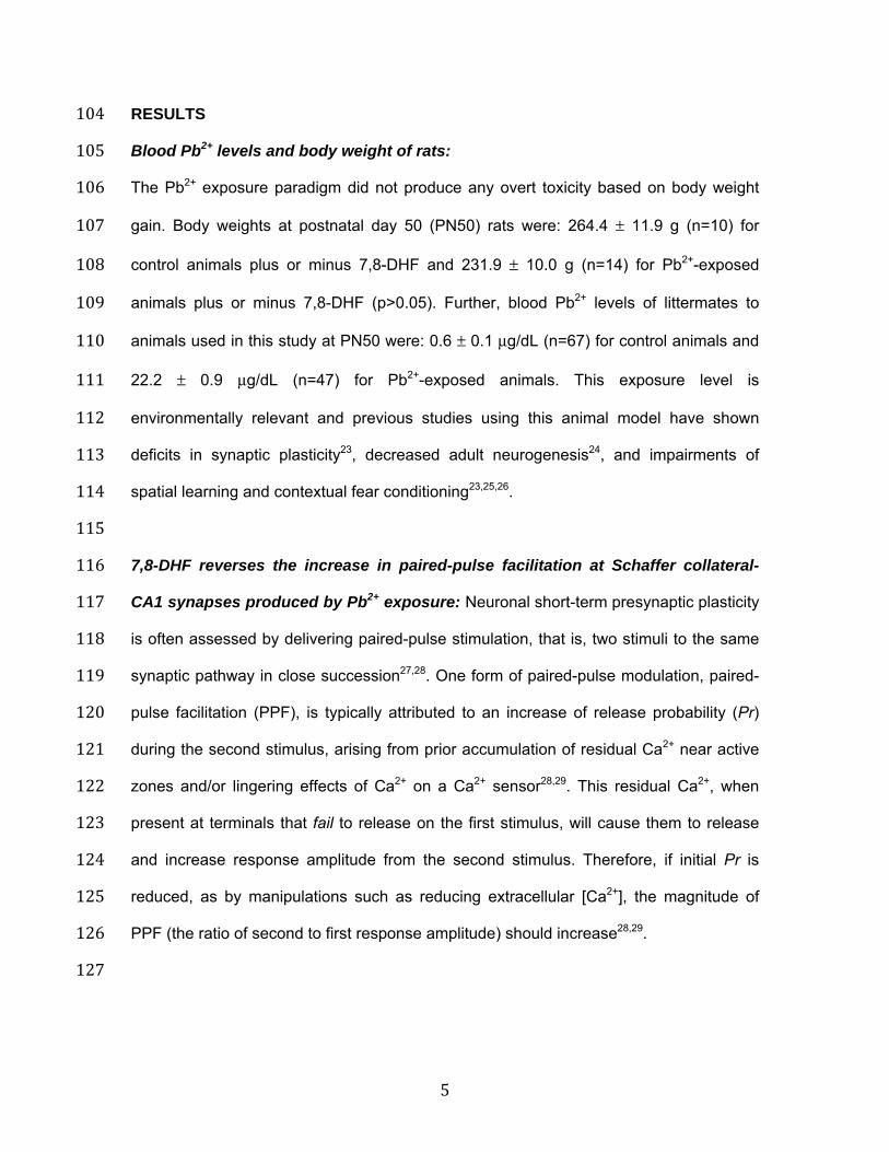
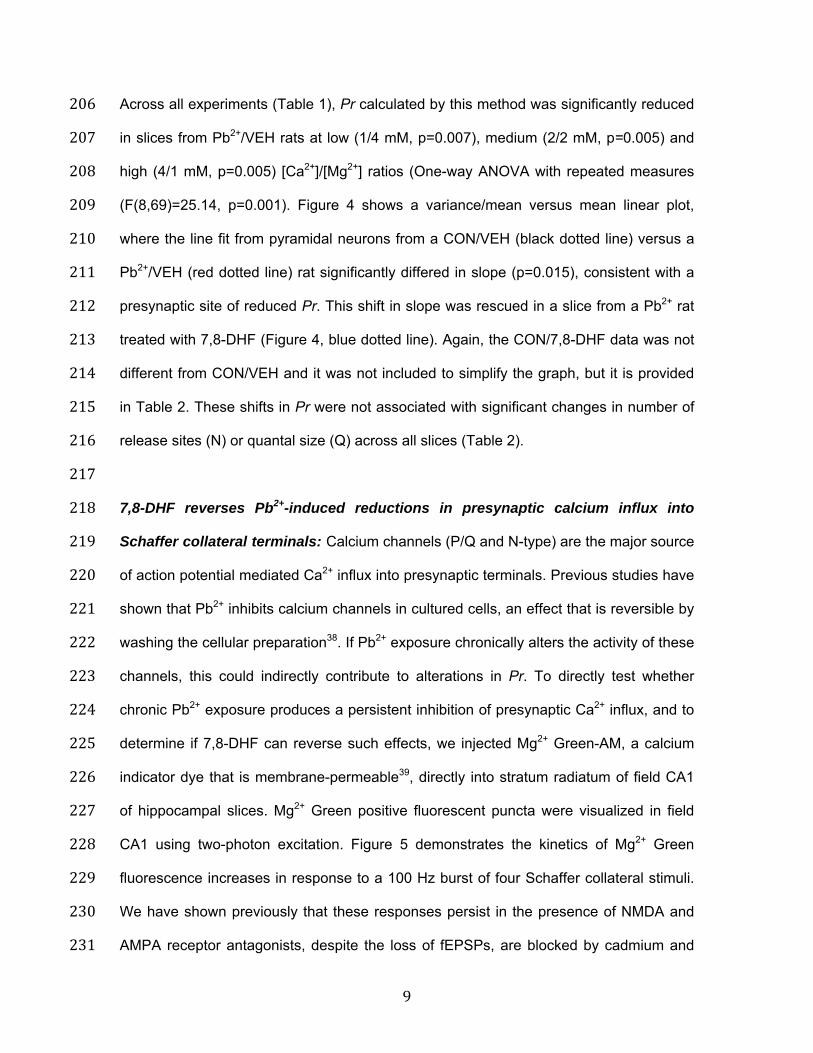
Across all experiments (Table 1), Pr calculated by this method was significantly reduced 206 in slices from Pb2+/VEH rats at low (1/4 mM, p=0.007), medium (2/2 mM, p=0.005) and 207 high (4/1 mM, p=0.005) [Ca2+]/[Mg2+] ratios (One-way ANOVA with repeated measures 208 (F(8,69)=25.14, p=0.001). Figure 4 shows a variance/mean versus mean linear plot, 209 where the line fit from pyramidal neurons from a CON/VEH (black dotted line) versus a 210 Pb2+/VEH (red dotted line) rat significantly differed in slope (p=0.015), consistent with a 211 presynaptic site of reduced Pr. This shift in slope was rescued in a slice from a Pb2+ rat 212 treated with 7,8-DHF (Figure 4, blue dotted line). Again, the CON/7,8-DHF data was not 213 different from CON/VEH and it was not included to simplify the graph, but it is provided 214 in Table 2. These shifts in Pr were not associated with significant changes in number of 215 release sites (N) or quantal size (Q) across all slices (Table 2). 216 217 7,8-DHF reverses Pb2+-induced reductions in presynaptic calcium influx into 218 Schaffer collateral terminals: Calcium channels (P/Q and N-type) are the major source 219 of action potential mediated Ca2+ influx into presynaptic terminals. Previous studies have 220 shown that Pb2+ inhibits calcium channels in cultured cells, an effect that is reversible by 221 washing the cellular preparation38. If Pb2+ exposure chronically alters the activity of these 222 channels, this could indirectly contribute to alterations in Pr. To directly test whether 223 chronic Pb2+ exposure produces a persistent inhibition of presynaptic Ca2+ influx, and to 224 determine if 7,8-DHF can reverse such effects, we injected Mg2+ Green-AM, a calcium 225 indicator dye that is membrane-permeable39, directly into stratum radiatum of field CA1 226 of hippocampal slices. Mg2+ Green positive fluorescent puncta were visualized in field 227 CA1 using two-photon excitation. Figure 5 demonstrates the kinetics of Mg2+ Green 228 fluorescence increases in response to a 100 Hz burst of four Schaffer collateral stimuli. 229 We have shown previously that these responses persist in the presence of NMDA and 230 AMPA receptor antagonists, despite the loss of fEPSPs, are blocked by cadmium and 231
10
omega conotoxin, and co-localize with FM4-64, confirming a presynaptic nature for 232 these calcium transients40. 233 234 Comparison of mean fluorescence increases of representative stimulus-evoked 235 presynaptic Ca2+ influx transients in Schaffer collateral terminals in slices from vehicle 236 control (CON/VEH; Figure 5A black trace) or control treated with 7,8-DHF (CON/7,8-237 DHF; Figure 5A grey trace), versus Pb2+ rats (Pb2+/VEH; Figure 5A red trace) and Pb2+ 238 rats treated with 7,8-DHF (Pb2+/7,8-DHF; Figure 5A, blue trace), revealed that action 239 potential-dependent Ca2+ influx was reduced in amplitude by Pb2+ exposure, and that 240 7,8-DHF was able to reverse this reduction in presynaptic Ca2+ influx. Figure 5B 241 summarizes these results across all slices, showing that Schaffer collateral presynaptic 242 terminals in hippocampal slices from Pb2+ rats (red bar) exhibited reduced Ca2+ influx 243 (One-way ANOVA with repeated measures F(3,28)=5.233, p=0.0054) compared to 244 vehicle control slices (black bar, p=0.0129), 7,8-DHF treated slices (grey bar, p=0.0192), 245 or slices from rats exposed to Pb2+ plus 7,8-DHF injections (blue bar, p=0.0269). Taken 246 together, our data indicate that chronic exposure to Pb2+ during development results in a 247 persistent reduction in presynaptic Pr that may be due to both reduced Ca2+ influx, and 248 actions downstream of presynaptic Ca2+ influx at the level of SNARE protein-mediated 249 exocytosis16-18. Consistent with its effects in rescuing Pr, daily injection of 7,8-DHF was 250 also able to rescue the effects of Pb2+ exposure in reducing presynaptic Ca2+ influx at 251 Schaffer collateral terminals in the hippocampus. 252 253 254 255 256 257
11
DISCUSSION 258 Our current study identifies a novel therapeutic target with the potential to treat Pb2+-259 intoxicated children. Daily postnatal administration of the cell permeant TrkB receptor 260 agonist 7,8-DHF, which readily crosses the blood-brain barrier, completely rescued Pb2+-261 induced reductions in vesicular release and presynaptic Ca2+ influx, supporting its 262 potential as a novel treatment for the cognitive effects of early life Pb2+ exposure. 7,8-263 DHF is a naturally occurring small molecule in the flavonoid family of polyphenolic 264 compounds found in Godmania aesculifoloia, Tridax procumbens, and primula tree 265 leaves41,42. It has been shown to be neuroprotective in preclinical studies43,44. However, 266 further studies are needed to determine whether the rescue of transmitter release by 267 7,8-DHF is long-lasting or permanent, and whether it rescues behavioral impairments 268 associated with chronic developmental exposure to Pb2+. 269 270 Previous studies using acute exposure to Pb2+ in cultured cells have shown inhibition of 271 Ca2+ channels by Pb2+, an effect that is reversed upon washout of Pb2+ from the cells38. 272 There could be additional mechanisms by which Pb2+ persistently alters vesicular 273 release, such as changes in levels of SNARE proteins that we have shown 274 previously16,17. In this study, fluorescent imaging of presynaptic Ca2+ influx showed that 275 Pb2+ exposure was associated with reductions in voltage-dependent Ca2+ channel-276 mediated Ca2+ entry that were completely reversed by 7,8-DHF. Previously, we found 277 that presynaptic Ca2+ fluorescent signals evoked by brief 20 Hz bursts of stimulation 278 showed only a small, early reduction in amplitude of presynaptic Ca2+ signals in slices 279 from Pb2+ rats19, leading us to use higher frequency 100 Hz bursts of stimulation in this 280 study. Our present findings suggest that developmental Pb2+ exposure can persistently 281 impair presynaptic Ca2+ entry, and have additional downstream effects at the level of 282 vesicular SNARE protein-mediated docking, recycling, and long-term stability of the 283
12
release complex, consistent with our previous findings in hippocampal neuronal 284 cultures16, and hippocampal slices from Pb2+ rats19. 285 286 In this study, rats were exposed to Pb2+ chronically during gestation, postnatally and 287 continuing through to young adulthood. The artificial cerebrospinal fluid used to maintain 288 slice viability during experiments did not contain Pb2+, indicating that the effects 289 observed were the result of the in vivo Pb2+ exposure. Our studies have previously 290 shown16 that BDNF synthesis and release are decreased in cultured hippocampal 291 neurons exposed to Pb2+, and are associated with reductions in levels of SNARE 292 proteins and inhibition of vesicular release. These effects of in vitro Pb2+ exposure, were 293 rescued by exogenous BDNF, consistent with our present findings. Stansfield et al.18, 294 using the same Pb2+ exposure paradigm in cultured neurons, showed that Pb2+ may 295 impair the transport of BDNF-containing vesicles, possibly by altering Huntingtin 296 phosphorylation at a site promoting anterograde BDNF vesicle movement. This effect of 297 Pb2+ resulted in impaired BDNF release, decreasing TrkB activation, and 298 phosphorylation of synapsin I. Our current findings further support the hypothesis that 299 BDNF receptor agonists and treatments such as enriched environments that increase 300 BDNF levels and release45,46, may be able to rescue the effects of chronic Pb2+ exposure 301 we observed in more intact hippocampal synaptic networks. Further, previous studies 302 from our laboratory have shown that environmental enrichment can reverse Pb2+-303 induced impairments of spatial learning in rats of similar age and Pb2+ treatment47. This 304 study also showed that Pb2+-exposed rats placed in an enriched environment that 305 reverses learning deficits, also exhibit increased BDNF gene expression in the 306 hippocampus47, supporting our current data implicating the BDNF-TrkB system in Pb2+ 307 neurotoxicity and suggesting the BDNF mimetic 7,8-DHF as a potential therapy for Pb2+-308 intoxicated children. 309
13
310 METHODS 311 Chemicals: Chemicals for extra- and intracellular solutions were purchased from Sigma-312 Aldrich (St. Louis, MO). Neurotransmitter receptor antagonists were purchased from 313 Tocris Cookson (Minneapolis, MN), and FM1-43 from Invitrogen (Grand Island, NY). 314 315 Blood Pb2+ analysis: Blood Pb2+ levels in samples from littermates were measured using 316 the LeadCare system (Magellan Diagnostics, N. Billerica, MA). 317 318 Animals: Adult female Long-Evans rats (250 g) were purchased from Charles River, Inc. 319 (Wilmington, MA) and randomly placed on diet containing 0 (control) or 1500 ppm lead 320 acetate (PbAc) (Dyets, Bethlehem, PA) 10 days prior to breeding with non-exposed 321 Long-Evans males (300 g). Litters were culled to 10 pups on postnatal day 1 (PN1). 322 Dams were maintained on their respective diet and at PN21 male pups were weaned 323 onto the same diet and maintained until PN50. All rats are housed in plastic cages at 22 324 ± 2°C on a 12/12 light:dark cycle. Food and water were allowed ad libitum. Each litter is 325 a single experimental unit for statistical purposes, so that for each experiment only one 326 animal per litter was used for one data point. All studies were conducted in accordance 327 with the United States Public Health Service’s Policy on Human Care and Use of 328 Laboratory Animals under protocols approved by Institutional Animal Care and Use 329 Committees from each university. 330 331 Hippocampal slice electrophysiology: Experiments were conducted as described 332 previously19,30,31. At 50 ± 2 days of age, rats were deeply anesthetized with isoflurane, 333 decapitated and their brains rapidly removed and submerged in ice-cold artificial 334 cerebrospinal fluid (ACSF, 2–4 °C), containing (in mM): 124 NaCl, 4 KCl, 2 MgCl2, 2 335
14
CaCl2, 1.25 NaH2PO4, 26 NaHCO3, 10 glucose; at pH 7.4, gassed continuously with 336 95% O2/5% CO2). Brains were hemisected, the frontal lobes cut off, and individual 337 hemispheres glued using cyanoacrylate adhesive onto a stage immersed in ice-cold 338 ACSF gassed continuously with 95% O2/5% CO2 during slicing. We cut 400 µm thick 339 coronal slices using a vibratome (Leica VT1200S), and transferred them to an interface 340 holding chamber for incubation at room temperature for a minimum of 1 hr before 341 transferring to a submerged recording chamber continuously on a Zeiss Axioskop 342 microscope continuously perfused at 3 ml/min with oxygenated ACSF at 32 ± 0.5 °C. 343 344 Whole cell patch-clamp recordings were performed in CA1 pyramidal neurons using 345 standard techniques. Patch pipettes (R=3-4 MΩ) were filled with recording solution 346 containing (in mM): 135 CsMeSO3, 8 NaCl, 10 HEPES, 2 Mg-ATP, 0.3 Na-GTP, 0.5 347 EGTA, and 1 QX-314 (275 mOsm, pH 7.25 adjusted with Cs(OH)2). Access resistance 348 was carefully monitored, and only cells with stable access resistance (<5% change) 349 were included in analyses. CA1 pyramidal cells were recorded under voltage clamp 350 using a MultiClamp 700B (Axon Instruments) with Clampex (v9). Recording signals were 351 filtered through an eight-pole Bessel low-pass filter with a 3 kHz cutoff frequency, 352 digitized at 10 kHz, and sampled using Clampex (v9). Neurons were clamped at –60 353 mV, and Schaffer collateral-evoked EPSCs were delivered by a bipolar stimulating 354 electrode (FHC, USA, 50-100 pA, 100 µs duration). EPSC slopes were calculated by 355 linear interpolation of the initial downward current from 20% to 80% of the maximum 356 EPSC amplitude. Paired-pulse facilitation was assessed by applying a pair of Schaffer 357 collateral stimuli at intervals of 10-125 msec, and the ratio of slopes of the second to the 358 first response was calculated, so that numbers greater than 1.0 represented facilitation, 359 less than 1.0 inhibition. 360 361
15
362 363 Two-photon laser scanning microscopy 364 Vesicular release FM1-43 fluorescence measurements: Fluorescence was visualized 365 using a customized two-photon laser-scanning Olympus BX61WI microscope with a 366 60x/0.90W water immersion infrared objective lens and an Olympus multispectral 367 confocal laser scan unit. The light source was a Mai-Tai™ laser (Solid-State Laser Co., 368 Mountain View, CA), tuned to 860 nm for exciting Magnesium Green and 820 nm for 369 exciting FM1-43. Epifluorescence was detected with photomultiplier tubes of the 370 confocal laser scan head with pinhole maximally opened and emission spectral window 371 optimized for signal over background. In the transfluorescent pathway, a 565 nm dichroic 372 mirror was used to separate green and red fluorescence to eliminate transmitted or 373 reflected excitation light (Chroma Technology, Rockingham, VT). After confirming the 374 presence of Schaffer collateral-evoked fEPSPs >1 mV in amplitude in CA1 stratum 375 radiatum, and inducing LTP, 10 µM 6-cyano-7-nitroquinoxaline-2,3-dione (CNQX) was 376 bath-applied throughout the rest of the experiment to prevent synaptically-driven action 377 potentials in CA3 pyramidal neurons from accelerating dye release. Presynaptic boutons 378 were loaded by bath-applying 5 µM FM1-43 (Molecular Probes) in hypertonic ACSF 379 supplemented with sucrose to 800 mOsm for 25 sec to selectively load the rapidly-380 recycling pool (RRP)30,31, then returned to normal ACSF. Stimulus-induced destaining 381 was measured after 30 min perfusion with dye-free ACSF, by bursts of 10 Hz bipolar 382 stimuli (150 µs DC pulses) for 2 sec applied once each 30 sec. We fitted a single 383 exponential to the first 6 fluorescence time course values, and decay time constants 384 between groups compared by two-tailed Student’s t-test, as we have shown previously 385 that the early release reflects vesicular release from the RRP prior to recycling and 386 reuse of vesicles30,31. 387
16
388 Presynaptic Ca2+ influx fluorescence measurements: Using established methods for 389 measuring [Ca2+] transients48, we filled Schaffer collateral presynaptic fibres with 390 Magnesium Green AM. Briefly, an ejection electrode (tip diameter, 5-10 µm) containing 391 Magnesium Green AM (1 mM Magnesium Green AM, 10% DMSO, 1% pluronic acid in 392 ACSF) was lowered into the Schaffer collateral pathway between the stimulating 393 electrode and the presynaptic terminal field to be observed, air pressure pulses (6-9 psi, 394 100-200 msec) controlled by a Picospritzer (General Valve Corp. USA) were applied to 395 the electrode until a small bright spot (≈10 mm in diameter) was observed. Thirty 396 minutes elapsed to allow dye to sufficiently diffuse into presynaptic boutons prior to 397 commencing imaging. To verify that magnesium green selectively loaded presynaptic 398 terminals, FM4-64 was loaded with high [K+]o at the end of the experiment. To measure 399 Ca2+ dynamics, stimulus-evoked fluorescence signals were collected by scanning at 200 400 Hz in surface-scanning mode (XYT). Baseline fluorescence (F0) was averaged over four 401 images, and ∆F/F calculated as (∆F/F)(t)=(F(t)-F0)/F0. 402 403 Estimation of presynaptic release probability by variance-mean (VM) analysis: Variance-404 mean (VM) analysis according to a binomial model of synaptic transmission is a method 405 that has been employed to study transmitter release at many synapses32,33,49. It is mainly 406 applied to steady-state sequences of evoked EPSCs recorded under a variety of 407 conditions by varying extracellular [Ca2+], or delivering long repetitive trains of stimulation 408 of different frequencies, each resulting in a range of mean response size34-37. 409 410 We used three ratios of [Ca2+]/[Mg2+] in ACSF (4/1, 2/2, and 1/4 mM) to alter release 411 probability at Schaffer collateral synapses. Experiments began by establishing stable 412 whole-cell recording from a CA1 pyramidal neuron, and then perfusing the slice with 4/1 413
17
[Ca2+]/[Mg2+] ACSF. Cells were voltage-clamped at -65 mV, and 100 µs constant-current 414 stimulus pulses were delivered to Schaffer collateral/commissural fiber axons every 10 415 sec to evoke an EPSC. Stable recordings for 8-10 min were made in 4/1 [Ca2+]/[Mg2+], 416 before replacing the perfusate with 1/4 mM [Ca2+]/[Mg2+] ACSF. After EPSCs decreased 417 in amplitude and restabilized, which usually took 5-8 min, EPSCs were recorded for an 418 additional 8 min. Slices were then perfused with 2/2 mM [Ca2+]/[Mg2+] ACSF. After EPSC 419 amplitudes had again stabilized, another 8 min of recordings were made. To induce 420 LTD, slices were exposed to either 10 µM NMDA or 25 µM DHPG in 2/2 mM 421 [Ca2+]/[Mg2+] ACSF for three or five min, respectively, durations which reliably induced 422 LTD lasting hours. After drug exposure, slices were perfused with 2/2 mM [Ca2+]/[Mg2+] 423 ACSF for >30 min, to verify expression of LTD, and then the same sequence of 424 [Ca2+]/[Mg2+] ACSF applications was repeated. To ensure that postsynaptic AMPA 425 receptors were responding to non-saturating glutamate concentration, a requirement for 426 VM analysis, experiments were performed in a low concentration of the AMPA receptor 427 antagonist 6,7-dinitroquinoxaline-2,3-dione (DNQX, 100 nM). 428 429 7,8-DHF administration: 430 7,8-Dihydroxyflavone hydrate (DHF, Sigma-Aldrich, St Louis, MO) was dissolved in 431 phosphate-buffered saline (PBS) containing 17% dimethylsulfoxide (DMSO). Male rats 432 received daily intraperitoneal injections of 5 mg/kg 7,8-DHF or 17% DMSO vehicle daily 433 for 14-15 consecutive days starting when they were 35-42 days of age. Rats were 434 sacrificed for slice preparation twenty-four hours after the last 7,8-DHF administration. 435 436 Statistics: Power analysis showed a group size of 6 animals per treatment group with 437 significance level pre-set to p<0.05 could detect between group differences of 10% for 438 vesicular transmitter release time constants and release probability at a power of 85% 439
18
with typical parameter standard deviations. Data sets did not deviate significantly from 440 normal distribution (D’Agostino-Pearson omnibus normality test), and did not exhibit 441 significant differences in parameter variances (F-test). Slices and treatments were 442 randomized, with treated and control slices examined in parallel on the same or 443 sequential days. While all analyses were automated, the investigator was not blinded to 444 treatment group. Student’s t-test was used to determine differences between the control 445 and Pb2+ treated groups for each particular measure. In analyses requiring comparisons 446 between multiple groups, a one-way ANOVA with Sidak’s Multiple Comparisons analysis 447 was used with post-hoc Tukey’s test for individual group comparisons. Significance level 448 was preset to p< 0.05. 449 450 Data Availability: Following calculation of EPSP slopes, paired-pulse ratios or optical 451 time courses of vesicular release and presynaptic calcium influx, all data will be made 452 available upon request. Image analysis was performed with ImageJ (NIH), statistical 453 analyses with GraphPad Prism v6 (La Jolla, CA), and custom-built, proprietary software 454 was used to control the multi-photon laser scanner for FM1-43 imaging experiments, 455 which is copyrighted by the designer and cannot be made available. 456 457 458 459 460 461 462 463 464 465
19
466 467 468 ACKNOWLEDGEMENTS: This work was supported by grants ES006189 and 469 ES020465 from the National Institute of Environmental Health Sciences to TRG. 470 471 AUTHOR CONTRIBUTIONS: 472 X.Z.: experimental planning, analysis and execution; writing and editing of manuscript. 473 J.L.M.: experimental planning and execution; writing and editing of manuscript. O.M.: 474 experimental execution; K.H.S.: experimental execution; M.K.L.: experimental execution; 475 P.K.S.: experimental planning, analysis, and direction; writing and editing of manuscript; 476 T.R.G.: conceptualized overall studies and received funding, experimental planning and 477 direction of study; writing and editing of manuscript. 478 479 COMPETING INTERESTS: 480 The authors declare no competing financial interests. 481 482 483
20
REFERENCES 484 1. Tong, S., von Schirnding, Y.E. & Prapamontol, T. Environmental lead exposure: a 485
public health problem of global dimensions. Bull. World Hlth. Org. 78, 1068-1077 486 (2000). 487
2. Toscano, C.D. & Guilarte, T.R. Lead Neurotoxicity: From Exposure to Molecular 488 Effects. Brain Res. Rev. 49, 529-554 (2005). 489
3. Hanna-Atisha, M., LaChance, J., Sadler, R.C. & Champney Schnepp, A. Elevated 490 blood lead levels in children associated with the Flint drinking water crisis: A spatial 491 analysis of risk and public health response. Am. J. Public Health 106, 283-290 492 (2016). 493
4. Bellinger, D.C., Stiles, K.M. & Needleman, H.L. Low level lead exposure, intelligence 494 and academic achievement: a long term follow up. Pediatrics 90, 855-861 (1992). 495
5. Canfield, R.L., Henderson, C.R. Jr., Cory-Slechta, D.A., Cox, C., Jusko, T.A. & 496 Lanphear, B.P. Intellectual impairment in children with blood lead concentrations 497 below 10 ug per deciliter. N. Engl. J. Med. 348, 1517-1526 (2003). 498
6. Lanphear, B.P., Hornung, R., Khoury, J., Yolton, K., Baghurst, P., Bellinger, D.C., 499 Canfield, R.L., Dietrich, K.N., Bornschein, R., Greene, T., Rothenberg, S.J., 500 Needleman, H.L., Schnaas, L., Wasserman, G., Graziano, J. & Roberts, R. Low-level 501 environmental lead exposure and children’s intellectual function: An international 502 pooled analysis. Environ. Health Perspect. 113, 894-899 (2005). 503
7. Jusko, T.A., Henderson, C.R., Lanphear, B.P., Cory-Slechta, D.A., Parsons, P.J. & 504 Canfield, R.L. Blood lead concentrations < 10 ug/dl and child intelligence at 6 years 505 of age. Environ. Health Perspect. 116, 243-248 (2008). 506
8. Schwartz, B.S., Stewart, W.F., Bolla, K.I., Simon, P.D., Bandeen-Roche, K., Gordon, 507 P.B., Links, J.M. & Todd, A.C. Past adult lead exposure is associated with 508 longitudinal decline in cognitive function. Neurology 55, 1144-1150 (2000). 509
21
9. Magzamen, S., Imm, P., Amato, M.S., Havlena, J.A., Anderson, H.A., Moore, C.F. & 510 Kanarek, M.S. Moderate lead exposure and elementary school end-of-grade 511 examination performance. Ann. Epidemiol. 23, 700-707 (2013). 512
10. Evens, A., Hryhorczuk, D., Lanphear, B.P., Rankin, K.M., Lewis, D.A., Forst, L. & 513 Rosenberg, D. The impact of low-level lead toxicity on school performance among 514 children in the Chicago Public Schools: a population-based retrospective cohort 515 study. Environ. Health 14, 21 (2015). 516
11. Stewart, W.F., Schwartz, B.S., Davatzikos, C., Shen, D., Liu, D., Wu, X., Todd, A.C., 517 Shi, W., Bassett, S. & Youssem, D. Past adult lead exposure is linked to 518 neurodegeneration measured by brain MRI. Neurology 66, 1476-1484 (2006). 519
12. Cecil, K.M., Brubaker, C.J., Adler, C.M., Dietrich, K.N., Altaye, M., Egelhoff, J.C., 520 Wessel, S., Elangovan, I., Hornung, R., Jarvis, K. & Lanphear, B.P. Decreased brain 521 volume in adults with childhood lead exposure. PLoS Med. 5, e112 (2008). 522
13. Rizzoli, S.O. & Betz, W.J. Synaptic vesicle pools. Nat. Rev. Neurosci. 6, 57-69 523 (2005). 524
14. Bouchard, M.F., Bellinger, D.C, Weuve, J., Matthews-Bellinger, J., Gilman, S.E., 525 Wright, R.O., Schwartz, J. & Weisskopf, M.G. Blood lead levels and major 526 depressive disorder, panic disorder, and generalized anxiety disorder in US young 527 adults. Arch. Gen. Psychiatry 66, 1313-1319 (2009). 528
15. Guilarte, T.R., Opler, M. & Pletnikov, M. Is lead exposure in early life an 529 environmental risk factor for schizophrenia? Neurobiological connections and 530 testable hypotheses. Neurotoxicology 33, 560-574 (2012). 531
16. Neal, A.P., Stansfield, K.H., Worley, P.F., Thompson, R.E. & Guilarte, T.R. Lead 532 exposure during synaptogenesis alters vesicular release proteins and impairs 533 vesicular release: potential role of NMDA receptor-dependent BDNF signaling. 534 Toxicol. Sci. 116, 249-263 (2010). 535
22
17. Neal, A.P. & Guilarte, T.R. Molecular Neurobiology of Pb2+: effects on synaptic 536 function. Mol. Neurobiol. 42, 151-160 (2010). 537
18. Stansfield, K.H., Pilsner, J.R., Lu, Q., Wright, R.O. & Guilarte, T.R. Dysregulation of 538 BDNF-TrkB signaling in developing hippocampal neurons by Pb2+: implications for an 539 environmental basis of neurodevelopmental disorders. Toxicol. Sci. 127, 277-295 540 (2012). 541
19. Zhang, X.L., Guariglia, S.R., McGlothan, J.L., Stansfield, K.H., Stanton, P.K. & 542 Guilarte, T.R. Presynaptic mechanisms of lead neurotoxicity: effects on vesicular 543 release, vesicle clustering and mitochondrial number. PLoS ONE 10, e0127461 544 (2015). 545
20. Liu, C., Chan, C.B. & Ye, K. 7,8-dihydroxyflavone, a small molecular TrkB agonist, is 546 useful for treating various BDNF-implicated human disorders. Transl. Neurodegener. 547 5, 2 (2016). 548
21. Devi, L. & Ohno, M. 7,8-dihyrdoxyflavone, a small-molecule TrkB agonist, reverses 549 memory deficits and BACE1 elevation in a mouse model of Alzheimer’s disease. 550 Neuropsychopharmacology 37, 434-444 (2012). 551
22. Jiang, M., Peng, Q., Liu, X., Jin, J., Hou, Z., Zhang, J., Mori, S., Ross, C.A., Ye, K. & 552 Duan, W. Small-molecule TrkB receptor agonists improve motor function and extend 553 survival in a mouse model of Huntington’s disease. Hum. Mol. Genet. 22, 2462-2470 554 (2013). 555
23. Nihei, M.K., Desmond, N.L., McGlothan, J.L., Kuhlmann, A.C. & Guilarte, T.R. N-556 methyl-D-asparatate receptor subunit changes are associated with lead-induced 557 deficits of long-term potentiation and spatial learning. Neuroscience 99, 233-242 558 (2000). 559
23
24. Verina,.T., Rohde, C.A. & Guilarte, T.R. Environmental lead exposure during early 560 life alters granule cell neurogenesis and morphology in the hippocampus of young 561 adult rats. Neuroscience 145, 1037-1047 (2007). 562
25. Kuhlmann, A.C., McGlothan, J.L. & Guilarte, T.R. Developmental lead exposure 563 causes spatial learning deficits in adult rats. Neurosci. Lett. 233, 101-104 (1997). 564
26. McGlothan, J.L., Karcz-Kubicha, M. & Guilarte, T.R. Developmental lead exposure 565 impairs extinction of conditioned fear in young adult rats. NeuroToxicology 29, 1127-566 1130 (2008). 567
27. Andersen, P. & Lømo, T. Control of hippocampal output by afferent volley frequency. 568 Progress in Brain Research: Structure and Function of the Limbic System, eds 569 Tokizane, T. & Adey, R.W. (Elsevier Science), 400-412 (1967). 570
28. Zucker, R.S. Short-term plasticity. Annu. Rev. Neurosci. 12, 13-31 (1989). 571 29. Neher, E. Vesicle pools and Ca2+ microdomains: new tools for understanding their 572
roles in neurotransmitter release. Neuron 20, 389-399 (1998). 573 30. Stanton, P.K., Winterer, J. & Müller, W. Imaging LTP of presynaptic release of FM1-574
43 from the rapidly-recycling vesicle pool at Schaffer collateral-CA1 synapses in rat 575 hippocampal slices. Eur. J. Neurosci. 22, 2451-2461 (2005). 576
31. Stanton, P.K., Winterer, J., Bailey, C.P., Kyrozis, A., Raginov, I., Laube, G., Veh, 577 R.W., Nguyen, C.Q. & Müller, W. Long-term depression of presynaptic release from 578 the readily-releasable vesicle pool induced by NMDA receptor-dependent retrograde 579 nitric oxide. J. Neurosci. 23, 5936-5944 (2003). 580
32. Clements, J.D. & Silver, R.A. Unveiling synaptic plasticity: a new graphical and 581 analytical approach. Trends Neurosci. 23, 105-113 (2000). 582
33. Silver, R.A. Estimation of nonuniform quantal parameters with multiple-probability 583 fluctuation analysis: theory, application and limitations. J. Neurosci. Methods 130, 584 127-141 (2003). 585
24
34. Silver, R.A., Momiyama, A. & Cull-Candy, S.G. Locus of frequency-dependent 586 depression identified with multiple-probability fluctuation analysis at rat climbing fibre-587 Purkinje cell synapses. J. Physiol. 510, 881-902 (1998). 588
35. Reid, C.A. & Clements, J.D. Postsynaptic expression of long-term potentiation in the 589 rat dentate gyrus demonstrated by variance-mean analysis. J. Physiol. 518, 121-30 590 (1999). 591
36.Oleskevich, S., Clements, J. & Walmsley, B. Release probability modulates short-592 term plasticity at a rat giant terminal. J. Physiol. 524, 513-523 (2000). 593
37. Foster, K.A. & Regehr, W.G. Variance-mean analysis in the presence of a rapid 594 antagonist indicates vesicle depletion underlies depression at the climbing fiber 595 synapse. Neuron 43, 119-131 (2004). 596
38. Peng, S., Hajela, R.K. & Atchison, W.D. Characteristics of block by Pb2+ of function 597 of human neuronal L-, N-, and R-type Ca2+ channels transiently expressed in human 598 embryonic kidney 293 cells. Mol. Pharm. 62, 1418-1430 (2002). 599
39. Brustein, E., Marandi, N., Kovalchuk, Y., Drapeau, P. & Konnerth, A. "In vivo" 600 monitoring of neuronal network activity in zebrafish by two-photon Ca2+ imaging. 601 Pflugers Arch. 446, 766-773 (2003). 602
40. Zhang, X.L., Upreti, C. & Stanton, P.K. Gβγ and the C-terminus of SNAP-25 are 603 necessary for long-term depression of transmitter release. PLoS One 6, e20500 604 (2011). 605
41. Taddei, A. & Rosas-Romero, A.J. Bioactivity studies of extracts from Tridax 606 procumbens. Phytomedicine 7, 235-238 (2000). 607
42. Bhutia, T.D. & Valant-Vetschera, K.M. Diversification of exudate flavonoid profiles in 608 further Primula spp. Nat. Prod. Commun. 7, 587-589 (2012). 609
25
43. Jang, S.W., Liu, X., Yepes, M., Shepherd, K.R., Miller, G.W., Liu, Y., Wilson, W.D., 610 Xiao, G., Blanchi, B., Sun, Y.E. & Ye, K. A selective TrkB agonist with potent 611 neurotrophic activities by 7,8-dihydroxyflavone. Proc. Natl. Acad. Sci. USA 107, 612 2687-2692 (2010). 613
44. Zhang, Z., Liu, X., Schroeder, J.P., Chan, C.B., Song, M., Yu, S.P., Weinshenker, D. 614 & Ye, K. 7,8-dihydroxyflavone prevents synaptic loss and memory deficits in a 615 mouse model of Alzheimer’s disease. Neuropsychopharmacology 39, 638-650 616 (2014). 617
45. Ickes, B.R., Pham, T.M., Sanders, L.A., Albeck, D.S., Mohammed, A.H. & Granholm, 618 A.C. Long-term environmental enrichment leads to regional increases in 619 neurotrophin levels in rat brain. Exp. Neurol. 164, 45-52 (2000). 620
46. Rossi, C., Angelucci, A., Costantin, L., Braschi, C., Mazzantini, M., Babbini, F., 621 Fabbri, M.E., Tessarollo, L., Maffei, L., Berardi, N. & Caleo, M. Brain-derived 622 neurotrophic factor (BDNF) is required for the enhancement of hippocampal 623 neurogenesis following environmental enrichment. Eur. J. Neurosci. 24, 1850-1856 624 (2006). 625
47. Guilarte, T.R., Toscano, C.D., McGlothan, J.L. & Weaver, S.A. Environmental 626 enrichment reverses cognitive and molecular deficits induced by developmental lead 627 exposure. Ann. Neurol. 53, 50-56 (2003). 628
48. Regehr, W. & Tank, D. Selective fura-2 loading of presynaptic terminals and nerve 629 cell processes by local perfusion in mammalian brain slice. J. Neurosci. Meth. 37, 630 111-119 (1991). 631
49. Zhang, X.L., Zhou, Z.Y., Winterer, J., Müller, W. & Stanton, P.K. NMDA-dependent, 632 but not group I mGluR-dependent, LTD at Schaffer collateral-CA1 synapses is 633 associated with long-term reduction of release from the rapidly recycling presynaptic 634 vesicle pool. J. Neurosci. 26, 10270-10280 (2006). 635
26
636 637 FIGURES 638
Figure 1: 7,8-DHF reverses the 639 increase in paired-pulse facilitation 640 (PPF) produced by Pb2+ exposure at 641 Schaffer collateral-CA1 synapses in rat 642 hippocampus. (A) Representative 643 excitatory postsynaptic currents 644 (EPSC) in field CA1 in response to 645 Schaffer collateral paired-pulse stimuli 646 at a 30 ms interstimulus interval (ISI) in 647 slices from Control (CON/VEH; black 648 trace), Pb2+-treated (Pb2+/VEH; red 649 trace) and Pb2+ + 7,8-DHF-treated rats 650 (Pb2+/7,8-DHF; blue trace), illustrating 651 the ability of 7,8-DHF to reverse Pb2+-652 induced increases in PPF. (B) Mean ± 653 SEM EPSC PPF P2/P1 ratio as a 654 function of ISI, where PPF was 655 significantly enhanced for ISI 20-70 ms 656 in slices from Pb2+-treated (filled red 657 circles, N=9 slices) versus controls 658 (open black circles, N=13 slices), and 659 this effect was rescued by treatment of 660
Pb2+ rats with 7,8-DHF (filled blue circles, N=10 slices) (p<0.05). (C) Mean ± SEM ratio 661
27
of P2/P1 (30 ms ISI) at Schaffer collateral-CA1 synapses in slices from control (open 662 bar), Pb2+-treated (red bar), and Pb2+ plus 7,8-DHF-treated rats (blue bar), showing that 663 increased PPF in Pb2+-treated rats (p<0.05) was rescued by 7,8-DHF co-administration. 664 665
666 Figure 2: Two-photon laser scanning microscopic (TPLSM) images of FM1-43 vesicular 667 release from Schaffer collateral terminals in field CA1 of hippocampal slices show that 668 Pb2+-induced persistent reduction in release probability is rescued by 7,8-DHF. (A) 669 Representative TPLSM pseudocolor images of FM1-43 loaded presynaptic terminals in 670 stratum radiatum of field CA1 in hippocampal slices from a Pb2+ rat (Pb2+/VEH), versus a 671
28
rat treated with Pb2+ plus 7,8-DHF (Pb2+/7,8-DHF), and one treated with DHF alone 672 (CON/7,8-DHF), or control vehicle (CON/VEH) imaged prior to (Baseline) and after 12 673 min 2 Hz Schaffer collateral stimulation (Calibration Bar: 5 µm). (B) Time course (Mean 674 ± SEM) of stimulus-evoked FM1-43 de-staining from puncta in field CA1 of hippocampal 675 slices in response to 2 Hz Schaffer collateral stimulation in slices from control rats (open 676 circles, N=8 slices, 35 puncta) versus Pb2+-treated (red circles, N=6 slices, 30 puncta), 677 and Pb2+ plus 7,8-DHF-treated rats (grey diamonds, N=8 slices, 36 puncta). (C) Mean ± 678 SEM of initial fluorescence decay time constant in slices from Pb2+-treated rats 679 (Pb2+/VEH), versus rats treated with Pb2+ plus 7,8-DHF (Pb2+/7,8-DHF), and control rats 680 treated with 7,8-DHF alone (CON/7,8-DHF) or vehicle (CON/VEH). All slices from Pb2+ 681 rats showed significantly slower de-staining of Schaffer collateral terminals (p<0.05) 682 compared to control slices, and this reduction was completely rescued by 7,8-DHF. 683
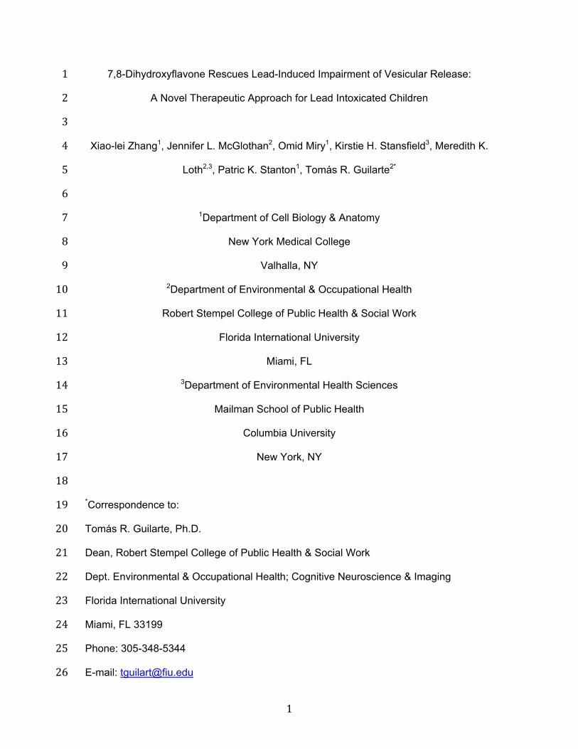
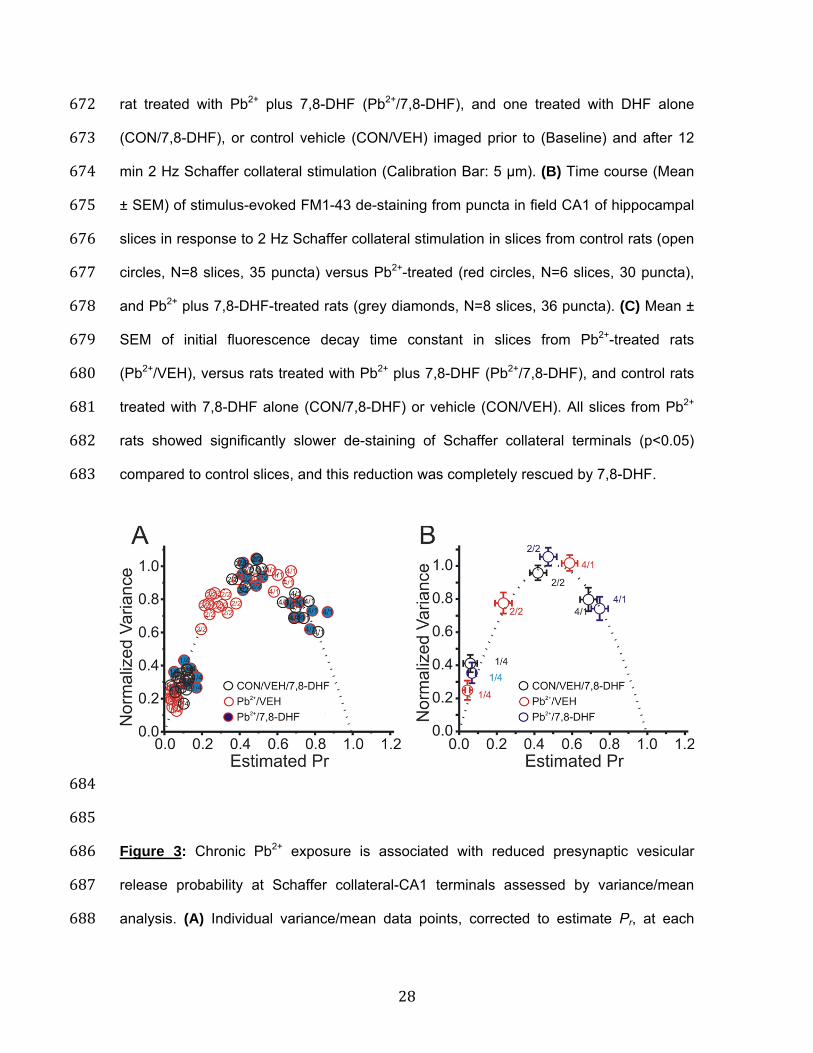
684 685 Figure 3: Chronic Pb2+ exposure is associated with reduced presynaptic vesicular 686 release probability at Schaffer collateral-CA1 terminals assessed by variance/mean 687 analysis. (A) Individual variance/mean data points, corrected to estimate Pr, at each 688
29
[Ca2+]o for each CON/VEH/7,8-DHF (open black circles), each slice from a Pb2+ rat 689 (Pb2+/VEH; open red circles), and each slice from a Pb2+ rat administered 7,8-DHF 690 (Pb2+/7,8-DHF; blue circles). Data from all groups of slices were well fit by a single 691 parabola forced to pass through 0,0 with Pb2+ synapses shifted to the left, consistent 692 with a presynaptic reduction in Pr from chronic Pb2+ exposure. This shift was rescued by 693 7,8-DHF. (B) Mean ± SEM of variance/mean points in slices from CON/VEH/7,8-DHF 694 (black circles; N=11), Pb2+/VEH (red circles; N=8), and Pb2+/7,8-DHF (blue circles; N=8), 695 normalized to the maximal peak amplitude recorded at 4 mM [Ca2+]o. 696 697
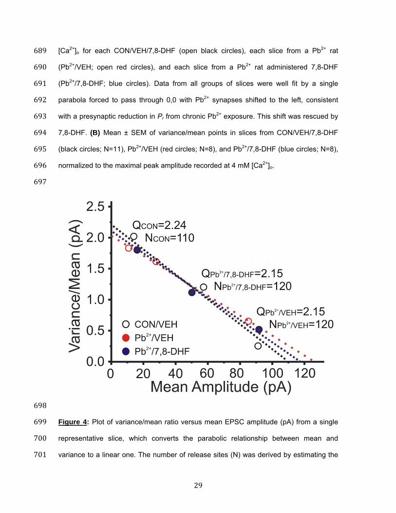
698 Figure 4: Plot of variance/mean ratio versus mean EPSC amplitude (pA) from a single 699 representative slice, which converts the parabolic relationship between mean and 700 variance to a linear one. The number of release sites (N) was derived by estimating the 701
30
slope of the linear fit, while the y-intercept denotes quantal size (Q) of the EPSC. The 702 reduction in slope indicates that chronic Pb2+ exposure (Pb2+/VEH; red dotted line) was 703 associated with a reduction in presynaptic Pr compared to a control slice (CON/VEH; 704 dotted black line), that was partially reversed in a slice from a Pb2+-exposed rat 705 administered 7,8-DHF (Pb2+/7,8-DHF; dotted blue line). 706 707
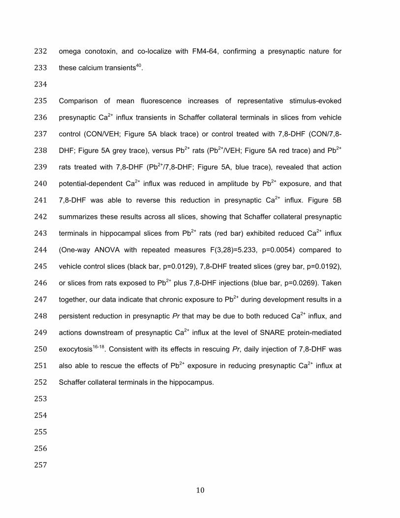
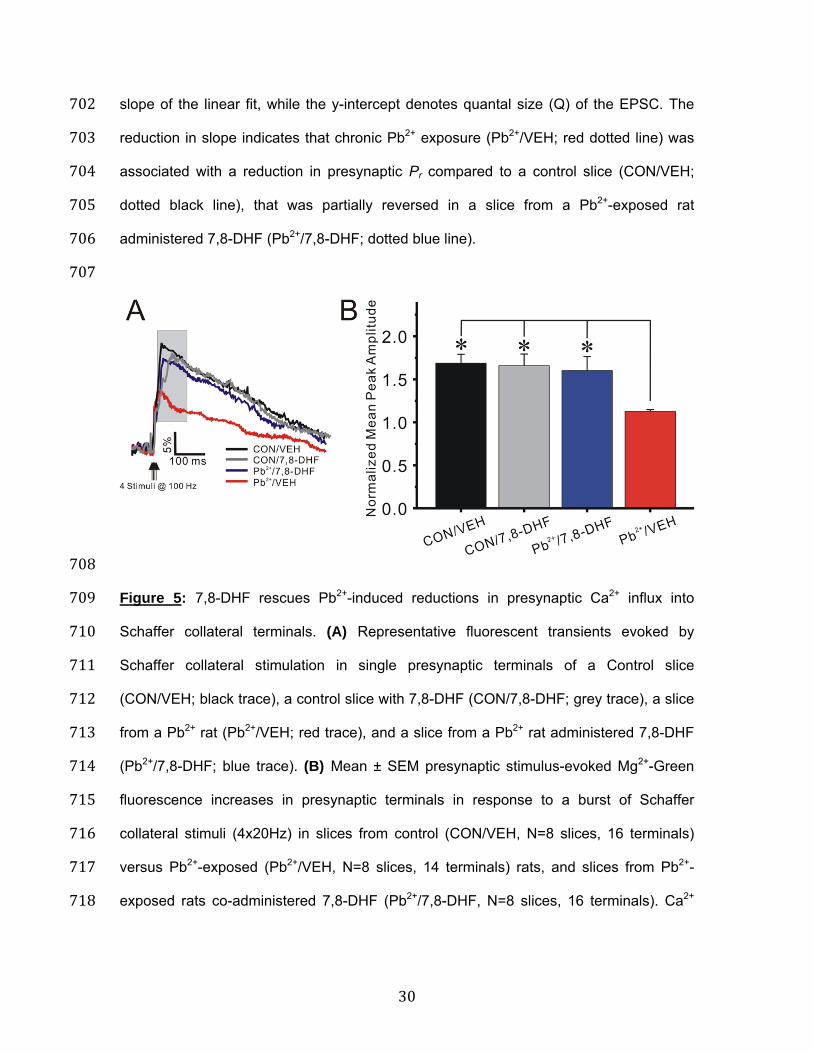
708 Figure 5: 7,8-DHF rescues Pb2+-induced reductions in presynaptic Ca2+ influx into 709 Schaffer collateral terminals. (A) Representative fluorescent transients evoked by 710 Schaffer collateral stimulation in single presynaptic terminals of a Control slice 711 (CON/VEH; black trace), a control slice with 7,8-DHF (CON/7,8-DHF; grey trace), a slice 712 from a Pb2+ rat (Pb2+/VEH; red trace), and a slice from a Pb2+ rat administered 7,8-DHF 713 (Pb2+/7,8-DHF; blue trace). (B) Mean ± SEM presynaptic stimulus-evoked Mg2+-Green 714 fluorescence increases in presynaptic terminals in response to a burst of Schaffer 715 collateral stimuli (4x20Hz) in slices from control (CON/VEH, N=8 slices, 16 terminals) 716 versus Pb2+-exposed (Pb2+/VEH, N=8 slices, 14 terminals) rats, and slices from Pb2+-717 exposed rats co-administered 7,8-DHF (Pb2+/7,8-DHF, N=8 slices, 16 terminals). Ca2+ 718
31
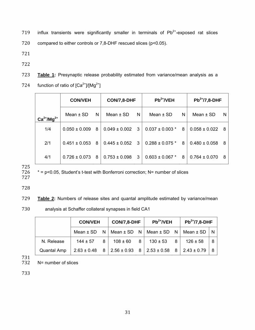
influx transients were significantly smaller in terminals of Pb2+-exposed rat slices 719 compared to either controls or 7,8-DHF rescued slices (p<0.05). 720 721 722 Table 1: Presynaptic release probability estimated from variance/mean analysis as a 723 function of ratio of [Ca2+]/[Mg2+] 724 Ca2+/Mg2+
CON/VEH CON/7,8-DHF Pb2+/VEH Pb2+/7,8-DHF
Mean ± SD N Mean ± SD N Mean ± SD N Mean ± SD N
1/4 0.050 ± 0.009 8 0.049 ± 0.002 3 0.037 ± 0.003 * 8 0.058 ± 0.022 8
2/1 0.451 ± 0.053 8 0.445 ± 0.052 3 0.288 ± 0.075 * 8 0.480 ± 0.058 8
4/1 0.726 ± 0.073 8 0.753 ± 0.098 3 0.603 ± 0.067 * 8 0.764 ± 0.070 8
725 * = p<0.05, Student’s t-test with Bonferroni correction; N= number of slices 726 727 728 Table 2: Numbers of release sites and quantal amplitude estimated by variance/mean 729
analysis at Schaffer collateral synapses in field CA1 730
CON/VEH CON/7,8-DHF Pb2+/VEH Pb2+/7,8-DHF
Mean ± SD N Mean ± SD N Mean ± SD N Mean ± SD N
N. Release 144 ± 57 8 108 ± 60 8 130 ± 53 8 126 ± 58 8
Quantal Amp 2.63 ± 0.48 8 2.56 ± 0.93 8 2.53 ± 0.58 8 2.43 ± 0.79 8
731 N= number of slices 732 733