Embed Size (px)
Citation preview
[Frontiers in Bioscience 7, d442-457, Feburary 1, 2002]
442
PERSISTENT INFECTIONS AND IMMUNITY IN CYSTIC FIBROSIS
Hongwei Yu and Nathan E. Head
Department of Microbiology, Immunology and Molecular Genetics, Joan C. Edwards School of Medicine at Marshall University,1542 Spring Valley Drive, Huntington, WV 25704
TABLE OF CONTENTS
1. Abstract2. Cystic fibrosis3. P. aeruginosa4. Chronic infection phenotypes: biofilm and mucoidy5. Genetic diversity and genomic islands6. Aerosol infection mouse model7. Persistent infection phenotype and immunity8. Future challenges9. Acknowledgements10. References
1. ABSTRACT
Cystic fibrosis (CF) is the most commonautosomal recessive lethal disease in the Caucasianpopulation. Chronic respiratory infections withPseudomonas aeruginosa, neutrophil-dominated airwayinflammation and progressive lung damage are the majorcauses of morbidity and mortality in CF. Two persistentinfection phenotypes expressed by this bacterium arebiofilm and mucoidy. Biofilm, also called the microcolonymode of growth is the surface-associated adherent bacterialcommunity, while mucoidy refers to a phenotypeconducive to copious amounts of mucoidexopolysaccharide (MEP)/alginate that provides a matrixfor mature biofilms conferring resistance to host defensesand antibiotics. Recent completion of the whole genomicsequence of the standard reference strain P. aeruginosaPAO1 has led to discoveries that many clinical isolates ofthis species possess unique genomic sequences (genomicislands) due to horizontal gene transfer. We propose thistype of genetic exchange may play an important role incausing intrinsic genomic diversity of this organism.Therefore, the diversity, as revealed through profiles ofrestriction fragment length polymorphism (RFLP), may belinked to an array of novel and unexplored pathogenicmechanisms in P. aeruginosa. CF mouse models, whiledisplaying many clinical similarities to human CF, have yetto demonstrate a chronic pulmonary disease phenotype.This review is intended to provide an overview of P.aeruginosa persistent infection phenotypes (biofilm andmucoidy) and an aerosol infection mouse model for CF.
Genomic diversity of P. aeruginosa and its implications inthe pathogenesis in CF will also be discussed.
2. CYSTIC FIBROSIS
Approximately 30,000 children and adults in theUnited States have CF. One in 31 Americans, more than10 million people are asymptomatic carriers of the mutantCF gene. Despite many promising advances in CFmedicine, the median life expectancy for CF patients is 30years (1). Like other genetic diseases, the predisposingfactor is a mutation of an otherwise functional gene. TheCF gene has been identified, and the gene product is namedthe CF transmembrane conductance regulator (CFTR) (2).Since the initial discovery, close to 1,000 different types ofdisease-causing mutations within this gene have beendocumented (http://www.genet.sickkids.on.ca/). By far themost common, which accounts for almost 70% of allknown mutations is a three base pair deletion resulting inthe removal of a phenylalanine residue at position 508 ofCFTR (deltaF508).
CFTR is a multifunctional protein located on theapical plasma membrane. It is an anion channel and/or achannel regulator that controls the flow of Cl-, Na+ andother ions across the membrane. Defects in CFTR result indisruption of normal Na+ and Cl- ion transport (3).Recently, CFTR has been linked with the control of Cl--coupled bicarbonate concentration across the lumen of the
P. aeruginosa infections in cystic fibrosis
443
lung epithelium for maintaining the proper acidity (4). Thecellular defects associated with the CFTR mutations causemany clinical problems, but the majority are confined tothose organs rich in exocrine glands such as respiratory,gastrointestinal and genitourinary tracts (5).Approximately 85% of CF patients are characterized withpancreatic insufficiency (PI) while 10–15% of them have aclinical condition called meconium ileus (MI), an intestinalblockage due to malabsorption (6). Patients homozygousfor the deltaF508 mutation tend to have more severepancreatic insufficiency than those carrying other types ofmutations (6). Conditions such as vitamin deficiency (A,D, E and K) and chronic malnutrition are common in CF(7). The clinical manifestation of CF is mainlydemonstrated through involvement of the respiratory tracts.The electrolytic imbalance combined with acidic pH due todefective bicarbonate transport results in changes in thecomposition of airway surface liquid (ASL), a thin layer offluid that covers the upper and lower respiratory tract, andretention of an abnormal dehydrated viscous mucous withinthe lung (4). These conditions leave the CF lungs prone tobecoming infected with microbial organisms (8).
Pulmonary infections in CF are associated withchronic colonization by several bacterial pathogens anddebilitating exacerbations as a result of bacterial and viralinfections superimposed upon progressive lung damage (9).Children with CF are often infected by Staphylococcusaureus and Haemophilus influenzae, both within thepatient’s first 3 years (10). Since the development ofaggressive antibiotic therapy, these organisms can betreated before any long-term damage is inflicted. Whileboth S. aureus and H. influenzae will re-emerge in the lungthroughout the remainder of the disease, P. aeruginosaultimately becomes the major inhabitant for the remainderof the CF patient's life (9). In fact, nearly all CF patientswill show evidence of P. aeruginosa colonizations by age 3(11). Chronic lung infections with P. aeruginosa areresponsible for the majority of early deaths in CF (12).
Though CF biogenesis is clear, the relationshipbetween CFTR mutations and pulmonary disease is elusive.Particularly, how the genetic defect leads to theestablishment of chronic bacterial colonization in the lungsis not fully understood. Several concurrent and sometimesconflicting proposals have been offered (13-18) to addressthis relationship. For example, reduced sialylation ofglycoconjugates on the surface of epithelial cells has beensuggested to promote P. aeruginosa adhesion in CF (18).However, a recent paper has questioned the role of thisglycolipid as the receptor for fresh clinical isolates of P.aeruginosa since the antibodies raised against asialo-GM1for confirming its interaction with P. aeruginosa also bindto multiple P. aeruginosa surface antigens (19).
The CF epithelial secretions have been reportedto display reduced bactericidal properties due to the alteredsalt content (14, 17). CFTR has also been proposed to beinvolved in P. aeruginosa uptake by respiratory epithelialcells (16). These models rely mainly on the knownfunctions of CFTR as an ion channel and/or channelregulator (6) and on its presumed pleiotropic effects (20).
Other studies, focusing on cytokine profiles in broncho-alveolar lavage fluids of CF patients, have suggested thatthe propensity for excessive inflammation in CF may beattributed to endogenously increased levels ofproinflammatory cytokines such as tumor necrosis factor(TNF)-alpha, interleukin (IL)-8, and IL-1 (13), andconcurrent decreased level of anti-inflammatory cytokineIL-10 (105). According to some reports, the levels of thesecytokines may be altered in CF even before a bacterialinfection begins (15, 21). Despite these promising leads,our understanding of host-pathogen interactions in CF isminimal. Even though the most obvious physiologicaldefect in CF is linked with ion transport, pathologicalconsequences due to a CFTR mutation appear to be morecomplex.
3. P. AERUGINOSA
P. aeruginosa is a ubiquitous Gram-negativeenvironmental bacterium that can grow in a wide variety ofmoist habitats such as soil, rivers, hospital sinks and oilfields and has an extremely versatile metabolic demand.This organism can colonize humans, invertebrates, insects,and plants. It is considered an opportunistic pathogen sincethe infections only occur in individuals with underlyingcompromised host defense systems (22). Besides beingassociated with fatal pneumonia in CF, P. aeruginosa cancause other types of infections ranging from acute andsystemic bacterimia to chronic catcher-associated urinarytract infections (8).
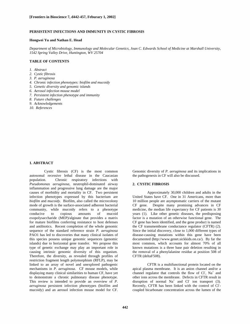
The genome of the P. aeruginosa strain PAO1has recently been completely sequenced and released to thepublic domain (http://www.pseudomonas.com/) (figure 1and ref. 23). A quick comparison of this genome to aselected group of bacterial genomes is listed in table 1. Thegenome size of P. aeruginosa is the largest among thecurrently available pathogenic bacteria in GenBank, andhas a coding capacity for 5,570 open reading frames(ORF’s; ref. 23). Initial analysis of the PAO1 genomesuggests the overall gene regulation is more complicatedthan that of the E. coli strain MG1655. For example, thenumber of the two-component signal transduction systemsin PAO1 is 144 (23 per Mb) whereas that of E. coli is only62 (13 per Mb; ref. 23). The inherent drug resistance ofthis pathogen can be in part attributed to the increasednumber of efflux pumps encoded within the genome (97).The P. aeruginosa strain PAO1 was originally isolatedfrom a wound (24), and is considered a standard referencestrain well adapted to the laboratory growth conditions.The O5 serotype to which PAO1 belongs represents a smallfraction (2.5%) of the clinical CF isolates surveyed duringthe late 1980’s (personal communication with Dr. JosephLam). According to the pulsed-field gel electrophoresis(PFGE) analysis, the genomic profile of PAO1 is notsimilar to that of the majority of the clinical CF isolates.Therefore, it is unclear how representative this genome iscompared to those of clinical isolates since the number ofmobile genetic elements (genomic islands, phages andtransposons) within each genome of clinical origin remainsunknown.
P. aeruginosa infections in cystic fibrosis
444
Figure 1. Schematic diagram of the entire genome of P. aeruginosa strain PAO1 according to http://www.pseudomonas.com/.The scale on the outmost circle is in Mb. Outer and inner circles consist of SpeI and XbaI digested fragments assembled in order,respectively. Known SpeI fragments (also see table 2) are labeled in the outer circle with respective alphabetic letters. Locationsof 2 probable bacterial phages (23), P. aeruginosa genomic island-1 (PAGI-1) (78), a flagellin glycosylation island (77), and 3genetic loci (23) proposed to be involved in the regulation of alginate production along with algD and algU loci are noted.
4. CHRONIC INFECTION PHENOTYPES: BIOFILMAND MUCOIDY
In response to changing environments,microorganisms have evolved various adaptivemechanisms and as a result they often express differentphenotypes. Bacterial biofilms are one of the persistentinfection phenotypes (1). P. aeruginosa biofilms and CFchronic pulmonary infections are closely related (25, 26).Biofilm is often defined as exopolysaccharide-surroundedbacteria, or microcolonies, growing on biotic or abioticsurfaces (27). Formation of biofilms has been shown to bethe preferred mode of bacterial growth in nature as thesessile population exceeds that of planktonic (free floating)biomass by 2-4 log10 units (28). The two modes of growthcomplement each other; the mobile phase provides a meansfor spread and colonization, while the biofilms affordprotection against protozoans, phages, and antibiotics inenvironments (28).
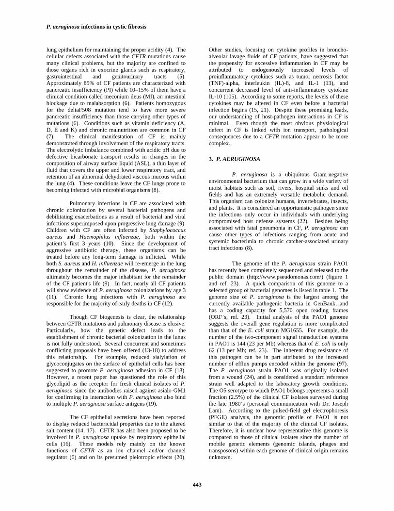
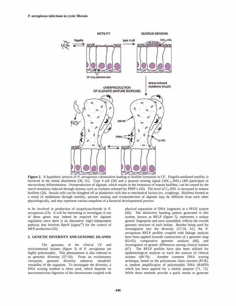
Biofilm formation is a dynamic and complexprocess that can be roughly divided into four phases (1): i)initial attachment to the surface (transition from planktoniccells to transiently attached cells); ii) cell proliferationforming a monolayer; iii) formation of a microcolony; andiv) development of mature biofilms. In terms of biofilmformation within the CF lungs, it is still not fullyunderstood how this process is initiated. Even so, ahypothetical and perhaps oversimplified scheme is
presented in figure 2. Based on several studies monitoringbiofilm formation on abiotic surfaces, motility is importantfor the initial attachment to the surface since it provides theforces required to overcome the surface repulsion forbacteria (29). Type IV pili mutant of P. aeruginosa formsthin and undifferentiated monolayers, but fails todifferentiate into microcolonies (30). Other factors mayalso be required since a hyperpiliated P. aeruginosa strainwithout twitching ability is still able to form thick biofilms(31). Some surface-associated proteins such as curli andouter membrane proteins (32) also play a role in the initialattachment. Very recently, through screening the Tn5insertion library for surface adhesion deficiency in the typeIV pili mutant background of P. aeruginosa PAK, Vallet etal. (33) reported the identification of a novelchaperone/usher pathway involved in the assembly of newclasses of adhesins that are needed for early biofilmformation, suggesting that multiple adhesion factors arepresent on the cell surface of P. aeruginosa. Furthermore,many global regulators such as the crc gene involved incarbon metabolism (34) and polyphosphate kinase (35), andmore recently, a two-component regulatory systemGacA/GacS (36), are also known to participate in the initialbiofilm formation.
After initial attachment, P. aeruginosa biofilmformation requires participation of a cell density-dependentresponse, known as quorum sensing consisting of twodistinct but interrelated systems, las and rhl (37). These
P. aeruginosa infections in cystic fibrosis
445
Table 1. Completed genomes from a representative group of pathogenic bacteria1
Organism Genome size ORF # Accession # Release DateH. influenzae Rd 1,830,138 1,709 NC_000907 07/25/95E. coli MG1655 4,639,221 4,289 NC_000913 10/13/98Mycobacterium tuberculosis H37Rv 4,411,529 3,918 NC_000962 6/11/98Vibrio cholera O1 El Tor 4,033,464 2,736
1,092NC_002505NC_002506
6/14/00
P. aeruginosa PAO1 6,264,403 5,5702 NC_002516 5/16/00E. coli O157:H7 5,529,376 5,283 NC_002655 1/25/01M. leprae 3,268,203 2,720 NC_002677 2/20/01
1 based on http://www.ncbi.nlm.nih.gov/PMGifs/Genomes/bact.html, 2 based on http://www.pseudomonas.com/
systems work coordinately to regulate a number of factorsthat enable this pathogen to survive in highly diverseenvironments. Two molecules produced are N-(3-oxododecanoyl)-L-homoserine lactone (3OC12-HSL) andN-butyryl-L-HSL (C4-HSL), which serve as signals forregulation of the cell-to-cell communication (38, 39).Particularly, 3OC12-HSL is involved in microcolonydifferentiation (40). Recently, using a sensitive radiometricassay coupled with a reverse phase high performance liquidchromatography to directly measure the level of acyl-HSLs,Singh et al. (26) reported that CF sputum carrying P.aeruginosa has more C4-HSL than 3OC12-HSL, which issimilar to PAO1 grown in the biofilm mode, suggestingthat C4-HSL may serve as a biomarker for mature biofilms(figure 2). Although alginate helps to maintain threedimensional structure of biofilms, alginate per se does notappear to be required for the initial P. aeruginosa biofilmdevelopment (31, 41). Furthermore, the wild type and lasImutant of PAO1 produce an equal amount of alginate whenforming biofilm in vitro (40). This appears to support thenotion that control of alginate production is independent ofat least 3OC12-HSL mediated quorum sensing pathway.Therefore, the initial biofilm formation and MEP basedbiofilms may represent two separate phenomena and/or twodifferent stages of biofilm development. To distinguish theinitial biofilm formation from those microcoloniesembedded with a mucoid coat as commonly seen in CF, weintroduce a term called “mature” biofilms to describe thosecovered with excessive amounts of alginate (figures 2 and4).
The major component of the mucoid capsuleproduced by P. aeruginosa is alginate that consists ofnegatively-charged polyuronic acid (9). During chronicrespiratory tract colonization, a subset of initial nonmucoidcolonizing P. aeruginosa strains may acquire mutationscausing conversion to mucoidy (42). The transition fromthe initial non-mucoid strain to the mucoid variant hasrecently been reproduced in vitro with repeated exposure toa sublethal concentration of hydrogen peroxide, a commonoxidant in polymorphonuclear neutrophils (PMN’s) (43).Overproduction of alginate in P. aeruginosa facilitatesdevelopment of mature biofilms that contribute to immuneevasion and antibiotic resistance (figures 2 and 4) (1). Thisunique phenotype (figure 3) is the main diagnostic indicatorfor the chronic onset of Pseudomonas lung infections in CF(44). The mucoid phenotype is usually unstable, but it ispossible to maintain this phenotype in vitro using certain
laboratory media (figure 3). Mucoidy is of importance tothe persistence of P. aeruginosa in CF. Protective,opsonizing antibodies against alginate are present only inuncolonized patients (45). The mucoid variant caninfluence the pulmonary outcome in CF patients (46).Alginate also has other pathogenic roles such as inhibitionof phagocytosis, suppression of neutrophil chemotaxis, andscavenging of oxidative radicals (9).
Persistence of P. aeruginosa and mucoidy aretwo inseparable clinical conditions since emergence ofmucoid phenotype in CF-affected lungs correlates withonset of pulmonary function deterioration (47). One ofthe molecular mechanisms related to mucoidy conversionis up-regulation of an alternative sigma factor(AlgU/AlgT) that in turn activates expression of algD, thebiosynthetic gene for alginate production (48). AlgU, astress-related sigma factor with extracytoplasmic function(ECF), is negatively regulated by its immediatedownstream gene cluster, mucABCD where MucA is thecognate antisigma factor for AlgU (48). The AlgUortholog in E. coli is RpoE (sigmaE) (66% identity and91% overall similarity). The E. coli rpoE gene canfunctionally complement the algU mutation and restoremucoidy in P. aeruginosa (49). Furthermore, up-regulation of algU inhibits flagellum synthesis (50).Recently, we have found that inactivation of algU andrpoE in P. aeruginosa PAO1 and E. coli K12,respectively, could lead to increased flagellar activity inboth organisms, thereby causing increased initial biofilmformation in vitro and in vivo (51). Conversely,overproduction of sigmaE in E. coli K12 causes reducedinitial biofilm formation (51). This is interesting since anearlier report indicates that inactivation of algU in PAO1caused increased virulence in a systemic infection mousemodel (52). This elevated virulence as a result of algUinactivation may be due to increased initial colonizationof the mutant. Two recent analyses using proteomics tocompare global protein expression coupled with mucoidyindicate that production of several proteins such as outermembrane protein porin F (OprF) and disulfide bondisomerase (DsbA), which may not be involved in alginateproduction, is affected (53, 54). These candidates mayplay a role in induction of mucoidy-coupled host-derivedhuman beta-defensin (55). As a result of thebioinformatic analysis of the PAO1 genomic sequence,three additional genetic loci in this genome (figure 1)have been proposed
P. aeruginosa infections in cystic fibrosis
446
Figure 2. A hypothetic process of P. aeruginosa colonization leading to biofilm formation in CF. Flagella-mediated motility isinvolved in the initial attachment (30, 51). Type 4 pili (30) and a quorum sensing signal (3OC12-HSL) (40) participate inmicrocolony differentiation. Overproduction of alginate, which results in the formation of mature biofilms, can be caused by themucA mutations induced through stresses such as oxidants released by PMN’s (43). The level of C4-HSL is increased in maturebiofilms (26). Sessile cells can be sloughed off as planktonic cells due to mechanical factors (ex. coughing). Biofilms formed asa result of mediations through motility, quorum sensing and overproduction of alginate may be different from each otherphysiologically, and may represent various snapshots of a bacterial developmental process.
to be involved in production of exopolysaccharide in P.aeruginosa (23). It will be interesting to investigate if anyof these genes may indeed be required for alginateregulation since there is an alternative AlgU-independentpathway that involves RpoN (sigma54) for the control ofMEP production (56).
5. GENETIC DIVERSITY AND GENOMIC ISLANDS
The genomes of the clinical CF andenvironmental isolates (figure 5) of P. aeruginosa arehighly polymorphic. This phenomenon is also referred toas genomic diversity (57-59). From an evolutionaryviewpoint, genomic diversity enhances metabolicversatility of the organism. To investigate the diversity, aDNA scoring method is often used, which depends onmacrorestriction digestion of the chromosome coupled with
physical separation of DNA fragments in a PFGE system(60). The distinctive banding pattern generated in thissystem, known as RFLP (figure 5), represents a uniquegenetic fingerprint and once assembled, reflects the overallgenomic structure of each isolate. Besides being used forinvestigation into the diversity (57-59, 61), the P.aeruginosa RFLP profiles coupled with linkage analysishave been applied towards construction of a genomic map(62-65), comparative genomic analysis (66), andinvestigation of genetic differences among clinical isolates(67). The RFLP profiles have also been utilized forepidemiological analysis to track the sources of clinicalisolates (68-70). Another common DNA scoringtechnique, based on the polymerase chain reaction (PCR),is random amplification of polymorphic DNA (RAPD)which has been applied for a similar purpose (71, 72).While these methods provide a quick means to generate

P. aeruginosa infections in cystic fibrosis
447
Figure 3. A persistent infection phenotype expressed by P.aeruginosa due to the excessive production of alginate.Alginate helps to maintain the three dimensionalarchitecture of mature biofilms known to be resistant tohost defenses and antibiotics. Shown is a mucoid colonymorphology from a clinical CF isolate of P. aeruginosagrowing on a laboratory media.
Figure 4. Transmission electron micrograph of a thin-section of a postmortem lung sample from a CF patient.Shown is P. aeruginosa in mature biofilms, a persistentinfection phenotype closely associated with chronicbacterial lung infections in CF. P. aeruginosa is embeddedwithin a mucous matrix produced by bacteria and host[Reproduced with permission from Lam et al. (25)].
genomic comparison, they are often limited by lack ofinformation on individual DNA fragments produced withinthese systems.
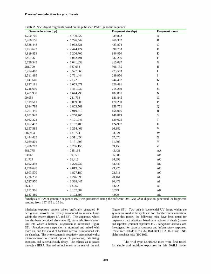
The availability of the PAO1 genomic sequencefacilitates utilization of PFGE more effectively to studygenomic diversity among various clinical isolates. Forexample, it has been previously reported that completedigestion of the PAO1 genome with restrictionendonuclease SpeI generated 37 fragments in a PFGEsystem (64). While the estimation of fragment sizes wasnot always accurate, one can deduce a precise range offragments based on the PAO1 genomic sequence. Thesebands, listed in table 2, may be used as a standard referencefor comparison with clinical CF isolates. In our initial
PFGE analysis of 101 clinical CF isolates, which werecollected over a wide range of geographical regions andcompared with the standard banding pattern of PAO1 (table2), we identified 75 unique SpeI-digested genomic profileswith 23 ± 12 bands (n = 75; 99% confidence interval; ref.73). Of these isolates, 51% have at least one bandsignificantly larger than the largest fragment of PAO1(Spe-A = 539,862 bp). We observed 74% genomicdiversity among the CF isolates in our collection. Thisnumber perhaps under-estimates the genomic diversity ofP. aeruginosa since the remaining 26% non-uniquegenomic profiles were sequential, sibling or same-sourceisolates. It appears that a particular isolate remains fairlystable in its genome after it colonizes the CF lung (figure5), which is consistent with a previous observation that themajority of CF patients are colonized with one strain (74).However it is possible for a single patient to be colonizedby 2 different strains at the same time (71, 74). Horizontaltransmission between patients is possible since identicalgenomic profiles have been seen between CF isolates andan isolate from aquatic habitats (59, 66).
The ultimate cause of genomic diversity thatexists throughout clinical CF isolates of P. aeruginosa isattributed to mutation (75). One of the virulencemechanisms employed by many pathogenic bacteria is theacquisition of auxiliary DNA sequences from within orbetween species or even genera (76). There is compellingevidence to suggest that the event of horizontal genetransfer occurs in P. aeruginosa (77-79). Mobile geneticelements such as bacterial phages are probably present inthe PAO1 genome (figure 1 and ref. 23). Some of thePseudomonas phages have the genetic capacity of serotypeconversion (80). Tummler et al. (66) were among the firstto notice that the genome of one aquatic/CF isolate of P.aeruginosa was significantly larger than that of PAO1. Asubsequent study with a clinical wound isolate of P.aeruginosa, UCBPP-PA14 (PA14), has reported thediscovery of several unique chromosomal regions found inthis strain but not present in the PAO1 genome (81-83).
Recently, Liang et al. (78) have reported theidentification of the first genomic island (PAGI-1; figure1), in the genome of a clinical urinary tract infection isolateP. aeruginosa X24509. This genetic island is absent inPAO1, but is present in 85% of the clinical isolatesincluding those from CF patients. It is composed of 48,893bp with a coding capacity for 51 ORF’s. Throughrecombination, this island replaces a region of 6,729 bpsequence derived from the PAO1 genome that is thought tobe prone to genetic alterations. Interestingly, this PAGI-1-replaced locus is located within a previously identifiedregion with unusually low G+C content (PA2221-PA2228,G+C: 49.2% vs. 66.6% overall; figure 1) (23). While thefunctions of PAGI-1 need to be elucidated, there are at leasttwo potential transcriptional regulators located on theisland, one of which is the homologue of RpoN-dependenttranscriptional activators. Furthermore, two homologs ofE. coli, which are known to be involved in counteractingoxidative stress, are also encoded on PAGI-1. Since RpoNhas been implicated in the regulation of mucoidy (56) andother surface molecules such as flagella and pili, it is

P. aeruginosa infections in cystic fibrosis
448
Figure 5. Separation of SpeI digested P. aeruginosachromosomal DNA by PFGE. (A). CF006 and CF007, twoclinical CF isolates display the genomic diversity that existsthroughout the majority of clinical isolates. Environmentalstrains (ENV42: Japan, and PAOH: Ohio River) of P.aeruginosa also exhibit a diverse chromosome. (B). Burnisolates PAO1 and PA14 also possess diversity along thechromosome. Clinical sequential isolation of P.aeruginosa shows conservation of genome throughoutcolonization (CF041: initial isolate in 1990, CF042:isolated in 1994, CF043: isolated in 1998).
conceivable that some genes encoded on PAGI-1 may havespecific virulence roles including modulation of chronicinfection phenotypes.
The discovery of PAGI-1 suggests that an islandof this nature may contribute to causing the genomicdiversity observed with PFGE. For example, within thePAGI-1 DNA sequence (GenBank accession# AF241171),there are five positions recognizable by endonucleases SpeIand XbaI (4 sites) that are often used for PFGE analysis ofP. aeruginosa genomes. Therefore, a genomic island islinked with a cause of genomic diversity of P. aeruginosa.A recent publication of the E. coli O157:H7 genome
sequence indicates that this virulent strain, unlike thelaboratory counterpart E. coli K12, has an additional 1.34Mb encoding 1,387 new genes in 177 O157:H7-specificgenomic islands interspersed in a K12 genome (84). Basedon the overall sequence homology between P. aeruginosaPAO1 and E. coli K12 (23), it is reasonable to assume thatmore genomic islands are to be identified throughout the P.aeruginosa genomes. It is known that severity of thepulmonary disease in CF is poorly correlated with thegenotype (6). This variability can be attributed to manyfactors of environmental and/or host origin. Perhaps it canalso be caused by factors of bacterial origin through thediversity. Genomic diversity resulting from horizontalgene transfer suggests a possibility of acquisition of novelvirulence traits in P. aeruginosa. For example, flagella ofsome P. aeruginosa strains are often glycosylated (85).Very recently, it has been reported (77) that anothergenomic island of ca. 16 kb located immediately upstreamof the fliC gene encoding the structural flagellin protein isresponsible for this action. Although the pathogenicsignificance of this glycosylation is still unknown, thisdiscovery may become the first example demonstrating thathorizontal gene transfer introduces a novel virulencemechanism into this organism.
6. AEROSOL INFECTION MOUSE MODEL
An appropriate animal model is essential forsimulating the respiratory tract infection in CF. Theavailability of CFTR transgenic mice has enabled us toinvestigate CF-related bacterial pulmonary infections in asurrogate host. Currently, there are several genetic mousemodels for CF (86-93). These CF mice, while displayingseveral similar characteristics of intestinal disease in humanCF, fail to develop respiratory infections or other signs ofovert lung disease. This disappointing limitation of theCFTR transgenic mice, has been linked to the presence ofalternative Cl- and Na+ channels in mice that couldcompensate for the loss of the CFTR lesion (94). Noapparent differences in the ionic compositions of ASL havebeen found between CFTR mutant mice and their littermatecontrols (95). With the recent development of sensitivemethods to directly measure the contents of ASL such asthe use of a cryoprobe (96) and staining with fluorescention indicators (97), there is still no significant difference inASL salt content between wild type and CF mice. Moreimportantly, spontaneous colonization with P. aeruginosahas not been detected in the CF animals (98). While thereare numerous mouse infection models for CF, each modelhas its pros and cons (8, 99). For example, the agar-beadmodel, in which P. aeruginosa is artificially embedded inagar beads, simulates a situation similar to mature biofilminfection. On the other hand, the aerosol infection modeldescribed below may be more useful in modeling the innatehost response immediately after initial Pseudomonascolonization.
A Bacterial Aerosol Induced Lung Infection(BAILI) mouse model has been developed for simulation ofbacterial pulmonary infections in CF (100-102). Thismodel utilizes the aerosol technology originally developedfor tuberculosis studies (103). The core of this model is an
P. aeruginosa infections in cystic fibrosis
449
Table 2. SpeI digest fragments based on the published PAO1 genomic sequence1
Genome location (bp) Fragment size (bp) Fragment name4,250,766 - 4,790,627 539,862 A5,266,156 - 5,726,542 460,387 B3,538,448 - 3,962,321 423,874 C2,053,672 - 2,444,424 390,753 D4,819,853 - 5,206,702 386,850 E725,196 - 1,062,491 337,296 F5,726,543 - 6,041,639 315,097 G281,799 - 587,953 306,155 H3,254,467 - 3,527,969 273,503 I2,511,495 - 2,761,444 249,950 J6,041,640 - 21,723 244,487 K1,827,181 - 2,053,671 226,491 L1,246,699 - 1,461,937 215,239 M1,461,938 - 1,644,798 182,861 N99,954 - 281,798 181,845 O2,919,511 - 3,089,800 170,290 P1,644,799 - 1,803,569 158,771 Q2,761,445 - 2,919,510 158,066 R4,101,947 - 4,250,765 148,819 S3,962,322 - 4,101,946 139,625 T1,062,492 - 1,187,488 124,997 U3,157,585 - 3,254,466 96,882 V587,954 - 681,774 93,821 W2,444,425 - 2,511,494 67,070 X3,089,801 - 3,151,305 61,505 Y5,206,703 - 5,266,155 59,453 Z681,775 - 725,195 43,421 AA63,068 - 99,953 36,886 AB21,724 - 56,415 34,692 AC1,192,398 - 1,226,237 33,840 AD4,790,628 - 4,819,852 29,225 AE1,803,570 - 1,827,180 23,611 AG1,226,238 - 1,246,698 20,461 AH3,527,970 - 3,538,447 10,478 AI56,416 - 63,067 6,652 AJ3,151,306 - 3,157,584 6,279 AK1,187,489 - 1,192,397 4,909 AL1Analysis of PAO1 genomic sequence (97) was performed using the software OMIGA; XbaI digestion generated 99 fragmentsranging from 237,113 to 25 bp.
inhalation exposure system where artificially generated P.aeruginosa aerosols are evenly introduced to murine lungswithin the system (figure 6A and 6B). This apparatus, whichhas also been described elsewhere (8), has a nebulizer-Venturiunit into which a bacterial suspension is introduced (figure6B). Pseudomonas suspension is atomized and mixed withroom air, and this cloud of bacterial aerosol is introduced intothe chamber. The whole system is negatively pressurized with amicroprocessor to control cycles of preheating, nebulizing,exposure, and bacterial cloudy decay. The exhaust air is passedthrough a HEPA filter and an incinerator in the rear of the unit
(figure 6B). Two built-in bactericidal UV lamps within thesystem are used at the cycle end for chamber decontamination.Using this model, the following mice have been tested forrespiratory tract infections, based on a regimen of single (innate)and repeated (chronic) exposures to P. aeruginosa aerosols, andinvestigated for bacterial clearance and inflammatory responses.These mice include C57BL/6J, BALB/cJ, DBA, IL-10 and TNF-alpha knockout mice (100-102).
The wild type C57BL/6J mice were first testedfor single and multiple exposures in this BAILI model

P. aeruginosa infections in cystic fibrosis
450
Figure 6. A. The core of the bacterial aerosol infectionmouse model (BAILI): An inhalation exposure system.The BAILI mouse model (6, 115, 116) is a whole-bodyaerosol-based infection model where artificially generatedP. aeruginosa aerosols can be evenly introduced to themurine lungs within the chamber, thereby causing a lung-specific infection and inflammation in mice. The chamberholds up to 100 mice within 5 separate compartments andcan be used as an efficient high-throughput screeningdevice for monitoring pulmonary clearance andinflammatory responses from the host.B. Schematicdiagram of the aerosol inhalation machine system.Utilizing a negative pressured system powered by avacuum pump at the end of the arrangement, room air ispassed through a HEPA filter (#1) before entering thesystem. A compressor produces the air pressure needed toaerosolize the bacterial suspension, as shown in the inset.A valve controls the flow of the compressed air, regulatingthe level of aerosolization. The main air is responsible forcarrying the aerosol from the nebulizer-Venturi unit to theinhalation chamber. Exhaust air with bacteria is filtered(HEPA filter #2) and incinerated. UV lamps destroyresidual bacteria within the chamber as well as on theanimal coats.
(101). While a single exposure to P. aeruginosa aerosolresulted in only mild histopathological changes, repeatedexposures caused significant lung pathology in C57BL/6Jmice. Mucoid cells (mucA22) were cleared several-foldless efficiently than isogenic nonmucoid (mucA+) cellsduring the initial stages of aerosol exposure. However,microscopic pathology findings and proinflammatorycytokine levels were similar in mice exposed to nonmucoidand mucoid P. aeruginosa throughout the infection. Lunghistopathology and proinflammatory cytokines were testedin IL-10 deficient transgenic mice (IL-10T). Significantmortality was seen in the IL-10T mice on initial challengewith P. aeruginosa. Increased pathology was detectedupon repeated challenge with P. aeruginosa in the IL-10Tmice relative to the C57BL/6J mice. In an chronicinfection agar-bead model with IL-10 knockout mice, asimilar finding was noted (104). These observationssuggest that anti-inflammatory cytokines may play a role insuppressing P. aeruginosa-induced tissue damage duringchronic infection, and that reduced IL-10 levels in the lungsof CF patients (105) may be of significance for therespiratory sequelae in this disease.
The CF mice have also been tested in this modelfor their ability to clear P. aeruginosa (102). This result isapparently in keeping with other observations using theagar-bead chronic infection model (106, 107) or anotheraerosol infection model (108). These mice presented twoextremes: either clearing or not clearing P. aeruginosa.This finding seems to be associated with variations inmouse body weight and nutritional status. WhenCFTR
mlUNC-/- mice had their intestinal defect corrected by a
functional human CFTR gene expressed from a ratintestinal fatty acid-binding protein gene promoterCFTR
mlUNC-/- (FABP-hCFTR mouse; ref. 92), P. aeruginosa
was efficiently cleared from the lung and variability was nolonger observed. While these observations may suggestthat repairing the CFTR defect in the intestinal tractimproves lung defense against P. aeruginosa, they alsoshow that CF transgenic mice may be incapable of clearingPseudomonas lung infections under certain conditions.
Recently, we have tested the role of malnutritionin host pulmonary defenses against P. aeruginosacolonizations in a group of C57BL/6J mice which was firstinduced with protein energy malnutrition (PEM) and thenexposed to P. aeruginosa aerosol (102). PEM resulted in a11-fold increase of P. aeruginosa survival in the lungs ofthe C57BL/6J mice. PEM also contributed to excessiveinflammation upon chronic infections with P. aeruginosa.The repeatedly infected malnourished mice did not produceIL-10 in their lungs. In addition to increased levels ofproinflammatory cytokines and neutrophil infiltration,another CF hallmark is a low level of the major anti-inflammatory cytokine IL-10 in the bronchio-alveolar fluid(105). Using a model of a repeated exposure with P.aeruginosa (101), we observed significant amounts of IL-10 production in the well-nourished mice 22 days followingthe initiation of the regimen of repeated exposures. Incontrast, the malnourished animals had no detectable IL-10.These results demonstrate that malnutrition compromisespulmonary defenses against P. aeruginosa colonization and
P. aeruginosa infections in cystic fibrosis
451
is conducive to excessive inflammation in response to P.aeruginosa infection, resembling the situation in CF.Furthermore, neutrophil infiltration in the lungs ofmalnourished animals did not result in increased bacteriallung clearance and instead was a correlate of anunproductive inflammatory response. In two separatereports using a sepsis model induced by P. aeruginosa(109) and a chronic infection model with Pseudomonas-laden agar beads (110), respectively, increased neutrophilinfiltration and increased bacterial load in the lungs weredetected at the same time. These results suggest thatneutrophils under these conditions (e.g., PEM, sepsis andchronic infection) may not be fully activated or functionalin the process of eliminating Pseudomonas from the lungs.Therefore, neutrophil-mediated bactericidal mechanismsneed to be further investigated since pulmonary alveolarmacrophages have recently been shown to play a marginalrole in defense against P. aeruginosa infections in mice(111).
7. PERSISTENT INFECTION PHENOTYPES ANDIMMUNITY
The major characteristic of chronic lunginfections in CF is the persistence of bacterial pathogensdespite excessive immune response from the host (112).The normal innate pulmonary defense systems involvemucociliary clearance and cough, phagocytes andantimicrobial factors such as defensins (113). In normalindividuals, an effective inflammatory response involvingproinflammatory cytokines is needed immediately afterexposure to P. aeruginosa. However, the successful initialcolonization of P. aeruginosa in the CF lungs suggests thatthe innate immunity that is required for Pseudomonasclearance is defective in CF. When this fails, biofilmformation, accompanied by excessive inflammation, occursresulting in chronic infection. Chronic pulmonaryinflammation, caused by excessive neutrophil infiltrationand an unproductive immune response, is one of the majorclinical manifestations in CF. The CF patients apparentlyhave the ability to elicit humoral and cell-mediated immuneresponses. The phagocytic cells of CF patients appear tolack any major functional abnormalities (44). Furthermore,CF patients are able to mount an antibody response sincethe proteinaceous antigens of P. aeruginosa are processedby antigen presenting cells and presented to T-cells asevidenced by the presence of anti-toxin (112) and morerecently, anti-type-III-protein antibodies (114) in the serumof CF patients. The problem encountered in combating P.aeruginosa persistent infections is due to the deficiency ofan effective removal system of MEP enclosedmicrocolonies of P. aeruginosa and therefore causingincreased and prolonged host inflammatory responses(115). The antibody-bacterial complex aggregates, whichare too large to be phagocytized, could act as foci for“frustrated phagocytosis” (116). Fc-receptor mediateddegranulation and oxidative burst due to the presence ofcontinuous infiltration of neutrophils occur, resulting inexcessive inflammation and tissue destruction.
Biofilm formation, particularly conversion to themucoid phenotype leading to development of mature
biofilms, impairs the normal immune response process inCF patients. Alginate is immunogenic, and can act as anefficient polyclonal B-cell activator (117) leading to aclinical condition mainly present in older CF patients:hyperimmunoglobulinemia (44). Another peculiar propertyof alginate is that excessive amounts in the experimentalanimals induces production of non-protective and lowopsonizing antibodies (45). Using serum from a patientundergoing an exacerbation, it was demonstrated by crossimmunoelectrophoresis that the serum recognized morethan 30 distinct P. aeruginosa antigens (118). In contrast,serum from a patient in remission, who had been infectedfor 16 years, only recognized five antigens (119). Thisprovided prima facie evidence of masking P. aeruginosaantigens by biofilm-forming bacteria.
The seminal work performed to establish thebasic concept for P. aeruginosa biofilms was done in theearly 1980s. Using direct electron microscopic analysis ofpost-mortem lung tissues from CF patients, Lam et al.(118) demonstrated that P. aeruginosa in CF lungs formaggregates or microcolonies (figure 4). Excessive amountsof alginate surrounding microcolonies during the course ofchronic infections in CF directly impair antigenpresentation. Antibody response to iron-regulated outermembrane proteins (IROMPs) in CF patients does notappear early in the infection and is apparently associatedwith the advanced stage of the disease (120). The roughLPS mutants of P. aeruginosa emerge during the course ofchronic infections in CF patients who have a high titer ofanti-LPS antibodies, but the immune systems of CFpatients fail to eradicate the serum-sensitive mucoid variantP. aeruginosa infections (44). Efforts for vaccinedevelopment have been mainly focused on the followingcandidates: O-polysaccharide, toxins, flagella, outermembrane proteins and alginate (121). Though somecandidates are promising, an effective anti-Pseudomonasvaccine is still not available. Recently, it has been reportedthat a DNA vaccine carrying oprF allows the immunizedmice to produce a significantly higher level of opsonicIgG1 antibody, confering protection in these mice (122).Genetic vaccines combined with molecular adjuvants mayoffer a new approach for future development of vaccinesagainst P. aeruginosa infections in CF.
8. FUTURE CHALLENGES
Recent completion of the entire PAO1 genomicsequence accompanied by discoveries of genomic islandsfrom clinical isolates spurs further investigation of thegenetic diversity in P. aeruginosa. Some of the relevantquestions to now address include i) the number of islandspresent per genome; ii) the relationship between the islandsand diversity; iii) identification of any CF- and/or otherdisease-specific islands; and iv) elucidation of specificvirulence roles for the islands including their possibleinvolvement in biofilm formation. Some of the challengingtopics related to biofilm research include i) identificationand characterization of new adhesins, and ii) control ofbiofilm formation by quorum sensing and other globalregulators. To establish a relationship between in vitro andin vivo biofilm formation, it is necessary to evaluate biofilm
P. aeruginosa infections in cystic fibrosis
452
formation defective mutants in a proper animal model. Agood CF infection mouse model will allow us to reproducethe chronic infection process, i.e., from initial nonmucoidcell colonization to in vivo mucoidy conversion leading todevelopment of mature biofilms coupled with excessiveneutrophilic inflammation and inflammatory cytokineprofiles similar to what is seen in human CF. Developmentof a true CF disease animal model and elucidation of themolecular mechanisms underlying the transition to thepersistent infection phenotypes in this organism could leadto novel therapeutic strategies against chronic P.aeruginosa lung infections in CF.
9. ACKNOWLEDGEMENTS
We thank D. Speert, F. Ausubel, P. Phibbs, andC. Somerville for providing the clinical CF, burn andenvironmental isolates used in this study; T. W. Fenger forsharing the PFGE apparatus. This work is supported by anoperating grant to H.Y. from the Joan C. Edwards Schoolof Medicine at Marshall University.
10. REFERENCES
1. Costerton, J. W., P. S. Stewart, & E. P. Greenberg:Bacterial biofilms: a common cause of persistent infections.Science 284, 1318-1322 (1999)
2. Collins, F. S.: Cystic fibrosis: molecular biology andtherapeutic implications. Science 256, 774-779 (1992)
3. Wine, J. J.: The genesis of cystic fibrosis lung disease.Journal of Clinical Investigation 103, 309-312 (1999)
4. Choi, J. Y., D. Muallem, K. Kiselyov, M. G. Lee, P. J.Thomas, & S. Muallem: Aberrant CFTR-dependent HCO-
3transport in mutations associated with cystic fibrosis.Nature 410, 94-97 (2001)
5. Tomashefski Jr., J. F., C. R. Abramowsky, & B. B.Dahms: The pathology of cystic fibrosis. In: Lung Biologyin Health and Disease. Eds: Davis, P. B., Marcel Dekker,Inc., New York p. 435-489 (1993)
6. Welsh, M. J., B. W. Ramsey, F. Accurso, & G. R.Cutting: Cystic fibrosis. In: The metabolic & molecularbases of inherited disease. Eds: Valle, D., McGraw-Hill,New York p. 5121-5188 (2001)
7. Pencharz, P., & P. Durie: Nutritional management ofcystic fibrosis. Ann Rev Nutr 13, 111-136 (1993)
8. Deretic, V.: Pseudomonas aeruginosa infections. In:Persistent bacterial infections. Eds: Cunningham-Rundles,S., ASM Press, Washington, DC p. 305-326 (2000)
9. Govan, J. R. W., & V. Deretic: Microbial pathogenesisin cystic fibrosis: mucoid Pseudomonas aeruginosa andBurkholderia cepacia. Microbiol Rev 60, 539-574 (1996)
10. Konstan, M. W., & M. Berger: Infection andinflammation of the lung in cystic fibrosis. In: Cystic
fibrosis. Eds: Davis, P. B., Marcel Dekker, Inc., New Yorkp. 219-276 (1993)
11. Burns, J. L., R. L. Gibson, S. McNamara, D. Yim, J.Emerson, M. Rosenfeld, P. Hiatt, K. McCoy, R. Castile, A.L. Smith, & B. W. Ramsey: Longitudinal assessment ofPseudomonas aeruginosa in young children with cysticfibrosis. J Infect Dis 183, 444-452 (2001)
12. Pier, G. B.: Role of the cystic fibrosis transmembraneconductance regulator in innate immunity to Pseudomonasaeruginosa infections. Proc Natl Acad Sci USA 97, 8822-8828 (2000)
13. Bonfield, T. L., J. R. Panuska, M. W. Konstan, K. A.Hillard, J. B. Hillard, H. Ghnaim, & M. Berger:Inflammatory cytokines in cystic fibrosis lungs. Am JRespir Crit Care Med 152, 2111-2118 (1995)
14. Goldman, M. J., G. M. Anderson, E. D. Stolzenberg,U. P. Kari, M. Zasloff, & J. M. Wilson: Human b-Defensin-1 is a salt-sensitive antibiotic in lung that isinactivated in cystic fibrosis. Cell 88, 553-560 (1997)
15. Khan, T. Z., J. S. Wagener, T. Bost, J. Martinez, F. J.Accurso, & D. W. H. Riches: Early pulmonaryinflammation in infants with cystic fibrosis. Am J RespirCrit Care Med 151, 1075-1082 (1995)
16. Pier, G. B., M. Grout, T. S. Zaidi, J. C. Olsen, L. G.Johnson, J. R. Yankaska, & J. B. Goldberg: Role of mutantCFTR in hypersusceptibility of cystic fibrosis patients tolung infections. Science 271, 64-67 (1996)
17. Smith, J. J., S. M. Travis, E. P. Greenberg, & M. J.Welsh: Cystic fibrosis airway epithelia fail to kill bacteriabecause of abnormal airway surface fluid. Cell 85, 1-20(1996)
18. Zar, H., L. Saiman, L. Quittell, & A. Prince: Bindingof Pseudomonas aeruginosa to respiratory epithelial cellsfrom patients with various mutations in the cystic fibrosistransmembrane regulator. J Pediatr 126, 230-233 (1995)
19. Schroeder, T. H., T. Zaidi, & G. B. Pier: Lack ofadherence of clinical isolates of Pseudomonas aeruginosato asialo-GM1 on epithelial cells. Infect Immun 69, 719-729(2001)
20. Biwersi, J., & A. S. Verkman: Functions of CFTRother than as a plasma membrane chloride channel. In:Cystic fibrosis-current topics, Vol 2. Eds: Widdicombe, J.H., John Wiley & Sons Ltd, Chichester p. 155-171 (1994).
21. Tirouvanziam, R., S. de Bentzmann, C. Hubeau, J.Hinnrasky, J. Jacquot, B. Peault, & E. Puchelle:Inflammation and infection in naive human cystic fibrosisairway grafts. Am J Respir Cell Mol Biol 23, 121-127(2000)
22. Wilson, R., & R. B. Dowling: Pseudomonasaeruginosa and other related species. Thorax 53, 213-219(1998)
P. aeruginosa infections in cystic fibrosis
453
23. Stover, C. K., X. Q. Pham, A. L. Erwin, S. D.Mizoguchi, P. Warrener, M. J. Hickey, F. S. Brinkman, W.O. Hufnagle, D. J. Kowalik, M. Lagrou, R. L. Garber, L.Goltry, E. Tolentino, S. Westbrock-Wadman, Y. Yuan, L.L. Brody, S. N. Coulter, K. R. Folger, A. Kas, K. Larbig, R.Lim, K. Smith, D. Spencer, G. K. Wong, Z. Wu, & I. T.Paulsen: Complete genome sequence of Pseudomonasaeruginosa PA01, an opportunistic pathogen. Nature 406,959-64 (2000)
24. Holloway, B. W.: Genetic recombination inPseudomonas aeruginosa. J Gen Microbiol 13, 572-581(1955)
25. Lam, J., R. Chan, K. Lam, & J. W. Costerton:Production of mucoid microcolonies by Pseudomonasaeruginosa within infected lungs in cystic fibrosis. InfectImmun 28, 546-556 (1980)
26. Singh, P. K., A. L. Schaefer, M. R. Parsek, T. O.Moninger, M. J. Welsh, & E. P. Greenberg: Quorum-sensing signals indicate that cystic fibrosis lungs areinfected with bacterial biofilms. Nature 407, 762-764(2000)
27. Pratt, L. A., & R. Kolter: Genetic analysis of bacterialbiofilm formation. Current opinion in microbiology 2, 598-603 (1999)
28. Costerton, J. W., K. Cheng, G. G. Geesey, T. I. Ladd,J. C. Nickel, M. Dasgupta, & T. J. Marrie: Bacterialbiofilms in nature and disease. Ann Rev Microbiol 41, 435-464 (1987)
29. Stickler, D.: Biofilms. Curr Opin Microbiol 2, 270-5(1999)
30. O'Toole, G. A., & R. Kolter: Flagellar and twichingmotilities are necessary for Pseudomonas aeruginosabiofilm development. Mol Microbiol 30, 295-304 (1998)
31. Deziel, E., Y. Comeau, & R. Villemur: Initiation ofbiofilm formation by Pseudomonas aeruginosa 57RPcorrelates with emergence of hyperpiliated and highlyadherent phenotypic variants deficient in swimming,swarming, and twitching motilities. J Bacteriol 183, 1195-1204 (2001)
32. Vidal, O., R. Longin, C. Prigent-Combaret, C. Dorel,M. Hooreman, & P. Lejeune: Isolation of an Escherichiacoli K-12 mutant strain able to form biofilms on insertsurfaces: Involvement of a new ompR allele that increasescurli expression. J Bacteriol 180, 2442-2449 (1998)
33. Vallet, I., J. W. Olson, S. Lory, A. Lazdunski, & A.Filloux: The chaperone/usher pathways of Pseudomonasaeruginosa: identification of fimbrial gene clusters (cup)and their involvement in biofilm formation. Proc Natl AcadSci U S A 98, 6911-6 (2001)
34. O'Toole, G. A., K. A. Gibbs, P. W. Hager, P. V.Phibbs, Jr., & R. Kolter: The global carbon metabolism
regulator Crc is a component of a signal transductionpathway required for biofilm development by Pseudomonasaeruginosa. J Bacteriol 182, 425-31 (2000)
35. Rashid, M. H., K. Rumbaugh, L. Passador, D. G.Davies, N. H. Abdul, B. H. Iglewski, & A. Kornberg:Polyphosphate kinase is essential for biofilm development,quorum sensing, and virulence of Pseudomonasaeruginosa. Proc Natl Acad Sci USA 97, 9636-9641 (2000)
36. Parkins, M. D., H. Ceri, & D. G. Storey: Pseudomonasaeruginosa GacA, a factor in multihost virulence, is alsoessential for biofilm formation. Mol Microbiol 40, 1215-26(2001)
37. Pearson, J. P., C. Van Delden, & B. H. Iglewski: Rolesof Pseudomonas aeruginosa las and rhl quorum-sensingsystems in control of elastase and rhamnolipid biosynthesisgenes. J Bacteriol 179, 5756-5767 (1997)
38. Parsek, M. R., & E. P. Greenberg: Acyl-homeserinelactone quorum sensing in Gram-negative bacteria: Asignaling mechanism involved in associations with higherorganisms. Proc Natl Acad Sci USA 97, 8789-8793 (2000)
39. Parsek, M. R., D. L. Val, B. L. Hanzelka, J. E. J.Cronan, & E. P. Greenberg: Acyl homoserine-lactonequorum-sensing signal generation. Proc Natl Acad Sci USA1999, 4360-4365 (1999)
40. Davies, D. G., M. R. Parsek, J. P. Pearson, B. H.Iglewski, J. W. Costerton, & E. P. Greenberg: Theinvolvement of cell-to-cell signals in the development of abacterial biofilm. Science 280, 295-298 (1998)
41. Nivens, D. E., D. E. Ohman, J. Williams, & M. J.Franklin: Role of aliginate and its O acetylation information of Pseudomonas aeruginosa microcolonies andbiofilms. J Bacteriol 183, 1047-1057 (2001)
42. Martin, D. W., M. J. Schurr, M. H. Mudd, J. R. W.Govan, B. W. Holloway, & V. Deretic: Mechanism ofconversion to mucoidy in Pseudomonas aeruginosainfecting cystic fibrosis patients. Proc Natl Acad Sci USA90, 8377-8381 (1993)
43. Mathee, K., O. Ciofu, C. Sternberg, P. Lindum, J. I. A.Campbell, P. Jensen, A. H. Johnsen, M. Givskow, D. E.Ohman, S. Molin, N. Hoiby, & A. Kharazmi: Mucoidconversion of Pseudomonas aeruginosa by hydrogenperoxide: a mechanism for virulence activation in the cysticfibrosis lung. Microbiology 145, 1349-1357 (1999)
44. Speert, D. P.: Pseudomonas aeruginosa infections inpatients with cystic fibrosis. In: Pseudomonas aeruginosainfections and treatment. Eds: Smith, R. P., Marcel Dekker,Inc., New York p. 183-236 (1994)
45. Pier, G. B., G. J. Small, & H. B. Warren: Protectionagainst mucoid Pseudomonas aeruginosa in rodent modelsof endobronchial infections. Science 249, 537-540 (1990)
P. aeruginosa infections in cystic fibrosis
454
46. Parad, R. B., C. J. Gerard, D. Zurakowski, D. P.Nichols, & G. B. Pier: Pulmonary outcome in cysticfibrosis is influenced primarily by mucoid Pseudomonasaeruginosa infection and immune status and only modestlyby genotype. Infect Immun 67, 4744-4750 (1999)
47. Demko, C. A., P. J. Byard, & P. B. Davis: Genderdifferences in cystic fibrosis: Pseudomonas aeruginosainfection. J Clin Epidemiol 48, 1041-9 (1995)
48. Yu, H., M. J. Schurr, J. C. Boucher, J. M. Martinez-Salazar, D. W. Martin, & V. Deretic: Molecular mechanismof conversion to mucoidy in Pseudomonas aeruginosa. In:Molecular biology of pseudomonads. Eds: Haas, D., ASMPress, Washington, D.C. p. 384-397 (1996)
49. Yu, H., M. J. Schurr, & V. Deretic: Functionalequivalence of Escherichia coli sE and Pseudomonasaeruginosa AlgU: E. coli rpoE restores mucoidy andreduces sensitivity to reactive oxygen intermediates in algUmutants of P. aeruginosa. J Bacteriol 177, 3259-3268(1995)
50. Garrett, E. S., D. Perlegas, & D. J. Wozniak: Negativecontrol of flagellum synthesis in Pseudomonas aeruginosais modulated by the alternative sigma factor AlgT. JBacteriol 181, 7401-7404 (1999)
51. Head, N. E., N. D. Noureddine, & H. Yu.(Unpublished)
52. Yu, H., J. C. Boucher, N. S. Hibler, & V. Deretic:Virulence properties of Pseudomonas aeruginosa lackingthe extreme-stress sigma factor AlgU (sE). Infect Immun 64,2774-2781 (1996)
53. Hanna, S. L., N. E. Sherman, M. T. Kinter, & J. B.Goldberg: Comparison of proteins expressed byPseudomonas aeruginosa strains representing initial andchronic isolates from a cystic fibrosis patient: an analysisby 2-D gel electrophoresis and capillary column liquidchromatography-tandem mass spectrometry. Microbiology146, 2495-508 (2000)
54. Malhotra, S., L. A. Silo-Suh, K. Mathee, & D. E.Ohman: Proteome analysis of the effect of mucoidconversion on global protein expression in Pseudomonasaeruginosa strain PAO1 shows induction of the disulfidebond isomerase, DsbA. J Bacteriol 182, 6999-7006 (2000)
55. Harder, J., U. Meyer-Hoffert, L. M. Teran, L.Schwichtenberg, J. Bartels, S. Maune, & J. M. Schroder:Mucoid Pseudomonas aeruginosa, TNF-a, and IL-1b, butnot IL-6, induces human b-defensin-2 in respiratoryepithelia. Am J Respir Cell Mol Biol 22, 714-721 (2000)
56. Boucher, J. C., M. J. Schurr, & V. Deretic: Dualregulation of mucoidy in Pseudomonas aeruginosa andsigma factor antagonism. Mol Microbiol 36, 341-351(2000)
57. Kiewitz, C., & B. Tummler: Sequence diversity ofPseudomonas aeruginosa: impact on population structureand genome evolution. J Bacteriol 182, 3125-35 (2000)
58. Romling, U., J. Grelpel, & B. Tummler: Gradients ofgenomic diversity in the Pseudomonas aeruginosachromosome. Mol Microbiol 17, 323-332 (1995)
59. Romling, U., J. Wingender, H. Muller, & B. Tummler:A major Pseudomonas aeruginosa clone common topatients and aquatic habitats. Appl Environ Microbiol 60,1734-8 (1994)
60. Romling, U., T. Heuer, & B. Tummler: Bacterialgenome analysis by pulsed field gel electrophoresistechniques. In: Advances in electrophoresis. Eds: Radola,VCH Publishers, Inc., Weinheim, Germany p. 353-406(1994)
61. Ruimy, R., E. Genauzeau, C. Barnabe, A. Beaulieu, M.Tibayrenc, & A. Andremont: Genetic diversity ofPseudomonas aeruginosa strains isolated from ventilatedpatients with nosocomial pneumonia, cancer patients withbacteremia, and environmental water. Infect Immun 69,584-588 (2001)
62. Holloway, B. W., U. Romling, & B. Tummler:Genomic mapping of Pseudomonas aeruginosa PAO.Microbiology 140, 2907-2929 (1994)
63. Liao, X., I. Charlebois, C. Ouellet, M. J. Morency, K.Dewar, J. Lightfoot, J. Foster, R. Siehnel, H. Schweizer, J.S. Lam, R. E. W. Hancock, & R. C. Levesque: Physicalmapping of 32 genetic markers on the Pseudomonasaeruginosa PAO1 chromosome. Microbiology 142, 79-86(1996)
64. Ratnaningsih, E. S., S. Dharmsthiti, V. Krishnapillai,A. Morgan, M. Sinclair, & B. W. Holloway: A combinedphysical and genetic map of Pseudomonas aeruginosaPAO. J Gen Microbiol 136, 2351-2357 (1990)
65. Shortridge, V. D., M. L. Pato, A. I. Vasil, & M. L.Vasil: Physical mapping of virulence-associated genes inPseudomonas aeruginosa by transverse alternating-fieldelectrophoresis. Infect Immun 59, 3596-3603 (1991)
66. Schmidt, K. D., B. Tummler, & U. Romling:Comparative genome mapping of Pseudomonas aeruginosaPAO with P. aeruginosa C, which belongs to a major clonein cystic fibrosis patients and aquatic habitats. J Bacteriol178, 85-93 (1996)
67. Schmidt, K. D., T. Schmidt-Rose, U. Romling, & B.Tummler: Differential genome analysis of bacterial bygenomic substractive hybridization and pulsed field gelelectrophoresis. Electrophoresis 19, 509-514 (1998)
68. Hla, S. W., K. P. Hui, W. C. Tan, & B. Ho: Genomemacrorestriction analysis of sequential Pseudomonasaeruginosa isolates from bronchiectasis patients withoutcystic fibrosis. J Clin Microbiol 34, 575-578 (1996)
69. Matsuda, J., Y. Hirakata, F. Iori, C. Mochida, Y.Ozaki, M. Nakano, K. Izumikawa, T. Yamaguchi, R.Yoshida, Y. Miyazaki, S. Maesaki, K. Tomono, Y.

P. aeruginosa infections in cystic fibrosis
455
Yamada, S. Kohno, & S. Kamihira: Genetic relationshipbetween blood and nonblood isolates from bacteremicpatients determined by pulsed-field gel electrophoresis. JClin Microbiol 36, 3081-3084 (1998)
70. Pujana, I., L. Gallego, M. F. Lopez, J. Canduela, & R.Cisterna: Epiemiological analysis of sequentialPseudomonas aeruginosa isolates from chronicbronchiectasis patients without cystic fibrosis. J ClinMicrobiol 37, 2071-2073 (1999)
71. Mahenthiralingam, E., M. E. Campbell, J. Foster, J. S.Lam, & D. P. Speert: Random amplified polymorphic DNAtyping of Pseudomonas aeruginosa isolates recovered frompatients with cystic fibrosis. J Clin Microbiol 34, 1129-1135 (1996)
72. Render, N., U. Romling, H. Verbrugh, & A. VanBelkum: Comparative typing of Pseudomonas aeruginosaby random amplification of polymorphic DNA or pulsed-field gel electrophoresis of DNA macrorestrition fragments.J Clin Microbiol 34, 3190-3195 (1996)
73. Head, N. E., & H. Yu. (Unpublished data)
74. Oliver, A., R. Canton, P. Campo, F. Baquero, & J.Blazquez: High Frequency of Hypermutable Pseudomonasaeruginosa in Cystic Fibrosis Lung Infection. Science 288,1251-1253 (2000)
75. Spiers, A. J., A. Buckling, & P. B. Rainey: The causesof Pseudomonas diversity. Microbiology 146, 2345-2350(2000)
76. Hacker, J., & J. B. Kaper: The concept ofpathogenicity islands. In: Pathogenicity islands and othermobile virulence elements. Eds: Hacker, J., ASM Press,Washington, DC p. 1-11 (1999)
77. Arora, S. K., M. Bangera, S. Lory, & R. Ramphal: Agenomic island in Pseudomonas aeruginosa carries thedeterminants of flagellin glycosylation. Proc Natl Acad SciU S A 98, 9342-7 (2001).
78. Liang, X., X. T. Pham, M. V. Olson, & S. Lory:Identification of a Genomic Island Present in the Majorityof Pathogenic Isolates of Pseudomonas aeruginosa. JBacteriol 183, 843-853 (2001)
79. Wilderman, P. J., A. I. Vasil, Z. Johnson, & M. L.Vasil: Genetic and biochemical analysis of a eukaryotic-like phopholipase D of Pseudomonas aeruginosa suggesthorizontal acquisition and a role for persistence in a chronicpulmonary infection model. Mol Microbiol 39, 291-303(2001)
80. Kropinski, A. M.: Sequence of the genome of thetemperate, serotype-converting, Pseudomonas aeruginosabacteriophage D3. J Bacteriol 182, 6066-6074 (2000)
81. Mahajan-Miklos, S., M. W. Tan, L. G. Rahme, & F. M.Ausubel: Molecular mechanisms of bacterial virulence
elucidated using a Pseudomonas aeruginosa-Caenorhabditis elegans pathogenesis model. Cell 96, 47-56(1999)
82. Tan, M. W., S. Mahajan-Miklos, & F. M. Ausubel:Killing of Caenorhabditis elegans by Pseudomonasaeruginosa used to model mammalian bacterialpathogenesis. Proc Natl Acad Sci U S A 96, 715-20 (1999)
83. Tan, M. W., L. G. Rahme, J. A. Sternberg, R. G.Tompkins, & F. M. Ausubel: Pseudomonas aeruginosakilling of Caenorhabditis elegans used to identify P.aeruginosa virulence factors. Proc Natl Acad Sci U S A 96,2408-13 (1999)
84. Perna, N. T., G. Plunkett, V. Burland, B. Mau, J. D.Glasner, D. J. Rose, G. F. Mayhew, P. E. Evans, J. Gregor,H. A. Kirkpatrick, G. Posfai, J. Hackett, S. Klink, A.Boutin, Y. Shao, L. Miller, E. J. Grotbeck, N. W. Davis, A.Lim, E. T. Dimalanta, K. D. Potamousis, J. Apodaca, T. S.Anantharaman, J. Lin, G. Yen, D. C. Schwartz, R. A.Welch, & F. R. Blattner: Genome sequence ofenterohaemorrhagic Escherichia coli O157:H7. Nature409, 529-533 (2001)
85. Brimer, C. D., & T. C. Montie: Cloning andcomparison of fliC genes and identification ofglycosylation in the flagellin of Pseudomonas aeruginosaa-type strains. J Bacteriol 180, 3209-17 (1998)
86. Colledge, W., R. Ratcliffe, D. Foster, R. Williamson,& M. J. Evans: Cystic fibrosis mouse with intestinalobstruction. Lancet 340, 680 (1992)
87. Colledge, W. H., B. S. Abella, K. W. Southern, R.Ratcliff, C. Jiang, S. H. Cheng, L. J. MacVinish, J. R.Anderson, A. W. Cuthbert, & M. J. Evans: Generation andcharacterization of a D508 cystic fibrosis mouse model.Nat Genet 10, 445-452 (1995)
88. Dorin, J. R., P. Dickinson, E. W. F. W. Alton, S. N.Smith, D. M. Geddes, B. J. Stevenson, W. L. Kimber, S.Fleming, A. R. Clarke, M. L. Hooper, L. Anderson, R. S. P.Beddington, & D. J. Porteous: Cystic fibrosis in the mouseby target insertional mutagenesis. Nature 359, 211-215(1992)
89. O'Neal, W. K., P. Hasty, P. B. McCray, B. Casey, J.Rivera-Perez, M. J. Welsh, A. L. Beaudet, & A. Bradley: Asevere phenotype in mice with a duplication of exon 3 inthe cystic fibrosis locus. Hum Mol Genet 2, 1561-1569(1993)
90. Snouwaert, J. N., K. K. Brigman, A. M. Latour, N. N.Malouf, R. C. Boucher, O. Smithies, & B. H. Koller: Ananimal model for cystic fibrosis made by gene targeting.Science 257, 1083-1088 (1992)
91. van Doorninck, J. H., P. J. French, E. Verbeek, R. H.Peters, H. Morreau, J. Bijman, & B. J. Scholte: A mousemodel for the cystic fibrosis delta F508 mutation. EMBO J14, 4403-4411 (1995)

P. aeruginosa infections in cystic fibrosis
456
92. Zeiher, B. G., E. Eichwald, J. Zabner, J. J. Smith, A. P.Puga, P. B. McCray, M. R. Capecchi, M. J. Welsh, & K. R.Thomas: A mouse model for the delta F508 allele of cysticfibrosis. J Clin Invest 96, 2051-2064 (1995)
93. Zhou, L., C. R. Dey, S. E. Wert, M. D. DuVall, R. A.Frizzell, & J. A. Whitsett: Correction of lethal intestinaldefect in a mouse model of cystic fibrosis by human CFTR.Science 266, 1705-1708 (1994)
94. Clarke, L. L., B. R. Grubb, J. R. Yankaskas, C. U.Cotton, A. McKenzie, & R. C. Boucher: Relationship of anon-cystic fibrosis transmembrane conductance regulator-mediated chloride conductance to organ level disease inCFTR(-?-) mice. Proc Natl Acad Sci USA 91, 479-483(1994)
95. Cowley, E. A., K. Govindaraju, C. Guilbault, D.Radzioch, & D. H. Eidelman: Airway surface liquidcomposition in mice. Am J Physiol Lung Cell Mol Physiol278, L1213-L1220 (2000)
96. Zahm, J. M., S. Baconnais, D. J. Davidson, S. Webb, J.Dorin, N. Bonnet, G. Balossier, & E. Puchelle: X-raymicroanalysis of airway surface liquid collected in cysticfibrosis mice. Am J Physiol Lung Cell Mol Physiol 281,L309-13 (2001)
97. Verkman, A. S.: Lung disease in cystic fibrosis: isairway surface liquid composition abnormal? Am J PhysiolLung Cell Mol Physiol 281, L306-8 (2001)
98. Kent, G., R. Iles, C. E. Bear, L. Huan, U. Griesenbach,C. McKerlie, H. Frndova, C. Ackerley, D. Gosselin, D.Razioch, H. O'Brodovich, L. Tsui, M. Buchwald, & A. K.Tanswell: Lung disease in mice with cystic fibrosis. J ClinInvest 100, 3060-3069 (1997)
99. Yu, H., J. C. Boucher, & V. Deretic: Molecularanalysis of Pseudomonas aeruginosa virulence. In:Methods in microbiology: bacterial pathogenesis. Eds:Salmond, G. P. C., Academic Press, London p. 383-393(1998)
100. Boucher, J. C., H. Yu, M. H. Mudd, & V. Deretic:Mucoid Pseudomonas aeruginosa in cystic fibrosis:characterization of muc mutations in clinical isolates andanalysis of clearance in a mouse model of respiratoryinfection. Infect Immun 65, 3838-3846 (1997)
101. Yu, H., M. Hanes, C. E. Chrisp, J. C. Boucher, & V.Deretic: Microbial pathogenesis in cystic fibrosis:pulmonary clearance of mucoid Pseudomonas aeruginosaand inflammation in a mouse model of repeated respiratorychallenge. Infect Immun 66, 280-288 (1998)
102. Yu, H., S. Z. Nasr, & V. Deretic: Innate lung defensesand compromised Pseudomonas aeruginosa clearance inthe malnourished mouse model of respiratory infections incystic fibrosis. Infect Immun 68, 2142-2147 (2000)
103. Orme, I. M., & F. M. Collins: Mouse model oftuberculosis. In: Tuberculosis: pathogenesis, protection andcontrol. Eds: Bloom, B. R., ASM Press, Washington, D.C.p. 113-134 (1994)
104. Chmiel, J. F., M. W. Konstan, J. E. Knesebeck, J. B.Hilliard, T. L. Bonfield, D. V. Dawson, & M. Berger: IL-10attenuates excessive inflammation in chronic Pseudomonasinfection in mice. Am J Respir Crit Care Med 160, 2040-2047 (1999)
105. Bonfield, T. L., M. W. Konstan, P. Burfeind, J. R.Panuska, J. B. Hilliard, & M. Berger: Normal bronchialepithelial cells constitutively produce the anti-inflammatorycytokine interleukin-10, which is downregulated in cysticfibrosis. Am J Respir Cell Mol Biol 13, 257-261 (1995)
106. Gosselin, D., M. M. Stevenson, E. A. Cowley, U.Griesenbach, D. H. Eidelman, M. Boule, M. F. Tam, G.Kent, E. Skamene, L. C. Tsui, & D. Radzioch: Impairedability of Cftr knockout mice to control lung infection withPseudomonas aeruginosa. Am J Respir Crit Care Med 157,1253-1262 (1998)
107. van Heeckeren, A., R. Walenga, M. W. Konstan, T.Bonfield, P. B. Davis, & T. Ferkol: Excessiveinflammatory response of cystic fibrosis mice tobronchopulmonary infection with Pseudomonasaeruginosa. J Clin Invest 100, 2810-2815 (1997)
108. McCray Jr., P. B., J. Zabner, H. P. Jia, M. J. Welsh, &P. S. Thorne: Efficient killing of inhaled bacteria in deltaF508 mice: role of airway surface liquid composition. Am JPhysiol Lung Cell Mol Physiol 21, L183-L190 (1999)
109. Steinhauser, M. L., C. M. Hogaboam, S. L. Kunkel,N. W. Lukacs, R. M. Strieter, & T. J. Standiford: IL-10 is amajor mediator of sepsis-induced impairment in lungantibacterial host defense. J Immunol 162, 392-399 (1999)
110. van Heeckeren, A. M., J. Tscheikuna, R. Walenga, M.W. Konstan, P. B. Davis, B. Erokwu, M. A. Haxhiu, & T.Ferkol: Effect of Pseudomonas aeruginosa infection onweight loss, lung mechanics, and cytokines in mice. Am JRespir Crit Care Med 161, 271-279 (2000)
111. Cheung, D. O. Y., K. Halsey, & D. P. Speert: Role ofpulmonary alveolar macrophages in defense of the lungagainst Pseudomonas aeruginosa. Infect Immun 68, 4585-4592 (2000)
112. Hoiby, N., A. Fomsgaard, E. T. Jensen, H. K.Johansen, G. Kronborg, S. S. Pedersen, T. Pressler, & A.Kharazmi: The immune response to bacterial biofilms. In:Microbial biofilms. Eds: Costerton, J. W., CambrigeUniversity Press, Cambridge p. 233-250 (1995)
113. Travis, S. M., P. K. Singh, & M. J. Welsh:Antimicrbial peptides and proteins in the innate defense ofthe airway surface. Current opinion in immunology 13, 89-95 (2001)
P. aeruginosa infections in cystic fibrosis
457
114. Moss, J., M. E. Ehrmantraut, B. D. Banwart, D. W.Frank, & J. T. Barbieri: Sera from adult patients with cysticfibrosis contain antibodies to Pseudomonas aeruginosatype III apparatus. Infect Immun 69, 1185-1188 (2001)
115. Muhlebach, M. S., P. W. Stewart, M. W. Leigh, & T.L. Noah: Quantitation of inflammatory responses tobacteria in young cystic fibrosis and control patients. Am JRespir Crit Care Med 160, 186-191 (1999)
116. Govan, J. R., & G. S. Harris: Pseudomonasaeruginosa and cystic fibrosis: unusual bacterial adaptationand pathogenesis. Microbiol Sci 3, 302-308 (1986)
117. Daley, L., G. B. Pier, J. D. Liporace, & D. D.Eardley: Polyclonal B cell stimulation and interleukin 1induction by the mucoid exopolysaccharide ofPseudomonas aeruginosa associated with cystic fibrosis. JImmunol 134, 3089-3093 (1986)
118. Lam, J. S., L. M. Mutharia, R. E. W. Hancock, N.Hoiby, K. Lam, B. L., & J. W. Costerton: Immunogenicityof Pseudomonas aeruginosa outer membrane antigensexamined by crossed immunoelectrophoresis. Infect Immun42, 88-98 (1983)
119. Costerton, J. W., T. J. Marrie, & J. C. Nickel: Therole of bacterial glycocalyx and the biofilm mode ofgrowth. Roche seminars of bacteria (1986)
120. Shand, G. H., S. S. Pedersen, M. R. W. Brown, & N.Hoiby: Serum antibodies to Pseudomonas aeruginosaouter-membrane proteins and iron-regulated membraneproteins at different stages of chronic cystic fibrosis lunginfection. J Med Microbiol 34, 203-212 (1991)
121. Cryz, S. J.: Vaccines, immunoglobulins, andmonoclonal antibodies for the prevention and treatment ofPseudomonas aeruginosa infections. In: Pseudomonasaeruginosa: Infection and treatment. Eds: Smith, R. P.,Marcel Dekker, New York, NY p. 519-545 (1994)
122. Price, B. M., D. R. Galloway, N. R. Baker, L. B.Gilleland, J. Staczek, & H. E. Gilleland: Protection againstPseudomoans aeruginosa chronic lung infection in mice bygenetic immunization against outer membrane protein F(OprF) of P. aeruginosa. Infect Immun 69, 3510-3515(2001)
Key Words: Cystic Fibrosis; Chronic Infections;Pseudomonas aeruginosa: Biofilm; Mucoidy; PFGE;Genomic Diversity; Genomic Island; Aerosol Infection;Mouse Model, Review
Send Correspondence to: Dr. Hongwei Yu, Departmentof Microbiology, Immunology and Molecular Genetics,Joan C. Edwards School of Medicine at MarshallUniversity, 1542 Spring Valley Drive, Huntington, WV25704, Tel: 304-696-7356, Fax: 304-696-7207, E-mail:[email protected]