Embed Size (px)
Citation preview
The Egyptian Journal of Hospital Medicine Vol., 6 : 63 – 79 March 2002
I.S.S.N: 12084
1687-2002
Fluoxetine Hcl Induced Intrauterine Foetal Growth Retardation And
Skeletal Malformation In Pregnant Mice
Ali,M.O. ;Sharf-El Deen, U.A.. ; El Menshawy,O.M. and Bakry,S.A.
Zoology Dept. Fac. Sci., Al-Azhar Univ., Nasr city , Cairo, Egypt.
Abstract Fluoxetine is antidepressant drug which widely known as Prozac
®, is a fluorinated methyl
phenoxy derivative of phenylpropylamine. Fluoxetine is a bicyclic antidepressant that differs structurally and pharmacologically from other currently available antidepressant agents. 80
pregnant mice were administrated oral doses of Fluoxetine (0.052, 0.104 & 0.208 mg/mouse
/day) From 6th up to both 15
th and 19
th days of gestation.
The pregnant mice treated with Fluoxetine HCl showed states of instability, nervousness,
twitching of head, agitation, hazy movement and marked reduction in food intake as well as
reduction in the body weight. The results of uteri examination of pregnant mice groups on both
15th and 19
th days of gestation showed remarkable reduction in their size, dismorphology, length
and number of implantation sites as well as large reduction in the number of still live embryos.
Increase in the number of dead and resorbed mouse embryos was dose dependant. Also,
the results showed reduction in both body weight and crown rump of mouse embryos. The treated mouse foetuses showed several malformations as diminution in size, exenocephalia and
skeletal malformations.
Key words: Fluoxetine, Mice, Teratogenicity, Malformations, Dead and resorbed embryos.
Introduction The problem of psychotropic drug teratogenicity has apparent in the very
recent years and directed a great attention
of the authors toward this problem. Over 10.000 cases of congenital anomalies in
infants in Germany attributed to
Thalidomide (one individual of Tranquilizers) have been recorded between
1960 and 1962. Sommi et al. (1987) and El-
Nahass, 1988 ) showed that when rats,
administrated doses up to nine times of Fluoxetine of those recommended for
humans did not affect fertility but caused
reduced maternal food intake and weight gain and a slight reduction in neonatal
survival. No teratogenic effects have been
observed in vivo. Fluoxetine and its
metabolite Norfluoxetine have been shown to cross the placenta in the rats and are at
highest levels in foetal brain and thymus
(Pohland et al., 1989). The same results were reported by many investigators
(Cooper, 1988; Hoyt,1989; Byrd &
Markham ,1994 and Vorhees et al., 1994). Isenberg (1990) revealed that, Fluoxetine
and its metabolite (Norfluoxetine) secreted in human breast milk. Limited data indicate
that Fluoxetine and Norfluoxetine concentr-
ations are (20-30%) concurrent maternal plasma drug concentration. Fluoxetine was
not teratogenic in rat and rabbits at doses
equivalent to (9 and 11) fold the maximum daily human therapeutic dose respectively.
A slight decrease in maternal survival was
seen in rats exposed to (5 and 9) times the
maximum human therapeutic dose; this may have been due to maternal toxicity
(Goldstein, 1990). The exposure of mouse
embryos in whole embryo culture to Sertarline (SSRI) at concentration, which
produced no evidence of general embry-
otoxicity, caused by craniofacial malfor-
mations consistent with direct action of 5-HT (5-hydroxytryptamine) uptake sites
(Shuey et al., 1992).
Both Fluoxetine and Sertarline are inhibit the proliferation of cardiac mesenchyme
endocardium. These effects were most
pronounced when exposure began at embryo day 9
th. These results suggested that
63
Fluoxetine Hcl Induced Intrauterine Foetal Growth……..
64
5-HT may play a role in cardiac morph-
ogenesis during endocardial cushion
formation (Sadler et al., 1993). Mary et al. (1993) revealed that there
have been no reported cases of Prozac
toxicity in neonate. The mother was
prescribed 20 mg of Prozac each day during most of her pregnancy by her Psychiatrist
for sever depression. She had a good
prenatal care and a normal labor. (Pastuszak et al., 1993) suggested that the use of
Fluoxetine during the first trimester is not
associated with major malformations. Also,
women exposed to Fluoxetine and tricyclic antidepressant (TCA) tend to report higher
rates of miscarriage. Stanford and Patton,
(1993) stated that Fluoxetine HCl (Prozac) might cause adverse vascular effects such
as hematoma in rats exposed in utero.
When pregnant rats administrated 5.62 mg/kg Fluoxetine HCl by oral gaving
beginning on the 7th day of gestation and
ending on the birthday. Higher frequency of
skin hematoma was found in exposed pups. Fluoxetine did not produce significant
neurodevelopmental effects in the offspring
of Sprague - Dawley rats given oral doses up to 12 mg/kg on 7
th day up to 20
th day of
gestation. The highest dose resulted in
reduced litter size and increased neonatal mortality, but was also toxic to the dam, as
evidenced by maternal weight loss during
gestation (Vorhees et al., 1994).
On the other hand, Koren et al. (1994) revealed that the use of antidepressant
Fluoxetine during embryogenesis is not
associated with an increased risk of major malformation and further studies are needed
to clarify the teratogenic effect of this drug
which affects a central nervous system neu-
rotransmitter. Also, (Goldberg and Nissim, 1994) showed that, there is no controlled
studies have ever been done in pregnant
women to truly prove their safety, it is appears that most, but not all, current
psychotropic drugs appears fairly safe for
use in pregnancy. In a study by David et al. (1997) they found that maternal Fluoxetine
exposure dose not represents a significant
risk of teratogenicity in rats, rabbits and
mice. Recently, many investigatores revea-
led the embryotoxcity and side effects of
the anti-depressant drugs during the
different developmental periods on the
experimental animals and human being ( Moiseiwitsch, 2000; Sharama, 2001; Fung
& Ferrill, 2001; Mitchell et al., 2001). They
reported that the Antidepressant drugs,
especially tricyclics have been widely used in the treatment of chronic pain, but not in
acute pain. Because of numerous undes-
irable side effects, the selective serotonin reuptake inhibitors (SSRIs), with their
favorable side effect profile, are preferred
nowadays. The former one , reported that
the children of patients who have received selective serotonergic re-uptake inhibitors
(such as Prozac and Zoloft) or the less
selective tricyclic anti-depressant drugs (such as Elavil) would be at a higher risk
for developmental dental defects such as
anodontia and hypodontia. Who reported that the 5-HT induced intercellular Ca (2+)
which inhibit the Ca metabolism. So, this
study was planned to study the effect of
prozac (the most widely used psychatropic drug in Egypt) on foetal growth and skeletal
formation in mice at different times of
gestation. And to follow up any abnorm-alities may be done
Materials and Methods The present study was carried out
using mature male and female mice (Swiss
westeber) of an average body weight (25-30 g.). They were apparently normal, healthy
animals and obtained from Helwan
Laboratory Farms of Egyptian Organization for Vaccine and Biological preparations.
This work was conducted on 80 pregnant
mice, divided into 2 main groups (belonging to stage 15
th & 19
th days of
gestation) each consists of 4 subgroups of
pregnant mice, groups 1&5 consist of
control gravid mice belonging to stage 15th
& 19th days of gestation respectively. The
drug was administered by oral intubation
route dissolved in distilled water. Mature males were caged at the afternoon for
overnight with virgin females in a ratio of
one male/two females/cage. At the next morning the presence of the vaginal plug
was considered or designated as the first
day of the pregnancy according to (Sanyal
& Eric, 1979, Barlow et al., 1980 and Ali et
Ali,M.O. et al
65
al., 1989). Where they remained undist-
urbed until the morning of the 6th day of
gestation. The pregnant females were trans-ferred from the mating cages and rehoused
in groups; each group consists of 10
animals as the following GI&GV design-
ated as control groups for 15th and 19
th days
of gestation rspectively, while GII, GIII and
GIV were treated with Fluoxetine HCl
doses (0.052, 0.104 & 0.208 mg/mouse /day) From 6
th up to both 15
th days of
gestation and GVI, GVII and GVIII were
treated with Fluoxetine HCl doses (0.052,
0.104 & 0.208 mg/mouse /day) from 6th up
to 19th day of gestation. The pregnant mice
were killed by the cervical dislocation after
12 hrs from the last dose, the gravid uteri were dissected out, and foetuses were rem-
oved, weighted and examined for the live,
dead and resorbed foetus numbers accord-ing to Cook & Farweather (1968) and
Barlow et al. (1980). A numbers of embr-
yos from each pregnant mouse were placed
in Bouins solution for a maximum of 2 days before assessment of external malform-
ations and measuring of crown-rump
length. Then embryos were photographed with standard scale, in accordance to
Kaplan & Grabowski (1967). All animals in
the pervious 8 groups were weighted and behaviorally observed daily to estimate the
changes in the behavior and body weight
during the experiment. Some foetuses from
each group were stained with Alizarin red S accordance to the methods of (McColl et
al., 1963 and Globus & Gibson, 1968) in
order to study the skeletal malformations.
Results Among the experimental groups rash
or urticaria and facial oedema appeared in some individuals. Also, diarrhea, dizziness
and convulsions were observed in most
individuals of the experimental groups.
More events were appeared in the all treated groups with nervousness, twitching
of head, agitation and hizzy movem-
ent.Food intake was also observed among the treated groups, which is gradually
decreased by increasing of the dose and
days of gestation, food intake was compl-etely reduced in most groups before
sacrificing.
The effects of Fluoxetine HCl doses
on the body weights of the pregnant mice
were shown in table (1) . The data indicated a decrease in the body weight of the
experimental groups when compared with
the corresponding control. These reductions
in the body weights were statistically highly significant (P < 0.01) and dose dependant.
The isolated uteri of control pregnant
mice at15th day of gestation (GI) one uterus
shown in Plate 2 (A) clarifies the equal
distribution of the embryos in both uterine
horns, without any signs of resorption or
other abnormalities.While Plate 2 (B,C & D) showing the unequal distribution , a
symmetrical distribution early and late
resorbed embryos in the uterus isolated from treated group GII, GIII and GIV,
which reflect the signs of dismorphology.
On the other side, the isolated uteri at the 19
th day of gestation from GV, which
designated as a control group represented in
Plate 2 ( E ), manifested with the distrib-
ution of normal embryos in both uterine horns, without any signs of abnormalities.
The examination of isolated uteri obtained
from GVI, GVII and GVIII revealed that the most common characters are the
reduction in the number of embryos,
dismorphology of the uterine horns, rem-arkable decrease in the number of embryos
and shortness of whole uterus as well as
early resorption of embryos in both horns in
comparison with control uterus as represe-nted in Plate 2 (F ,G& H ).
Fluoxetine HCl with various doses
administrated orally to the pregnant mice from 6
th day up to both 15
th and 19
th days
of gestation induced reduction in the
number of implantation sites , number of
still lives mouse embryos as well as embryo body weight and crown rump of the mouse
embryos. While induces increase in the
number of both dead and resorbed embryos as well as several fetuses malformations
which manifested in diminution of size ,
anencephaly , subcutaneous hecatomb , edematous cites as well as protrusion of
both eyes and brain. These teratogenicity
items was dose dependant when compared
with the corresponding control belonging to each stage as shown in table (1) and plates
1(A-D).
Fluoxetine Hcl Induced Intrauterine Foetal Growth……..
66
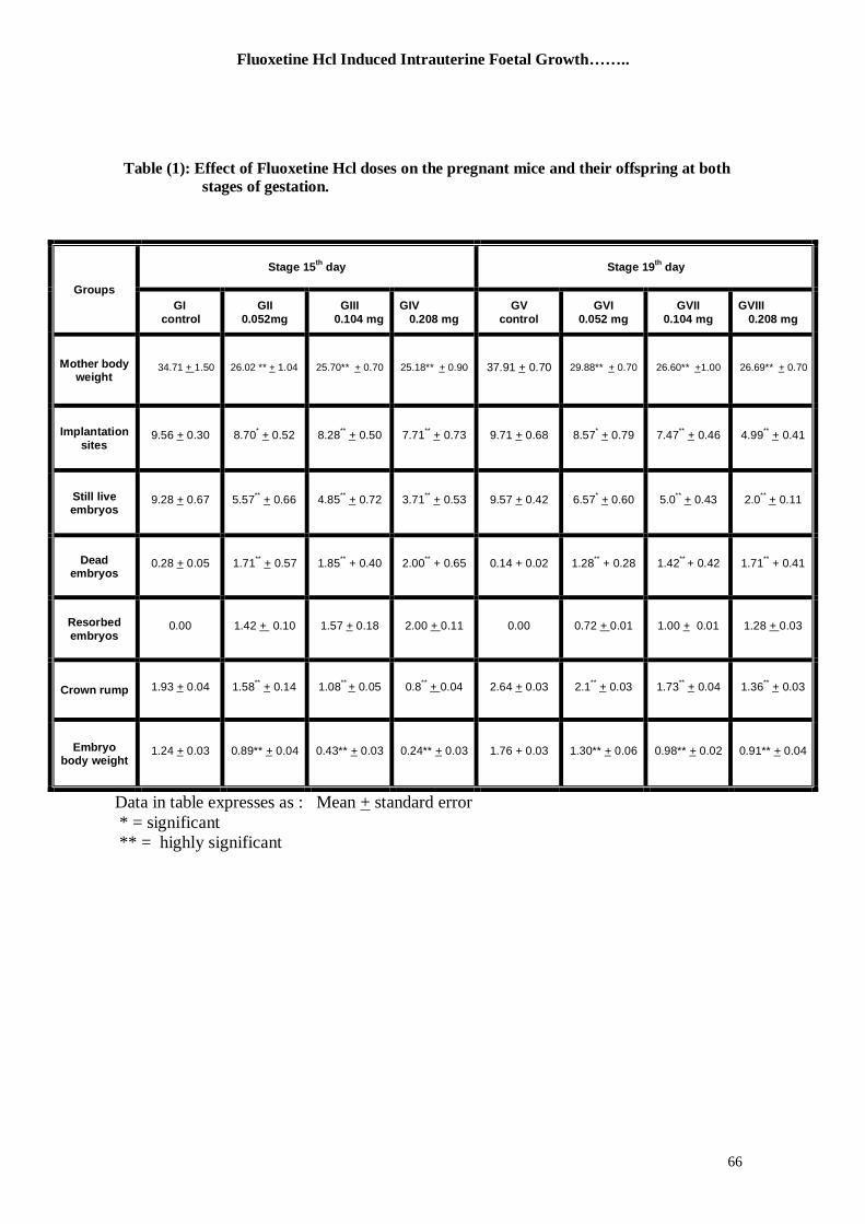
Table (1): Effect of Fluoxetine Hcl doses on the pregnant mice and their offspring at both
stages of gestation.
Data in table expresses as : Mean + standard error
* = significant
** = highly significant
Groups
Stage 15th
day Stage 19th
day
GI
control
GII
0.052mg
GIII
0.104 mg
GIV
0.208 mg
GV
control
GVI
0.052 mg
GVII
0.104 mg
GVIII
0.208 mg
Mother body weight
1.50+ 34.71 26.02 ** + 1.04 0.70 +25.70** 0.90 +25.18** 0.70 +37.91 0.70 +29.88** 1.00+26.60** 0.70 +26.69**
Implantation
sites
0.30 +9.56
8.70* + 0.52
0.50 + **
8.28
0.73 + **
7.71
0.68 +9.71
0.79 + *
8.57
0.46 + **
7.47
0.41 + **
4.99
Still live embryos
0.67 +9.28 5.57** + 0.66 0.72 +
**4.85 0.53 +
**3.71 0.42 +9.57 0.60 +
*6.57 0.43 +
**5.0 0.11 +
**2.0
Dead embryos
0.05 +0.28 0.57 + **
1.71 1.85** + 0.40 2.00
** + 0.65 0.14 + 0.02 1.28
** + 0.28 1.42
** + 0.42 1.71
** + 0.41
Resorbed
embryos 0.00 0.10 + 1.42 0.18 +1.57 0.11+ 2.00 0.00 0.01+ 0.72 0.01 +1.00 0.03+ 1.28
Crown rump 0.04 +1.93 1.58** + 0.14 0.05 +
** 1.08 0.04+
**0.8 0.03 +2.64 0.03 +
**2.1 0.04 +
**1.73 0.03 +
**1.36
Embryo body weight
0.03 +1.24 0.04 +0.89** 0.03 +0.43** 0.03 +0.24** 1.76 + 0.03 0.06 +1.30** 0.02 +0.98** 0.04 +0.91**
Ali,M.O. et al
67
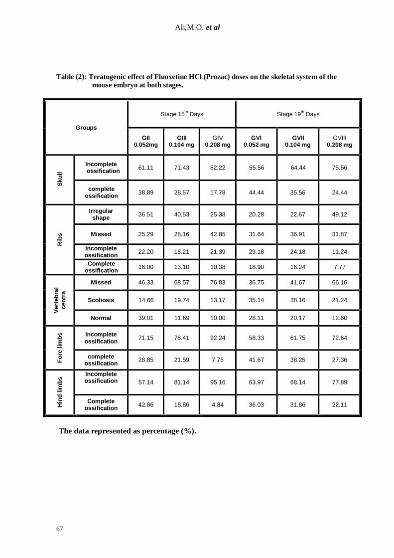
Table (2): Teratogenic effect of Fluoxetine HCl (Prozac) doses on the skeletal system of the
mouse embryo at both stages.
Groups
Stage 15th Days Stage 19
th Days
GII 0.052mg
GIII 0.104 mg
GIV 0.208 mg
GVI 0.052 mg
GVII 0.104 mg
GVIII 0.208 mg
Sku
ll
Incomplete ossification
61.11 71.43 82.22 55.56 64.44 75.56
complete ossification
38.89 28.57 17.78 44.44 35.56 24.44
Rib
s
Irregular shape
36.51 40.53 25.38 20.28 22.67 49.12
Missed 25.29 28.16 42.85 31.64 36.91 31.87
Incomplete ossification
22.20 18.21 21.39 29.18 24.18 11.24
Complete ossification
16.00 13.10 10.38 18.90 16.24 7.77
Vert
eb
ral
cen
tra
Missed 46.33 68.57 76.83 36.75 41.67 66.16
Scoliosis 14.66 19.74 13.17 35.14 38.16 21.24
Normal 39.01 11.69 10.00 28.11 20.17 12.60
Fo
re lim
bs
Incomplete ossification
71.15 78.41 92.24 58.33 61.75 72.64
complete ossification
28.85 21.59 7.76 41.67 38.25 27.36
Hin
d lim
bs Incomplete
ossification 57.14 81.14 95.16 63.97 68.14 77.89
Complete ossification
42.86 18.86 4.84 36.03 31.86 22.11
The data represented as percentage (%).
Fluoxetine Hcl Induced Intrauterine Foetal Growth……..
68
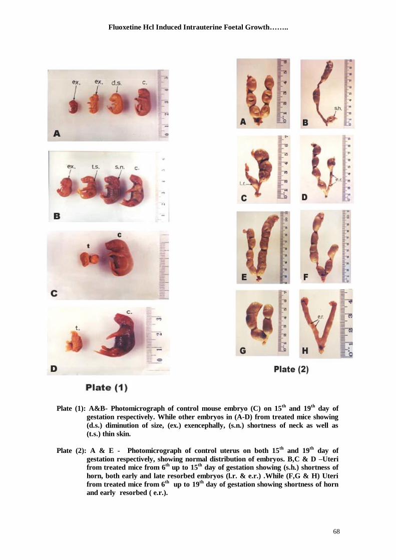
Plate (1): A&B- Photomicrograph of control mouse embryo (C) on 15
th and 19
th day of
gestation respectively. While other embryos in (A-D) from treated mice showing
(d.s.) diminution of size, (ex.) exencephally, (s.n.) shortness of neck as well as
(t.s.) thin skin.
Plate (2): A & E - Photomicrograph of control uterus on both 15th
and 19th
day of
gestation respectively, showing normal distribution of embryos. B,C & D –Uteri
from treated mice from 6th
up to 15th
day of gestation showing (s.h.) shortness of
horn, both early and late resorbed embryos (l.r. & e.r.) .While (F,G & H) Uteri
from treated mice from 6th
up to 19th
day of gestation showing shortness of horn
and early resorbed ( e.r.).
Ali,M.O. et al
69
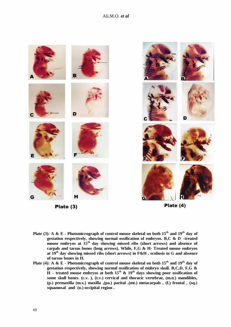
Plate (3): A & E - Photomicrograph of control mouse skeletal on both 15th
and 19th
day of
gestation respectively, showing normal ossification of embryos. B,C & D –treated
mouse embryos at 15th
day showing missed ribs (short arrows) and absence of
carpals and tarsus bones (long arrows). While, F,G & H- Treated mouse embryos
at 19th
day showing missed ribs (short arrows) in F&H , scoliosis in G and absence
of tarsus bones in H.
Plate (4): A & E - Photomicrograph of control mouse skeletal on both 15th
and 19th
day of
gestation respectively, showing normal ossification of embryo skull. B,C,D, F,G &
H – treated mouse embryos at both 15th
& 19th
days showing poor ossification of
some skull bones. (c.v. ), (t.v.) cervical and thoracic vertebrae, (m.n.) mandibles,
(p.) premaxilla (m.x.) maxilla ,(pa.) parital ,(mt.) metacarpals , (f.) frontal , (sq.)
squamosal and (o.) occipital region .
Fluoxetine Hcl Induced Intrauterine Foetal Growth……..
70
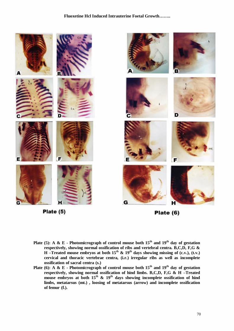
Plate (5): A & E - Photomicrograph of control mouse both 15
th and 19
th day of gestation
respectively, showing normal ossification of ribs and vertebral centra. B,C,D, F,G &
H –Treated mouse embryos at both 15th
& 19th
days showing missing of (c.v.), (t.v.)
cervical and thoracic vertebrae centra, (i.r.) irregular ribs as well as incomplete
ossification of sacral centra (s.)
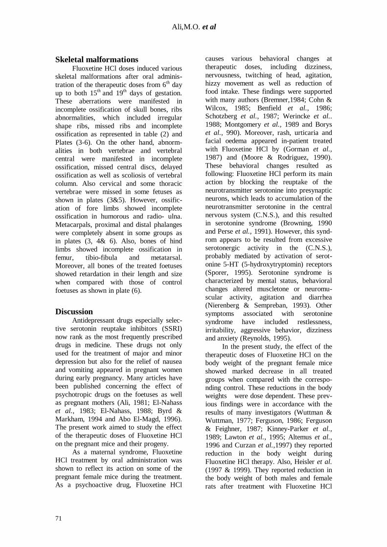
Plate (6): A & E - Photomicrograph of control mouse both 15th
and 19th
day of gestation
respectively, showing normal ossification of hind limbs. B,C,D, F,G & H –Treated
mouse embryos at both 15th
& 19th
days showing incomplete ossification of hind
limbs, metatarsus (mt.) , loosing of metatarsus (arrow) and incomplete ossification
of femur (f.).
Ali,M.O. et al
71
Skeletal malformations Fluoxetine HCl doses induced various skeletal malformations after oral adminis-
tration of the therapeutic doses from 6th day
up to both 15th
and 19th days of gestation.
These aberrations were manifested in
incomplete ossification of skull bones, ribs
abnormalities, which included irregular
shape ribs, missed ribs and incomplete ossification as represented in table (2) and
Plates (3-6). On the other hand, abnorm-
alities in both vertebrae and vertebral central were manifested in incomplete
ossification, missed central discs, delayed
ossification as well as scoliosis of vertebral
column. Also cervical and some thoracic vertebrae were missed in some fetuses as
shown in plates (3&5). However, ossific-
ation of fore limbs showed incomplete ossification in humorous and radio- ulna.
Metacarpals, proximal and distal phalanges
were completely absent in some groups as in plates (3, 4& 6). Also, bones of hind
limbs showed incomplete ossification in
femur, tibio-fibula and metatarsal.
Moreover, all bones of the treated foetuses showed retardation in their length and size
when compared with those of control
foetuses as shown in plate (6).
Discussion Antidepressant drugs especially selec-
tive serotonin reuptake inhibitors (SSRI)
now rank as the most frequently prescribed drugs in medicine. These drugs not only
used for the treatment of major and minor
depression but also for the relief of nausea
and vomiting appeared in pregnant women during early pregnancy. Many articles have
been published concerning the effect of
psychotropic drugs on the foetuses as well as pregnant mothers (Ali, 1981; El-Nahass
et al., 1983; El-Nahass, 1988; Byrd &
Markham, 1994 and Abo El-Magd, 1996). The present work aimed to study the effect
of the therapeutic doses of Fluoxetine HCl
on the pregnant mice and their progeny.
As a maternal syndrome, Fluoxetine HCl treatment by oral administration was
shown to reflect its action on some of the
pregnant female mice during the treatment. As a psychoactive drug, Fluoxetine HCl
causes various behavioral changes at
therapeutic doses, including dizziness,
nervousness, twitching of head, agitation, hizzy movement as well as reduction of
food intake. These findings were supported
with many authors (Bremner,1984; Cohn &
Wilcox, 1985; Benfield et al., 1986; Schotzberg et al., 1987; Werincke et al..
1988; Montgomery et al., 1989 and Borys
et al., 990). Moreover, rash, urticaria and facial oedema appeared in-patient treated
with Fluoxetine HCl by (Gorman et al.,
1987) and (Moore & Rodriguez, 1990).
These behavioral changes resulted as following: Fluoxetine HCl perform its main
action by blocking the reuptake of the
neurotransmitter serotonine into presynaptic neurons, which leads to accumulation of the
neurotransmitter serotonine in the central
nervous system (C.N.S.), and this resulted in serotonine syndrome (Browning, 1990
and Perse et al., 1991). However, this synd-
rom appears to be resulted from excessive
serotonergic activity in the (C.N.S.), probably mediated by activation of serot-
onine 5-HT (5-hydroxytryptomin) receptors
(Sporer, 1995). Serotonine syndrome is characterized by mental status, behavioral
changes altered muscletone or neuromu-
scular activity, agitation and diarrhea (Nierenberg & Sempreban, 1993). Other
symptoms associated with serotonine
syndrome have included restlessness,
irritability, aggressive behavior, dizziness and anxiety (Reynolds, 1995).
In the present study, the effect of the
therapeutic doses of Fluoxetine HCl on the body weight of the pregnant female mice
showed marked decrease in all treated
groups when compared with the correspo-
nding control. These reductions in the body weights were dose dependent. These prev-
ious findings were in accordance with the
results of many investigators (Wuttman & Wuttman, 1977; Ferguson, 1986; Ferguson
& Feighner, 1987; Kinney-Parker et al.,
1989; Lawton et al., 1995; Altemus et al., 1996 and Curzan et al.,1997) they reported
reduction in the body weight during
Fluoxetine HCl therapy. Also, Heisler et al.
(1997 & 1999). They reported reduction in the body weight of both males and female
rats after treatment with Fluoxetine HCl
Fluoxetine Hcl Induced Intrauterine Foetal Growth……..
72
due to decreases of fat and protein intakes.
Moreover, Dryden et al. (1996) reported
and explained how and why can Fluoxetine HCl causes decrease in the body weight?
They suggest that: serotonine and neurop-
eptide Y neurons in the hypothalamus,
respectively inhibit and stimulate food intake. Fluoxetine HCl not only inhibits
various aspects of the activity of the
neuropeptide Yergic arcuato-paraventric-ular neurons but also, reduce neuropeptide
Y release in the paraventricular nucleus, a
major site of neuropeptide Y release which,
is highly sensitive to appetite stimulating actions of neuropeptide Y. They also,
suggested that serotonine may influence
food intake and energy balance by inhibiting arcuato-paroventricular project-
ion, and that the two neurotransmitters
(serotonine and neuropeptide Y) may act together to regulate feeding and energy
homeostasis. The reduction in the body
weight in this study may be due to one of
the pervious reasons, which is confirmed by Curzan et al. (1997) they also, reported the
interaction between Fluoxetine HCl and
neuropeptide Y in the hypothalamus. Both phenomena of uterine
contraction and dismorphology of uterine
horns were induced by Fluoxetine HCl in all treated experimental groups when
compared with the corresponding control.
These findings were also observed by Ali,
1981 and El-Nahass, 1988 who used antide-pressant drugs namely Motival, Ludiomil
and Imitriptyline HCl during embryonic
development in the rats, and Abo El-Magd (1996) who used Tranxene and Lexotanil
(tranquilizers) with pregnant rats.The
previous phenomenae may be resulted from
one of the following results, the first one may be due to the ability of Fluoxetine HCl,
as a psychoactive drug, to blockage of 5-
HT reuptake in the smooth muscles of the myometerium layer in the uteri from the
treated mothers. The second reason
reported by Ficicioglu et al. (1995) who states that, Fluoxetine HCl induced
hyperprolactinemia in female rats, and the
high concentration of serum prolactine level
may causes degeneration of myometrial cells that resulted in myometrial invasion
by endometrial stroma. This invasion
eventually progress to adenomyosis.
In the present study, oral adminis-tration of therapeutic doses of Fluoxetine
HCl to the pregnant mice induced various
teratogenic pictures in mouse embryos.
These pictures were statistically significant when compared with the control ones.
These pictures were manifested in reduction
of foetal body weight, reduction in crown-rump length, still live embryos, delaying of
implantation, intrauterine death as well as
resorption of embryos. The embryotoxicity
effect was dose dependent. It can be said that, the previous prevailing phenomenae of
teratogenicity may attributed to toxicity of
Fluoxetine HCl due to accumulation of this drug in certain organs, placental barriers,
endometrial layer of uteri as well as
placental dysfunction. Similar results were recorded by many authors using Fluoxetine
HCl as Stanford & Patton (1993) who used
pregnant rats and resulted in subcutaneous
hematoma of neonates. Also, Vorhees et al. (1994), Vendittelli et al. (1995), Chambers
et al. (1996) and Blayac et al. (1997)
reported several prenatal and postnatal problems when the pregnant subjects
treated with Fluoxetine HCl.
David et al. (1998) and Lanczik et al. (1998) reported that, the use of
Psychotropic drugs during pregnancy may
cause three complications:1- Teratogen-
icity, 2- prenatal syndromes (neonatal toxicity), and 3- postnatal behavioral
squalae behavioral toxicity. They also
added, the exposure to certain psychotropic drugs in utero may increase the risk for
some specific congenital anomalies, and the
intrauterine exposure to the Psychotropic
drugs during the second and third trimester can lead to postnatal complications; for
example, floppy-infant syndrome induced
after administration of Benzodiazepines (Tranquilizers) to pregnant dams.
Moreover, Isenberg (1990) proved the post-
natal complication following Fluoxetine HCl therapy during pregnancy in human
being. In contradictory to the present
findings, Sommi et al., 1987; Goldstein,
1990; Pastuszak et al., 1993; Koren et al., 1994; David et al., 1997 and Nulman et al.,
Ali,M.O. et al
73
1997 reported that, no teratogenic effects
resulted from Fluoxetine HCl have been
observed in vivo. On the other hand, both Shuey et al. (1992) and Sadler et al. (1993)
proved the teratoge-nicity of Fluoxetine
HCl in vitro, and stated that the developm-
ental defects that resulted from exposure of mouse embryos in whole embryo culture to
Fluoxetine HCl induced craniofacial malfo-
rmation consistent with direct action at 5-HT uptake sites. Such developmental
defects may produced from inhibition of 5-
HT uptake during the embryogenesis by
interference with serotonergic regulation of growing epithelial mesenchymal embry-
onic cells.
Also, Shader (1992) examined two groups of women (totaling 133) who were
during pregnance exposed to Fluoxetine
HCl and documented that 3 of 44 pregn-ancies resulted in major abnormalities at
birth. The present findings are in parallel
with these results from Haloperidol
(Tranquilizer) treated experimental animals. Haloperidal administrated to rats and mice
after mating resulted in delay implantation
of 50% of exposed animals from 2nd
to 8th
day of gestation (Tuchmann-Duplessis &
Mercier-Parot, 1971). In higher doses,
Haloperidol can increase resorption in the pregnant rats and induce cleft palate in mice
(Vichi, 1969). Increased rates of embryonic
death and malformation have been observed
among the offspring of hamsters treated with Haloperidol during pregnancy (Gill et
al., 1982). Also, reduction in the foetal
body weight was recorded in foetuses obtained from treated mothers with
Haloperidol during pregnancy (Holson et
al., 1994).
Convening to the reduction in body weight of foetuses due to treatment with
Fluoxetine HCl the results were statistically
highly significant in comparison with the control ones. These findings were confir-
med by the results of Foritz et al. (1976)
and El-Nahass et al. (1983). Also, crown-rump of foetuses showed great reductions
in Fluoxetine HCl treated groups, these
reductions were statistically highly signify-
cant when compared with the control. These findings were in harmony with that
of Ali (1981), El-Nahass (1988) and Abo
El-Magd (1996) who used Psychotropic
drugs as Motival and Ludiomil by the first
author and Amitriptyline HCl by the second author as antidepressant drugs while, the
last author was used both Tranxene and
Lexotanil as a tranquilizers. It could be
suggested that: during the early embryonic development, the blastocytes lies free in the
uterus, it absorbs nutrients from the surrou-
nding fluid, which have high glycogen content. After implantation, the embryo
utilizes special fluid, the embryotroph,
which is mixture of uterine fluids, destro-
yed epithelial cells as well as blood cells (Tuchmann-Duplesis, 1975). The intraute-
rine development of the embryo is highly
dependent on the food of mother (Giroud, 1970 & 1973). The present result showed
that; the level of food intake by treated
mothers was severely induced. Hence, the nutrients reached to the embryo via
placenta was decreased. It was also noticed
that during the embryonic development, a
lack or an excess of a specific nutrient, result in sever impairment of pregnancy,
including embryonic death and congenital
malformation. Similar results were reported by various investigators (Fichter,1993 ;
Fung & Ferrill, 2001 ; Mitchell et al.,2001 ;
Sharma, 2001). During the foetal stage that charact-
erized by an intense general growth, a
specific nutritional deficiency will result in
growth inhibition. This inhibition was recorded by Heisler et al. (1997 & 1999)
who proved that Fluoxetine HCl decreases
fat and protein intake but not carbohydrates intake in both male and female rats. The
inhibition or retardation of the growth In
the present study may be resulted to one or
more of the previous reasons. Malformation of foetuses considered a
major part of the results of the present
work. Oral administration of the therapeutic doses of Fluoxetine HCl to pregnant female
mice from 6th day up to 15
th and 19
th days of
gestation induced several foetal malform-ations. These malformations represented by
subcutaneous hematoma, which appeared in
all foetuses obtained from all treated mice
as well as thin skin and diminution of size. Also, exencephally was recorded in GIII
and GIV which, treated orally from
Fluoxetine Hcl Induced Intrauterine Foetal Growth……..
74
gestation 6th day up to 15
th day of gestation
by 0.104 mg Fluoxetine HCl. Shortness of
tail, microcephally, scoliosis as well as oedematous ascites were observed. These
results were similar to those described by
Ali (1981), Sanyal & El-Nahass (1983) and
James et al. (1986), who worked on antid-epressant drugs as Motival, Ludiomil and
Imipiramin, respectively. Also, these find-
ings were in accordance with Ibraheem et al. (1998) when found teratogenic effect of
Nefopam HCl (sedative drug) on the
embryonic development of the rats.
Skeletal defects of foetuses obtained from Fluoxetine HCl treated mothers that
were sacrificed on 15th day and 19
th day of
gestation have been observed. These defects included incomplete ossification of
most skull bones, irregular and missed ribs,
absence of vertebral centra and scoliosis as well as incomplete ossification of both fore
and hind limbs. The major skeletal defects
were skull anomalies, including non
ossification centers of several skull bones maxilla, nasal, parital, interparital, suprao-
ceipital and basioccipital. The percentages
of these defects were dose dependent. Ali (1981) reported similar result
using antidepressant drugs namely Motival
and Ludiomil. Also, these results confirmed by those of McColl et al, (1963) and
Dwornik & Moore (1965) When described
a delay or incomplete ossification of skull
and sternum of rat foetuses treated with Thalidomide (Tranquilizer). Moreover,
Dipaolo (1963) and Dipaolo et al. (1964)
reported that, Thalidomide administrated to mice caused sever spinal abnormalities. The
deficient growth of the base of the skull
specially that of basioccipital has been
considered to be caused by primary meso-dermal insufficiency (Marin-Padilla, 1966).
Also, injury to the paraxial mesoderm
during early embryonic development (organogenesis) could result in a primary
mesodermal in sufficiency which in itself,
may be cause a variety of developmental abnormalities involving the various axial
structures. This findings also, were quite
confirmed by El-Nahass et al. (1983) who
reported that the use of Fluphenazine HCl during pregnancy induced absence of 5
th
centrum in rat foetuses and delayed or
induced incomplete ossification of the
sternum and vertebral column. Also, the
present findings are in agreement with those obtained by Rumea-Roquette et al.
(1977) who reported that Fluphenazine HCl
(Tranquilizer) caused inhibition or delayed
the ossification in vertebrae, ribs and scoliosis in vertebral column in human
foetuses.
In the present study, there was no ossification especially metacarpals and
metatarsals except in some individual
foetuses in GVII when compared with
normal ones and these also were dose dependent. These results in agreement with
those reported by Ali (1981), El-Nahass
(1988) and Abo El-Magd (1996). It may suggested that malformations of foetal
skeletal system attributed to the decreased
absorption rate of calcium from the intestine and/or injuries induced in the liver
of female pregnant rats previously treated
by Chlorpromazine (Othman et al., 1981).
Liver hepatitis was found to be decrease the formation of vitamin D metabolite
(monohydroxy vitamin D) which in renal
tissues will be rehydroxylated (1, 25 dihydroxy vitamin D) forming active
principle metabolite responsible for calcium
absorption from intestine. The present findings supported by Azmitia et al. (1990)
who studied the effects of 3,4-methylene
dioxymethamphetamine (MDMA) on
cultured serotonergic neurons of foetal raph neurons of rats to evidence for Ca
2+
dependent toxicity linked. They found that
5-HT (5-hydroxytryptamine) receptor is linked to increased intracellular Ca
2+, this
linkage inhibited Fluoxetine HCl action.
Also, Moiseiwitsch et al. (1998)
reported that, serotonine (5-HT) and its serotonergic ligands (which blocked by
Fluoxetine HCl) regulate the calcium
binding protein and tenascin (extracellular matrix molecule) which, together are
important in cranio-facial development, and
cartilage proteogylcan core protein. This regulation is stimulated by serotonine (5-
HT) in tooth-germ development in embry-
onic mouse mandibular explant in cultures,
which inhibited by Fluoxetine HCl. The present study suggested that the oral
administration of Fluoxetine HCl to the
Ali,M.O. et al
75
pregnant mice induced delaying of the
ossification and sever skeletal anomalies
which may be due to mesenchymal conde-nsation during embryonic development, or
may be due to resorption of cartilage,
during embryonic development, which
precedes endochondral ossification, which adversely affected by Fluoxetine HCl
doses. These results were also confirmed by
Cancholal - Martinez et al. (1997) they reported that certain Psychotropic drugs
inhibits calcium-calmodulin-system in rat
neonates as a systemic action of these
Psychotropic drugs(Verapamal, Haloperidol and Penfluridal). The present work sugge-
sted that Fluoxetine HCl either inhibits Ca+2
metabolism in both extrac-ellular and intracellular matrix molecule or caused
blocking of serotonergic ligands that
regulate calcium binding protein and this may be lead to the inhibition of serotonin 5-
HT receptor gene or to response of certain
genetic factors to interacting with the anti-
depressent drug (Fluoxtine). These sugges-tion were supported by many authors
(Moiseiwitsch ,2000).The former one
reported that the prozac and less selective tricyclic antideperessant drug like Elavil
would be at a higher risk for develo-
pmental dental defects such as anodontia & hypodontia and will cause major cranio-
ficial malformation. While Okada reported
that A human serotonin (5-HT)(2C) recep-
tor gene polymorphism leads to the substitution of cysteine for serine at codon
23 (Cys23Ser); the frequency of the Ser23
allele in unrelated Caucasians is approxim-ately 0.13. Although the amplitude of the 5-
HT-induced intracellular Ca(2+) peak did
not differ between the alleles, Ser23 requ-
ired higher 5-HT concentrations to elicit the same response. These results indicate that
the Ser23 allele may be constitutively more
active than Cys23. Thus, Ser23 appears to be an abundant candidate allele capable of
directly influencing inter-individual varia-
tion in behavior, susceptibility to mental disorder, and response to drugs including
atypical antipsychotic and some antide-
pressant drugs that are potent 5-HT(2C)
inverse agonists or antagonists. Eventialy , the present work awarance
the medical community and drug manufac-
turers that it should make a concerted effort
to protect women and their unborn babies
from risk associated with fetal exposure to teratogenic drugs, which can lead to
unneccessary abortions.
References 1. Abo El-Magd, M.H. (1996): Possible
Teratogenicity and mutagenicity induced by
some Benzodiazepines in rat embryos
Ph.D. Thesis. Fac. Sci. Al-Azhar Univ.
2. Ali, M.O. (1981): Studies on some embryological aspects induced by certain
antidepressant drugs in mammalian
embryos. Ph.D. Thesis, Fac. Sci., Al-Azhar
Univ.
3. Ali, M.O.; El Nahass, E.; Diamond, M.O.
and Desouki, G. (1989): Embryotoxic
effect of Diabetes mellitus. Al-Azhar
Medical J. 17(4): 421-428.
4. Altemus, M.; Glowa, J.R.; Galliven, E.;
Leong, Y.M. and Murphy, D.L.(1996): Effects of Serotonergic agent on food restr-iction induced hyperactivity. Pharmacol-
Biochem. - Behav, 53(1): 123-131.
5. Azmitia, E.C.; Murphy, R.B. and
Whitaker- Azmitia, P.M. (1990): MDMA
(ecstasy) effects on culture serotonergic
neurons: evidence for Ca2+ dependent
toxicity linked to release. Brain Res.
510(1): 97-103.
6. Barlow, M.S.; Knight, A. F. and Sullivan,
F.M. (1980): Diazepam- induced Cleft
palate in the Mouse: The Role of
Endogenous Maternal Corticosterone. Teratology, 21: 149-155.
7. Benfield, P.; Heel, R.C. and Lewis, S.P.
(1986): Fluoxetine: a review of its
pharmacodynamic and pharmacokinetic
properties, and therapeutic efficacy in
depressive illness. Drug. 32:481-508.
8. Blayac, J.P.; Hillaire - Buys, D. and
Peyriere, H. (1997): Pharmacovigilance of
new antidepressants: evaluation of neuro-
psychobehavioral disorders. Therapie,
52(2): 117-122.
9. Borys, D.J.; Setzer, S.C. and Ling, L.J.
(1990): Acute Fluoxetine overdose: a report
of 234 cases. Am. J. Emerg. Med.; 10:115-
120.
10. Bremner, J.D. (1984): Fluoxetine in
depressed patients: a comparison with
imipramine. J. Clin Psychiatry 45:414-419.
11. Browning, W.N. (1990): Exacerbation of
symptoms of multiple scerosis in a patient
taking Fluoxetine (Letter). Am J.
Psychiatry. 147:1089.
Fluoxetine Hcl Induced Intrauterine Foetal Growth……..
76
12. Byrd, A.R. and Markham, J.K. (1994): Developmental toxicology studies of
Fluoxetine HCl administrated orally to rats
and rabbits. Fundam. Appl. Toxicol., 22
(4): 511-518.
13. Canchola - Martinez, E.; Vergara -
Onofre, M.; Rodriguez - Medina, M.A.
and Mercado - Sanchez, G. (1997): Inhibitors of calcium - calmodulin system
and hypothalamic sex differentiation in rats.
Biochemical parameters. Ginecol - Obstet - Mex. 65:508-514.
14. Chambers, C.D.; Johnson, K.A. and
Dick, L.M. (1996): Birth outcome in
pregnant women taking Fluoxetine. N.
Engl. J. Med. 335:1010-1015.
15. Cohn, J.B. and Wilcox, C. (1985): Comparison of Fluoxetine, Imipramine and
Placebo in patients with major depressive
disorder. J. Clin Psychiatry, 46 : 26-31.
16. Cook, M. and Farweather, F. (1968): Methods used in teratogenic testing Lab. Anim. 2:219-228.
17. Curzan, G.; Gibson, E.L. and Oluyomi,
A.O. (1997): Appetite suppression by
commonly used drugs depends on 5-HT
receptors but not on 5-HT availability.
Trends - Pharmacol. Sci. 18(1):21-25.
18. David, J.; Lois, A.; Goldstein, R.N.;
Corbin, R.N. and Sundell, L.K. (1997): Effect of first trimester Fluoxetine exposure
on the newborn. Obsetetrics and
Gynecology. 89(5) 1:713-718. 19. Dipaolo, J.A. (1963): Congenital
malformation in strain A mice. J.A.M.A.
183: 139-141.
20. Dipaolo, J.A.; Gatzek, H. and Picken, J.
(1964): Malformations induced in mouse
by thalidomide. Ama. Rec. 149:149-156.
21. Dryden, S.; Frankish, H.M.; Wang, Q.;
Pickavance, L. and Williams, G. (1996): The serotonergic agent Fluoxetine reduces
neuropeptide Y levels and neuropeptide Y
secretion in the hypothalamus of lean and
obese rats. Neurosciance. 72(2):557-566.
22. Dwornik, J.J. and Moore, K.L. (1965):
Skeletal malformations in the Holtzman rat
embryo following the administration of
thalidomide. J. Embryol. Exp. Morphal.
13:181.
23. El-Nahass, A.M. (1988): Effect of certain
Psychotropic drugs on pregnant rats and
their offspring's. Ms.D. Thesis, Fac. Sci.
Al-Azhar Univ.
24. El-Nahass, S.M.; Ali, M.O. and El-
Nahass, E. (1983): Congenital abnormal-lities in rat embryos associated with
maternal use of fluphenazine HCl during
pregnancy. Egypt, J. Genet. Cytol. 12:449-
458.
25. Ferguson, J.M. (1986): Fluoxetine induced
weight loss in over weight, nondepressed
subjects. Am. J. Psychiatry, 143:1496
(Letter).
26. Ferguson, J.M. and Feighner, J.P.
(1987): Fluoxetine induced weight loss in
over weight non-depressed humans. Int. J.
Obese. 11(3):163-170.
27. Fichter , M.M.(1993): Drug treatment of anorexia nervosa and bulimia nervosa. A
review. Nervenarzt. 64(1):21 – 35.
28. Ficicioglu, C.; Tekin, H.I.; Arioglu, P.F.
and Okar, I. (1995): A murine model of
adenomyosis: the effect of
hyperprolactinemia induced by Fluoxetine
HCl, a selective serotonin reputake
inhibitor, on adenomyosis in induction in
Wister albino rats. Acta. Eur. Fertil. 26(2):
75-79.
29. Foritz, H.D.; Mudler and Hess, R. (1976): Comparative study of teratogenicity
of Phenobarbitone, Diphenylydantation and
Carbamazepine in mice. Toxicology. 6(3):
323-330.
30. Fung, S.M. and Ferrill, M.J.( 2001): Treatment of bulimia nervosa with
ondansetron. Ann Pharmacother. 35(10):
1270-3.
31. Giroud, A. (1970): The nutrition of the
embryo. Thomas, Spring Field : 10-51.
32. Giroud, A. (1973): Nutritional requirements of the embryo. World review
of nutrition and dietetics, 18:195-196.
33. Globus, M. and Gibson, M.A. (1968): A
histological study of the development of the
sternum in thalidomide treated rats.
Teratology. 1: 235-256.
34. Goldberg, H.L. and Nissim, R. (1994): Psychotropic drugs in pregnancy and
lactation. Int. J. Psychiatry Med. 24
(2):129-147.
35. Goldstein, D.J. (1990): Outcome of
Fluoxetine exposed pregnancies. Am. J. Hum. Genet. 47: A136.
36. Gorman, J.M.; Liebowitz, M.R. and
Fyer, A.J. (1987): An open trial of
Fluoxetine in the treatment of panic attacks.
J.Clin. Psychopharmacal. 7:329-332.
37. Heisler, L.K.; Kanare K.R.B. and
Gerstein, A. (1997): Fluoxetine decreases
Fat and protein intakes but not carbohydrate
intake in male rats. Pharmacal. Biochem.
Behav. 58(3): 676-773.
38. Heisler, L.K.; Kanare K.R.B. and Gerstein, A. (1999): Reduction of fat and
protein intakes but not carbohydrate intake
Ali,M.O. et al
77
following acute and chronic administration
of Fluoxetine in female rats. Pharmacal.
Biochem. Behav. 63 (3) : 377 – 385.
39. Holson, R.R.; Webb, P.J.; Grafton, T.F.
and Hansen, D.K. (1994): Prenatal
neuroleptic exposure and growth stunting in
the rat: an in vivo and vitro examination of
sensitive periods and possible mechanisms.
Teratology. 50 (2): 125-136.
40. Ibraheem, M.A.; Zaki, T.Z. and Nadia,
H.I. (1998): Tertagoenic and histopathological effect of Nefopam HCl on
rat foetuses. J. Egypt. Ger. Soc. Zool. 25
(B), 185-239.
41. Isenberg, K.E. (1990): Excretion of
Fluoxetine in human breast milk. J. Clin.
Psychiatry. 51:169 (Letter).
42. James, R.; Harmon, Peggy, J.; Webb;
Gary, L.; Kimmel and Robert, R. (1986): Effect of prenatal imipramine exposure on
development of the postnatal rat heart and
brain. Teratogenesis, Carcinogenesis and Mutagenesis. Med. J. 18: (8) : 416-424.
43. Kaplan, S. and Grabowski, C.T. (1967): Analysis of Trypan blue-induced
rumplessness in chick embryos. J. Exp.
Zool. 165:325-336.
44. Kinney-Parker, J.L.; Smith, D. and Ingle
S.F. (1989): Fluoxetine and weight: Some
thing last and some thing gained? Clin.
Pharm. 8:727-733.
45. Koren, G.; Pastuszak, A.; Jasobson, S.;
Schick, B.; Donnenfeld, A.; Feldkamp,
M.; Zuber, C.; M.G. Cormack, M.;
Jones, K. and Gardener, H.A. (1994):
The safety of antidepressants in pregnancy.
Maternal fetal Toxicology. A Clincans -
Guide second edtion. Koren, G. ed. New
York, NY-U.S.A Marcel - Dekker, Inc.
2:59-76.
46. Lanczik, M.; Knoche, M. and Fritz, J.
(1998): Psychopharmacotherapy during
pregnancy and lactation.1: pregnancy.
Nervenarzt. 69(1): 1-9.
47. Lawton, C.L.; Wales, J.K; Hill, A. J. and Bundell, J.E. (1995): Serotonergic
manipulation, meal – induced satiety and
eating pattern: Effect of Fluoxetine in obese
female subjects. Obese-Res. 3 (4): 345-356.
48. McColl, J.D.; Globus, M. and Robinson,
S. (1963): Drug induced skeletal
malformations in rat. Experintia (Basel).
19:183.
49. Mitchell, J.E.; Peterson, C.B.; Myers, T.
and Wonderlich, S.(2001): Combining
pharmacotherapy and psychotherapy in the treatment of patients with eating disorders.
Psychiatr. Clin. North. Am. Jun. 24(2):315-
23.
50. Moiseiwitsch, J.R. (2000):The role of
serotonin and neurotransmitters during
craniofacial development. Crit Rev Oral
Biol Med. 11(2):230-9.
51. Moiseiwitsch, J.R.; Raymond, J.R.;
Tamir, H. and Lauder, J.M. (1998):
Regulation by serotonin of tooth-germ
morphogenesis and gene expression in
mouse mandibular explant cultures. Arch - oral Biol. 43(10): 789 - 800.
52. Montgomery, S.A.; Dufour, H. and
Brion, S. (1989): Prophylactic efficacy of
Fluoxetine in unipolar depression. Br. J.
Psychiatry. 153 (3): 69-76.
53. Moore, J.L. and Rodriguez, R. (1990): Toxicity of Fluoxetine in over dose. Am. J.
Psychiatry, 147:1089.
54. Nierenberg, D.W. and Sempreban, M.
(1993): The central nervous system
serotonin syndrom. Clin. Pharmacal Ther. 53:84-88.
55. Nulman, I.; Rovet, J.; Stewart, D.E.;
Wolpin, J.; Gardner H.A.; Thesis,
J.G.W.; Kulin, N. and Koren, G. (1997): Neurodevelopment of children exposed in
utero to antidepressant drugs. N. Eng. J.
Med. 336:258-262.
56. Othman, A.B.; El-Seddik and El-Kholy,
W. (1981): Studies on calcium metabolism
in chlorpromazine treated female and
pregnant rats. Ain Shams Medical J. Ain Shams University Hospitals, Abbasia,
Cairo.
57. Pastuszak, A.; Schick-Boschetto, B. and
Zuber, C. (1993): Pregnancy outcome
following first. Trimester exposure to
Fluoxetine (Prozac). JAMA. 269:2246-
2248.
58. Perse, T.; Meyers, F. and Hegyvary, C.
(1991): New Pespatives on Fluoxetine
(Prozac): Promixe, Problems and Potential
dangers. Emerg. Med. Repots. 12:73-80.
59. Reynolds, R.D. (1995): Serotonin syndrom: what family physicians need to
known. Am. Fam. Physician; 52:1263-
1271.
60. Rumeau-Rouquette, C.; Goujard, J. and
Heul, G. (1977): Possible teratogenic effect
of phenothiazine in human beings.
Teratology. 15:57-64.
61. Sadler, T.W.; Shuey, D.L. and Lauder,
J.M. (1993): Serotonine and cardiac
morphogenesis in the mouse embryo.
Teratology. 47 (6) : 573-584.
62. Sanyal, M.K. and Eric, A.W. (1979): Oxygen requirement in Vitro growth and
Fluoxetine Hcl Induced Intrauterine Foetal Growth……..
78
differentiation of the rat conceptus during
organogenesis phase of embryo
development. Biology of Reproduction. 20:
639- 647.
63. Schotzberg, A.F.; Dessain, E. and O`Neil, P. (1987): Recent studies on selective
serotonergic antidepressants: trazodone,
Fluoxetine and Fluvoxamine. J. Clin
Psychopharmacal. 7:445-495.
64. Shader, R. (1992): A patient of mine
missed aperiod while taking Fluoxetine; it turned out that she was not pregnant.
However, the question arose about the
safety of Fluoxetine during pregnancy.
What is known? (Response). J. Clin.
Psychopharma, 12:213.
65. Sharma, A.( 2001): Anorexia nervosa and
bulimia nervosa: An appraisal. Drugs
Today (Barc). 37(4):229-236.
66. Shuey, D.L.; Sadler, T.W. and Lauder,
J.M. (1992): Serotonin as a regulator of
Craniofacial morphogenesis: Site specific malformations following exposure to
serotonin uptake inhibitors.
67. Sommi, R.W.; Crismon, M.L. and
Bowden CL. (1987): Fluoxetine: a
serotonin specific, second generation
antidepressant. Pharmacotherapy. 7:1-15.
68. Sporer, K.A. (1995): The serotonine
syndrome. Implicated drugs,
Pathologysiology and management. Drug
Saf. 13:94-104.
69. Stanford, M.S. and Patton, J.H. (1993): In utero exposure to Fluoxetine HCl
increases haematoma frequency at birth.
Pharmacy. Biochem. Behav. 45(4): 459-
962.Teratology, 46 (4): 367-378.
70. Tuchmann-Duplesis, H. (1975): Durg
effects on the Foetus. ADIS. Press. Sydney:
15-57.
71. Tuchmann-Duplessis, H. and Mercier -
Parot, L. (1971): Influence of neuroleptics
on prenatal development in mammals. In :
Tuchmann - Duplessis, H. (Eds):
Malformations, Tumors and mental
Dafects, pathogenetic correlations. Carlo Erba Foundation. Milan.
72. Vendittelli, F.; Alain, J. and Nouaille, Y.
(1995): A case of lipomeningocele reported
with Fluoxetine (and alprazolam, vitamins
B1 and B6, heptaminol) prescribed during
pregnancy. Eur. J. Obstet. Gynecol.
Reprod. Biol. 58:85-86.
73. Vichi, F. (1969): Neurolepic drugs in
experimental teratogenesis. In Bertelli and
Donati, (eds): Teratology Proceedings,
Amesterdam, Excepta. Medica. : 87-101.
74. Vorhees, C.V.; Acuff-Smith, K.D. and
Schilling, M.A. (1994): A developmental
neurotoxicity evalution of the effects of
prenatal exposure to Fluoxetine in rats.
Fundam. Appl. Toxicol. 23 (2) : 194-205.
75. Wernicke, J.F.; Dunlop, S.R. and
Dornseif, B.E. (1988): Fixed dose of
Fluoxetine therapy for depression.
Psychopharmacal. Bull. 23:164-168.
76. Wuttman, J.J. and Wuttman, R.J.
(1977): Fenfluramine and Fluoxetine spare protein consumption while suppressing
caloric intake by rats. Science. 198:1178-
1180.
Ali,M.O. et al
79
ر النمى الجنيني و التشىهبت الهيكلية المحدث بىاسطة الفلىكسيتيهتأخ
هيدروكلىريد في الفئران الحىامل
محمد عثمبن عل ، أسبمة شرف الديه ، عمر الصديق المنشبوي ، سيد أحمد بكري
جاهعة الازر –كلة العلم –قطن علن الحاى
يي اضييعة اانمييا ه يي الفلكطييني يي الأييي االيية الوتييالو ل كن ييا
عبا و عي هميناا هيي ه ي الفل ننيا الوميناة هيي بي ر ي . البرزاكالفلكطيييني ييي هيييي االييية الوتيييالو ل كن يييا الييي نلييي ركبيييا . اهيييي
قي اضن يم ب . ه بلجا عي عض اااع ااخر هي االة الوتالو ل كن ا
، 0.0.2هيييي الف يييراى الحاهييي ييين هعييياللنن لرعيييا ه نلفييية ييي 00ث ا البحييم هييي الفلكطييني عليي هييرلألني الاليي هييي الييم / بيي /هلليين 0.200، 0.100
الطالش لأن الم ال اهص عمر ال اة هي الم الطالش لأن الم الناضع عمر هي
كطييني ي كل ييي لييلأ عيييم الف ييراى الحاهيي الوعالليية اضيي ة الفل. الحويي اضييينارا لأيييالنن زيييالو عليييبنن النيييك الوطييينور الحركييية الويييسزو اليييا
فحي اا لأيام . الوطنور بي ياا الاي ام هويا يلل الي اليا بي زى اللطين
الو نلفة للف راى الحاه ب الولاهع الوعاللة هيي اليم ا لطيالش الي اليم ال ياهص ل الم الناضع عمر هي الحوي قيي جيي اى ي اا لأيام يارو بي الحلين عمر إ
ال ا عيم النواث ب كلا الرلأوي عيم الاي و علي بيا ااجية ويا اليا زيالو عييل ااجية الونية الوونلية عنويي علي جرعية .الكبر ب عييل ااجية الحية
قييي جييي اتييا بيي الف ييراى الوعالليية . بحييث اليييام الوطيين يهة الوعنويييو بيي يي ا ال
الفلكطني ى اك ما ب اللاز الكل ه ار لألو ظ ما (.اكطضفالا)ب الراش