Embed Size (px)
Citation preview
RESEARCH REVIEWInternational Microbiology (2014) 17:65-73doi:10.2436/20.1501.01.208 ISSN (print): 1139-6709. e-ISSN: 1618-1095www.im.microbios.org
Functional amyloids in bacteria
Diego Romero,1*§ Roberto Kolter2§
1Institute of Subtropical and Mediterranean Hortofruticulture “La Mayora”- CSIC, and Department of Microbiology, University of Malaga, Malaga, Spain.
2Department of Microbiology and Immunobiology, Harvard Medical School, Boston, MA, USA
Received 12 June 2014 · Accepted 28 June 2014
Summary. The term amyloidosis is used to refer to a family of pathologies altering the homeostasis of human organs. Despite having a name that alludes to starch content, the amyloid accumulations are made up of proteins that polymerize as long and rigid fibers. Amyloid proteins vary widely with respect to their amino acid sequences but they share similarities in their quaternary structure; the amyloid fibers are enriched in β-sheets arranged perpendicular to the axis of the fiber. This structural feature provides great robustness, remarkable stability, and insolubility. In addition, amyloid proteins specifically stain with certain dyes such as Congo red and thioflavin-T. The aggregation into amyloid fibers, however, it is not restricted to pathogenic processes, rather it seems to be widely distributed among proteins and polypeptides. Amyloid fibers are present in insects, fungi and bacteria, and they are important in maintaining the homeostasis of the organism. Such findings have motivated the use of the term “functional amyloid” to differentiate these amyloid proteins from their toxic siblings. This review focuses on systems that have evolved in bacteria that control the expression and assembly of amyloid proteins on cell surfaces, such that the robustness of amyloid proteins are used towards a beneficial end. [Int Microbiol 2014; 17(2):65-73]
Keywords: Bacillus subtilis · bacterial biofilms · extracellular matrix · TasA amyloid-like fibers
*Corresponding author: D. RomeroDepartamento de Microbiología. Facultad de CienciasUniversidad de Málaga-Instituto de Hortofruticultura Subtropical y Mediterránea “La Mayora”(IHSM-UMA-CSIC)Bulevar Louis Pasteur-Campus Universitario de Teatinos, s/n29071 Málaga, SpainTel. +34-952134274 E-mail: [email protected]
§ Both authors are equal contributors
Amyloids in history
The term amyloidosis is used to refer to a family of pathologies altering the homeostasis of human organs. Written descrip tions of what most likely were amyloidoses dates back to the late 17th century. An autopsy report describing a spleen full of white stones can be considered the first description of amyloi-dosis, which is now known as “sago spleen” to describe the
starch-like granules that grow in the organ [20]. The term amyloid, meaning resembling starch, was first used two cen-turies later. The German chemist Rudolph Virchow discov-ered that the corporea amylacea of the nervous system stained with Congo red in a similar way as did cellulose and starch [41]. There was a long-standing controversy on the chemical nature of such amyloid plaques with some maintaining that they were made of starch (thus amyloid) and others arguing that they were more akin to lard [20]. It was not until the 20th century that chemical analyses revealed that such accumula-tions consisted of protein. By then, however, the medical term had gained a stronghold and to date these proteins are referred to as amyloids, despite their having no starch content whatso-ever. However, the early descriptions of amyloid proteins al-ready revealed a peculiarity; under the electron microscope amyloids appeared as long and rigid fibers [20].
Int. Microbiol. Vol. 17, 2014 ROMERO, KOLTER.66
The intense research on amyloid proteins has shown that even though they vary widely with respect to their amino acid sequences, they share similarities in their quaternary structure. Amyloid protein fibers are enriched in β-sheets arranged per-β-sheets arranged per--sheets arranged per-pendicular to the axis of the fiber [50]. This structure provides great robustness, which is the defining feature of amyloid pro-tein. Amyloids have remarkable stability, insolubility and spe-cifically stain with certain dyes such as Congo red and thiofla-vin-T. The fact that amyloid fibers had always been associated with human pathologies led to the perception that the amyloid state was due to an erratic processing or misfolding of soluble and functional proteins [44]. However, the aggregation into amyloid fibers seems to be widely distributed among proteins and polypeptides, and in some cases, these amyloid fibers are important in maintaining the homeostasis of the organism [10,23]. Two outstanding examples of amyloids non-related to pathologies in humans are: The protein pMel17, which form amyloid fibers to eliminate intermediate aggregates that may be toxic to the organism, and proteins or peptides hormones of the
human endocrine system, which are efficiently stored in secre-tory granules in an amyloid-state, thus contributing to the nor-mal physiology of cells [19,23]. Amyloid fibers are also pres-ent in insects, fungi and in bacteria, and they participate in pro-tection, interaction with surfaces, and detoxification (Table 1). Prions, another group of proteins with propensity to fold into amyloid fibers but with the astonishing ability of self-propa-gating, are mostly known for their pathological implications, although exceptions to this rule are arising [27,49]. This hap-pens to be with the translation regulation protein CPEB (cyto-plasmic polyadenylation element binding protein) in the mol-lusk Aplysia. As other prions, this protein possesses the ability to acquire different functional conformations, and it appears that the prion dominant form contributes to stabilizing the long-term stimulated synapses in memory storage [42]. An-other example of a non-pathological prion is the protein HET-s in the fungus Podospora anserine [26,55], which in the prion state seems to contribute to build a physical barrier that impede the transfer of deleterious elements between genetically in-
Table 1. Amyloid proteins
1A. Pathogenic amyloids
Name Precursor Disease Ref.
AA Serum amyloid Systemic amyloidosis [10]
Aβ β-Protein (APP) Alzheimer’s disease [10]
AIAPP Islet amyloid polypeptide (Type 2 diabetes and insuloma) [10]
α-Synuclein α-Synuclein Parkinson’s disease [10]
1B. Functional amyloids
Species Protein Function
Humans Pmel17 Elimination of toxic intermediates during melanin synthesis [19]
Fungi Hydrophobins Formation of fungal coat [56]
Escherichia coli, Salmonella enteritidis Curli Interaction with host, biofilm formation [9,15]
Pseudomonas sp. FapC Biofilm formation [12]
Streptomyces coelicolor Chaplins Formation of aerial structures [13]
Bacillus subtilis TasA Biofilm formation [31]
Klebsiella pneumoniae Microcin E492 Antimicrobial [40]
Staphylococcus aureus PSM Biofilm formation [37]
Streptococcus mutans Adhesin P1 Biofilm formation [29]
Mycobacterium tuberculosis MTP Host interaction [1]
Xanthomonas axonopodis Harpin Virulence factor, multicellularity [28,39]
Int. Microbiol. Vol. 17, 2014BACTERIAL AMYLOIDS 67
compatible strains [26]. All these findings have motivated the use of the term “functional amyloid” to differentiate these amyloid proteins from their toxic siblings [14]. In this review we focus on systems that have evolved in bacteria that control the expression and assembly of amyloid proteins on cell sur-faces, such that the robustness of amyloid proteins are used towards a beneficial end.
Functional amyloids in bacteria
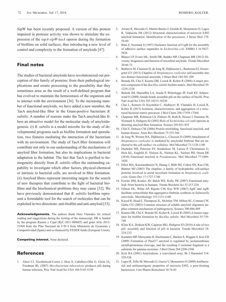
One of the first reports of functional amyloid proteins in bac-teria was a study of the curli pili of an uropathogenic strain of Escherichia coli. These pili were initially described as fibro-nectin-binding organelles on the surface of cells [30]. Subse-quently, Chapman and collaborators demonstrated that the curli fibers that emerged from the surfaces of E. coli cells had the same physical properties as the well-studied amyloid pro-teins responsible for human amyloidoses, e.g., staining with specific dyes (Fig.1) [9]. Escherichia coli uses curli amyloid-like fibers for a variety of physiological functions. Among these are interactions with host tissues, biofilm formation, and evasion of the immune system [4]. This first description of a bacterial functional amyloid opened the possibility that amy-
loid proteins could be present in other bacterial species. In-deed, Larsen and collaborators carried out an immunolabeling study with diverse bacterial species and suggested that amy-loid proteins were present as constituents of bacterial biofilms in a variety of environmental samples [22]. However, there is still just a limited number of examples where the direct impli-cation of amyloids in biofilm formation has been demonstrat-ed: Tafi, the curli homolog in Salmonella, FapC in many Pseudomonas species, TasA in Bacillus subtilis, and the re-cently found phenol soluble modulins (PSMs) in Staphylo-coccus aureus and the adhesin protein P1 in Streptococcus mutans in dental plaque biofilms [12,15,29,31,37].
Amyloid proteins may be utilized to perform other physio-l ogical processes in bacteria. For example, chaplins of Strep-tomyces coelicolor not only serve to interact with surfaces, but they also facilitate the rising of the aerial hyphae [5,11]. In Mycobacterium tuberculosis, amyloid pili (MTP) are neces-sary to interact with the host during pathogenesis [1]. Another example of an amyloid having a role during pathogenesis in-volves the harpins, important virulence factors of Xanthomon-as axonopodis and other plant pathogenic bacteria. The harpin Hpag of Xanthomonas has been demonstrated to form amy-loid fibers in vitro, and to be related to the hypersensitive re-sponse (HR) caused in the host and most recently shown to
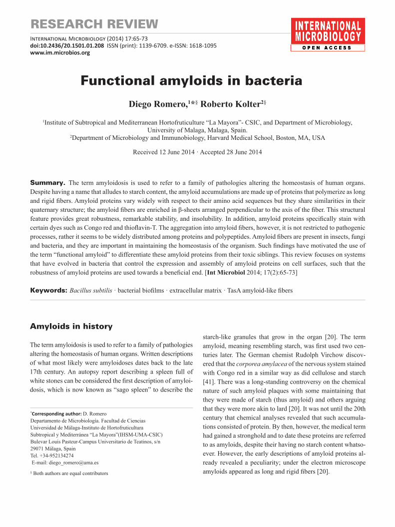
Fig. 1. Top row: Escherichia coli and Bacillus subtilis colonies stain with the amyloid specific dye Congo red. The pictures were taken after 72 h of growth in MSgg (minimal salts glycerol glutamate) agar (B. subtilis) or YESCA agar (E. coli) supplemented with Congo red. Bottom row: Transmission electron micrographs of curli amyloid fibers in E. coli (left) and TasA amyloid-like fibers in B. subtilis (right). Bars equal 200 nm. In
t Mic
robi
ol
Int. Microbiol. Vol. 17, 2014 ROMERO, KOLTER.68
participate in the multicellular behavior of this bacterial spe-cies [28,39]. In this case, it was demonstrated that tetrameric oligomers were the main protein species found in hypersensi-tive responses. These findings contribute to the debate on the toxicity of amyloids. Historically, fibers have been considered as the etiological agents of amyloidosis. However, intermedi-ate aggregates have a high propensity to insert into biological membranes and induce structural destabilization [16,51]. Thus, it is now thought that the intermediate aggregates, rath-er that the fibers themselves, are responsible for disease devel-opment [51].
Other studies demonstrate that, besides their role in devel-opmental programs or interaction with the host cells, amy-loids can be exploited as a detoxifying system. An interesting example of how the toxicity of amyloids can be modulated in self-benefit is that of the toxin microcin E492 produced by Klebsiella pneumoniae [40]. This toxin is a small peptide that oligomerizes into the cytoplasmic membranes of Enterobacte-riceae, causing pores that result in cell death [21]. The toxicity of microcin E492 was observed to vary depending on the state of growth of the producing cells; it was higher during expo-nential growth and decreased progressively with the aging of the culture. Not that the reduction of toxicity was associated to the transition of the toxin from an oligomeric state to fur-ther polymerization into amyloid fibers. This observation led to the conclusion that the toxic oligomers of microcin E492 resemble the intermediate aggregates of the protein Aβ associ-β associ- associ-ated with Alzheimer’s disease [2,24].
The inclusion bodies (IBs) that form during heterologous expression of proteins in bacteria are also an example of pro-teins with an amyloid conformation. The IBs have been his-torically considered the bottleneck that reduces the yield of overexpressed soluble and functional proteins. However, re-cent research demonstrates that peptides can be recovered from the IBs and still retain functionality. Besides the biologi-cal role of IBs as scavengers of putative toxic molecules for the bacterial cells, they offer an exciting new way for the ef-ficient and controlled delivery of drugs in chemotherapy [52,53].
Amyloidogenesis as a sophisticated process of protein aggregation
The discovery that amyloid proteins can carry out important physiological functions in bacteria has forced the re-evalua-tion of the concept that amyloidogenesis is always an erratic process of protein aggregation. As we describe in this section,
rather than being an uncontrolled process, it is now clear that in bacteria, and probably in other organisms, sophisticated machineries have evolved that direct the polymerization of these amyloid fibers outside the cell, thus avoiding toxicity yet providing the structural robustness to produce very stable organelles [36].
Curli: a paradigm for bacterial amyloidogenesis. Amyloid proteins have the intrinsic propensity to polymerize from the native monomeric state to the ordered and insoluble amyloid fibers [50]. During the formation of the fibers, amy-loids go through diverse stages of aggregation with variable biochemical and morphological features. The kinetics of amy-loid polymerization follow a typical sigmoidal curve with an initial lag phase, followed by an exponential phase of growth and a final plateau, where the fibers saturate and do not grow any further [25]. What allows the members of this family of proteins to form fibers? The study of curli in E. coli has clari-fied much of the molecular mechanisms that direct amyloido-genesis. The first clear difference with pathogenic amyloids is that curli formation is a highly regulated process. The protein products of two divergent operons are directly involved in the formation of curli fibers: csgABC and csgDEFG. The prod-ucts of csgA and csgB are the main components of the fibers and the other proteins participate in regulating gene expres-sion or control the proper secretion and polymerization of the fibers. CsgD regulates the expression of csgABC. CsgG forms a pore-like structure in the outer membrane, and allows the translocation of CsgA and CsgB from the periplasm to the outside of the cell. GsgF helps expose CsgB to the surfaces, and CsgE is thought to facilitate the access of CsgA to the secretion complex formed by CsgG. Finally, CsgC, which also has oxidoreductase activity, is thought to control the for-mation of the CsgG pore-like structure. The fibrillation of curli follows a nucleation-polymerization model, which means that the mixture of single mutants csgA and csgB com-plement each other extracellularly.
This is a fascinating result indicating that there is no need to produce both subunits in the same cell. The detailed analy-sis of the curlin subunits CsgA and CsgB demonstrated the existence of an amyloidogenic core, composed of four imper-fect repeats within the proteins’ amino acid sequence. Thus, when CsgB encounters CsgA, it induces a conformational change towards the amyloidogenic state making its polymer-ization into fibers possible. The reaction between the two sub-units is mediated by CsgC [4]. All this knowledge on curli biogenesis has served to establish an elegant E. coli cell-based methodology to evaluate the potential amyloidogenic proper-
Int. Microbiol. Vol. 17, 2014BACTERIAL AMYLOIDS 69
ties of proteins [43]. In this study, human or yeast amyloid proteins were shown to be targeted to the envelop of E. coli cells via the curli system, where they propagated fibers with amyloid properties.
How conserved is the mechanism of polymerization among bacterial amyloids is a question that needs intensive investigation. In the closely related bacterium Salmonella ty-phimurium, homologs to each curli gene have been identified and thus the polymerization is hypothesized to follow a simi-lar scheme [58]. Consistent with the above idea, a recent study demonstrated that homologs of CsgA found in E. coli and S. typhimurium, among other Gram-negative bacteria, can cross seed fiber formation in vitro. As stated by the au-thors, this observation leads to the idea that these heteroge-neous curli fibers may be produced in mixed-species biofilms in natural settings [57]. In the case of the amyloid protein FapC of Pseudomonas species, although no similarity in se-quence with curli genes is observed, the presence of genes that could play similar roles to each component of the machinery dedicated to curli in E. coli has been demonstrated [12]. Stud-ies on other bacterial filaments described as pilli and fimbriae have demonstrated differences in the specific way that they are formed in Gram-positive and Gram-negative bacteria which should come as no surprise given the dramatic differences in the cell envelopes of these two general classes of bacteria [18].
Amyloid fibers in Gram-positive bacteria: TasA in Bacillus subtilis. A good model for the study of amy-loid proteins of Gram-positive bacteria is TasA in B. subtilis.
The story of TasA is quite intriguing. TasA was first described in the late 1990s, by two separate groups, as a protein that was both secreted into the medium during stationary phase and as a constituent of the spores [38,46]. The absence of TasA did not affect the viability of the spores but the spores appeared morphologically altered. In addition, Stover and Driks [46] reported an intriguing result for TasA; when overexpressed in E. coli, it had broad-spectrum of antimicrobial activity. The name of TasA reflects the findings of this protein as a translo-cation-dependent antimicrobial spore component. Later, TasA was shown to be a major component of the extracellular ma-trix of B. subtilis biofilms and also required for the formation of biofilms [6]. This functionality was further described as being related to the amyloid-like nature of TasA (Fig. 1) [31]. Note that although not demonstrated or even predicted, the observation of TasA’s antimicrobial activity when produced in E. coli pointed towards one of the putative features of TasA as an amyloid protein: when produced in large quantities and in the absence of the additional elements necessary for the assembly in fibers, TasA may form toxic aggregates that like other amyloids, may cause cell death [46].
As introduced earlier, it can be proposed that B. subtilis uses TasA to produce amyloid fibers outside the cell for two purposes: (i) to detoxify the possible accumulation of toxic aggregates of this protein in the cytoplasm, and (ii) to form the protein-scaffold that supports the assembly of the extra-cellular matrix, the network of molecules necessary for the formation of bacterial biofilms. Thus, the formation of such fibers needs to be highly regulated. There is a complex regula-
Int M
icro
biol
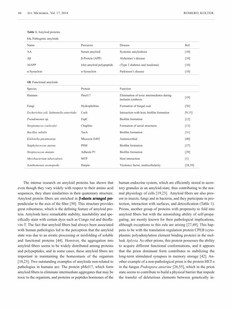
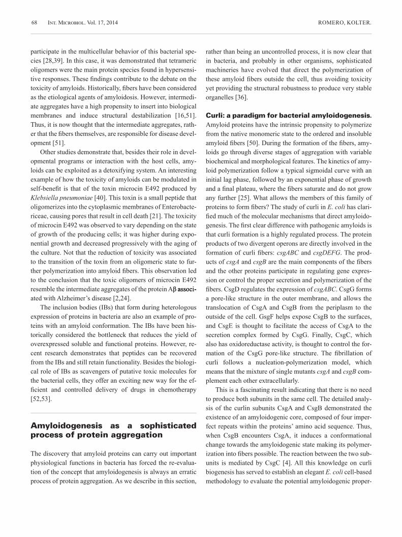
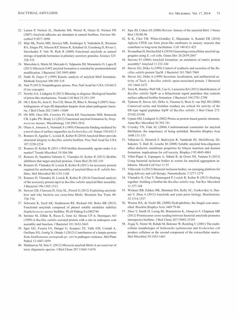
Fig. 2. Fibers of TasA purified from Bacillus subtilis as seen in electron microscopy and negative staining. Bar equals 100 nm.
Int. Microbiol. Vol. 17, 2014 ROMERO, KOLTER.70
tory network that controls and connects the expression of TasA with other bacterial factors [54]. To summarize, the master regulator SinR directly represses the expression of tasA until conditions are propitious for biofilm development [17]. One operon containing three genes, tapA-sipW-tasA, is necessary for the formation of the amyloid fibers [31]. This is markedly different from the chaplin amyloid fibers in the Gram-positive bacterium Streptomyces coelicolor, where eight chaplin genes have been described [13]. Streptomyces coelicolor shows a complex developmental program and it is thought that the diversity of chaplins play important roles in the different stages of the S. coelicolor life cycle [5].
TasA is the main component of the B. subtilis amyloid fibers. When purified directly from B. subtilis, TasA retains amyloid properties, such as self-aggregation into insoluble fibers (Fig. 2). TasA fibers can be depolymerized with aggressive acid treatments and then they can be observed to repolymer-ize. Such a property has been useful to study the kinetics of polymerization of TasA, which reflects a dynamic transition through different stages of aggregation. In agreement with its amyloid nature, some of these intermediate aggregates, but not the fiber nor the monomers, reacts with an antibody that specifically detects the toxic aggregates of the amyloid pro-tein Aβ associated with Alzheimer’s disease [31]. However, TasA is not purified from B. subtilis in its monomeric form. Rather, we obtain a homogenous suspension of stable oligo-
mers, which could aggregate as different morphotypes de-pending on the physical properties of the medium: (i) fibers form on hydrophobic surfaces but (ii) plaques form under acidic conditions. This is an appealing discovery that could reflect a way in which bacteria can adapt to different habitats, modifying the state of aggregation of an external protein and probably the final arrangement of the extracellular matrix de-pending on environmental cues such as pH and surface hydro-phobicity [8].
Bacterial cell surfaces are hydrophobic, and we have seen that this physical feature promotes the formation of fibers [8]. However, additional factors must be dedicated to increase the efficiency of the fiber polymerization process outside the cells. To build the curli fibers, E. coli uses CsgA and the nu-cleator protein CsgB [3]. As stated above, tasA is part of a three gene operon that encodes another protein, TapA, which is also necessary for the assembly of biofilms (Fig. 3A) [6,34]. The failure of a tapA deletion mutant to form biofilms has been related to the absence of TasA fibers and even attach-ment of TasA to cell surfaces [34]. Note that, differently from what was reported for curli, TapA and TasA have to be pro-duced in the same cell in order to form the fibers, a process similar to the formation of pili in B. cereus [7].
Immunolocalization studies proved that TapA localizes, as foci, at certain regions of the cell wall, where presumably it directs the polymerization of TasA into fibers. In addition,
Int M
icro
biol
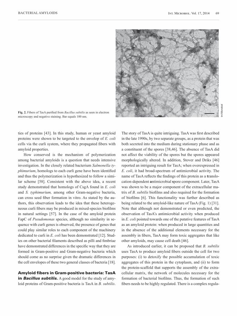
Fig. 3. The formation of biofilms of Bacillus subilis and TasA amyloid-like fibers in B. subtilis depend on the proteins TasA and TapA. (A) Top row: Highly wrinkly colony of wild type and featureless colony of tasA or tapA mutants after 72 h of growth at 30 ºC in MSgg agar plates. Bottom row: wrinkly pellicles of wild type and absence of pellicle of tasA and tapA mutants grown in MSgg broth for 24 h at 30 ºC. (B) Transmission electron microscopy and co-immunolabeling with anti-TapA (15 nm gold particles) and anti-TasA (10 nm gold particles) antibodies of TasA fibers in B. subtilis biofilms. Bar equals 100 nm.
Int. Microbiol. Vol. 17, 2014BACTERIAL AMYLOIDS 71
TapA appears distributed along the TasA fibers (Fig. 3B), al-though at much less abundance (the ratio of TasA:TapA is 100:1) [34]. Based on these findings, we proposed that TapA is an accessory protein that promotes the efficient polymeriza-tion of TasA at the cell envelope and contributes to the organi-zation of the growing fibers. In addition, the localization of TapA at the base of the TasA fibers, and its close association to the cell wall led to the idea that TapA is the connector of the fibers to the cell envelope [34].
Consistent with this conclusion, the absence of TapA causes a decrease in the amount of detectable extracellular TasA, which in addition appears as small and disorganized fibrils that are disconnected from the cell envelope. The name TapA was coined to refer to these functions: TasA anchoring and assembly protein [34].
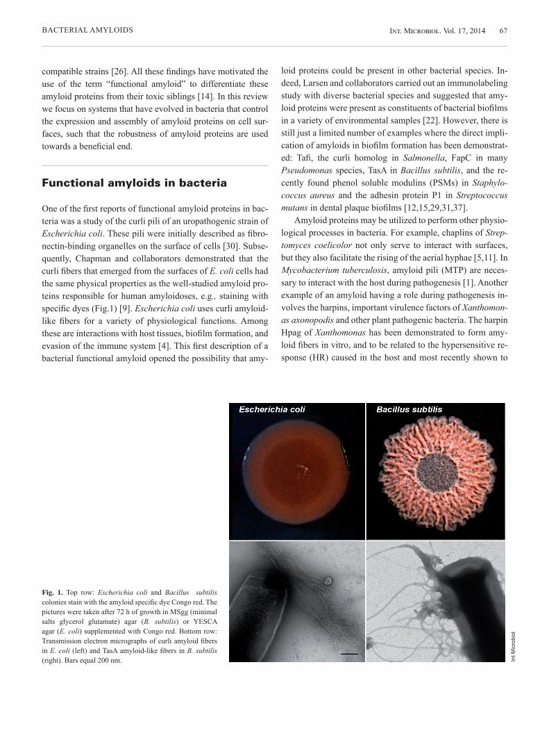
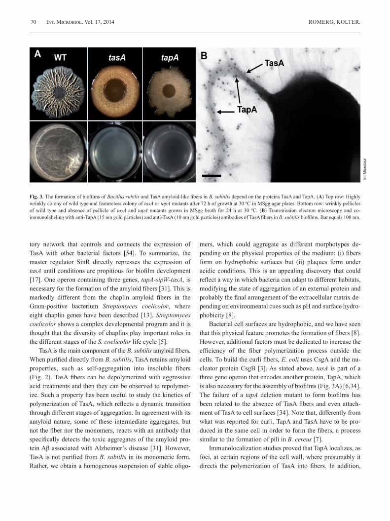
In a recent publication we showed that indeed TapA ac-celerates the polymerization of TasA in vitro (Fig. 4) [35]. How it is that TapA facilitates the polymerization of TasA into amyloid fibers is still unclear. What we do know is that a mu-tant with a tapA allele lacking an eight-amino-acid sequence in the N-terminal half of the protein failed to form biofilms and failed to promote TasA fiber formation [35]. By compari-son with curli in E. coli, we propose that this domain in the N-terminal half of TapA can either promote the processing of TasA or induce a conformational change of TasA toward its amyloidogenic state.
The knowledge of the polymerization of pili in other Gram-positive bacteria can be compared and contrasted with TasA fiber formation. The formation of pili in B. cereus needs two subunits, BcpA and BcpB. The major subunit, BcpA is processed by a specific sortase, and the product is linked to the minor subunit BcpB [7]. However, a hypothetical process-ing of TasA and TapA would have to involve other proteins than the putative sortases identified in B. subtilis because mu-tants in those sortases had no defect in biofilm formation [34]. The anchoring of TasA fibers mediated by TapA could be thought to be similar to other pili in Gram-positive bacteria. However, two lines of evidence discard this hypothesis: (i) The C-terminal region of TapA does not contain a canonical sorting signal for anchoring to the peptidoglycan, and (ii) the deletion of the two putative sortases, the enzymes that recog-nize the sorting signals, did not alter biofilm formation [34]. These findings open the possibility that there exist diverse mechanisms of attachment of fibers to the cell wall of Gram-positive bacteria.
Finally, the formation of TasA fibers depends on a third protein, SipW, also encoded in the same operon. This is a sig-nal peptidase that processes TasA and TapA to their mature form before sorting to the cell envelope [45,46]. SipW is an atypical signal peptidase, given that it resembles more those found in the endoplasmic reticulum of eukaryotic cells than typical bacterial signal peptidases [48]. An additional role for
Int M
icro
biol
Fig. 4. A feature of TasA amyoid-like fibers is the binding of the amyloid specific fluorescent dye thioflavin T (structure on top). Thioflavin T fluo-rescence is used to follow the kinetics of polymerization of amyloid pro-teins. The increase of the fluorescence intensity indicates an increase in the β-sheet content of the protein, typical in amyloid fibers. Purified TasA follows a typical amyloid kinetics of polymerization (Ñ), and the addi-tion of TapA accelerates the polymerization of TasA into amyloid-like fi-bers (¡).
Int. Microbiol. Vol. 17, 2014 ROMERO, KOLTER.72
SipW has been recently proposed. A version of this protein impaired in protease activity was shown to stimulate the ex-pression of the tapA-sipW-tasA operon during the formation of biofilms on solid surfaces, thus introducing a new level of control and complexity to the formation of amyloids [47].
Final notes
The studies of bacterial amyloids have revolutionized our per-ception of this family of proteins: from their pathological im-plications and erratic processing to the possibility that they sometimes arise as the result of a well-defined program that has evolved to maintain the homeostasis of the organism and to interact with the environment [36]. To the increasing num-ber of functional amyloids, we have added a new member, the TasA amyloid-like fiber in the Gram-positive bacterium B. subtilis. A number of reasons make the TasA amyloid-like fi-bers an attractive model for the molecular study of amyloido-genesis. (i) B. subtilis is a model microbe for the study of de-velopmental programs such as biofilm formation and sporula-tion, two features mediating the interaction of the bacterium with its environment. The study of TasA fiber formation will contribute not only to our understanding of the mechanisms of amyloid fiber formation, but also its implications in bacterial adaptation to the habitat. The fact that TasA is purified to ho-mogeneity directly from B. subtilis offers the outstanding ca-pability to investigate which other factors, physical-chemical or intrinsic to bacterial cells, are involved in fiber formation. (ii) Amyloid fibers represent interesting targets for the search of new therapies that contribute to the fight of bacterial bio-films and the biochemical problems they may cause [32]. We have previously demonstrated that B. subtilis biofilms repre-sent a formidable tool for the search of molecules that can be exploited in two directions: anti-biofilm and anti-amyloid [33].
Acknowledgements. The authors thank Hera Vlamakis for critical reading and suggestions during the writing of the manuscript. DR is funded by the program Ramón y Cajal (RyC-2011-080605) and grant AGL-2012-31968 from the Plan Nacional de I+D+I from Ministerio de Economía y Competitividad (Spain) and co-financed by FEDER funds (European Union).
Competing interest. None declared.
References
1. Alteri CJ, Xicohténcatl-Cortes J, Hess S, Caballero-Olín G, Girón JA, Friedman RL (2007) Mycobacterium tuberculosis produces pili during human infection. Proc Natl Acad Sci USA 104:5145-5150
2. Arranz R, Mercado G, Martin-Benito J, Giraldo R, Monasterio O, Lagos R, Valpuesta JM (2012) Structural characterization of microcin E492 amyloid formation: Identification of the precursors. J Struct Biol 178: 54-60
3. Bian Z, Normark S (1997) Nucleator function of CsgB for the assembly of adhesive surface organelles in Escherichia coli. EMBO J 16:5827-5836
4. Blanco LP, Evans ML, Smith DR, Badtke MP, Chapman MR (2012) Di-versity, biogenesis and function of microbial amyloids. Trends Microbiol 20:66-73
5. Bokhove M, Claessen D, de Jong W, Dijkhuizen L, Boekema EJ, Ooster-getel GT (2013) Chaplins of Streptomyces coelicolor self-assemble into two distinct functional amyloids. J Struct Biol 184:301-309
6. Branda SS, Chu F, Kearns DB, Losick R, Kolter R (2006) A major pro-tein component of the Bacillus subtilis biofilm matrix. Mol Microbiol 59: 1229-1238
7. Budzik JM, Marraffini LA, Souda P, Whitelegge JP, Faull KF, Schnee-wind O (2008) Amide bonds assemble pili on the surface of bacilli. Proc Natl Acad Sci USA 105:10215-10220
8. Chai L, Romero D, Kayatekin C, Akabayov B, Vlamakis H, Losick R, Kolter R (2013) Isolation, characterization, and aggregation of a struc-tured bacterial matrix precursor. J Biol Chem 288:17559-17568
9. Chapman MR, Robinson LS, Pinkner JS, Roth R, Heuser J, Hammar M, Normark S, Hultgren SJ (2002) Role of Escherichia coli curli operons in directing amyloid fiber formation. Science 295:851-855
10. Chiti F, Dobson CM (2006) Protein misfolding, functional amyloid, and human disease. Annu Rev Biochem 75:333-366
11. de Jong W, Wosten HA, Dijkhuizen L, Claessen D (2009) Attachment of Streptomyces coelicolor is mediated by amyloidal fimbriae that are an-chored to the cell surface via cellulose. Mol Microbiol 73:1128-1140
12. Dueholm MS, Petersen SV, Sonderkaer M, Larsen P, Christiansen G, Hein KL, Enghild JJ, Nielsen JL, Nielsen KL, Nielsen PH, Otzen DE (2010) Functional amyloid in Pseudomonas. Mol Microbiol 77:1009-1020
13. Elliot MA, Karoonuthaisiri N, Huang J, Bibb MJ, Cohen SN, Kao CM, Buttner MJ (2003) The chaplins: a family of hydrophobic cell-surface proteins involved in aerial mycelium formation in Streptomyces coeli-color. Genes Dev 17:1727-1740
14. Fowler DM, Koulov AV, Balch WE, Kelly JW (2007) Functional amy-loid--from bacteria to humans. Trends Biochem Sci 32:217-224
15. Gibson DL, White AP, Rajotte CM, Kay WW (2007) AgfC and AgfE facilitate extracellular thin aggregative fimbriae synthesis in Salmonella enteritidis. Microbiology 153:1131-1140
16. Kayed R, Head E, Thompson JL, McIntire TM, Milton SC, Cotman CW, Glabe CG (2003) Common structure of soluble amyloid oligomers im-plies common mechanism of pathogenesis. Science 300:486-489
17. Kearns DB, Chu F, Branda SS, Kolter R, Losick R (2005) A master regu-lator for biofilm formation by Bacillus subtilis. Mol Microbiol 55:739-749
18. Kline KA, Dodson KW, Caparon MG, Hultgren SJ (2010) A tale of two pili: assembly and function of pili in bacteria. Trends Microbiol 18: 224-232
19. Kummer MP, Maruyama H, Huelsmann C, Baches S, Weggen S, Koo EH (2009) Formation of Pmel17 amyloid is regulated by juxtamembrane metalloproteinase cleavage, and the resulting C-terminal fragment is a substrate for gamma-secretase. J Biol Chem 284:2296-2306
20. Kyle RA (2001) Amyloidosis: a convoluted story. Br J Haematol 114: 529-538
21. Lagos R, Tello M, Mercado G, Garcia V, Monasterio O (2009) Antibacte-rial and antitumorigenic properties of microcin E492, a pore-forming bacteriocin. Curr Pharm Biotechnol 10:74-85
Int. Microbiol. Vol. 17, 2014BACTERIAL AMYLOIDS 73
22. Larsen P, Nielsen JL, Dueholm MS, Wetzel R, Otzen D, Nielsen PH (2007) Amyloid adhesins are abundant in natural biofilms. Environ Mi-crobiol 9:3077-3090
23. Maji SK, Perrin MH, Sawaya MR, Jessberger S, Vadodaria K, Rissman RA, Singru PS, Nilsson KP, Simon R, Schubert D, Eisenberg D, Rivier J, Sawchenko P, Vale W, Riek R (2009) Functional amyloids as natural storage of peptide hormones in pituitary secretory granules. Science 325: 328-332
24. Marcoleta A, Marin M, Mercado G, Valpuesta JM, Monasterio O, Lagos R (2013) Microcin E492 amyloid formation is retarded by posttranslational modification. J Bacteriol 195:3995-4004
25. Naiki H, Gejyo F (1999) Kinetic analysis of amyloid fibril formation. Methods Enzymol 309:305-318
26. Nair P (2013) Nonpathogenic prions. Proc Natl Acad Sci USA 110:6612 (Core concepts)
27. Newby GA, Lindquist S (2013) Blessing in disguise: Biological benefits of prion-like mechanisms. Trends Cell Biol 23:251-259
28. Oh J, Kim JG, Jeon E, Yoo CH, Moon JS, Rhee S, Hwang I (2007) Amy-loidogenesis of type III-dependent harpins from plant pathogenic bacte-ria. J Biol Chem 282:13601-13609
29. Oli MW, Otoo HN, Crowley PJ, Heim KP, Nascimento MM, Ramsook CB, Lipke PN, Brady LJ (2012) Functional amyloid formation by Strep-tococcus mutans. Microbiology 158:2903-2916
30. Olsen A, Jonsson A, Normark S (1989) Fibronectin binding mediated by a novel class of surface organelles on Escherichia coli. Nature 338:652-5
31. Romero D, Aguilar C, Losick R, Kolter R (2010) Amyloid fibers provide structural integrity to Bacillus subtilis biofilms. Proc Natl Acad Sci USA 107:2230-2234
32. Romero D, Kolter R (2011.) Will biofilm disassembly agents make it to market? Trends Microbiol 19:304-306
33. Romero D, Sanabria-Valentin E, Vlamakis H, Kolter R (2013) Biofilm inhibitors that target amyloid proteins. Chem Biol 20:102-110
34. Romero D, Vlamakis H, Losick R, Kolter R (2011) An accessory protein required for anchoring and assembly of amyloid fibres in B. subtilis bio-films. Mol Microbiol 80:1155-1168
35. Romero D, Vlamakis H, Losick R, Kolter R (2014) Functional analysis of the accessory protein tapA in Bacillus subtilis amyloid fiber assembly. J Bacteriol 196:1505-1513
36. Sawyer EB, Claessen D, Gras SL, Perrett S (2012) Exploiting amyloid: how and why bacteria use cross-beta fibrils. Biochem Soc Trans 40: 728-734
37. Schwartz K, Syed AK, Stephenson RE, Rickard AH, Boles BR (2012) Functional amyloids composed of phenol soluble modulins stabilize Staphylococcus aureus biofilms. PLoS Pathog 8:e1002744
38. Serrano M, Zilhão R, Ricca E, Ozin AJ, Moran CP Jr, Henriques AO (1999) A Bacillus subtilis secreted protein with a role in endospore coat assembly and function. J Bacteriol 181:3632-3643
39. Sgro GG, Ficarra FA, Dunger G, Scarpeci TE, Valle EM, Cortadi A, Orellano EG, Gottig N, Ottado J (2012) Contribution of a harpin protein from Xanthomonas axonopodis pv. citri to pathogen virulence. Mol Plant Pathol 13:1047-1059
40. Shahnawaz M, Soto C (2012) Microcin amyloid fibrils A are reservoir of toxic oligomeric species. J Biol Chem 287:11665-11676
41. Sipe JD, Cohen AS (2000) Review: history of the amyloid fibril. J Struct Biol 130:88-98
42. Si K, Choi YB, White-Grindley E, Majumdar A, Kandel ER (2010) Aplysia CPEB can form prion-like multimers in sensory neurons that contribute to long-term facilitation. Cell 140:421-425
43. Sivanathan H, Hochschild A (2010) Generating extracellular amyloid ag-gregates using E. coli cells. Genes Dev 26:2659-2667
44. Stevens FJ (2004) Amyloid formation: an emulation of matrix protein assembly? Amyloid 11:232-244
45. Stover AG, Driks A (1999) Control of synthesis and secretion of the Ba-cillus subtilis protein YqxM. J Bacteriol 181:7065-7069
46. Stover AG, Driks A (1999) Secretion, localization, and antibacterial ac-tivity of TasA, a Bacillus subtilis spore-associated protein. J Bacteriol 181:1664-1672
47. Terra R, Stanley-Wall NR, Cao G, Lazazzera BA (2012) Identification of Bacillus subtilis SipW as a bifunctional signal peptidase that controls surface-adhered biofilm formation. J Bacteriol 194:2781-2790
48. Tjalsma H, Stover AG, Driks A, Venema G, Bron S, van Dijl JM (2000) Conserved serine and histidine residues are critical for activity of the ER-type signal peptidase SipW of Bacillus subtilis. J Biol Chem 275: 25102-25108
49. Uptain SM, Lindquist S (2002) Prions as protein-based genetic elements. Annu Rev Microbiol 56:703-741
50. Uversky VN, Fink AL (2004) Conformational constraints for amyloid fibrillation: the importance of being unfolded. Biochim Biophys Acta 1698:131-153
51. Valincius G, Heinrich F, Budvytyte R, Vanderah DJ, McGillivray DJ, Sokolov Y, Hall JE, Losche M (2008) Soluble amyloid beta-oligomers affect dielectric membrane properties by bilayer insertion and domain formation: implications for cell toxicity. Biophys J 95:4845-4861
52. Villar-Piqué A, Espargaro A, Sabaté R, de Groot NS, Ventura S (2012) Using bacterial inclusion bodies to screen for amyloid aggregation in-hibitors. Microb Cell Fact 11:55
53. Villaverde A (2012) Bacterial inclusion bodies: an emerging platform for drug delivery and cell therapy. Nanomedicine 7:1277-1279
54. Vlamakis H, Chai Y, Beauregard P, Losick R, Kolter R (2013) Sticking together: building a biofilm the Bacillus subtilis way. Nat Rev Microbiol 11:157-168
55. Wickner RB, Edskes HK, Bateman DA, Kelly AC, Gorkovskiy A, Day-ani Y, Zhou A (2013) Amyloids and yeast prion biology. Biochemistry 52:1514-1527
56. Wosten HA, de Vocht ML (2000) Hydrophobins, the fungal coat unrav-elled. Biochim Biophys Acta 1469:79-86
57. Zhou Y, Smith D, Leong BJ, Brannstrom K, Almqvist F, Chapman MR (2012) Promiscuous cross-seeding between bacterial amyloids promotes interspecies biofilms. J Biol Chem 287:35092-35103
58. Zogaj X, Nimtz M, Rohde M, Bokranz W, Romling U (2001) The multi-cellular morphotypes of Salmonella typhimurium and Escherichia coli produce cellulose as the second component of the extracellular matrix. Mol Microbiol 39:1452-1463