Embed Size (px)
Citation preview
Vol. 13, No. 7MOLECULAR AND CELLULAR BIOLOGY, JUlY 1993, P. 3964-39740270-7306/93/073964-11$02.00/0Copyright X 1993, American Society for Microbiology
Functional and Physical Associations between NF-KB andC/EBP Family Members: a Rel Domain-bZIP Interaction
BERND STEIN,1 PATRICIA C. COGSWELL,1 AND ALBERT S. BALDWIN, JR.1,2*Lineberger Comprehensive Cancer Center, 1 and Department of Biology, 2 University of
North Carolina at Chapel Hill, Chapel Hill, North Carolina 27599
Received 22 February 1993/Returned for modification 31 March 1993/Accepted 9 April 1993
NF-KB and C/EBP represent distinct families of transcription factors that target unique DNA enhancerelements. The heterodimeric NF-KB complex is composed of two subunits, a 50- and a 65-kDa protein. Allmembers of the NF-cB family, including the product of the proto-oncogene c-rel, are characterized by theirhighly homologous -300-amino-acid N-terminal region. This Rel homology domain mediates DNA binding,dimerization, and nuclear targeting of these proteins. C/EBP contains the bZIP region, which is characterizedby two motifs in the C-terminal half of the protein: a basic region involved in DNA binding and a leucine zippermotif involved in dimerization. The C/EBP family consists of several related proteins, C/EBPa, C/EBP3,C/EBP-y, and C/EBP8, that form homodimers and that form heterodimers with each other. We now
demonstrate the unexpected cross-coupling of members of the NF-cB family with three members of the C/EBPfamily. NF-cB p65, p50, and Rel functionally synergize with C/EBPa, C/EBPf3, and C/EBP8. Thiscross-coupling results in the inhibition of promoters with KB enhancer motifs and in the synergistic stimulationof promoters with C/EBP binding sites. These studies demonstrate that NF-KB augments gene expressionmediated by a multimerized c-fos serum response element in the presence of C/EBP. We show a direct physicalassociation of the bZIP region ofC/EBP with the Rel homology domain of NF-KB. The cross-coupling of NF-cBwith C/EBP highlights a mechanism of gene regulation involving an interaction between distinct transcriptionfactor families.
Transcriptional regulation of gene expression is controlledby factors that bind to specific DNA sequences. Many ofthese factors can be classified into distinct families based oncommon structural motifs. These families include the homeo-domain family (30), the zinc finger proteins (26, 46), theleucine zipper family (49, 84), and the helix-loop-helix family(59, 60). Many of these transcription factors bind to theircognate DNA motifs as homodimers and, moreover, thereare multiple examples of heterodimer formation within thesame family of transcription factors. However, there isgrowing evidence that these dimerization motifs are alsoinvolved in interactions between transcription factors fromdifferent families. Examples are the interaction of the Junleucine zipper region with the helix-loop-helix motif ofMyoD (12) or with the DNA binding and ligand bindingregions of the glucocorticoid receptor (24, 42, 74, 87). Thiscross-coupling between different families might lead to theformation of novel transcription factor complexes displayingaltered biological activities.The transcription factor NF-KB binds to KB enhancer
motifs found in a variety of genes in response to varioussignals (5 and references therein, 35, 51). NF-KB DNAbinding activity typically involves two subunits, a 50-kDaprotein (17, 31, 45) and a 65-kDa protein (63, 72). NF-KB p50and NF-KB p65 share a highly homologous 300-residueN-terminal region. This region is also found in the productsof the proto-oncogene c-rel and of the dorsal maternal effectgene of Drosophila melanogaster (16, 31, 32, 38, 80). TheRel homology region of these proteins mediates DNA bind-ing, dimerization, nuclear targeting, and interactions withIKB (9, 11, 17, 31, 33, 45, 63, 72).The C/EBP family of transcription factors belongs, to-
* Corresponding author.
gether with AP-1 and ATF/CREB, to a class ofDNA bindingproteins named bZIP proteins (84). These proteins are char-acterized by their leucine zipper structure and the adjacentDNA binding basic region, both located in the C-terminalhalf of the respective proteins. The originally characterizedCCAAT/enhancer-binding protein CIEBP (41) is now namedC/EBPa. C/EBPI (20) is the new term for LAP (23), IL6-DBP (67), CRP2 (86), AGP/EBP (21), and NF-IL6 (2).C/EBP-y replaces the old name Ig/EBP-1 (71). C/EBPa,C/EBP,3, C/EBP-y, and C/EBPb (20) form homodimers andheterodimers with each other in vitro and bind with a similaraffinity to various C/EBP binding sites (20, 48). All membersof the C/EBP family are encoded by different genes, and theamino acid sequences of the N-terminal two-thirds of theseproteins are not related. C/EBPot expression is tissue re-stricted, with the highest expression occurring in liver,adipose, and intestinal tissue (14). C/EBPa is an activator ofspecific gene expression (22, 28, 29, 43, 65, 81). Further-more, C/EBPa plays a general role in cellular growth inhi-bition (82) and has a hypothetical responsiveness to hor-mones that modulate energy balance (54). The c-fos serumresponse element (SRE) contains overlapping binding sitesfor the serum response factor and C/EBPP (56). Cyclic AMP(cAMP) stimulates phosphorylation and translocation ofC/EBPI to the nucleus in PC12 cells. These events induceC/EBP,B binding to the SRE and activate c-fos expression(55). Interestingly, several promoters of immune or acute-phase response genes, such as the interleukin-6 (IL-6) gene,the IL-8 gene, and the angiotensinogen gene, have adjacentor overlapping binding sites for C/EBP and NF-KB (19, 40,58).
In this study, we show that members of the NF-KB/Relfamily can interact with three members of the C/EBP family.This cross-coupling results in the inhibition of promoters
3964

CROSS-COUPLING OF NF-KB WITH C/EBP 3965
with KB enhancer motifs and the synergistic stimulation ofpromoters with C/EBP binding sites. Specifically, NF-KBaugments gene expression mediated by a multimerized c-fosSRE in the presence of C/EBP. A direct physical interactionbetween the bZIP region of C/EBP and the Rel homologydomain of NF-KB is demonstrated.
MATERIALS AND METHODS
Plasmid constructs. The KB-TATA-CAT and SRE-TATA-CAT reporter plasmids have been described elsewhere (42,79). The C/EBP-TATA-CAT reporter was generated bycloning a single copy of an oligonucleotide with a C/EBPbinding site derived from the c-fos SRE (5' AGCTTGATTAGGACATCG 3') (binding site printed in boldface type) intoHindIII-BamHI-cut TATA-CAT. The CRE-TK-CAT re-porter contains a single copy of a CREB binding site (5'AGCT'lGTGACGTCACCCG 3') (binding site printed inboldface type) cloned into HindIII-BamHI-cut pBLCAT4(79). The Myb-TK-CAT reporter contains five copies of aMyb binding site cloned in front of the herpes simplex virusthymidine kinase promoter (46a). The pCMV4T plasmid wasderived from pCMV4 (3) by inserting an oligonucleotide withstop codons in all three reading frames into the SmaI site.The pCMV4T-p65 and pCMV4T-p50 plasmids containcDNAs encoding human NF-KB p65 (72) and NF-KB p50(amino acids 1 to 462) (45), respectively. Plasmid pCMV4T-p65mLZ was constructed by polymerase chain reaction-assisted site-directed mutagenesis of NF-KB p65, resulting inthe change of amino acids 436-LSEALLQLQFDDEDL-450to 436-ASEAALQAQFDDEDA-450 (underlining highlightschanges). Plasmid pCMV4T-p65A10 encodes an alterna-tively spliced form of NF-KB p65 with an internal deletion ofamino acid residues 222 to 231 (61, 73). Plasmid pCMV4T-p65Eco contains a cDNA encoding the Rel homology do-main of NF-KB p65 (amino acids 1 to 282). Expressionvectors pCMV4T-vRel and pCMV4T-hcRel are as describedpreviously (9). Plasmid pCMV4T-rC/EBP3 contains a cDNAencoding rat C/EBPP (67) cloned into pCMV4T. Cytomega-lovirus (CMV) enhancer-driven expression vectors for hu-man C/EBPa, C/EBPP, and C/EBPb were a gift of Steven L.McKnight. The mouse c-Jun, human c-Fos, and humanCREB pCMV4T expression vectors contain full-length cod-ing sequences of the corresponding T7 polymerase-drivenvectors (13, 69, 70) cloned downstream of the CMV imme-diate-early-region promoter. The wild-type Myb expressionvector (pRmb3SVneo) and the corresponding frameshiftcontrol pJTmyb-fs have been described elsewhere (25). Thebacterial FLAG-heart muscle kinase (HMK) fusion proteinexpression vectors were constructed by cloning the cDNAsfor NF-KB p65, NF-KB p50 (amino acids 1 to 462), and ratC/EBPP as polymerase chain reaction-amplified fragmentsinto the EcoRI site of the T7 polymerase-driven FLAG-HMK vector (15). The bacterial glutathione S-transferase(GST) fusion protein expression vectors were constructedby cloning the cDNAs for rat C/EBPP and the bZIP trunca-tion of rat C/EBPP (C/EBPPbZIP; encoding amino acids 187to 297) as polymerase chain reaction-amplified fragments inframe into the EcoRI site of pGEX-1N (Amrad). Expressionvectors for in vitro transcription were generated by cloningthe cDNAs for NF-KB p65, NF-KB p65A10, and NF-KB p50(amino acids 1 to 462) into pGEM4 (Promega). In vitro-translated proteins NF-KB p65Eco (amino acids 1 to 282) andNF-KB p65Bgl (amino acids 1 to 194) were generated byrunoff transcription terminated at internal EcoRI and BglIIsites, respectively.
Transfection of cells and analysis of CAT activity. All celllines were cultured in Iscove's Dulbecco modified Eaglemedium supplemented with 7.5% fetal calf serum and anti-biotics. Mouse F9 embryonal carcinoma cells were tran-siently transfected by the calcium phosphate method (34).Monkey COS cells were transiently transfected with plasmidDNA by the DEAE-dextran method (44). Chloramphenicolacetyltransferase (CAT) enzymatic activity was assayed aspreviously described (62) and normalized for protein recov-ery (18).
Bacterial expression of proteins. Human C/EBPabZIP,C/EBPPbZIP, and C/EBP&bZIP proteins expressed in bac-teria (20, 78) were a generous gift of Steven L. McKnight.The FLAG-HMK fusion proteins were expressed in Esche-richia coli BL21 (LysS). The GST fusion proteins wereexpressed in E. coli JM109. Bacteria were grown overnight,diluted 1:10 and, after another hour of growth, stimulatedwith 0.4 mM isopropyl-3-D-thiogalactopyranoside (IPTG)for 3 h at 32°C. Bacteria were then harvested, resuspended ina 0.1 volume of phosphate-buffered saline (PBS), and soni-cated for 1 min in PBS, and the extracts were cleared bycentrifugation.
Purification and 32p labeling of proteins. Cytosolic NF-KBfrom HeLa cells was purified as previously described (9).Bacterially expressed human C/EBP proteins containingonly the bZIP domain were purified by column chromato-graphic techniques as described previously (20, 78). Bacte-rially expressed FLAG-HMK fusion proteins were purifiedby low-pH elution from an M2 anti-FLAG antibody column(IBI-Kodak, New Haven, Conn.) in accordance with themanufacturer's instructions. FLAG-HMK fusion proteinswere labeled in vitro with [_y-32P]ATP by use of HMK(Sigma) in buffer containing 20 mM Tris (pH 7.6), 100 mMNaCl, 12 mM MgCl2, and 1 mM dithiothreitol for 60 min at37°C (15). Whole-cell extracts from COS cells were preparedby a method used to prepare CAT assay extracts (4) 3 daysposttransfection.EMSA. Electrophoretic mobility shift assays (EMSA)
were performed as previously described (79). The bindingbuffer contained 12 mM N-2-hydroxyethylpiperazine-N'-2-ethanesulfonic acid (HEPES)-NaOH (pH 7.9), 4 mM Tris(pH 7.9), 60 mM KCl, 5 mM MgCl2, 0.6 mM EDTA, 12%glycerol, and 5 mM dithiothreitol. The human immunodefi-ciency virus type 1 (HIV-1) KB probe was as previouslydescribed (79). The C/EBP binding site found as a coresequence within the c-fos SRE was created by annealing theoligonucleotides 5' AGC1TGATTAGGACATCG 3' and 5'GATCCGATGTCCTAATCA 3' (binding site printed in bold-face type).
Coimmunoprecipitation. For coimmunoprecipitation anal-yses, 0.6 ,ul of 32P-labeled FLAG-HMK fusion protein wascombined with 5 p,l of unlabeled bacterial protein in EMSAbinding buffer. After 15 min of incubation at room tempera-ture (RT), the chemical cross-linker dithio-bis(succinimidyl-propionate) (DSP) (Pierce) was added at a final concentra-tion of 2 mM, and the mixture was incubated for 30 min atRT. The cross-linking reaction was squelched by the addi-tion of 100 mM ethanolamine. After the volume was in-creased with RIPA buffer, normal rabbit serum and proteinA-Sepharose were added, and the mixture was incubated for1 h at 4°C. The immunoreaction was then cleared, specificantibody and protein A-Sepharose were added, and themixture was incubated for 2 h at 4°C. After the immunocom-plexes were washed, the cross-linked complexes were re-duced with Laemmli sodium dodecyl sulfate-polyacrylamidegel electrophoresis (SDS-PAGE) loading buffer (47) contain-
VOL. 13, 1993

3966 STEIN ET AL.
A18,
16-14-
'S 12-
< 10-
*, 8-(5i6-
4-
2-
0 1 2 3 4 5 EFg C/EBPB expression vector
0 1 2 3 4 5
1ig p65 expression vector
C
cI-
C.)e)Cu
350_ C/EBP-TATA-CAT )
300-
250-
200-
150-
100-
50-
O ...0 1 2 3 4 5 6 7 E
Rg p65 expression vector
FIG. 1. Cross-coupling of NF-KB p65 with C/EBPI. Mouse F9 embryonal carcinoma cells were transiently cotransfected with 8 p,g of theindicated CAT reporter plasmids (KB-TATA-CAT, containing one copy of the duplicated HIV-1 KB enhancer motif from the HIV-1 longterminal repeat [positions -105 to -79] linked to the TATA box of the X. laevis albumin gene; SRE-TATA-CAT, containing four copies ofthe SRE of the human c-fos promoter [-320 to -2991; and C/EBP-TATA-CAT, containing one copy of a C/EBP binding site and 0.1 p,g ofpCMV4T-p65 (A), 1 p1g of pCMV4T-rC/EBP, (B and C), and increasing amounts (0 to 6 ,g) of pCMV4T-rC/EBP,B (A) or pCMV4T-p65 (Band C). The total amount of transfected DNA was kept constant by the addition of the parental pCMV4T vector. At 6 h after transfection,CAT activity was determined. The mean levels ofCAT activity determined in at least three independent experiments are presented as the foldinduction relative to the activity obtained with transfected pCMV4T alone. The standard errors were always less than 20% of the means.
ing 15% ,3-mercaptoethanol, boiled, and subjected to SDS-PAGE.GST fusion protein interaction assay. Glutathione-
Sepharose 4B beads (Pharmacia) were equilibrated inPBS-1% Triton X-100 and then mixed with 20 volumes ofbacterial GST fusion proteins on a rotary shaker for 20 minat RT. The beads were washed three times with PBS-1%Triton X-100 and then equilibrated in 20 mM HEPES-NaOH(pH 7.9)-25 mM NaCl-2.5 mM MgCl2-0.1 mM EDTA-0.05% Nonidet P-40-1% Triton X-100 (LSBT). Five micro-liters of a 1:1 bead slurry in PBS was combined with 5 ,ul ofa 35S-labeled reticulocyte lysate in a final volume of 200 ,ul ofLSBT-1 mM dithiothreitol-1 mM phenylmethylsulfonyl flu-oride on a rotary shaker for 60 min at RT. The beads werethen washed four times with LSBT and once with 50 mMTris (pH 6.8). The bound proteins were eluted by boiling inLaemmli SDS-PAGE loading buffer (47) and subjected toSDS-PAGE.Western blot (immunoblot) analysis. Nuclear extracts from
-1 x 10⁶ COS cells prepared 3 days after transfection asdescribed previously (79) were fractionated by SDS-PAGE,blotted onto a nitrocellulose membrane, and immunostainedby use of an Amersham enhanced chemiluminescence (ECL)kit in accordance with the manufacturer's instructions.
RESULTSNF-KB functionally acts in synergy with C/EBP-related
proteins. NF-KB activity is increased in response to IL-1(64), and C/EBPf is activated by IL-6 (66). Interestingly,synergy between IL-1 and IL-6 in several physiologicalresponses, including the acute-phase response, in T-cellactivation, and in immunoglobulin secretion has been re-
ported (1, 37, 83). To test for possible cross-coupling be-tween the NF-KB and C/EBP families of transcription fac-tors, we performed transfection experiments with mouse F9
embryonal carcinoma cells. F9 cells were selected becauseof their exceedingly low levels of endogenous nuclearNF-KB (39, 78a). F9 cells were transfected with a CATreporter plasmid composed of the duplicated HIV-1 KBenhancer motif linked to the TATA box of the Xenopuslaevis albumin gene (KB-TATA-CAT). This KB enhancermotif is sufficient to mediate activation by various membersof the NF-KB transcription factor family (8, 78a). Unexpect-edly, cotransfection of a rat C/EBPP expression vectorstrongly reduced the induction of KB-TATA-CAT by NF-KBp65 in a dose-dependent manner (Fig. 1A). Therefore, wewere interested in whether C/EBPP functions as a positiveregulator of gene expression in F9 cells when tested onreporter plasmids containing C/EBP binding sites. SRE-TATA-CAT is a reporter plasmid in which the KB enhancermotifs are replaced with four repeats of the c-fos SRE thatcontains in its core a C/EBP binding site (55, 56). Expressionof rat C/EBP,B in F9 cells caused dose-dependent activationof this reporter plasmid (data not shown and Fig. 1B). Toanalyze whether a single C/EBP binding site is sufficient tomediate the stimulation of CAT activity by C/EBP, we
transfected F9 cells with C/EBP-TATA-CAT, a reporterplasmid with one copy of a core C/EBP binding site. Asexpected, cotransfection of a rat C/EBPt3 expression vectorled to the strong activation of this reporter plasmid in a
dose-dependent manner (data not shown). These results alsoindicate that the inhibitory effect of C/EBPI on KB-TATA-CAT gene transcription was dependent on the KB enhancermotifs. We then transfected a combination of NF-KB p65 andrat C/EBPPI and observed strong synergistic stimulation ofSRE-TATA-CAT (Fig. 1B), while NF-KB p65 by itself wasunable to affect the expression of SRE-TATA-CAT (data notshown). Similar results were obtained when the C/EBP-TATA-CAT reporter plasmid with a single C/EBP bindingsite was used (Fig. 1C). To detect possible differences in
KB-TATA-CAT
MOL. CELL. BIOL.

CROSS-COUPLING OF NF-KB WITH C/EBP 3967
A
90
80 -
.R 70-
3 60-C.
1- 50-
0 40-
& 30-
20-
10-
0-
orKB-TATA-CAT
NF-KB p65
C
* pCMV4T
El rat C/EBPP
E hum C/EBPa
E hum C/EBPOE hum C/EBP6
B
2O._C.)
._)a0c
CMV NF-KB p65 CMV hC/EBPa CMV hC/EBPJi CMV hC/EBPb
kD O-c n200-
68-
43-
29-
1 2 3 4 5 6 7 8 9 10 11 12 13 14
FIG. 2. Cross-coupling of NF-KB p65 with C/EBP-related proteins. (A) Mouse F9 embryonal carcinoma cells were transientlycotransfected with 8 ,ug of CAT reporter plasmid KB-TATA-CAT (containing one copy of the duplicated HIV-1 KB enhancer motif from theHIV-1 long terminal repeat [positions -105 to -79] linked to the TATA box of the X. Iaevis albumin gene), 0.1 p,g of pCMV4T-p65, and 6pg of pCMV4T or CMV expression vectors for rat C/EBP3 or human C/EBPa, C/EBPI, and C/EBP8. CAT activity is presented as percentinhibition relative to 100% activity obtained with transfected pCMV4T alone. (B) Mouse F9 embryonal carcinoma cells were transientlycotransfected with 8 p,g of CAT reporter plasmid SRE-TATA-CAT (containing four copies of the SRE of the human c-fos promoter [-320to -299]) and 1 pg of CMV expression vectors for rat C/EBP,B or human C/EBPa, C/EBPP, and C/EBPB in the presence or absence of 3 pgof pCMV4T-p65. The total amount of transfected DNA was kept constant by the addition of the parental pCMV4T vector. At 6 h aftertransfection, CAT activity was determined. CAT activity is presented as the fold induction relative to the activity obtained with transfectedpCMV4T alone. (C) Monkey COS cells were transiently transfected with 12 1.g of pCMV4T (lanes 1, 6, 9, and 12); a combination of 400 ngof pCMV4T-p65 and 12 Fg of pCMV4T, pCMV-C/EBPx, pCMV-C/EBPO, or pCMV-C/EBPB (lanes 2 to 5); a combination of 4 pg ofpCMV-C/EBPa and 12 pg of pCMV4T or pCMV4T-p65 (lanes 7 and 8); a combination of 4 pg of pCMV-C/EBP3 and 12 pg of pCMV4T orpCMV4T-p65 (lanes 10 and 11); or a combination of 4 ,ug of pCMV-C/EBPB and 12 p,g of pCMV4T or pCMV4T-p65 (lanes 13 and 14). TheSDS-PAGE-fractionated and blotted nuclear proteins prepared from cells 3 days after transfection were immunostained with antibodiesspecific for NF-KB p65 (lanes 1 to 5), C/EBPa (lanes 6 to 8), C/EBP, (lanes 9 to 11), and C/EBPB (lanes 12 to 14). The arrowheads indicatethe positions of the expressed proteins.
transcriptional activity among three members of the C/EBPfamily, we compared the effect of rat C/EBP,B and humanC/EBPa, C/EBPI, and C/EBPb on the induction of KB-TATA-CAT by NF-KB p65 in F9 cells. All three isoforms ofhuman C/EBP were able to strongly inhibit a KB enhancer-dependent promoter (Fig. 2A). Similarly, NF-KB p65 wasable to synergistically activate SRE-TATA-CAT in the pres-ence of human C/EBPa, C/EBPj, or C/EBPb (Fig. 2B).Western blot analyses of nuclear extracts from cells trans-
fected with combinations of NF-KB p65 and C/EBP showedthat the decreased activation of KB-TATA-CAT by NF-KBp65 in the presence of cotransfected C/EBP expression
vectors was not the result of an inhibition of NF-KB p65expression. Vice versa, the expression of C/EBP familymembers was not increased by the cotransfected NF-KB p65expression vector (Fig. 2C).
Several members of the NF-KB/Rel family cross-couple withC/EBP,B. The functional interaction between the NF-KB/Reland C/EBP transcription factor families was further con-firmed by cotransfection of plasmid KB-TATA-CAT withexpression vectors for different members of the NF-KBfamily (Table 1) in the absence or presence of rat C/EBPI. Inaddition, we also tested a modified form of NF-KB p65,NF-KB p65mLZ, containing a point mutation in a recently
VOL. 13, 1993

3968 STEIN ET AL.
TABLE 1. Inhibition of NF-KB Rel transcriptionfactors by C/EBPP'
Inhibition by rat C/EBPPTranscription (% of induced level) with:
factor 4 pg of 6 pg ofvector vector
NF-KB p65 29.4 ± 6.1 18.6 ± 3.5NF-KB p65mLZ 37.5 ± 3.2 25.5 ± 6.3NF-KB p50 91.3 ± 27.8 65.2 ± 1.2v-Rel 58.4 ± 20.3 40.5 ± 18.6
a Mouse F9 embryonal carcinoma cells were transiently cotransfected with8 ,ug of KB-TATA-CAT (containing one copy of the KB enhancer from theHIV-1 long terminal repeat [positions -105 to -79] linked to the TATA boxof the X laevis albumin gene), different amounts of expression vectors forNF-KB p65, NF-KB p65mLZ, NF-KB p5O, and v-Rel, and different amounts (0,4, and 6 pg) of the pCMV4T-rC/EBP,B expression vector. The total amount oftransfected DNA was kept constant by the addition of the parental pCMV4Tvector. At 6 h after transfection, CAT activity was determined. The meanlevels of CAT activity and standard errors determined in at least threeindependent experiments are presented as percent inhibition relative to 100%activity obtained with transfected pCMV4T alone.
described leucine zipper-like structure (73). For comparisonof the effects of rat C/EBPP on KB enhancer-dependent geneexpression, the amounts of transfected expression vectorsfor NF-KB proteins were adjusted to obtain similar levels ofinduced CAT activity. As shown in Table 1, rat C/EBP,inhibited induction by NF-KB p65, NF-KB p65mLZ, andv-Rel. The v-Rel oncoprotein functions as a KB-specifictranscriptional activator in the undifferentiated F9 cells usedfor these transfection analyses (85). NF-KB p5O was a veryweak transcriptional activator, even when we transfectedlarge amounts of expression vector. This result and theabsence of endogenous NF-KB activity in F9 cells explainswhy we obtained only weak inhibition of promoter activityby rat C/EBPI when we used NF-KB p50.
Next, we were interested in whether these members of theNF-KB/Rel family of transcription factors can synergisticallystimulate the SRE-TATA-CAT reporter in the presence ofrat C/EBPP. While transfecting a small amount of the ratC/EBP,B expression plasmid with the control vectorpCMV4T yielded about 20-fold stimulation of CAT activity,cotransfection of NF-KB p65, NF-KB p65mLZ, NF-KB p50,v-Rel, or human c-Rel resulted in 3- to 7-fold higher levels ofgene expression (Table 2). Interestingly, NF-KB p65A10, anaturally occurring variant of NF-KB p65 that has an internal10-amino-acid deletion and that can dimerize only veryweakly (61, 78a), and NF-KB p65Eco, a C-terminal trunca-tion encompassing only the Rel homology domain and notthe C-terminal transactivation domain, were not able toefficiently act in synergy with rat C/EBP, (Table 2). Bothproteins were expressed at levels comparable to those ofNF-KB p65, as shown by Western blot analysis (data notshown).The synergistic stimulation of C/EBP is limited to mem-
bers of the NF-KB/Rel family. Several members of othertranscription factor families, such as c-Jun, c-Fos, CREB,and Myb, were not able to synergistically stimulate theSRE-TATA-CAT reporter in the presence of rat C/EBP,B(Table 2). These transcription factors were expressed atlevels similar to that of NF-KB p65 (as shown by Westernblot analysis; data not shown) and were able to transactivatepromoter-CAT constructs with their cognate binding sites(Table 3 and data not shown). The specificity of the cross-coupling between NF-KB and C/EBP was further shown by
TABLE 2. Synergistic stimulation of C/EBP,B by NF-KB Reltranscription factorsa
Transcription factor Fold activation byrat C/EBPf3
pCMV4T..................................... 20.4 ± 2.6NF-KB p65 ..................................... 133.6 ± 26.4NF-KB p65mLZ ..................................... 113.2 ± 26.8NF-KB p65Al10 ...................... ............... 39.1 ± 10.1NF-KB p65Eco ......... 11.1 ± 2.3NF-KB p50 ..................................... 82.7 ± 12.8v-Rel ..................................... 67.8 ± 11.1hc-Rel ..................................... 70.0 ± 8.3c-Jun ..................................... 30.7 ± 4.6c-Fos..................................... 33.9 ± 7.5CREB ..................................... 29.9 ± 8.5Myb ..................................... 39.3 ± 3.5
a Mouse F9 embryonal carcinoma cells were transiently cotransfected with8 ,ug of SRE-TATA-CAT (containing four copies of the SRE of the humanc-fos promoter [positions -320 to -299]), 1 ,ug of pCMV4T-rC/EBP,B, and 3,ug ofpCMV4T or expression vectors for NF-KB p65, NF-KB p65mLZ, NF-KBp65A10, NF-KB p65Eco, NF-KB p50, v-Rel, hc-Rel, c-Jun, c-Fos, CREB, andMyb. The total amount of transfected DNA was kept constant by the additionof the parental pCMV4T vector. At 6 h after transfection, CAT activity wasdetermined. The mean levels of CAT activity and standard errors determinedin at least three independent experiments are presented as the fold inductionrelative to the activity obtained with transfected pCMV4T alone.
the lack of synergistic stimulation of the CRE-TK-CATreporter by NF-KB p65 in the presence of CREB and of theMyb-TK-CAT reporter in the presence of Myb (Table 3).Requirement of the Rel homology domain and the bZIP
region for alterations in the DNA binding of NF-cB andC/EBP family members. Our in vivo experiments showed afunctional interplay between two transcription factor fami-lies, NF-KB/Rel and C/EBP. This interplay resulted in thesynergistic stimulation of reporter plasmids with C/EBPbinding sites and in the inhibition of reporter plasmids withKB enhancer motifs. Thus, these data raised the possibilitythat NF-KB and C/EBP might physically interact to form anovel transcription factor complex that exhibits decreasedDNA binding activity and/or transcriptional activity whentested with KB enhancer motifs but displays increased DNA
TABLE 3. Specificity for C/EBP of synergistic activationby NF-KB p65a
Expt (CAT reporter) Transcription factor Relative CAT activity
A (CRE-TK-CAT) pCMV4T 1.0CREB 3.7 ± 1.4NF-KB p65 1.6 ± 0.3NF-KB p65 + CREB 1.7 ± 0.5
B (Myb-TK-CAT) Control 1.0Myb 8.4 ± 2.5NF-KB p65 1.1 ± 0.8NF-KB p65 + Myb 8.6 ± 3.4
a Mouse F9 embryonal carcinoma cells were transiently cotransfected inexperiment A with 8 p,g of CRE-TK-CAT (containing one copy of thefibronectin promoter CREB binding site [positions -174 to -162]), 2 ,ug ofpCMV4T-CREB, and/or 1 ,ug of pCMV4T-p65 and in experiment B with 8 ,gof Myb-TK-CAT (containing five copies of a Myb binding site), 0.8 ,g ofRSV-Myb, and/or 1 pg of pCMV4T-p65. The total amount of transfectedDNA was kept constant by the addition of the corresponding parental vectors.At 6 h after transfection, CAT activity was determined. The mean levels ofCAT activity and standard errors determined in at least three independentexperiments are presented as the fold induction relative to the activityobtained with transfected pCMV4T alone.
MOL. CELL. BIOL.
CROSS-COUPLING OF NF-KB WITH C/EBP 3969
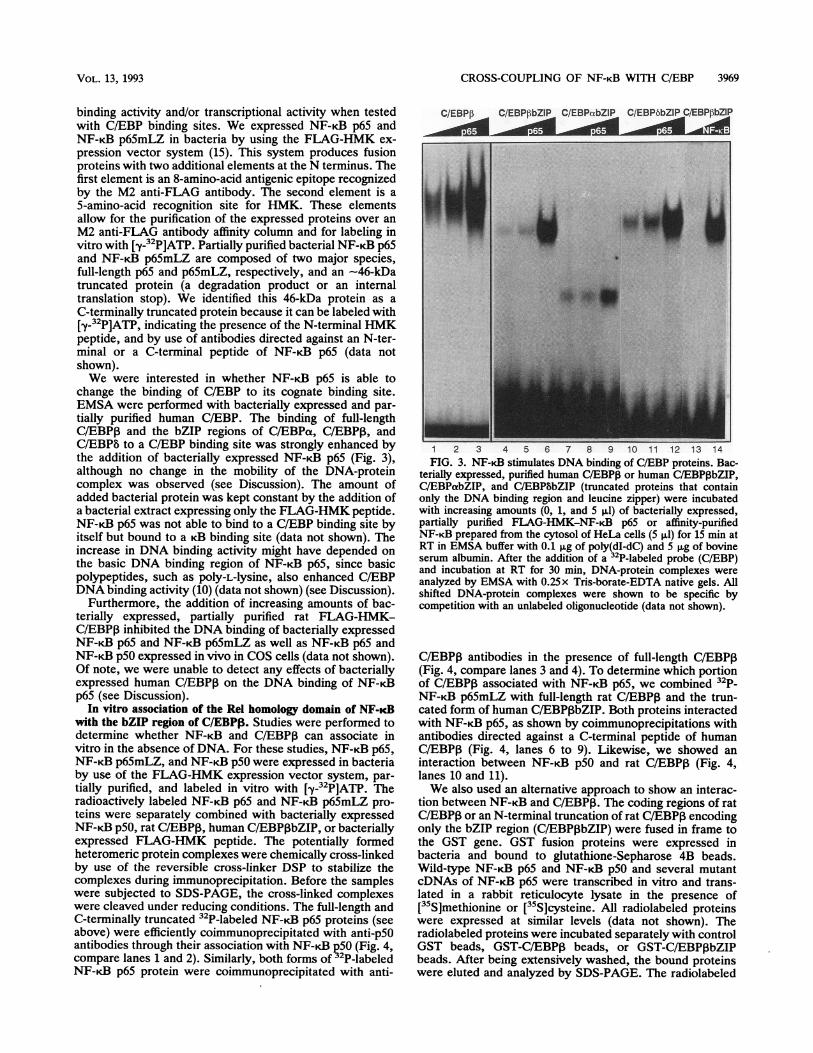
binding activity and/or transcriptional activity when testedwith C/EBP binding sites. We expressed NF-KB p65 andNF-KB p65mLZ in bacteria by using the FLAG-HMK ex-pression vector system (15). This system produces fusionproteins with two additional elements at the N terminus. Thefirst element is an 8-amino-acid antigenic epitope recognizedby the M2 anti-FLAG antibody. The second element is a5-amino-acid recognition site for HMK. These elementsallow for the purification of the expressed proteins over anM2 anti-FLAG antibody affinity column and for labeling invitro with [y-32P]ATP. Partially purified bacterial NF-KB p65and NF-KB p65mLZ are composed of two major species,full-length p65 and p65mLZ, respectively, and an -46-kDatruncated protein (a degradation product or an internaltranslation stop). We identified this 46-kDa protein as aC-terminally truncated protein because it can be labeled with[y-32P]ATP, indicating the presence of the N-terminal HMKpeptide, and by use of antibodies directed against an N-ter-minal or a C-terminal peptide of NF-,B p65 (data notshown).We were interested in whether NF-iB p65 is able to
change the binding of C/EBP to its cognate binding site.EMSA were performed with bacterially expressed and par-tially purified human C/EBP. The binding of full-lengthC/EBP3 and the bZIP regions of C/EBPa, C/EBPI, andC/EBPb to a C/EBP binding site was strongly enhanced bythe addition of bacterially expressed NF-KB p65 (Fig. 3),although no change in the mobility of the DNA-proteincomplex was observed (see Discussion). The amount ofadded bacterial protein was kept constant by the addition ofa bacterial extract expressing only the FLAG-HMK peptide.NF-K.B p65 was not able to bind to a C/EBP binding site byitself but bound to a rB binding site (data not shown). Theincrease in DNA binding activity might have depended onthe basic DNA binding region of NF-iB p65, since basicpolypeptides, such aspoly-L-lysine, also enhanced C/EBPDNA binding activity (10) (data not shown) (see Discussion).
Furthermore, the addition of increasing amounts of bac-terially expressed, partially purified rat FLAG-HMK-C/EBP,B inhibited the DNA binding of bacterially expressedNF-KB p65 and NF-KB p65mLZ as well as NF-KB p65 andNF-KB p50 expressed in vivo in COS cells (data not shown).Of note, we were unable to detect any effects of bacteriallyexpressed human C/EBPP on the DNA binding of NF-KBp65 (see Discussion).
In vitro association of the Rel homology domain of NF-KBwith the bZIP region of C/EBP3. Studies were performed todetermine whether NF-KB and C/EBPI can associate invitro in the absence of DNA. For these studies, NF-KB p65,NF-KB p65mLZ, and NF-KB p50 were expressed in bacteriaby use of the FLAG-HMK expression vector system, par-
tially purified, and labeled in vitro with [y_32p]ATp. Theradioactively labeled NF-KB p65 and NF-KB p65mLZ pro-
teins were separately combined with bacterially expressedNF-KB p50, rat C/EBP,B , human C/EBPPbZIP, or bacteriallyexpressed FLAG-HMK peptide. The potentially formedheteromeric protein complexes were chemically cross-linkedby use of the reversible cross-linker DSP to stabilize thecomplexes during immunoprecipitation. Before the sampleswere subjected to SDS-PAGE, the cross-linked complexeswere cleaved under reducing conditions. The full-length andC-terminally truncated 32p-labeled NF-KB p65 proteins (seeabove) were efficiently coimmunoprecipitated with anti-p5Oantibodies through their association with NF-KB p50 (Fig. 4,compare lanes 1 and 2). Similarly, both forms of 32p-labeledNF-KB p65 protein were coimmunoprecipitated with anti-
C/EBPP C/EBPPbZIP C/EBPabZIP C/EBP6bZIP CIEBPPbZ1P
1 2 3 4 5 6 7 8 9 10 11 12 13 14FIG. 3. NF-KB stimulates DNA binding of C/EBP proteins. Bac-
terially expressed, purified human C/EBP,B or human C/EBPIbZIP,C/EBPabZIP, and C/EBP8bZIP (truncated proteins that containonly the DNA binding region and leucine zipper) were incubatedwith increasing amounts (0, 1, and 5 pl) of bacterially expressed,partially purified FLAG-HMK-NF-KB p65 or affinity-purifiedNF-KB prepared from the cytosol of HeLa cells (5p,l) for 15 min atRT in EMSA buffer with 0.1 pg of poly(dI-dC) and 5pLg of bovineserum albumin. After the addition of a2P-labeled probe (C/EBP)and incubation at RT for 30 min, DNA-protein complexes wereanalyzed by EMSA with 0.25 x Tris-borate-EDTA native gels. Allshifted DNA-protein complexes were shown to be specific bycompetition with an unlabeled oligonucleotide (data not shown).
C/EBP3 antibodies in the presence of full-length C/EBPP(Fig. 4, compare lanes 3 and 4). To determine which portionof C/EBPP associated with NF-KB p65, we combined32p-NF-KB p65mLZ with full-length rat C/EBP,B and the trun-cated form of human C/EBP,BbZIP. Both proteins interactedwith NF-KB p65, as shown by coimmunoprecipitations withantibodies directed against a C-terminal peptide of humanC/EBPI (Fig. 4, lanes 6 to 9). Likewise, we showed aninteraction between NF-KB p50 and rat C/EBPP (Fig. 4,lanes 10 and 11).We also used an alternative approach to show an interac-
tion between NF-KB and C/EBP,B. The coding regions of ratC/EBP,B or an N-terminal truncation of rat CIEBPI encodingonly the bZIP region (CIEBP,bZIP) were fused in frame tothe GST gene. GST fusion proteins were expressed inbacteria and bound to glutathione-Sepharose 4B beads.Wild-type NF-KB p65 and NF-KB p50 and several mutantcDNAs of NF-KB p65 were transcribed in vitro and trans-lated in a rabbit reticulocyte lysate in the presence of[[35Smethionine or[35S]cysteine. All radiolabeled proteinswere expressed at similar levels (data not shown). Theradiolabeled proteins were incubated separately with controlGST beads, GST-C/EBP,B beads, or GST-C/EBPIbZIPbeads. After being extensively washed, the bound proteinswere eluted and analyzed by SDS-PAGE. The radiolabeled
VOL. 13, 1993

3970 STEIN ET AL.
anti-p65 0anti-C/EBPI- * * * 0 * * * Oanti-p50 0 0 032P-FLAG-p5O S .32P-FLAG-p65mLZ S S S 032P-FLAG-p65 0 * 0 0 0C/EBPPbZIP 0FLAG-C/EBPP . 0 0
A
35S- I
200-97-68 -
43-
29-
GST GST-CiEBPI GST-C/EBPibZIP
44)4) 4)~ 1 4~ '~(O9,CoCo 9, 4) 4) 4) 4e lI o Co$ o CoI? Co
I Ialm----
2 3 6 7 9_- ...
1 2 3 4 5 6 7 8 9 10 11 121 2 3 4 5 6 7 8 9 10 11 12
FIG. 4. In vitro association of NF-KB with C/EBPP. Bacteriallyexpressed, purified proteins were incubated as indicated with 32p_labeled bacterial NF-KB p65 (lanes 1 to 5), NF-KB p65mLZ (lanes 6to 9), and NF-KB p50 (lanes 10 to 12). After being cross-linked withDSP, the protein complexes were immunoprecipitated with antibod-ies directed against NF-KB p50 (lanes 1, 2, and 12), anti-C/EBPP(lanes 3, 4, and 6 to 11), and antibodies directed against NF-KB p65(lane 5). The immunocomplexes were reduced and subjected toSDS-PAGE. The arrowheads indicate the positions of the 32P-labeled proteins. Numbers to the left of the gel are in kilodaltons.
proteins bound only weakly or not at all to the control GSTbeads (Fig. 5A, lanes 2 to 5). As expected, full-length NF-KBp65 and a C-terminal truncation of NF-KB p65 encompassingonly the Rel homology domain (NF-KB p65Eco) boundefficiently to GST-C/EBPO (Fig. 5A, lanes 6 and 8) or toGST-C/EBP,BbZIP (Fig. 5A, lanes 10 and 11). A shortertruncation of NF-KB p65 encompassing only a portion of theRel homology domain (NF-KB p65Bgl) was unable to bind toGST-C/EBPI (Fig. SA, lane 9) or to GST-C/EBPIbZIP (Fig.5A, lane 12). These results identify the cross-coupling ofC/EBP,B with NF-KB as a bZIP-Rel homology domain inter-action. The alternatively spliced form of NF-KB p65 (NF-KBp65A10), which had only weak effects on C/EBP,B transacti-vation in vivo (Table 2), was not able to efficiently interactwith C/EBPI in vitro (Fig. 5A, lane 7). We also demon-strated the physical association of NF-KB p50 with GST-C/EBP,B and GST-C/EBPPbZIP (data not shown).The observed physical interaction between NF-KB p65
and C/EBP,B indicates it to be of an affinity similar to that ofthe homodimer formation between 35S-radiolabeled NF-KBp65 and GST-NF-KB p65 beads (compare Fig. SA, lanes 6and 8, with Fig. SB, lanes 4 and 5). The specificity of thisinteraction was further proven by mixing 5S-radiolabeledNF-KB p65Eco and c-Fos proteins and applying this mixtureto GST-C/EBPPI beads. As expected, NF-KB p65Eco boundspecifically to GST-C/EBP,B and GST-C/EBP,BbZIP (Fig.5C, lanes 3 to 5), while c-Fos, in accordance with our in vivodata (Table 2), did not specifically interact with C/EBPI.
DISCUSSIONIn this report, we demonstrate the functional and physical
associations of members of two important transcriptionfactor families, NF-KB and C/EBP. The functional associa-tions are characterized by (i) the inhibition of NF-KB-induced expression of promoters containing NF-KB bindingsites by the expression of C/EBP family members and (ii) thepositive synergistic action of NF-KB family members withC/EBP family members on promoters containing C/EBPbinding sites. Evidence is presented that NF-KB subunits
B
20097
C
1 2 3 4 5
Input
-o Al,44 AC9
c 9
C) CC)
.4a,-.. ";i
&*x
l 2 .3 4 5
FIG. 5. The Rel homology domain of NF-KB interacts with thebZIP region of C/EBP,B in vitro. (A) Radiolabeled, rabbit reticulo-cyte lysate-translated proteins (5 ,ul) were incubated as indicatedwith bacterially expressed GST (lanes 2 to 5), GST-C/EBPI (lanes 6to 9), and GST-C/EBPPbZIP (lanes 10 to 12) immobilized onglutathione-Sepharose 4B beads. (B) Radiolabeled NF-KB p65 orNF-KB p65Eco (5 ,ul) was incubated separately with control GSTbeads (lanes 2 and 3) or GST-NF-KB p65 beads (lanes 4 and 5). (C)Radiolabeled c-Fos and NF-KB p65Eco (5 pA each) were mixed andincubated with control GST beads, GST-C/EBPI beads, or GST-C/EBP1bZIP beads. Lanes 1 and 2 show one-fifth of the input amountsused for lanes 3 to 5. After being extensively washed, the boundproteins were eluted and analyzed by SDS-PAGE. Equal amounts ofbacterially expressed fusion proteins were bound to glutathione-Sepharose 4B beads. All radiolabeled proteins were expressed in therabbit reticulocyte lysate at similar levels (data not shown). Num-bers to the left of the gels are in kilodaltons.
strongly act in synergy with members of the C/EBP family oftranscription factors in regulating gene expression mediatedby the multimerized c-fos SRE. In addition, we demonstratea physical interaction between the two transcription factorfamilies through contacts on the Rel homology domain ofNF-KB and on the bZIP region of C/EBP. The experimentsthat revealed that several functional NF-KB subunits (p50,p65, and c-Rel) interact with three different isoforms ofC/EBP (a, 1, and b) indicated a general interaction betweenthese families and not merely a limited interaction betweenindividual members. Interestingly, and supporting our data,the work of LeClair et al. (50) demonstrated that screening ofa Xgtll expression library with radiolabeled NF-KB p50identified a cDNA encoding C/EBP3. The functional syn-ergy was limited to members of the NF-KB and C/EBPfamilies, since other transcription factors, such as c-Jun,c-Fos, CREB, and Myb, were not able to act in synergy withC/EBP and NF-KB p65 did not act in synergy with CREB
MOL. CELL. BIOL.
CROSS-COUPLING OF NF-KB WITH C/EBP 3971
and Myb (Tables 2 and 3). Our data also suggest an inhibitionof CREB-induced promoter activity by NF-KB p65.The positive synergy observed between the NF-KB and
C/EBP families of transcription factors in regulating geneexpression (Fig. 1B and C, Fig. 2B, and Table 2) presumablyis based on the direct physical interaction between thesefactors. We found that NF-KB p65 increases the formation ofcomplexes between C/EBP and its DNA binding site (Fig. 3).It was of interest that the mobility of the C/EBP-DNAcomplexes was not altered by the addition of NF-KB p65,suggesting that NF-KB p65 is not part of the complexesdetected by EMSA. Thus, it is possible that NF-KB p65functions to stabilize the C/EBP-DNA interaction by form-ing a ternary complex in solution but that any NF-KB p65associated with this complex becomes separated duringelectrophoresis. A similar observation was reported recentlyfor the interaction between the homeodomain protein Phoxland the serum response factor (36). Another possibility isthat NF-KB p65 transiently interacts with CIEBP and causesa conformational change in C/EBP that then results inincreased DNA binding of C/EBP accompanied by therelease of NF-KB p65 from the complex. A similar mecha-nism has been postulated for the increase in MyoDl DNAbinding by HSP90 (75). Furthermore, it has been shown thatC/EBP DNA binding is also increased by small basic poly-peptides, such as poly-L-lysine (10). Therefore, the increasein C/EBP DNA binding by NF-KB p65 might depend on thebasic DNA binding region of NF-KB p65. Experiments witha truncated form of NF-KB missing the C-terminal transac-tivation domain (NF-KB p65Eco; Table 2) indicated that thefunctional synergy in vivo not only was a result of increasedDNA binding of the C/EBP-NF-KB p65 complex but alsodepended on the transactivation domain of NF-KB p65.Our experiments clearly demonstrate the direct physical
association between NF-KB and C/EBP through an interac-tion of the Rel homology domain and the bZIP region (Fig. 4and 5). This interaction is detected in the absence of bindingsite DNA. A truncated form of NF-KB p65 expressing onlythe Rel homology domain (NF-KB p65Eco) efficiently asso-ciates with a truncated form of C/EBPP expressing only thebZIP region (C/EBPIbZIP) (Fig. SA). Further deletion of theNF-KB p65 Rel homology region (NF-KB p65Bgl) or aninternal deletion of 10 amino acids within the Rel homologyregion (NF-KB p65A10) results in a loss of the interactionwith C/EBPI (Fig. SA). This result leads to the conclusionthat the same motifs that play a role in dimer formationwithin the NF-KB and C/EBP families of transcription fac-tors are also used for complex formation between the C/EBPand NF-KB families. Alternatively, since these mutations inthe Rel homology domain also prevent homodimer formationby NF-KB p65, it is possible that a monomeric form ofNF-KB p65 does not interact with C/EBP,B. Chimeric pro-teins containing the GAL4 DNA binding and dimerizationdomain (amino acids 1 to 147) fused in frame to NF-KBp65Eco or NF-KB p65Bgl are able to form homodimersthrough the GALA domain, but only GAL4-NF-KB p65Ecois able to interact with C/EBP (data not shown). This resultsuggests either that amino acids 195 to 282 of NF-KB p65,missing in GALA NF-KB p65Bgl, are the target for thephysical interaction between these two families or thatdeletion of the C-terminal portion of the Rel homologydomain changes the conformation of the residual protein,preventing an interaction with C/EBP.
If NF-KB and C/EBP physically interact in vitro andsynergistically activate promoters in vivo, then they mayalso physically interact in vivo. We have not detected an in
vivo interaction, probably because of the very low intracel-lular concentrations of NF-KB and C/EBP family members.Furthermore, the interaction appears to be stabilized bybinding site DNA (see below). The specificity of the physicalinteraction between NF-KB and C/EBP is also evident fromour protein mixing experiments proving that c-Fos, whichdoes not cross-couple with C/EBP, is not able to bind toGST-C/EBPI beads (Fig. SC) and from experiments showingthat NF-KB p65 binds with similar affinities to GST-NF-KBp65 beads and to GST-C/EBP,B beads (Fig. SB).The basis for the inhibition of NF-KB transactivation of
gene expression by C/EBP (Fig. 1A, Fig. 2A, and Table 1) isnot known. One possibility is that C/EBP blocks the abilityof NF-KB to interact with a critical transcriptional cofactor.This process is termed "squelching" and has been describedelsewhere (57, 68). If this hypothesis is true, it is curious whysquelching was not observed when a C/EBP-responsivereporter plasmid was used in the presence of the twotranscription factors. A second explanation is the possibleinduction of an inhibitor of NF-KB function by the expres-sion of C/EBP in F9 cells. Although we cannot rule out thesemechanisms, we favor a third explanation: the formationbetween C/EBP and NF-KB of a novel transcription factorcomplex with reduced transcriptional activity at KB enhanc-ers. The latter could be manifested by a reduced affinity forKB binding sites or the inability of the complex to stimulatetranscriptional activity. We have observed that bacteriallyexpressed rat C/EBPP inhibits the ability of NF-KB p65 andNF-KB p50 to bind to its cognate sequence, yet bacteriallyexpressed human C/EBPI does not alter the ability ofNF-KB to bind to DNA (data not shown). It is not clearwhether this result is caused by the presence of the N-ter-minal FLAG-HMK region on the rat protein or whether itreflects differences in the concentrations or state of purifi-cation of these proteins. It is important to note that threeisoforms of human C/EBP (a, 1B, and b) are able, like ratC/EBPP, to inhibit NF-KB-induced gene activation (Fig.2A).The nature of the molecular complex formed between
NF-KB and C/EBP is not clear, but evidence points to aninteraction between the diner forms of the two factors.First, a mutation in a putative NF-KB p65 leucine zipperdoes not block the functional or physical interaction betweenthis protein and C/EBP (Tables 1 and 2 and Fig. 4). Inaddition, no leucine zipper-like structure has been reportedin NF-KB p50, v-Rel, or c-Rel, yet these proteins function-ally and physically interact with C/EBP family members(Tables 1 and 2 and Fig. 4). Second, an alternatively splicedform of NF-KB p65 (NF-KB p65A10) and a C-terminallytruncated form (NF-KB p65Bgl) that cannot efficiently dimer-ize (61, 78a) do not functionally or physically interact withC/EBP subunits (Table 2 and Fig. 5). This result suggeststhat the same motif that is necessary for dimer formation isalso involved in cross-dimerization between these two fam-ilies. Of particular importance is the observation that a singleC/EBP binding site is sufficient to function as a target for thefunctional synergy between NF-KB and C/EBP (Fig. 1C).Thus, it is unlikely that NF-KB makes DNA contacts on thesingle C/EBP binding site. This idea supports the model of adimer of a C/EBP family member binding to DNA andinteracting through its bZIP region with the Rel homologyregion of an NF-KB family member. Presumably, an alteredconfiguration of the transcription factor complex is stabilizedand ultimately is more efficient in interacting with the basictranscriptional machinery.What is the functional significance of the interactions
VOL. 13, 1993

3972 STEIN ET AL.
observed between NF-KB and C/EBP? Of particular interestis the observation that NF-KB can augment gene expressionmediated through the multimerized c-fos SRE in the pres-ence of C/EBP. Thus, NF-KB subunits, which are stronglyimplicated in the control of cellular proliferation and areinduced in the Go-to-G1 transition (7), may functionallystimulate c-fos gene expression when C/EBP is present.C/EBP is induced by compounds that activate cAMP (55),and evidence has been presented that increased cAMP levelsmay induce preexisting NF-KB as well (76, 77). Thus, anincrease in cAMP levels may coinduce C/EBP and NF-KB,allowing for enhanced expression from C/EBP binding sitesand, potentially, a decrease in gene expression mediated byNF-KB binding sites. A basis for the functional interaction ofNF-KB and C/EBP is also supported by studies with IL-1 andIL-6. IL-1 is known to induce NF-KB (64), and IL-6 activatesC/EBPP (66). The physical and functional interactions ob-served between NF-KB and C/EBPI may explain the func-tional synergy observed between IL-1 and IL-6 in severalphysiological responses, including the acute-phase response,in T-cell activation, and in immunoglobulin secretion (1, 37,83). In this regard, the IL-6 promoter contains both anNF-KB binding site and a C/EBP binding site (37, 40, 52).Both sites have been shown to be targets for IL-1 and tumornecrosis factor alpha stimulation (37); thus, it is likely thatthe induction of NF-KB by IL-1 and tumor necrosis factoralpha functions to stimulate gene expression from each site.Furthermore, the IL-8 promoter contains adjacent bindingsites for C/EBP and NF-KB that cooperate in gene activation(58), and the angiotensinogen promoter contains overlappingbinding sites for these two transcription factors (19). Prelim-inary data obtained with the IL-8 promoter show coopera-tive binding of NF-KB and C/EBP to their adjacent bindingsites, resulting in a dose-dependent synergistic activation ofthe IL-8 promoter (78a). Functional interactions betweenC/EBP and NF-KB subunits could have profound influenceson cellular growth control. It has been demonstrated thatC/EBPa is growth inhibitory and likely functions in theterminal differentiation of certain cells (27, 82). The expres-sion of C/EBP isoforms could alter the ability of NF-KBsubunits to control cellular proliferation.There is growing evidence that the regulation of gene
expression is not mediated solely by the presence or absenceof a particular set of transcription factors. The function of atranscription factor can be inhibited by the presence of aspecific inhibitor that blocks the ability of the transcriptionfactor to bind to DNA (6). In addition, the interplay oftranscription factors themselves likely functions as a criticalregulatory component of gene expression. Physical andfunctional interactions, usually dimerization, of factorswithin a particular family are well documented. However,recent reports indicate cross-family interactions of specifictranscription factors. One example is the interaction be-tween the glucocorticoid receptor and the AP-1 proteins Fosand Jun (24, 42, 53, 74, 87). A second example is the recentreport of the interaction between the leucine zipper proteinJun and the helix-loop-helix protein MyoD (12). Our datademonstrate that the important transcription factor familiesC/EBP and NF-KB functionally and physically interact. Thisobservation, along with those made previously, highlights aunique mechanism by which the levels of one transcriptionfactor can strongly modulate the functional activity of an-other.
ACKNOWLEDGMENTS
We thank Dean Ballard, Michael Blanar, John Cogswell, MichaelGilman, Alain Israel, Steven McKnight, Michael Kuehl, ValeriaPoli, David Ron, Craig Rosen, and Inder Verma for valuablereagents. We also thank Amer Beg, Adrienne Brown, and ShannonKenney for helpful discussions and Kenneth LeClair and PhillipSharp for communicating results prior to publication.
This research was supported by a grant (to A.S.B.) from theNational Institutes of Health (CA 52515), by a March of Dimes BasilO'Connor Research Award, by an RJR-Nabisco Scholar Award inImmunology, and by an ACS Junior Faculty Research Award(JFRA-309).
ADDENDUM IN PROOFThe plasmids described as encoding human C/EBP forms
as well as the recombinant proteins encoded by their respec-tive cDNAs were erroneously named and are actually rat(C/EBPa) or mouse (C/EBP, and C/EBP8). This does notalter the interpretations of any of the data.
REFERENCES1. Akira, S., T. Hirano, T. Taga, and T. Kishimoto. 1990. Biology
of multifunctional cytokines: IL-6 and related molecules (IL-1and TNF). FASEB J. 4:2860-2867.
2. Akira, S., H. Isshiki, T. Sugita, 0. Tanabe, S. Kinoshita, Y.Nishio, T. Nakajima, T. Hirano, and T. Kishimoto. 1990. Anuclear factor for IL-6 expression (NF-IL6) is a member of aC/EBP family. EMBO J. 9:1897-1906.
3. Andersson, S., D. L. Davis, H. Dahlbick, H. Jornvall, and D. W.Russel. 1989. Cloning, structure, and expression of the mito-chondrial cytochrome P-450 sterol 26-hydroxylase, a bile acidbiosynthetic enzyme. J. Biol. Chem. 264:8222-8229.
4. Angel, P., I. Baumann, B. Stein, H. Delius, H. J. Rahmsdorf, andP. Herrlich. 1987. 12-O-Tetradecanoyl-phorbol-13-acetate in-duction of the human collagenase gene is mediated by aninducible enhancer element located in the 5'-flanking region.Mol. Cell. Biol. 7:2256-2266.
5. Baeuerle, P. A. 1991. The inducible transcription activatorNF-KB: regulation by distinct protein subunits. Biochim. Bio-phys. Acta 1072:63-80.
6. Baeuerle, P. A., and D. Baltimore. 1988. IKB: a specific inhibitorof the NFKB transcription factor. Science 242:540-546.
7. Baldwin, A. S., Jr., J. C. Azizkhan, D. E. Jensen, A. A. Beg, andL. R. Coodly. 1991. Induction of NF-KB DNA-binding activityduring the Go-to-G1 transition in mouse fibroblasts. Mol. Cell.Biol. 11:4943-4951.
8. Ballard, D. W., E. P. Dixon, N. J. Peffer, H. Bogerd, S. Doerre,B. Stein, and W. C. Greene. 1992. The 65-kDa subunit of humanNF-KB functions as a potent transcriptional activator and atarget for v-Rel-mediated repression. Proc. Natl. Acad. Sci.USA 89:1875-1879.
9. Ballard, D. W., W. H. Walker, S. Doerre, P. Sista, J. A. Molitor,E. P. Dixon, N. J. Peffer, M. Hannink, and W. C. Greene. 1990.The v-rel oncogene encodes a KB enhancer binding protein thatinhibits NF-KB function. Cell 63:803-814.
10. Bannister, A. J., and T. Kouzarides. 1992. Basic peptidesenhance protein/DNA interaction in vitro. Nucleic Acids Res.20:3523.
11. Beg, A. A., S. M. Ruben, R. I. Scheinman, S. Haskill, C. A.Rosen, and A. S. Baldwin, Jr. 1992. IcB interacts with thenuclear localization sequences of the subunits of NF-KB: amechanism for cytoplasmic retention. Genes Dev. 6:1899-1913.
12. Bengal, E., L. Ransone, R. Scharfmann, V. J. Dwarki, S. J.Tapscott, H. Weintraub, and I. M. Verma. 1992. Functionalantagonism between c-Jun and MyoD proteins: a direct physicalassociation. Cell 68:507-519.
13. Berkowitz, L. A., and M. Z. Gilman. 1990. Two distinct forms ofactive transcription factor CREB (cAMP response elementbinding protein). Proc. Natl. Acad. Sci. USA 87:5258-5262.
14. Birkenmeier, E. H., B. Gwynn, S. Howard, J. Jerry, J. I.Gordon, W. H. Landschulz, and S. L. McKnight. 1989. Tissue-
MOL. CELL. BIOL.

CROSS-COUPLING OF NF-KB WITH C/EBP 3973
specific expression, developmental regulation, and genetic map-ping of the gene encoding CCAAT/enhancer binding protein.Genes Dev. 3:1146-1156.
15. Blanar, M. A., and W. J. Rutter. 1992. Interaction cloning:identification of a helix-loop-helix zipper protein that interactswith c-Fos. Science 256:1014-1018.
16. Blank, V., P. Kourilsky, and A. Israel. 1992. NF-KB and relatedproteins: Rel/dorsal homologies meet ankyrin-like repeats.Trends Biochem. Sci. 17:135-140.
17. Bours, V., J. Villalobos, P. R. Burd, K. Kelly, and U. Siebenlist.1990. Cloning of a mitogen-inducible gene encoding a KBDNA-binding protein with homology to the rel oncogene and tocell-cycle motifs. Nature (London) 348:76-80.
18. Bradford, M. M. 1976. A rapid and sensitive method for thequantitation of microgram quantities of protein utilizing theprinciple of protein-dye binding. Anal. Biochem. 72:248-254.
19. Brasier, A. R., D. Ron, J. E. Tate, and J. F. Habener. 1990. Afamily of constitutive C/EBP-like DNA binding proteins atten-uate the IL-la induced NF-KB mediated trans-activation of theangiotensinogen gene acute-phase response element. EMBO J.9:3933-3944.
20. Cao, Z., R. M. Umek, and S. L. McKnight. 1991. Regulatedexpression of three C/EBP isoforms during adipose conversionof 3T3-L1 cells. Genes Dev. 5:1538-1552.
21. Chang, C.-J., T.-T. Chen, H.-Y. Lei, D.-S. Chen, and S.-C. Lee.1990. Molecular cloning of a transcription factor, AGP/EBP,that belongs to members of the C/EBP family. Mol. Cell. Biol.10:6642-6653.
22. Christy, R. J., V. W. Yang, J. M. Ntambi, D. E. Geiman, W. H.Landschulz, A. D. Friedman, Y. Nakabeppu, T. J. Kelly, andM. D. Lane. 1989. Differentiation-induced gene expression in3T3-L1 preadipocytes: CCAAT/enhancer binding protein inter-acts with and activates the promoters of two adipocyte-specificgenes. Genes Dev. 3:1323-1335.
23. Descombes, P., M. Chojkier, S. Lichtsteiner, E. Falvey, and U.Schibler. 1990. LAP, a novel member of the C/EBP gene family,encodes a liver-enriched transcriptional activator protein.Genes Dev. 4:1541-1551.
24. Diamond, M. I., J. N. Miner, S. K. Yoshinaga, and K. R.Yamamoto. 1990. Transcription factor interactions: selectors ofpositive or negative regulation from a single DNA element.Science 249:1266-1272.
25. Evans, J. L., T. L. Moore, W. M. Kuehl, T. Bender, and J. P.-Y.Ting. 1990. Functional analysis of c-Myb protein in T-lympho-cytic cell lines shows that it trans-activates the c-myc promoter.Mol. Cell. Biol. 10:5747-5752.
26. Evans, R. M. 1988. The steroid and thyroid hormone receptorsuperfamily. Science 240:889-895.
27. Freytag, S. O., and T. J. Geddes. 1992. Reciprocal regulation ofadipogenesis by Myc and C/EBPa. Science 256:379-382.
28. Friedman, A. D., W. H. Landschulz, and S. L. McKnight. 1989.CCAAT/enhancer binding protein activates the promoter of theserum albumin gene in cultured hepatoma cells. Genes Dev.3:1314-1322.
29. Friedman, A. D., and S. L. McKnight. 1990. Identification oftwo polypeptide segments of CCAAT/enhancer-binding proteinrequired for transcriptional activation of the serum albumingene. Genes Dev. 4:1416-1426.
30. Gehring, W. J. 1987. Homeoboxes in the study of development.Science 236:1245-1252.
31. Ghosh, S., A. M. Gifford, L. R. Riviere, P. Tempst, G. P. Nolan,and D. Baltimore. 1990. Cloning of the p50 DNA binding subunitof NF-K<B: homology to rel and dorsal. Cell 62:1019-1029.
32. Gilmore, T. D. 1990. NF-KB, KBF1, dorsal, and related mat-ters. Cell 62:841-843.
33. Gilmore, T. D., and H. M. Temin. 1988. v-rel oncoproteins inthe nucleus and in the cytoplasm transform chicken spleen cells.J. Virol. 62:703-714.
34. Graham, F. L., and A. J. van der Eb. 1973. A new technique forthe assay of infectivity of human adenovirus 5 DNA. Virology52:456-467.
35. Greene, W. C., E. Bdhnlein, and D. W. Ballard. 1989. HIV-1,HTLV-I and normal T-cell growth: transcriptional strategies
and surprises. Immunol. Today 10:272-278.36. Gruenenberg, D. A., S. Natesan, C. Alexandre, and M. Z.
Gilman. 1992. Human and Drosophila homeodomain proteinsthat enhance the DNA-binding activity of serum responsefactor. Science 257:1089-1095.
37. Hirano, T., S. Akira, T. Taga, and T. Kishimoto. 1990. Biolog-ical and clinical aspects of interleukin 6. Immunol. Today11:443-449.
38. Ip, Y. T., R. Kraut, M. Levine, and C. A. Rushlow. 1991. Thedorsal morphogen is a sequence-specific DNA-binding proteinthat interacts with a long-range repression element in Dros-ophila. Cell 64:439-446.
39. Israel, A., 0. Yano, F. Logeat, M. Kieran, and P. Kourilsky.1989. Two purified factors bind to the same sequence in theenhancer of mouse MHC class I genes: one of them is a positiveregulator induced upon differentiation of teratocarcinoma cells.Nucleic Acids Res. 17:5245-5257.
40. Isshiki, H., S. Akira, 0. Tanabe, T. Nakajima, T. Shimamoto, T.Hirano, and T. Kishimoto. 1990. Constitutive and interleukin-1(IL-1)-inducible factors interact with the IL-1-responsive ele-ment in the IL-6 gene. Mol. Cell. Biol. 10:2757-2764.
41. Johnson, P. F., W. H. Landschulz, B. J. Graver, and S. L.McKnight. 1987. Identification of a rat liver nuclear protein thatbinds to the enhancer core element of three animal viruses.Genes Dev. 1:133-146.
42. Jonat, C., H. J. Rahmsdorf, K.-K. Park, A. C. B. Cato, S. Gebel,H. Ponta, and P. Herrlich. 1990. Antitumor promotion andantiinflammation: down-modulation of AP-1 (Fos/Jun) activityby glucocorticoid hormone. Cell 62:1189-1204.
43. Kaestner, K. H., R. J. Christy, and M. D. Lane. 1990. Mouseinsulin-responsive glucose transporter gene: characterization ofthe gene and trans-activation by the CCAAT/enhancer bindingprotein. Proc. Natl. Acad. Sci. USA 87:251-255.
44. Kawai, S., and M. Nishizawa. 1984. New procedure for DNAtransfection with polycation and dimethyl sulfoxide. Mol. Cell.Biol. 4:1172-1174.
45. Kieran, M., V. Blank, F. Logeat, J. Vandekerckhove, F. Lott-speich, 0. Le Bail, M. Urban, P. Kourilsky, P. A. Baeuerie, andA. Israel. 1990. The DNA binding subunit of NF-KB is identicalto factor KBF1 and homologous to the rel oncogene product.Cell 62:1007-1018.
46. Klug, A., and D. Rhodes. 1987. Zinc fingers: a novel proteinmotif for nucleic acid recognition. Trends Biochem. Sci. 12:464-469.
46a.Kuehl, M. Unpublished data.47. Laemmli, U. K. 1970. Cleavage of structural proteins during the
assembly of the head of bacteriophage T4. Nature (London)227:680-685.
48. Lamb, P., and S. L. McKnight. 1991. Diversity and specificity intranscriptional regulation: the benefits of heterotypic dimeriza-tion. Trends Biochem. Sci. 16:417-422.
49. Landschulz, W., P. Johnson, and S. McKnight. 1988. Theleucine zipper: a hypothetical structure common to a new classof DNA binding proteins. Science 240:1759-1764.
50. LeClair, K. P., M. A. Blanar, and P. A. Sharp. 1992. The p50subunit of NF-KB associates with the NF-IL6 transcriptionfactor. Proc. Natl. Acad. Sci. USA 89:8145-8149.
51. Lenardo, M. J., and D. Baltimore. 1989. NF-KB: a pleiotropicmediator of inducible and tissue-specific gene control. Cell58:227-229.
52. Libermann, T. A., and D. Baltimore. 1990. Activation of inter-leukin-6 gene expression through the NF-KB transcription fac-tor. Mol. Cell. Biol. 10:2327-2334.
53. Lucibello, F. C., E. P. Slater, K. U. Jooss, M. Beato, and R.Muller. 1990. Mutual transrepression of Fos and the glucocor-ticoid receptor: involvement of a functional domain in Fos thatis absent from FosB. EMBO J. 9:2828-2834.
54. McKnight, S. L., M. D. Lane, and S. Gluecksohn-Waelsch. 1989.Is CCAAT/enhancer-binding protein a central regulator of en-ergy metabolism? Genes Dev. 3:2021-2024.
55. Metz, R., and E. Ziff. 1991. cAMP stimulates the C/EBP-relatedtranscription factor rNFIL-6 to trans-locate to the nucleus andinduce c-fos transcription. Genes Dev. 5:1754-1766.
VOL. 13, 1993
3974 STEIN ET AL.
56. Metz, R., and E. Ziff. 1991. The helix-loop-helix protein rE12and the C/EBP-related factor rNFIL-6 bind to neighboring siteswithin the c-fos serum response element. Oncogene 6:2165-2178.
57. Meyer, M.-E., H. Gronemeyer, B. Turcotte, M.-T. Bocquel, D.Tasset, and P. Chambon. 1989. Steroid hormone receptorscompete for factors that mediate their enhancer function. Cell57:433-442.
58. Mukaida, N., Y. Mahe, and K. Matsushima. 1990. Cooperativeinteraction of nuclear factor-KB and cis-regulatory enhancerbinding protein-like factor binding elements in activating theinterleukin-8 gene by pro-inflammatory cytokines. J. Biol.Chem. 265:21128-21133.
59. Murre, C., P. S. McCaw, and D. Baltimore. 1989. A new DNAbinding and dimerization motif in immunoglobulin enhancerbinding, daughterless, MyoD, and myc proteins. Cell 56:777-783.
60. Murre, C., P. S. McCaw, H. Vaessin, M. Caudy, L. Y. Jan, Y. N.Jan, C. V. Cabrera, J. N. Buskin, S. D. Hauschka, A. B. Lassar,H. Weintraub, and D. Baltimore. 1989. Interactions betweenheterologous helix-loop-helix proteins generate complexes thatbind specifically to a common DNA sequence. Cell 58:537-544.
61. Narayanan, R., J. F. Klement, S. M. Ruben, K. A. Higgins, andC. A. Rosen. 1992. Identification of a naturally occurring trans-forming variant of the p65 subunit of NF-KB. Science 256:367-370.
62. Neumann, J. R., C. A. Morency, and K. 0. Russian. 1987. Anovel rapid assay for chloramphenicol acetyltransferase geneexpression. BioTechniques 5:444 448.
63. Nolan, G. P., S. Ghosh, H.-C. Liou, P. Tempst, and D. Balti-more. 1991. DNA binding and IKB inhibition of the cloned p65subunit of NF-KB, a rel-related polypeptide. Cell 64:961-969.
64. Osborn, L., S. Kunkel, and G. J. Nabel. 1989. Tumor necrosisfactor a and interleukin 1 stimulate the human immunodefi-ciency virus enhancer by activation of the nuclear factor KB.Proc. Natl. Acad. Sci. USA 86:2336-2340.
65. Park, E. A., W. J. Roesler, J. Liu, D. J. Klemm, A. L. Gurney,J. D. Thatcher, J. Shuman, A. Friedman, and R. W. Hanson.1990. The role of the CCAAT/enhancer-binding protein in thetranscriptional regulation of the gene for phosphoenolpyruvatecarboxykinase (GTP). Mol. Cell. Biol. 10:6264-6272.
66. Poli, V., and R. Cortese. 1989. Interleukin 6 induces a liver-specific nuclear protein that binds to the promoter of acute-phase genes. Proc. Natl. Acad. Sci. USA 86:8202-8206.
67. Poli, V., F. P. Mancini, and R. Cortese. 1990. IL-6DBP, anuclear protein involved in interleukin-6 signal transduction,defines a new family of leucine zipper proteins related toC/EBP. Cell 63:643-653.
68. Ptashne, M. 1988. How eukaryotic transcriptional activatorswork. Nature (London) 335:683-689.
69. Ransone, L. J., J. Visvader, P. Sassone-Corsi, and I. M. Verma.1989. Fos-Jun interaction: mutational analysis of the leucinezipper domain of both proteins. Genes Dev. 3:770-781.
70. Ransone, L. J., J. Visvader, P. Wamsley, and I. M. Verma. 1990.Trans-dominant negative mutants of Fos and Jun. Proc. Natl.Acad. Sci. USA 87:3806-3810.
71. Roman, C., J. S. Platero, J. D. Shuman, and K. Calame. 1990.Ig/EBP-1: a ubiquitously expressed immunoglobulin enhancerbinding protein that is similar to C/EBP and heterodimerizeswith C/EBP. Genes Dev. 4:1404-1415.
72. Ruben, S. M., P. J. Dillon, R. Schreck, T. Henkel, C.-H. Chen,
M. Maher, P. A. Baeuerle, and C. A. Rosen. 1991. Isolation of arel-related human cDNA that potentially encodes the 65-kDsubunit of NF-KB. Science 251:1490-1493.
73. Ruben, S. M., R. Narayanan, J. F. Klement, C.-H. Chen, andC. A. Rosen. 1992. Functional characterization of the NF-KBp65 transcriptional activator and an alternatively spliced deriv-ative. Mol. Cell. Biol. 12:444-454.
74. Schule, R., P. Rangarajan, S. Kliewer, L. J. Ransone, J. Bolado,N. Yang, I. M. Verma, and R. M. Evans. 1990. Functionalantagonism between oncoprotein c-Jun and the glucocorticoidreceptor. Cell 62:1217-1226.
75. Shaknovich, R., G. Shue, and S. Kohtz. 1992. Conformationalactivation of a basic helix-loop-helix protein (MyoDl) by theC-terminal region of murine HSP90 (HSP84). Mol. Cell. Biol.12:5059-5068.
76. Shirakawa, F., M. Chedid, J. Suttles, B. A. Pollok, and S. B.Mizel. 1989. Interleukin 1 and cyclic AMP induce K immuno-globulin light-chain expression via activation of an NF-KB-likeDNA-binding protein. Mol. Cell. Biol. 9:959-964.
77. Shirakawa, F., and S. B. Mizel. 1989. In vitro activation andnuclear translocation of NF-KB catalyzed by cyclic AMP-dependent protein kinase and protein kinase C. Mol. Cell. Biol.9:2424-2430.
78. Shuman, J. D., C. R. Vinson, and S. L. McKnight. 1990.Evidence of changes in protease sensitivity and subunit ex-change rate on DNA binding by C/EBP. Science 249:771-774.
78a.Stein, B. Unpublished data.79. Stein, B., H. J. Rahmsdorf, A. Steffen, M. Litfin, and P.
Herrlich. 1989. UV-induced DNA damage is an intermediate inthe UV-induced expression of human immunodeficiency virustype 1, collagenase, c-fos, and metallothionein. Mol. Cell. Biol.9:5169-5181.
80. Steward, R. 1987. Dorsal, an embryonic polarity gene in Dros-ophila, is homologous to the vertebrate proto-oncogene, c-rel.Science 238:692-694.
81. Trus, M., N. Benvenisty, H. Cohen, and L. Reshef. 1990.Developmentally regulated interactions of liver nuclear factorswith the rat phosphoenolpyruvate carboxykinase promoter.Mol. Cell. Biol. 10:2418-2422.
82. Umek, R. M., A. D. Friedman, and S. L. McKnight. 1991.CCAAT/enhancer binding protein: a component of a differenti-ation switch. Science 251:288-292.
83. Van Snick, J. 1990. Interleukin-6: an overview. Annu. Rev.Immunol. 8:253-278.
84. Vinson, C. R., P. B. Sigler, and S. L. McKnight. 1989. Scissors-grip model for DNA recognition by a family of leucine zipperproteins. Science 246:911-916.
85. Walker, W. H., B. Stein, P. A. Ganchi, J. A. Hoffman, P. A.Kaufman, D. W. Ballard, M. Hannink, and W. C. Greene. 1992.The v-rel oncogene: insights into the mechanism of transcrip-tional activation, repression, and transformation. J. Virol. 66:5018-5029.
86. Williams, S. C., C. A. Cantwell, and P. F. Johnson. 1991. Afamily of C/EBP-related proteins capable of forming covalentlylinked leucine zipper dimers in vitro. Genes Dev. 5:1553-1567.
87. Yang-Yen, H.-F., J.-C. Chambard, Y.-L. Sun, T. Smeal, T. J.Schmidt, J. Drouin, and M. Karin. 1990. Transcriptional inter-ference between c-Jun and the glucocorticoid receptor: mutualinhibition of DNA binding due to direct protein-protein interac-tion. Cell 62:1205-1215.
MOL. CELL. BIOL.


![FA/BRCA DNA damage repair pathway is reglated by NF-[kappa]B and](https://img.pdfslide.net/doc/110x75/620f4286843897096d43e0a4/fabrca-dna-damage-repair-pathway-is-reglated-by-nf-kappab-and.jpg)