Embed Size (px)
Citation preview

University of Arkansas, Fayetteville University of Arkansas, Fayetteville
ScholarWorks@UARK ScholarWorks@UARK
Graduate Theses and Dissertations
12-2020
Functional and Physiological Role of Extra-Hypothalamic Functional and Physiological Role of Extra-Hypothalamic
Corticotropin Releasing Hormone Neurons in the Nucleus of the Corticotropin Releasing Hormone Neurons in the Nucleus of the
Hippocampal Commissure in Regulation of Stress Response Hippocampal Commissure in Regulation of Stress Response
Hakeem Kadhim University of Arkansas, Fayetteville
Follow this and additional works at: https://scholarworks.uark.edu/etd
Part of the Endocrinology, Diabetes, and Metabolism Commons, Molecular Biology Commons, and the
Neurosciences Commons
Citation Citation Kadhim, H. (2020). Functional and Physiological Role of Extra-Hypothalamic Corticotropin Releasing Hormone Neurons in the Nucleus of the Hippocampal Commissure in Regulation of Stress Response. Graduate Theses and Dissertations Retrieved from https://scholarworks.uark.edu/etd/3840
This Dissertation is brought to you for free and open access by ScholarWorks@UARK. It has been accepted for inclusion in Graduate Theses and Dissertations by an authorized administrator of ScholarWorks@UARK. For more information, please contact [email protected].

Functional and Physiological Role of Extra-Hypothalamic Corticotropin Releasing Hormone Neurons in the Nucleus of the Hippocampal Commissure in Regulation of Stress Response
A dissertation submitted in partial fulfillment of the requirements for the degree of
Doctor of Philosophy in Cell and Molecular Biology
by
Hakeem Kadhim University of Baghdad
Bachelor of Veterinary Medicine, 2002 Al Nahrain University
Master of Science in Physiology, 2012
December 2020 University of Arkansas
This dissertation is approved for recommendation to the Graduate Council.
_________________________________ Wayne Kuenzel, Ph.D. Dissertation Director ____________________________________ _________________________________ Walter Bottje, Ph.D. Timothy Evans, Ph.D. Committee Member Committee Member _____________________________________ _________________________________ Sami Dridi, Ph.D. Young Min Kwon, Ph. D. Committee Member Committee Member

Abstract
Corticotropin releasing hormone (CRH) neurons located within the paraventricular nucleus
(PVN) are known to be involved in regulation of stress responses. Recently, CRH neurons were
identified above the PVN within the nucleus of the hippocampal commissure (NHpC) that located
in the septum. We hypothesized that CRH neurons in the NHpC play a critical role in the stress
response due to their rapid activation and could be a part of the traditional hypothalamo-pituitary-
adrenal (HPA) axis. The dissertation addresses the role of 1) CRH expressing neurons in the NHpC
compared with those within the PVN utilizing two different stressors, food deprivation (FD) and
immobilization stress, 2) arginine vasotocin (AVT) neurons in the late phase of stress responses to
sustain CRH neuron activities, 3) CRH and AVT receptors within the NHpC, PVN, and anterior
pituitary (APit), 4) brain derived neurotrophic factor, BDNF, in the regulation of the stress
response, particularly, interactions of CRH and AVT and their, major receptors, CRHR1 and
V1aR, and 5) the glucocorticoid receptor (GR) and its role in regulating CRH neurons in the NHpC
and PVN, and POMC transcripts within the APit. Results showed that CRH neurons in the NHpC
are activated rapidly and help initiate the general response of both types of stressors investigated,
namely, FD and immobilization. rapid activation of CRH neurons in the NHpC indicated that the
NHpC contributes significantly in the initial upregulation of POMC transcripts and plasma CORT
concertation increase; however, persistence of high CORT levels seemed to be attributed to both
CRH and AVT activation in the PVN demonstrating that the two neuropeptides are working
together to maintain a response to continued stress. Additionally, a delayed increase of AVT
expression in the PVN is associated with upregulation of its major receptor, V1aR, showing a
positive feedback indicating that AVT and V1aR are involved when a stressor persists. CRH and
AVT receptors within the two structures, NHpC and PVN, are regulated differentially during the

stress response. Specifically, CRH and its major receptor, CRHR1, are regulated negatively in the
NHpC and positively within the PVN; however, CRHR2 has a positive feedback with its ligand in
both neural structures. Importantly, BDNF appeared to play a critical role in the upregulation of
CRH followed by AVT activation in the PVN as well as for the positive feedback relationship
between CRH and CRHR1 and AVT and V1aR within the PVN. Additionally, the V1bR mRNA
was detected and shown upregulated within the NHpC and PVN. Increased neuronal secretion
during stress downregulated CRHR1 and V1aR gene expression in the APit resulting in an absence
of stimulating POMC transcripts thereby reducing their effect on CORT release. In contrast,
upregulation of the V1bR in the APit maintains a significant CORT release when stressors persist.
Upregulation of GR within the brain functions to inhibit CRH neurons in the NHpC followed by
those in the PVN in order to decrease peak plasma levels of CORT. Hence, CRH neurons in the
NHpC function to assist in initiating the stress response and, therefore, play a significant role in
the early phase of HPA axis activation. CRH and AVT in the PVN sustain the stress response as
evidenced by plasma CORT levels. The GR functions to dampen peak levels of CORT thereby
effecting a homeostatic response to persistent stressors.

©2020 by Hakeem Kadhim All Rights Reserved

Acknowledgments
First and foremost, I would like to express my deep appreciation and sincere gratitude to my
advisor, Dr. Wayne Kuenzel, for providing me the great opportunity. It is really an honor to learn
under his supervision the basic research in neuroendocrinology, differences in brain structures,
and how to perform and conduct research. It was him, who guided, supported, and encouraged me
throughout my graduate journey. Without his comments, this dissertation would never be done. I
cannot express enough thankfulness to him for his enormous help and genius guidance. He has
become a wonderful role model for me to follow, and he has taught me many essential skills that
will guide me in my future endeavors.
Second, I like to thank my committee members: Dr. Bottje, Dr. Evens, Dr. Dridi and Dr. Kwon
who provide tremendous help and guided me through the course of this dissertation research. I
would like also to thank Dr. Seong Kang for his help.
I would really like to thank Dr. Dridi for allowing me to conduct all molecular biology
techniques using his laboratory facility and equipment. Special thanks to Dr. Liz Greene for
teaching, guiding me with molecular biology techniques through troubleshoots and providing help
with smile.
I would like to thank my country, Iraq, and my sponsor, HCED, for giving me an opportunity
to complete my education and helping me throughout six years journey.
I am grateful to the biological science department for providing me one semester assistantship,
specially, Dr. Lora Shadwich. I like to thank the Cell and Molecular biology program (CEMB) for
their support and providing me assistantship for one semester also.

My special thanks to the United States of America and Americans for being kind and nice to
me and my family. The USA is going to be always in my heart, and I will miss it.
Last but not least, I owe too many thanks to my loving family: my parents, my brothers and
sisters, for their love and support during the long years of my education. Their love and
encouragement give me the strength and energy to finish my PhD study.

Dedication
This dissertation is dedicated to my family, especially my amazing wife and my children. Without
their unconditional love and support this work would not have been possible.

Table of Contents
Chapter 1: Literature Review ...................................................................................................... 1
1. Stress Response System: Hypothalamo-Pituitary-Adrenal (HPA) axis. ................................. 1
2. Major drivers of the HPA axis ................................................................................................. 3
3. Stress Response and CORT release. ...................................................................................... 11
4. Neural pathways and inputs for PVN .................................................................................... 14
5. Neurotrophic factors involved of stress response .................................................................. 17
6. Peripheral regulation of stress response ................................................................................ 19
7. Rationale and aims of the dissertation ................................................................................... 22
References ..................................................................................................................................... 26
Chapter 2 ..................................................................................................................................... 40
Corticotropin releasing hormone neurons in the nucleus of the hippocampal commissure
initiate stress response. ............................................................................................................... 40
Abstract ......................................................................................................................................... 40
1. Introduction ........................................................................................................................... 41
2. Materials and methods ........................................................................................................... 45
3. Results ................................................................................................................................... 49
4. Discussion .............................................................................................................................. 52
References ..................................................................................................................................... 63
Chapter 3 ..................................................................................................................................... 67
Arginine vasotocin and its receptors in septo-hypothalamic brain structures and anterior
pituitary sustain HPA axis functions during acute stress. ...................................................... 67
Abstract ......................................................................................................................................... 67
1. Introduction ........................................................................................................................... 69
2. Material and methods ............................................................................................................ 71
3. Results ................................................................................................................................... 77
4. Discussion .............................................................................................................................. 81
5. Conclusion ............................................................................................................................. 87
References ..................................................................................................................................... 93
Chapter 4 ..................................................................................................................................... 97

Activation of corticotropin releasing hormone system in the nucleus of hippocampal
commissure during immobilization stress ................................................................................ 97
Abstract ......................................................................................................................................... 97
1. Introduction ........................................................................................................................... 98
2. Material and methods .......................................................................................................... 101
3. Results ................................................................................................................................. 106
4. Discussion ............................................................................................................................ 113
5. Conclusion ........................................................................................................................... 120
References ................................................................................................................................... 130
Appendix ..................................................................................................................................... 135
Chapter 5 ................................................................................................................................... 136
Conclusion ................................................................................................................................. 136
Appendix ..................................................................................................................................... 139

List of Tables
Chapter 2
Table 1. Fold changes, % increase/decrease of mRNA CRH, CRHR1 and CRHR2 in NHpC and PVN after feed deprivation for 1h and 2h……………………………………………….…….…62

List of Figures (Short titles)
Chapter 1
Fig. 1. Localization of CRH neurons in the NHpC.…………………….…………………………5
Fig. 2. CRH mechanism of action on corticotrpes……………………………….……...…...…….7
Fig. 3. Side view (Sagittal view) of the HPA axis in avian species ………………………….…..13
Fig. 4. C-fos activation in the NHpC during stress response…………………………………….24
Chapter 2
Fig. 1. Plasma corticosterone concentrations during food deprivation………………….……….57
Fig. 2. Relative mRNA levels of CRH, CRHR1, CRHR2 in the NHpC and PVN……………….58
Fig. 3. Relative gene expression levels of BDNF and GDNF in the NHpC and PVN………….…59
Fig. 4. Glucocorticoid receptor gene expression in the NHpC and PVN…………………………60
Fig. 5. POMC mRNA and hn POMC expression levels in the anterior pituitary following FD….61
Chapter 3
Fig. 1. AVT-ir and V1aR-ir in the NHpC, PVN, and MBHv/ME…….………………………….88
Fig. 2. Plasma corticosterone concentrations during food deprivation ………………………….89
Fig. 3. Gene expression of AVT and its receptors in the NHpC and PVN……………………….90
Fig. 4. Gene expression of AVT and its receptors in the MBHv/ME…………………………….91
Fig. 5. Gene expression of AVT receptors in the anterior pituitary……………………………….92
Chapter 4
Fig. 1. Plasma corticosterone concentrations during immobilization stress.…………….….….122
Fig. 2. CRH and its receptors expression in the NHpC and PVN…………………….….…….123
Fig. 3. AVT and V1aR mRNA expression in the NHpC and PVN.……………………….…...124
Fig. 4. BDNF and GR gene expression in the NHpC and PVN….…………………………….125

Fig. 5. Expression of POMC mRNA and hn POMC in the anterior pituitary…………….……126
Fig. 6. CRH, AVT, and GR receptors expression in the APit………………………….……....127
Fig. 7. CRHR1 and GR protein levels in the NHpC and PVN……………………...………….128
Fig. 8. CRHR1 and GR protein levels in the APit……………………………………………...129

List of Abbreviations
ABC - avidin biotin complex
ACTH - adrenocorticotropic hormone
AVP - arginine vasopressin
AVT - arginine vasotocin or vasotocin
BDNF - brain derived neurotropic factor
CORT - corticosterone
CRH - corticotropin releasing hormone
CRHR1- corticotropin releasing hormone receptor 1
CRHR2- corticotropin releasing hormone receptor 2
DAB - diaminobenzidine
FD - food deprivation
GDNF- glial cell-derived neurotropic facto
GR-glucocorticoid receptors
hn-heteronuclear
HRP - horseradish peroxidase
MBHv/ME-ventral mediobasal hypothalamus/median eminence
NHpC - nucleus of the hippocampal commissure
Ni - nickle
POMC – proopiomelanocortin
PVN - paraventricular nucleus
V1aR - vasotocin 1 a receptor
V1bR - vasotocin 1 b receptor

List of Published Papers
1. Kadhim, H.J., Kang, S.W., Kuenzel, W.J., 2019. Differential and temporal expression of corticotropin releasing hormone and its receptors in the nucleus of the hippocampal commissure and paraventricular nucleus during the stress response. Brain Res. 1714, 1–7 (Published, Chapter 2).
2. Kadhim, H.J., Kidd M. Jr., Kang, S. W., Kuenzel, W. J., 2020. Differential delayed responses of arginine vasotocin and its receptors in septo-hypothalamic brain structures and anterior pituitary that sustain hypothalamic–pituitary-adrenal (HPA) axis functions during acute stress. Gen. Comp. Endocrinol. 286, 113302 (Published, Chapter 3).
3. Kadhim, H.J., Kang, S.W., Kuenzel, W.J., 2020. Brain derived neurotrophic factor and extra-hypothalamic corticotropin releasing hormone neurons in the nucleus of hippocampal commissure play functional roles in the neuroendocrine regulation of stress. (Submitted to Stress Journal on 7/31/2020)

1
Chapter 1
Literature Review
1. Stress Response System: Hypothalamo-Pituitary-Adrenal (HPA) axis.
Even though often considered as a negative factor, the stress response is essential for survival
and adaptation of an organism to environmental threats. The main function of the stress response
is to destabilize the prospective stressor and restore homeostasis. Early work reported that
releasing of adrenaline and adrenal cortical hormones was due to any kind of threat to homeostasis
(Selye, 1937). Two major stress response systems in mammals, the sympatho-adrenomedullary
and HPA axis, work to restore homeostasis. Studies in the early 20th century characterized that the
autonomic nervous system initiates an immediate response - the “flight or fight” response. Once
the autonomic stress response is activated of by a stressor, there is an increase in adrenaline or
noradrenaline that causes an increase of heart rate, vasoconstriction, and energy mobilization. The
major brain regions involved in the autonomic nervous system response are the brainstem,
hypothalamus and the circumventricular organs (Ulrich-Lai and Herman, 2009). In parallel, but
slower, compared to the autonomic nervous system, activation of the HPA axis involves both the
central nervous system and endocrine system responsible for the neuroendocrine, sustained
adaptation component of the stress response.
The hypothalamus, pituitary and adrenals work together in a regulated cascade of events in
response to stress. In the diencephalon, the hypothalamus is located and is composed of specialized
nuclei that control all endocrine systems and regulate hormone secretion targeting many organs
(Skinner, 2003). Communication between the hypothalamus and endocrine system occurs through
specialized neurons that synthesize and release their products (neurohormones) directly into blood

2
vessels targeting specific organs. Many neurohormones target the pituitary (hypophysis) gland that
is cradled in the sphenoid bone of the skull and attached to the base of the hypothalamus by a stem
called infundibulum or pituitary stalk.
The pituitary gland consists of two lobes, posterior pituitary (neurohypophysis) and anterior
pituitary (APit, adenohypophysis), that originate embryologically from neural tissue and primitive
digestive tract, respectively. The pituitary gland is one of the most important endocrine glands
secreting different kinds of hormones or neuropeptides that control several biological functions.
Additionally, functions of the pituitary gland are controlled by the hypothalamus based on
information from other brain regions. The communication between hypothalamus and anterior
pituitary occurs through chemicals that are produced by the hypothalamus and delivered to the
APit through blood vessels system called hypophyseal portal veins. In contrast, hormones
produced by cell bodies of neurosecretory cells within hypothalamus are packaged in vesicles and
transported through the axon and stored in the axon terminals that are located in the posterior
pituitary. When the neurosecretory cells are stimulated, the release of the stored hormones from
the axon terminals to a capillary network within the posterior pituitary occurs. The posterior
pituitary portion releases two hormones, oxytocin (OT) and arginine vasopressin or anti-diuretic
hormone (AVP or ADH). On other hand, the anterior portion of the pituitary contains six types of
specialized cells each producing a specific hormone: corticotropes (adrenocorticotrophic hormone,
ACTH), lactotrophs (prolactin, PRL), somatotrophs (growth hormone, GH), gonadotrophs (follicle
stimulating hormone, FSH, and luteinizing hormone, LH), thyrotropes (thyroid stimulating
hormone, TSH), and melanotrophs (melanocyte stimulating hormone, MSH) (Carsia, 2015). Once
APit cells are activated via specialized hypothalamic neuropeptides released from different
hypothalamic nuclei, pituitary hormones are produced and secreted into the bloodstream targeting
3
peripheral endocrine glands. Hormones from peripheral organs feedback to the hypothalamus and
pituitary and are continually monitored and regulated by the brain.
2. Major drivers of the HPA axis
2.1 Corticotropin releasing hormone neurons
Parvocellular neurons located within PVN produce corticotropin-releasing hormone/factor
(CRH/F). CRH contains 41-amino acids with an amidated C terminus, vital for physiological
activity produced in the brain. It is chemically classified as a neuropeptide hormone, a protein-like
molecule, because it is made up of a short chain of amino acids. CRH has a critical role in the
regulation of the HPA axis modulating fight-or-flight responses to stress (Vale et al., 1981). CRH
was first isolated from sheep’s hypothalamus in 1981 and named for its stimulatory actions on
corticotropin release by the APit. Thereafter, it was confirmed in other species, including human,
mouse, rat, pigs, amphibians, and chicken (Vale et al., 1981; Holsboer, 1999). The chicken CRH
gene is located on chromosome # 2 and consists of two exon and one intron. Interestingly,
Vandenborne et al. (2005) found that the amino acid sequence of chicken CRH is identical to CRH
in human and rat. When CRH neurons get activated in response to a stressor, an increase of stress
hormone in the blood is the outcome. Furthermore, CRH acts as neuromodulator in the brain and
regulates the immune system, autonomic nervous system, and endocrine system in response to a
stress response (Lovejoy and Balment, 1999; Orozco-Cabal et al., 2006). In addition to the stress
response, CRH is involved in multiple physiological functions such as regulation of body
temperature, growth, suppression of food intake, metamorphosis, reproduction, metabolism,
diuresis, and learning and memory consolidation (Croiset et al., 2000; Crespi and Denver, 2004;
Crespi et al., 2004; Mastorakos and Zapanti, 2004; Gulpinar and Yegen, 2005; Amano, 2016).

4
CRH is widely distributed in body tissues. Within the central nervous system (CNS), the major
concentration of CRH immunoreactive (ir) neurons has been identified in the hypothalamus,
several nuclei of the basal forebrain and brain stem, the cerebral cortex, part of the limbic system,
preoptic septal area, thalamus, and the spinal cord (Deussing and Chen, 2018). Furthermore, CRH-
ir has been observed outside of the CNS. Specifically, CRH-ir has been found in endocrine cells
of pancreas, gastrointestinal system, liver, pituitary, adrenal gland, lung, ovary, testes, thymus,
spleen, heart, and placenta (Petrusz et al., 1985; Suda et al., 1993; Muglia et al., 1994; Boorse and
Denver, 2006).
In the avian brain, Richard et al. (2004) examined the distribution of CRH and found CRH
fibers and/or perikarya in the hyperpallium, hippocampus, nidopallium, medial striatum,
arcopallium, nucleus taeniae of the amygdala, nucleus accumbens, nucleus of the stria terminalis,
and ventral pallidum. Furthermore, CRH neurons have been identified in the NHpC within chicken
brain (Nagarajan et al., 2014). The CRH neurons in the NHpC are large and multipolar neurons
(Fig. 1). However, less attention has been devoted to the roles of CRH neurons outside the
hypothalamus. It is important to note that another CRH called CRH2 has been documented
recently. Specifically, CRH2 has been identified in avian species (Bu et al., 2019). Of interest,
CRH2 shares 63% of its amino acid sequence with the original avian CRH peptide and has one
less amino acid (40AA).

5
Fig. 1. Corticotropin releasing hormone neurons located in the nucleus of hippocampal commissure (NHpC) are showing at the level A8.2 adopted from (Nagarajan et al., 2017). CA-anterior commissure, PPoN- periventricular preoptic nucleus; L – Lateral group of neurons.
2.1.1. Corticotropin releasing hormone receptors
CRH actions are initiated and mediated by binding to two heptahelical receptors, corticotropin
releasing hormone receptor 1 and 2 (CRHR1 and -2). The binding site is located along the seventh
transmembrane domain (7TMD) of each G – protein-coupled receptors. CRH has a tenfold higher
affinity to CRHR1 than CRHR2 (de Souza and Grigoriadis, 2002; De Kloet et al., 2005; Hauger
et al., 2008). However, CRH2 has higher affinity for CRHR2 than CRHR1. It has been proposed
that CRH2 activates the hypothalamic- pituitary- thyroid (HPT) axis as well as stimulates ACTH
secretion (Bu et al., 2019). The two receptors, CRHR1 and CRHR2, are encoded by two different
genes, and they share roughly 70% of their amino acid sequences. CRHR1 and CRHR2 consist of
420 (48.6 kDa) and 412 (47.6 kDa) amino acids, respectively. A third type of receptor, CRHR3,
has been identified in the catfish species Ameiurus nebulosus (Arai et al., 2001) that is more similar

6
in its amino acid sequence with CRHR1 than CRHR2. However, it appears to be a rare receptor
due to a lack of reports identifying the receptor in other species of vertebrates.
CRHR1 is widely distributed in body organs including the brain, pituitary gland, and
peripheral tissues such as the testis, ovary, skin, and uterus. In contrast, CRHR2 is mainly
expressed in peripheral tissues, specifically in cardiac myocytes, lung, skeletal muscle, ovary, and
gastrointestinal tract. In the brain, CRHR2 was found with higher concentration in the forebrain,
limbic structures, amygdala, cerebellar cortex, and diencephalon. However, the highest densities
of CRHR2 in the brain were observed in the PVN, amygdala, and lateral septum (Hillhouse et al.,
2002). Of the two receptors, CRHR1 has been implicated in facilitating the normal stress response.
While CRHR2 appeared to be involved in maintaining HPA drive and modified the recovery phase
of the HPA response as CORT levels remain elevated 90 minutes after stress termination in mice
lacking CRHR2 (CRHR2-/-) (Coste et al., 2000). Interestingly, the two receptors seem to have
opposite effects regarding behavior of animals. For example, regarding anxiety regulation,
anxiogenic actions of CRH were mediated through CRHR1, while CRHR2 displayed anxiolytic
properties, opposite to the properties of CRHR1.
The most common, cellular mechanism of action for CRH, but not solely, is that binding of
CRH to the CRHR1 or CRHR2 in most, but not all, tissues activates adenylyl cyclase leading to
an increase of cAMP and activation of protein kinase (PKA) called the cAMP/PKA signaling
pathway (Fig. 2) that is involved in activation of POMC gene and ACTH release from pituitary
corticotropes (Reisine et al., 1985; Aguilera and Liu, 2012). Therefore, the biological activity of
CRH is mediated by its two receptors. Note, however, that CRH availability is controlled by CRH
binding protein (CRH-BP), 345 amino acids molecule (38.427 kDa), designed to sequester the
7
peptide. The CRH-BP complex binds to CRH and neutralizes its biological activity to prevent
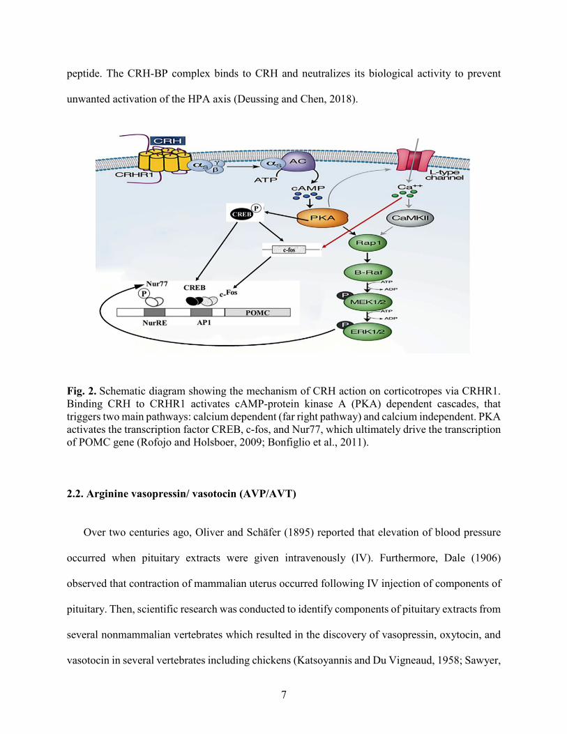
unwanted activation of the HPA axis (Deussing and Chen, 2018).
Fig. 2. Schematic diagram showing the mechanism of CRH action on corticotropes via CRHR1. Binding CRH to CRHR1 activates cAMP-protein kinase A (PKA) dependent cascades, that triggers two main pathways: calcium dependent (far right pathway) and calcium independent. PKA activates the transcription factor CREB, c-fos, and Nur77, which ultimately drive the transcription of POMC gene (Rofojo and Holsboer, 2009; Bonfiglio et al., 2011).
2.2. Arginine vasopressin/ vasotocin (AVP/AVT)
Over two centuries ago, Oliver and Schäfer (1895) reported that elevation of blood pressure
occurred when pituitary extracts were given intravenously (IV). Furthermore, Dale (1906)
observed that contraction of mammalian uterus occurred following IV injection of components of
pituitary. Then, scientific research was conducted to identify components of pituitary extracts from
several nonmammalian vertebrates which resulted in the discovery of vasopressin, oxytocin, and
vasotocin in several vertebrates including chickens (Katsoyannis and Du Vigneaud, 1958; Sawyer,

8
1960). Studies to identify roles of nine peptide (nonapeptide) hormones discovered that AVP was
associated with different physiological functions including blood pressure, anti-diuresis/osmotic
regulation, and reproduction (Sawyer, 1960; Heller and Pickering, 1961). One hormone was
named anti-diuretic hormone, a classical name, due to its ability to regulate extracellular fluid
volume via acting on the collecting duct of nephrons to increase water reabsorption and retention
of body water (Grantham and Burg, 1966). In birds, AVT, a homolog of AVP in mammals, has
been historically recognized as the avian physiological regulator of water balance (Munsick et al.,
1960). Similar to mammals, AVT is primarily synthesized in specialized neurons and transported
to the internal zone of ME to be released from neurosecretory neurons into the neurohypophysis.
From there, it is secreted as active hormone into the general circulation to execute its function in
the kidney so that reabsorption of water occurred to prevent water loss in the body (Skadhauge
and Schmidt-Nielsen, 1967).
Neurons synthesizing AVP/AVT are mainly found in two subpopulations on either side of the
hypothalamus, SO and PVN (Swanson and Sawchenko, 1983). Utilizing IHC methods, AVP-ir
neurons in the rodent brain were identified (Vandesande and Dierickx, 1975). In rodents,
magnocellular AVP-ir was observed in the SO, PVN, medial preoptic area, bed nucleus of the stria
terminalis (BNST), and lateral hypothalamic (LH) area. Most thick terminal fields projecting to
ME originate from perikarya of the SO or PVN. Additionally, a large number of parvocellular
AVP-ir neurons was observed in the suprachiasmatic nucleus (Valesky et al., 2012).
In several avian species, AVT-ir neurons were studied using a specific antibody targeting AVT
and reported occurring in different types of neurons (Goossens et al., 1977; Bons, 1980; Berk et
al., 1982; Tennyson et al., 1985; Kiss et al., 1987; Panzica et al., 1999; Fabris et al., 2004;

9
Montagnese et al., 2015). First, large or magnocellular AVT-ir neurons were identified as the
source of neurohormone release into the peripheral system (Mikami et al., 1978; Mikami, 1986).
Second, another small type of AVP/AVT neuron called parvocellular neurons was detected in the
hypothalamus. Parvocellular neurons project either into the ME where their terminals release
peptides into the portal system to modulate physiological responses (by binding to receptors
located on pituitary cells), or to brain stem and spinal cord to modulate autonomic functions.
AVP/AVT has different functions, such as osmoregulation, blood regulation, reproduction
behavior, and the stress response. AVT neurons were identified in the hypothalamus and extra-
hypothalamic structures in both mammals and birds. Nonetheless, both hypothalamic and extra-
hypothalamic distributions of AVT-ir neurons are presumed to play distinct roles in the physiology
and behavior of birds.
2.2.1. AVP/AVT receptors
AVP/AVT neuropeptides initiate their functions and effects via binding to specific receptors,
G-protein coupled receptors, that are located on the cell membrane and distributed in a variety of
cells including the cardiovascular system, kidney, brain, pituitary and blood platelets. Different
functions of vasopressin receptors have been identified including those in the visceral system and
central nervous system to facilitate physiological as well as behavioral functions (Koshimizu et
al., 2012; De Wied et al., 1984). Over the past decade, four different types of vasotocin receptors
have been identified in vertebrates, namely V1a, V1b, V2 and V3 (oxytocin receptor). These
receptors are conserved between mammalian and avian species throughout evolution (Ocampo et
al., 2012; Yamaguchi et al., 2013). The distributions of AVP receptor subtypes in the CNS show
significant differences among species. In rodents, the V1a and OT receptor subtypes are
10
abundantly expressed receptors in the brain (Johnson et al., 1993; Tribollet et al., 1999).
Furthermore, V2 receptor mRNA has been reported within mammalian brains, particularly, AVP
producing neurons possess the V2 that mediates autocrine role of somatodendritic release of AVP
in rat vasopressin neurons under hypo-osmotic conditions (Sato et al., 2011). The V1b receptor in
the mouse CNS is found most prominently in the hippocampus, cerebral cortex, amygdala,
olfactory bulb, and hypothalamus, including the PVN. Although, the AVP/AVT receptors are
conserved, the second messenger system within the cells varies to a smaller extent depending upon
the receptor subtype. In a variety of cells, the V1aR, V1bR and oxytocin receptor (V3) were found
to have a signal transduction pathway associated with phosphatidylinositol breakdown leading to
calcium signaling (Woods et al., 1986; Hatton et al., 1992; Dayanithi et al., 1996; Cornett et al.,
2003), while V2 receptors are involved in activating adenylate cyclase leading to the release of
cAMP serving as the second messenger.
In birds, the first type of vasotocin receptor identified was the VT1 receptor and found in the
eggshell gland and brain of chickens (Tan et al., 2000). Although, V2 receptors have been
identified in the kidney of mammals (Bankir, 2001) and associated with the regulation of ionic
balance, its function in the avian kidney is unknown. The second receptor type detected in birds
was the VT2 receptor. Based upon its similar sequence to the mammalian V1b receptor gene, it
was suggested to be equivalent to the mammalian V1b receptor (Cornett et al., 2003). Studies with
an antibody to the avian VT2R (V1bR) showed that the receptor protein occurred primarily on
corticotropes in the chicken APit (Jurkevich et al., 2005, 2008). Unlike, mammalian V1b, the avian
V1b receptor has not been detected in the chick brain utilizing IHC (Jurkevich et al., 2005). In
contrast, the avian VT4R was proposed to be homologous to the mammalian V1a, based upon its
specific immunoreactivity shown on corticotropes in the APit and presence in specific neurons as
11
well as in circumventricular organs within the brain of chickens (Selvam et al., 2013; 2015) as
well as songbird in the brains (Leung et al., 2009). The last of the four different types, the avian
VT3 receptor subtype also known as the mesotocin receptor and was proposed to be comparable
to the mammalian oxytocin receptor, due to its presence in the shell gland of birds (Gubrij et al.,
2005). Although VT3 receptors have not been studied in the chicken brain, evidence using in situ
hybridization showed that VT3 receptors are expressed in several brain regions of the white-
throated sparrow (Zonotrichia albicollis) and zebra finch (Taeniopygia guttata) (Leung et al.,
2011).
3. Stress Response and CORT release.
Stress was introduced by Hans Selye as “the triphasic general adaptation syndrome (GAS)”.
In the response to stress, there are many stages: the first stage is the initial alarm reaction where
the body prepares itself for “fight or flight”; the second is the stage of resistance involving
adaptation to the stressor; and exhaustion is the last stage which might lead to an organism’s death
(Wang et al., 2017). While investigating the endocrinology of stress, Selye (1937) was one of first
scientists who recognized the relationship between stress and adrenocortical activation. The HPA
axis sensitivity and activity depend on the type, duration, and intensity of stressors (Pacák and
Palkovits, 2001) and predefined by exposure to CORT (Buckingham, 2006).
Early studies showed that the HPA axis activation was associated with hypothalamic factors.
First, it was found that arginine vasopressin (AVP)/ vasotocin (AVT) enhanced ACTH release
from APit cells with lower efficiency compared with other hypothalamic or pituitary stalk extracts
(Gillies et al., 1978; 1982). Upon the discovery of a 41 amino acids peptide hormone in the early
1980’s (Vale et al., 1981), the major increase of ACTH release by hypothalamic extracts was

12
attributed for CRH. Eventually, it was found that CRH and AVP/AVT work synergistically to
augment the release of ACTH (Castro et al., 1986), and are present in a sub-population of neurons
within the medial parvocellular division of the PVN (Sawchenko et al., 1992). Thereafter,
immunohistochemical (IHC) data showed AVP and CRH co-localization within some
parvocellular neurons in the PVN and about 50% of the cells contain both CRH and AVP within
the cell bodies. Similarly, the axon terminals of parvocellular neurons displayed co-localization of
the two peptides in the external zone of the median eminence (ME) (Sawchenko et al., 1984;
Whitnall et al., 1985; Antoni, 1993; Whitnall, 1993, Aste et al., 1998). Later, co-localization of
both neuropeptides in neurons was reported in birds (Kuenzel and Jurkevich, 2010). In vertebrates,
AVP/AVT and CRH produced by parvocellular neurons are released and transported to the APit
to trigger ACTH release from corticotropes (Antoni, 1993). However, CRH and AVT genes within
parvocellular neurons have different sensitivities for stress. For example, ether inhalation stress, a
potent stressor, triggers expression of hnCRH primary transcripts (hn-heteronuclear) as early as
5m followed by hnAVP expression at 1h (Kovács and Sawchenko, 1996; Ma et al., 1997).
Likewise, temporal mRNA expression of CRH and AVP following stressors were also reported in
a number of studies (Lightman and Young, 1989;Baitanusz et al., 1993; Nagarajan et al., 2017a).
Parvocellular neurons producing CRH and AVP/AVT are controlled by distinct cells in other brain
areas (described in 4. Section below).
In the anterior pituitary, CRH and AVP/AVT act on corticotropes within APit via their
respective receptors (CRH – CRHR1/CRHR2 and AVT – V1a/V1b) to stimulate synthesis and
release of ACTH into the systemic circulation. Upon binding of CRH and AVP/AVT to their
receptors, dimerization of CRHR1 and V1b receptors occurs on corticotropes of mammals (Young
et al., 2007) and birds (Mikhailova et al., 2007). The dimerization of CRHR1 and V1bR provides

13
structural evidence that CRH and AVP/AVT are working together in response to stress stimuli not
only at the level of hypothalamus but also at the levels of the APit. Activation of receptors by their
ligands leads to stimulations of cyclic adenosine monophosphate (cAMP) dependent pathway
causing the increase of proopiomelanocortin (POMC) synthesis. POMC is a polypeptide (241
amino acid residues) termed a prohormone because it contains multiple peptide sequences which
when processed results in ACTH, α- melanocyte stimulating hormone (α-MSH), β--lipotropin, β-
-endorphin, and some other unknown fragments. Once ACTH, a 39 amino acid, reaches the adrenal
glands via the general circulation, it binds to its receptor, melanocortin receptor 2 (MC2), on the
zona fasciculata (mammals) or adrenocortical cell (birds) causing cortisol/ corticosterone release
(Fig. 3) , the end product of the HPA axis, produced by the adrenal glands (Carsia, 2015).
Fig. 3. Side view (Sagittal view) of the HPA axis in avian species adopted from (Nagarajan et al., 2017a). The NHpC and PVN have neurons (CRH and AVT type) that project to the median eminence, a structure just dorsal to the anterior pituitary. The anterior pituitary releases ACTH and within the avian adrenal gland, the interrenal tissue produces the product, corticosterone (CORT), transported throughout the body by the cardiovascular system.

14
4. Neural pathways and inputs for PVN
The hypothalamic PVN is an integrative site linking autonomic and neuroendocrine systems
during stress conditions. Stress stimuli are either transmitted directly to the PVN or integrated by
the limbic system and conveyed to the parvocellular neurons located in the PVN. An immediate
response for systemic physical and metabolic stressors utilizes monosynaptic ascending pathways
projections directly from the brain stem and spinal cord to the PVN (Herman et al., 2003). In
addition to direct projection to the PVN, neurons within brain stem project and also interact with
other limbic areas within the brain such as, the dorsal raphe, dorsomedial hypothalamic nucleus
(DMH), and forebrain. In addition to monosynaptic ascending pathways, complex polysynaptic
pathways have been identified that activate or inhibit neurons within the PVN arise from different
brain areas such as the prefrontal cortex (PFC), hippocampus, amygdala, and BNST. Activation
of afferent neural pathways terminating on the PVN during stress results in rapid release of
neuropeptides followed by an increase of their transcription and de novo synthesis of peptides. For
example, a rapid activation of neural afferent pathways is caused by acute stress leading to rapid
release of CRH followed by increasing CRH transcription and de novo synthesis of CRH. Also, an
increasing of AVP expression in CRH neurons was reported during stress and adrenalectomy
(Whitnall, 1989). Furthermore, the fast release of CRH and AVP/AVT is followed by a rapid
increase of gene transcription documented by steady-state mRNA level elevation at 4h after acute
stress. In stress studies, CRH gene expression precedes or is followed by AVP gene activation
(Kovács and Sawchenko, 1996; Ma et al., 1997; Herman et al., 2003).
Parvocellular neurons located in the PVN are the main neurons responsible for the stress
response and regulated by several inputs such as, noradrenergic, glutamatergic, GABAergic, and

15
peptidergic neural pathways (Aguilera and Liu, 2012). The PVN receives abundant ascending
adrenergic or noradrenergic projections that innervate parvocellular neurons originating from the
brain stem (Cunningham and Sawchenko, 1988; Füzesi et al., 2007). Major ascending
noradrenergic neurons originate from the nucleus tractus solitarius (NTS) and locus coeruleus
(LC). Additionally, adrenergic α1 and α2 receptors were identified on the CRH neurons and
parvocellular neurons (Cummings and Seybold,, 1988; Little et al., 1992). Electrical stimulation
of the ascending noradrenergic bundle and intra-PVN or intracerebroventricular application of
norepinephrine activate the HPA axis and cause a significant increase in the CRH gene expression
and CORT concentration. However, stress-induced ACTH and CORT releases were reduced after
administration of an α1-adrenoceptor antagonist in the PVN (Plotsky, 1987; Itoi, 1994; Itoi et al.,
1999; Helmreich et al., 2001; Cole and Sawchenko, 2002). Furthermore, despite an intra-PVN
glutamate injection, an impaired HPA response to stress, particularly, decreased ACTH and CORT
response occurred when noradrenergic inputs to the PVN were reduced (Feldman and Weidenfeld,
1997; Bienkowski and Rinaman, 2008). However, an activation of noradrenergic terminals in the
PVN resulted in CORT hypersecretion (Laorden et al., 2002). The noradrenergic afferents are
positively regulating the HPA axis and are activated by systemic sensory stimulation or
physiological stress signals, such as immune system activation and hypoglycemia (Ritter et al.,
2003). In addition to direct projections to the PVN and synapses with CRH neurons, noradrenergic
and adrenergic neurons within the brain stem project and interact with other limbic areas within
the brain such as, dorsal raphe; that regulates serotoninergic activity, dorsomedial hypothalamic
nucleus (DMH); which control autonomic activity, and the forebrain.
In addition to the excitatory noradrenergic inputs to the PVN, parvocellular neurons within the
PVN receive another excitatory input from glutamatergic neurons. Glutamatergic inputs to the

16
PVN originate from intrahypothalamic glutamatergic interneurons or the peri-PVN area (Boudaba
et al., 1997; Daftary et al., 1998, 2000) to control the activity of neuroendocrine responses
(Boudaba et al., 1997; Herman et al., 2004; Iremonger et al., 2010). All main subtypes of the
ionotropic glutamatergic receptors were found within or around the PVN (Day et al., 1999;
Herman et al., 2000). It has been demonstrated that an enhanced secretion of ACTH and CORT
was reported when glutamate was injected directly into the PVN (Darlington et al., 1989; Feldman
and Weidenfeld, 1997). However, a weaker activation of the HPA axis following restraint stress,
as measured by plasma corticosterone level, was observed after bilateral injection of a
glutamatergic receptor antagonist (Ziegler and Herman, 2000).
Unlike the noradrenergic and glutamatergic inputs, parvocellular neurons in the PVN receive
inhibitory inputs which are GABAergic neurons that originate from interneurons located in the
surrounding area of the PVN (Boudaba et al., 1996; Herman et al., 2002). The GABA interneurons
in the peri-PVN region orchestrate the information from limbic inputs originating mainly from
several brain areas such as, the hippocampus-ventral subiculum, prefrontal cortex, medial
amygdala, lateral septum, paraventricular thalamus, and suprachiasmatic nucleus. The other
proportion of the GABAergic terminals within the PVN originates from limbic and diencephalic
regions such as the dorsomedial and medial preoptic nucleus and the bed nucleus of the stria
terminalis (Herman et al., 2002). It was reported that in vivo blockage of GABA A receptors within
the PVN caused a significant increase of CRH transcription resulted in the increase of plasma
glucocorticoid levels displaying that CRH neurons are under the inhibitory effects of GABAergic
inputs (Cole and Sawchenko, 2002). Furthermore, decreasing of ACTH secretion in response to
an acute stress was reported after bilateral injection off bicuculline which is a GABA A receptor
agonist into the PVN. Hence, the activation of the HPA axis when triggered by stress is inhibited

17
by GABAergic inputs (Stotz-Potter et al., 1996). Other neurons in the NTS express other
neuropeptides, such as neuropeptide (NPY), glucagon-like peptide 1 (GLP-1), inhibin-β,
somatostatin and enkephalin, that are able to influence parvocellular neurons and HPA axis
activities (Wahlestedt et al., 1987; Suda et al., 1993; Ziegler and Herman, 2000; Nakade et al.,
2007).
5. Neurotrophic factors involved of stress response
Stress results in a wide range of effects that influence many different factors, such as CREB
and brain-derived neurotrophic factor (BDNF) in the hippocampus and other brain regions. BDNF,
246 amino acid (27.715 kDa), is a neurotrophin widely expressed in the mammalian brain (Hofer
et al., 1990) and was initially purified from mammalian brains based on its ability to promote
neuronal survival in vitro (Barde et al., 1982). It is expressed highly in the hippocampus followed
by cortex, amygdala, and hypothalamus. Furthermore, it can be observed outside the brain in the
thymus, liver, spleen, heart, and lung (Pruunsild et al., 2011). BDNF gene is composed of 11 exons
and contains 9 functional promoters located on chromosome 11p13 producing 24 different
transcripts that are all translated to the same mature protein (Pruunsild et al., 2011). BDNF
transcription is regulated by many elements, including estrogens, promoter-specific methylation,
and the c-AMP response element-binding protein (CREB) (Sohrabji et al., 1995; Tao et al., 1998;
Aid et al., 2007). BDNF is essential to many facets of CNS functions, such as, neuronal
development and survival, migration, dendritic arborization, synaptic plasticity, and cognitive
function (Greenberg et al., 2009). BDNF has been able to prevent the negative effects of oxidative,
metabolic and excitotoxic stress on neurons in experimental models. Dysregulation of BDNF

18
signaling has been shown in several neurodegenerative disorders (Mattson et al., 2004; Marini et
al., 2007).
Differences in levels of BDNF expression have been investigated during social defeat
paradigm in many species. For this form of stress, BDNF expression decreased significantly in the
hippocampus and piriform cortex of golden hamster contributing to atrophy and decreased
neurogenesis (Arendt et al., 2012). However, an acute increase in BDNF expression was reported
in the PFC, nucleus accumbens (nAcc), amygdala, and ventral tegmental area (VTA) of rats
(Nikulina et al., 2012). Furthermore, social isolation induced a decrease in BDNF protein levels in
the midbrain, hypothalamus, PFC, and hippocampus in rats and mice (Berry et al., 2012). In rodent
model studies, expression of BNDF has been measured during restraint stress and found an
increase in BDNF mRNA preceding its protein level increase in the hippocampus (Marmigère et
al., 2003). In contrast, other studies demonstrated that the protein and mRNA levels decreased
during acute stress (Ueyama et al., 1997; Franklin and Perrot-Sinal, 2006; Mazon et al., 2006; Lee
et al., 2008; Roth and Sweatt, 2011). Researchers reported the increase in the BDNF mRNA
relative levels during stress compared to unstressed control (Nair et al., 2007; Alboni et al., 2011).
Effects of immobilization were not uniform across all brain regions. Specifically, some groups
reported a transient upregulation of BDNF in the hypothalamus (Rage et al., 2002), but others
demonstrated no effect of this form of stress on BDNF levels in the basolateral amygdala or PFC
(Roth and Sweatt, 2011). The results suggest a complex relationship between the type and duration
of stressors and do not indicated a clear results or exact conclusion on the expression of BDNF or
specific structures involved in the response.

19
In addition to BDNF, another important neurotrophic factor called glial-derived neurotrophic
factor (GDNF) was discovered in 1993 and found that GDNF is essential for midbrain
dopaminergic neurons. GDNF was initially identified and produced as proGDNF, 211 amino acid,
that is cleaved mature, active form by endoproteolytic enzymes into the 134 amino acid (Lin et al.,
1993). Uchida et al. (2011) found that individuals who cannot upregulate GDNF during stress
exhibit anxiety and avoidance of social interactions, possibly due to the negative consequences of
chronic stress on the dopaminergic circuits. Later, Buhusi et al. (2016) demonstrated that an
increase of vulnerability to stress was observed in GDNF heterozygous mice manifested by
alterations in their executive functions. Furthermore, intraventricular administration of GDNF
revealed its role in weight loss (Manfredsson et al., 2009). The mechanism for that results was due
to the ability of GDNF to phosphorylate an extracellular signal-regulated kinase (p-ERK) in a
small population of CRH neurons located specifically in the hypothalamus PVN. Activation of
these hypothalamic CRH via GDNF might enhance hypothalamo– pituitary–adrenal axis.
However, less is known about the mRNA expression pattern of GNDF during stress response.
6. Peripheral regulation of stress response
CRH neurons show different patterns of activity under resting and stress conditions. The
activity of CRH neurons is regulated by several stimulatory and inhibitory neural pathways as well
as hormonal pathways that originate peripherally. Several peripheral factors such as, sex steroids,
glucocorticoids, peptides, and cytokines, can affect the stress response through modulating CRH
neurons either positively or negatively. For example, CRH transcription activation depends mainly
on cAMP/PKA pathways occurred when phosphorylated cAMP response elements binding
proteins (pCREB) bind to the cAMP response element (CRE) of the CRH promoter (Seasholtz et

20
al., 1988). Moreover, binding sites of immediate early genes such as c-fos and NGFI-B (nerve
growth factor inducing factor- B) have been identified in the promoter regions of CRH and AVP
(Chan et al., 1993) and associated with the differences in expression patterns of the neuropeptides.
Also, it was found that CRH autoregulates itself via its receptor CRHR1, which is coupled to
adenylate cyclase (De Goeij et al., 1991; Di et al., 2003). CRHR1 activation by locally secreted
CRH would provide a source of cyclic AMP, which is necessary for activation of CRH
transcription. Also, during stress response, the release of pituitary adenylate cyclase activating
polypeptide (PACAP) in the brain could provide an additional cAMP stimulator in the CRH
neuron via PACAP innervation contacting CRH perikarya (Grinevich et al., 1997; Légrádi et al.,
1998). CRH neurons inhibition is essential for homeostasis and health. It is well known that CRH
transcription is inhibited by in vivo or in vitro glucocorticoids. However, the molecular mechanism
is not fully understood. Further, evidence showed that AVP and OXT could inhibit CRH secretion
and expression and attenuate c-fos mRNA in forebrain regions involved in the regulation of the
HPA axis, yet the mechanism has to be understood ( Plotsky et al., 1984; Windle et al., 1997, 2004;
Neumann et al., 2000; Ochedalski et al., 2007). The influence of different types of stressors is still
unidentified in the avian species because of a mixed population of magnocellular and parvocellular
neurons in the PVN. Furthermore, the stress response in the avian species regarding the activity of
CRH and AVT neurons in the PVN needs further research.
6.1. Corticosteroid and glucocorticoid receptors (GRs)
The final product of the HPA axis is a corticosteroid which is one of a class of steroid hormones
secreted by adrenal glands. Low density lipoprotein is a substrate that enters cells and is broken
down to a release of free cholesterol in the cytoplasm. Cholesterol, the precursor for corticosteroid,

21
enters a series of enzymatic processes in the mitochondria and endoplasmic reticulum leading to
steroidogenesis. Corticosteroids are not stored in cells nor packed into vesicles due to their
lipophilic nature. Therefore, they pass through the cell membrane into the blood and reversibly
bind to a carrier protein called corticosteroid- binding globulin (CBG) that carries them to target
tissues to induce their effects. Corticosteroids have essential roles in maintaining homeostasis in
response to stress, immune response, electrolyte balance, carbohydrate metabolism, emotion, and
cognition. Glucocorticoid refers to the product of glucose metabolism and synthesis within the
adrenal cortex that produces steroid. Glucocorticoids effects are mediated by two receptors, which
are mineralocorticoids (MRs) and glucocorticoids (GRs). The MRs have higher affinity for
corticosteroid than the GRs. Therefore, most MRs are fully occupied under basal conditions (Karst
et al., 2005). The MRs are highly expressed in the hippocampus, lateral septum, and brain stem
motor nuclei, and moderately expressed in the amygdala, PVN and locus coeruleus. However, GRs
are ubiquitously expressed on neurons and glia throughout the brain, particularly, in the
hippocampus, lateral septum, PVN, and pituitary (Joëls and Baram, 2009).
There are two ways for glucocorticoids to exert their effects on the cells, which are genomic
and nongenomic actions. The genomic way occurs after binding of a corticosteroid to the
cytoplasmic GR, the activated receptor is translocated to the nucleus and binds to a specific DNA
sequence located in the promotor region of the targeted gene known as the glucocorticoid response
element (GRE), and subsequent activation or repression of de novo synthesis of mRNA and protein
production (Hinz and Hirschelmann, 2000). Therefore, an activated GR is regulating targeted
genes by acting as a transcription factor or interacting with other transcription factors. In contrast,
the non-genomic effects of glucocorticoids occur when glucocorticoids bind to the membrane
associated GR. Unlike the slow genomic effect of glucocorticoids, the non-genomic effect is very

22
fast and usually affects neurons by changing synaptic transmission, occurs within minutes, and
induced by conjugated glucocorticoids that do not permeate the cell membrane. The non-genomic
effects of glucocorticoids do not alter gene transcription and protein synthesis (Keller-Wood,
2015).
During stressful situation, corticosteroids bind GRs in targeted tissues to enhance the body
utilizing stored energy more efficiently and increase the organisms’ performance. At the same
time, corticosteroids target GRs located in the brain and pituitary to inhibit corticosteroid release
and to prevent long-term exposure to high levels of corticosteroid. The main sites for negative
feedback are hippocampus, hypothalamus, and pituitary. The negative feedback occurs at the
hippocampus to shut down excitation signals, while it limits ACTH at the pituitary and inhibits
CRH and AVP/AVT release at the PVN (Buckingham, 2006). In addition, the presence of GRs on
CRH neurons indicate that glucocorticoids regulate CRH neurons directly via GRs (Uth et al.,
1988). Within the HPA axis, the expression of the POMC gene in the APit and the CRH gene in
the PVN parvocellular neurons is downregulated during the stress response by high CORT
utilizing GRs. Furthermore, it has been found that corticosterone and dexamethasone are able to
regulate glutamatergic and GABAergic inputs to the PVN via the non-genomic way in a process
called glucocorticoid-induced suppression of excitation. Glucocorticoid-induced suppression of
excitation can be found in CRH, VP and OT neurons (Di et al., 2003; 2009).
7. Rationale and aims of the dissertation
Maintenance of internal milieu in the presence of real or perceived challenges is carried out
during the physiological stress response. Sustaining homeostasis of an organism is conducted by
interaction of different body systems that cause the release of different biochemical molecules.

23
Even though there are two major stress response systems in mammals, the sympatho-
adrenomedullary and HPA axis response, the nervous system plays critical roles in both systems.
Several types of neurons located within different brain nuclei are activated in response to stressors.
In particular, parvocellular neurons within the hypothalamic PVN that co-express CRH and
AVP/AVT play essential roles in the regulating the HPA axis. In addition to their long-established
role of parvocellular neurons as the main regulators of the HPA axis, CRH and AVT expressing
neurons were also identified in several brain structures (Deussing and Chen, 2018, De Souza et
al., 1985), and they act as a neurotransmitter or neuromodulator at diverse ‘extra-hypothalamic’
sites within the central nervous system (CNS) to induce rapid autonomic and behavioral responses
to a stressor (Dunn and Berridge, 1990; Reghunandanan et al., 1998; Van Bockstaele and
Valentino, 2009). Similarly, in the avian species, CRH neurons have been observed in many brain
structures (Richard et al., 2004). Interestingly, one of the brain structures containing CRH neurons
was discovered in the septum of birds in a structure called the NHpC (Nagarajan et al., 2014).
Studies thereafter suggested an interaction of the CRH neurons in the NHpC with CRH and AVT
neurons in the PVN regarding their roles in the regulation of the avian stress response (Nagarajan
et al., 2017a, b). Furthermore, CRH and AVP/AVT receptors within different levels of the HPA
axis play a critical role in the stress response. For instance, receptors located at the level of APit
connect between nervous and endocrine systems to regulate ACTH release. Therefore, the major
drivers of neuroendocrine stress response, CRH and AVP/AVT neurons and their receptors as well
as BDNF, will be in the focus of the current study within two brain structures, the NHpC and PVN.
The former is a septal extra-hypothalamic nucleus, while the latter is the well-known, major
hypothalamic nucleus regulating stress. The main focus of the research will address CRH neurons
within the NHpC. Choosing the NHpC enables the structure to serve as a model for any other
24
extra-hypothalamic brain structure that contains CRH neurons. One reason is that the NHpC is
located right above the hypothalamic PVN and separated from the PVN by the anterior
commissure (AC). Another reason for the NHpC to be an excellent candidate is that an activation
of the c-fos gene (an early activated neuron marker) in the NHpC was observed during a stress
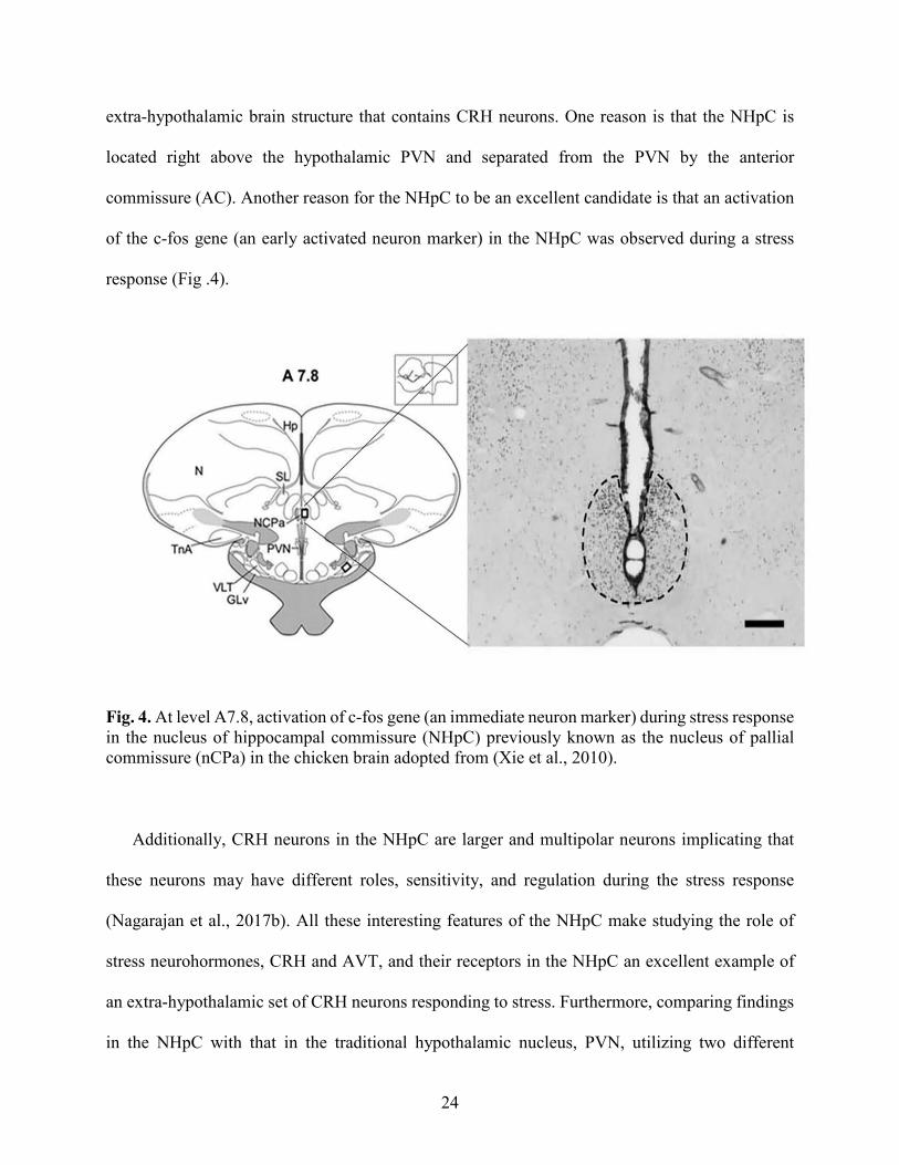
response (Fig .4).
Fig. 4. At level A7.8, activation of c-fos gene (an immediate neuron marker) during stress response in the nucleus of hippocampal commissure (NHpC) previously known as the nucleus of pallial commissure (nCPa) in the chicken brain adopted from (Xie et al., 2010).
Additionally, CRH neurons in the NHpC are larger and multipolar neurons implicating that
these neurons may have different roles, sensitivity, and regulation during the stress response
(Nagarajan et al., 2017b). All these interesting features of the NHpC make studying the role of
stress neurohormones, CRH and AVT, and their receptors in the NHpC an excellent example of
an extra-hypothalamic set of CRH neurons responding to stress. Furthermore, comparing findings
in the NHpC with that in the traditional hypothalamic nucleus, PVN, utilizing two different

25
stressors, feed deprivation and immobilization stress, would be intriguing to establish and
understand the importance of extra-hypothalamic CRH neurons in an avian species as a model for
those who may be examining the stress pathway in the humans or other vertebrate species.
Therefore, the dissertation will explore the role of CRH neurons in the NHpC to determine if
these neurons are involved in the regulation of the stress response in birds as a model for the role
of extra-hypothalamic CRH neurons. The research also will determine the sequence of structure
activation involved in the neuroendocrine regulation of the avian stress response as well as provide
further evidences whether the NHpC is a structure involved in the traditional HPA axis.
Furthermore, the dissertation will investigate the relationship between CRH and its receptors in
different brain structures, and it will help understanding the role of BNDF in the stress response
within different brain structures during different stress trials, feed deprivation versus
immobilization stress. The data from this dissertation will improve the understanding of the role
of extra-hypothalamic structures, particularly, the NHpC in the stress response.

26
References
Aguilera, G., Liu, Y. 2012. The molecular physiology of CRH neurons. Front. Neuroendocrinol. 33(1), 67 – 84.
Aid, T., Kazantseva, A., Piirsoo, M., Palm, K., Timmusk, T. 2007. Mouse and rat BDNF gene structure and expression revisited. J. Neurosci. Res. 85 (3), 525 – 35.
Alboni, S., Tascedda, F., Corsini, D., Benatti, C., Caggia, F., Capone, G., Barden, N., Blom, J., Brunello, N., 2011. Stress induces altered CRE/CREB pathway activity and BDNF expression in the hippocampus of glucocorticoid receptor-impaired mice. Neuropharmacology 60 (7–8), 1337 – 46.
Amano, M. 2016. Corticotropin-releasing hormone. In: Takei Y, Ando H, Tsutsui K, eds. Handbook of Hormones: Comp Endocrinology for Basic and Clinical Research. 1st ed. Oxford, United Kingdom: Academic Press; 23-5.
Antoni, F. A. 1993. Vasopressinergic control of pituitary adrenocorticotropin secretion comes of age. Front. Neuroendocrinol. 14 (2), 76 –122.
Arai, M., Assil, I. Q., Abou-Samra., A. B., 2001. Characterization of three corticotropin-releasing factor receptors in catfish: a novel third receptor is predominantly expressed in pituitary and urophysis. Endocrinology. 142 (1), 446 – 54.
Arendt, D. H., Smith, J. P., Bastida, C. C., Prasad, M. S., Oliver, K. D., Eyster, K. M., Summers, T. R., Delville, Y., Summers, C. H., 2012. Contrasting hippocampal and amygdalar expression of genes related to neural plasticity during escape from social aggression. Physiol. Behav. 107(5), 670 –79.
Aste, N., Balthazart, J., Absil, P., Grossmann, R., Mülhbauer, E., Viglietti-Panzica, C., Panzica., G. C. 1998. Anatomical and neurochemical definition of the nucleus of the stria terminalis in japanese quail (Coturnix Japonica). J. Comp.Neurol. 396 (2), 141 – 57.
Baitanusz, V., Aubry, J.M., Jezova, D., Baffi, J., Kiss. J.Z., 1993. Upregulation of vasopressin mRNA in paraventricular hypophysiotrophic neurons after acute immobilization stress. Neuroendocrinology 58 (6), 625 – 9.
Bankir, L. 2001. Antidiuretic action of vasopressin: quantitative aspects and interaction between V1a and V2 receptor-mediated effects. Cardiovasc. Res. 51(3), 372 – 90.
Barde, Y. A., Edgar, D., Thoenen, H., 1982. Purification of a new neurotrophic factor from mammalian brain. EMBOJ. 1 (5), 549 – 53.
Berk, M. L., Reaves, T. A., Hayward, J. N., Finkelstein. J. A., 1982. The localization of vasotocin and neurophysin neurons in the diencephalon of the pigeon, Columba Livia. J. Comp. Neurol. 204 (4), 392 – 406.

27
Berry, A, Bellisario, V., Capoccia, S., Tirassa, P., Calza, A., Alleva, E., Cirulli, F., 2012. Social Deprivation stress is a triggering factor for the emergence of anxiety- and depression-like behaviours and leads to reduced brain BDNF levels in C57BL/6J mice. Psychoneuroendocrinology 37 (6), 762 –72.
Bienkowski, M. S., Rinaman. L., 2008. Noradrenergic inputs to the paraventricular hypothalamus contribute to hypothalamic-pituitary-adrenal axis and central fos activation in rats after acute systemic endotoxin exposure. Neuroscience 156 (4), 1093 – 102
Bonfiglio J. J., Inda C., Refojo, D., Holsboer, F., Arzt, E., Silberstein, S., 2011. The corticotropin-releasing hormone network and the hypothalamic-pituitary-adrenal axis: molecular and cellular mechanisms involved. Neuroendocrinology 94, 12–20
Bons, N.. 1980. The topography of mesotocin and vasotocin systems in the brain of the domestic mallard and japanese quail: immunocytochemical identification. Cell Tissue Res. 213 (1), 37 – 51.
Boorse, G. C., Denver, R. J. 2006. Widespread tissue distribution and diverse functions of corticotropin releasing factor and related peptides. Gen. Comp. Endocrinol. 146 (1), 9 –18.
Boudaba, C., Schrader, L. A., Tasker, J. G.1997. Physiological evidence for local excitatory synaptic circuits in the rat hypothalamus. J. Neurophysiol. 77 (6), 3396 – 400.
Boudaba, C., Szabó, K., Tasker, J. G., 1996. Physiological mapping of local inhibitory inputs to the hypothalamic paraventricular nucleus. J. Neurosci. 16(22), 7151– 60.
Bu, G., Fan, J., Yang, M., Lv, C., Lin, Y., Li, J., Meng, F., Du, X., Zeng, X., Zhang, J., Li, J., Wang, Y., 2019. Identification of a novel functional corticotropin-releasing hormone (CRH2) in chickens and its roles in stimulating pituitary TSHb expression and ACTH secretion. Front. Endocrinol. 10, 595.
Buckingham, J.C., 2006. Glucocorticoids: exemplars of multi-tasking. Br. J. Pharmacol. 147(SUPPL. 1), 258 – 68.
Buhusi, M., Olsen, K., Yang, B. Z., Buhusi, C. V., 2016. Stress-Induced Executive Dysfunction in GDNF-Deficient Mice, A Mouse Model of Parkinsonism. Frontiers in behavioral neuroscience, 10, 114.
Carsia, R. V. 2015. Chapter 26 - Adrenals. In: Scanes, C.G. (Ed.), Sturkie's Avian Physiology (Sixth Edition). Academic Press, San Diego, pp. 577-611.
Castro, M. G., Estivariz, F. E., Iturriza, F. C. 1986. The regulation of the corticomelanotropic cell activity in Aves-II. Effect of various peptides on the release of ACTH from dispersed, perfused duck pituitary cells. Comp. Biochem. Physiol. A Comp. Physiol. 83 (1), 71– 5.
Chan, R. K. W., Brown, E. R., Ericsson, A., Kovacs, K. J., Sawchenko, P. E. 1993. A comparison of two immediate early genes, c-Fos and NGFI-B, as markers for functional activation in stress related neuroendocrine circuitry. J. Neurosci. 13 (12), 5126–38.

28
Cole, R. L., Sawchenko, P. E., 2002. Neurotransmitter regulation of cellular activation and neuropeptide gene expression in the paraventricular nucleus of the hypothalamus. J. Neurosci. 22 (3), 959 – 969.
Cornett, L. E., Kirby, J. D., Vizcarra, J. A., Ellison, J. C., Thrash, J., Mayeux, P. R., Crew, M. D., Jones, S. M., Ali, N., Baeyens, D. A., 2003. Molecular cloning and functional characterization of a vasotocin receptor subtype expressed in the pituitary gland of the domestic chicken (Gallus domesticus): avian homolog of the mammalian V1b-vasopressin receptor. Regul. Pept. 110 (3), 231 – 9
Coste, S. C., Kesterson, R. A. , Heldwein, K. A., Stevens, S. L., Heard, A. D., Hollis, J. H. , Murray, S. E., Hill, J. K., Pantely, G. A., Hohimer, A. R., Hatton, D. C., Philips, T. J., Finn, D. A., Low, M. J., Rittenberg, M. B., Stenzel, P., Stenzel-Poore, M. P., 2000. Abnormal adaptations to stress and impaired cardiovascular function in mice lacking corticotropin-releasing hormone receptor-2. Nat Genet 24:403–409
Crespi, E. J., Denver, R. J., 2004. Ontogeny of corticotropin releasing factor effects on locomotion and foraging in the Western spadefoot toad (Spea hammondii). Horm. Behav. 46 (4), 399 – 410.
Crespi, E. J., Vaudry, H., Denver, R. J., 2004. Roles of corticotropin releasing factor, neuropeptide Y and corticosterone in the regulation on food intake in xenopus laevis. J. Neuroendocrinol. 16 (3), 279 – 88.
Croiset, G., Nijsen, M. J., Kamphuis, P. J. 2000. Role of corticotropin-releasing factor, vasopressin and the autonomic nervous system in learning and memory. Eur. J. Pharmacol. 405 (1-3), 225 – 34.
Cummings, S., Seybold, V., 1988. Relationship of alpha-1- and alpha-2-adrenergic-binding sites to regions of the paraventricular nucleus of the hypothalamus containing corticotropin-releasing factor and vasopressin neurons. neuroendocrinology 47 (6), 523 – 32.
Cunningham, E. T., Sawchenko. P. E., 1988. Anatomical specificity of noradrenergic inputs to the paraventricular and supraoptic nuclei of the rat hypothalamus. J. Comp. Neurol. 274 (1), 60 – 76.
Daftary, S. S., Boudaba, C., Szabó, K., Tasker, J. G. 1998. Noradrenergic excitation of magnocellular neurons in the rat hypothalamic paraventricular nucleus via intranuclear glutamatergic circuits. J. Neurosci. 18 (24), 10619 – 28.
Daftary, S. S., Boudaba, C., Tasker, J. G., 2000. Noradrenergic regulation of parvocellular neurons in the rat hypothalamic paraventricular nucleus. Neuroscience 96 (4), 743 – 51.
Dale, H. H. 1906. On Some Physiological Actions of Ergot. J. Physiol. 34(3), 163–206.
Darlington, D. N., Miyamoto, M., Keil, L. C., Dallman, M. F.1989. Paraventricular stimulation with glutamate elicits bradycardia and pituitary responses. Am. J. Physiol. 256 (1 Pt 2), R112 – 9.
29
Day, H. E., Campeau, S. W., Watson, S. J. Jr, Akil, H., 1999. Expression of α(1b) adrenoceptor mRNA in corticotropin-releasing hormones-containing cells of the rat hypothalamus and its regulation by corticosterone. J. Neurosci. 19(22), 10098 – 106.
Dayanithi, G., Widmer, H., Richard, P.1996. Vasopressin induced intracellular Ca2+ increase in isolated rat supraoptic cells. J. Physiol., 490 (3), 713 – 27.
De Goeij, D. C. E., Kvetnansky, R., Whitnall, M. H., Jezova, D., Berkenbosch, F., Tilders, F. J. H., 1991. Repeated stress-induced activation of corticotropin-releasing factor neurons enhances vasopressin stores and colocalization with corticotropin-releasing factor in the median eminence of rats. Neuroendocrinology 53 (2), 150 – 9.
De Kloet, Ron, E., Joëls, M., Holsboer, F., 2005. Stress and the brain: from adaptation to disease. Nat. Rev. Neurosci. 6 (6), 463 – 75.
De Souza, E. B., Grigoriadis. D. E., 2002. Corticotropin-releasing factor: physiology, pharmacology and role in central nervous system disorders. Psychoneuroendocrinology 20 (8), 789 – 819.
De Souza, E. B., Insel, T. R., Perrin, M. H., Rivier, J., Vale, W. W., Kuhar, M. J., 1985. Corticotropin-releasing factor receptors are widely distributed within the rat central nervous system: an autoradiographic study. J. Neurosci. 5 (12), 3189 – 203.
De Wied, D., Gaffori, O., Van Ree, J. M., De Jong, W., 1984. Central target for the behavioural effects of vasopressin neuropeptides. Nature 308 (5956), 276 – 8.
Deussing, J. M., Chen, A., 2018. The corticotropin-releasing factor family: physiology of the stress response. Physiol. Rev. 98 (4), 2225 – 86.
Di, S., Malcher-Lopes, R., Halmos, K., Tasker, J. G. 2003. Nongenomic glucocorticoid inhibition via endocannabinoid release in the hypothalamus: a fast feedback mechanism. J. Neurosci. 23 (12), 4850 – 57.
Di, S., Maxson, M. M., Franco, A., Tasker. J.G. 2009. Glucocorticoids regulate glutamate and GABA synapse-specific retrograde transmission via divergent nongenomic signaling pathways. J. Neurosci. 29 (2), 393 – 401,
Dunn, A. J., Berridge, C. W. 1990. Physiological and behavioral responses to corticotropin releasing factor administration: is CRF a mediator of anxiety or stress responses? Brain Res. Brain Res. Rev. 15(2), 71–100.
Fabris, C., Ballarin, C., Massa, R., Granato, , Fabiani, O., Panzica, G. C., Cozzi, B., 2004. The vasotocinergic system in the hypothalamus and limbic region of the budgerigar (Melopsittacus undulatus). Eur. J. Histochem. 48 (4), 367 –72.
Feldman, S., Weidenfeld, J., 1997. Hypothalamic mechanisms mediating glutamate effects on the hypothalamo-pituitary-adrenocortical axis. J. Neural. Transm., 104 (6-7), 633 – 42.

30
Franklin, T. B., Perrot-Sinal. T. S., 2006. Sex and ovarian steroids modulate brain-derived neurotrophic factor (BDNF) protein levels in rat hippocampus under stressful and non-stressful conditions. Psychoneuroendocrinology, 31(1), 38 – 48.
Füzesi, T., Wittmann, G., Liposits, Z., Lechan, R. M., Fekete, C., 2007. Contribution of noradrenergic and adrenergic cell groups of the brainstem and agouti-related protein-synthesizing neurons of the arcuate nucleus to neuropeptide-Y innervation of corticotropin-releasing hormone neurons in hypothalamic paraventricular nucle. Endocrinology 148 (11), 5442 –50.
Gillies, G. E., Linton, E. A., Lowry, P. J. 1982. Corticotropin releasing activity of the new CRF is potentiated several times by vasopressin. Nature 299 (5881), 355 – 7.
Gillies, G., Greidanus, T. B., Lowry, P. J., 1978. Characterization of rat stalk median eminence vasopressin and its involvement in adrenocorticotropin release. Endocrinology 104 (2), 528 –534
Goossens, N., Blähser, S., Oksche, A., Vandesande, F., Dierickx, K., 1977. Immunocytochemical investigation of the hypothalamo-neurohypophysial system in birds. Cell Tissue Res. 184 (1), 1–13.
Grantham, J.J., Burg, M.B., 1966. Effect of vasopressin and cyclic AMP on permeability of isolated collecting tubules. Am. J. Physiol. 211, 255 – 259.
Greenberg, M. E., Xu, B., Lu, B., Hempstead, B. L., 2009. New insights in the biology of BDNF synthesis and release: implications in CNS function. J. Neurosci. 29(41), 12764 – 67.
Grinevich, V., Fournier, A., Pelletier, G.,1997. Effects of pituitary adenylate cyclase-activating polypeptide (PACAP) on corticotropin-releasing hormone (CRH) gene expression in the rat hypothalamic paraventricular nucleus. Brain Res. 773 (1–2), 190 – 96.
Gubrij, K. I., Chaturvedi, C. M., Ali, N., Cornett, L. E., Kirby, J. D., Wilkerson, J., Mikhailova, M., Turner, M. L., Baeyens, D. A. 2005. Molecular cloning of an oxytocin-like receptor expressed in the chicken shell gland. Comp. Biochem. Physiol. B Biochem. Mol. Biol. 142 (1), 37 – 45.
Gulpinar, M., Yegen, B. 2005. The physiology of learning and memory: role of peptides and stress. Curr. Protein Pept. Sci. 5(6), 457 –73.
Hatton, G. I., Bicknell, R. J., Hoyland, J., Bunting, R., Mason, W. T., 1992. Arginine vasopressin mobilises intracellular calcium via V1-receptor activation in astrocytes (Pituicytes) cultured from adult rat neural lobes. Brain Res. 588 (1), 75 – 83.
Hauger, R., Risbrough, V., Brauns, O., Dautzenberg, F., 2008. Corticotropin releasing factor (CRF) receptor signaling in the central nervous system: new molecular targets. CNS Neurol. Disord. Drug Targets 5 (4), 453 –79.

31
Heller, H., Pickering, B., 1961. Neurohypophysial hormones of non‐mammalian vertebrates. J. Physiol. 155, 98 – 114.
Helmreich, D. L., Itoi, K., Lopez-Figueroa, M. O., Akil, H., Watson, S. J., 2001. Norepinephrine-induced CRH and AVP gene transcription within the hypothalamus: differential regulation by corticosterone. Mol. Brain Res. 88 (1-2), 62 – 73.
Herman, J. P., Eyigor, O., Ziegler, D. R., Jennes, L., 2000. Expression of ionotropic glutamate receptor subunit mRNAs in the hypothalamic paraventricular nucleus of the rat. J. Comp. Neurol. 422 (3), 352-62.
Herman, J. P., Figueiredo, H., Mueller, N. K., Ulrich-Lai, Y., Ostrander, M. M., Choi, D. C., Cullinan, W. E., 2003. Central mechanisms of stress integration: hierarchical circuitry controlling hypothalamo-pituitary-adrenocortical responsiveness. Front. Neuroendocrinol. 24 (3), 151 – 80.
Herman, J. P., Mueller, N. K. Figueiredo, H., 2004. Role of GABA and glutamate circuitry in hypothalamo-pituitary-adrenocortical stress integration. Ann. NY Acad. Sci. 1018, 35 –45.
Herman, J. P., Tasker, J.G., Ziegler, D.R., Cullinan, W.E., 2002. Local circuit regulation ofparaventricular nucleus stress integration: glutamate-GABA connections. Pharmacol. Biochem. Behav. 71 (3), 457 – 468.
Hillhouse, E. W., Randeva, H., Ladds, G., Grammatopoulos, D., 2002. Corticotropin-releasing hormone receptors. Biochem. Soc. Trans. 30 (4), 428 – 32.
Hinz, B., Hirschelmann, R., 2000. Rapid non-genomic feedback effects of glucocorticoids on CRF-induced ACTH secretion in rats. Pharm. Res. 17 (10), 1273 – 1277.
Hofer, M., Pagliusi, S. R., Hohn, A., Leibrock, J., Barde, Y. A., 1990. Regional distribution of brain-derived neurotrophic factor mRNA in the adult mouse brain. The EMBOJ. 9 (8), 2459 – 64.
Holsboer, F. 1999. The rationale for corticotropin-releasing hormone receptor (CRH-R) antagonists to treat depression and anxiety. J. Psychiatr. Res. 33 (3), 181– 214.
Iremonger, K. J., Benediktsson, A. M., Bains. J. S., 2010. Glutamatergic synaptic transmission in neuroendocrine cells: basic principles and mechanisms of plasticity. Front. Neuroendocrinol. 31 (3), 296 – 306.
Itoi, K., Suda, T., Tozawa, F., Dobashi, I., Ohmori, N., Sakai, Y., Abe, K., Demura, H., 1994. Microinjection of Norepinephrine into the paraventricular nucleus of the hypothalamus stimulates corticotropin-releasing factor gene expression in conscious rats. Endocrinology 135 (5), 2177 – 2182.
Itoi, K.i, Helmreich, D. L., Lopez-Figueroa, M. O., Watson, S. J. 1999. Differential regulation of corticotropin-releasing hormone and vasopressin gene transcription in the hypothalamus by norepinephrine. J. Neurosci. 19 (13), 5464 – 72.

32
Joëls, M., Baram, T. Z., 2009. The neuro-symphony of stress. Nat. Rev. Neurosci. 10 (6), 459 – 66.
Johnson, A.E., Audigier, S., Rossi, F., Jard, S., Tribollet, E., Barberis, C. (1993). Localization and characterization of vasopressin binding sites in the rat brain using an iodinated linear AVP antagonist. Brain Res. 622, 9 –16.
Jurkevich, A., Berghman, L. R., Cornett, L. E., Kuenzel, W. J., 2005. Characterization and immunohistochemical visualization of the vasotocin VT2 receptor in the pituitary gland of the chicken, Gallus gallus. Gen. Comp. Endocrinol.143 (1 SPEC. ISS.), 82 – 91.
Karst, H., Berger, S., Turiault, M., Tronche, F., Schütz, G., Joëls, M., 2005. Mineralocorticoid receptors are indispensable for nongenomic modulation of hippocampal glutamate transmission by corticosterone. PNAS, 102 (52), 19204 – 7.
Katsoyannis, P. G., Du Vigneaud, V., 1958. Arginine-vasotocin, a synthetic analogue of the posterior pituitary hormones containing the ring of oxytocin and the side chain of vasopressin. J. Biol. Chem. 233 (6), 1352– 4.
Keller-Wood, W., 2015. Hypothalamic-pituitary-adrenal axis—feedback control. Compr. Physiol. 5 (3), 1161–1182.
Kiss, J. Z., Voorhuis, T. A. M., J van Eekelen, . A. M., de Kloet, E. R., de Wied, D., 1987. Organization of vasotocin‐immunoreactive cells and fibers in the canary brain. J. Comp. Neurol. 263 (3), 347– 64.
Koshimizu, T., Nakamura, K., Egashira, N., Hiroyama, M., Nonoguchi, H., Tanoue. A., 2012. Vasopressin V1a and V1b receptors: from molecules to physiological systems. Physiol. Rev. 92 (4),1813 – 64.
Kovács, K. J., Sawchenko, P. E., 1996. Sequence of stress-induced alterations in indices of synaptic and transcriptional activation in parvocellular neurosecretory neurons. J. Neurosci. 16 (1), 262 – 73.
Kuenzel, W. J., Jurkevich, A. 2010. Molecular neuroendocrine events during stress in poultry. Poult. Sci. 89 (4), 832 – 40.
Laorden, M. L., Castells, M. T., Milanés, M. V., 2002. Effects of morphine and morphine withdrawal on brainstem neurons innervating hypothalamic nuclei that control the pituitary-adrenocortical axis in rats. Br. J. Pharmacol. 136 (1), 67 – 75.
Lee, T., Saruta, J., Sasaguri, K., Sato, S., Tsukinoki, K., 2008. Allowing animals to bite reverses the effects of immobilization stress on hippocampal neurotrophin expression. Brain Res. 1195, 43 – 49.
Légrádi, G., Hannibal, J., Lechan, R. M.,. 1998. Pituitary adenylate cyclase-activating polypeptide-nerve terminals densely innervate corticotropin-releasing hormone-neurons in the hypothalamic paraventricular nucleus of the rat. Neurosci. Lett. 246 (3), 145 – 8.

33
Leung, C. H., Abebe, D. F., Earp, S. E., Goode, C. T., Grozhik, A. V., Mididoddi, P., Maney, D. L., 2011. Neural distribution of vasotocin receptor mRNA in two species of songbird. Endocrinology 152 (12), 4865 – 81.
Leung, C. H., Goode, C. T., Young, L. J., Maney, D. L., 2009. Neural distribution of nonapeptide binding sites in two species of songbird. J. Comp. Neurol. 513 (2), 197 – 208.
Lightman, S. L., Young, W. S., 1989. Influence of steroids on the hypothalamic corticotropin-releasing factor and preproenkephalin mRNA responses to stress. Proc. Natl. Acad. Sci. USA. 86 (11), 4306 –10.
Lin L. F., Doherty D. H., Lile J. D., Bektesh S., Collins F. (1993). GDNF: a glial cell line-derived neurotrophic factor for midbrain dopaminergic neurons. Science 260, 1130–1132.
Little, K. Y., Duncan, G. E., Breese, G. R., Stump, W. E., 1992. β-adrenergic receptor binding in human and rat hypothalamus. Biol. Psychiatry. 32 (6), 512 – 22.
Lovejoy, D. A., Balment, R J.,. 1999. Evolution and physiology of the corticotropin-releasing factor (CRH) family of neuropeptides in vertebrates. Gen. Comp Endocrinol. 115 (1), 1 –22.
Ma, X. M., Levy, A., Lightman, S. L., 1997. Rapid changes in heteronuclear RNA for corticotrophin-releasing hormone and arginine vasopressin in response to acute stress. J Endocrinol. 152 (1), 81 – 89.
Manfredsson, F., Tumer, N., Edros, B., Landa, T., Broxson, C., Sullivan, L., Rising, A., Foust, K., Zhang, Y., Muzyczka, N., Gorbatuk, O., Scarpace, P., and Mandel, R., 2009. Bilateral nigral rAAV5-mediated GDNF over-expression activates CRH neurons in the paraventricular nucleus of the hypothalamus and induces robust weight loss in obese leptin-resistant rats. Mol. Ther. 17, S73.
Marini, A. M., Jiang, X., Wu, X., Pan, H., Guo, Z., Mattson, M. P., Blondeau, N., Novelli, A., Lipsky, R. H., 2007. Preconditioning and neurotrophins: a model for brain adaptation to seizures, ischemia and other stressful stimuli. Amino Acids. 32 (3), 299 – 304.
Marmigère, F., Givalois, L., Rage, F., Arancibia, S., Tapia-Arancibia, L., 2003. Rapid induction of BDNF expression in the hippocampus during immobilization stress challenge in adult rats. Hippocampus 13 (5), 646 – 55.
Mastorakos, G., Zapanti, E., 2004. The hypothalamic-pituitary-adrenal axis in the neuroendocrine regulation of food intake and obesity: the role of corticotropin releasing hormone. Nutr Neurosci. 7 (5-6), 271– 80.
Mattson, M. P., Duan, W., Wan, R., Guo, Z., 2004. Prophylactic activation of neuroprotective stress response pathways by dietary and behavioral manipulations. NeuroRx. 1 (1), 111 –116.

34
Mazon, A. F., Verburg-van Kemenade, B. M. L., Flik, G., Huising, M. O., 2006. Corticotropin-releasing hormone-receptor 1 (CRH-R1) and CRH-binding protein (CRH-BP) are expressed in the gills and skin of common carp Cyprinus carpio L. and respond to acute stress and infection. J. Exp. Biol. 209 (3), 510 – 17.
Mikami, S., 1986. Immunocytochemistry of the avian hypothalamus and adenohypophysis. Int. Rev. Cytol. 103, 189 – 248
Mikami, S., Tokado, H., Farner, D. S., 1978. The hypothalamic neurosecretory systems of the japanese quail as revealed by retrograde transport of horseradish peroxidase. Cell Tissue Res. 194 (1), 1–15.
Mikhailova, M. V., Mayeux, P. R., Jurkevich, A., Kuenzel, W. J., Madison, F., Periasamy, A., Chen, Y., Cornett, L. E., 2007. Heterooligomerization between vasotocin and corticotropin-releasing hormone (CRH) receptors augments CRH-stimulated 3′,5′-cyclic adenosine monophosphate production. Mol. Endocrinol. 21 (9), 2178 – 88.
Montagnese, C. M., Székely, T., Csillag, A., Zachar, G., 2015. Distribution of vasotocin- and vasoactive intestinal peptide-like immunoreactivity in the brain of blue tit (Cyanistes coeruleus). Front. Neuroanat. 9, 90.
Muglia, L. J., Jenkins, N. A., Gilbert, D. J., Copeland, N. G., Majzoub, J. A., 1994. Expression of the mouse corticotropin-releasing hormone gene in vivo and targeted inactivation in embryonic stem cells. J. Clin. Invest. 93 (5), 2066 – 2072.
Munsick, R. A., Sawyer, W. H., Van Dyke, H. B., 1960. Avian neurohypophysial hormones: pharmacological properties and tentative identification. Endocrinology 66, 860 – 871.
Nagarajan, G., Kang, S. W., Kuenzel, W. J., 2017. Functional evidence that the nucleus of the hippocampal commissure shows an earlier activation from a stressor than the paraventricular nucleus: implication of an additional structural component of the avian hypothalamo-pituitary-adrenal axis. Neurosci. Lett. 6 (642), 14 – 19.
Nagarajan, G., Tessaro, B. A., Kang, S. W., Kuenzel, W. J., 2014. Identification of arginine vasotocin (AVT) neurons activated by acute and chronic restraint stress in the avian septum and anterior diencephalon. Gen. Comp. Endocrinol. 202, 59 – 68.
Nair, A., Vadodaria, K. C. Banerjee, S. B., Benekareddy, M., Dias, B.G., Duman, R. S., Vaidya, V. A., 2007. Stressor-specific regulation of distinct brain-derived neurotrophic factor transcripts and cyclic AMP response element-binding protein expression in the postnatal and adult rat hippocampus. Neuropsychopharmacology 32 (7), 1504 – 19.
Nakade, Y., Tsukamoto, K., Iwa, M., Pappas, T. N., Takahashi, T., 2007. Glucagon like peptide-1 accelerates colonic transit via central CRF and peripheral vagal pathways in conscious rats. Auton Neurosci. 31 (1-2), 50 – 6.

35
Neumann, I. D., Wigger, A., Torner, L., Holsboer, F., Landgraf, R., 2000. Brain oxytocin inhibits basal and stress-induced activity of the hypothalamo-pituitary-adrenal axis in male and female rats: partial action within the paraventricular nucleus. J. Neuroendocrinol. 12 (3), 235 – 43.
Nikulina, E. M., Lacagnina, M. J., Fanous, S., Wang, J. , Hammer, R. P., 2012. Intermittent social defeat stress enhances mesocorticolimbic ΔFosB/BDNF co-expression and persistently activates corticotegmental neurons: implication for vulnerability to psychostimulants. Neuroscience 212, 38 – 48.
Ocampo Daza, D., Lewicka, M., Larhammar, D., 2012. The oxytocin/vasopressin receptor family has at least five members in the gnathostome lineage, inclucing two distinct V2 subtypes.” Gen. Comp. Endocrinol. 175 (1), 135 – 43.
Ochedalski, T., Subburaju, S., Wynn, P. C., Aguilera, G., 2007. Interaction between oestrogen and oxytocin on hypothalamic-pituitary-adrenal axis activity. J. Neuroendocrinol. 19 (3), 189 – 97.
Oliver, G., Schäfer, E. A., 1895. On the physiological action of extracts of pituitary body and certain other glandular organs: preliminary communication. J. Physiol. 18 (3), 277 – 9.
Orozco-Cabal, L., Pollandt, S., Liu, J., Shinnick-Gallagher, P., Gallagher, J. P., 2006. Regulation of synaptic transmission by CRF receptors. Rev. Neurosci. 17 (3), 279 – 307.
Pacák, K., Palkovits, M., 2001. Stressor specificity of central neuroendocrine responses: implications for stress-related disorders. Endocr. Rev. 22 (4), 502 – 48.
Panzica, G. C., Plumari, L., García-Ojeda, E., Deviche, P., 1999. Central vasotocin-immunoreactive system in a male passerine bird (Junco hyemalis). J. Comp. Neurol. 409 (1), 105 – 17.
Petrusz, P., Merchenthaler, I., Maderdrut, J. L., Heitz, P. U., 1985. Central and peripheral distribution of corticotropin-releasing factor. Fed. Proc. 44 (1 Pt 2), 229 – 35.
Plotsky, P. M. 1987. Facilitation of immunoreactive corticotropin-releasing factor secretion into the hypophysial-portal circulation after activation of catecholaminergic pathways or central norepinephrine injection. Endocrinology 121(3), 924 – 30.
Plotsky, P. M., Bruhn, T. O., Vale, W., 1984. Central modulation of immunoreactive corticotropin-releasing factor secretion by arginine vasopressin. Endocrinology 115 (4), 1639 – 41.
Pruunsild, P., Sepp, M., Orav, E., Koppel, I., Timmusk, T., 2011. Identification of Cis-elements and transcription factors regulating neuronal activity-dependent transcription of human BDNF gene. J. Neurosci. 31 (9), 3295 – 308.
Rage, F., Givalois, L., Marmigère, F., Tapia-Arancibia, L., Arancibia, S. 2002. “Immobilization stress rapidly modulates BDNF mRNA expression in the hypothalamus of adult male rats.” Neuroscience 112 (2), 309 –18.
36
Refojo, D., Holsboer, F., 2009. CRH signaling. Molecular specificity for drug targeting in the CNS. Ann. NY. Acad. Sci.1179, 106–119.
Reghunandanan, V., Reghunandanan, R., Mahajan, K. K., 1998. Arginine vasopressin as a neurotransmitter in brain. Indian J. Exp. Biol., 36 (7), 635 – 43.
Reisine, T., Rougon, G., Barbet, J., Affolter, H. U., 1985. Corticotropin-releasing factor-induced adrenocorticotropin hormone release and synthesis is blocked by incorporation of the inhibitor of cyclic amp-dependent protein kinase into anterior pituitary tumor cells by liposomes. Proc Natl Acad Sci USA. 82 (23), 8261– 5.
Richard, S., Martínez-García, F., Lanuza, E., Davies, D. C., 2004. Distribution of corticotropin-releasing factor-immunoreactive neurons in the central nervous system of the domestic chicken and Japanese quail. J. Comp. Neurol. 469 (4), 559 – 80.
Ritter, S., Watts, A. G., Dinh, T. T., Sanchez-Watts, G., Pedrow, C.. 2003. Immunotoxin lesion of hypothalamically projecting norepinephrine and epinephrine neurons differentially affects circadian and stressor-stimulated corticosterone secretion. Endocrinology 144 (4), 1357 –67.
Roth, T. L., Sweatt, J. D., 2011. Epigenetic marking of the bdnf gene by early-life adverse experiences. Horm. Behav. 59 (3), 315 – 20.
Sato, K., Numata, T., Saito, T., Ueta, Y., Okada, Y. (2011). V2 receptor-mediated autocrine role of somatodendritic release of AVP in rat vasopressin neurons under hypo-osmotic conditions. Sci. Signal. 4 (157).
Sawchenko, P. E., Imaki, T., Vale, W., 1992. Co-localization of neuroactive substances in the endocrine hypothalamus. Ciba. Found. Symp. 168, 16 – 30.
Sawchenko, P. E., Swanson, L. W., Vale, W. W., 1984. Co-expression of corticotropin-releasing factor and vasopressin immunoreactivity in parvocellular neurosecretory neurons of the adrenalectomized rat. PNAS USA., 81 (6), 1883 – 7.
Sawyer, W. H., 1960. Increased water permeability of the bullfrog (Rana catesbiana) bladder in vitro in response to synthetic oxytocin and arginine vasotocin and to neurohypophysial extracts from nonmammalian vertebrates. Endocrinology 66, 112 – 20.
Seasholtz, A. F., Thompson, R. C., Douglass, J. O., 1988. Identification of a cyclic adenosine monophosphate-responsive element in the rat corticotropin-releasing hormone gene. Mol Endocrinol. 2 (12), 1311 – 9.
Selvam, R., Jurkevich, A., Kang, S. W., Mikhailova, M. V., Cornett, L. E., Kuenzel. W. J., 2013. “Distribution of the vasotocin subtype four receptor (VT4R) in the anterior pituitary gland of the chicken, gallus gallus, and its possible role in the avian stress response. J. Neuroendocrinol. 25 (1), 56 – 66.

37
Selvam, R., Jurkevich, A., Kuenzel. W. J., 2015. Distribution of the vasotocin type 4 receptor throughout the brain of the chicken, gallus gallus. J. Comp. Neurol. 523 (2), 335 – 58.
Selye, H., 1937. Studies on Adaptation. Endocrinology. 21 (2), 169 – 188.
Skadhauge, E., Schmidt-Nielsen, B., 1967. Renal function in domestic fowl. Am. J. Physiol. 212, 793 – 798.
Skinner, D. C., 2003. Behavioral endocrinology . Second Edition. Edited by Becker, J. B., Breedlove, S. M., Crews, D., and McCarthy, M. M. A Bradford Book. Cambridge (Massachusetts): MIT Press
Sohrabji, F., Miranda, R. C. G., Dominique Toran-Allerand, C., 1995. Identification of a putative estrogen response element in the gene encoding brain-derived neurotrophic factor. PNAS USA, 92 (24), 11110 –14.
Stotz-Potter, E. H., Morin, S. M., DiMicco, J. A., 1996. Effect of microinjection of muscimol into the dorsomedial or paraventricular hypothalamic nucleus on air stress-induced neuroendocrine and cardiovascular changes in rats. Brain Res. 742 (1-2), 219 – 24.
Suda, T., Tozawa, F., Iwai, I., Sato, Y., Sumitomo, T., Nakano, Y., Yamada, M., Demura, H., 1993. Neuropeptide Y increases the corticotropin-releasing factor messenger ribonucleic acid level in the rat hypothalamus. Mol. Brain Res. 18 (4), 311– 315.
Swanson, L. W. and Sawchenko. P. E., 1983. Hypothalamic integration: organization of the paraventricular and supraoptic nuclei. Annu. Rev. Neurosci. 6 (1), 269 – 324.
Tan, F., Lolait, S. J., Brownstein, M. J., Saito, N., MacLeod, V., Baeyens, D. A., Mayeux, P. R., Jones, S. M., Cornett. L. E., 2000. Molecular cloning and functional characterization of a vasotocin receptor subtype that is expressed in the shell gland and brain of the domestic chicken1. Biol. Reprod. 62 (1), 8 – 15.
Tao, X., Finkbeiner, S., Arnold, D. B., Shaywitz, A. J., Greenberg, M. E., 1998. Ca2+ influx regulates BDNF transcription by a CREB family transcription factor-dependent mechanism. Neuron 20 (4), 709 – 26.
Tennyson, V. M., Hou-Yu, A., Nilaver, G.,. Zimmerman, E. A., 1985. Immunocytochemical studies of vasotocin and mesotocin in the hypothalamo-hypophysial system of the chicken. Cell Tissue Res. 239 (2), 279 – 91.
Tribollet, E., Raufaste, D., Maffrand, J., Serradeil-Le Gal, C. (1999). Binding of the non-peptide vasopressin V1a receptor antagonist SR-49059 in the rat brain: an in vitro and in vivo autoradiographic study. Neuroendocrinology 69, 113–120.
Uchida, S., Hara, K., Kobayashi, A., Otsuki, K., Yamagata, H., Hobara, T., Suzuki, T., Miyata, N., Watanabe, Y., 2011. Epigenetic status of GDNF in the ventral striatum determines susceptibility and adaptation to daily stressful events. Neuron 69, 359–372.

38
Ueyama, T., Kawai, Y., Nemoto, K., Sekimoto, M., Toné, S., Senba, E., 1997. Immobilization stress reduced the expression of neurotrophins and their receptors in the rat brain. Neurosci Res. 28 (2), 103 –110.
Ulrich-Lai, Y. M., Herman, J.P., 2009. Neural regulation of endocrine and autonomic stress responses. Nat. Rev. Neurosci. 10 (6), 397 – 409.
Uth, R. M., McKelvy, J. F., Harrison, R. W., Bohn, M. C., 1988. Demonstration of glucocorticoid receptor‐like immunoreactivity in glucocorticoid‐sensitive vasopressin and corticotropin‐releasing factor neurons in the hypothalamic paraventricular nucleus. J. Neurosci. Res. 19 (4), 405 – 11 .
Vale, W., Spiess, J., Rivier, C., Rivier, J., 1981. Characterization of a 41-residue ovine hypothalamic peptide that stimulates secretion of corticotropin and β-endorphin. Science. 213 (4514), 1394 –7.
Valesky, E. M., Burda, H., Kaufmann, R., Oelschläger. H. H. A.2012. Distribution of oxytocin- and vasopressin-immunoreactive neurons in the brain of the eusocial mole rat (Fukomys anselli). Anat. Rec. 295 (3), 474 – 80.
Van Bockstaele, E. J., Valentino, R. J., 2009. Corticotropin releasing factor and the brain norepinephrine system. Hormones, Brain and Behavior (Second Edition), Pp: 1669 – 1689.
Vandenborne, K., De Groef, B., Geelissen, S. M. E., Boorse, G.C., Denver, R. J., Kühn, E. R., Darras, V. M., Geyten, S. V., 2005. Molecular cloning and developmental expression of corticotropin-releasing factor in the chicken. Endocrinology 146 (1), 301 –308.
Vandesande, F., Dierickx, K., 1975. Identification of the vasopressin producing and of the oxytocin producing neurons in the hypothalamic magnocellular neurosecretory system of the rat. Cell Tissue Res. 164 (2),153 – 62.
Wahlestedt, C., Skagerberg, G., Ekman, R., Heilig, M., Sundler, F., Håkanson, R., 1987. Neuropeptide Y (NPY) in the area of the hypothalamic paraventricular nucleus activates the pituitary-adrenocortical axis in the rat. Brain Res. 417 (1), 33 – 8.
Wang, F., Pan, F., Shapiro, L. A., Huang, J. H., 2017. Stress induced neuroplasticity and mental disorders. Neural Plast., 5382537.
Whitnall, M. H., 1989. Stress selectively activates the vasopressin-containing subset of corticotropin-releasing hormone neurons. Neuroendocrinology, 50 (6), 702 –7.
Whitnall, M. H., 1993. Regulation of the hypothalamic corticotropin-releasing hormone neurosecretory system. Prog. Neurobiol. 40 (5), 573 – 629.
Whitnall, M. H., Mezey, E., Gainer, H., 1985. Co-localization of corticotropin-releasing factor and vasopressin in median eminence neurosecretory vesicles. Nature 317 (6034), 248 – 50.
39
Windle, R. J., Kershaw, Y. M., Shanks, N., Wood, S. A., Lightman, S. L., Ingram, C. D., 2004. Oxytocin attenuates stress-induced c-fos mRNA expression in specific forebrain regions associated with modulation of hypothalamo-pituitary-adrenal activity. J. Neurosci. 24 (12), 2974 – 82.
Windle, R. J., Shanks, N., Lightman, S. L., Ingram, C. D., 1997. Central oxytocin administration reduces stress-induced corticosterone release and anxiety behavior in rats. Endocrinology 138 (7), 2829 – 34.
Woods, N. M., Cuthbertson, K. S., Cobbold, P.H., 1986. Repetitive transient rises in cytoplasmic free calcium in hormone-stimulated hepatocytes. Nature 319 (6054), 600 – 2.
Xie, J., Kuenzel, W., Anthony, N., Jurkevich, A., 2010. Subpallial and hypothalamic areas activated following sexual and agonistic encounters in male chickens. Physiol. Behav. 10, 344 – 359.
Yamaguchi, Y., Suzuki, T., Mizoro, Y., Kori, H., Okada, K., Chen, Y., Fustin, J. M., Yamazaki, F., Mizuguchi, N., Zhang, J., Dong, X., Tsujimoto, G., Okuno, Y., Doi, M., Okamura, H., 2013. Mice genetically deficient in vasopressin V1a and V1b receptors are resistant to jet lag. Science 342 (6154), 85 – 90.
Young, S. F., Griffante, C., Aguilera, G., 2007. Dimerization between vasopressin V1b and corticotropin releasing hormone type 1 receptors. Cell. Mol. Neurobiol. 27(4), 439 – 61.
Ziegler, D. R., Herman, J. P., 2000. Local integration of glutamate signaling in the hypothalamic paraventricular region: regulation of glucocorticoid stress reponses. Endocrinology, 141(12), 4801 – 4.

40
Chapter 2
Corticotropin releasing hormone neurons in the nucleus of the hippocampal commissure
initiate stress response.
This chapter is adopted from “Kadhim, H.J., Kang, S.W., Kuenzel, W.J., 2019. Differential and temporal expression of corticotropin releasing hormone and its receptors in the nucleus of the hippocampal commissure and paraventricular nucleus during the stress response in chickens (Gallus gallus). Brain Res. 1714, 1–7.”
(Reproduced with permission from reference (Kadhim et al., 2019), copyright (2019) Brain Research)
Abstract
Recently, in addition to the paraventricular nucleus (PVN), the nucleus of the hippocampal
commissure (NHpC) has been proposed to regulate stress in birds due to the discovery of
corticotropin releasing hormone (CRH) neurons in the NHpC. Expression of CRH, CRHR1,
CRHR2 and glucocorticoid receptors (GRs) was determined within the NHpC compared to the
PVN. Additionally, two levels of the hypothalamo-pituitary-adrenal (HPA) axis: 1) anterior
pituitary (APit) and 2) adrenal gland were examined following food deprivation (FD) stress
including proopiomelanocortin (POMC) expression and plasma corticosterone (CORT),
respectively. CRH expression in the NHpC increased rapidly; however, it quickly returned to
control levels, showing a negative feedback with CRHR1. In contrast, CRH expression in the
PVN and its receptor CRHR1, steadily increased throughout the sampling period showing a
positive feedback with CRH. Of interest, brain-derived neurotrophic factor (BDNF) mRNA was
significantly elevated in the PVN while no significant change in BDNF mRNA was observed in
the NHpC. The rapid increase in BDNF expression that matched the pattern shown by CRHR1 in

41
the PVN may play a role in the positive feedback of CRH and its receptor. GRs were
downregulated in both the NHpC and PVN throughout the study. POMC hnRNA and mRNA were
significantly elevated in the APit from 1-4h of FD compared to controls. A significant increase in
plasma CORT levels occurred at 2h and persisted to the end of the experiment, suggesting that
CRH neurons in the NHpC initiated, while PVN CRH neurons sustained the early response of the
HPA axis to stress.
Keywords: corticotropin releasing hormone receptor 1 and 2, septum, hypothalamus, food
deprivation, BDNF, corticosterone.
1. Introduction
In birds as in mammals, a major regulator of the hypothalamo-pituitary-adrenal (HPA) axis is
corticotropin-releasing factor or hormone (CRF or CRH) (Herman and Cullinan, 1997), which is
a 41-amino-acid peptide (Vale et al., 1981). CRH synthesized in parvocellular neurons of the
paraventricular nucleus (PVN) of the hypothalamus and released in the external zone of the median
eminence is well-known to be involved in autonomic (Lehnert et al.,1998), and behavioral stress
responses (Arborelius et al., 1999; Mattson, 2000). Once reaching the anterior pituitary (APit) via
the portal vessels, neural secretions of CRH stimulate corticotropes in the APit to synthesize a
preprohormone, proopiomelanocortin (POMC), that is further processed to adrenocorticotropic
hormone (ACTH) and released into the general circulation (Bonfiglio et al., 2011; Blas, 2015). In
the adrenal glands, ACTH initiates the production and release of glucocorticoids (GCs), i.e.
42
cortisol, in most mammals and fish, and corticosterone (CORT) in, rodents, birds, reptiles and
amphibians (Romero, 2004; Carsia, 2015; Herman et al., 2016). The HPA axis is dependent upon
a negative feedback system, in which the binding of GCs to glucocorticoid receptors (GRs) at
several levels within the axis can inhibit its activity (De Kloet et al., 2005; Vandenborne et al.,
2005; Chrousos, 2009; Keller-Wood, 2011). Classically, the PVN is considered the primary
structure containing CRH neurons for regulating the HPA axis. In mammals, these neurons within
the PVN respond to a variety of stressors such as maternal deprivation (Chen et al., 2012), osmotic
and metabolic stress (Yadawa and Chaturvedi, 2016), and restraint stress (Girotti et al., 2006). In
the PVN, CRH acts via two types of receptors which are CRH type-1 and type-2 receptors (CRHR1
and CRHR2) (Potter et al., 1994). In birds as in mammals, CRH neurons in the PVN respond to
different stressors, such as hyperosmotic stress (Sharp et al., 1995; Jaccoby et al., 1997; Aman et
al., 2016), heat stress (Cramer et al., 2015), and social stress (Xie et al., 2010) to initiate the stress
response. In birds, CRH exerts its effect through two receptors, CRHR1 and CRHR2. Utilizing an
acute and chronic psychogenic stressor; immobilization, revealed significant changes in the
CRHR1 and CRHR2 within the avian anterior pituitary and significant increases in plasma CORT
(Kuenzel et al., 2013; Kang and Kuenzel, 2014).
Previous studies in our laboratory demonstrated that exposure of birds to various acute
stressors showed significant elevation of FOS-ir neurons in the medial septal brain structure termed
the nucleus of the hippocampal commissure (NHpC), previously known as the nucleus of pallial

43
commissure (Puelles et al., 2007; Xie et al., 2010; Nagarajan et al., 2014). Utilizing colchicine
administration revealed that the NHpC contained a high density of CRH-ir neurons (Nagarajan et
al., 2014; 2017a). To date, a structure homologous to the avian NHpC has not been identified in
mammals or reptiles. Possible candidates, located in a similar position to the avian NHpC, at
midline and directly above the anterior commissure (AC) would be the medial septal nucleus
(Merchenthaler, 1984) or triangular septal nucleus in the rat brain (Sperlagh et al., 1998) and two
septal structures (central part and dorsal part of the dorsal septal nucleus and nucleus of the anterior
commissure) in the reptilian brain (Font et al., 1998). In reviewing the literature, one possibility is
that the medial septal nucleus in the rat’s brain may be equivalent to the NHpC, since CRH-like
immunoreactive cells have been identified in that structure (Merchenthaler, 1984). To the best of
our knowledge, the expression pattern of CRH mRNA in the medial septal nucleus during a stress
response has not been addressed. One study in rats involved lesion directed to the medial septal
nucleus was conducted, thereafter, rats were exposure to ether, photic or acoustic stressors and
plasma CORT levels were measured. However, no differences in plasma CORT were detected
between intact controls and septal lesioned rats (Feldman and Conforti, 1980).
Recently our laboratory has utilized another type of stressor, feed deprivation (FD) in our
studies. It is different from past, previous stressors, used in that it is gradual and becomes more
intense over time due to persistent nutrient deficits in the birds. Utilizing that stressor enabled us
to show what appeared to be a specific sequence in gene activation beginning with CRH expression
in the NHpC, CRH activation in the PVN and a third, delayed peak of arginine vasotocin (AVT)
gene expression in the PVN (Nagarajan et al., 2017b). Since our previous study suggested that
CRH neurons in the NHpC showed earlier gene expression than CRH neurons in the PVN and at
the anterior pituitary level, heteronuclear (hn) POMC but not mRNA POMC was activated at 2h

44
and 4h following FD (Nagarajan et al., 2017b), we wished to examine those temporal changes in
gene expression again by sampling more frequently. If we could demonstrate an earlier activation
of CRH mRNA in the NHpC coupled with earlier activation of hn POMC and mRNA at the level
of the anterior pituitary, this would further support our prior suggestion that the NHpC may be part
of the traditional HPA axis in avian species (Nagarajan et al., 2017b). We therefore wished to
follow up that work to determine if CRH neurons in the NHpC are truly the first responders to FD
stress. Additionally, by determining gene expression of CRHR1 and CRHR2 in the NHpC and
PVN may elucidate whether CRH neurons within the two structures are differentially regulated.
Currently, the distribution of CRHR1 and CRHR2 in the avian brain is unknown. Careful
dissection of NHpC and PVN followed by demonstrating gene expression of the two CRH
receptors would be evidence that both receptor types occur within the two structures. Importantly,
studies on rats demonstrated that brain derived neurotrophic factor (BDNF) activates CRH neurons
(Givalois et al., 2004). Additionally, Manfredsson et al. (2009) showed that overexpression of a
glial cell line-derived neurotrophic factor, GDNF, induced activation of phosphorylated
extracellular signal-regulated kinase (p-ERK), an important signaling event in the activation of the
CRH releasing neurons in the PVN and the subsequent enhancement of hypothalamo– pituitary–
adrenal axis. Previous work in our lab found that the avian NHpC contains a dense number of glial
cells (Nagarajan et al., 2017a). Therefore, we hypothesize that CRH neurons in the NHpC are
involved in the early activation of the neuroendocrine regulation of the stress response. Secondly,
BDNF and/or GDNF may activate CRH neurons in the NHpC and/or PVN to initiate or sustain
the avian stress response. To test our hypotheses, a study was designed to determine: 1) the timing
of significant changes in plasma CORT levels, 2) gene expression patterns of CRH, CRHR1,

45
CRHR2, BDNF, GDNF, and GRs in the NHpC and PVN during the stress response, and, 3) gene
expression of hnRNA and mRNA POMC within the anterior pituitary gland during stress.
2. Materials and methods
2.1. Animals and sample collection
Day old male Cobb 500 chicks (Gallus gallus) were obtained from a commercial hatchery and
raised in brooder batteries starting with a temperature of 32 ℃. Temperature was reduced 2.5 ℃
weekly until reaching 27 ℃. Water and a standard broiler starter diet (22% protein and
metabolizable energy of 3100 kcal/kg) were provided ad libitum. A constant photoperiod of 16 h
light, 8 h dark with lights on at 6 AM was initiated when birds were 3 days of age. At 12 days of
age, birds were weighed and randomly distributed to cages (3 birds/cage). A metabolic stressor,
food deprivation (FD), was initiated when birds were 14 and 15 days of age (with unlimited access
to water). Chicks were subjected to one of the following FD treatments, beginning at 8 AM: 0h
(control), 1h, 2h, 3h, 4h, and 8h (n = 12 birds/group). Blood samples were first collected. Each
bird was cervically dislocated, and its brain and anterior pituitary were rapidly dissected. Brain
samples were immediately frozen by immersion in two-methyl butane at −30 ℃ for 15 second
followed by dry ice to maintain structural morphology of the brain for cryosectioning. All samples
(brain and anterior pituitary) were stored at−80 ℃ until processed. Experimental procedures were
approved by the University of Arkansas Institutional Animal Care and Use Committee.
Plasma was separated from each blood sample by centrifugation at 3000 rpm for 20 min at 4
℃ and stored at −20 ℃ until processed. Cross sections of brain samples were cut at 100 µm using
a cryostat (Leica CM3050 S, Leica Microsystems, Frisco, TX) and regions targeted were punched

46
(brain punch, Palkovits, 1973) using a glass pipette including (1) the NHpC, 1.4 mm diameter
including atlas plates A8.2–A7.6 and (2) the PVN, 1.4 mm diameter including atlas plates A8.0–
A6.4 (Kuenzel and Masson, 1988). All brain sections were dissected inside the cryostat chamber
maintained at −15 ℃. The anterior commissure (AC) was used as a landmark and served as the
ventral border for dissections of the NHpC (Kuenzel and Masson, 1988). To determine the
accuracy of the boundaries of punched or dissected structures, immediately after each punch, the
sections were examined under a dissecting microscope (Leica MZ 125). Punches were collected
in Trizol and stored at −20 ℃ until processed for RNA extraction.
2. 2. RNA isolation and gene expression assay
Total RNA was extracted from micro dissected brain tissue and anterior pituitaries (n=12
birds/group) using Trizol-chloroform (Life Technologies) according to the protocol provided by
the supplier. Total RNA was purified using a RNeasy mini kit (Qiagen), and RNA concentration
was estimated using Synergy HT multi-mode micro plate reader (Biotek). Each experimental
group ended up with 8-9 samples due to insufficient amount of RNA extracted from micro-
punches. First-strand cDNA was synthesized in 40 μl from total RNA (300ng of NHpC, 600ng of
PVN and 800ng of anterior pituitary) for each sample treated with DNase I (Ambion, Austin, TX,
USA) using Superscript® III reverse transcriptase (Invitrogen) according to the manufacturer’s
protocol. In brief, RNA was incubated with 2 μl Oligo (DT) and 2 μl dNTPs at 65 ℃/ 5 min. Then,
the mixture was transferred to the ice for 2-3 min. After that, 20 μl cDNA synthesis mix (10X RT
Buffer, 25 mM MgCl2, 0.1 M DTT, RNaseOUT, and superscript III RT) was added and incubated
at 50 ℃/5min. Then, the reaction was terminated by 85 ℃/ 5min, and the RNA was removed by
adding 2 μl RNase H and incubated at 37 ◦C/20 min. The best primer pair was chosen depending

47
on past studies in our lab or selected from several pairs based on PCR product quality and lengths
after electrophoresis on a 3% agarose gel. Primer sets used in the assays were (name, accession #,
primer set, product size): CRH mRNA, NM 001123031, forward 5′GCCCACAGCAA
CAGGAAAC3′ and reverse 5′GTGATGGCTCTG GTGCTGA C3′, 98 bp; CRH-R1 mRNA,
NM_20432, forward 5′CCCTGCCCCGAGTATT TCTA3′ and reverse 5′CTT GCTCCTCTTCTC
CTCACTG3′; CRH-R2 mRNA, XM_015281046, forward 5′GCAGTCTTTTCAGGGTTTCTT
TG3′ and reverse 5′CGGTGCCATCTTTTCCTG G3′, 87bp; BDNF mRNA, NM_001031616,
forward 5′ GACATGGCAGCTTGGCTTAC3′ and reverse 5′GTTTTCCTCACTGGGCTGGA3′,
167bp; GDNF mRNA, XM_015277532, forward 5′CACAGCCTTTGACG ATGACC3′ and
reverse 5′CAGCGCACAAGAGTCAGACA3′, 107bp; GR mRNA, XM_015294033, forward
5′GCCATCGTGAAAAGAGAAGG 3′ and reverse 5′TTTCAACCACATCGTGCAT3′, 94
POMC hnRNA, NM_001031098, forward 5′ATTTTACGCTTCCATTTCGC3′ and reverse
5′ATGGCTCATCACGTACTTGC3′, 141 bp; POMC mRNA, NM_001031098, forward
5′GCCAGACCCCGCTGATG3′ and reverse 5′CTTGTAGGCGCTTTTGACGAT3′, 56 bp;
GADPH mRNA, NM_204305, forward 5′CTTTGGCATTGTGGAGGGTC3′ and reverse
5′ACGCTGGGATGATGTTCTGG3′, bp 128. Power SYBR green PCR Master Mix was mixed
with sample products and primers and amplified using real-time quantitative PCR (Applied
Biosystems 7500Real-Time PCR system). The assay was achieved in duplicate (30 μl) using the
following conditions: 1 cycle at 95 ℃ for 10 m and amplified for 40 cycles at 95 ℃ for 30s, 60 ℃
for 1 m, and 72 ℃ for 30 s. The chicken glyceraldehyde 3-phosphate dehydrogenase (GAPDH)
and beta actin (βA) were used as internal controls to normalize the data. We chose the internal
control that was most consistent in showing no differences between control and treatment groups.

48
Relative gene expression levels of each specific gene were determined by the 2−ΔΔCt method
(Schmittgen and Livak, 2008).
2.3. Radioimmunoassay
A radioimmunoassay procedure was used to measure plasma CORT concentration from each
bird (Madison et al., 2008). A rabbit polyclonal primary antibody against CORT (Fitzgerald Inc.,
Concord, MA, USA) and secondary antibody (sheep anti-rabbit) were used in the assay. The
CORT isotope, 125I, (MP Biomedicals Inc.) was used for the competitive binding assay. Of the 12
birds sampled per group, 8-10 samples/group were assayed in duplicate due to insufficient plasma
volume or excess hemolyzed red blood cells noted in plasma samples. Intra-assay coefficient of
variation was 12.8%.
2.4. Statistical analysis
Statistical analyses of both gene expression data and hormone assay were performed using
JMPR pro 13.0 (SAS Institute Inc., NC). A normal distribution was first tested, and thereafter one-
way analysis of variance (ANOVA) was used to evaluate significant treatment effects among six
independent groups. Samples obtained from the structures, NHpC and PVN, were tested separately
using ANOVA. An F value and degrees of freedom were provided for each structure (NHpC, PVN,
and anterior pituitary) and plasma CORT. A mean separation test, Tukey’s Kramer HSD
procedure, was used to find significant differences among all treatments (feed deprivation groups
and control group and between each time-point with other time-points) in plasma CORT
concentration and relative changes of gene expression for each gene within the NHpC, PVN, and
49
anterior pituitary. Data are presented as the mean ± SEM. A probability level of p < 0.05 was
considered statistically significant.
3. Results
3.1. Food deprivation is stressful as indicated by increasing corticosterone concentrations
Corticosterone (CORT) concentrations were measured from plasma samples of birds
subjected to FD at 0h (control), 1h, 2h, 3h, 4h, and 8h. Food deprived birds showed an overall
significant difference among treatment groups (F (5, 53) = 34.48, p < 0.0001). Although, a slight
but non-significant increase in CORT following FD can be seen as early as 1h (p = 0.07), the first
significant difference was observed at 2h (P=0.02) and remained significantly elevated at 3h (p <
0.001), 4h (p < 0.0001), and 8h (p < 0.0001) (Fig. 1). Hence, FD is gradually stressful and takes
2h for CORT to be significantly elevated. The major receptors of CRH were therefore examined
along with neurotrophic factors and glucocorticoid receptors to determine possible differences in
their expression patterns between the NHpC and PVN over the 8h period of the gradually
increasing stressor, FD, due to continued utilization of past-consumed nutrients and/or body
reserves.
3.2. Gene expression patterns in the NHpC and PVN following Stress
3.2.1. CRH, CRHR1, and CRHR2
Gene expression levels of CRH, CRH-R1, and CRH-R2 were evaluated in the NHpC and PVN
after FD for 1h, 2h, 3h, 4h, and 8h compared to controls (0h) (Fig.2). Both structures responded to
the stressor with significant increases in relative CRH mRNA [F (5, 47) = 46.7, p < 0.0001 in the
NHpC and F (5, 50) = 17.8, p < 0.0001 in the PVN]. Of interest, the patterns of CRH and CRHR1

50
in the NHpC were very different compared to the same two genes determined for the PVN. CRH
expression in the NHpC was significantly upregulated after 1h of FD compared to its control (p <
0.0001). Peak gene expression for the NHpC occurred at 2h and thereafter, expression levels
declined being non-significant from controls at 8 h of FD (p = 0.98). In contrast, the PVN showed
a different pattern with a gradual and significant increase in CRH gene expression that started at
1h (p = 0.034) with a peak response at 8h (p < 0.0001) after FD. Similarly, expression levels of
the CRHR1 in the NHpC and PVN were significantly expressed and completely different between
two structures [F (5, 47) = 52.8, p < 0.001 in the NHpC; F (5, 50) = 44.8, p < 0.001 in the PVN].
In the NHpC, CRHR1 expression was significantly downregulated at 1h (p < 0.0001) and remained
significantly lower than controls through 3h (p < 0.0001). At 4h there was no difference from
controls (p = 0.99) while at 8h CRH-R1 was significantly upregulated compared to controls (p <
0.0001). In marked contrast, CRHR1 expression in the PVN increased at 1h and 2h (p < 0.0001),
peaked at 3h and remained significantly elevated through the remaining sampling periods (Fig.
2B). CRHR2 expression in the NHpC and PVN showed a similar pattern of upregulation of gene
expression [F (5, 47) = 63.02, p < 0.0001 in the NHpC; F (5, 50) = 62.7, p <0.0001 in the PVN]
that increased from controls at 2h (p < 0.001) and remained significantly elevated in both structures
through the remaining sampling periods (Fig. 2C).
3.2.2. BDNF and GDNF
Past studies in mammals showed that central administration of BDNF increased CRH
expression in rats (Givalois et al., 2004) and GDNF overexpression induced activation of CRH
neurons in the rodent PVN (Mannfredsson et al., 2009). We therefore examined the changes in
neurotrophic factor mRNA over the 8h sampling period to determine if differential expression was

51
shown for BDNF or GDNF between the NHpC and the PVN. Indeed, striking differences were
obtained in BDNF gene expression in the PVN throughout the entire 8h sampling period [F (5, 50)
=33.6, p < 0.0001] following FD compared to controls (Fig. 3A). In contrast, no changes in gene
expression for BDNF occurred in the NHpC during the same sampling periods [F (5, 47) = 2.6, p
= 0.037]. Gene expression for GDNF was similar for both structures [F (5, 47) = 28.59, p<0.0001
in the NHpC; F (5, 50) = 6.65, p = 0.0002 in the PVN]. Note, however, that no significant changes
in gene expression occurred between controls (0h) through 4h of FD (p > 0.05). For both the NHpC
and PVN, a significant increase in gene expression was only determined at the last sampling time
of 8h (p < 0.0001) (Fig. 3B).
3.2.3. Glucocorticoid receptors (GRs)
Expression of glucocorticoid receptors (GRs) was determined in the NHpC and PVN,
regarding their possible negative feedback role. Gene expression was significantly decreased in
the NHpC for all time points up to the 4h sampling period [F (5, 47) = 39.35, p < 0.0001].
Thereafter at 8h, GR mRNA returned to control levels (p = 0.90), the same time point when CRH
mRNA in the NHpC returned to basal, control levels. In contrast, expression for GRs was
significantly downregulated throughout all sampling times for the PVN [F (5, 50) =26.28, p <
0.0001] (Fig. 4).
3.3. POMC expression in the anterior pituitary
POMC expression was determined as an indirect measure for activation of corticotropes in the
anterior pituitary gland and therefore whether an early rise in CRH mRNA from the NHpC may
have influenced the initial gene expression of the HPA axis at the level of the pituitary gland.

52
Relative gene expression levels of hnRNA POMC and mRNA POMC in the anterior pituitary
mirrored each other throughout the sampling periods with the hnRNA showing slightly more
dynamic changes. Significant increases in both hnRNA and mRNA of food deprived birds
compared with the control group occurred at 1h (p < 0.008 for hnRNA) and (p < 0.003 for mRNA)
from 1- 4h sampling periods with a peak response occurring at 2h (p < 0.0001). Food deprived
birds showed an overall significant effect among treatment groups [F (5, 53) = 25.62, p < 0.0001]
for POMC mRNA and [F (5, 53) =31.65, p < 0.0001 for hnRNA] (Fig. 5). Both hnRNA and mRNA
returned to control levels at 8h (p = 0.30 and p = 0.93, respectively).
4. Discussion
4.1. Early activation of CRH neurons in the NHpC
The current study compared the expression of CRH and its main receptors, CRHR1 and
CRHR2, which respond to stress located within two different structures, the NHpC and the PVN,
in the chick brain. The former is an extra-hypothalamic structure located in the septum, while the
latter is regarded as the major hypothalamic nucleus associated with stress. Our current data
provide additional evidence suggesting participation of septal CRH neurons within the NHpC in
the neuroendocrine regulation of stress in birds. Specifically, activation of NHpC CRH neurons
appears to occur prior to CRH neurons in the hypothalamic PVN thereby initiating the stress
response. Data show that activation of gene expression in the NHpC is more dynamic, evidenced
by a 79% increase in CRH mRNA compared with a 44% increase in CRH mRNA in the PVN
during the first 2h of FD (Table. 1; Fig. 2A). Similarly, POMC, A prepro-product that produces
the pituitary stress hormone ACTH, showed significantly increased hn and mRNA POMC at 1h
following FD with a peak increase at 2h FD (Fig.5) supporting previous evidence that the avian

53
NHpC appears to be the first responder following the onset of a stressor (Nagarajan et al., 2017b).
Importantly, the pattern of hnPOMC and POMC mRNA in the anterior pituitary, regarding its peak
gene expression and return to control levels at 8h (Fig. 5), matched the pattern of CRH mRNA in
the NHpC (Fig. 2A). Significant increases in CRH mRNA by both the NHpC and PVN resulted in
a significant increase in plasma CORT both at 1h and 2h after FD which was significantly different
from controls at 2h (p < 0.05). Thereafter, continued increases in CORT appeared to be the result
of sustained, enhanced levels of CRH mRNA produced mainly by the PVN (Fig. 1, Fig. 2A).
4.2. CRHR1 in the hypothalamic PVN is positively upregulated while CRHR1 in the septal
NHpC displays a negative feedback
The distribution of the CRHR1 in the brain of avian species is currently unknown. In
mammals, CRHR1 is regarded as the major receptor for the HPA axis in that it has been shown to
occur in the anterior pituitary and present specifically in corticotropes (Bonfiglio et al., 2011). In
the current study, we focused on examining expression of both the CRHR1 and CRHR2 in the
septal structure, the NHpC, and compared the results to those obtained for the hypothalamic PVN
following nutritional stress. Expression of CRHR1 was different between the two structures.
CRHR1 mRNA levels in the NHpC were downregulated when CRH expression was high and
upregulated (Fig. 2B) when CRH mRNA was low, suggesting that CRHR1 has a negative feedback
relationship with CRH responsible for shutting down expression of CRH neurons within the
NHpC. In contrast, CRHR1 expression in the PVN of the chick hypothalamus was upregulated or
potentiated in response to feed deprivation stress over the 8h sampling period (Fig. 2B) showing a
positive relationship between CRH and its receptor. This positive feedback was originally
discovered in rodents (Imaki et al., 1996, 2001) suggesting that in mammals, CRH neurons in the
54
PVN play a role in sustaining the stress response due to continued increase in the expression of its
major receptor. Our data also revealed another difference in gene expression between the NHpC
and the PVN. Note that gene expression for the neurotrophic factor BDNF, determined throughout
the 8h of sampling, showed no differences (p > 0.05) in samples taken from the NHpC (Fig. 3A).
On the other hand, BDNF showed significantly elevated gene expression at 1h through 8h in
samples containing the PVN (Fig. 3A). It is known that intraventricular administration of BDNF
significantly increases CRH expression and activates the HPA axis in rats (Givalois et al., 2004).
Utilizing targeted deletion of glucocorticoid receptors (GR) in the PVN of transgenic mice,
resulted in an impairment of GR capability of reducing CRH expression followed by upregulation
of hypothalamic levels of BDNF. The overall effect was a disinhibition of the HPA axis
(Jeanneteau et al., 2012). Our gene expression data for PVN samples obtained from chicks
subjected to stress showed similar gene expression responses including a sustained significant
downregulation of GR from 1-8h (Fig. 4) coupled with a continuous, significant increase in CRH
mRNA, BDNF mRNA and CRHR1 throughout the same time periods. Our data in chicks support
the data in gene edited mice that specifically reduced the downregulation capability of their
glucocorticoid receptors (Jeanneteau et al., 2012). Perhaps the molecular mechanism identified by
Jeanneteau et al. (2012) involving compromising the ability of GR to inhibit expression of CRH
by upregulation of hypothalamic levels of BDNF may be responsible for CRHR1 showing a
positive feedback on CRH expression within the PVN in our results. Furthermore, we addressed
the role of glial cell-derived neurotrophic factor (GDNF) in the chicken brain exposed to
nutritional stress and found that expression of GDNF increased significantly only at the very last
sampling time (8h) (Fig.3 B) in both brain structures. The role of GDNF in the NHpC and PVN is
not clear based upon our current results.

55
4.3. CRHR2 in both the NHpC and PVN are upregulated following FD stress
Data on the effects of stress- or glucocorticoids on the regulation of central (brain) expression
of CRHR2 in avian species or mammals are limited. In this study, CRHR2 mRNA showed a
significant upregulation in both neural structures, NHpC and PVN, beginning at 2h through the
last sample taken at 8h of FD (Fig. 2C). Our observation that downregulation of CRHR1 and
upregulation of CRHR2 in the NHpC and upregulation of CRHR1 and CRHR2 in the PVN after
feed deprivation stress support the hypothesis that both CRHR1 and CRHR2 may be regulated
differentially and structure specific by CRH (Brunson et al., 2002). Moreover, our data in the PVN
suggest that upregulation of CRH mRNA is responsible for upregulation of CRHR2 mRNA in that
structure. Other studies conducted in mammals support our observation in chicks. Korosi et al.,
(2006) reported in mice that elevation of CRH levels was associated with upregulation of CRHR2
mRNA in the dorsal part of lateral septum. Also, Greetfeld et al. (2009) showed rapid and strong
upregulation of CRHR2 mRNA in stress-relevant brain structures, one of them was paraventricular
nucleus (PVN) in mice after restraint stress. Furthermore, not only an increased expression of
CRHR2 mRNA may be caused by upregulation of CRH expression but also by other factors known
to be involved in stress-related neuronal activities, such as glucocorticoids. It has been reported
that corticosterone administration increases CRHR2 mRNA in rat brain (Makino et al., 1998). Past
studies showed that targeted deletion of CRHR2 (CRHR2-/-) in mice resulted in early termination
of ACTH release suggesting that CRHR2 is involved in maintaining HPA drive and shaping stress
responses initiated by CRHR1 via sustaining ACTH elevation for extended time periods instead
of rising and dropping quickly as shown by the mutant line of mice (Coste et al., 2000).
Additionally, Coste et al. (2000) found that CORT levels at their last sampling period were

56
significantly higher in CRHR2-/- mice indicating that CRHR2 may be required for proper
maintenance and recovery of the HPA after a stressor is imposed.
In summary, food deprivation stress imposed on an avian species resulted in an early, rapid
activation of CRH gene expression in the NHpC as well as a steady and sustained significant
increase in CRH gene expression in the PVN. The rapid increase and decline in CRH mRNA
within the NHpC suggest a role of the NHpC in initiating anterior pituitary POMC and production
of the stress hormone, CORT. A sustained output of CORT appears to be due to the positive
feedback of CRH mRNA and gene expression of its CRHR1 gene expression in the PVN. Data
also showed that CRH and its receptors, CRHR1 and CRHR2, have a different and tissue specific
relationship. In the NHpC, CRH has a negative feedback particularly with CRHR1. In marked
contrast, the PVN displays a positive relationship with CRHR1, CRHR2, and BDNF. Upregulation
of BDNF in the PVN over the sampling period could be a factor responsible for the upregulation
of CRH and its receptors, CRHR1 and CRHR2. CRH neurons in the NHpC or PVN do not appear
to be negatively impacted by GRs located within the two structures during the 8h FD stressor
utilized in the study except at the last sampling point in the NHpC.

57
Fig. 1. Plasma corticosterone concentrations (ng/ml) in the control group (0h) and food deprivation (FD) groups (1h, 2h, 3h, 4h, and 8h) were measured by RIA (n=8-10 birds/group, duplicate). Significance level used in all analyses was p < 0.05 using ANOVA. Then, mean separation comparisons, Tukey-Kramer HSD test, was used to compare each time-point with control and with other time-points. Data are shown as mean ± SEM of CORT concentration. Different letters above each time point show significant differences compared to other treatment groups.
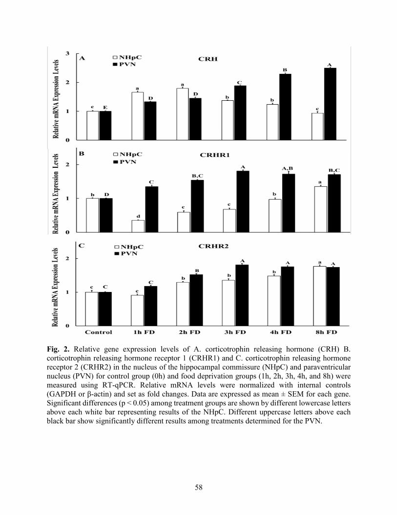
58
Fig. 2. Relative gene expression levels of A. corticotrophin releasing hormone (CRH) B. corticotrophin releasing hormone receptor 1 (CRHR1) and C. corticotrophin releasing hormone receptor 2 (CRHR2) in the nucleus of the hippocampal commissure (NHpC) and paraventricular nucleus (PVN) for control group (0h) and food deprivation groups (1h, 2h, 3h, 4h, and 8h) were measured using RT-qPCR. Relative mRNA levels were normalized with internal controls (GAPDH or β-actin) and set as fold changes. Data are expressed as mean ± SEM for each gene. Significant differences (p < 0.05) among treatment groups are shown by different lowercase letters above each white bar representing results of the NHpC. Different uppercase letters above each black bar show significantly different results among treatments determined for the PVN.

59
Fig. 3. Relative gene expression levels of A. Brain derived neurotropic factor (BDNF), and B. Glial cell-derived neurotropic factor (GDNF) in the nucleus of the hippocampal commissure (NHpC) and paraventricular nucleus (PVN) for the control group (0h) and food deprivation groups (1h, 2h, 3h, 4h, and 8h) were measured using RT-qPCR. Relative mRNA levels were quantified and normalized with internal controls (GAPDH or β-actin). Data were set as fold changes of relative expression levels using the 2-ΔΔCt method after normalization. Data were expressed as mean ± SEM for each gene. Significant differences (p < 0.05) among all groups are shown by different lowercase letters above the white bars representing the NHpC. Different uppercase letters above each black bar show significant differences among time points for the PVN.

60
Fig. 4. Relative gene expression levels of the glucocorticoid receptor (GR) in the nucleus of the hippocampal commissure (NHpC) and paraventricular nucleus (PVN) for the control (0h) and food deprivation groups (1h, 2h, 3h, 4h, and 8h) were measured using RT-qPCR. Relative mRNA levels were quantified and normalized with internal controls (GAPDH or β-actin). Data were set as fold changes of relative expression levels using the 2-ΔΔCt method after normalization. Data were expressed as mean ± SEM for each gene. A level of significance difference (p < 0.05) among groups is shown by different lowercase letters above each white bar for the NHpC. Different uppercase letters above each black bar show differences among treatment groups for the PVN.

61
Fig. 5. Pro-opiomelanocortin (POMC) mRNA (black bars) and heteronuclear (hn) POMC (striped bars) expression levels in the anterior pituitary following food deprivation. Relative mRNA levels were quantified and normalized with internal controls (GAPDH or β-actin). Data were set as fold changes of relative expression levels using the 2-ΔΔCt method after normalization. Data for each gene was expressed as mean ± SEM. Different lowercase letters above striped bars represent hnRNA POMC while uppercase letters above black bars represent mRNA POMC (p < 0.05 among treatment groups).

62
Table 1. Fold changes, % increase/decrease of mRNA CRH, CRHR1 and CRHR2 in NHpC and PVN after feed deprivation for 1h and 2h.
Gene NHpC1 PVN1 Control 1h %2 2h %2 Control 1h %2 2h %2
CRH 1.00 1.65 +65 1.79 +79 1.00 1.33 +33 1.44 +44 CRHR1 1.00 0.35 -65 0.59 - 41 1.00 1.35 +35 1.54 +54 CRHR2 1.00 0.90 -10 1.29 +29 1.00 1.17 +17 1.52 +52
Abbreviations: 1Nucleus of hippocampal commissure, Paraventricular nucleus 2Percent increase (+) or decrease (-) from control.

63
References
Aman, N., Nagarajan, G., Kang, S., Hancock, M., Kuenzel, W., 2016. Differential responses of the vasotocin 1a receptor (V1aR) and osmoreceptors to immobilization and osmotic stress in sensory circumventricular organs of the chicken (Gallus gallus) brain. Brain Res.1649, 67-78.
Arborelius, L., Owens, M., Plotsky, P., Nemeroff, C., 1999. The role of corticotropin releasing factor in depression and anxiety disorders. J. Endocrinol. 160, 1–12.
Blas, J. (2015). Stress in birds, In: Scanes, C. G. (ed.), Sturkie’s Avian Physiology, sixth ed. Academic Press, San Diego, pp. 769-810.
Bonfiglio J. J., Inda C., Refojo, D., Holsboer, F., Arzt, E., Silberstein, S., 2011. The corticotropin-releasing hormone network and the hypothalamic-pituitary-adrenal axis: molecular and cellular mechanisms involved. Neuroendocrinology 94, 12–20
Brunson, K.L., Grigoriadis, D.E., Lorang, M.T., Baram, T.Z., 2002. Corticotropin-releasing hormone (CRH) downregulates the function of its receptor (CRF1) and induces CRF1 expression in hippocampal and cortical regions of the immature rat brain. Exp. Neurol. 176, 75 – 86.
Carsia, R.V., 2015. Adrenals. Scanes, C. G. (ed.), Sturkie’s Avian Physiology, sixth ed. Academic Press, San Diego, pp. 577 – 611.
Chen, J., Evans, A. N., Liu, Y., Honda, M., Saavedra, J. M., Aguilera, G., 2012. Maternal deprivation in rats is associated with corticotrophin-releasing hormone (CRH) promoter hypomethylation and enhances CRH transcriptional responses to stress in adulthood. J. Neuroendocrinol. 24, 1055 –1064.
Chrousos, G., 2009. Stress and disorders of the stress system. Nat. Rev. Endocrinol. 5: 374-381.
Coste, S. C., Kesterson, R. A., Heldwein, K. A., Stevens, S. L., Heard, A. D., Hollis, J. H., Murray, S. E., Hill, J. K., Pantely, G. A., Hohimer, A. R., Hatton, D. C., Phillips, T. J., Finn, D. A., Low, M. J., Rittenberg, M. B., Stenzel, P., Stenzel-Poore, M. P., 2000. Abnormal adaptations to stress and impaired cardiovascular function in mice lacking corticotropin-releasing hormone receptor-2. Nat. Genet. 24, 403 – 409.
Cramer, T., Kisliouk, T., Yeshurun, S., Meiri, N., 2015. The balance between stress resilience and vulnerability is regulated by corticotropin-releasing hormone during the critical postnatal period for sensory development, Dev. Neurobiol.75, 842 – 853.
De Kloet, E., Joels, M., Holsboer, F., 2005. Stress and the brain: from adaptation to disease. Nat. Rev. Neurosci. 6, 463 – 475.
Feldman, S. Conforti, N., 1980. The role of the medial septa1 nucleus in mediating adrenocortical responses to somatosensory stimulation. J. Neurosci. Res. 5, 19 – 23.

64
Font, C., Lanuza, E., Martinez-Marcos, A., Hoogland, P. V., Martinez-Garcia, F., 1998. Septal complex of the telencephalon of lizards: III. Efferent connections and general discussion. J. Comp. Neurol. 401, 525 – 548.
Girotti, M., Pace, T., Gaylord, R., Rubin, B., Herman, J., Spencer, R., 2006. Habituation to repeated restraint stress is associated with lack of stress-induced c-fos expression in primary sensory processing areas of the rat brain. Neuroscience 138, 1067 – 1081.
Givalois L, Naert G, Rage F, Ixart G, Arancibia S, Tapia-Arancibia L., 2004. A single brain-derived neurotrophic factor injection modifies hypothalamo-pituitary-adrenocortical axis activity in adult male rats. Mol. Cell Neurosci. 27, 280 – 295.
Greetfeld, M., Schmidt, M. V., Ganea, K., Sterlemann, V., Liebl, C., Müller, M. B., 2009. A single episode of restraint stress regulates central corticotrophin-releasing hormone receptor expression and binding in specific areas of the mouse brain. J. Neuroendocrinol. 21, 473–480.
Herman, J., Cullinan, W., 1997. Neurocircuitry of stress: central control of the hypothalamo-pituitary-adrenal axis. Trends Neurosci. 20:78 – 84.
Herman, J., McKlveen, J., Ghosal, S., Kopp, B., Wulsin, A., Makinson, R., Scheimann, J., Myers, B., 2016. Regulation of the hypothalamic-pituitary adrenocortical stress response. Comp. Physiol. 6, 603 – 621.
Imaki, T. Naruse, M. Harada, S. Chikada, N Imaki, J. Onodera, H. Demura, H., Vale. W., 1996. Corticotropin-releasing factor up-regulates its own receptor mRNA in the paraventricular nucleus of the hypothalamus. Mol. Brain Res. 38, 166 – 170.
Imaki, T., Katsumata, H., Miyata, M., Naruse, M., Imaki, J., Minami, S., 2001. Expression of corticotropin-releasing hormone type 1 receptor in paraventricular nucleus after acute stress. Neuroendocrinology 73, 293 – 301.
Jaccoby, S., Singh, A., Cornett, L., Koike, T., 1997. Arginine vasotocin gene expression and secretion during osmotic stimulation and hemorrhagic hypotension in hens, Gen. Comp. Endocrinol. 106, 327 – 337.
Jeanneteau, F., Lambert, W., Ismaili, N., Bath, K., Lee, F., Garabedian, M., Moses, V., Chaoa, M., 2012. BDNF and glucocorticoids regulate corticotrophin releasing hormone (CRH) homeostasis in the hypothalamus. Pro. Natl. Acad. Sci. USA 109, 1305 – 1310.
Kang, S., Kuenzel, W., 2014. Regulation of gene expression of vasotocin and Corticotropin-releasing hormone receptors in the avian anterior pituitary by corticosterone. Gen. Comp. Endocrinol. 204, 25 – 32.
Keller-Wood, M., 2011. Hypothalamic-pituitary-adrenal Axis feedback control. Comp. Physiol. 5, 1161 –1182.

65
Korosi, A., Veening, J. G., Kozicz, T., Henckens, M., Dederen, J., Groenink, L., van der Gugten, J., Olivier, B., Roubos, E.W., 2006. Distribution and expression of CRF receptor 1 and 2 mRNAs in the CRF over-expressing mouse brain. Brain Res. 1072, 46 – 54.
Kuenzel, W. J., Kang, S. W., Jurkevich, A., 2013. Neuroendocrine regulation of stress in birds with an emphasis on vasotocin receptors (VTRs). Gen. Comp. Endocrinol. 190, 18 – 23.
Kuenzel, W., Masson, M., 1988. A stereotaxic atlas of the brain of the chick (Gallus domesticus). The Johns Hopkins University Press, Baltimore, p. 166.
Lehnert, H., Schulz, C., Dieterich, K., 1998. Physiological and neurochemical aspects of corticotropin-releasing factor actions in the brain: the role of the locus coeruleus. Neurochem. Res. 23, 1039 –1052.
Madison, F., Jurkevich, A., Kuenzel, W., 2008. Sex differences in plasma corticosterone release in undisturbed chickens (Gallus gallus) in response to arginine vasotocin and corticotropin releasing hormone. Gen. Comp. Endocrinol. 155, 566 – 573.
Makino, S., Nishiyama, M., Asaba, K., Gold, P.W., Hashimoto, K., 1998. Altered expression of type 2 CRH receptor mRNA in the VMH by glucocorticoids and starvation. Am. J. Physiol. 275, 1138 – 1145.
Manfredsson, F., Tumer, N., Edros, B., Landa, T., Broxson, C., Sullivan, L., Rising, A., Foust, K., Zhang, Y., Muzyczka, N., Gorbatuk, O., Scarpace, P., and Mandel, R., 2009. Bilateral nigral rAAV5-mediated GDNF over-expression activates CRH neurons in the paraventricular nucleus of the hypothalamus and induces robust weight loss in obese leptin-resistant rats. Mol. Ther. 17, S73.
Mattson, M., 2000. Turning off the anxiety master switch. Trends Neurosci. 23, 333.
Merchenthaler, I., 1984. Corticotropin releasing factor (CRF)-like immunoreactivity in the rat central nervous system. Extra-hypothalamic distribution. Peptides 5, 53 – 69.
Nagarajan, G., Jurkevich, A., Kang, S., Kuenzel, W. 2017a. Anatomical and functional implications of corticotrophin releasing hormone neurones in a septal nucleus of the avian brain: an emphasis on glial-neuronal interaction via V1a receptors in vitro. J. Neuroendocrinol. 29, 1–11.
Nagarajan, G., Kang, S., Kuenzel, W., 2017b. Functional evidence that the nucleus of the hippocampal commissure shows an earlier activation from a stressor than the paraventricular nucleus: Implication of an additional structural component of the avian hypothalamo-pituitary-adrenal axis. Neurosci. Lett. 642, 14 – 19.
Nagarajan, G., Tessaro, B.A., Kang, S.W., Kuenzel, W.J., 2014. Identification of arginine vasotocin (AVT) neurons activated by acute and chronic restraint stress in the avian septum and anterior diencephalon. Gen. Comp. Endocrinol. 202, 59 – 68.
66
Palkovits, M., 1973. Isolated removal of hypothalamic or other brain nuclei of the rat. Brain Res. 59, 449 – 450.
Potter, E., Sutton, S., Donaldson, C., Chen, R., Perrin, M., Lewis, K., Sawchenko, P., Vale, W., 1994. Distribution of corticotropin releasing factor receptor mRNA expression in the rat brain and pituitary. Proc. Natl. Acad. Sci. USA 91, 8777 – 8781.
Puelles, L., Martinez-de-la-Torre, M., Paxinos, G., Watson, C., Martínez, S., 2007. The chick brain in stereotaxic coordinates: an atlas featuring neurometric subdivisions and mammalian homologies. Academic Press, San Diego.
Romero, L.M., 2004. Physiological stress in ecology: lessons from biomedical research. Trends Ecol. Evol. 19, 249 –255.
Schmittgen, T., Livak, K., 2008. Analyzing real-time PCR data by the comparative CT method. Nat. Protocols 3, 1101 – 1108.
Sharp, P., Li, Q., Talbot, R., Barker, P., Huskisson, N., Lea, R., 1995. Identification of hypothalamic nuclei involved in osmoregulation using fos immunocytochemistry in the domestic hen (Gallus domesticus), Ring dove (Streptopelia risoria) Japanese quail (Coturnix japonica) and Zebra finch (Taenopygia guttata), Cell Tissue Res. 282, 351–361.
Sperlagh, B., Maglóczky, Z.S., Vizi, E.S., Freund, T.F., 1998. The triangular septal nucleus as the major source of ATP release in the rat habenula: a combined neurochemical and morphological study. Neuroscience 86, 1195 –1207.
Vale, W., Spiess, J., Rivier, C., Rivier, J., 1981. Characterization of a 41-residue ovine hypothalamic peptide that stimulates secretion of corticotropin and endorphin. Science 213, 1394 –1397.
Vandenborne, K., De Groef, B., Geelissen, S., Kühn, E., Darras, V., Van der Geyten, S., 2005. Corticosterone-induced negative feedback mechanisms within the hypothalamic pituitary adrenal axis of the chicken. J. Endocrinol. 185, 383 –391.
Xie, J., Kuenzel, W., Anthony, N., Jurkevich, A., 2010. Subpallial and hypothalamic areas activated following sexual and agonistic encounters in male chickens. Physiol. Behav. 10, 344 – 359.
Yadawa, A., Chandra, M., 2016. Expression of stress hormones AVP and CRH in the hypothalamus of Mus musculus following water and food deprivation. Gen. Comp. Endocrinol. 239, 13 – 20.

67
Chapter 3
Arginine vasotocin and its receptors in septo-hypothalamic brain structures and anterior
pituitary sustain HPA axis functions during acute stress.
This chapter is excerpted from “Kadhim, H.J., Kidd M. Jr., Kang, S. W., Kuenzel, W. J., 2020. Differential delayed responses of arginine vasotocin and its receptors in septo-hypothalamic brain structures and anterior pituitary that sustain hypothalamic–pituitary-adrenal (HPA) axis functions during acute stress”. Gen. Comp. Endocrinol. 286, 113302.”
(Reproduced with permission from reference (Kadhim et al., 2020), copyright (2020) General and Comparative Endocrinology)
Abstract
Recently, we proposed that corticotropin releasing hormone (CRH) neurons in the nucleus of
hippocampal commissure (NHpC), located in the septum, function as a part of the traditional
hypothalamo-pituitary-adrenal (HPA) axis in avian species. CRH and its receptor, CRHR1, are
regulated differently in the NHpC compared to the paraventricular nucleus (PVN) following feed
deprivation (FD). Therefore, we followed up our work by examining arginine vasotocin (AVT),
the other major ACTH secretagogue, and its receptors, V1aR and V1bR, gene expression during
FD stress in the NHpC, PVN, and ventral mediobasal hypothalamus/median eminence
(MBHv/ME). The objectives were to 1) identify AVT perikarya, fibers and its two major receptors,
V1aR and V1bR, in the NHpC, PVN, and MBHv/ME using immunohistochemistry (IHC), 2)
determine the effect of stress on AVT, V1aR and V1bR mRNA expression in the same three brain
structures, NHpC, PVN, and MBHv/ME; and 3) ascertain the expression pattern of V1aR and
V1bR mRNA in the anterior pituitary (APit) and measure plasma stress hormone, corticosterone
(CORT), concentration following FD stress. Male chicks (Cobb 500), 14 days of age, were divided

68
into six groups (10 birds/treatment) and subjected to different times of FD stress: (Control, 1h, 2h,
3h, 4h, and 8h). For each bird, blood, brain, and APit were sampled and frozen immediately. The
NHpC, PVN, and MBHv/ME were micro-dissected for RT-PCR. Data were analyzed using one-
way ANOVA followed by Tukey Kramer HSD test with a significance level of p < 0.05. Perikarya
of AVT neurons were identified in the PVN but not in the NHpC nor MBHv/ME, and only V1aR-
immunoreactivity (ir) was observed in the three structures; however, gene expression data for AVT
and its two receptors were obtained in all structures. Both AVT and V1aR mRNA are expressed
and increased significantly in the PVN following FD stress (p < 0.01). For the first time, V1bR
mRNA was documented in the avian brain and specifically upregulated in the NHpC and PVN (p
< 0.01) following stress. Additionally, a delayed significant gene expression of AVT and its
receptors in the PVN showed a positive feedback relationship responsible for maintaining CORT
release. In contrast, a significant downregulation of AVT mRNA and upregulation of V1aR mRNA
occurred in the NHpC (p < 0.01) during FD showing a negative feedback relationship between
AVT and its receptors, V1aR and V1bR. Within the MBHv/ME and anterior pituitary, a gradual
increase of AVT mRNA in PVN as well as MBHv/ME was associated with significant
upregulation of V1bR (p < 0. 01) and downregulation of V1aR (p <0.01) in both MBHv/ME and
anterior pituitary indicating that AVT regulates its receptors differentially to sustain CORT release
and control overstimulation of the anterior pituitary during a stress response.
Key words: nucleus of hippocampal commissure, paraventricular nucleus, V1aR, V1bR, feed
deprivation, corticosterone.

69
1. Introduction
The central neural components of the classical hypothalamic pituitary adrenal (HPA) axis
regulating stress responses in avian species, similar to mammals, comprise two types of neurons,
corticotropin releasing hormone (CRH) and arginine vasotocin (AVT), the latter regarded as a
homolog of mammalian arginine vasopressin (AVP). Previous studies reported that AVT/AVP is
considered a stress neural hormone because of its ability to induce CORT release when it is injected
alone or in combination with CRH. Anterior pituitary adrenocorticotropic hormone (ACTH) and
the ultimate stress hormone, corticosterone (birds, mice and rats) and cortisol (most mammalian
species), increased significantly after central administration of CRH or AVT (Yayou et al., 2003,
2007, 2008; Madison et al., 2008; Pryce et al., 2011). Studies in our laboratory also showed that
immobilization stress in chickens caused a significant increase in plasma AVT concentration
(Aman et al., 2016). Furthermore, utilizing immunohistochemistry, it was reported that AVT
neurons in the avian PVN were activated in both acute and chronic restraint stress (Nagarajan et
al., 2014). AVP/AVT regulation of the HPA axis is coordinated by two vasotocin receptors, V1aR
and V1bR. Mapping studies of the avian V1aR in chicks showed a widespread distribution within
corticotropes of the anterior pituitary and brain suggesting that the V1aR was involved in the stress
response (Selvam et al., 2013, 2015). A second chicken vasotocin receptor, V1bR, was likewise
shown to be associated with stress. The receptor plays an important role in mediating the
stimulatory effects of AVT on ACTH secretion from corticotropes within anterior pituitary, where
it regulates ACTH release, based upon the presence of the immunoreactive V1bR in corticotropes
(Jurkevich et al., 2005, 2008) and heterodimerization with CRHR1 (Mikhailova et al., 2007)
indicating a synergistic effect of AVT and CRH on plasma corticosterone release (Kuenzel et al.,
2010; Cornett et al., 2013).

70
Of the various stress models available for the study of the effects of stress, the feed deprivation
(FD) model has been recently employed in our lab. Feed deprivation differs from other stressors
in that it is gradual and becomes more intense over time as bodily demands for nutrients increase.
This stressor was used to study the sequence of gene activation beginning with CRH expression in
the nucleus of the hippocampal commissure (NHpC), a septal structure recently proposed as a
functional part of the avian hypothalamic-pituitary-adrenal (HPA) axis (Nagarajan et al., 2017a,
2017b), followed by CRH activation in the PVN, and a third delayed peak of arginine vasotocin
(AVT) gene expression in the PVN (Nagarajan et al., 2017b). Our recent publication (Kadhim et
al., 2019) utilized the same stressor, FD, and focused upon the relationship between CRH
expression and its two receptors, corticotropin releasing hormone receptor 1 (CRHR1) and
corticotropin releasing hormone receptor 2 (CRHR2), in the NHpC and PVN. We showed that
rapid activation of CRH neurons via increasing mRNA in the NHpC was associated with
downregulation of its major receptor, CRHR1, demonstrating a negative feedback. Whereas
increasing of CRH mRNA in the hypothalamic PVN resulted in upregulation of CRHR1, showing
positive feedback. In other words, CRH regulation of its receptors in the brain appears to be
structure specific.
Hence, the study was designed to determine whether a similar relationship occurs with the
other peptide system, AVT and its receptor, V1aR, known to be activated by stress in brain
structures located within the avian neuroendocrine stress axis comprising the NHpC, PVN, and
ventral mediobasal hypothalamus/median eminence (MBHv/ME). We hypothesize that the stress-
related secretagogue, AVT and its receptor, V1aR, within the PVN would likewise show a positive
feedback. In contrast, we also hypothesized that gene expression of AVT within the NHpC would
display a negative feedback with its major receptor, the V1aR. Additionally, by determining gene

71
expression of AVT’s major receptor, V1aR, in stress-related brain structures, NHpC and PVN,
may help us understand the role of vasotocin in regulating the central HPA axis during stress.
Curiously, a past, unpublished finding of ours was that V1bR mRNA was found in the avian brain
(Kang, unpublished). However, the actual V1b receptor protein, utilizing immunohistochemistry
was not detected in the chick brain (Jurkevich et al., 2005) nor the songbird brain (Leung et al.,
2011). Since the V1bR is the major neuroendocrine receptor involved with stress for the peptide,
vasopressin, in the mammalian PVN, we hypothesized that the V1bR mRNA would also be found
within the PVN and perhaps the NHpC of birds following FD stress. Three specific aims were
designed to 1) identify AVT perikarya and its two major receptors, V1aR and V1bR, in the NHpC,
PVN, and MBHv/ME using immunohistochemistry; 2) determine the effect of stress on AVT,
V1aR and V1bR mRNA expression in the same three brain structures, NHpC, PVN, and
MBHv/ME; and, 3) ascertain the effect of stress on V1aR and V1bR mRNA levels in the anterior
pituitary.
2. Material and methods
2.1. Animals
Male Cobb 500 chicks, one-day old, (Gallus gallus) were obtained from a commercial hatchery
and provided with feed (a standard, broiler starter diet) and water ad libitum. Birds were raised in
brooder batteries for the first ten days. Chicks were exposed to continuous light for the first three
days so that they could locate both the feed and water. Thereafter, birds were maintained under
16h: 8h light/dark cycle (lights on at 6:00 AM) with controlled heat (32 ℃). Heat was reduced
2.5oC weekly until reaching 27 ℃. On day 10, birds were weighted and distributed randomly to
cages (3 birds/cage). At 14 days of age, experiments were initiated, and sampling occurred between

72
8:00 AM and 4:00 PM. All procedures utilized were approved by the University of Arkansas
Institutional Animal Care and Use Committee.
2.2. Sample collection for gene expression
Food deprivation (FD), a stressor, was initiated on day 14, however, water was available ad
libitium. Chicks were subjected to one of the following FD treatments, beginning at 8 AM: 0h
(control), 1h, 2h, 3h, 4h, and 8h with each experimental group containing ten to twelve birds. Blood
samples were first collected within 2 minutes for each bird from the brachial vein. Each bird was
then cervically dislocated, and brain and anterior pituitary were rapidly dissected. Brain samples
were immediately frozen by immersion in two-methyl butane at −30 ℃ for 15 seconds followed
by dry ice to maintain structural morphology of the brain for cryosectioning. All samples (brain
and anterior pituitary) were stored at−80 ℃ until processed. Cross sections of brain samples were
cut at 100 µm using a cryostat (Leica CM3050 S, Leica Microsystems, Frisco, TX) and the targeted
regions were punched (brain punch, Palkovits, 1973) using a glass pipette including (1) the NHpC,
1.4 mm diameter from atlas plates A8.2–A7.6 ; (2) the PVN, 1.4 mm diameter including atlas
plates A8.0–A6.4; and (3) MBHv/ME, 1.4 mm diameter including atlas plates A5.8-A4.4
(Kuenzel and Masson, 1988). All brain sections were dissected inside the cryostat chamber
maintained at −15 oC. The anterior commissure (AC) was used as a landmark and served as the
ventral border for dissections of the NHpC. Stereotaxic atlas plates were used to position the
pipette for dissecting the NHpC, PVN and MBH/ME. Punches of the PVN and MBH/ME were
initiated at atlas plate A8.0 and A5.8, respectively, using a chick stereotaxic atlas reference
(Kuenzel and Masson, 1988). To determine the accuracy of the boundaries of punched or dissected
structures, immediately after each punch, the sections were examined under a dissecting
73
microscope (Leica MZ 125). Punches were collected in Trizol and stored at −20 oC until processed
for RNA extraction.
2.3. Tissue preparation for immunohistochemistry
Male chicks, 14 days old, were anesthetized with an intravenous (i.v.) injection of sodium
pentobarbital (27 mg/kg). Each anesthetized bird was perfused through the left ventricle of the
heart (100 ml) followed by 150 ml via the carotid arteries using ice cold heparinized 0.1M
phosphate buffer (PB) containing 0.1% sodium nitrite, pH 7.4 and lithium heparin (71 mg/L PB,
Sigma). Immediately, thereafter, birds were perfused via carotid arteries with 250 ml of freshly
prepared and filtered ice-cold Zamboni’s fixative solution, containing 4% paraformaldehyde with
15% picric acid in 0.1M PB at pH 7.4. Brains were blocked in a stereotaxic instrument (Kopf
Instrument, Tujunga, CA) and post-fixed in the same fixative overnight at 4 °C. Blocked brains
were cryoprotected using 30% sucrose in 0.1M PB at 4 °C until they sank. Brains were wrapped
in parafilm and aluminum foil and stored at -20 °C overnight, then they were transferred to the -
80 °C until sectioned. Blocked brains were taken out of -80 °C and equilibrated at -20 °C in a
cryostat (LeicaCM3050S, Leica Microsystems, Austin, TX, USA) before embedded in Jung OTC
medium (freezing media, Leica Microsystems, Wetzlar, Germany). Cross-sections were cut at 40
μm using the cryostat. NHpC, PVN, and MBHv/ME sections were collected in a 24-well plate
containing 2 ml of 0.02M PBS (pH 7.4). Sections were free-floating in 0.02 M PBS (one per well
in a 24-well plate) and treated with 0.6% of hydrogen peroxide to attenuate endogenous peroxide
activity (for non-fluorescent sections) followed by 0.02M PBS containing 0.4% Triton X-100 for
30 minutes for permeabilization. Sections were transferred to 5% normal goat serum (NGS) in
0.02M PBS with 0.4% Triton-X-100 and 0.1 % sodium azide for 30 minutes to block nonspecific

74
binding sites, followed by incubation for at least 48h with polyclonal rabbit anti-AVT antibody,
kindly provided by Dr. Gray (1:10000 dilution, Gray and Simon, 1983). Polyclonal antibodies
made in rabbit, anti-V1aR antibody (RRID: AB_2336062, 1:2,000, Selvam et al., 2015) and anti-
V1bR antibody (1:2000 dilution, Custom made antibody, Jurkevich et al., 2005, 2008) in 0.02M
PBS containing 1% NGS, 0.2% Triton X-100, and 0.1% sodium azide were also utilized. Sections
were incubated for 90 minutes with biotinylated goat anti-rabbit IgG (dilution 1:500 in 0.02M PBS
with 0.2% Triton X-100; Vector Laboratories), followed by incubation with Vectastain Elite ABC
peroxidase complex diluted 1:5 for 90m (Vector Laboratories). Sections of anterior pituitary were
used as positive control as past data showed the avian V1bR is found on corticotropes of anterior
pituitary (Jurkevich et al., 2005). The AVT, V1aR, and V1bR immunoreactive (-ir) cells were
visualized with glucose oxidase-catalyzed diaminobenzidine–nickel complex. Sections were then
rinsed in PBS, mounted onto clean glass slides, coverslipped with histomount (National Diagnostic
Laboratories, Atlanta, GA), and visualized using a Zeiss Imager M2 microscope and digital camera
(Axiocam 105 color).
2.4. Hormone assay
Blood samples were taken from the brachial vein of chicks in all treatment groups (n=10/
treatment). Plasma was obtained from the heparinized blood via centrifugation at 3000 rpm for 20
min at 4 ℃. Hemolyzed samples (1-2 samples/ group) were excluded. Plasma was stored at - 20
℃ until analysis of CORT concentrations by radioimmunoassay (Madison et al., 2008; Proudman
and Opel, 1989). Each plasma sample was analyzed in duplicate. Briefly, plasma samples (200 μl)
were first extracted by vortex for 30 min at room temperature with 2 ml of ethyl ether. Then, the
water phase was separated in methanol/ dry ice bath. The liquid phase of each sample was

75
transferred to a new tube and dried at 38 ℃ water bath. Dried extracts were reconstituted with 400
μl of assay buffer, vortexed for 5m, and equilibrated overnight at 4 ℃. Then, 100 μl of the primary
antibody, polyclonal rabbit anti-CORT # 377, (kindly provided by Dr. Proudman, Schmeling and
Nockels, 1978) and 100 μl of 125I corticosterone tracer purchased from MP Biomedicals Inc.
(Orangeburg, NY, USA) were added to each sample and incubated for at least 24 h at 4 ℃. Sheep
anti-rabbit antibody (200 μl) was used as secondary antibody (MP Biomedicals Inc., Orangeburg,
NY, USA) and 6% of polyethylene glycol (500 μl) was used added and centrifuged to precipitate
bound tracers. The supernatant of each sample and standard were discarded, the remaining liquid
dried, and counts/tube were determined using a Perkin Elmer Wizard gamma-counter. Data were
analyzed by one-way ANOVA followed by a Tukey’s Kramer HSD test and expressed as the mean
± SEM of each group. In all studies, p < 0.05 was considered statistically significant. Intra
experimental coefficient of variance was less than 13%.
2.5. RNA extraction from brain structures and anterior pituitaries
Total RNA was extracted from frozen micro-dissected brain tissue and anterior pituitaries
(n=10 birds/group) using Trizol-chloroform (Life Technologies) according to the protocol
provided by the supplier. Total RNA was purified using a RNeasy mini kit (Qiagen), and RNA
concentration was estimated using Synergy HT multi-mode micro plate reader (BioTek). Samples
(2-3 per each group) were excluded because of insufficient amount of RNA extracted from micro-
punches. Therefore, we finished with 7-8 birds per group. First-strand cDNA was synthesized in
60 μl from total RNA (200ng of NHpC, 500ng of PVN, 300ng of ME and 600ng of anterior
pituitary) for each sample treated with DNase I (Ambion, Austin, TX, USA) using Superscript®
III reverse transcriptase (Invitrogen) according to the manufacturer’s protocol. In brief, RNA was

76
incubated with 3 μl Oligo (DT) and 3 μl dNTPs at 65 ℃/ 5m. After that, the mixture was incubated
with 30 μl cDNA synthesis mix (10X RT Buffer, 25 mM MgCl2, 0.1 M DTT, RNaseOUT, and
superscript III RT) at 50 ℃/ 60m, and the reaction was terminated by 85 ℃/ 5m. Finally, the RNA
was removed by adding 3 μl RNase H and incubated at 37 ℃/20m. The best primer pair was
chosen depending on past studies in our lab or selected from several pairs based on PCR product
quality and lengths after electrophoresis on a 3% agarose gel. Primer sets used in the assays were
(name, accession #, primer set, product size): AVT mRNA, NM_205185, forward
5′CCTTCCCCGAACGCATAG 3′ and reverse 5′GGGCAGTTCTGG ATGTAGCAG3′, 117 bp;
V1aR mRNA, NM_001110438, forward 5′GGTTGCAGTGTTTTCAGAGTCG3′ and reverse
5′CAAGATCCGCACCGTCAA G3′; V1bR mRNA, NM_001110438, forward 5’GGTTGCAGT
GTTTTCAGAGTCG3’ and reverse 5’CAAGATCCGCACCGTCAAG3’, 137bp, GAPDH
mRNA, NM_204305, forward 5′CTTTGGCATTGTGGAGGGTC3′ and reverse 5′ACGCTGG
GATGATGTTCTGG3′, bp 128; β- actin L_08165, forward, 5’CACAATGTACCCTGGCA
TTG3’ and reverse 5’ACATCTGCT GGAAG GTGGAC3’ ,158 bp. Power SYBR green PCR
Master Mix was mixed with cDNA and primers and amplified using real-time quantitative PCR
(Applied Biosystems 7500 Real-Time PCR system). The assay was run in triplicate and achieved
in 30 μl using the following conditions: 1 cycle at 95 ℃ for 10m and amplified for 40 cycles at 95
℃ for 30s, 60 ℃ for 1m, and 72 ℃ for 30s. The chicken glyceraldehyde3-phosphate (GAPDH)
or beta actin (βA) was used as internal controls to normalize the data. We chose the internal control
that displayed the most consistent levels, less than one Ct value, across all treatments for a given
structure. Relative gene expression levels of each specific gene were determined by the 2−ΔΔCt
method (Schmittgen and Livak, 2008). Relative expression of control groups was set to 1.

77
2.6. Statistical analysis
Statistical analyses were performed using JMPR pro 14.0 (SAS Institute Inc., NC). A normal
distribution was first tested and thereafter differences among six groups for the NHpC, PVN,
MBHv/ME, anterior pituitary, and plasma CORT concentration were analyzed separately using
one-way analysis of variance (ANOVA) to find the F- value and degrees of freedom for each
structure. Comparison for all pairs using Tukey’s Kramer HSD test were used to evaluate plasma
CORT concentration and relative changes of gene expression between control and FD groups.
Data are presented as the mean ± SEM. A probability level of p < 0.05 was considered statistically
significant.
3. Results
3.1. AVT cell bodies and terminal fields in the NHpC, PVN, and MBHv/ME
Immunohistochemical data revealed that no AVT cell bodies were identified in the NHpC, but
terminal fields of AVT fibers were confirmed ( Nagarajan et al., 2017a) innervating the core of the
NHpC and appeared more dense in the dorsal region (Fig. 1A). The origin/s of the nerve terminal
fields found in the NHpC is/are unknown. In contrast, the PVN showed a dense group of AVT cell
bodies near midline with a stream of cell bodies that moved laterally from the third ventricle and
a less dense thin group of AVT neurons occurred along the midline just parallel to the third
ventricule (Fig.1B). Within the ventral MBHv/ME no AVT cell bodies were displayed, however,
dense terminal fields of AVT fibers were seen in the external and internal zones of the ME as
shown in (Fig1.C) confirming past studies (Tennyson et al., 1986). In terms of V1aR and V1bR
immunoreativity (ir) in brain structures located within the septohypothamic area, V1aR-ir was
78
identified in all three structures. Specifically, in the NHpC, a dense V1aR-ir was observed. and the
majority of immunoreactivity was found in glial cells (Fig. 1D) confirming results from a previous
study (Selvam et al. 2015). However, the V1aR-ir in the PVN was not as dense as in the NHpC.
The PVN V1aR-ir was associated with glia and denser along the edges of 3V and became less
prominent on glial processes moving latterally (Fig. 1E). Similar to the NHpC, cells in the
MBHv/ME, a circumventricular organ (CVO), showed V1aR-ir (Fig. 1F) on tanycytes as
previously documented (Selvam et al., 2015). Regarding V1bR-ir in brain structures, similar to
previous studies in our lab (Jurkevich et al., 2005), we were unable to detect any immunoreactive
signal in neural tissue.
3.2. Food deprivation induced elevated CORT concentrations in blood plasma
The time course for plasma corticosterone (CORT) concentration of birds subjected to food
deprivation at 0h (control), 1h, 2h, 3h, 4h, and 8h were measured by RIA (Fig. 2). Food deprived
birds showed an overall significant difference among treatment groups (F (5, 53) = 34.48, p<0.01).
A significant increase was first shown at 2h (p = 0.02) and steadily increased thereafter with a peak
level at 4h (p < 0.01), which was maintained at 8h (p < 0.01), the last sampling time.
3.3. Gene expression results within brain structures following stress
3.3.1. AVT, V1aR, and V1bR mRNA expression in the NHpC and PVN
The relative mRNA expression levels of AVT and its receptors, V1aR and V1bR, were
assessed in the NHpC and PVN after feed deprivation for 1h, 2h, 3h, 4h, and 8h compared to
unstressed controls (Fig. 3). AVT mRNA levels displayed different patterns of expression in the
NHpC and PVN [F (5, 44) = 40.30, p < 0.01 in the NHpC and F (5, 44) = 20.10, p < 0.01 in the

79
PVN]. Specifically, the NHpC showed significant downregulation of AVT mRNA after 1h (p <
0.01) which remained significantly lower throughout all sampling periods (p < 0.01) with the
lowest AVT mRNA at 8h (Fig. 3A). In contrast, AVT mRNA expression in the PVN did not
significantly increase until 3h, peaked at 4h, and persisted significantly higher at 8h of FD (p <
0.01) (Fig. 3A).
In terms of the two major receptors of AVT, V1aR and V1bR, in the NHpC, V1aR gene
expression was significantly downregulated during 1h and 2h of FD. Thereafter, V1aR mRNA
levels recovered at 3h and 4h; however, showed no significant differences compared with controls.
Interestingly, a significant upregulation of V1aR mRNA in the NHpC was observed only at 8h of
FD (Fig. 3B). In contrast to V1aR mRNA expression in the NHpC, V1bR mRNA expression in
the NHpC was upregulated significantly at 2h through 4h with a peak response at 8h (Fig 3C).
Overall V1aR mRNA and V1bR mRNA expression were significant in the NHpC [F (5, 44) =
53.89, p < 0.01 and F (5, 44) = 31.30, p < 0.01), respectively]. The pattern of the V1aR and V1bR
mRNA expression was striking in the PVN in that they matched gene expression of AVT.
Specifically, no change in mRNA levels from controls occurred until after 2h of FD (Fig. 3B, 3C).
Then, a significant increase in V1aR and V1bR mRNA was shown at 3h of FD and continued at
4h and 8h (p< 0.01). Overall, V1aR mRNA and V1bR mRNA levels were significantly expressed
in the PVN [F (5, 44) = 23.774, p<0.01 and F (5, 44) = 17.8, p < 0.01, respectively].
3.3.2. AVT, V1aR, and V1bR mRNA expression in the ventral mediobasal hypothalamus/
median eminence (MBHv/ME)
Expression of AVT mRNA within neuronal terminal fields located in the MBHv/ME displayed
a significant increase in gene expression after stress [F (5, 44) = 37.20, p< 0.01, Fig. 4A]. The
80
significant increase of AVT mRNA was observed at 2h (p < 0.01) of FD and continued to climb
at 3h and 4h (p < 0.01) and peaked at 8h (p < 0.01). On the other hand, V1aR mRNA expression
in the MBHv/ME was downregulated slightly but significantly among FD treatments compared to
controls [F (5, 44) = 4.32, p < 0.01, Fig. 4B]. Specifically, the first significant downregulation of
V1aR mRNA levels was demonstrated after 1h of FD and continued through 8h (p < 0.05). In
contrast, the overall expression of V1bR mRNA was significant after FD [F (5, 44) = 30.64, p <
0.01; Fig. 4B]. The significant upregulation of V1bR mRNA occurred at 1h (p < 0.05) of FD and
gradually climbed to peak levels at 8h (p < 0.001) matching the expression pattern of AVT in the
ME (Fig. 4A, 4B).
3.4. Expression of V1aR and V1bR mRNA in the anterior pituitary
The relative mRNA levels of V1aR and V1bR were examined in the anterior pituitary after FD
at different time points (1h, 2h, 3h, 4h, and 8h) compared to unstressed (control) birds (Fig. 5).
The two receptors, V1aR and V1bR, responded differently to the stressor, FD [ F (5, 54) = 37.94,
p < 0.01 for V1aR and F (5, 54) = 46.94, p < 0.01 for the V1bR]. V1aR mRNA levels were
downregulated significantly at 1h FD (p < 0.02) and remained lower than control groups through
the last time point of sampling (p < 0.01). In contrast, the expression of V1bR mRNA exhibited a
slight non-significant increase of mRNA transcripts at 1h of FD with the first significant
upregulation at 2h FD (p < 0.05). Peak gene expression occurred at 3h (p < 0.01) and thereafter
declined at 4h and 8h, however, remained significantly higher than controls (p < 0.01).
81
4. Discussion
4.1. Anatomical localization of AVT perikarya, fibers, V1aR and V1bR in the NHpC, PVN,
and MBHv/ME
Similar to previous studies in our lab, the NHpC contained a terminal field of AVT fibers,
mostly in its dorsal region and no evidence of AVT perikarya (Nagarajan et al., 2017a) while the
PVN contained a large population of AVT perikarya (Fig. 1A, 1B). No AVT perikarya were
observed in the MBHv, while dense neural fiber tracts were seen in the ME occupying the internal
and external zones of that structure (Fig. 1C). With respect to the avian V1aR, the NHpC, PVN,
and MBHv/ME displayed V1aR-ir. The receptor, V1aR, was shown to occur in glia (Fig. 1D, 1F),
confirming the study of Selvam et al. (2015). Similarly, however in much less density, the PVN
likewise showed glia originating from the third ventricle and their processes passed into the PVN
(Fig. 1E).
4.2. AVT gene expression in the PVN displays a delay in activation following FD stress and
sustains the stress response initiated by CRH
The study utilized a gradually increasing stress effect of food absence for 8h, as measured by
the stress hormone CORT, mRNA expression of arginine vasotocin (AVT), and its two main
receptors, V1aR and V1bR. Of Interest, gene expression data revealed that the neuroendocrine
regulation of the stress response from FD in avian species comprises and activates not only CRH
and its receptors but also AVT and its receptors. Both AVT mRNA expression and CORT data
illustrate that AVT is involved in the late stage of this stressor (3h of FD and beyond) (Fig. 2, Fig.
3A). Our previous work demonstrated that CRH neurons in the NHpC and PVN show a significant

82
gene expression increase within both structures, initially at 1h FD. CRH mRNA peaked at 2h FD
in the NHpC and thereafter, CRH gene expression decreased significantly and returned to control
levels at 8h FD (Kadhim et al., 2019). Interestingly, at the same time period when downregulation
of CRH was documented in the NHpC (Kadhim et al., 2019), significant upregulation of AVT
mRNA levels occurred in the PVN (Fig. 3). Data suggest that in response to FD stress, CRH
mRNA is initially activated in the NHpC and PVN while no response from AVT neurons occurs.
The markedly increased AVT mRNA in the PVN at 3h of FD suggests that the delayed AVT gene
expression occurs to sustain the stress response to the persistent increasing FD stress to maintain
the overall output of the HPA axis. Our findings support previous evidence obtained in both in
vivo and in vitro studies showing that CRH gene expression precedes AVP/AVT gene expression
in mammals and birds following stress (Herman et al., 1992; Kovacs and Sawchenko 1996, Ma,
et al., 1997; Nagarajan et al., 2017a). Additionally, in the current study, a delayed but consistent
rise in AVT mRNA expression pattern in the PVN is responsible for sustaining the elevated plateau
of CORT suggesting that AVT is able to release CORT alone or potentiate CRH action during
stress. Our AVT data obtained from gene expression in the PVN are supported by other studies in
mammals and birds that found upregulation of AVT mRNA after stress (Yadawa and Chaturvedi,
2016; Nagarajan et al., 2017a).
4.3. Downregulation of AVT mRNA within vasotocin terminals located in the NHpC
associated with delayed upregulation of V1aR and V1bR mRNA showing a negative
feedback
Immunohistochemical data provide further evidence that terminal fields of AVT fibers
occurred in the NHpC and innervated the core of that structure (Fig.1A). However, the origin of

83
vasotocin treminls in that structure is unknown. Furthermore, mRNA expression in the NHpC
revealed that AVT mRNA was expressed and responded to the stressor, FD. Unexpectedly, AVT
gene expression in the NHpC was downregulated through all time points with lowest expression
at the last sampling point (Fig.3A). The possible explanation for the downregulation of AVT
mRNA levels within the NHpC is that the AVT mRNA might be transported from soma located
in other brain areas projecting to the NHpC and translated locally. Our data in avian, in agreement
with mammalian studies (Jung et al., 2013), showed that both growing and mature axons possess
local mRNA. Specifically, vasopressin (VP) mRNA was detected in the VP terminal field of the
neurohypophysis using RT-PCR, in situ hybridization and northern blot in mammals (Mohr et al.,
1991; Mohr and Richter, 1992; Trembleau et al., 1996). It would therefore be important to identify
the sources of vasotocin inputs into the NHpC. In terms of receptors within the NHpC, similar to
the PVN, V1aR-ir was observed in the glial cells (Fig.1E). The NHpC, at its posterier region,
contains the subseptal organ, homologus to the mammalian subfornical organ, one of the
recognized circumventricular organs (CVOs) (Selvam et al., 2015). It has been proposed that
CVOs possess specialized glia that serve as chemosensors (Bolborea and Dale, 2013). Therefore,
the NHpC possibly utilizes the V1aR to monitor AVT signals during the stress response. Gene
expression data demonstrated that both receptors responded to the stressor and displayed an
associated negative feedback with their ligand, AVT, in the NHpC (Fig.3B; 3C). Specifically,
V1bR mRNA levels were upregulated significantly starting at 2h, peaked at the last sampling point
(8h), while V1aR mRNA levels in the NHpC displayed a different pattern of expression initially
showing a similar decreased expression over the first 2h of FD along with AVT mRNA, and
thereafter displaying a negative feedback with AVT mRNA (Fig 3A, 3B). Indeed, it has been
hypothesized in mammals that central V1aR expression via arginine vasopressin (AVP) signaling
84
contributes to the regulation or control of overstimulation of the HPA axis during stress responses
(Gray et al, 2012). Therefore, the delayed upregulation of both receptors in that structure indicated
that the NHpC is involved in regulating stress responses.
4.4. AVT mRNA and its receptors, V1aR and V1bR mRNA, at the level of the PVN are
regulated positively during stress responses
Gene expression data in the PVN showed that AVT and its receptors are positively associated
during FD stress (Fig. 3). Immunohistochemical data, however, revealed that only V1aR protein
(Fig. 1D-1F), but not the V1bR protein was shown to be expressed in the chicken brain confirming
a previous publication in our lab (Jurkevich et al., 2005). Specifically, V1aR-ir in the PVN
occurred in glial cells located along the edges of third ventricle (3V) with extended processes
identified in the PVN. Having an inconsistency between protein expression and gene expression
is not unusual (Herkenham, 1987; Greenbaum et al., 2003; Vogel and Marcotte, 2012). It can be
explained by the miss-match hypothesis (Herkenham, 1987), limitation of the antibody and its IHC
sensitivity for quantification, abundance of the protein, and its stability in a particular tissue. Of
interest, gene expression data showed that a significant upregulation of the V1aR and V1bR
mRNA levels occurred within the hypothalamic PVN after 3h of FD showing a delay that matched
the significant increase in AVT mRNA in that structure (Fig. 3B and 3C). Data strongly suggest a
positive feedback regulation of AVT with its two receptors, the V1aR and V1bR in the PVN.
Interestingly, our previous study examining CRH and its two receptors, the CRHR1 and CRHR2,
in the PVN likewise showed increased gene expression of both receptors that matched increased
CRH mRNA over a comparable FD period of 8h, documenting a positive feedback between CRH
mRNA and its receptors, CRHR1 and CRHR2, in the PVN (Kadhim et al., 2019). Data suggest
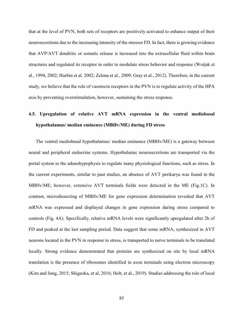
85
that at the level of PVN, both sets of receptors are positively activated to enhance output of their
neurosecretions due to the increasing intensity of the stressor FD. In fact, there is growing evidence
that AVP/AVT dendritic or somatic release is increased into the extracellular fluid within brain
structures and regulated its receptor in order to modulate stress behavior and response (Wotjak et
al., 1994, 2002; Hurbin et al. 2002; Zelena et al., 2009; Gray et al., 2012). Therefore, in the current
study, we believe that the role of vasotocin receptors in the PVN is to regulate activity of the HPA
axis by preventing overstimulation, however, sustaining the stress response.
4.5. Upregulation of relative AVT mRNA expression in the ventral mediobasal
hypothalamus/ median eminence (MBHv/ME) during FD stress
The ventral mediobasal hypothalamus/ median eminence (MBHv/ME) is a gateway between
neural and peripheral endocrine systems. Hypothalamic neurosecretions are transported via the
portal system to the adenohypophysis to regulate many physiological functions, such as stress. In
the current experiments, similar to past studies, an absence of AVT perikarya was found in the
MBHv/ME; however, extensive AVT terminals fields were detected in the ME (Fig.1C). In
contrast, microdissecting of MBHv/ME for gene expression determination revealed that AVT
mRNA was expressed and displayed changes in gene expression during stress compared to
controls (Fig. 4A). Specifically, relative mRNA levels were significantly upregulated after 2h of
FD and peaked at the last sampling period. Data suggest that some mRNA, synthesized in AVT
neurons located in the PVN in response to stress, is transported to nerve terminals to be translated
locally. Strong evidence demonstrated that proteins are synthesized on site by local mRNA
translation is the presence of ribosomes identified in axon terminals using electron microscopy
(Kim and Jung, 2015; Shigeoka, et al, 2016; Holt, et al., 2019). Studies addressing the role of local

86
protein synthesis indicated that local translation is required for functional and structural synaptic
plasticity in vivo (Holt, et al., 2019).
4.6. At the level of the MBHv/ME and anterior pituitary, downregulation of V1aR mRNA
and upregulation of V1bR mRNA appear to function to regulate the stress response and
CORT release
At the MBHv/ME, similar to the NHpC, V1aR-ir was found in glia. In the MBHv/ME
specialized glial cells called tanycytes occur at the base of and adjacent to the third ventricle (3V,
Fig.1F). Tanycytes in that location originate in the 3V and usually consist of a single process that
passes laterally and ventrally through brain tissue toward the ME and portal blood vessels. Directly
beneath the ME is the anterior pituitary, where both the V1aR and V1bR were identified on
corticotropes and are involved in the avian stress response (Jurkevich et al., 2005, 2008; Selvam
et al., 2013). Importantly, in both the MBHv/ME and anterior pituitary, V1aR mRNA expression
decreased, while gene expression of V1bR increased during FD stress (Fig. 4B and 5). As shown
in Fig. 4A, a gradual but steady increase in AVT mRNA occurred throughout the 8h FD period.
The increase was associated with upregulation of V1bR mRNA in the MBHv/ME and anterior
pituitary and concurrently downregulation of V1aR apparently to prevent enhanced CORT release.
A suggested means by which increased AVT secretion effects during the avian stress response
could be attenuated would be by endocytosis of V1a receptors on corticotrope membranes
(Jurkevich et al., 2005, 2008; Selvam et al., 2013). Consistent with that, mammalian studies
reported that upregulation of AVP mRNA in the PVN is responsible for upregulation of V1bR in
the anterior pituitary showing a positive relationship between AVP and its receptor, V1bR, and a
targeted deletion of V1bR was associated with attenuation of the stress response and production

87
of ACTH (Aguilera et al., 2008). Similarly, in birds, at the level of the anterior pituitary, not only
was upregulation of V1bR mRNA and downregulation of the V1aR revealed after FD stress,
previous data utilizing a different stressor resulted in the same response of the two receptors (Kang
and Kuenzel, 2014; Selvam et al., 2013) suggesting a generalized response in mammals and birds
to regulate the stress response.
5. Conclusion
An anatomical study of three brain areas showed that AVT cell bodies are found in the PVN,
not in the NHpC nor MBHv/ME. A major receptor of AVT, the V1aR, was identified by
immunohistochemistry in all three brain structures and located primarily in glia. Gene expression
data revealed that AVT, V1aR and V1bR mRNA are expressed in all three brain structures and
responded differentially to FD stress. Results showed that AVT, V1aR and V1bR are involved in
the late phase of FD stress after 3h. A negative feedback in gene expression between AVT and its
receptors, V1bR and V1aR, was found within the NHpC during FD stress. In contrast, AVT and
its two receptors showed a positive feedback at the level of the PVN during FD stress. At central
neuroendocrine level of the MBHv/ME and anterior pituitary response, upregulation of relative
AVT mRNA expression with positive expression of V1bR and decreased V1aR mRNA occurred
in both anatomical levels (MBHv/ME and APit). Overall, the response of AVT and its two major
receptors in the current model of the avian neuroendocrine stress pathway suggest that the balance
in function of the two receptors preserve and regulate ACTH secretion from the anterior pituitary
and ultimately plasma CORT to prevent overstimulation of the HPA axis during FD stress
responses.

88
Fig. 1. Chick brain sections show arginine vasotocin (AVT) and V1aR immunoreactive neurons or glia in the nucleus of the hippocampal commissure (NHpC), paraventricular nucleus (PVN), and ventral mediobasal hypothalamus/median eminence (MBHv/ME) (A) Neural terminal fields in the NHpC (B) AVT cell bodies in the PVN (C) AVT nerve terminal fields localized in the ME showing internal and external zones. D) V1aR-ir in the NHpC, E) V1aR-ir in the PVN, and, F) V1aR-ir in the MBHv/ME occurred in glia.

89
Fig. 2. Plasma corticosterone concentrations (ng/ml) following food deprivation (FD) for 1h, 2h, 3h, 4h, and 8h compared to controls (0h) were measured by RIA (n= 8-10 birds/group). Significance level used in all analyses was p < 0.05 using ANOVA followed by comparisons made among all pairs using the Tukey-Kramer HSD test. Data shown as mean ± SEM. Different letters above each time point show significant differences among the treatment groups.

90
Fig. 3. Relative gene expression levels of A. arginine vasotocin (AVT) B. vasotocin 1a receptor (V1aR) and C. vasotocin 1b receptor (V1bR) in the nucleus of the hippocampal commissure (NHpC) and paraventricular nucleus (PVN) following food deprivation for 1h, 2h, 3h, 4h, and 8h using RT-qPCR. Controls were set to 1, and data were expressed as mean ± SEM for each gene. Significance level set in all analyses was p < 0.05 using ANOVA and treatment comparisons for all pairs using Tukey-Kramer HSD test between control and treatment groups. Different letters above each bar within a set representing an anatomical structure (capital and lower-case letters) show significant (p<0.05) differences among treatment groups within that set.

91
Fig. 4. Relative gene expression levels of, A. arginine vasotocin (AVT). Time points connected with the same letter are not different (p > 0.05), B. Vasotocin 1a receptor (V1aR) and vasotocin 1b receptor (V1bR) in the ventral mediobasal hypothalamus/median eminence (MBHv/ME) following food deprivation for 1h, 2h, 3h, 4h, 8h and control (0h) using RT-qPCR. Controls were set to 1, and data were expressed as mean ± SEM for each gene. Significance level used in all analyses was p < 0.05 using ANOVA and comparisons for all pairs using Tukey-Kramer HSD test. Different letters above each bar within a set for an anatomical structure represent significant (p < 0.05) differences among those treatment groups.

92
Fig. 5. V1aR and V1bR mRNA expression levels in the anterior pituitary following food deprivation (FD). Data were analyzed using one-way ANOVA and comparisons for all pairs used the Tukey-Kramer HSD test. Relative mRNA levels were quantified, and data were set as relative expression levels using the 2−ΔΔCt method with GAPDH and β-actin serving as internal controls. Controls were set to 1, and data were expressed as mean ± SEM for each gene. Different letters above each bar for each receptor represent significant differences among treatment groups (p < 0.05).

93
References
Aguilera, G., Subburaju, S., Young, S., Chen, J., 2008. The parvocellular vasopressinergic system and responsiveness of the hypothalamic pituitary adrenal axis during chronic stress. Prog. Brain Res. 170, 29 – 39.
Aman, N., Nagarajan, G., Kang, S., Hancock, M., Kuenzel, W.K, 2016. Differential responses of the vasotocin 1a receptor (V1aR) and osmoreceptors to immobilization and osmotic stress in sensory circumventricular organs of the chicken (Gallus gallus) brain. Brain Res.1649, 67-78.
Bolborea, M., Dale, N., 2013. Hypothalamic tanycytes: potential roles in the control of feeding and energy balance. Trends Neurosci. 36, 91–100.
Cornett, L. E., Kang, S. W., Kuenzel, W. J., 2013. A possible mechanism contributing to the synergistic action of vasotocin (VT) and corticotropin-releasing hormone (CRH) receptors on corticosterone release in birds. Gen. Comp. Endocrinol. 188, 46 – 53.
Gray, D. A., Simon, E., 1983. Mammalian and avian antidiuretic hormone: studies related to possible species variation in osmoregulatory systems. J. Comp. Physiol. 151, 241–246.
Gray, M., Innala, L., Viau, V., 2012. Central vasopressin V1A receptor blockade impedes hypothalamic-pituitary-adrenal habituation to repeated restraint stress exposure in adult male rats. Neuropsychopharmacology 37, 2712 – 19.
Greenbaum, D., Colangelo, C., Williams, K., Gerstein, M., 2003. Comparing protein abundance and mRNA expression levels on a genomic scale. Genome Biol.4, 117.
Herkenham, M., 1987. Mismatches between neurotransmitter and receptor localizations in brain: observations and implications. Neuroscience 23, 1 – 38.
Herman, J.P., Schafer, M.K., Thompson, R.C., Watson, S.J., 1992. Rapid regulation of corticotropin-releasing hormone gene transcription in vivo. Mol. Endocrinol. 6, 1061–1069.
Holt, C.E., Martin, K.C., Schuman, E.M., 2019. Local translation in neurons: visualization and function. Nat. Struct. Biol. 26, 557 – 566.
Hurbin, A., Orcel, H., Alonso, G., Moos, F., Rabie, A., 2002. The vasopressin receptors colocalize with vasopressin in the magnocellular neurons of the rat supraoptic nucleus and are modulated by water balance. Endocrinology 143, 456 – 466.
Jung, H., Yoon, B.C., Holt, C.E., 2012. Axonal mRNA localization and local protein synthesis in nervous system assembly, maintenance and repair. Nat. Rev. Neurosci. 13, 308 – 324.

94
Jurkevich, A., Berghman, L.R., Cornett, L.E., Kuenzel, W.J., 2005. Characterization and immunohistochemical visualization of the vasotocin VT2 receptor in the pituitary gland of the chicken, Gallus gallus. Gen. Comp. Endocrinol. 143, 82 – 91.
Jurkevich, A., Berghman, L.R., Cornett, L.E., Kuenzel, W.J., 2008. Immunohistochemical characterization of chicken pituitary cells containing the vasotocin VT2 receptor. Cell Tissue Res. 333, 253 – 262.
Kadhim, H.J., Kang, S.W., Kuenzel, W.J., 2019. Differential and temporal expression of corticotropin releasing hormone and its receptors in the nucleus of the hippocampal commissure and paraventricular nucleus during the stress response in chickens (Gallus gallus). Brain Res. 1714, 1 – 7.
Kang, S., Kuenzel, W.K., 2014. Regulation of gene expression of vasotocin and corticotropin-releasing hormone receptors in the avian anterior pituitary by corticosterone. Gen. Comp. Endocrinol. 204, 25 – 32.
Kim, E., Jung, H., 2015. Local protein synthesis in neuronal axons: why and how we study. BMB Rep. 48, 139 – 146.
Kovacs, K.J., Sawchenko, P.E., 1996. Sequence of stress-induced alterations in indices of synaptic and transcriptional activation in parvocellular neurosecretory neurons. J. Neurosci. 16, 262 – 273.
Kuenzel, W.J., Jurkevich, A., 2010. Molecular neuroendocrine events during stress in poultry. Poult. Sci. 89, 832 – 840.
Kuenzel, W.J., Masson M., 1988. A stereotaxic atlas of the brain of chick (Gallus domesticus) Johns Hopkins University Press, Baltimore.
Leung, C.H., Abebe, D.F., Earp, S.E., Goode, C.T., Grozhik, A.V., Mididoddi, P., Maney, D.L., 2011. Neural distribution of vasotocin receptor mRNA in two species of songbird. Endocrinology 152, 4865 – 4881.
Ma, X-M., Levy, A., Lightman, S.L., 1997 Rapid changes in heteronuclear RNA for corticotrophin-releasing hormone and arginine vasopressin in response to acute stress. J. Endocrinol. 152, 81– 89.
Madison, F.N., Jurkevich, A., Kuenzel, W.J., 2008. Sex differences in plasma corticosterone release in undisturbed chickens (Gallus gallus) in response to arginine vasotocin and corticotropin releasing hormone. Gen. Comp. Endocrinol. 155, 566 – 573.
Mikhailova, M.V., Mayeux, P.R., Jurkevich, A., Kuenzel, W.J., Madison, F., Periasamy, A., Chen, Y., Cornett, L.E., 2007. Heterooligomerization between vasotocin and corticotropin-releasing hormone (CRH) receptors augments CRH-stimulated 3’, 50’cyclic adenosine monophosphate production. Mol. Endocrinol. 21, 2178 – 2188.

95
Mohr, E., Fehr, S., Richter, D., 1991. Axonal transport of neuropeptide encoding mRNAs within the hypothalamo–hypophyseal tract of rats. EMBO J. 10, 2419 – 2424.
Mohr, E., Richter, D., 1992. Diversity of mRNAs in the axonal compartment of peptidergic neurons in the rat. Eur. J. Neurosci. 4, 870 – 876.
Nagarajan, G., Jurkevich, A., Kang, S.W., Kuenzel, W.J., 2017a. Anatomical and functional implications of CRH neurons in the nucleus of the hippocampal commissure of the avian brain: an emphasis on glia-neuron interaction via the V1a receptors. J. Neuroendocrinol. 29, 1 – 11.
Nagarajan, G., Kang, S. W., Kuenzel, W.J., 2017b. Functional evidence that the nucleus of the hippocampal commissure shows an earlier activation from a stressor than the paraventricular nucleus: Implication of an additional structural component of the avian hypothalamo-pituitary-adrenal axis. Neurosci. Lett. 642, 14 –19.
Nagarajan, G., Tessaro, B.A., Kang, S.W., Kuenzel, W.J., 2014. Identification of arginine vasotocin (AVT) neurons activated by acute and chonic restraint stress in the avian septum and anterior diencephalon. Gen. Comp. Endocrinol. 202, 59 – 68.
Palkovits, M., 1973. Isolated removal of hypothalamic or other brain nuclei of the rat. Brain Res. 59, 449 – 450.
Proudman, J.A., Opel, H., 1989. Daily changes in plasma prolactin, corticosterone, and luteinizing hormone in the unrestrained, ovariectomized turkey hen. Poult. Sci. 68, 177–184.
Pryce, C.R., Siegl, S., Mayer, R., Rahmanzadeh, G., McAllister, K.H., 2011. Endocrine and behavioral responses to acute central CRF challenge are antagonized in the periphery and CNS, respectively, in C57BL/6 mice. Neuropharmacology 60, 318– 327.
Schmeling, S. K., Nockels, C. F., 1978. Effects of age, sex, and ascorbic acid ingestion on chicken plasma corticosterone levels. Poult. Sci. 57, 527 –533.
Schmittgen, T.D., Livak, K.J., 2008. Analyzing real-time PCR data by the comparative CT method. Nat. Protoc. 3, 1101 –1108.
Selvam, R., Jurkevich, A., Kang, S., Mikhailova, M., Cornett, L., Kuenzel, W., 2013. Distribution of the vasotocin subtype four receptor (VT4R) in the anterior pituitary gland of the chicken, Gallus gallus, and its possible role in the avian stress response. J. Neuroendocrinol. 25, 56 – 66.
Selvam, R., Jurkevich, A., Kuenzel, W.J., 2015. Distribution of the vasotocin type 4 receptor throughout the brain of the chicken, Gallus gallus. J. Comp. Neurol. 523, 335 – 358.
Shigeoka, T., Jung, H., Jung, J., Turner-Bridger, B., Ohk, J., Lin, J. Q., Amieux, P. S Holt, C.E., 2016. Dynamic axonal translation in developing and mature visual circuits. Cell 166, 181–192.

96
Tennyson, V. M., Nilaver, G., Hou-Yu, A., Valiquette, G., Zimmerman, E. A., 1986. Immunocytochemical study of the development of vasotocin/mesotocin in the hypothalamo-hypophysial system of the chick embryo. Cell Tissue Res. 243, 15 – 31.
Trembleau, A., Morales, M., Bloom, F., 1996. Differential compartmentalization of vasopressin messenger RNA and neuropeptide within the rat hypothalamo–neurohypophysial axonal tracts: light and electron microscopic evidence. Neuroscience 70, 113–125.
Vogel, C., Marcotte, E. M., 2012. Insights into the regulation of protein abundance from proteomic and transcriptomic analyses. Nat. Rev. Genet. 13, 227–232.
Wotjak, C.T., Ludwig, M., Ebner, K., Russell, J.A., Singewald, N., Landgraf, R., Engelmann, M., 2002. Vasopressin from hypothalamic magnocellular neurons has opposite actions at the adenohypophysis and in the supraoptic nucleus on ACTH secretion. Eur. J. Neurosci. 16, 477 – 485.
Wotjak, C.T., Ludwig, M., Landgraf, R., 1994. Vasopressin facilitates its own receptors within the rat supraoptic nucleus in vivo. Neuroreport 5, 1181–1184.
Yadawa, A.k., Chaturvedi, C.M., 2016. Expression of stress hormones AVP and CRH in the hypothalamus of Mus musculus following water and food deprivation. Gen. Comp. Endocrinol. 239, 13 – 20.
Yayou, K., Otani, H., Takusari, N., Uetake, K., Okamoto, T., 2003. Effects of intracerebroventricular infusions of corticotropin-releasing hormone in sheep. Anim. Sci. J. 74, 37– 44.
Yayou, K., Sato, Y., Ito, S., Nakamura, M., 2008. Comparison between the central effects of CRH and AVP in steers. Physiol. Behav. 93, 537–545.
Yayou, K., Seo, T., Uetake, K., Ito, S., Nakamura, M., 2007. Effects of intracerebroventricular infusions of arginine vasopressin in sheep. Physiol. Behav. 90, 376–381.
Zelena, D, Langnaese K, Domokos A, Pinter O, Landgraf R, Makara G, Engelmann, M., 2009. Vasopressin administration into the paraventricular nucleus normalizes plasma oxytocin and corticosterone levels in Brattleboro rats. Endocrinology 150, 2791–2998.
Vogel, C., Marcotte, E. M., 2012. Insights into the regulation of protein abundance from proteomic and transcriptomic analyses. Nat. Rev. Genet. 13, 227–232.

97
Chapter 4
Activation of corticotropin releasing hormone system in the nucleus of hippocampal
commissure during immobilization stress.
This chapter is excerpted from “Kadhim, H. J., Kang S. W., Kuenzel, W. J., 2020. Brain derived neurotrophic factor and extra-hypothalamic corticotropin releasing hormone neurons in the nucleus of hippocampal commissure play functional roles in the avian neuroendocrine regulation of stress.” Submitted to stress Journal on 7/31/2020
Abstract
Corticotropin releasing hormone (CRH) neurons located in the nucleus of hippocampal
commissure (NHpC) have been proposed to be involved in the avian neuroendocrine regulation of
stress and appear to respond prior to CRH neurons in the hypothalamic paraventricular nucleus
(PVN). The response, however, has been documented solely from applying one type of stressor,
food deprivation (FD). We therefore wanted to test whether the response of CRH neurons in the
NHpC was stressor specific. Additionally, since the response of the NHpC was rapid and short-
lived, was it regulated differentially from the PVN? We therefore applied a psychogenic stressor,
immobilization, to determine gene expression of CRH and a panel of stress-related genes in the
NHpC, PVN, and anterior pituitary (APit) and assayed the final stress product, plasma
corticosterone (CORT). Furthermore, brain derived neurotrophic factor (BDNF) and
glucocorticoid receptor (GR) were examined regarding their positive/negative roles in the
regulation of CRH neurons. Data showed that rapid activation of CRH mRNA in the NHpC
occurred and preceded a slower gene activation in the PVN resulting in subsequent upregulation
of proopiomelanocortin (POMC) transcripts in the APit associated with significant increases in

98
plasma CORT concentrations. Results suggested BDNF’s role in negative feedback observed
between CRH and CRHR1 in the NHpC and positive feedback between CRH and CRHR1 as well
as AVT and V1aR in the PVN. The GR mRNA expression and protein levels revealed that the
CRH neurons in the NHpC are regulated by CORT before those in the PVN. In the APit, V1bR
activation appeared responsible for sustaining POMC gene expression for continued CORT release
when stress persists. Overall, the data suggest that the NHpC functions as part of the traditional
HPA axis of birds and perhaps a comparable extra-hypothalamic structure may also be present in
mammals and other vertebrates.
Keywords: - immobilization stress, corticotropin releasing hormone receptors, paraventricular
nucleus, glucocorticoid receptors, V1b receptor.
1. Introduction
The hypothalamic- pituitary- adrenocortical (HPA) axis is the traditional regulator of the stress
response in vertebrates. Parvocellular neurons located mainly within the hypothalamic
paraventricular nucleus (PVN) produce corticotropin releasing hormone (CRH) and arginine
vasopressin (AVP) in mammals and arginine vasotocin (AVT) in avian species (Whitnall et al.,
1985; Kuenzel and Jurkevich, 2010). Both neuropeptides have a set of two G-protein coupled
receptors, CRHR1 and CRHR2 for CRH and V1aR and V1bR for AVP/AVT. Activation of
parvocellular neurons by stress increases synthesis of CRH and AVT and release of each
neuropeptide from the external zone of the median eminence. Once transported to the anterior
pituitary (APit) via hypophyseal portal vessels, they bind to their receptors located on corticotropes
and stimulate proopiomelanocortin (POMC) that is further processed to the adrenocorticotropic
hormone (ACTH) (Bonfiglio et al., 2011; Blas, 2015). When ACTH reaches adrenal glands via

99
blood circulation, it activates the interrenal tissue to increase synthesis and release of the stress
hormone, corticosterone (CORT) in birds (Romero, 2004, Carsia, 2015; Herman et al., 2016;).
Stress hormone binds to glucocorticoid receptors (GRs) located on different tissues to provide
energy for immediate use (McEwen, 2007) as well as induces a negative feedback that regulates
HPA axis activity (Lovejoy and Balment, 1999; De Kloet et al., 2005; Vandenborne et al., 2005;
Chrousos, 2009; Keller-Wood, 2015).
Immunohistochemical studies have reported activation of c-fos gene (a neuronal activation
marker) in the nucleus of the hippocampal commissure (NHpC), previously called the nucleus of
the pallial commissure, located in the septum just above the anterior commissure (AC), following
a variety of stressors suggesting that the NHpC could be involved in the regulation of the stress
response ( Xie et al., 2010; Nagarajan et al., 2014). Furthermore, we identified CRH neurons were
identified in the NHpC for the first time colchicine injection (Nagarajan et al., 2014). The CRH
neurons in the NHpC are different from those in the PVN, they are larger and multipolar neurons
compared to CRH cells in the PVN suggesting these neurons may have various roles (Nagarajan
et al., 2017a). To date, a structure homologous to the avian NHpC has not been identified in
mammals or other vertebrate species.
Utilizing food deprivation (FD) stress enabled us to study the sequence of gene expression of
CRH in the NHpC and showed that CRH gene expression in the NHpC activated rapidly and
appeared to precede those in the PVN indicating that CRH neurons in the NHpC are early
responders that initiate the stress response to that specific stressor, FD ( Nagarajan et al., 2017b;
Kadhim et al., 2019). Our recent publication utilizing FD showed that CRH gene expression peaks
early in the NHpC and returned to control levels. In contrast, a steady increase in gene expression

100
of CRH and its major receptor, CRHR1, occurred in the PVN suggesting a positive feedback
(Kadhim et al., 2019). A similar, positive relationship was found with the neuropeptide, AVT and
its receptors in the PVN after FD stress, but not in the NHpC (Kadhim et al., 2020). It has been
found that neurotrophic factors, specifically brain derived neurotrophic factor (BDNF), could be
involved in the regulation of the stress response (Bath et al., 2013) and CRH neurons (Jeanneteau
et al., 2012) because the BDNF gene promotor site has many binding sites for the cyclic adenosine
monophosphate (cAMP) response element protein (CREB) (Blendy, 2006; Tardito et al., 2006).
Research addressing the sequence of activation of CRH and AVT neurons and their receptors
within the NHpC and PVN has to date only involved a single stressor, FD. We therefore utilized
a totally different stressor, immobilization, to determine whether the neuroendocrine stress
response sequence follows the same pattern and is initiated by activation of the NHpC. Our
previous stressor, FD, affects the energetic state of an animal and intensifies with time. In contrast,
immobilization, a neurogenic stressor, seems to have a more profound effect initially. However,
an animal appears to adapt to it over time if water and/or food are available. Applying this stressor
over a short time span should not impact significantly its energetic state and would be a valid test
to determine whether a similar pattern of stress genes occurs providing further evidence that the
NHpC functions within the avian HPA axis and is not stressor specific. Our hypothesis is that CRH
neurons in the NHpC would be the first responder to immobilization stress followed by sequence
of stress related genes and receptors that would match the pattern previously determined for FD
stress in the same avian species. If the hypothesis is proven to occur, this would support the
inclusion of the NHpC in the avian traditional HPA axis. Additionally, we focused on BDNF to
determine its possible role in regulating expression within the NHpC and PVN.

101
2. Material and methods
2.1. Animals
Male Cobb 500 chicks (Gallus gallus) one day old, were raised in brooder batteries for their
first ten days and provided food (a standard, broiler starter diet) and water ad libitum. Continuous
light was used for the first 3 days enabling birds to find both food and water. Thereafter, a light
program of 16h of light (L): 8h of dark (D) was initiated with lights on at 6:00 AM. Heat provided
to the chicks was initially 32 ℃ that was reduced 2.5 ℃ weekly until reaching 24 ℃. On day 10,
after weighing them, birds were distributed randomly to cages (3 birds/cage). At 5 weeks of age,
experiments were initiated, and sampling occurred between 8:00 AM and 4:00 PM. All procedures
utilized (i. e. immobilization, housing conditions, handling, and sampling) were approved by the
University of Arkansas Institutional Animal Care and Use Committee.
2.2. Antibody production against chicken CRH receptor 1
Polyclonal antibodies to chicken CRHR1 were produced in guinea pigs by a commercial
company (21st Century Biochemicals Inc., Marlboro, MA, USA). Briefly, a cohort of guinea pigs
(n = 4) were injected with synthetic peptides comprising 13 or 14 amino acids from the 1st
extracellular domain of chicken CRHR1 (residues 180-193; TMNPEVHESNVVWC). Two
versions of the same peptide sequence were used, one had a cysteine added to the N-terminus, and
the other peptide had the cysteine added to the C-terminus, enabling conjugation to KLH (keyhole
limpet hemocyanin) via both the N- and C-termini. The amino acid sequence of the protein was
probed against the nonredundant GenBank protein database using NCBI-BLAST software (NCBI
reference sequence: NP_989652.1). The chosen sequence was specific to the chicken CRHR1 (See

102
Appendix A. Supplementary data). To enhance the immune system, the peptide was conjugated to
a carrier protein, keyhole limpet hemocyanin. The peptides were mixed and injected with complete
Freund’s adjuvant at days 0, with boosts at 14, 28, 42 and 63 using incomplete Freund’s adjuvant.
Production bleeds were taken at days 49, 70 and 77. The final bleed from one of the guinea pigs
was affinity purified. The peptide sequence was confirmed using tandem mass spectrophotometry
(MS).
2.3. Stress procedure and sample collection for gene expression
A stressor, immobilization, was initiated on week 5, with food and water provided ad libitium.
Chicks were randomly assigned to one of six treatment groups (n = 12/treatment): control (no
stress), 15 minutes (m), 30m, 60m, 90m, and 120m. Birds were secured in a harness where they
could not move their wings nor stand, however, did have access to water and food during the period
of restraint. Directly following restraint, blood samples were taken from the brachial vein and
collected in heparinized tubes for each bird. After cervical dislocation, brain and APit were rapidly
dissected. To maintain structural morphology of the brain for cryosectioning, brain samples were
immediately immersed in two-methyl butane at −30 ℃ for 15s, placed in dry ice and stored at −80
℃ until processed. Coronal sections of brain samples were cut at 100 µm using a cryostat (Leica
CM3050 S, Leica Microsystems, Frisco, TX) and the targeted structures were punched (brain
punch, Palkovits, 1973) using a glass pipette including (1) the NHpC, (previously labeled nCPa),
1.4 mm diameter from atlas plates A8.2–A7.6; (2) the PVN, 1.4 mm diameter including atlas plates
A8.0–A6.4 (Kuenzel and Masson, 1988). For the NHpC dissection, the anterior commissure (AC)
was used as a landmark immediately ventral to the NHpC. NHpC and PVN were punched,

103
transferred to separate vials containing Trizol, and stored at −20 ℃ until processed for RNA
extraction.
2.4. Radioimmunoassay (RIA)
Blood samples were taken from the brachial vein of chicks in all treatment groups (n=12/
treatment). Plasma was obtained from heparinized blood via centrifugation at 3000 rpm for 20m
at 4 ℃. Hemolyzed samples (1-2 samples/ group) were excluded. Plasma was stored at - 20 ℃
until analysis of CORT concentrations by radioimmunoassay (Proudman and Opel, 1989; Madison
et al., 2008; Kadhim et al., 2020). Briefly, 100 μl of the primary antibody, polyclonal rabbit anti-
CORT # 377, (kindly provided by from Dr. Proudman) and 100 μl of 125I corticosterone tracer
purchased from MP Biomedicals Inc. (Orangeburg, NY, USA) were added to each sample and
incubated for at least 24h at 4 ℃. Sheep anti-rabbit antibody (200 μl) was used as the secondary
antibody (MP Biomedicals Inc., Orangeburg, NY, USA). Counts/tube were determined using a
Perkin Elmer Wizard gamma-counter. Samples were assayed in duplicate. Data were analyzed by
one-way ANOVA followed by a Tukey’s Kramer HSD test and expressed as the mean ± SEM of
each group. In all studies, p < 0.05 was considered statistically significant. Intra experimental
coefficient of variance was less than 11 %.
2.5. RNA extraction from collected samples
RNA extraction from frozen micro-dissected brain tissue and anterior pituitaries (n=12
birds/group) were described previously (Kadhim et al., 2019; Kadhim et al., 2020). Briefly, Trizol-
chloroform (Life Technologies) according to the protocol provided by the supplier was used to
extract total RNA followed by a treatment to eliminate contamination with genomic DNA using

104
RNase-free DNase I (Ambion, Austin, TX, USA). Purification was then conducted using a RNeasy
mini kit (Qiagen), and Synergy HT multi-mode micro plate reader (BioTek) was used to estimate
RNA concentration. Samples with insufficient amount RNA (1-3 samples) were excluded. First-
strand cDNA was synthesized in 60 μl from total RNA (400 ng of NHpC, 700 ng of PVN, and 600
ng of APit) using Superscript® III reverse transcriptase (Invitrogen) according to the
manufacturer’s protocol. The best primer pair was chosen depending on past studies in our lab or
selected from several pairs based on PCR product quality and lengths after electrophoresis on a
3% agarose gel ( Kang and Kuenzel; 2014; Kadhim et al., 2019; Kadhim et al., 2020). The assay
was run in triplicate and achieved in 30 μl using the following conditions: 1 cycle at 95 ℃ for 10m
and amplified for 40 cycles at 95 ℃ for 30s, 60 ℃ for 1m, and 72 ℃ for 30s. The chicken
glyceraldehyde-3-phosphate (GAPDH) or beta actin (βA) were used as internal controls to
normalize the mRNA levels. Relative gene expression levels of each specific gene were
determined by the 2−ΔΔCt method (Schmittgen and Livak 2008). Relative expression of control
groups was set to 1.
2.6. Western blot experiment
Protein content analysis was conducted for APit glands and brains collected from birds after
being immobilized for different time lengths. Brain samples were immediately frozen by
immersion in two-methyl butane at −30 ℃ for 15s. Both organs were stored at −80 ℃ until
processed. Brains were sectioned in a cryostat (−15 ℃). NHpC and PVN were collected in separate
1.5 test tubes. Total proteins were extracted using RIPA Lysis and extraction Buffer (Thermo
Fisher scientific) mixed with protease and phosphatase inhibitor cocktails (Thermo Fisher
scientific) in accordance with the manufacturer’s instructions. Tissues were homogenized with the

105
buffer using glass beads and centrifuged at 14000 rpm/ 20m at 4 ℃. The supernatant was taken,
and a Bradford protein assay (Bio-Rad protein assay kit, Hercules, CA, USA) was utilized to
determine the concentration of the protein using Synergy HT multimode microplate reader
(BioTek, Winooski, VT). Bovine serum albumin (BSA) was used as a standard (Bio-Rad,
Hercules, CA, USA). Total protein from the NHpC (40 µg), PVN (60 µg) and anterior pituitaries
(60 µg) were diluted in loading LDs (Lithium dodecyl sulfate) sample buffer containing a reducing
agent (Thermo Fisher scientific). Samples were heated for 10m at 70 ℃ before loaded onto a
NuPAGE™ 4-12% Bis-Tris Protein gel (Thermo Fisher scientific) and separated at 150 V until
reaching the ends of the gel (Bio-Rad). Proteins were transferred to an Amersham hybond P 0.2
PVDF membrane (GE healthcare life sciences) by applying a current of 30 V for 75m (Bio-Rad)
in Tris-Glycine HCl buffer (pH 7.6). Nonspecific binding was blocked by incubating the
membrane for 2h/ RT in 5% nonfat milk (Sigma, St Louis, MO, USA). The membrane was then
incubated overnight with primary antibody guinea pig anti-chicken CRHR1 (polyclonal, dilution
1: 5000) and mouse anti-GR (polyclonal, 1: 200 dilution, sc-393232, Santa Cruz Biotechnology,
Inc.). After washing the membrane twice in Tris-buffered saline with 0.1% Tween-20 (TBS-T),
the membrane was incubated with horseradish peroxidase-conjugated anti-mouse, or anti-guinea-
pig immunoglobulin IgG (1:5000) for 1h at room temperature (Cell signaling technology, USA).
The membrane was rinsed twice with TBS-T. An Enhanced Chemiluminescence Kit (Immun-
star™ WesternC™ Kit, Bio-Rad, USA) was used to visualize the signal and captured by
FluorChem M MultiFluor system (protein sample). Rabbit anti-GAPDH (dilution 1:5000, NB300-
327, Novus Biologicals) was used as the reference protein. Image acquisition and analysis were
performed by alpha view software (Version 3.4.0, 1993-2011, Protein Simple, Santa Clara, CA).

106
2.7. Statistical analysis
JMPR pro 14.0 (SAS Institute Inc., NC) was used to perform statistical analyses for gene
expression and hormone assays. A normal distribution was first tested and thereafter differences
among six independent groups for the NHpC, PVN, APit, and plasma CORT concentration were
analyzed separately using one-way analysis of variance (ANOVA). An F- value and degrees of
freedom were provided for each structure (NHpC, PVN, and APit) and CORT concentration.
Comparison for all pairs using Tukey’s Kramer HSD test were used to evaluate plasma CORT
concentration, changes in protein levels, and relative changes of genes expression between the
control and each immobilized group. Data are presented as the mean ± SEM. A probability level
of p < 0.05 was considered statistically significant.
3. Results
3.1. Immobilization induced CORT concentration increase in blood plasma
During immobilization, a robust and rapid increase of CORT plasma concentration occurred
(Fig. 1). Specifically, plasma CORT concentration was increased after 15 min of immobilization
with peak levels reached at 30 m (p < 0.001). Thereafter, CORT levels declined after 60m
compared with 15m and 30m, although it remained significantly higher than the initial control
values at time 0. The overall significance of CORT concentration changes between immobilized
groups compared to the control group was [F (5, 47) = 14.48, p < 0.001].
107
3.2. Gene Expression in the NHpC and PVN during immobilization stress
3.2.1. CRH, CRHR1, and CRHR2
The overall gene expression data for CRH showed significant differences among treatment
groups compared to controls [F (5, 48) = 34.18, p < 0.001 in the NHpC; F (5, 48) = 21.08, p <
0.001 in the PVN]. In the NHpC, CRH mRNA levels increased rapidly and significantly at 15m
of stress initiation by over 100% of the control value and remained significantly higher at 30m of
restraint stress. Thereafter, at 60m, 90m, and 120m of immobilization, CRH gene expression
returned to control levels (Fig. 2A). In contrast, gene expression of CRH in the PVN displayed a
gradual upregulation by less than 30% at 15m. A peak level of mRNA occurred at 90m of
immobilization stress followed by more than a 50% decline by 120m, although it remained
significantly higher than controls (Fig. 2A). Gene expression of CRHR1 compared to CRHR2
within the NHpC demonstrated different patterns across the 2 h study. Both receptors showed a
significant treatment effect [F (5, 48) = 30.17, p < 0.001] for CRHR1 and [F (5, 48) = 28.88, p <
0.001] for CRHR2. Specifically, the pattern of CRHR1 mRNA expression in the NHpC was
downregulation at 15m of immobilization stress, followed by a rapid recovery to basal, control
levels at 30m and 60m, and ending with significant increases at 90m and 120m of stress (Fig. 2B).
In contrast, mRNA expression of CRHR2 in the NHpC did not change in the 15m period of stress.
Thereafter, the first significant upregulation was seen at 30m and persisted at that level of
expression through 90m with a second upregulation at 120m of treatments (Fig. 2C). In the PVN,
the two receptors, CRHR1 and CRHR2, were upregulated significantly [F (5, 48) = 39.37, p <
0.001] for CRHR1 and [ F (5, 48) = 48.10, p < 0.001] for CRHR2. Of particular interest, the p
expression levels and response for CRHR1 and CRHR2 mRNA were nearly the same showing

108
their first significant increase at 30m of immobilization stress which continued to increase and
peak at 90m, then decreased at 120m, however, remained significantly higher than controls (Fig.
2B, 2C). Importantly, both receptors were upregulated in the PVN matching the upregulation of
their ligand, CRH.
3.2.2. AVT and V1aR
Gene expression of arginine vasotocin (AVT) and its major receptor, V1aR, was measured in
the NHpC and PVN during the stressor. Different patterns of AVT mRNA expression occurred
over treatment times [F (5, 48) = 18.4, p < 0.001 in the NHpC; F (5, 48) = 32.4, p < 0.001 in the
PVN]. Specifically, AVT mRNA levels in the NHpC decreased significantly and never increased
above control levels throughout the 2h stress treatment (Fig. 3A). In contrast to the NHpC, AVT
mRNA expression in the PVN displayed no significant change in gene expression until 60m of
immobilization. At 60, 90, and 120m there occurred a consistent, step up in gene expression that
peaked at the end of the stress treatments (Fig. 3A). Regarding the V1aR in the NHpC, it initially
significantly decreased its gene expression similar to the response of AVT. Thereafter, however,
its gene expression significantly increased from its low level at 15m to its peak gene expression at
120m. In contrast, within the PVN, gene expression of the V1aR matched nearly perfectly the
expression of AVT throughout the period of restraint stress (Fig. 3B). The V1aR gene expression
in stressed birds displayed an overall significant effect among treatment groups [F (5, 48) = 13.72,
p<0.01 in the NHpC and F (5, 48) =16.51, p < 0.01 in the PVN].

109
3.2.3. Brain derived neurotrophic factor (BDNF) and glucocorticoid receptor (GR)
Gene expression of brain derived neurotrophic factor (BDNF) in the NHpC and PVN showed
significant differences among groups [F (5, 48) = 11.38, p < 0.01 in the NHpC; F (5, 48) = 17.17,
p < 0.01 in the PVN]. BDNF mRNA levels in the NHpC decreased at 15m, recovered to the basal
level at 30m and remained around the basal levels through 90m, then increased significantly at
120m of immobilization (Fig. 4A). In contrast to the NHpC, relative mRNA expression of BDNF
in the PVN was upregulated significantly at 15m of stress initiation, remained at that elevated
plateau until the end of the sampling period (120m). Interestingly, matching between CRHR1
and BDNF mRNA expression was observed in both the NHpC and PVN structures (Fig. 2B and
4A).
In the NHpC, the relative mRNA expression of the glucocorticoid receptor (GR) decreased
to a nearly significant level at 30m followed by the first significant upregulation demonstrated at
90m with a peak occurring at 120m (Fig. 4B). In the PVN, GR mRNA levels showed a slight
non-significant downregulation from 15 to 90m followed by its only a significant upregulation
at the end (120m) of restraint stress (Fig. 4B). Nonetheless the overall gene expression of the GR
in the NHpC and PVN showed significant differences among groups [F (5, 48) = 15.38, p < 0.01
in the NHpC; F (5, 48) = 13.17, p < 0.01 in the PVN].
3.3. Gene expression in the anterior pituitary (APit)
3.3.1. Heteronuclear POMC and mRNA POMC
Heteronuclear proopiomelanocortin (hnPOMC) and mRNA expression were measured in the
APit gland as an indicator of corticotrope activation during immobilization stress. Its timing and

110
rate of upregulation would help determine if the increase in CRH mRNA in the NHpC was
responsible for or contributed to the initiation of the neuroendocrine stress response at the level of
the APit. Immobilized birds showed an overall significant effect among treatment groups [F (5,
55) = 19.62, p<0.001 for POMC mRNA; F (5, 55) =21.73, p < 0.001 for hnRNA]. In the APit,
POMC transcripts (hn and mRNA) displayed a similar pattern over the sampling points (Fig. 5).
In details, POMC (hn and mRNA) relative gene expression levels upregulated significantly at 15m
of immobilization stress and matched the significant increase of CRH mRNA in the NHpC. The
hnPOMC gene expression downregulated significantly at 60m of restraint until the last sampling
point (120m). On other hand, the mRNA POMC expression upregulated at 15m followed by a
significant downregulation at 60m and subsequent fluctuation that were not significantly different
from pre-stress control levels.
3.3.2. CRHR1, CRHR2, V1aR, V1bR, and GR in the Anterior pituitary
Gene expression of the CRH receptors (CRHR1 and CRHR2), AVT receptors (V1aR and
V1bR) and GR in the APit displayed different patterns during immobilization stress (Fig. 6).
Specifically, the CRHR1 mRNA downregulated significantly at 60m of immobilization and
remained lower than the initial, basal level until the end of restraint treatment (Fig. 6A). In contrast
to the CRHR1, the expression of CRHR2 mRNA upregulated significantly at 15m and continued
increasing throughout the entire sampling period. Additionally, stressed birds exhibited an overall
significant effect among treatment groups [F (5, 55) = 23.22, p < 0.001 for CRHR1; F (5, 55)
=18.13, p < 0.0001 for CRHR2]. Like the CRH receptors, AVT receptors, V1aR and V1bR,
displayed an expression pattern similar to that of the CRH receptors (Fig. 6B). Expression of the
V1aR decreased significantly at 30m (p < 0.01) and 60m (p < 0.001). Thereafter, the mRNA levels

111
recovered to basal levels at 90m and 120m. In contrast, the expression levels of the V1bR mRNA
increased significantly at 15m and continued to rise and peaked at 90m. Additionally, stressed
birds exhibited an overall significant effect among treatment groups [F (5, 55) = 27.12, p<0.01 for
V1aR; F (5, 55) =20.33, p < 0. 01 for V1bR].
To understand the possible negative regulation via glucocorticoid receptors at the level of the
APit, a detailed time course of glucocorticoid receptor (GR) mRNA expression was documented
(Fig. 6C). GR gene expression in stressed birds showed an overall significant affect among
treatment groups compared with non-immobilized controls (F (5, 55) = 12.22, p < 0.001).
Specifically, GR mRNA expression displayed a non-significant increase at 15m following stress
initiation (p = 0.07), then downregulated rapidly and significantly at 30m and 60m. Thereafter,
mRNA relative levels returned to basal levels comparable to controls.
3.4. Western blot results
3.4.1. CRHR1 and GR protein levels in the NHpC and PVN
The proteins of corticotropin releasing hormone receptor1 (CRHR1) and glucocorticoid
receptor (GR) were detected in the NHpC and PVN extracted from birds subjected to different
lengths of immobilization stress and compared with non-immobilized birds (controls). Data
showed clear bands of CRHR1 and GR located at 49 kDa and 83 kDa, respectively, in both the
NHpC and PVN (Fig. 7). It is interesting to note that the CRHR1 bands were very dense in the
NHpC compared with the CRHR1 bands in the PVN despite uploading less protein concentration
(40 µg for the NHpC and 60 µg for the PVN) and showed different densities over the
immobilization stress time periods (Fig. 7A and 7B). In the NHpC, the analysis of band densities
112
using the alpha view SA program showed that CRHR1 protein levels decreased significantly at
15m of immobilization (p < 0.01), returned to the basal, control levels at 30 and 60m and increased
at 90m and 120m (p < 0.001) (Fig. 7A). Remarkably, the protein levels of CRHR1 in the NHpC
match the gene expression pattern of CRHR1 (Fig. 7A and 2B). In contrast to the NHpC, the
protein levels of CRHR1 in the PVN never decreased through all treatments group compared with
control groups. Specifically, the first significant increase of CRHR1 was observed at 60m (p <
0.01) and continued to the last sample taken at 120m (Fig. 7B). Unlike the NHpC, the gene
expression of CRHR1 in the PVN preceded the increased protein content (Fig. 2B and 7B). The
overall significant difference for CRHR1 was calculated between stressed and control chicks for
each structure [F (5, 30) = 11.26 p < 0. 01 in the NHpC; F (5, 30) = 9.13, p < 0.01 in the PVN].
Regarding glucocorticoid receptors (GRs), the GR protein levels remained at basal levels in the
NHpC at 15m and 30, then increased significantly at 60m (p < 0.001) through 120m (Fig. 7A).
Gene expression and protein levels of GRs in the NHpC followed the same pattern (Fig. 4B and
7A). While in the PVN, the significant increase in GR protein levels occurred only at 120m (p <
0.001), a time point matching its GR gene expression (Fig. 7B and 4B). The overall significance
for the GR among all treated groups for each structure was [F (5, 30) = 8.76 p < 0. 01 in the NHpC;
F (5, 30) = 12.43, p < 0.01 in the PVN].
3.4.2. CRHR1 and GR protein levels in the anterior pituitary (APit)
The protein levels for the CRHR1 and GRs were detected and quantified in the APit during
immobilization stress (Fig. 8). CRHR1 and GR proteins showed clear bands located at the 49 kDa
and 83 kDa positions, respectively, on the gel, matching the molecular mass of both receptors in
Gallus gallus. The CRHR1 protein levels decreased significantly at 60m and remained at that level

113
through 120m (Fig. 8). Similarly, the relative expression of CRHR1 mRNA in the same tissue
downregulated at 60m compared to the control group indicating that the downregulation of
CRHR1 gene expression matches the decreasing of protein levels (Fig 6A and 8). In contrast,
glucocorticoids receptor (GRs) in the APit showed significantly increased protein levels at 15m of
stress followed by a decrease to basal levels through to the last sampling period (Fig. 8).
4. Discussion
4.1. CRH neurons in the NHpC play a major role in initiating a neuroendocrine stress
response involving the APit and CORT release
The time course of CRH gene activation in the NHpC and PVN following immobilization
stress showed that CRH mRNA expression in the NHpC was rapidly upregulated, peaked at 15m,
remained significantly higher at 30m, and then returned to basal, control levels throughout the
remaining sampling time periods. In contrast, CRH mRNA levels in the hypothalamic PVN
increased gradually and peaked at 90m near the end of the stress treatment period (Fig. 2A). Data
demonstrated that septal CRH neurons within the NHpC display a more rapid response to that
stressor than those in the PVN. Specifically, CRH mRNA transcripts in the NHpC increased more
than 100% at 15m of stress compared to less than a 30% increase in the PVN. Remarkably, the
pattern of gene expression of APit hnPOMC matched that of CRH mRNA in the NHpC (Compare
Fig. 2A with Fig. 5 and 1). Importantly, plasma CORT levels increased more than 8-fold from
controls at the 15m sampling period suggesting strongly that a major contributor to CORT increase
came from CRH neurons within the NHpC. A past study utilized a totally different stressor, food
deprivation (FD), where a much longer time period is required before significant elevations in gene
expression of CRH, hnPOMC and ultimately plasma CORT can be realized from activation of the
114
neuroendocrine stress axis. Interestingly, results from two independent studies showed a similar
response at the neural, APit and blood response levels occurred showing a major input from CRH
neurons within the NHpC (Compare Fig. 2A, with Fig. 5 and 1 in Kadhim et al., 2019 and Fig. 2
and 3 in Nagarajan et al., 2017a). Overall, the major and early significant contribution of increased
transcripts of mRNA from the NHpC that complemented the gradual rise in CRH gene expression
in the PVN utilizing two very different stressors, in our judgment, provide sufficient data to
validate our earlier suggestion that the NHpC should be included as one of the key neural structures
activating the neuroendocrine regulation of stress in birds (Nagarajan et al., 2017a).
4.2. The role of BDNF and GRs in effecting positive feedback and negative feedback of CRH
and its receptor, CRHR1 in the PVN and NHpC, respectively
The PVN displayed a positive feedback in gene expression of CRH and its major receptor,
CRHR1, throughout the stress period. Both CRHR1 mRNA and its protein quantification were
significantly elevated at 30m and 60m, respectively, and maintained that response to the end of
treatments. Of interest, BDNF within the PVN showed a significant increase in gene expression
from 15m to the end of immobilization. Most importantly, its significant increase preceded that of
the significant increase in CRHR1 mRNA. Data of the study support a mammalian experiment in
which intracerebroventricular injections of BDNF resulted in a gradual increase in CRH mRNA
suggesting that BDNF is a stress-responsive intercellular messenger (Givalois et al., 2004). In
contrast, the NHpC showed a negative feedback in gene expression between CRH and its receptor,
CRHR1. CRH mRNA significantly increased at 15m while CRHR1 mRNA significantly
decreased. Of particular interest, BDNF mRNA significantly declined at 15m, the same time
period when CRH mRNA peaked in the NHpC.

115
Hence, the response of BDNF in the PVN was essentially opposite that observed within the
NHpC. In contrast, when gene expression of BDNF and CRHR1 was compared throughout the 2h
stress period in the NHpC, both genes showed a similar pattern. When the same two genes were
compared in the PVN, their pattern matched as well. Therefore, the matching of CRHR1 and
BDNF gene expression in two different populations of CRH neurons suggests an important, direct
functional relationship between that receptor and a neurotrophic factor within each structure.
BDNF appears to play a significant role in determining the relationship between CRH, and its
major receptor, CRHR1. Mammalian studies reported that downregulation of BDNF occurred
when CRHR1 was blocked centrally (Bayatti et al., 2005; de la Tremblaye et al., 2016). Similarly,
the natural downregulation of BDNF mRNA in the NHpC matched the decline of the CRHR1 at
the 15min sampling point clearly displaying a negative feedback between CRH and CRHR1 in
that structure.
Additionally, a distinct interaction between BDNF and GR occurred in the PVN compared to
the NHpC. The GR mRNA and its protein content in the PVN showed its expected no change from
controls through its first four stress periods thereby exerting little, if any, negative control on the
impressive rise in BDNF, CRH and CRHR1 mRNA. Gene expression of GR finally showed a
significant increase at the last sampling period, the same time when CRH and CRHR1 mRNA
showed clear declines from their previous peak responses at 90m. In contrast, BDNF mRNA was
significantly reduced in the NHpC when its CRH peaked, while GR mRNA and its protein showed
significant increased gene expression and protein content, thereafter from 60m to 120m of
immobilization stress (Fig. 4B and 7A), resulting in a significant decline in CRH gene expression
caused by high CORT levels via GRs. This occurred initially in CRH expressing neurons in the
NHpC and later in the PVN supporting past data that glucocorticoids restrain the HPA axis activity

116
through GR (Ginsberg et al., 2006; Yao and Denver, 2007; Noguchi et al., 2010). In summary, it
appears that BDNF has two effects on CRH and its major receptor. BDNF’s initial positive or
negative gene expression can determine if a positive or negative feedback will occur. Secondly,
the subsequent interaction of BDNF with GRs and CORT can influence the length of time CRH
mRNA will display significant upregulation.
4.3. Role of CRHR2 in the PVN and NHpC
The positive feedback response of CRHR2 gene expression with CRH in the PVN matched
almost perfectly the significant, increased CRHR1 mRNA response (Compare Fig. 2C with 2B).
Although increased gene expression for CRHR2 occurred in the NHpC as well, it is clear that a
more dynamic action occurred in the PVN compared to the NHpC (Fig. 2C). It is well known that
the major site of CRHR2 occurs on thyrotropes, not corticotropes in the anterior pituitary (De
Groef et al., 2003). Indeed, a recent study in our lab has shown that when broiler chicks are exposed
to 2h of FD stress, not only is the HPA axis significantly activated, but the hypothalamic pituitary
thyroid (HPT) axis as well (Kidd Jr. et al., 2020, submitted). Importantly, when the NHpC is
surgically disrupted with electrolytic lesions, the HPT axis response to 2h of food deprivation is
not significantly different compared to controls having an intact NHpC and PVN structures (Kidd
Jr et al., 2020). Although thyroid stimulating hormone beta (TSHβ) mRNA was not measured in
our current study, our recent study suggests that the PVN, not the NHpC contains the major
population of neurons responsible for activating the HPT axis. Furthermore, mammalian studies
demonstrated that the extracellular release of CRH upregulates CRHR2 within brain structures
(Korosi et al., 2006; Greetfeld et al., 2009) resulting in, possibly, the activation of thyrotropin
releasing neurons. Data herein suggest that stimulation of CRH neurons results in upregulation of

117
CRHR2 and the HPA and HPT axes thereby linking them in the avian neuroendocrine stress
response.
4.4. Activation of BDNF preceded an enhanced gene expression of AVT and V1aR in the
PVN
Gene expression of AVT and its major receptor, V1aR, in the PVN upregulated significantly
at 60m that lasted to the end of immobilization stress at 120m with a peak response (Fig.3). The
match between the increase in mRNA transcripts of AVT and its major receptor, V1aR, in the
PVN, throughout the imposed immobilization stress indicated a positive feedback occurred,
supporting our previous findings utilizing FD stress (Kadhim et al., 2020). Interestingly,
upregulation of BDNF mRNA levels in the PVN preceded the increase of AVT and V1aR mRNA
levels in that structure. The pattern of upregulation displayed by BDNF, V1aR and AVT matched
well the increased gene expression for BDNF, CRHR1 and CRH, discussed in a previous Sub-
heading (4.2). In contrast, BDNF mRNA level in the NHpC was downregulated initially at the
15m sampling period which appeared to eliminate the possibility for positive feedback within that
structure. Increased BDNF and AVT mRNA in the avian PVN support findings showing that
BDNF transcripts were upregulated in mammals subjected to osmotic stress and documented prior
to the increasing of AVP gene expression within the PVN and supraoptic nucleus. The results in
mammals suggest that BDNF has an autocrine or paracrine action in the regulation of AVP (Aliaga
et al., 2002). Hence, the continued increase in gene expression of CRH in the PVN followed by
the significant increase in AVT mRNA strongly suggests that BDNF differentially regulates CRH
and AVT within the PVN compared to the NHpC.

118
4.5. Differential regulation of CRH and AVT receptors in the APit downregulates POMC
transcripts but sustains CORT release
At the hypothalamic level, it was observed that CRH and AVT gene expression show a positive
feedback with their receptors in the PVN (Fig. 2 and 3). Therefore, one would expect a continued
increase in plasma CORT levels beyond its peak level at the 30m sampling time (Fig. 1). This,
however, did not occur. Nonetheless, the decreased CORT levels in the stressed birds did remain
significantly higher than controls throughout the study. Therefore, at the APit level, a required
downregulation of POMC was needed to modulate that robust, increased gene expression from
CRH and AVT neurons within the hypothalamic PVN. Five receptors in the chick APit related to
stress were examined: CRHR1, CRHR2, V1aR, V1bR and GR. CRHR1, regarded as the major
avian stress receptor, was significantly downregulated from 30 to 120m of immobilization stress
(Fig. 6A) as well as its protein product (Fig. 8) supporting previous findings in birds (Løtvedt et
al., 2017). A study of restraint stress in rats reported a similar significant downregulation of APit
CRHR1 mRNA and its protein levels and a mechanism that perhaps accounts for our results.
Specifically, Nemoto et al. (2013) identified short RNA molecules, microRNA (miRNA), regarded
as post-transcriptional regulators, that played a role in glucocorticoid downregulation of CRHR1
mRNA and CRHR1 protein in corticotropes following restraint stress. The second APit receptor
showing continued downregulation was the V1aR (Fig. 6B). It is located on avian corticotropes
(Selvam et al., 2013) and previously was found to be downregulated in both acute and chronic
stress (Kang and Kuenzel, 2014). The consistent downregulation of the avian CRHR1 and V1aR
throughout most of the time points of the current stress study clearly played a dominant role in
repressing the overall CORT production. However, there were two receptors, CRHR2 and V1bR
in the APit that displayed significantly increased gene expression (Fig. 6A and 6B). CRHR2 has

119
been reported solely on thyrotropes in birds (De Groef et al., 2003); hence, it functions as part of
the hypothalamic-pituitary-thyroid (HPT) axis, and it does not directly activate POMC and
glucocorticoid production in stress. The fourth receptor, the V1bR, appears to be a critical one as
it is the only corticotrope receptor we examined that is significantly elevated throughout the entire
period of immobilization (Fig. 6B) and therefore could function to maintain the elevated avian
stress response. Thus, activation of the V1bR and its resistance to the negative feedback of CORT
compensate for the downregulation of CRHR1 and V1aR enabling the stress response to persist as
evidenced by the continued elevated CORT levels. Consistent with our data, mammalian data
showed that V1b receptor mRNA levels are stimulated by glucocorticoids, which may contribute
to the refractoriness of AVP-stimulated ACTH secretion to glucocorticoid feedback (Rabadan-
Diehl and Aguilera, 1998). Indeed, the last receptor examined in the APit was GR regarding its
role in mediating the negative feedback of CORT (Fig. 6C and 8). The well-known mechanism
involving GR binding to a negative glucocorticoid response element (nGRE) in the promoter
region of the POMC gene mediates glucocorticoid-dependent repression of POMC gene
transcription (Drouin et al., 1989; 1993). Note, however, the significant downregulation of GR
gene expression at 30m and 60m and persistent decrease in its protein levels below controls from
30m to 120m suggest that its reduction will complement the activation of the V1bR to maintain
sufficient output of POMC expression. Immobilization stress as well as its initial high CORT
concentration impacted GR mRNA levels and its protein contents similar to in vitro studies in
which glucocorticoid agonists repressed GR mRNA levels and inhibited transcription initiation in
a dose dependent manner (Shimojo et al., 1995; Ramamoorthy and Cidlowski, 2013). Overall, at
the level of the APit, a single receptor, the V1bR was upregulated while two receptors, CRHR1
and V1aR were downregulated. Coupled with the downregulation of the negative effects of the

120
GR, enabled the V1bR to activate sufficient POMC production to maintain plasma glucocorticoids
significantly higher than controls throughout the stress period.
4.6. Sequence of the avian neuroendocrine regulation of stress
Examining two different stressors, immobilization and food restriction revealed a sequence of
gene activation responsible for the ultimate production of the stress hormone CORT. The sequence
of gene activation for both stressors was the following: 1) early activation of CRH mRNA within
the NHpC that displayed a rapid increase that peaked early, followed by a negative feedback with
its CRHR1 resulting in a return to baseline levels; 2) early, but gradual increase in CRH mRNA
within the PVN displaying a positive feedback with its CRHR1 over a sustained period; 3) a
delayed activation AVT mRNA within the PVN just prior to the peak of CRH mRNA within that
structure; 4) a delayed activation of the V1aR mRNA that matches the activation pattern of AVT
thereby showing a positive feedback within the PVN; 5) a pattern of APit hnPOMC and POMC
mRNA that resembles the initial temporal expression of NHpC CRH mRNA and its subsequent
return to baseline; and, 6) a pattern of CORT release that initially matches the pattern of NHpC
CRH mRNA, however, its return to baseline is variable due to the persistence of the stressor and
its effect on subsequent gene expression of PVN CRH and AVT and their respective receptors
thereafter.
5. Conclusion
Effects of immobilization stress on gene expression of two neuropeptides, CRH and AVT, and
a neurotrophic factor, BDNF, within two structures, NHpC and PVN, showed an early, rapid and
significant upregulation of CRH gene expression in the NHpC associated with a significant
121
increase in APit POMC transcripts and a robust increase in plasma CORT. Hence, evidence
indicates that CRH neurons in the NHpC initiate the avian stress response not only in initial studies
using food deprivation stress as the stressor, but also for the current study using a psychogenic
stressor, immobilization. For sustaining the CORT release, both CRH and AVT expressing
neurons in the PVN were primarily responsible. The rapid and significant increase of BDNF gene
expression in the PVN preceded the significant upregulation in CRHR1 and V1aR and appeared
responsible for the positive feedback between CRH and CRHR1 gene expression as well as AVT
and V1aR mRNA in the PVN. BDNF, CRH, and AVT work in a cascade-like way during the avian
stress response to maintain CORT release when stressors persist. At the level of the APit, increased
neural secretion of CRH and AVT during immobilization stress resulted in two receptors, CRHR1
and V1aR, being significantly downregulated while the V1bR was significantly upregulated. The
latter plays a critical role for preserving corticotropes activation to produce the essential level of
POMC gene expression to maintain the appropriate function of the HPA axis.

122
Fig. 1. Changes in plasma corticosterone concentration in response to different times, minutes (m), of immobilization stress: 15m, 30m, 60m, 90m, and 120m compared to controls (unstressed) (n=10-12 birds/group) were measured using a radioimmunoassay (RIA). One-way ANOVA followed by comparisons for all pairs using the Tukey-Kramer HSD test with a significance level p < 0.05 between control and stressed (immobilized) groups. The values are shown as mean ± SEM. Significant differences among the treatment groups were indicated by different letters above each group.
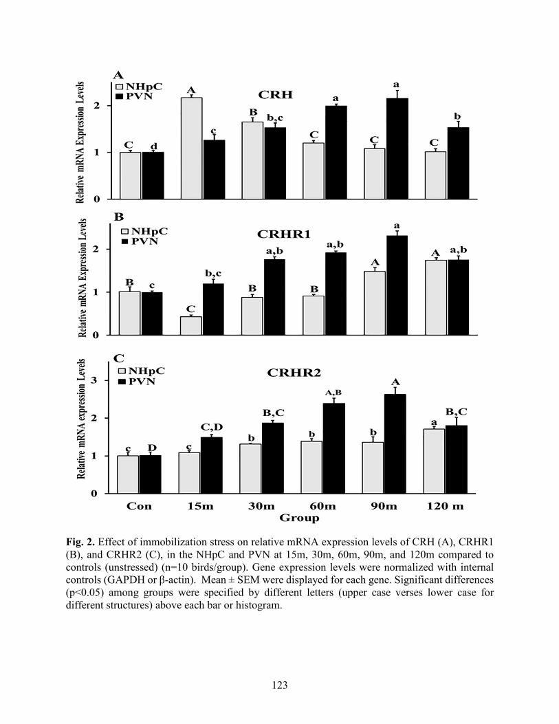
123
Fig. 2. Effect of immobilization stress on relative mRNA expression levels of CRH (A), CRHR1 (B), and CRHR2 (C), in the NHpC and PVN at 15m, 30m, 60m, 90m, and 120m compared to controls (unstressed) (n=10 birds/group). Gene expression levels were normalized with internal controls (GAPDH or β-actin). Mean ± SEM were displayed for each gene. Significant differences (p<0.05) among groups were specified by different letters (upper case verses lower case for different structures) above each bar or histogram.

124
Fig. 3. Changes in the relative gene expression of AVT (A) and its major receptor, V1aR (B), in the NHpC and PVN during immobilization stress at 15m, 30m, 60m, 90m, and 120m using RT-qPCR compared to the controls (unstressed) (n = 10 birds/group). GAPDH and β-actin gene expression levels served as internal controls to normalize the RT-PCR data. Mean ± SEM were expressed for each gene. Significant differences (p < 0.05) among groups were specified by different letters above each bar.

125
Fig. 4. Effects of immobilization stress on the relative gene expression of A) brain derived neurotrophic factor (BDNF) and B) glucocorticoid receptor (GR) in the NHpC and PVN at 15m, 30m, 60m, 90m, and 120m compared to controls (unstressed) (n=10 birds/group). Gene expression levels were normalized with GAPDH or β-actin. Mean ± SEM were expressed for each gene. Significant differences (p<0.05) among groups were specified by different letters above each bar.
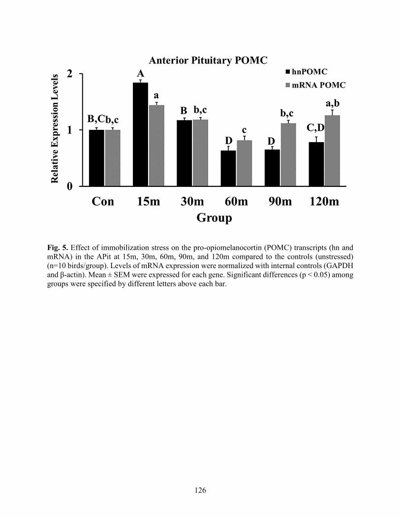
126
Fig. 5. Effect of immobilization stress on the pro-opiomelanocortin (POMC) transcripts (hn and mRNA) in the APit at 15m, 30m, 60m, 90m, and 120m compared to the controls (unstressed) (n=10 birds/group). Levels of mRNA expression were normalized with internal controls (GAPDH and β-actin). Mean ± SEM were expressed for each gene. Significant differences (p < 0.05) among groups were specified by different letters above each bar.

127
Fig. 6. Immobilization stress effects on mRNA expression levels of CRHR1 and CRHR2 (A), V1aR and V1bR (B), and GR (C) in the APit following immobilization stress for 15m, 30m, 60m, 90m, and 120m compared to the controls (unstressed) (n=10 birds/group). Levels of mRNA expression were normalized with internal controls (GAPDH or β-actin). Mean ± SEM were expressed for each gene. Significant differences (p < 0.05) among groups were specified by different letters above each bar.

128
Fig. 7. Total proteins were analyzed from the NHpC (A) and PVN (B) for CRHR1 and GR detection and quantification using Western blot throughout the period of immobilization stress (Control-120m). The density of bands was quantified using the alpha view SA program. Mean ± SEM changes in protein quantity between stressed birds compared to non-stressed controls throughout 120m of immobilization stress. Significant differences (p < 0.05) among groups were specified by different letters above each bar. The number of samples in each group was six. The upper panels in each graph show representative Western blots for GR, CRHR1, and GAPDH in the NHpC (A) and the PVN (B) during the stress period.

129
Fig. 8. Total proteins extracted from the anterior pituitaries (APit) during immobilization stress (Control-120m) were electrophoresed and blotted on PVD membrane. CRHR1 and GR were detected, and the density of bands was analyzed using alpha view SA program. Mean ± SEM changes in protein quantity between stressed birds compared to non-stressed controls throughout 120m of immobilization stress. Significant differences (p < 0.05) among groups were specified by different letters above each bar. The number of samples in each group was six. The upper panels in each graph show representative bands for GR, CRHR1, and GAPDH in the APit during the immobilization stress (control-120m).

130
References
Aliaga, E., Arancibia, S., Givalois, L., Tapia-Arancibia, L., 2002. Osmotic stress increases brain-derived neurotrophic factor messenger RNA expression in the hypothalamic supraoptic nucleus with differential regulation of its transcripts. Relation to arginine vasopressin content. Neuroscience 112 (4), 841 – 850.
Bath, K. G., Schilit, A., Lee, F. S., 2013. Stress effects on BDNF expression: effects of age, sex, and form of stress. Neuroscience 239, 149 –56.
Bayatti, N., Hermann, H., Lutz, B., Behl, C., 2005. Corticotropin-releasing hormone-mediated induction of intracellular signaling pathways and brain-derived neurotrophic factor expression is inhibited by the activation of the endocannabinoid system. Endocrinology 146 (3), 1205 –13.
Blas, J., 2015. Chapter 33 - Stress in Birds. In: Scanes, C.G. (Ed.), Sturkie's Avian Physiology (Sixth Edition). Academic Press, San Diego, pp. 769 – 810.
Blendy, J. A. 2006. The role of CREB in depression and antidepressant treatment. Biol. Psychiatry. 59 (12), 1144 – 1150.
Bonfiglio, J. J., Inda, C., Refojo, D., Holesboer, F., Arzt, E., Silberstein, S., 2011. The corticotropin-releasing hormone network and the hypothalamic-pituitary-adrenal axis: molecular and cellular mechanisms involved. Neuroendocrinology 94 (1), 12 – 20.
Carsia, R. V. 2015. Chapter 26 - Adrenals. In: Scanes, C.G. (Ed.), Sturkie's Avian Physiology (Sixth Edition). Academic Press, San Diego, pp. 577 – 611.
Chrousos, G. P., 2009. Stress and Disorders of the Stress System. Nat. Rev. Endocrinol. 5 (7), 374 – 81.
De Groef, B., Goris, N., Arckens, L., Kühn, E.R., Darras, V.M., 2003. Corticotropin-releasing hormone (CRH)-induced thyrotropin release is directly mediated through CRH receptor type 2 on thyrotropes. Endocrinology 144, 5537 – 5544.
De Kloet, E., Joels, M., Holsboer, F., 2005. Stress and the brain: from adaptation to disease. Nat. Rev. Neurosci. 6, 463 – 475.
De la Tremblaye, P. B., Linares, N. N., Schock, S., Plamondon, H., 2016. Activation of CRHR1 receptors regulates social and depressive-like behaviors and expression of BDNF and TrkB in mesocorticolimbic regions following global cerebral ischemia. Exp. Neurol. 284, 84 –97.
Drouin, J., Sun, Y. L., Chamberland, M., Gauthier, Y., De Lean, A., Nemer, M., Schmidt, T. J., 1993. Novel glucocorticoid receptor complex with DNA element of the hormone-repressed POMC gene. The EMBOJ. 12 (1), 145 –156.

131
Drouin, J., Trifiro, M. A., Plante, R. K., Nemer, M., Eriksson, P., Wrange, O., 1989. Glucocorticoid receptor binding to a specific DNA sequence is required for hormone-dependent repression of pro-opiomelanocortin gene transcription. Mol. Cell. Biol. 9 (12), 5305 –14.
Ginsberg, A. B., Frank, M. G., Francis, A.B., Rubin, B.A., O’Connor, K. A., Spencer, R. L. 2006. Specific and time-dependent effects of glucocorticoid receptor agonist RU28362 on stress-induced pro-opiomelanocortin hnRNA, c-Fos mRNA and Zif268 mRNA in the pituitary. J. Neuroendocrinol. 18 (2), 129 – 38.
Givalois, L., Naert, G., Rage, F., Ixart, G., Arancibia, S., Tapia-Arancibia, L., 2004. A single brain-derived neurotrophic factor injection modifies hypothalamo-pituitary-adreno- cortical axis activity in adult male rats. Mol. Cell Neurosci. 27, 280 – 295.
Greetfeld, M., Schmidt, M.V., Ganea, K., Sterlemann, V., Liebl, C., Müller, M.B., 2009. A single episode of restraint stress regulates central corticotrophin-releasing hormone receptor expression and binding in specific areas of the mouse brain. J. Neuroendocrinol. 21, 473–480.
Herman, J., McKlveen, J., Ghosal, S., Kopp, B., Wulsin, A., Makinson, R., Scheimann, J., Myers, B., 2016. Regulation of the hypothalamic-pituitary adrenocortical stress response. Comp. Physiol. 6, 603 – 621.
Jeanneteau, F., Lambert, W., Ismaili, N., Bath, K., Lee, F., Garabedian, M., Moses, V., Chaoa, M., 2012. BDNF and glucocorticoids regulate corticotrophin releasing hormone (CRH) homeostasis in the hypothalamus. Pro. Natl. Acad. Sci. USA 109, 1305 –1310.
Kadhim, H.J., Kang, S.W., Kuenzel, W.J., 2019. Differential and temporal expression of corticotropin releasing hormone and its receptors in the nucleus of the hippocampal commissure and paraventricular nucleus during the stress response in chickens (Gallus gallus). Brain Res. 1714, 1 – 7.
Kadhim, H.J., Kidd M. Jr., Kang, S. W., Kuenzel, W. J., 2020. Differential delayed responses of arginine vasotocin and its receptors in septo-hypothalamic brain structures and anterior pituitary that sustain hypothalamic–pituitary-adrenal (HPA) axis functions during acute stress. Gen. Comp. Endocrinol. 286, 113302.
Kang, S. W., Kuenzel, W.K., 2014. Regulation of gene expression of vasotocin and cortico- tropin-releasing hormone receptors in the avian anterior pituitary by corticosterone. Gen. Comp. Endocrinol. 204, 25 – 32.
Keller-Wood, M., 2015. Hypothalamic-pituitary-adrenal axis-feedback control. Compr. Physiol. 5 (3), 1161– 82.
Kidd Jr. M. T., Kang, S. W., Kadhim, H. J., Kuenzel, W. J., 2020. A functional role of the nucleus of the hippocampal commissure involving activation of both the hypothalamo-pituitary-adrenal and the hypothalamo-pituitary- thyroid axes during stress in birds. Gen. Comp. Endocrinol. (Submitted).

132
Korosi, A., Veening, J.G., Kozicz, T., Henckens, M., Dederen, J., Groenink, L., van der Gugten, J., Olivier, B., Roubos, E.W., 2006. Distribution and expression of CRF receptor 1 and 2 mRNAs in the CRF over-expressing mouse brain. Brain Res. 1072, 46 – 54.
Kovács, K. J., Sawchenko, P. E., 1996. Sequence of stress-induced alterations in indices of synaptic and transcriptional activation in parvocellular neurosecretory neurons. J. Neurosci. 16 (1), 262 – 73.
Kuenzel, W. J., Jurkevich. A., 2010. Molecular neuroendocrine events during stress in poultry. Poult. Sci. 89 (4), 832–40.
Kuenzel, W.J., Masson M., 1988. A stereotaxic atlas of the brain of chick (Gallus domesticus) Johns Hopkins University Press, Baltimore.
Løtvedt, P. Fallahshahroudi, A, Bektic, L., Altimiras, J., Jensen, P., 2017. Chicken domestication changes expression of stress-related genes in brain, pituitary and adrenals. Neurobio. stress 7, 113 – 121.
Lovejoy, D. A., Balment, R. J. 1999. Evolution and physiology of the corticotropin-releasing factor (CRF) family of neuropeptides in vertebrates. Gen. Comp. Endocrinol. 115 (1), 1 – 22.
Ma, X. M., Levy, A., Lightman., S. L., 1997. Rapid changes in heteronuclear RNA for corticotrophin-releasing hormone and arginine vasopressin in response to acute stress. J. Endocrinol. 152 (1), 81 – 89.
Madison, F. N., Jurkevich, A., Kuenzel, W. J., 2008. Sex differences in plasma corticosterone release in undisturbed chickens (Gallus gallus) in response to arginine vasotocin and corticotropin releasing hormone. Gen. Comp. Endocrinol. 155 (3), 566 – 73.
McEwen, Bruce S. 2007. Physiology and neurobiology of stress and adaptation: central role of the brain. Physiol. Rev. 87(3), 873 – 904.
Nagarajan, G., Jurkevich, A., Kang, S., Kuenzel, W., 2017a. Anatomical and functional implications of corticotrophin releasing hormone neurones in a septal nucleus of the avian brain: an emphasis on glial-neuronal interaction via V1a receptors in vitro. J. Neuroendocrinol. 29, 1 – 11.
Nagarajan, G., Kang, S., Kuenzel, W., 2017b. Functional evidence that the nucleus of the hippocampal commissure shows an earlier activation from a stressor than the paraventricular nucleus: Implication of an additional structural component of the avian hypothalamo-pituitary-adrenal axis. Neurosci. Lett. 642, 14 – 19.
Nagarajan, G., Tessaro, B.A., Kang, S.W., Kuenzel, W.J., 2014. Identification of arginine vasotocin (AVT) neurons activated by acute and chronic restraint stress in the avian septum and anterior diencephalon. Gen. Comp. Endocrinol. 202, 59 – 68.

133
Nemoto, T., Mano, A., Shibasaki, T. 2013. MiR-449a contributes to glucocorticoid-induced CRF-R1 downregulation in the pituitary during stress. Mol. Endocrinol. 27 (10), 1593 – 1602.
Noguchi, T., Makino, S., Matsumoto, R., Nakayama, S., Nishiyma, M., Terada, Y., Hashimoto, K, 2010. Regulation of glucocorticoid receptor transcription and nuclear translocation during single and repeated immobilization stress. Endocrinology 151 (9), 4344 – 55.
Palkovits, M. 1973. Isolated removal of hypothalamic or other brain nuclei of the rat. Brain Res. 59, 449 – 450.
Proudman, J. A., Opel, H. 1989. Daily Changes in plasma prolactin, corticosterone, and luteinizing hormone in the unrestrained, ovariectomized turkey hen. Poult. Sci. 68 (1), 177 – 84.
Rabadan-Diehl, C., Aguilera, G., 1998. Glucocorticoids increase vasopressin V1b receptor coupling to phospholipase C. Endocrinology 139(7), 3220 – 6.
Ramamoorthy, S., Cidlowski, J. A., 2013. Ligand-induced repression of the glucocorticoid receptor gene is mediated by an NCoR1 repression complex formed by long-range chromatin interactions with intragenic glucocorticoid response elements. Mol. Cell. Biol. 33 (9), 1711 – 1722.
Romero, L. M., 2004. Physiological stress in ecology: Lessons from biomedical research. Trends Ecol. Evol. 19 (5), 249 – 255.
Schmittgen, T. D., Livak, K. J., 2008. Analyzing real-time PCR data by the comparative CT Method. Nat. Protoc. 3 (6), 1101– 8.
Selvam, R., Jurkevich, A., Kang, S. W., Mikhailova, M. V., Cornett, L. E., Kuenzel. W. J., 2013. Distribution of the vasotocin subtype four receptor (VT4R) in the anterior pituitary gland of the chicken, Gallus gallus, and its possible role in the avian stress response. J. Neuroendocrinol. 25(1), 56 – 66.
Shimojo, M., Hiroi, N., Yakushiji, F., Ueshiba, H., Yamaguchi, N., Miyachi, Y., 1995. Differences in down-regulation of glucocorticoid receptor mRNA by cortisol prednisolone and dexamethasone in Hela cells. Endocr. J. 42 (5), 629 –636.
Tardito, D., Perez, J., Tiraboschi, E., Musazzi, L., Racagni, G., Popoli, M. 2006. Signaling pathways regulating gene expression, neuroplasticity, and neurotrophic mechanisms in the action of antidepressants: a critical overview. Pharmacol. Rev. 58 (1), 115 –34.
Vandenborne, K., De Groef, B., Geelissen, S., Kühn, E., Darras, V., Van der Geyten, S., 2005. Corticosterone-induced negative feedback mechanisms within the hypothalamic pituitary adrenal axis of the chicken. J. Endocrinol. 185, 383 –391.
Whitnall, Mark H., Mezey, E., Gainer, H. 1985. Co-localization of corticotropin-releasing factor and vasopressin in median eminence neurosecretory vesicles. Nature 317 (6034), 248–250.

134
Xie, J., Kuenzel, W., Anthony, N., Jurkevich, A., 2010. Subpallial and hypothalamic areas activated following sexual and agonistic encounters in male chickens. Physiol. Behav. 10, 344 –359.
Yadawa, A.k., Chaturvedi, C.M., 2016. Expression of stress hormones AVP and CRH in the hypothalamus of Mus musculus following water and food deprivation. Gen. Comp. Endocrinol. 239, 13 –20.
Yao, M., Denver, R. J., 2007. Regulation of vertebrate corticotropin-releasing factor genes. Gen. Comp. Endocrinol. 153 (1–3), 200 –216.

135
Appendix
Supplementary. (A) Amino acid sequence of the corticotropin releasing hormone receptor 1 (CRHR1) with the peptide synthesized for antibody production shown in red (1st extracellular domain). (B) Amino acid sequence alignment of the 1st extracellular domain of the CEHR1 with the synthesized peptide.

136
Chapter 5
Conclusion
The physiological and functional roles of corticotropin releasing hormone (CRH) neurons
located in the nucleus of hippocampal commissure (NHpC) in the neuroendocrine regulation of
stress were reported from several experiments for this dissertation. The first experiment utilized
feed deprivation (FD) stress because it is a practical method utilized in broiler breeders to maintain
birds on a growth curve known to maintain a functional reproductive system throughout their
lifetime. From an animal welfare perspective, it is important to determine what neuroendocrine
structures and receptors are involved following food withdrawal. This particular stressor becomes
more intense overtime; therefore, it enabled us to study the sequences of gene expression within
the NHpC and compared the findings with those obtained from the major structure involved in
stress, the paraventricular nucleus (PVN). Interestingly, nutritional stress (FD) resulted in an early,
rapid activation of CRH mRNA levels in the NHpC as well as a gradual increase in CRH mRNA
in the PVN. The rapid increase in CRH mRNA within the NHpC was associated with higher
POMC transcripts in the anterior pituitary and ultimately increased secretion of the stress hormone,
CORT. A sustained output of CORT appears to due to the positive feedback of CRH mRNA and
gene expression of its major receptor, CRHR1, in the PVN. Data, also, showed that CRH and its
receptors, CRHR1 and CRHR2, have a different and tissue specific relationship. In the NHpC,
CRH has a negative feedback particularly with CRHR1. In marked contrast, the PVN displays a
positive relationship with CRHR1, CRHR2, and BDNF. Upregulation of BDNF in the PVN over
the sampling period could be a factor responsible for the upregulation of CRH and its receptors,
CRHR1 and CRHR2.

137
The second experiment showed that AVT cell bodies are found in the PVN, not in the NHpC
nor MBHv/ME utilizing IHC. A major receptor of AVT, the V1aR, was identified in all three brain
structures and located primarily in glia. Gene expression data revealed that AVT, V1aR and V1bR
mRNA are expressed in all three brain structures and responded differentially to FD stress. Results
showed that AVT, V1aR and V1bR are involved in the late phase, 3h or longer of FD stress. A
negative feedback in gene expression between AVT and its receptors, V1bR and V1aR, was found
within the NHpC during FD stress. In contrast, AVT and its two receptors showed a positive
feedback at the level of the PVN during FD stress. At the final, central neuroendocrine level of the
MBHv/ME and level of the anterior pituitary, upregulation of relative AVT mRNA expression
with positive expression of V1bR and decreased V1aR mRNA occurred at both anatomical levels.
Overall, the response of AVT and its two major receptors in the current model of the avian
neuroendocrine stress pathway suggest that the balance in function of the two receptors preserve
and regulate ACTH secretion from the anterior pituitary and ultimately plasma CORT to prevent
overstimulation of the HPA axis during stress responses.
The last experiment was conducted to identify whether CRH neurons in the NHpC are stress
specific or involved following any type of stressor. Therefore, a very different stressor,
immobilization, was imposed on birds and the neuroendocrine response was examined including
the structural response of the NHpC and PVN. Data showed a rapid and significant upregulation
of CRH gene expression in the NHpC associated with a significant increase of POMC transcripts
and higher plasma CORT similar to what was determined following FD. Within the PVN, a gradual
increase of CRH gene expression was documented. Thereafter, a delayed upregulation of AVT
indicated that the two neuropeptides worked synergistically to sustain the CORT release from
immobilization stress. An increase of BNDF in the PVN mRNA appeared to enhance CRH mRNA
138
upregulation and preceded AVT mRNA levels in the PVN, but not in the NHpC suggesting that
BDNF has a positive effect on CRH and AVT expression in the PVN during the stress response.
Importantly, upregulation of BDNF gene expression was associated with CRHR1 and V1aR
mRNA increase showing a positive relationship between BDNF and CRHR1 as well as V1aR
within the PVN. The relationship between CRH and its CRHR1in the NHpC was a negative one,
both at the gene expression and protein levels. However, gene expression as well as protein levels
of CRHR1 indicated that CRH and CRHR1 have a positive relationship within the PVN. The
CRHR2 gene expression was upregulated significantly in both structures, NHpC and PVN,
suggesting that CRHR2 has a role to activate hypothalamus-pituitary-thyroid (HPT) axis in
respond to stress stimuli. Furthermore, in the NHpC, downregulation of AVT mRNA and
upregulation of V1aR gene expression observed during immobilization stress indicated that the
NHpC utilizes V1aR located in the glial cells to monitor the response to the stressor. However, the
delayed increase of both AVT and V1aR in the PVN was to enhance CRH activities during stress
response and maintain higher CORT levels when stressors persisted for long time. Differential
gene expression of CRH receptors and AVT receptors in the APit revealed that the two sets of
receptors are working together to maintain activity of the HPA axis and prevent corticotrope
overactivation. Increasing CORT concentration that occurred during the stress response induced a
negative feedback through GRs to inhibit POMC transcripts at the level of the APit first, followed
by downregulation of CRH mRNA in the NHpC and PVN, respectively.
Hence, data within the dissertation provide additional support showing that CRH neurons in
the NHpC function within the traditional HPA axis to initiate the neuroendocrine stress response.
Other vertebrate species therefore may have a similar extra-hypothalamic structure containing
CRH neurons that likewise functions to initiate the stress response.

139
Appendix