Embed Size (px)
Citation preview

Functional Aspects of Colour Processing
within the Human Brain
By
Andrei Georgescu, BSc PT
A thesis Submitted to the College of Graduate
Studies and Research
in Partial Fulfillment of the Requirements
for the Degree of
Master of Science
University of Saskatchewan
© Copyright by Andrei Georgescu, April 2006
i
Permission to use
In presenting this thesis in partial fulfillment of the requirements for a
Postgraduate degree from the University of Saskatchewan, I agree that the
Libraries of this University may make it freely available for inspection. I further
agree that permission for copying of this thesis in any manner, in whole or in part,
for scholarly purposes may be granted by the professors who supervised my thesis
work or, in their absence, by the head of the Department or the Dean of the
College in which my thesis work was done. It is understood that any copying or
publication or use of this thesis or parts thereof for financial gain shall not be
allowed without my written permission. It is also understood that due recognition
shall be given to me and to the University of Saskatchewan in any scholarly use
which may be made of any material in my thesis.
Requests for permission to copy or to make other uses of material in this
thesis in whole or in part should be addressed to:
Head of the Department of Kinesiology
University of Saskatchewan
Saskatoon, Saskatchewan
S7N 5B3

ii
Abstract
In a seminal work, Ungerleider and Mishkin (1982) offered substantial evidence
that two separate visual pathways – coding what/where-- exist within the primate
brain. Recently, human evidence has resulted in the “what/where” pathways being
reconsidered in terms of ventral stream (vision for perception) and dorsal stream
(vision for action; Goodale & Milner, 1992). Consistently, many studies have
demonstrated that there is an overrepresentation of magnocellular (luminance)
information within the dorsal stream; parvocellular input (colour, shape,
consistancy) represents the primary source of information for the ventral stream.
Although luminance contrast is important in perceiving moving objects, colour
discrepancies help the visual system to identify the detailed characteristics of the
environment and, subsequently, to prepare the motor system for action. This thesis
endeavors to determine the role played by colour, in contrast with luminance, in
influencing the programming and control movement production. Using a grasping
paradigm and two different luminance conditions (iso-luminance vs.
heteroluminance) within two separate experiments (experiment 1 – programming;
experiment 2 – online control), we show that chromatic information can be
successfully be used by motor circuits to complete the grasping task faultlessly.
Although significant temporal delays in terms of reaction time and movement time
between colour and luminance processing are identified, the human visual system
seems able to fully integrate colour features for action with no significant spatial
error cost.
iii
Acknowledgements
I would like to thank and express my gratitude towards my supervisor, Dr.
Gordon Binsted. His leadership, support, understanding and knowledge have set
an example of expertise I hope to match some day.
I would also like to thank my Advisory Committee members, Dr. Deb Saucier,
Dr. Phil Chilibeck, and Dr. Brian Maraj for their inclusive and pertinent comments
and advice they provided at all levels of the research project.
Further, I thank my friends and colleagues, Kyle Brownell, Nick Clarke and
Tyler Rolheiser for their day by day help, friendship and fully support.
Finally, I would like to thank my family for their fully support and in
particular, I must acknowledge my wife, Magda, without whose love,
encouragement, patience and trust I would not have finished this thesis.
iv
Table of Contents
Permission to use .............................................................................................. i
Abstract.............................................................................................................. ii
Acknowledgements............................................................................................ iii
Table of contents................................................................................................ iv
Chapter I
Thesis introduction............................................................................................. 1
Literature review................................................................................................ 4
General Context................................................................................................. 4
Dorsal/Ventral dissociation; Psycho-behavioural models................................. 5
Vision - Physiological and anatomical aspects................................................. 9
Fig. 1.................................................................................................................. 10
Fig. 2.................................................................................................................. 11
Parvo(P) and Magno (M) pathways................................................................... 11
Konio pathway - a possible explanation?.......................................................... 14
The blindsight condition.................................................................................... 16
Present research.................................................................................................. 18
Objectives and Hypotheses................................................................................ 20
Chapter II
Pilot study.......................................................................................................... 22

v
Introduction........................................................................................................ 22
Methods.............................................................................................................. 25
Participants......................................................................................................... 25
Task.................................................................................................................... 26
Stimuli................................................................................................................ 26
Apparatus........................................................................................................... 27
Procedure........................................................................................................... 28
Data reduction and analysis............................................................................... 30
Results................................................................................................................ 31
Discussion.......................................................................................................... 32
Table 1................................................................................................................ 35
Figure caption................................................................................................... 36
Fig. 1.................................................................................................................. 38
Fig. 2.................................................................................................................. 39
Fig. 3.................................................................................................................. 40
Fig. 4.................................................................................................................. 41
Fig. 5.................................................................................................................. 42
Chapter III
Manuscript introduction..................................................................................... 43
Functional aspects of colour processing within the human brain...................... 45
Introduction........................................................................................................ 46

vi
Dual vision system theories............................................................................... 46
Colour processing.............................................................................................. 48
Experiment 1...................................................................................................... 52
Methods.............................................................................................................. 52
Participants......................................................................................................... 52
Task.................................................................................................................... 53
Stimuli................................................................................................................ 53
Apparatus........................................................................................................... 54
Procedure........................................................................................................... 55
Data Reduction and Analysis............................................................................. 57
Results................................................................................................................ 58
Discussion.......................................................................................................... 60
Experiment 2...................................................................................................... 64
Methods.............................................................................................................. 64
Participants......................................................................................................... 64
Procedure........................................................................................................... 65
Data Reduction and Analysis............................................................................. 66
Results................................................................................................................ 66
Discussion.......................................................................................................... 68
General Discussion............................................................................................ 69
Programming...................................................................................................... 70
vii
Online control.................................................................................................... 74
Figure caption.................................................................................................... 77
Figure 1.............................................................................................................. 80
Figure 2.............................................................................................................. 81
Figure 3.............................................................................................................. 82
Figure 4.............................................................................................................. 83
Figure 5.............................................................................................................. 84
Figure 6.............................................................................................................. 85
Figure 7.............................................................................................................. 86
Figure 8.............................................................................................................. 87
Figure 9 a, b....................................................................................................... 88
Figure 10............................................................................................................ 89
References.......................................................................................................... 90
Chapter IV
Thesis Summary................................................................................................. 97
Conclusions........................................................................................................ 97
Limitations and Future directions...................................................................... 100
References.......................................................................................................... 102

1
Chapter I
Thesis introduction
Colour is our perceptual response to a very narrow span of the total
electromagnetic radiation available with light. From the retina to the superior
command centres, the chromatic information has to travel through several
nervous structures that amplify and modulate the nervous impulse.
Ungerleider and Mishkin, (1982) first formulated the hypothesis that
objects are represented differently during action than they are for a purely
perceptual task. In brief, they argued that the brain’s visual pathways split into
two main streams: the dorsal “where” pathway and the ventral "what”
pathway. The “where” pathway runs dorsally from primary visual cortex into
posterior parietal cortex, and the “what” pathway runs ventrally from primary
visual cortex into inferotemporal cortex. The dorsal stream deals with the
information required for object location and the other is concerned with object
identification by analyzing its shape, texture or colour. In the recent years, a
new approach has been offered for reconsidering these two pathways in terms
of vision for perception and vision for action instead of “what” and “where”.
(perception - action model; Goodale & Milner, 1992). Another model that has
been proposed (Glover, 2004) is the planning-control model. This approach,
is not significantly different from the model proposed by Goodale and Milner,

2
but introduces a new perspective on how the two streams process visual
information during planning and action, independently. Although many
studies have been performed to investigate how the dorsal and the ventral
pathways process information (e.g., Milner & Goodale, 1995; Rizzolatti,
Fogassi, & Gallese, 1997; Jeannerod, 1988), there have been few projects
examining the chromatic properties of neurons in cortical areas belonging to
the dorsal and/or the ventral stream, and fewer still focusing on the use of
colour in controlling the motor system.
The present thesis consists of four chapters; the first chapter contains a
broad review of the previous literature regarding the two major visual streams
and colour processing mechanisms. The second chapter encloses a pilot study
examining the use of colour for preparing/performing a movement.
Specifically, we examined the precision of manual grasping towards a colour
object displayed on an isoluminant background. In general, even though
colour signals take slightly longer to be processed (Nowak & Bullier, 1997;
Tanaka & Shimojo, 1996; Tanne, Boussaoud, Boyer-Zeller, & Rouiller,
1995), they appear to be efficiently used by the motor system for a variety of
functions. The third part of this thesis contains two experiments which have
been written in a format suitable for publication and undertake to precisely
examine colour utilization for action. These experiments aimed to determine
the role of chromatic information for the real time modulation of action. In
particular, we examined the impact of chromatic information towards the
3
planning and the online control of the motor task within both heteroluminant
(different luminance) and isoluminant (same luminance) conditions. The last
section presents general conclusions of the thesis, future direction and
inherent limitations of the present studies.

4
Literature review
General context
A key feature of the primate visual system is the separation of visual
areas into two major cortico-cortical processing pathways: The dorsal and the
ventral streams. Ungerleider and Mishkin (1982) first proposed an anatomical
distinction between the ventral pathway and the dorsal pathway in the primate
visual system. The ventral stream projects from the primary visual cortex
toward infero-temporal cortex whereas the dorsal stream projects dorso-
laterally from the primary visual cortex to parietal areas. Ungerleider and
Mishkin based their anatomical distinction on neurophysiological and
behavioural evidence collected from the study of macaque monkeys. They
performed intrusive lesions in the ventral and in the dorsal pathway of the
visual system of macaque monkeys and found a double dissociation. Animals
with a lesion in the ventral pathway were impaired in the identification of
objects but they were relatively unimpaired in tasks of spatial orientation.
Conversely, animals with lesions of the dorsal areas showed deficits in
locating the target but their identification capacity was preserved. On this
basis, Ungerleider and Mishkin concluded that the ventral pathway of the
primate visual system is the What system and the dorsal pathway of the
primate visual system is the Where system.
5
Dorsal/Ventral dissociation; Psycho-behavioural models
Over a decade after the initial observation of Ungerleider and Mishkin,
Goodale and Milner (1992) provided a new conceptual account of how the
brain processes visual information. They proposed the perception - action
(PA) model and made two major assumptions: (1) The ventral stream
processes visual information for perception purposes; (2) perception and
action are two separate domains, the latter being an exclusive property of the
dorsal stream. According to Goodale and Milner, the ventral stream has its
main role in object recognition and preparing the first stage of information
processing (object identification), while the dorsal stream analyzes an object's
spatial location and coordinate movement. To support this notion they stress
the dissociation between the behaviour of the agnosic patient D.F. and that of
the ataxic patient A.T. (Perenin & Vighetto, 1988). A.T., suffering from optic
ataxia manifests a profound inability in reaching to targets under visual
guidance but no difficulty in observing and recognizing them whereas the
visual form agnosic patient D.F., has problems in recognizing objects
visually, but can still use visual information to guide her movements.
Although the PA model has been widely accepted as a pertinent explanation
of the dual system theory (humans included), some authors questioned its

6
validity and wonder if the strict dichotomy this model advances is perhaps too
inflexible.
The PA model point of view was in accordance with the classical notion
of the parietal cortex as the place for unitary space perception. The posterior
parietal cortex consists of a mixture of areas, each receiving specific efferent
information. These different types of sensory information are transformed into
information appropriate for action. Space perception appears to derive from
the joint activity of a series of sensorimotor fronto-parietal circuits which
encode the spatial location of an object and prepare it to be used for a future
action (Rizzolatti et al., 1997, Rizzolatti, Camarda, Fogassi, Gentilucci,
Luppino, & Matelli, 1998). Taken together, within the dorsal stream, there are
parallel cortico-cortical circuits, each of which elaborates a specific type of
visual information in order to guide different types of action.
In opposition with the PA model, Glover (2004) explored the evidence
for a distinction in human performance between the planning and on-line
control of actions. This new approach establishes an anatomical substrate of
the ideas offered previously by Woodworth (1899). Yet, the planning -
control (PC) model that he proposed facilitates a new distinction between the
visual and cognitive processes involved in planning and control. Brain
imaging studies support the dichotomy, in that planning in humans is linked
with activity in a distributed network including a visual representation in the
inferior parietal lobe, whereas control is associated with activity in a separate

7
network including a visual representation in the superior parietal lobe (SPL)
(Deiber, Ibanez, Sadato, & Hallett, 1996; Desmurget, Grea, Grethe, Prablanc,
Alexander, & Grafton, 2001; Grafton, Fagg, & Arbib, 1998; Grafton,
Mazziotta, Woods, & Phelps, 1992; Krams, Rushworth, Deiber, Frackowiak,
& Passingham, 1998). The inferior parietal lobe receives inputs from two
major sources: firstly, the visual information from the visual cortex is carried
out to the IPL via the temporal lobe or by a third stream directly from primary
visual cortex (V1) to IPL (Boussaoud, Ungerleider, & Desimone, 1990).
However, the temporal lobe inputs include the non-spatial (i.e., function,
fragility, weight, and colour) spatial (i.e., orientation, size, and shape) features
as well as information in the vicinity of the target (e.g., background, other
objects). Proprioceptive information from somatosensory association areas is
also integrated in the IPL together with information from frontal lobe (i.e.,
memories and past experiences). Once a motor act is planned, a copy of the
motor schema is sent over to SPL and cerebellum. These efferences are to be
used by the SPL once the movement is initiated. If needed, the whole motor
plan can be adjusted online using this efference copy.
There are few main distinctions that differentiate the PC model from
other models (including PA model) (Glover, 2004). The planning-control
model proposes separate visual representations underlying the two stages of
action by assigning the inferior and superior areas of the parietal lobes to their
specific roles in computing these visual representations. Furthermore, in

8
opposition with Jeannerod (1988), the PC model makes no specific distinction
between the information used during reaching versus grasping. It also takes
into consideration a gradual rather than discrete transition between the two
stages of action. Thus, the PC model differs from some models of motor
control that assume that control can only begin when feedback loops have had
time to close (Crossman & Goodeve 1983; Posner & Keele, 1968;
Woodworth 1899). Similarly to Wolpert, Ghahramani, and Jordan (1995), the
PC model suggests that an efference copy may be used to adjust movements
from any time after initiation.
There are two major features that differentiate the PC model from PA
model. Specifically, the first distinction resides in the fact that in the PC
model, the two stages of action utilize distinct visual representations in the
IPL and SPL, and those distinct representations result in interactions between
cognitive and visual information in planning but not in control. Conversely, in
the PA model (Goodale & Milner, 1992; Milner & Goodale, 1995), both
planning and control primarily utilize representations in the SPL with some
exceptions (e.g., pieces of perceptual information that may be imported from
the ventral stream; Haffenden & Goodale, 2000; Milner & Goodale, 1995). In
particular, the PA model suggests that parameters of movement dependent on
the spatial characteristics of the target will be both planned and controlled
independently of cognitive and perceptual influences. Second, while the
planning–control model assumes that the IPL is involved in the kinematic

9
computation of all movements, the perception-action model states that IPL
requires information regarding only the nonspatial target characteristics.
With deference to all these approaches, the question that has been raised
still encounters some difficulties. Are ventral features available to the dorsal
stream? Specifically, the point of interest to this study is that of colour as a
putative ventral attribute used to program and guide a motor schema in real
time.
Vision - Physiological and anatomical aspects
The capability of primates to see colour rises from the existence of two
chromatic channels (Derrington, Krauskopf, & Lennie, 1984) which
correspond to modulation of the response of the short wavelength (S) cones
and the difference between large wavelength (L) and middle wavelength (M)
cone activation; the achromatic (luminance) channel is derived from additive
combination of cone signals. Kaiser and Boynton, (1996) advanced the idea
of three colour channels. One channel carries out signals the sum of L and M
cone excitations and can be selectively activated by light/dark (e.g.,
white/black) grating stimuli. This channel is sometimes referred to as the
“luminance” channel. The two other channels, which are constant in
luminance, are referred to as “chromatic” channels.
Luminance, as a physical feature, is defined as “the integral over
wavelength of the radiance of a source, weighted by the spectral luminosity
V( ) “ (Gegenfurtner & Hawken, 1996, p. 395). Having said that, if the

10
quantity of light of a stimulus is attuned by modifying only the wavelength
and keeping constant the amount of light, the new stimulus will likely have a
different colour but the luminance will stay the same. The two stimuli will be
therefore isoluminant (See Fig. 1). If an organism uses a stimuli identifying
system based only on luminance discrepancies, then the two stimuli will
appear alike to that identifying system. Conversely, if the quantity of
wavelength of stimuli is modified and tuned to grayscale (e.g., achromatic
environment) without paying attention to luminance gradients, the new
stimuli will be easy to identify based on amount of light that reflects from its
surface ( i.e. luminance; see Fig. 2).
Fig. 1. Iso-luminant variance. By attuning the colour of an isoluminant scene
to a neutral achromatic value, the chromatic contrast disappears and the
components of the scene will appear alike.
11
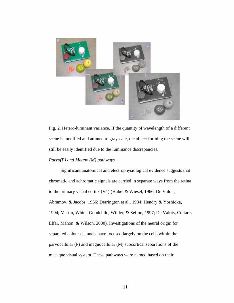
Fig. 2. Hetero-luminant variance. If the quantity of wavelength of a different
scene is modified and attuned to grayscale, the object forming the scene will
still be easily identified due to the luminance discrepancies.
Parvo(P) and Magno (M) pathways
Significant anatomical and electrophysiological evidence suggests that
chromatic and achromatic signals are carried in separate ways from the retina
to the primary visual cortex (V1) (Hubel & Wiesel, 1966; De Valois,
Abramov, & Jacobs, 1966; Derrington et al., 1984; Hendry & Yoshioka,
1994; Martin, White, Goodchild, Wilder, & Sefton, 1997; De Valois, Cottaris,
Elfar, Mahon, & Wilson, 2000). Investigations of the neural origin for
separated colour channels have focused largely on the cells within the
parvocellular (P) and magnocellular (M) subcortical separations of the
macaque visual system. These pathways were named based on their

12
anatomical distinction in the lateral geniculate nucleus (LGN) of the
thalamus; four LGN layers contain densely crowded, “parvo” small cells, and
two contain more sparsely placed, “magno” large cells, (Merigan & Maunsell,
1993; Dobkins & Albright, 1998). The P cells are involved in object vision,
colour, shape, and texture. They have low contrast sensitivity and are
sensitive only to large differences in brightness. The P cells are also slower
but have higher acuity and are embryological younger than the M cells.
Conversely, the M cells carry information regarding the spatial features such
as depth, motion and location of stimuli. They are more sensitive than the P
cells to differences in brightness and have high contrast sensitivity. Further M
cells are faster and serve both central and peripheral vision, but they are
colour blind. Livingstone and Hubel (1982; 1987) were the first to connect
this early segregation between the P and M pathways to neurochemical
compartmentalization of primate primary visual cortex (V1). By staining the
visual cortex with cytochrome oxidase (a large trans-membrane protein found
in the mitochondrion) Livingstone and Hubel found that cytochrome oxidase-
rich regions of striate cortex called blobs are specialized for colour processing
while the regions surrounding them (interblobs) are dominated by neurons
with high orientation selectivity but poor colour specificity. Livingstone and
Hubel proposed that the chromatic channels (P) supply the inputs to
mechanisms for colour perception, but have a modest contribution to early
processes for spatial orientation integration. Livingstone and Hubel further

13
made the assumption that there are two pathways that connect the LGN to V1
(i.e., P & M). They also proposed the idea that P and M channels remain
segregated well beyond the primary cortex. More recently, it has been
suggested that the ventral and dorsal projection streams identified by Mishkin
and Ungerleider (1982) might represent the continuations of the P and M
systems, respectively (Livingstone & Hubel, 1988).
Nevertheless, recent physiological evidence shows that in macaque V1
a significant proportion of cells respond intensely to combined modulation of
colour and luminance (Thorell, De Valois, & Albrecht 1984; Lennie,
Krauskopf, & Sclar, 1990; Leventhal, Thompson, Liu, Zhou, & Ault, 1995;
De Valois et al., 2000), including cells showing a high degree of orientation-
selectivity (Johnson, Hawken, & Shapley, 2001). Xiao and Felleman (2004)
studied the degree of the segregation between the two streams by
anatomical
tracing and demonstrated that both blobs and interblob project to the thin
stripes of V2. They debated the hypothesis due to V1 and V2 are connected in
parallel to form highly segregated functional streams. Moreover, recorded
data from a single neuron within area V2 did not confirmed the total
compartmentalization within V2 (Kiper, Fenstemaker, & Gegenfurtner,
1997). Although cells demonstrate colour specificity, the receptive-field
properties of cells located in V2 were generally similar for luminance and
chromatically defined stimuli. Studies in monkeys with discrete lesions of the
P and M layers of LGN also revealed a pattern of deficits that do not conform

14
to the distinction pointed out previously (Merigan & Maunsell, 1993; Schiller
& Logothesis, 1990). Lesions of the M pathway cause a decrease in contrast
sensitivity for stimuli of high temporal but low spatial frequency whereas
lesions of the P pathway results in a complementary pattern of deficits. These
results suggest that the specialization of the P and M cells, at least at the level
of the retina and LGN are best understood in terms of an exchange between
the different requirements of spatial wavelength and temporal processing
(Schiller & Logothesis, 1990). The additional processing at the level of V1
and V2 modifies the response characteristics of the magno and parvo inputs
enormously.
Konio pathway - a possible explanation?
Much more recently, long after parvo-cellular and magno-cellular
pathways in colour-motion processing were in unanimity accepted, a third
pathway, referred to as koniocellular (K) pathway has been proposed (Hendry
& Yoshioka 1994; Hendry & Reid, 2000). This pathway, first described in
primates (macaque; Casagrande, 1994) is thought now to process colour along
a dimension that activates the short-wavelength-selective S cones from the
retina. This geniculo-cortical stream selectively modulates the blue/yellow
dimension. The K pathway is anatomically distinct from the M and P
pathways and projects directly to the colour-selective blobs in area V1. The K
pathway therefore may offer another explanation by which colour information
could influences motion processing.
15
One important region for examining K pathway is the middle temporal
area (Dobkins, 2000). This region belonging to extrastriate visual cortex
receives an important input from V1 (Merigan & Maunsell, 1993). Until
recently, investigations of colour influences on motion responses in area MT
have focused exclusively on red/green input derived from signals in the L and
M cones (Gegenfurtner et al., 1994; Pisella, Arzi, & Rossetti, 1998). Wandell,
et al., (1999) used functional magnetic resonance imaging (fMRI) to
demonstrate S cone input to the motion-responsive region of human visual
cortex (referred to as MT1). These experiments were followed by single cell
recordings in macaque area MT, demonstrating a clear S cone contribution to
directionally selective responses (Seideman & Newsome, 1999).
Regardless the methodology or species, the S cone input to motion was
found to be significantly less powerful than light/dark input. The contrast
sensitivity of the neurons belonging to MT was significantly lower for
blue/yellow as compared to light/dark gradings. These results obtained for
stimuli modulated along the blue/yellow dimension are consistent with
previous results in MT obtained using red/green gratings (Dobkins &
Albright, 1994; Gegenfurtner et al., 1994). One explanation is that the
observed S cone influences on MT responses may reflect the functional input
from this third visual pathway to motion processing. Thus signals from the K
pathway could potentially reach area MT from the V1 blobs via V2 (Merigan
& Maunsell, 1993) or directly from K layers in the LGN to MT area (Hendry

16
& Reid, 2000). As a result the K pathway input to MT might advocate that the
S cone signals carried by this pathway could be used to identify the chromatic
features of an object as well as to help in motion detection. Another
possibility, however, is that the S cone signals might reach area MT by
interfering within the M pathway (Calkins, 2000, 2001). Although Calkins
provided evidence from retinal anatomy to support this possibility, other
neurophysiological studies argued against this hypothesis (Lee, Martin, &
Valberg, 1998; Dacey & Lee, 1994). Lee and colleagues, (1998) found that S
cone input to M pathway responses is insignificant if not absent.
The blindsight condition
The K pathway’s role in modulation of chromatic perception is not the
only account of colour processing within the dorsal/ventral dissociation
physiology. Weiskrantz (1986; 1997) brought into discussion a relatively new
neuropsychological condition called “blindsight”. Blindsight can occur due to
a lesion in the primary visual cortex. The lesion is located prior to the
bifurcation of the ventral and the dorsal streams. Blindsight is astonishing
because of the contrast between the incapacity in providing information about
the stimuli presented within the blind area, and the high rate of 80 to 90 %
(Marcel, 1983; Weiskrantz, 1997) of correct answers to questions concerning
those stimuli. A blindsighted person will report no visual experiences of
objects presented within the blind field (Marcel, 1983). Conversely, they are
able to give correct answers regarding optical stimuli presented in the blind

17
area when they are asked (forced choice) to decide between given
alternatives.
The extant neuropsychological literature suggests that, in blindsighted
patients the visual information is processed by subcortical pathways that
bypass the visual cortex and relay visual information to the motor cortex
(Schoenfeld, Heinze, & Woldorff, 2001; Parkin, 1996; Weiskrantz, 1998).
Indeed there are projections that connect LGN to non-striate visual cortex
(Schoenfeld et al., 2001). There are also many fibers (about 150,000 from
each eye) that do not project through the geniculo-striate pathway at all
(Weiskrantz, 1998). Of special interest are the approximately 100,000 fibers
that travel from the retina to the superior colliculus (SC) (Parkin, 1996). Many
of these fibers subsequently go through the pulvinar and onto the posterior
parietal cortex, which functions in visuomotor abilities. However, although
the existence of these neuronal circuits has been acknowledged (Weiskrantz,
1998) no evidence regarding the processing of colour has been forwarded
within these pathways.
18
Present research
Although there is a growing consensus that visual processing occurs in
parallel streams which eventually interact at different levels (Hubel &
Livingstone, 1987; Zeki, 1993; Milner & Goodale, 1995), the debate regarding
colour processing within the two visual streams (i.e. ventral, dorsal) has not
produced an unanimous accepted position. Specifically, there is not much
experimental research investigating a colour processing mechanism for spatial
motor action. However, recent studies have investigated the direct impact of
colour towards performing a motor task (Pisella, Arzi, & Rossetti, 1998;
Schmidt, 2002; Brenner & Smeets, 2004) and demonstrated inconclusive
findings.
Schmidt (2002), using a pointing prime-masking paradigm found that
coloured primes evidently altered the pointing responses. Chromatic
influences were observed in the first stages of the movement. Similarly,
Brenner and Smeets (2004) found that fast on-line adjustments during
pointing movements are possible based solely on chromatic information. They
showed that people are able to respond to colour almost as quickly as to
luminance contrast. Their obtained latency (120 ms) is consistent with the
previously reported latency of about 110 ms for responding to displacements
of luminance defined targets (Brenner & Smeets 1997; Prablanc & Martin
1992). Still, their findings are consistent with Schmidt (2002) and allude the

19
ability of the dorsal stream to use chromatic information. Brenner and Smeets
countered the results of Pisella, Arzi, and Rossetti, (1998) who proposed that
people cannot respond quickly to colour stimuli due to a slower processing
within the ventral streams. To address this point, Pisella et al., (1998) used a
pointing target perturbation paradigm (go/no go tasks) and found that colour
processing is slower than location processing. Further the earliest corrective
reactions to the perturbation target occurred sooner in the location-go than in
the location-no go condition. The roots of this debate seem therefore to have a
more semantic than concrete substrate since both studies (i.e. Brenner &
Smeets, 2004; Pisella et al., 1998) showed that colour processing requires
more time than location features during reaching tasks. The average latency
difference between location and colour processing is consistent with previous
research data (Nowak & Bullier, 1997) that confirmed that visual latency in
area V4 which send significant input to the ventral stream is 12 ms slower
than in area MT which in turn project major inputs to parietal areas from the
dorsal stream. Their findings are also consistent with visual latencies
reviewed by Nowak and Bullier, (1997). They defined the parietal areas as a
“fast brain” (having conduction latencies about 40 - 80 ms) and temporal
areas as the “slow brain” (temporal latencies of 100 - 150 ms). Although it
appears that colour is processed more slowly within the ventral pathway,
(Nowak et al., 1997; Tanaka & Shimojo, 1996; Tanne et al., 1995) the

20
discussion regarding the efficacy for the preparation and execution of a motor
task is still open.
Objectives and Hypotheses
The extent to which the primate motion system makes use of object
colour has been an issue of long debate in vision science. The largely
accepted view is that colour information should exert little or no influence on
motion detection, a notion arising from evidence for discrete processing in the
primate visual system (Ungerleider & Mishkin, 1982; Livingstone & Hubel,
1984; Milner & Goodale, 1995). However, several new lines of evidence
suggest the contrary: The motion system may be able to use colour
information in substantial ways (Merigan & Maunsell, 1993; Wandell et al.,
1999; Schmidt, 2002; Brenner & Smeets, 2004).
Although the existence of the two visual streams is largely accepted,
some problems regarding aspects of their interaction still exist. Are humans
able to clearly program an outgoing grasping movement based only on colour
attributes? Specifically, can colour be extracted from the various defining
attributes of an object and used by the dorsal stream in real time to modulate a
motor act? Our hypothesis underlies these questions. We suggest that colour
in isolation is able to offer sufficient information for planning and correcting
(if needed) an outgoing grasping movement. Our suggestion is that the
chromatic information can be used proficiently by the ventral and within the
latter regions by the dorsal stream to accomplish the planning phase of the

21
motor act. Once the movement is planned, we propose that the posterior
parietal areas are able to successfully: (1) integrate colour; (2) initiate; (3)
produce; and (4) complete the movement, even though significant temporal
delay of the chromatic corrections should occur due to the extensively
reported transmission latencies that characterize the colour system.

22
Chapter II
Pilot Study
Introduction
The visual system has two main pathways for processing visual
information; the ventral and the dorsal. Colour, texture and shape are
primarily analyzed in the ventral pathway, while motion and egocentric
position are analyzed in the dorsal pathway (Ungerleider & Mishkin, 1982).
Many neuropsychological studies on brain damaged human patients have
confirmed the significance of the anatomical duality between the ventral and
the dorsal pathways in the primate visual system. Relying on such
neuropsychological evidence, Milner and Goodale (1995) offered a
reinterpretation of the psychological and functional significance of the
anatomical segregation between the ventral and the dorsal streams within the
primate visual system. The PA model poses that the role of the ventral stream
is to allow object recognition and conscious visual perception, whereas the
dorsal stream is to provide the basis for the visual guidance of actions under
no conscious state. Another action-based model that has been proposed is the
PC model (Glover, 2004). Although this new approach reflects much of the
ideas proposed by Woodworth (1899), it offers a different approach of the
relationship between cognition and action. In particular, while the PC model

23
assumes that the Inferior Parietal Lobe (IPL) is involved in the kinematic
parametrization of all movements, the perception-action model states that IPL
requires information regarding only the non-spatial target characteristics.
As discussed before (see General Context), previous research using
pointing tasks pointed out that colour, although requiring more time than the
dorsal features to be processed (e.g., location), can influence human
movement to a certain degree (Schmidt, 2002; Brenner & Smeets, 2004). The
present experiment was designed to address the degree of association between
colour and luminance pathways and the dichotomy of the two parallel
streams. Are humans able to accurately program and execute an outgoing
movement based only on colour attributes? Specifically, we tested the
precision of manual grasping towards objects defined by colour, location
and/or size. Although neither the PC model nor the PA model make a
distinction between grasping and reaching (Glover, 2004), single cell
recordings (Rizzolatti et al., 1988; Gallese et al., 1996) have shown that
different neurons from the premotor area F 5 are involved in grasping and in
tasks associated with this act (i.e. holding, tearing, lifting). Grasping neurons
discharge in a different way as dependent on the type of grasping or the
temporal characteristics of the act. Some grasping neurons fire at the
beginning of the movement, others fire at the end or when the hand has
already pre-shaped the object (Rizzolatti et al., 1988). Some grasping neurons
discharge impulses during flexion of the fingers and still others when the

24
fingers are extended. Culham, Danckert, and Goodale, (2002) reported that
the anterior intraparietal (AIP) region of humans shows a larger response for
visually grasping tasks than for reaching tasks. Using functional magnetic
resonance imaging (fMRI) to separate the visual response and somatomotor
responses during delayed grasping and reaching tasks Culham et al., (2002)
found that the posterior subregion located at the junction of the intraparietal
sulcus and postcentral sulcus showed both visual and somatomotor responses.
Conversely, the anterior subregion in the PCS has been suggested to have a
large somatomotor response but modest visual reaction (Culham et al., 2002).
This segregation between the neural activation during reaching and
grasping within the dorsal areas was initiated by Jeannerod, (1981; 1988;
1995). He proposed dividing of the motor act into two schemas: The first
moment is transport (the hand makes the reaching movement towards the
target) and second is manipulation (grasping the object). Arbib, (1981), also
divided the motor act into two schemas: One for the slow phase of the reach
and the second for the enclose phase of the hand movement. Yet, subsequent
experiments showed this model to be inadequate. Paulignan et al., (1991)
found that when moving the target object at the beginning of a reach-to-grasp
movement to another place the participant was able to correct for this visual
perturbation and grasp accurately the displaced object. Although the task was
completed, this correction resulted in lengthening the duration of the reach by
about 100 ms. Hoff and Arbib, (1993), corrected their two schemas model and

25
proposed a new approach that postulated a two-way interaction between the
transport and grasp schemas. A third coordinating schema was therefore
introduced. This third element modulates the other two by estimating the time
needed to move the hand from its current location to the desired final one.
The schema hypothesis provides therefore a stronger framework for
segmenting grasping into elementary action units, and for relating these units
to the neural substrate (Jeannerod, Arbib, Rizzolatti, & Sakata, 1995).
Relying on this framework, a grasping paradigm was thereby utilized.
The luminance was controlled within all the trials. The participants had to
grasp a coloured object presented on the background. The size and the
location of the object were manipulated in order to observe the pattern of the
grasping movement. A 2000 ms preview of the initial location/ size of the
target was utilized to avoid conservative adaptive strategies by the
participants (e.g., a pop-up and search response) and to clearly examine the
planning of the motor act. If participants demonstrate a smooth transition
between the initial and perturbed target locations one can infer that the dorsal
stream is able to adapt and integrate ventral features during programming and
execution of an outgoing movement.
Methods
Participants
Eleven healthy participants (Range = 19-30 years old, mean = 24.5, SD =
3.5, five females and six males) took part in this study. All were naive to the

26
experimental question and were right hand dominant by self report. All
participants had normal or corrected to normal vision including normal colour
perception. Because the experiment required subjects to make a judgment
regarding the colour of the object displayed and to grasp it based on its
chromatic characteristics, all the participants were administrated the Ishihara
colour blindness test (Salvia & Ysseldyk, 1972) prior being admitted in the
study (See Fig. 1). The elements of the protocol have been previously
forwarded and approved by the University of Saskatchewan Behavioural
Science Research Ethics Board for ethical consideration in Human
Experimentation in accordance with Declaration of Helsinki (1964).
= = = = = = = = = = = = = = = = = = = =
Insert Figure 1 about here
= = = = = = = = = = = = = = = = = = = =
Task
The experiment consisted of 80 trials. Within every trial the participant
was to grasp a target cylindrical object as accurately as possible using a
precision grip (i.e., thumb and index).
Stimuli
Stimuli consisted of two plastic cylindrical objects, one large (radius =
2.2 cm, height = 3.5 cm) and second small (radius = 1.6 cm, height = 2.6 cm).
Both were painted using acrylic paint in red (R = 252, G = 68, B = 35). The
target object was presented on a purple cardboard background (R = 194, G =
27
58, B = 172). Both the background and the objects had the same luminance
(1.00 x 100 cd/m2). The luminance of both the objects and the background
was evaluated using TEKTRONIX Narrow Angle Inc. luminance meter. The
background was divided into two sections, one (80 cm x 91 cm) being
positioned horizontally on a table, and the other one (72 cm x 91 cm) being
placed vertically on the far side of the table from the participant in order to
ensure a homogenous “purple environment” as the research design required.
The distance between the floor and the near edge of the horizontal plane
surface was 80 cm (See Fig. 2)
= = = = = = = = = = = = = = = = = = = =
Insert Figure 2 about here
= = = = = = = = = = = = = = = = = = = =
Apparatus
The goal of this pilot study was to understand the modulation of motor
outputs during grasping movements; therefore, equipment for monitoring and
recording hand movements was used.
1. Motion Tracking. The movements were recorded by tracking infrared-
emitting diode (IREDs). Three markers were used, one placed on the distal
phalanx of the right thumb, the second on the distal phalanx of the right index
finger and the third on the region of the trapezium-metacarpal joint of the
right thumb (Jeannerod & Biguer, 1982). This allowed recording both grasp
and transport components of reaching. The position of the IREDs were

28
sampled for 3 s at 200 Hz following the auditory initiation cue using a
Visualeyez 3000 system (Phoenix Technologies Inc., Burnaby, BC, Canada).
2. Liquid Crystal Goggles. To achieve visual occlusion, Liquid Crystal
Goggles (Translucent Technologies Inc. Plato Model: P1) were utilized.
The motion tracking data was collected using a personal computer BOXX
Technologies Inc. running Visualeyez Soft 2.70. Randomized trials were
triggered by a second computer (IBM ThinkCentre) running E-Prime Studio
soft Version 1.1. The second computer triggered both the experiment and the
goggles.
Procedure
Participants were comfortably seated on a stationary chair located in front
of the horizontally positioned background in an illuminated room. The light
sources were placed above the experimental settings in order to avoid
shadows on the testing surface. At the beginning of the trials, the
experimental protocols were explained. In order to avoid particular strategies
from the participants, a randomization of trials occurred.
The experiment required participants to move “as rapidly and accurately as
possible” in response to a start tone. The participants were asked to grasp the
red object presented to them using a precision grip (i.e., thumb and index),
(Jeannerod, 1982). Participants had to perform 80 trials, starting at home
position and traveling 30 cm to the presented red target.

29
At the beginning of each trial, the participants were asked to put their
right hand on the home position. Following this step, the goggles closed for
5000 ms, during which the experimenter positioned the target object on the
background surface in concordance with the randomized order. After that
interval, the goggles opened for 2000 ms, allowing the participant to become
aware of the initial location or size of the object. The goggles were closed
again after this interval for another 5000 ms, time wherein the experimenter
repositioned the object on the background surface in concordance with the
randomized order. After this period the goggles opened concomitant with the
tone that indicated the participant to make the grasping movement. (Fig. 3).
= = = = = = = = = = = = = = = = = = = =
Insert Figure 3 about here
= = = = = = = = = = = = = = = = = = = =
Measurement began at the start tone and was terminated by the
experimenter only after the fingers securely grasped the target object. The
experiment consisted of eight conditions that were obtained by taking all
possible combinations regarding: (1) the target initial size (ISIZ; large, small);
(2) the target final size (FSIZ; large small); and (3) change or no change in
final location [SLOC (same location - right), DLOC (different location -
left)]. In 37.5 % of the trials (30 trials), the red targets randomly changed
either location (right to left) or size (large to small or small to large) or both,
the rest of 62.5 % being unperturbed targets (50 trials).
30
Data Reduction and Analysis
An average of 5% (4 trials, Range = 0 - 10) of the total number of trials
per participant were eliminated from the analysis due to missed performance
of the experimental task (i.e., fail to grasp the object). Position data were
filtered off-line using a second-order dual-pass Butterworth filter (low-pass,
15 Hz). Instantaneous velocities were calculated by differentiating
displacement data using a five-point central finite difference algorithm.
Movement initiation was defined as the first sample where the instantaneous
velocity exceeded 50 mm/s for more than 20 samples (Binsted & Elliott,
2001; Elliott, Heath, Binsted, Ricker, Roy, & Chua, 1999); reaction time was
the time that elapsed from the collection start to movement start. The end of
movement was defined as the first point below the absolute value of 50 mm/s,
where the following five points remained below this cut-off value.
A series of dependent variables were examined: reaction time (RT),
movement time (MT), peak acceleration, time to peak acceleration, peak
velocity, time to peak velocity, peak grip aperture (the maximal opening of
the precision grip), time to peak grip aperture.
All hand dependent measures were analyzed using a 2 initial sizes (ISIZ;
small, large) x 2 final sizes (FSIZ; small, large) x 2 final locations (SLOC -
right; DLOC - left) repeated measures ANOVA, with each score based on the
median of 5 trials for perturbed trials and 25 trials for control trials. Where
appropriate, F statistics were corrected for violations of the sphericity
31
assumption using the Huynh-Feldt correction. Simple effect analysis and
Bonferroni correction for multiple analyses were used when necessary to
specify the nature of any significant effect. Alpha was set at p = .05 for all
statistical analyses.
Results
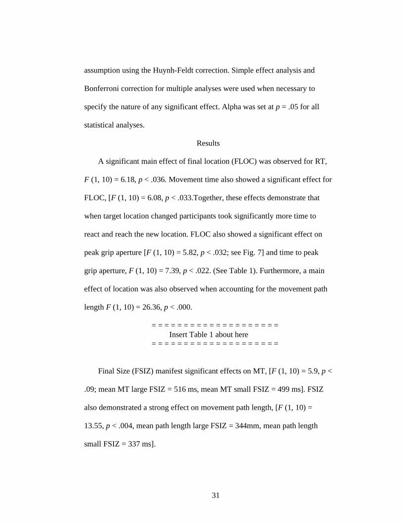
A significant main effect of final location (FLOC) was observed for RT,
F (1, 10) = 6.18, p < .036. Movement time also showed a significant effect for
FLOC, [F (1, 10) = 6.08, p < .033.Together, these effects demonstrate that
when target location changed participants took significantly more time to
react and reach the new location. FLOC also showed a significant effect on
peak grip aperture [F (1, 10) = 5.82, p < .032; see Fig. 7] and time to peak
grip aperture, F (1, 10) = 7.39, p < .022. (See Table 1). Furthermore, a main
effect of location was also observed when accounting for the movement path
length F (1, 10) = 26.36, p < .000.
= = = = = = = = = = = = = = = = = = = =
Insert Table 1 about here
= = = = = = = = = = = = = = = = = = = =
Final Size (FSIZ) manifest significant effects on MT, [F (1, 10) = 5.9, p <
.09; mean MT large FSIZ = 516 ms, mean MT small FSIZ = 499 ms]. FSIZ
also demonstrated a strong effect on movement path length, [F (1, 10) =
13.55, p < .004, mean path length large FSIZ = 344mm, mean path length
small FSIZ = 337 ms].
32
Discussion
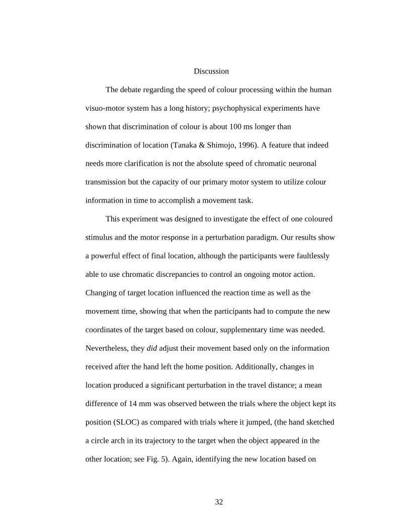
The debate regarding the speed of colour processing within the human
visuo-motor system has a long history; psychophysical experiments have
shown that discrimination of colour is about 100 ms longer than
discrimination of location (Tanaka & Shimojo, 1996). A feature that indeed
needs more clarification is not the absolute speed of chromatic neuronal
transmission but the capacity of our primary motor system to utilize colour
information in time to accomplish a movement task.
This experiment was designed to investigate the effect of one coloured
stimulus and the motor response in a perturbation paradigm. Our results show
a powerful effect of final location, although the participants were faultlessly
able to use chromatic discrepancies to control an ongoing motor action.
Changing of target location influenced the reaction time as well as the
movement time, showing that when the participants had to compute the new
coordinates of the target based on colour, supplementary time was needed.
Nevertheless, they did adjust their movement based only on the information
received after the hand left the home position. Additionally, changes in
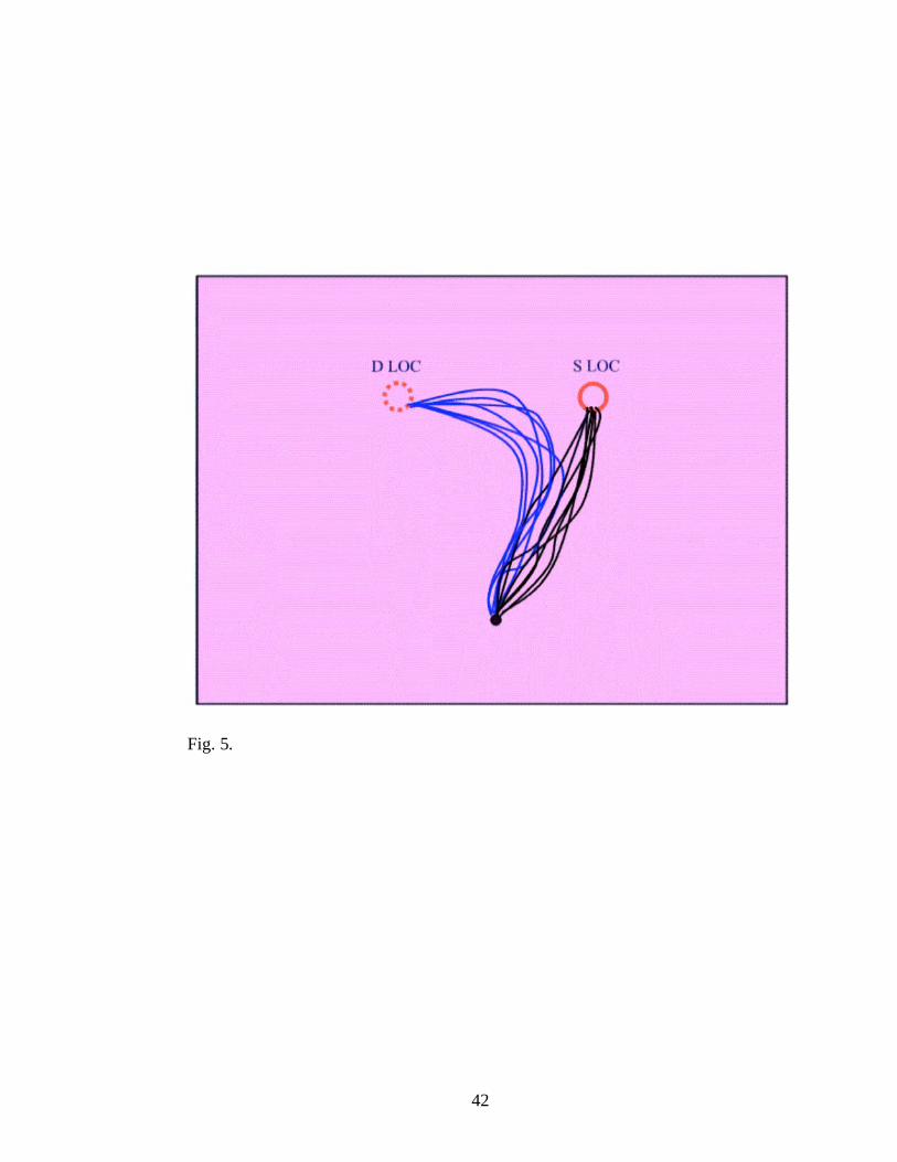
location produced a significant perturbation in the travel distance; a mean
difference of 14 mm was observed between the trials where the object kept its
position (SLOC) as compared with trials where it jumped, (the hand sketched
a circle arch in its trajectory to the target when the object appeared in the
other location; see Fig. 5). Again, identifying the new location based on

33
chromatic attributes showed a significant temporal cost in the absence of a
spatial error.
The size of the target also influenced the grasping parameters in a
similar manner (i.e., reduced efficiency but retained performance). Although
the initial size presentation did not disturb the kinematics of the movement,
when the target changed size from small to large (or vice-versa) an additional
processing of the stimulus was necessary; participants had to (1) locate the
object and then (2) compute its size based on its non spatial characteristics
(i.e., colour). Although our results revealed significant differences for the
trials where changing in size occurred, the task was completed faultlessly but
with significant temporal cost.
Overall, our data suggest that the participants were able to program
their dynamic parameters and use colour for controlling their movements
towards objects defined by location and/or size although significant delays in
processing the chromatic information were present. Pisella et al., (1998),
Schmidt (2002), Brenner and Smeets (2004) reached similar conclusions in
pointing studies in which they showed that even though coloured targets
influence movements to a certain point, participants were able to correct on
line the reaching parameters based only on chromatic information. Further,
although colour manipulation proved to have a significant effect on RT and
MT, our participants showed a more rapid adaptation as compared with the
results reported by Brenner and Smeets (2004). One possible explanation

34
could reside in the fact that it is almost impossible to control for a perfect iso-
luminance a real 3-dimensional paradigm. Given the real life environment,
even within a rigorous experimental design, a large variety of perturbing
factors might interfere with our sensory system (i.e., shadows, noise, depth of
the field). Still, our findings are consistent with the P/M visual pathways and
support the location/colour dissociation. Moreover, these findings suggest that
although a strong segregation between the P and M stream exists, these two
cortical streams may influence each other through the dense network of
cortico-cortical interconnections between different visual areas. The human
motor system appears to be almost wholly able to manage colour for
programming a grasping task.

35
Table 1
Summary of Median Values for Final Location (SLOC - same location;
DLOC - different location). Significant differences are reported, (p < .05).
Median SLOC DLOC
RT (ms) 278 292
MT (ms) 494 521
Movement Path Length (mm) 333 347
Time to Peak Grip Aperture (ms) 307 321

36
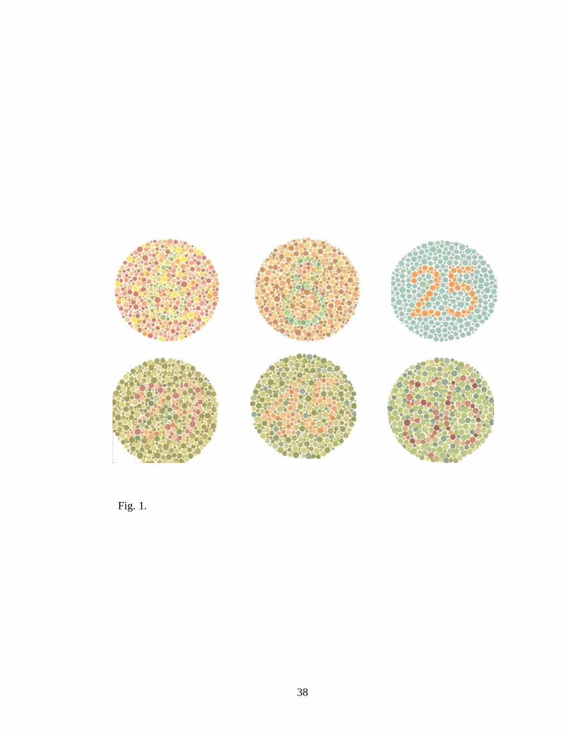
Figure Caption
Fig. 1. Ishihara colour blindness test. The test consisted of a series of coloured
cards; on each card is printed a circle made of many dots having different
sizes and different colours, spread randomly. Within the dot pattern, and
differentiated only by colour, is a number. A person having normal vision is
able to distinguish the number within the dot pattern.
Fig. 2. Experimental settings. Stimulus consisted of one red plastic cylindrical
object. The target was presented on a horizontally placed purple cardboard
background. In 50 % of the trials the target was large, (radius = 2.2 cm, height =
3.5 cm) and in half the target was small (radius = 1.6 cm, height = 2.6 cm). The
task required participant to grasp as accurately and quick as possible the target
object using the precision grip (i.e., thumb and index).
Fig. 3 Experimental design. At the beginning of each trial, the goggles closed
for 5000 ms, during which the target object was positioned on the background
surface in concordance with the randomized order. After this period the
goggles opened for 2000 ms, allowing the participant to observe the initial
location or size of the target object. The goggles were closed again after this
interval for another 5000 ms, time wherein the experimenter repositioned the
target object on the background surface in concordance with the randomized
order. After this period the goggles opened concomitant with the tone that
indicated the participant to make the grasping movement.

37
Fig. 4. Grip aperture across percents of movement, (SLOC = same location;
DLOC = different location).
Fig. 5. Movement pattern for the trials where the target changed location.
DLOC = different location. SLOC = same location. Plain circle represents the
initial target location (right); dot circle represents the position of the switched
location (left). Black lines represent the movement path towards the object
located in right; blue lines represent the movement path towards target located
in left.
38
Fig. 1.

39
Fig. 2.

40
Fig. 3.
Goggles
closed
Goggles
opened
Movement Target Set up
Tone
Goggles
closed
5000 ms 2000 ms 5000 ms
Goggles
opened
Target Set up
3000 ms

41
Grip Aperture (%)
0 20 40 60 80 100 120
Gi
At
()
50
55
60
65
70
75
80
85
90
SLOC
DLOC
Fig. 4.
42
Fig. 5.

43
Chapter III
Manuscript introduction
The pilot study emphasized the hypothesis that people are able to program
and execute a motor task (e.g., grasping) based on chromatic information.
Specifically, we showed that colour attributes can be successfully used by the
dorsal pathway for adjusting the motor programming schema. Although the
grasping task was completed successfully, a temporal dissociation occurred
when perturbation of final location/ size took place. This is consistent with
previous research (Nowak & Bullier, 1997; Tanaka & Shimojo, 1996; Tanne
et al., 1995) which proposed different latencies for dorsal vs. temporal areas.
Moreover, our results are consistent with previous researchers (Schmidt,
2002; Brenner & Smeets, 2004), showing that colour input is made available
to the dorsal stream during performance of an action at least at a satisfactory
level that allows movement to be corrected.
This first experiment was entirely controlled for luminance in order to
examine the impact of colour detection and correcting for changes in location.
We continue this line of inquiry by performing two experiments in which the
luminance is manipulated in half of the trials and the other half remains
isoluminent but vary in chromaticity (as in the Pilot Study). This manipulation
permits the examination of the relative contributions of M and P systems to
movement production. Moreover, in the first experiment, participants are
provided vision of the aiming environment only following the start tone and

44
prior to movement initiation, permitting the examination the relative roles of
luminance and chromaticity in movement planning. Based on the PC model
(Glover, 2004) one would expect that planning should be able to fully
integrate colour, but perhaps less so luminance. Conversely, the PA model
(Milner & Goodale, 1992) would predict the opposite. During experiment 2,
an open loop condition was introduced in order to observe how colour/
luminance modulates the on-line control of the movement. The goal here was
to identify the role played by the ventral stream during the execution of the
grasping task. Vision was only present during execution; no vision was
available prior to movement initiation. Contrary to experiment 1, both the PA
model and PC model would both predict preferential utilization of luminance
information.

45
Running head: COLOUR PROCESSING WITHIN THE HUMAN BRAIN
Functional aspects of colour processing within the human brain
Andrei Georgescu
Gordon Binsted
College of Kinesiology
University of Saskatchewan
Saskatoon, SK S7N 5B2
Canada

46
Introduction
Dual vision system theories
Ungerleider and Mishkin (1982) first proposed that our visual system has a
dual organization by advancing the anatomical distinction between the ventral and
the dorsal pathway within the primate visual system. The brain’s visual pathways
were therefore divided into two main pathways: The dorsal stream, referred as the
“where” pathway and the ventral stream known as the “what” pathway: The
“where” pathway projects from primary visual cortex to posterior parietal cortex,
while the “what” pathway connects to inferotemporal cortex. Predominantly, the
signals used for object localization seem to be generated mainly by the dorsal
system whereas the ventral stream is specialized for object recognition
(Ungerleider & Mishkin, 1982).
A decade later, Goodale and Milner (1992), Milner and Goodale (1995)
provided a new conceptual account of how the brain processes visual information.
They proposed the perception-action (PA) model and made two major
assumptions: (1) The dorsal stream processes visual information for motor
purposes, the dorsal path being engaged in guiding the body movements under
non-consciousness control (2) The ventral stream is involved in the object
perception by assembling the visual elements into a real, aware image. The visual
input is made available to the dorsal stream quickly for fast on-line adjustments
whereas the ventral stream activity is dominated by continuous interactions with
memory areas. In supporting these concepts, Milner and Goodale emphasize the

47
dissociation between the behaviour of the agnosic patient D.F. and that of the
ataxic patient A.T. (Perenin & Vighetto, 1988). A.T., suffers from optic ataxia
(consequence of a lesion of dorsal stream), manifests a profound incapability in
reaching and grasping targets under visual guidance but having no difficulty in
observing and recognizing them whereas the visual form agnosic patient D.F.
which has a lesion of the ventral structures shows difficulty in recognizing objects
visually, but can still use visual information to guide her movements.
Another pertinent approach of how the two streams process information was
proposed by Glover (2004). He explored the evidence for a distinction in human
performance between the planning and on-line control of action. The proposed
planning–control (PC) model offers a different approach with respect to the visual
and cognitive processes involved in movement production. This new model
establishes an anatomical substrate of the ideas offered previously by Woodworth
(1899), yet, it offers an inclusive analysis of the relationship between cognition
and action. The PC model takes for granted a gradual rather than discrete
transition between the two stages of action. Thus, planning in humans is linked
with activity in a dispersed network including a visual representation in the
inferior parietal lobe (IPL), whereas control is associated with activity in a
separate network including a visual representation in the superior parietal lobe
(SPL). During planning, information regarding the object’s shape, size,
orientation, texture, fragility and colour is sent out from the V1 to IPL via

48
temporal lobe. Once the motor act is planned, a blue-print copy is forwarded to the
SPL to be used in real time once the movement is initiated.
Whatever the approach, information regarding stimuli properties first reaches
V1 through separate neural pathways (Livingstone & Hubel, 1987, Merigan &
Maunsell, 1993). Anatomical studies have revealed that cells within the visual
system have different specializations, this being observed even from the retinal
level (Hubel & Wiesel, 1966; De Valois, Abramov, & Jacobs, 1966; Hendry &
Yoshioka, 1994; Martin, White, Goodchild, Wilder, & Sefton, 1997). Two types of
ganglion cells encode visual information; luminance and spatial location are
carried out through the philogenetically older Magno-pathway (M) having faster
neural conduction speed whereas the chromatic input is transported by the Parvo-
pathway (P). A discrete anatomical segregation between these projections can be
traced up to V1 (Livingstone, & Hubel, 1987). Although a complete mapping of
the M/P or luminance/colour onto the Dorsal/Ventral should seem plausible, there
is not enough evidence to support the total separation (for review see
Gegenfurtner, 2003).
Colour processing
The cortical processing of colour begins in the striate and extra-striate areas by
disassembling and analyzing of perceived image wave length components (Van
Essen & Zeki, 1978; Zeki & Marini, 1998). From V1 and V2 the information is
sent out to V4 (in primates; Zeki, 1978; Zeki & Shipp, 1989), and from V4 the
information is forwarded to inferio-temporal cortex. Zeki and Marini (1998)
49
proposed that colour processing occurs in three stages: (1) recording the colour
signal of every point on the visual field; (2) evaluation of the colour from one area
with the colour of surrounding area; (3) linking abstract colours to objects and
surfaces in the visual field. The first two stages represent the processing of the
impulse in V1 and V2 respectively whereas the last stage is the function of V4.
Although V4 has been considered for a long time to be the “colour centre” of the
visual brain (macaque; Zeki, 1993), its role in processing colour is not completely
understood. For example, V4 has also been proposed as the center which integrates
vision and cognition regarding the visual scene (Chelazzi, Miller, Duncan, &
Desimone, 2001).
However, even considering the possibility that V4 is not the only cortical
area involved in the superior stages of processing colour, chromatic information is
a distinctive attribute of the ventral stream. Many previous studies (Lennie, 1998;
Lennie, Krauskopf, & Sclar, 1990; Livingstone & Hubel, 1987; Milner & Goodale,
1995; Zeki, 1993) have showed that a large number of cells from the ventral
stream are sensitive to colour whereas within the dorsal stream there is a
significant proportion of neurons that respond to luminance discrepancies.
Nevertheless, extrapolating from neuro-anatomical data to real-time action
modulation, one might find this mapping difficult since during real life events
these neural circuits act together, synergistically, engaging in continuous
interaction and modulation. One question rises here: can colour only contribute in
controlling object-directed action? If we are to test this hypothesis through the

50
prism of the Milner and Goodale (1995) PA model, we should encounter
significant disruptions of the motor act when chromatic information has to be
perceived, integrated and processed during fast movements toward targets defined
by colour in absence of luminance discrepancies. Conversely, the PC model
(Glover 2004) would predict that the motor system should be able to fully
integrate both luminance and colour, as well as other object characteristics (e.g.,
shape, texture) for both planning and control phases of the movement. The usage
of colour seems therefore to encounter different interpretations in terms of
productivity for the action output.
Using a pointing paradigm, Brenner and Smeets (2004) tested this hypothesis
by asking participants to tap as quickly as possible a red target square which might
have inter-changed position with a green target. Luminance of the background was
either brighter, the same or darker than the targets. The main conclusion drawn by
Brenner and Smeets was that colour can contribute to fast online adjustments in
the presence of 120 ms delay, time required for the new location to be processed.
Brenner and Smeets countered the results of Pisella, Arzi, and Rossetti (1998) who
proposed that people cannot respond quickly to colour stimuli due to a slower
processing within the ventral streams. The roots of this debate seem therefore to
have a more semantic than concrete substrate since both studies showed that
colour processing requires more time than location features during reaching tasks.
Indeed, there is large body of literature showing that colour is processed more

51
slowly within the ventral pathway (Nowak & Bullier, 1997; Tanaka & Shimojo,
1996; Tanne, Boussaoud, Boyer-Zeller, & Rouiller, 1995).
Considering this discrepancy in interpretation we extended the research line
proposed by Brenner and Smeets (2004) and performed two experiments using a
grasping paradigm within a realistic 3-dimensional experimental design. Single
neuron recording studies (Rizzolatti, Camarda, Fogassi, Gentilucci, Luppino, &
Matelli, 1988 ; Gallese, Fadiga, Fogassi, & Rizzolatti, 1996) have shown that
different neurons from the premotor area F 5 are involved in grasping in a different
manner. Some grasping neurons fire at the beginning of the movement when the
hand just leaves the home position, and others fire at the end or when the hand has
already pre-shaped the object. Some grasping neurons discharge impulses during
flexion of the fingers and other when the fingers are extended. Culham, Danckert,
and Goodale, (2002) reported that the anterior intraparietal (AIP) region of humans
shows a larger response for visually grasping tasks than for reaching tasks. The
segregation hypothesis between the neural activation during reaching and grasping
within the dorsal areas has been intensely supported by Jeannerod (1981; 1988),
Jeannerod, Arbib, Rizzolatti, and Sakata (1995). Relying on the hypothesis that
reaching to a target is considered just a component of the grasping act we aimed to
measure the real-time capacity of our visuo-motor system to perceive, integrate
and modulate the chromatic information to (1) plan and (2) perform the grasping
movement. Within experiment one, participants were instructed to grasp, using a
precision grip, an iso-luminant red target displayed on the background in vicinity

52
of a green object distracter. By using a secondary condition where both the targets
and the background had different luminance gradients (e.g., black, white and grey)
we endeavored to determine the modulations that occur during planning and
execution of the motor task when colour in comparison with luminance had to be
computed.
Experiment 1
The first experiment (E1) was performed in order to investigate how chromatic
information in contrast with luminance information is able to manage the planning
of the grasping movement. Within this experiment vision was present only during
reaction time period and it was occluded after the participants started the
movement production.
Methods
Participants
Twelve university aged participants (eight males and four females) between
the ages of 19 and 30 years old (mean age 24.4 years old, SD = 3.1) volunteered to
participate in this study. All participants were right handed, in good health and
with normal or corrected to normal vision. Because the experiment required
subjects to make a judgment regarding the colour of the object displayed and to
grasp it based on its chromatic characteristics, all the participants were
administrated Ishihara colour blindness test (Salvia & Ysseldyk, 1972) prior being
admitted in the study. This investigation was carried out with the approval of the
University of Saskatchewan Behavioural Science Research Ethics Board for

53
ethical consideration in Human Experimentation in accordance with Declaration of
Helsinki (1964).
Task
The experiment consisted of two luminance/chromaticity conditions, each of
them having 80 trials (160 trials in total). Within every trial the participant had to
grasp a target cylindrical object as accurately as possible using a precision grip
(i.e. thumb and index).
Stimuli
Stimuli consisted of 8 plastic cylindrical objects, 4 large (radius = 2.2 cm,
height = 3.5 cm) and 4 small (radius = 1.6 cm, height = 2.6 cm). One of each size
was painted using oil paint in red, (R = 252, G = 68, B = 35), one in green, (R =
33, G = 136, B = 23), one in black, (R = 0, G = 0, B = 0) and one in white, (R =
255, G = 255, B = 255). Following painting, objects were coated with a matt finish
layer.
The experiment consisted of two main conditions. In the isoluminant
condition (RG) the red and the green objects were presented on a purple cardboard
background (R = 194, G = 58, B = 172). The heteroluminant condition (BW)
involved black and white objects; they were displayed on a gray background (R =
126, G = 126, B = 126).
The settings were similar for both conditions, the only exception being that the
luminance among the stimuli and the background varied from one condition to the
other (see figure. 1). Specifically, in RG condition the objects and the background

54
had the same luminance (20 cd/m2) whereas in the BW condition, the objects and
the background had different luminance (white target luminance = 136 cd/m2;
black target object luminance = 4 cd/m2; grey background luminance = 34 cd/m
2).
In both conditions the background (70 cm x 90 cm) was positioned on a 60
degrees inclined in front of the participant. The distance between the floor and the
near edge of the horizontal plane surface was 80 cm (see Fig. 1).
= = = = = = = = = = = = = = = = = = = =
Insert Figure 1 about here
= = = = = = = = = = = = = = = = = = = =
Apparatus
1. Motion Tracking. The movements were recorded by tracking the infrared-
emitting diode (IRED). Three markers were used, one being placed on the distal
phalanx of the right thumb, the second on the distal phalanx of right index finger
and the third on the region of the trapezium-metacarpal joint of the right thumb
(Jeannerod, 1981; Jeannerod & Biguer, 1982). This permitted deviation of grasp
and transport component of reaching. The position of the IRED was sampled for 3
s at 200 Hz following the auditory initiation cue using Visualeyez 3000 system
(Phoenix Technologies Inc., Burnaby, BC, Canada).
2. Liquid Crystal Goggles. To achieve visual occlusion where trials require
subjects to grasp the target under no vision interval, Liquid Crystal Goggles
(Translucent Technologies Inc. Plato Model: P1) were utilized.

55
3. The motion tracking data were collected using a personal computer BOXX
Technologies Inc. running Visualeyez Soft 2.70 (Phoenix Technologies Inc.,
Burnaby, BC, Canada). The randomization was determined by the second
computer (IBM ThinkCentre) running E-Prime Studio Soft Version 1.1
4. The luminance of the objects and the backgrounds was evaluated using
TEKTRONIX J-17 luminance meter (Narrow Angle Inc., Beaverton, OR, U.S.).
Procedure
The participants were comfortably seated on an immobile chair in front of
the inclined background in an illuminated room (1000 lumen/m2). The experiment
required participants to move “as rapidly and accurately as possible”, in response
to a start tone to the object presented to them and grasp it using a precision grip
(i.e., thumb and index). To avoid adaptive strategies to the targets the participants
used a chin rest during all the trials. At the beginning of each trial, the participants
were asked to put their right hand on the home position and press the trigger with
their index finger. The goggles were then closed for 5000 ms wherein the objects
were positioned on the background surface in concordance with the randomized
order. Following an additional randomized fore period of 1-3 sec, a starting tone
sounded indicating that the participant was to react with a grasping movement.
The tone also activated the goggles which opened and stayed open until the finger
lifted off. When the finger left the home position the goggles closed and the
grasping movement was made under no vision condition.

56
= = = = = = = = = = = = = = = = = = = =
Insert Figure 2 about here
= = = = = = = = = = = = = = = = = = = =
The measurement period was fixed for 3 seconds. After this interval a second
tone indicated participants that the measurement ended. Trials were considered
valid only if the fingers securely grasp the target object. The distance between the
home position and the target was 30 cm; within every trial two objects were
displayed on the background, one large and one small (green and red within RG
condition and black and white within BW condition). The distance between
objects was 14 cm. In each condition there were 4 secondary conditions that were
obtained by taking all possible combination regarding: (1) the target object size;
and (2) target object location. The experiment consisted of 160 trials, each
condition accounting for 80 trials. The conditions were counterbalanced across the
participants. The experiment lasted for approximately 60 minutes.
RG condition
The target object within the RG condition was the red object. Both the target
and the green distracter were positioned on the purple background. The trials
where the target object was located at the right were considered control trials and
they accounted for 75 % of the total number of trials (60 trials of the total of 80).
Before proceeding, the participants were informed that in 75% of the trials the
target is located at the right. The other two combinations (i.e., when the target
object was located at the left) accounted for 25 % of the total number of trials. In

57
half of the trials the target object was small and in half the target object was large.
In all the trials the distracter object had the opposite dimension (i.e., when the
target object was small the distracter object was large). To accomplish the task,
participants had to always grasp the red target using the precision grip.
BW condition
The target object for BW condition was the white object. Both the white and
the black objects were placed on the grey background. The procedure was
identical as for the RG condition.
Data Reduction and Analysis
The initiation of each reaching movement was identified interactively by
determining the first sample after which hand velocity attained and maintained a
value of 50 mm/s for ten consecutive frames (i.e., 50 ms; Binsted & Elliott, 2001;
Elliott, Heath, Binsted, Ricker, Roy, & Chua, 1999); reaction time (RT) was the
time that elapsed from the collection start to movement start. Movement offset was
the point at which fingers velocities fell below 50 mm/s and remained below this
criterion for ten consecutive frames (i.e., 50 ms). IRED position data were filtered
off-line using a second-order dual-pass Butterworth filter (low-pass, 15 Hz).
Instantaneous velocities were calculated by differentiating displacement data using
a five-point central finite difference algorithm.
A series of dependent variables were examined; these included: RT, movement
time (MT), the path length, peak acceleration, time to peak acceleration, peak

58
velocity, time to peak velocity, end wrist position, peak grip aperture, time to peak
grip aperture, the proportional grip aperture across the movement.
All hand dependent measures were analyzed using a two luminance conditions
[iso-luminance (RG) and hetero-luminance (BW)] x 2 locations (LOC; right, left)
x 2 sizes (SIZ; small, large) repeated measures ANOVA, with each score based on
the median of 10 trials for perturbed trials and 30 trials for control trials. Where
appropriate, F statistics were corrected for violations of the sphericity using
Huynh-Feldt correction. Significant effects/interactions were explored using
simple effects analyses and a Bonferroni correction for multiple comparisons
(alpha = 0.05). Only significant effects are reported (p < 0.05).
Results
With the exception of 14 trials (< 2.5 % of total) which were removed from
the analysis due to poor marker reading the participants exhibited consistent
patterns of movement and performed well across both conditions. Although vision
was not available during the movement task, all participants were able to complete
the task proficiently and only 4 trials (< 1 %) were missed trials (i.e.; unsuccessful
grasping of target object).
As predicted, the luminance/colour treatment demonstrated a significant
effect on RT [(BW mean RT = 273 ms, RG mean RT = 289 ms), F (1, 11) =
13.45, p < .003]. Moreover, an interaction between the RG/BW variation and
change in location occurred, F (1, 11) = 8.25, p < .014]. This behaviour was
manifest only when target object switched location with the perturbation object

59
forcing therefore a supplementary processing of target coordinates which turned
out to be significantly longer for colour as compared with luminance (See Fig. 3).
= = = = = = = = = = = = = = = = = = = =
Insert Figure 3 about here
= = = = = = = = = = = = = = = = = = = =
An interesting dynamic evolution occurred after the participants lifted the
fingers from the trigger and produced the grasping movement under the no vision
condition. A significant interaction was observed between colour/luminance
variation and size [F (1, 11) = 125.55, p < .000]. Within the RG condition the
participants scaled grip aperture appropriated for target size maximal grip aperture
(RG mean peak grip aperture large object = 92.78 mm, RG small object = 82.43
mm), whereas for the BW condition the scaling was insensitive to target size
variation (BW mean peak grip aperture for large object = 89.16 mm, BW mean
peak grip aperture for small object = 88.67 mm; see Fig. 4).
= = = = = = = = = = = = = = = = = = = =
Insert Figure 4 about here
= = = = = = = = = = = = = = = = = = = =
A powerful effect of changing location was also manifest for global
movement time (F (1, 11) = 25.33, p < .000) with a mean difference of 79 ms
between the condition where the target object jumped and those where initial
location was preserved (SLOC mean MT = 630 ms, DLOC mean MT = 709 ms).
The path length also suffered in similar manner when location was switched
having a mean difference of 57 mm, F (1, 11) = 19.80, p < .001. Final location
also showed an effect on all dynamic parameters, including peak velocity, time to

60
peak velocity, peak acceleration and time to peak acceleration [F (1, 11) = 4.78, p
< .049, F (1, 11) = 18.34, p < .001, F (1, 11) = 8.42, p < .013, F (1, 11) = 12.77, p
< .004 respectively). The participants reached all kinematic markers later and with
lower magnitude when the target objects switched location.
The size of the target object influenced the kinematics variables as well.
Significant effects of size were found for reaction time [F (1, 11) = 11.72 , p <
.005], path length [F (1, 11) = 6.89, p < .022], and peak velocity [F (1, 11) = 5.6 ,
p < .036]. As expected, participants started moving with increased latency when a
small target object was displayed (small size mean RT = 285 ms, large size mean
RT = 277 ms). Once the movement was initiated, the participants continued to
move more rapidly towards the larger target. The path movement length was
shorter for the small object (small target mean path length = 302 mm, large target
mean path length = 314 mm).
Discussion
Experiment 1 asked whether colour can be used efficiently for programming a
grasping movement. Although the participants successfully completed the task
important disruptions occurred. Significant delays in reaction time were
encountered when participants had to locate the target within RG settings as
compared with the BW condition. Our results are consistent with the dualistic
functionality of M/P; luminance are carried out through separate neural pathways
at least up to V1 (Hubel & Wiesel, 1966; De Valois et al., 1966; Merigan &
Maunsell, 1993; Livingstone & Hubel, 1987; Silveira & Perry, 1991). Specifically,

61
the observed slower colour processing is consistent with the large body of
evidence suggesting that colour information is transported slower through the P
pathway (Livingstone & Hubel, 1987; Merigan & Maunsell, 1993). Therefore,
even though both signals would eventually contribute to the production of the
motor act, the luminance information is made available to the motor areas more
quickly via the M pathway. Moreover, the observed latencies in reaction time are
consistent with previous findings (Nowak et al., 1997; Tanaka & Shimojo, 1996;
Tanne et al, 1995) which proposed different delays for dorsal vs. temporal areas.
Not only was the reaction time influenced by the manipulation of
luminance/colour but also the grip aperture. Although vision was not present after
the participants lifted their finger off the trigger, the movement was completely
performed without visual feed-back, the maximal grip aperture suffered significant
effects. Thus, the participants used a correct pre-shaping of the grip to grasp the
iso-luminant red target (i.e., RG condition) by adopting a conservative strategy,
scaling the grip widely but proportional with the object size. Conversely, within
the BW the grip was scaled independent of target size. This proficient grip
kinematics behaviour observed for the iso-luminant target during no vision
paradigm brings us back to the findings of Jeannerod et al., (1995). It has been
suggested that different populations of motor neurons fire differentially in regards
to the type of action they are engaged in. If during reaching, programming the
motor schema does not necessarily imply any supplementary processing of the 3-
dimensional shape of the target, during grasping this processing is mandatory and

62
it might occur even before the action takes place (Mountcastle, Lynch,
Georgopoulos, Sakata, & Acuna, 1975; Jeannerod et al., 1995). Secondly, it is
known that the control of target-directed movements relies on the visual
representations within the posterior parietal areas belonging to the dorsal stream
(Milner & Goodale, 1995; Sakata, Taira, Kusunoki, Murata, Tanaka, & Tsutsui,
1998; Goodale & Haffenden, 1998). However, it has also been suggested that
perceptual mechanisms stored within the ventral stream may be accessed during
movements when the target is not visible during a motor act (Westwood &
Goodale, 2003; Glover, 2004). Since the RG dimension proved to elicit correct
and accurate motor responses we may assume that scaling of grip aperture is the
product of two separate but interconnected processes: (1) it is planned well before
the initiation of movement, within specific populations of neurons from IPL which
encode the spatial characteristics of the target but do not fire until the movement
begins; and (2) it is the product of a ventral stored information which is accessed
by the control circuits in the event that vision is not available for guiding the
action.
However, our results are consistent with the PA model (Milner & Goodale,
1995) which proposes that the response selection stage of planning is mainly
served by the dorsal stream. Indeed, the faster movement initiation reported in the
BW condition implies that luminance information is preferentially used by the
dorsal stream to perceive the action scene and program the movement. Still, our
participants were able to plan their task relying the planning of the movement only

63
on chromatic information. It is plausible to suggest therefore that both luminance
and chromatic information reach the motor cortex; our obtained latencies being the
expression of the luminance higher speed processing through the M pathway
(Merigan & Maunsell, 1993; Maunsell, Ghose, Assad, Mc Adams, Boudreau, &
Noerager, 1999). The temporal dissociation may also reflect the fact that that
luminance information may have bypassed V1 through the superior colliculo-
thalamic route, thus creating a more efficient pathway to the primary motor cortex
(Parkin, 1996).
Our findings are consistent to a certain point with previous research lines
(Pisella et al., 1998; Brenner & Smeets, 2004). Participants were able to use colour
for programming the motor task, but the entire movement production was delayed
when the target changed location. Although colour/luminance variation did
influenced the reaction time and peak grip aperture, it did not manifest a
significant effect on the other movement parameters. The movement showed
strong effects of changing location but no significant disruptions were observed in
regard to the colour/luminance manipulation. Participants were able to
successfully generate a grip aperture for both conditions with no significant cost
(See Fig. 5).
= = = = = = = = = = = = = = = = = = = =
Insert Figure 5 about here
= = = = = = = = = = = = = = = = = = = =
These few inconsistencies with above mentioned studies can be explained if we
are to consider two aspects: firstly, in our experiment a 3-dimensional design was
64
used, approach more difficult to control and thus more sensitive to external
disturbance factors. Secondly, in our study participants performed the movement
under no vision condition. This might explain the fact that once the movement was
planned and the execution began, the colour/luminance variation stopped
influencing on-line movement production.
Summing up, although the participants showed an increased adaptability in
processing luminance, the chromatic information was used successfully by the end
of the task. This suggests that colour can be used to plan the motor task where
necessary.
Experiment 2
Within the second experiment we attempted to examine the real time
capacity of our visuo-motor system to integrate colour for real time control of the
grasping task. The luminance variation was manipulated following the same
procedure as presented in Experiment 1; the only noticeable change being that the
full vision condition was present only during the execution of the movement (i.e.,
on-line control).
Methods
Participants
Thirteen participants (8 male and 5 female) took part in this study. They were
between the ages of 20 and 29 years old (mean age 25.5 years old, SD = 3.2). All
participants were strongly right handed, having normal or corrected to normal
vision and in good health. All participants were administrated the Ishihara colour
65
blindness test prior being admitted in the study. This investigation was carried out
with the approval of the University of Saskatchewan Behavioural Science
Research Ethics Board for ethical consideration in Human Experimentation in
accordance with Declaration of Helsinki (1964).
Procedure
The experiment consisted of two luminance/chromaticity conditions, each of
them having 80 trials (160 trials in total). The task, the stimuli, the settings and the
apparatus were identical with those presented in E1 (See Fig. 1).
As in Experiment 1 participants used a chin rest during all the trials to prevent
accommodation to the targets. At the beginning of each trial, the participants put
their right hand on the home position and pressed the trigger with their index
finger. The goggles were then closed for 5000 ms wherein the objects were placed
on the background surface in respecting the randomized order triggered by the
computer. Following an additional randomized fore-period of 1-3 sec, a starting
tone sounded indicating that the participant was to react with a grasping
movement. After the tone the goggles remained closed and stayed closed until the
finger lifted off. When the finger left the home position the goggles opened and
the grasping movement was made under full vision conditions (Fig. 6). The
measurement period was fixed for 3 seconds. After this interval a second tone
indicated participants that the measurement ended. Again, trials were validated
only if the grasping movement was successful.

66
= = = = = = = = = = = = = = = = = = = =
Insert Figure 6 about here
= = = = = = = = = = = = = = = = = = = =
Data Reduction and Analysis
Data filtering, reduction and the analysis followed the same schema as
presented in experiment 1. Only significant effects are reported (p < 0.05).
Results
All thirteen subjects were able to produce grasping movements with
surprisingly consistent accuracy for all eight experimental conditions. Although
some of the responses showed a small overshoot with respect to the perturbed
target, only 4% of the trials were eliminated from the analysis due to total missed
performance.
A significant interaction for MT was observed when luminance/colour was
manipulated along with location, [F (1, 12) = 7.11, p < .024]. Post hoc analysis
revealed that participants took more time to correct and complete the task when
adjusting their movement parameters based only on colour attributes as compared
with luminance features. The effect of colour/luminance was therefore observed
only during perturbed trials, when the red object switched position with the green
one (See Fig. 7).
= = = = = = = = = = = = = = = = = = = =
Insert Figure 7 about here
= = = = = = = = = = = = = = = = = = = =
67
Both size and location also showed also an effect on movement time.
Participants moved slowly on average when the size of the objects was smaller
compared with the perturbed target, [F (1, 12) = 44.97, p < .000; (mean MT for
small object 691 ms; mean MT for large object 657 ms)]. When the target
switched location to the left, it forced a delay in processing the new target
coordinates. This delay was manifest as a significant effect on the total movement
time, F (1, 12) = 93.61, p < .000, (mean MT same location 624 ms; mean MT
changed location 724 ms).
= = = = = = = = = = = = = = = = = = = =
Insert Figure 8 about here
= = = = = = = = = = = = = = = = = = = =
= = = = = = = = = = = = = = = = = = = =
Insert Figure 9 about here
= = = = = = = = = = = = = = = = = = = =
The movement path also suffered significant perturbations (Fig. 8). A
significant effect of location was manifest, F (1, 12) = 44.67, p < .000 as well as
an effect of size, F (1, 12) = 25.50, p < .000, (large object movement path length
307 mm; small object movement path length 334 mm). When the target kept its
initial position (i.e. right) the movement path was almost linear (See Fig 9a).
Conversely, in the case of target location perturbation, the hand showed a less
efficient trajectory increasing the path length (See Fig. 9 b), (same location
movement path length 286 mm; different location movement path length 355 mm).

68
A similar conclusion holds for the dynamic parameters. Changes in location
showed a significant effect on time to peak velocity and time to peak acceleration,
respectively, F (1, 12) = 33.70, p < .000, F (1, 12) = 9.34, p < .010. Location also
influenced the kinematic parameters of the grasp, both peak grip aperture and time
to peak grip aperture showing significant effects, F (1, 12) = 9.42, p < .012; F (1,
12) = 38.06, p < .000, (Fig. 9). Furthermore, a significant interaction between
changing in location and size occurred [F (1, 12) = 6.39, p < .030]. Post hoc
analyses suggest that the participants were able to scale their maximal aperture and
initialize the final step of the movement faster in the case of small object when this
was presented in the same location (Fig. 10).
= = = = = = = = = = = = = = = = = = = =
Insert Figure 10 about here
= = = = = = = = = = = = = = = = = = = =
Discussion
Our primary goal in E 2 was to test the hypothesis that colour itself can
provide enough information to the parietal cortex for controlling a grasping action
in real time.
Overall, all thirteen participants were able to clearly complete the task for
both conditions. Because vision of the targets was available only after the
participants lifted off the finger from the trigger button, no reaction time
differences were observed across the trials.
As expected, global movement suffered significant adjustments during
completion of the motor task. Although the general impression was that the

69
participants carried out the task with surprising consistency, the performance was
considerably altered within the perturbed trials when we manipulated the location.
Changes in location significantly influenced the dynamic parameters by forcing
the participant to acknowledge, observe and modify their movement parameters in
concordance with a given arrangement. Moreover, the global movement time
suffered significant changes across the trials when the iso-luminant (red-green-
purple) environment was present as compared with the situation when the
experimental settings were not controlled for luminance. These results are
consistent with the previous researchers who postulate that colour information is
transported by the parvo-cellular pathway whereas the luminance modulation is
the function of the mango-cellular path (Livingstone & Hubel, 1987; Merigan &
Mausell, 1993). Moreover, significant latencies were observed in terms of
movement time, a fact that demonstrates predominant usage of the luminance
feature in controlling the outgoing movement.
General Discussion
The present research endeavoured to investigate the functional mechanisms
of colour processing for the programming and execution of a motor task. We
aimed to identify the particular motor strategies that visual cortex and subsequent
pathways adopt when processing colour and luminance. Our two major questions
were: (1) Is colour, as a putative ventral attribute, able to be used in identification,
selection and programming a motor schema? (2) Can colour solely guide the on-
line control of the grasping task?

70
In respect to both experiments, the main conclusion is that both planning and
on-line control can rely on chromatic information. Our results suggest that colour
can be used proficiently by the dorsal structures to modulate the grasping task.
Using a 3-dimensional grasping task we showed that luminance information is
carried out from the retina to the command structures faster than the colour
attribute and it is involved as a primary source of information in controlling an
outgoing movement.
Further, in this investigation we focused only on red/green modulation and
avoided the yellow/blue dimension and konio (K) pathway inputs due to its
variable contribution to colour/motion modulation. Although there is evidence to
support the possibility that K signals might reach area MT (Wandell, Poirson,
Newsome, Baseler, Boynton, Huk, Gandhi, & Sharpe, 1999) and interferes within
the M pathway (Calkins, 2000), other neurophysiological studies argued against
this hypothesis (Lee, Martin, & Valberg, 1998; Dacey & Lee, 1994). Whether K
pathway contribution to colour/motion processing plays a significant role or if it is
just a subsidiary path along the P/M pathways, that stands for further examination.
Programming
In accordance with previous research lines (Hubel & Wiesel, 1966; De
Valois, Cottaris, Elfar, Mahon, & Wilson, 2000; Derrington et al., 1984; Hendry &
Yoshioka, 1994; Martin, White, Goodchild, Wilder, & Sefton, 1997), our results
are consistent with the P/M parallel pathways highlighting the idea that there is a
constant delay in the transportation, processing and handling of colour as a

71
defining feature of on target object. Indeed, the parvo-cellular attributes are
processed slower on average than the magno-cellular pathway; our participants
exhibited constant delays where colour had to be processed. Still, our participants
were able to program the task faultlessly, a result consistent with previous findings
(Pisella et al., 1998; Brener & Smeets, 2004). Using a more realistic approach (3-
dimensional grasping versus pointing and tapping a coloured light array), we
showed that colour can solely guide the complete programming schema of the
motor act.
More interestingly, our findings highlight the idea that the dorsal stream seems
to be involved in the movement programming as early as response selection stage
since the luminance manipulation showed faster adaptability to location change.
The superior efficacy of the luminance feature in movement production could be
the result of two cortical mechanisms. Luminance information may reach first the
visual cortex earlier and subsequently be sent to posterior parietal cortex via dorsal
connection with higher neural conduction speeds (Nowak & Bullier, 1997; Tanaka
& Shimojo, 1996; Tanne et al., 1995). Conversely, luminance feature information
might bypass the classical dorsal neural path (LGN - Visual Cortex - Posterior
Parietal Cortex) and reach the parietal structures faster through the colicullo-
pulvinar pathway (Parkin, 1966; Schoenfeld, Heinze, & Woldorff, 2001). In both
cases, the posterior parietal cortex shows that it is able to integrate the luminance
information and modulate the response selection stage based on this attribute, a
result that is consistent with the PA model (Milner & Goodale, 1995). This finding

72
is less consistent with the PC model (Glover, 2004) which postulates that the
planning of the movement, at least up to the stage of response programming, is the
function of the ventral stream.
An alternative account for our findings is that the total segregation between the
two main retino-geniculo-visual cortex streams up to V1 reduces after visual
striate cortex, (Thorell, De Valois, & Albrecht 1984; Leventhal, Thompson, Liu,
Zhou, & Ault, 1995; De Valois et al., 2000; Johnson, Hawken, & Shapley, 2001;
Xiao & Felleman, 2004). The corollary of this hypothesis is that both colour and
luminance should reach the parietal cortex and the ventral cortex. Further, both
should be ready to be used for programming the upcoming movement. Thus,
latency differences reported between the chromatic and the luminance utilization
may be only a matter of the speed of nervous conduction, not efficacy. In this case,
it is difficult to assume which stream actually controls the planning stages and to
dissociate between the PA model and the PC model since both utilize the same
visual representation but within different streams.
However, our results show that once the motor act was planned, the movement
did not reveal specific patterns to fully support the second mechanism discussed
previously. Although significant differences were present in all kinematics
parameters following target perturbation, luminance variation did not influence the
precision of the task. Online computation of movement occurred in a similar
manner for both BW and RG conditions under no vision circumstance. If
73
luminance information had been available to the ventral stream, then a powerful
effect should have been present during execution of the movement.
Although the luminance feature showed faster processing and superior
movement production, scaling the grip aperture based on chromatic attribute
demonstrated a surprisingly better performance. One explanation of this effect is
offered by the interpretation of Jeannerod et al., (1995). During planning, different
populations of grasping neurons from IPL encode the target properties before the
movement is initiated. Once the motor act begins, the movement dynamic
parameters are modulated by a dense network connection within the dorsal stream;
in the event that vision is not available for action, the control mechanisms must
access the stored representation of the target from the ventral stream, the same
visual representation which was encoded by the grasping neurons before
movement production.
Summing up, our results are consistent with the dual system theory.
Chromatic information can contribute to planning but its processing is totally
dependent on the ventral capacities in modulating the chromatic information. If
luminance discrepancies are present (as is likely to occur within a normal
environment) then the planning is governed by the luminance attribute. The main
finding in this study is therefore consistent with the idea that the dorsal stream is
involved in the planning and specifically in the response selection stage of
information processing. Moreover, it can program the up-coming actions with a
higher degree of spatial resolution than is available to the ventral stream.

74
Online control
A second question of interest was whether on-line control of grasping a
specified target can be completed based on colour attributes solely. Although the
task was accomplished, the movements showed significant discrepancies between
BW condition and RG condition. This is again consistent with dual visual systems
theory, which predicts that visually guided motor actions made directly on an
object utilize largely the dorsal stream. Participants showed that the integration of
luminance attribute takes less time than chromatic information and result in more
efficient execution.
Our results are consistent with previous lines of investigation regarding the
usage of chromatic information in controlling an out-going movement. Overall,
data suggest that participants were able to modulate the dynamic parameters and
use colour for controlling their movements towards objects defined by location
and/or size although significant delays in processing the chromatic information
were present. Pisella et al., (1998) and Brenner and Smeets (2004) reached similar
conclusions in pointing studies where they showed that coloured 2-dimensional
targets could influence human movements to a certain point. Our obtained delays
for target displacement (mean difference 100 ms) are consistent with their reported
value of 120 ms (Brenner & Smeets, 2004). Therefore we may assume that colour
features take more time to be integrated whatever the neural path involved.
These finding are again consistent up to a point with Milner and Goodale’s
(1995) PA model. Obviously, during action participants exhibited faster
75
movements on average when they could rely on the information received through
the magno-occipito-parietal pathway or via colliculo - thalamo - posterior parietal
cortex (Parkin, 1966; Schoenfeld et al., 2001). Because vision was available only
after the participants initiate the movement (i.e. lifted the finger off the trigger),
the coordination of the sequences required only on-line adjustment and the
information regarding the colour of the objects, shape and location had to be
rapidly forwarded from the visual association areas to the motor cortex. A constant
delay between the kinematic parameters was observed across all trials where
colour had to be processed to determine object location perturbation. Still, our
participants were able to complete the task faultlessly. This fact leads us to the
conclusion that colour information might bypass certain ventral processes to be
made available to dorsal structures for on line control modulation. Another
possibility is that the ventral streams cooperate efficiently with the dorsal streams
during online-control. Reconsidering the Milner and Goodale model, we would
have expected a larger effect of changing location for luminance processing as
compared with chromatic processing since visually guided tasks are largely
coordinated by the dorsal stream (Milner & Goodale, 1995).
On the other hand, based on the anatomical predictions of Glover’s (2004) PC
model, we should not have encountered such a significant interaction between
changes in location and luminance manipulation because the chromatic efference
copy forwarded from IPL should reach SPL in real time during the control phase
of movement (Glover, 2004). Given the fact that IPL information can be used at

76
any point to adjust and compute on-line a motor schema (Glover, 2004), the
movement should have been smooth and the colour information should have not
encountered any difficulty in being processed by the SPL during online control.
Our participants showed a considerable temporal delay across trials when
colour had to be processed as the only source of information. It is plausible to
suggest that during visually guided actions the dorsal stream controls movement
relying mainly on the information regarding spatial characteristics of the target
(i.e., luminance). If this attribute is carefully attuned to a neutral value for all
components of the environmental scene, our results showed that colour is able to
offer consistent information for completing the grasping task but with temporal
cost in lieu of spatial error.
77
Figures Caption
Fig. 1. BW and RG Condition; experimental settings. Stimuli consisted of 8 plastic
cylindrical objects, 4 large (r = 2.2 cm, h = 3.5 cm) and 4 small (r = 1.6 cm, h =
2.6 cm). One of each size was red, one was green, one black and one white. The
experiment consisted of two main conditions. In the isoluminant condition (RG)
the red and the green objects having different sizes were presented on a purple
cardboard background. The heteroluminant condition (BW) involved black and
white objects; they were displayed on a gray background.
Fig. 2. Experiment 1. Experimental Design. Within experiment 1 vision was
available only during reaction time. At the beginning of each trial, goggles closed
for 5000 ms wherein the objects were positioned on the background surface.
Following an additional randomized foreperiod of 1-3 sec, a starting tone sounded
indicating that the participant was to react. The tone also activated the goggles
which opened and stayed open until the finger lifted off. When the finger left the
home position the goggles closed and the grasping movement was made under no
vision condition.
Fig. 3. Experiment 1. Reaction Time vs. changing in location. BW = hetero-
luminant condition; RG = iso-luminant Condition. DLOC = different location.
SLOC = dame location. * Indicates significant effect, p < .05.
Fig. 4. Experiment 1. Peak grip aperture as a function of target size. BW = hetero-
luminant condition; RG = iso-luminant condition. Small = small object size; Large
= large object size. * Indicates significant effect, p < .05.
78
Fig. 5. Experiment 1. Peak Grip Aperture in percents of total movement. BW large
= Grip aperture for large object within Hetero-luminant condition; BW small =
Grip aperture for small object within the hetero-luminant condition; RG large =
Grip aperture for large object within the Iso-luminant condition. RG small = Grip
aperture for small object within the Iso-luminant condition.
Fig. 6. Experiment 2. Experimental Design. Within experiment 2, vision was
available only after participants lifted the finger off the trigger and started the
movement. At the beginning of each trial, goggles closed for 5000 ms wherein the
objects were positioned on the background surface. Following an additional
randomized fore-period of 1-3 sec, a starting tone sounded indicating that the
participant was to react. The goggles stayed closed until the finger lifted off. When
the finger left the home position the goggles were activated which opened and
stayed open until the finger movement was completed.
Fig. 7. Experiment 2. Movement Time vs. changes in location. BW = hetero-
luminant condition; RG = iso-luminant condition. DLOC = different location.
SLOC = same location. * Indicates significant effect, p < .05.
Fig. 8. Experiment 2. Movement path length as a function of size (1) and location
(2). Large = large object size. S = small object size. DLOC = different location.
SLOC = same location. * Indicates significant effect, p < .05.
Fig. 9. Experiment 2. Movement pattern.
a. When target kept its initial position hand movement was linear.

79
b. The hand corrected its trajectory by sketching an arch in its way to grasps the
target when it appeared in left.
Fig. 10. Experiment 2. Peak Grip Aperture in percents of total movement. DLOC
large = Grip aperture for large object when located in different location; SLOC
large = Grip aperture for large object when located in same location; DLOC small
= Grip aperture for small object when located in different location; SLOC large =
Grip aperture for large object when located in same location;

80
Fig. 1.

81
Fig. 2.
Goggles
closed
Goggles
opened
Goggles
closed
No Vision Full Vision No Vision
Movement Target Set up
Tone Finger lift off Trigger
RT
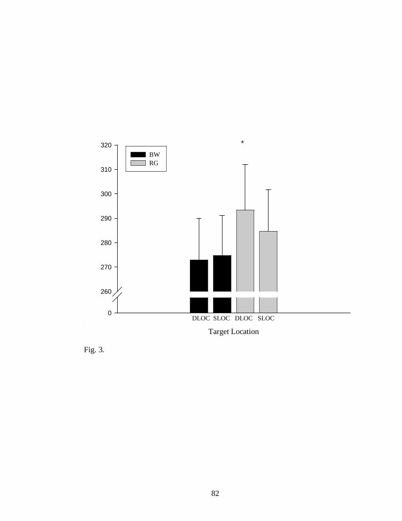
82
Ri
Ti
()
0
260
270
280
290
300
310
320
BW
RG
*
DLOC SLOC DLOC SLOC
Target Location
Fig. 3.

83
Pk G
i A
0
80
85
90
95
100
BW
RG
Small Large Small Large
*
Target Size
Fig. 4.

84
0 20 40 60 80 100 120
Gi
A (
)
30
40
50
60
70
80
90
100
BW large
BW small
RG large
RG small
Grip Aperture (% of Movement)
Fig. 5.

85
Fig. 6.
No Vision No Vision Full Vision
Movement Target Set up RT
Finger lift off Trigger Tone
Goggles
closed
Goggles
opened

86
0
600
625
650
675
700
725
750
775
800
BW
RG *
SLOC SLOC DLOC DLOC
Target Location
Fig. 7.

87
Mo
vem
ent p
ath len
gth
(mm
0
250
275
300
325
350
375
400
SIZE
LOC
(1 ) (2 )
Large Small DLOC SLOC
Target Size Target Location
Fig. 8.

88
Fig. 9 a.
Fig. 9 b.
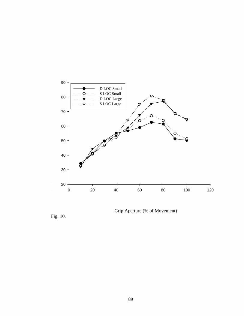
89
0 20 40 60 80 100 120
Gi
A (
)
20
30
40
50
60
70
80
90
D LOC Small
S LOC Small
D LOC Large
S LOC Large
Grip Aperture (% of Movement)
Fig. 10.

90
References
Binsted, G., & Elliott, D. (2001). Eye-hand coordination in goal directed aiming.
Human Movement Science, 20, 563-585.
Brenner, E., & Smeets, J., B., J. (2004) Colour vision can contribute to fast
corrections of arm movements. Experimental brain research, 158, 302-307.
Chelazzi, L., Miller, E., K., Duncan, J., & Desimone, R. (2001). Responses of
neurons in macaque area V4 during memory guided visual search. Cerebral
Cortex, 11, 761-762
Culham, J., C., Danckert, S., L., & Goodale, M., A. (2002). fMRI reveals a
dissociation of visual and somatomotor responses in human AIP during
delayed grasping. Journal of Vision, 2(7), 701-708.
De Valois, R., Abramov, I., & Jacobs, G. (1966). Analysis of response patterns of
LGN cells. Journal of the Optical Society of America, 56(7), 966-977.
De Valois, R., L., Cottaris, N., P., Elfar, S., D., Mahon, L., E., & Wilson, J., A.
(2000). Some transformations of color information from geniculate nucleus to
striate cortex. Proceedings of the National Academy of Sciences of the United
States of America, 97, 4997-5002.
Derrington, A., M., Krauskopf, J., & Lennie, P. (1984). Chromatic mechanisms in
lateral geniculate nucleus of macaque. Journal of Physiology (London), 357,
241-265.
91
Elliott, D., Heath, M., Binsted, G., Ricker, K., Roy, E., E., & Chua, R. (1999). Goal
directed aiming: Correcting force specification error with the right and left
hands. Journal of Motor Behaviour, 31 (4), 309-324.
Gallese, V., Fadiga, L., Fogassi, L., & Rizzolatti, G. (1996). Action recognition in the
premotor cortex. Brain, 119, 593-609.
Gegenfurtner, K. (2003). Cortical mechanisms of colour vision. Nature, 4, 563-572.
Glover, S. (2004). Separate visual representation in the planning and control of
action. Behavioural and Brain Science, 27, 3-78.
Goodale, M., A., & Haffenden, A., M. (1998). frames of reference for perception and
action in human visual system. Neuroscience and Behavioural Reviews, 22,
161-172.
Goodale, M., A., & Milner, A., D. (1992). Separate visual pathways for perception and
action. Trends in Neurosciences, 15, 20-25.
Hendry, S., H., & Yoshioka, T. (1994). A neurochemically distinct third channel in
the macaque dorsal lateral geniculate nucleus. Science, 264, 575-577.
Hubel, D., H., & Wiesel, T., N. (1966). Effects of varying stimulus size and color on
single lateral geniculate cells in Rhesus monkeys. Proceedings of the National
Academy of Sciences of the United States of America, 55, 1345-1346.
Jeannerod, M., Arbib, M., A., Rizzolatti, G., & Sakata, H. (1995). Grasping objects:
the cortical mechanisms of visuo-motor transformation. Trends in
Neuroscience, 18, 314–320.

92
Jeannerod, M. (1981). Intersegmental coordination during reaching at natural visual
objects. In: Long J, Baddeley A (Eds.) Attention and performance IX (153–
169). Lawrence Erlbaum, Hillsdale.
Jeannerod, M., & Biguer, B. (1982). Visuo-motor mechanisms in reaching within
extrapersonal space. In: Ingle D, Goodale MA, and Mansfield R. (Eds.).
Advances in the Analysis of Visual Behavior, (387-409). Boston, MA: MIT
Press.
Jeannerod, M. (1988). The neural and behavioural organization of goal-directed
movements. Oxford University Press.
Johnson, E., N., Hawken, M., J., & Shapley, R. (2001). The spatial transformation of
color in the primary visual cortex of the macaque monkey. Nature
Neuroscience, 4, 409-416.
Lennie, P. (1998). Single units and visual cortical organization. Perception, 27, 889-
935.
Lennie, P., Krauskopf, J., & Sclar, G. (1990). Chromatic mechanisms in striate cortex
of macaque. Journal of Neuroscience, 10, 649-669.
Leventhal, A., G., Thompson, K., G., Liu, D., Zhou, Y., & Ault, S., J. (1995).
Concomitant sensitivity to orientation, direction, and color of cells in layers 2,
3, and 4 of monkey striate cortex. Journal of Neuroscience, 15, 1808-1818.

93
Livingstone, S., M., & Hubel, D., H. (1987a). Psychophysical evidence for separate
channels for the perception of form, color, Movement and Depth. Journal of
Neuroscience, 7 (11), 3416-3468.
Livingstone M., S., & Hubel, D., H. (1987b). Connections between layer 4B of area
17 and the thick cytochrome oxidase stripes of area 18 in the squirrel monkey.
Journal of Neuroscience, 7, 3371-3377.
Livingstone M., S., & Hubel, D., H. (1982). Thalamic inputs to cytochrome oxidase-
rich regions in monkey visual cortex. Proceedings of the National Academy of
Sciences U S A, 79, 6098-6101.
Martin, P., R., White, A., J., R., Goodchild, A., K., Wilder, H., D., & Sefton, A., E.
(1997). Evidence that blue-on cells are part of the third geniculocortical
pathway in primates. European Journal of Neuroscience, 9, 1536-1541.
Maunsell, J., H., Ghose, G., M., Assad, J., A, McAdams, C., J., Boudreau, C., E., &
Noerager, B., D. (1999). Visual response latencies of magnocellular and
parvocellular LGN neurons in macaque monkeys. Visual Neuroscience, 16, 1-
14.
Merigan, W., & Maunsell, J., H. (1993). How parallel are the primate visual
pathways? Annual Review of Neuroscience, 16, 369-402.
Milner, A., D., & Goodale, M., A. (1995). The visual brain in action. Oxford,
England: Oxford University Press.

94
Mountcastle, V.,B., Lynch, J., C., Georgopoulos., A., Sakata, H., & Acuna, C,
(1975). Posterior parietal association cortex of the monkey: command
functions for operations within extrapersonal space. Journal of
Neurophysiology, 38, 871-908.
Nowak, L., G., & Bullier, J. (1997). The timing of information transfer in the visual
system. Cerebral cortex, 12, 205-241.
Parkin, A., J. (1996). Explorations in Cognitive Neuropsychology. Blackwell
Publishers Ltd.: Oxford.
Perenin, M., T., & Vighetto, A. (1988). Optic ataxia: a specific disruption in
visuomotor mechanisms. I. Different aspects of the deficit in reaching for
objects. Brain, 111, 643–674.
Pisella L., Arzi, M., & Rossetti, Y. (1998). The timing of color and location
processing in the motor context. Experimental Brain Research, 121, 270-276.
Rizzolatti, G., Camarda, R., Fogassi, L., Gentilucci, M., Luppino, G., & Matelli, M.
(1988). Functional organization of inferior area 6 in the macaque monkey: II.
Area F5 and the control of distal movements. Experimental Brain Research,
71, 491-507.
Salvia, J., & Ysseldyk, J. (1972). Criterion validity of 4 tests for green-red color
blindness. American Journal of Mental Deficiency, 76, 418-430.
Schoenfeld, M., A., Heinze, H., J., & Woldorff, M., G. (2001). Unmasking motion-
processing activity in human brain area V5/MT+ mediated by pathways that
bypass primary visual cortex. NeuroImage, 17, 769-779.

95
Silveira L., C., & Perry, V., H. (1991). The topography of magnocellular projecting
ganglion cells (M-ganglion cells) in the primate retina. Neuroscience 4, 217-
237.
Tanaka, Y., & Shimojo, S. (1996). Location vs. feature: reaction time reveals
dissociation between two visual functions. Vision Research, 36, 2125-2140.
Tanne, J., Boussaoud, D., Boyer-Zeller, N., & Rouiller, E. (1995). Direct visual
pathways for reaching movements in the macaque monkey. Neuroreport, 7,
267-272.
Thorell, L., G., De Valois, R., L., & Albrecht, D., G. (1984). Spatial mapping of
monkey V1 cells with pure color and luminance stimuli. Vision Research, 24,
751-769.
Ungerleider, L. G., & Mishkin, M. (1982). Two cortical visual systems. In D. J.
Ingle, R. J. W. Mansfield, & M. S. Goodale (Eds.), The Analysis of Visual
Behavior (549-586). Cambridge, Mass: MIT Press.
Van Essen, D., C., & Zeki, S., M. (1978). The topographic organization of rhesus
monkey prestriate cortex. Journal of Physiology, 277, 193-226.
Westwood, D., & Goodale, M., A. (2003). Perceptual illusion and the real-time
control of action, Spatial Vision, 16, 243-254.
Woodworth, R., S. (1899). The accuracy of voluntary movements. Psychological
Review Monograph, 3 (13) , 1-119.
Xiao, Y., & Felleman, D., J. (2004). Projections from primary visual cortex to
cytochrome oxidase thin stripes and interstripes of macaque visual area 2.

96
Proceedings of the National Academy of Sciences U S A 101 (18), 7147-
7151.
Zeki, S., M. (1978). Functional specialization in the visual cortex of the rhesus
monkey. Nature, 274, 423-428.
Zeki, S., M. (1993). A vision of the brain. Blackwell: Oxford.
Zeki, S., M., & Marini, L. (1998). Three cortical stages of colour processing in the
human brain. Brain, 121, 1669-1685.
Zeki, S. M., & Shipp, S. (1989). Modular connections between areas V2 and V4 of
macaque monkey visual cortex. European Journal of Neuroscience, 1 , 494-
506.

97
Chapter IV
Thesis summary
Conclusions
The present thesis consists of three experiments. Our key question on
developing this research line was whether colour can be used to plan and perform
a movement as the only source of information for the visual system. We tested our
hypotheses by designing and furthermore interpreting the results through the prism
of the two psycho-behavioural models, the Perception-Action (PA) model (Milner
and Goodale, 1995) and the Planning-Control (PC) model (Glover, 2004). While
the PA model postulates a strong dichotomy between the roles played by the two
cortical visual streams during perception and action, respectively, the PC model
offers a different interpretation of this interaction, yet based on similar
neuroanatomical substrates. Whereas the PA model emphasizes the importance of
the dorsal stream in perception (perception for action) and in the coordination of
the last stage of information processing, the PC model advances the idea of a
gradual rather than discrete transition between the stages of action by advancing
different concepts in terms of planning and control. Our aim was to investigate
how the two streams manage the performance of a grasping act when colour is
offered as the only source of information.
The pilot study was entirely controlled for iso-luminance in order to examine
the usage of colour for guiding a motor schema. Our experiment consisted of
manipulation of one target object which changed or did not change its location or

98
its size. This manipulation allowed us to make participants judge targets’
coordinates by computing only the chromatic information. The results revealed
that colour can contribute to adjusting the movement parameters during grasping,
although a constant temporal delay was observed across trials where colour had to
be used for reprogramming and adjusting the kinematic parameters. Our results are
consistent with previous lines of investigation (Pisella et al., 1998; Schmidt, 2002;
Brenner & Smeets, 2004). Using pointing/reaching tasks they showed that people
are able to faultlessly use chromatic information although temporal delays were
manifest. Our findings are also consistent with previous studies (Hubel & Wiesel,
1966; Derrington et al., 1984; Livingstone & Hubel, 1987; Hendry & Yoshioka,
1994) which postulate significant temporal conducting latencies between the P and
M pathways. Furthermore, the results support the dorsal/ventral delays in
processing information (Nowak & Bullier, 1997; Tanaka & Shimojo, 1996; Tanne
et al., 1995).
However, due to the fact that within the pilot study only one target object was
utilized, future investigation was required and two experiments were therefore
designed. The central foundation of this second experimental approach was to
explore the real time capacity of the visuo-motor system to use chromatic
information to program (Experiment 1) and control (Experiment 2) the grasping
movement having two coloured object displayed on the background. Luminance
was manipulated in half of the trials, the other half remaining iso-luminant varying
in chromaticity instead (similarly as in Pilot Study). This treatment allowed the

99
examination of the involvement of M and P systems in movement production and
furthermore the implication of the subsequent dorsal and ventral areas in
performing action.
Our findings are consistent with the results of the pilot study and
subsequently with previous lines of investigation (Pisella et al., 1998; Brenner &
Smeets, 2004) and reinforce the idea of a proficient usage of chromaticity for
adjusting the movement parameters when colour had to be processed for action.
Although participants were able to complete the grasping task, significant
differences were observed when comparing the iso-luminant condition with the
hetero-luminant condition. Both programming and on-line control showed an
increased adaptability in using luminance to guide the movement production. Still,
when the luminance attribute was adjusted to a neutral value for targets and
background, chromatic attributes offered substantial information for completing
the task.
Summing up, our findings are generally consistent with the PA model
proposed by Milner and Goodale (1995). While the dorsal stream is mainly
involved in the modulation of the motor act, the ventral stream represents a
reliable source of information when the task is performed under no vision
condition. The real-time hypothesis (Westwood & Goodale, 2003) is also
supported by our results. Scaling the grip during visually guided tasks occurs
under the command of the dorsal stream; conversely, when vision is occluded, the
ventral stream modulates the kinematics of the grasp based on previous recorded
100
experience, in our case colour being successfully used for controlling on-line the
movement.
Limitations and future directions
In order to design visual interfaces which afford better comprehension and
elicit faster and more accurate reactions, one must first understand how different
attributes of a target interact to influence perception and responding. The key
question which was raised along this thesis was whether humans can use colour
for preparing/performing a movement.
Although our results are consistent with previous lines of investigation
(Pisella et al., 1998; Brenner & Smeets, 2004; Nowak & Bullier, 1997; Tanaka &
Shimojo, 1996; Tanne et al., 1995) our obtained temporal latencies between the M
and P processing (i.e. location/colour) were less than the values reported
previously for pointing/reaching paradigms. One pertinent explanation could
reside in the fact that it is nearly unattainable to control for a perfect iso-luminance
a real 3-dimensional environment given that within a real life situation a large
variety of perturbation factors might interfere with our sensorial system (i.e.
shadows, noise, depth of the field). For example, future examination of monocular
guiding performance could provide supplementary data with respect to the degree
of segregation between M and P pathways when examining the grasping
movement (i.e. 3-dimesional design).
Another feature that might require supplementary examination is the
yellow/blue chromatic dimension. In this line of investigation we focused only on

101
red/green modulation and knowingly avoided the yellow/blue dimension and
konio (K) pathway inputs to movement production. Although existing evidence
supports the possibility that K signals might interfere with grasping circuits
(Wandell, Poirson, Newsome, Baseler, Boynton, Huk, Gandhi, & Sharpe, 1999;
Calkins, 2000), other neurophysiological studies argued against this hypothesis
(Lee, Martin, & Valberg, 1998; Dacey & Lee, 1994). Whether K contribution to
colour/motion modulation is significant or not remains to be examined.

102
References
Arbib, M., A. (1981). Perceptual Structures and Distributed Motor Control, in
Handbook of Physiology, Section 2: The Nervous System, Vol. II, Motor
Control, Part 1 (V.B. Brooks, Ed.), American Physiological Society , 1449-
1480.
Binsted, G., & Elliott, D. (2001). Eye-hand coordination in goal directed aiming.
Human Movement Science, 20, 563-585.
Boussaoud, D., Ungerleider, L., G., & Desimone, R. (1990). Pathways for motion
analysis: cortical connections of the medial superior temporal and fundus of
the superior temporal visual areas in the macaque. Journal of Comparative
Neurology, 296, 462-495.
Brenner, E., & Smeets, J., B., J. (2004) Colour vision can contribute to fast
corrections of arm movements. Experimental brain research, 158, 302-307.
Calkins, D., J. (2001). Seeing with S cones. Neuron, 24, 313-321.
Calkins, D., J. (2000). The representation of cone signals in the primate retina.
Journal of the Optical Society of America, 17, 597-606.
Carruthers, P. (1989). Brute Experience. Journal of Philosophy, 86, 258 - 269.
Casagrande, V., A. (1994). A third parallel visual pathway to primate area V1.Trends
in Neuroscience 17, 305–310.

103
Chelazzi, L., Miller , E., K., Duncan, J., & Desimone, R. (2001). Responses of
neurons in macaque area V4 during memory guided visual search. Cerebral
Cortex, (11), 761-762
Crossman, E., R., F., W., & Goodeve, P., J. (1983). Feedback control of hand-
movement and Fitts' law: Communication to the Experimental Society.
Journal of Experimental Psychology, 35A, 251-278.
Culham, J., C., Danckert, S., L., & Goodale, M., A. (2002). fMRI reveals a
dissociation of visual and somatomotor responses in human AIP during
delayed grasping. Journal of Vision, 2(7), 701-708.
Dacey, D., M., & Lee, B., B. (1994). The 'blue-on' opponent pathway in primate
retina originates from a distinct bistratified ganglion cell type. Nature 367,
731-735.
Deiber, M. P., Ibanez, V., Sadato, N., & Hallett, M. (1996). Cerebral structures
participating in motor preparation in humans: A positron emission
tomography study. Journal of Neurophysiology 75, 233–47
De Valois, R., Abramov, I., & Jacobs, G. (1966). Analysis of response patterns of
LGN cells. Journal of the Optical Society of America, 56(7), 966-977.
De Valois, R., L., Cottaris, N., P., Elfar, S., D., Mahon, L., E., & Wilson, J., A.
(2000). Some transformations of color information from geniculate nucleus to
striate cortex. Proceedings of the National Academy of Sciences of the United
States of America, 97, 4997-5002.

104
Derrington, A., M., Krauskopf, J., & Lennie, P. (1984). Chromatic mechanisms in
lateral geniculate nucleus of macaque. Journal of Physiology (London), 357,
241-265.
Desmurget, M., Grea, H., Grethe, J., S., Prablanc, C., Alexander, G., E., & Grafton,
S., T. (2001) Functional anatomy of nonvisual feedback loops during
reaching: A positron emission tomography study. Journal of Neuroscience,
21(8), 2919–28.
Dobkins, K., R. (2000). Moving colors in the lime light. Neuron, 25, 15-18.
Dobkins K., R., & Albright, T., D. (1994). What happens if it changes color when it
moves? The nature of chromatic input to macaque visual area MT. Journal of
Neuroscience, 14 , 4854-4870.
Egusa, H. (1983). Effects of brightness, hue, and saturation on perceived depth
between adjacent regions in the visual field. Perception 12, 167–175
Elliott, D., Heath, M., Binsted, G., Ricker, K., Roy, E., E., & Chua, R. (1999). Goal
directed aiming: Correcting force specification error with the right and left
hands. Journal of Motor Behavior, 31 (4), 309-324.
Faubert, J., & Von Grunau, M. (1995). The influence of two spatially distinct primers and
attribute
priming on motion induction. Vision Res. 35, 3119–3130
Gallese, V., Fadiga, L., Fogassi, L., & Rizzolatti, G. (1996). Action recognition in the
premotor cortex. Brain, 119, 593-609.
Gegenfurtner, K. (2003). Cortical mechanisms of colour vision. Nature, 4, 563-572.
105
Gegenfurtner, K., R., & Hawken, M., J. (1996) Interactions between color and
motion in the visual pathways. Trends in Neurosciences, 19, 394-401.
Gegenfurtner, K., R., Kiper, D., C., Beusmans, J., M., H., Caradini, M., Zaidi, Q., &
Movshon, J., A. (1994). Chromatic properties of neurons in macaque MT.
Visual Neuroscience, 11, 455-466.
Glover, S. (2004). Separate visual representation in the planning and control of
action.. Behavioural and Brain Science, 27, 3-78.
Grafton, S., T., Fagg, A., & Arbib, M., A. (1998). Dorsal premotor cortex and
conditional movement selection: A PET functional mapping study. Journal of
Neurophysiology 79, 1092–97.
Grafton, S., T., Mazziotta, J., Woods, R., & Phelps, M. (1992). Human functional
anatomy of visually guided finger movements. Brain 115, 565–87.
Haffenden A., M., & Goodale, M., A. (2000). Independent effects of pictorial
displays on perception and action. Vision Research, 40, 1597-1607.
Hendry, S., H., & Yoshioka, T. (1994). A neurochemically distinct third channel in
the macaque dorsal lateral geniculate nucleus. Science, 264, 575-577.
Hendry S., H., & Reid, R., C. (2000). The koniocellular pathway in primate vision.
Annual Review of Neuroscience, 23, 127-153.
Hoff, B., & Arbib, M., A. (1993). Models of trajectory formation and temporal
interaction of reach and grasp, Journal of Motor Behavior, 25, 175-192.
106
Jeannerod, M. (1984). The timing of natural prehension movements. Journal of
Motor Behavior, 16, 235–254.
Jeannerod, M., Arbib, M., A., Rizzolatti, G., & Sakata, H. (1995). Grasping objects:
the cortical mechanisms of visuo-motor transformation. Trends in
Neuroscience, 18, 314–320.
Jeannerod, M. (1981). Intersegmental coordination during reaching at natural visual
objects. In: Long J, Baddeley A (Eds.) Attention and performance IX (153–
169). Lawrence Erlbaum, Hillsdale.
Jeannerod, M., & Biguer, B. (1982). Visuo-motor mechanisms in reaching within
extrapersonal space. In: Ingle D, Goodale MA, and Mansfield R. (Eds.).
Advances in the Analysis of Visual Behavior, (387-409). Boston, MA: MIT
Press.
Jeannerod, M. (1988). The neural and behavioural organization of goal-directed
movements. Oxford University Press.
Johnson, E., N., Hawken, M., J., & Shapley, R. (2001). The spatial transformation of
color in the primary visual cortex of the macaque monkey. Nature
Neuroscience, 4, 409-416.
Kaiser, P., K., & Boynton, R., M. (1996). Human Color Vision. Washington, DC:
Optical Society of America.
Kanizsa, G. (1974). Contours without gradients or cognitive contours Italian J.
Psychoogy. 1, 93–113

107
Kiper, D., C., Fenstemaker, S., B., & Gegenfurtner, K., R. (1997). Chromatic
properties of neurons in macaque area V2. Visual Neuroscience, 14(6), 1061-
1072.
Krams, M., Rushworth, M., Deiber, M., P., Frackowiak, R., & Passingham, R.
(1998). The preparation, execution, and suppression of copied movements
inthe human brain. Experimental Brain Research 120, 386–98.
Lee, B., B., Martin, P., R., & Valberg, A. (1988). The physiological basis of
heterochromatic flicker photometry demonstrated in the ganglion cells of the
macaque retina. Journal of Physiology 404, 323-347.
Lennie, P., Krauskopf, J., & Sclar, G. (1990). Chromatic mechanisms in striate cortex
of macaque. Journal of Neuroscience, 10, 649-669.
Leventhal, A., G., Thompson, K., G., Liu, D., Zhou, Y., & Ault, S., J. (1995).
Concomitant sensitivity to orientation, direction, and color of cells in layers 2,
3, and 4 of monkey striate cortex. Journal of Neuroscience, 15, 1808-1818.
Livingstone, M., S., & Hubel, D., H. (1987). Psychophysical evidence for separate
channels for the perception of form, color, Movement and Depth. Journal of
Neuroscience, 7 (11), 3416-3468.
Livingstone M., S., & Hubel, D., H. (1987). Connections between layer 4B of area 17
and the thick cytochrome oxidase stripes of area 18 in the squirrel monkey.
Journal of Neuroscience, 7, 3371-3377.
108
Livingstone M., S., & Hubel, D., H. (1982). Thalamic inputs to cytochrome oxidase-
rich regions in monkey visual cortex. Proceedings of the National Academy of
Sciences U S A, 79, 6098-6101.
Marcel, A., J. (1983). Conscious and Unconscious Perception. Cognitive Psychology,
15, 197 - 300.
Martin, P., R., White, A., J., R., Goodchild, A., K., Wilder, H., D., & Sefton, A., E.
(1997). Evidence that blue-on cells are part of the third geniculocortical
pathway in primates. European Journal of Neuroscience, 9, 1536-1541.
Maunsell, J., H., Ghose, G., M., Assad, J., A., McAdams, C., J., Boudreau, C., E., &
Noerager, B., D. (1999). Visual response latencies of magnocellular and
parvocellular LGN neurons in macaque monkeys. Visual Neuroscience, 16, 1–
14
Milner, A., D., & Goodale, M., A. (1995). The visual brain in action. Oxford,
England: Oxford University Press.
Merigan, W., & Maunsell, J. (1993). How parallel are the primate isual pathways?
Annual Review of Neuroscience, 16, 369–402.
Morand, S., Thut, G., de Peralta,
R., G., Clarke, S., Khateb,
A., Landis,
T., & Michel,
C., M. (2000). Electrophysiological Evidence for Fast Visual Processing
through the Human Koniocellular Pathway when Stimuli Move. Cerebral
Cortex, 10 (8), 817-825.

109
Nowak, L., G., & Bullier, J. (1997). The timing of information transfer in the visual
system. Cerebral cortex, 12, 205-241.
Parkin, A., J. (1996). Explorations in Cognitive Neuropsychology. Blackwell
Publishers Ltd.: Oxford.
Paulignan, Y, MacKenzie, C., L., Marteniuk, R., G., & Jeannerod, M. (1991a).
Selective perturbation of visual input during prehension movements. II.
Effects of changing object size. Experimental Brain Research, 87, 407-420.
Pessoa, L., Beck, J., & Mingolla, E. (1996) Perceived texture segregation in
chromatic element-arrangement patterns: High Intensity interference Vision
Res. 36, 1745–1760
Posner, M., I., & Keele, S., W. (1968). On the genesis of abstract ideas. Journal of
Experimental Psychology, 77, 353-63.
Rizzolatti, G., & Gentilucci, M. (1988). Motor and visual-motor functions of the
premotor cortex. In Rakic, P., & Singer, W. (Eds.), Neurobiology of neocortex
(269-284). Chichester: Wiley.
Rizzolatti, G., Camarda, R., Fogassi, L., Gentilucci, M., Luppino, G., & Matelli, M.
(1988). Functional organization of inferior area 6 in the macaque monkey: II.
Area F5 and the control of distal movements. Experimental Brain Research,
71, 491-507.
Rizzolatti, G., Fadiga, L., Fogassi, L., & Gallese, V. (1996). Premotor cortex and the
recognition of motor actions. Cognitive Brain Research, 3, 131-141.
110
Rizzolatti, G., Fogassi, L., & Gallese, V. (1997). Parietal cortex: from sight to action.
Currrent Opinion in Neurobiology, 7, 562-567.
Sakata, H., Taira, M ., Kusunoki, M., Murata, A., Tanaka, Y., & Tsutsui, K. (1998).
Neural coding of 3D features of objects for and action in the parietal cortex of
monkeys. Phil Trans Roy Soc London B, Biological Science, 353, 1363-1373.
Salvia, J., & Ysseldyk, J. (1972). Criterion validity of 4 tests for green-red color
blindness. American Journal of Mental Deficiency, 76, 418-430.
Schiller, P., H., & Logothetis, N., K. (1990). The color opponent and broadband
channels of the primate visual system. Trends in Neurosciences, 13, 392-398.
Schmidt, T. (2002). The finger in flight: real-time motor control by visually masked
color stimuli. Psychoogical Science, 13, 112-118.
Schoenfeld, M., A., Heinze, H., J., & Woldorff, M., G. (2001). Unmasking motion-
processing activity in human brain area V5/MT+ mediated by pathways that
bypass primary visual cortex. NeuroImage, 17, 769-779.
Seideman, E., & Newsome, W., T. (1999). Effect of spatial attention on responses of
area MT neurons. Journal of Neurophysiology, 81, 1783-1794.
Tanaka, Y., & Shimojo, S. (1996). Location vs. feature: reaction time reveals
dissociation between two visual functions. Vision Research, 36, 2125-2140.
Tanne, J., Boussaoud, D., Boyer-Zeller, N., & Rouiller, E. (1995). Direct visual
pathways for reaching movements in the macaque monkey. Neuroreport, 7,
267-272.

111
Thorell, L., G., De Valois, R., L., & Albrecht, D., G. (1984). Spatial mapping of
monkey V1 cells with pure color and luminance stimuli. Vision Research, 24,
751-769.
Ungerleider, L., G., & Mishkin, M. (1982). Two cortical visual systems. In D. J.
Ingle, R. J. W. Mansfield, & M. S. Goodale (Eds.), The Analysis of Visual
Behavior (549-586). Cambridge, Mass: MIT Press.
Wandell, B., A., Poirson, A., B., Newsome, W., T., Baseler, H., A., Boynton, G., M.,
Huk, A., Gandhi, S., & Sharpe, L., T. (1999). Color signals in human motion-
selective cortex. Neuron 24, 901-909.
Weiskrantz, L. (1986). Blindsight: A Case Study and Implications. Clarendon Press:
Oxford.
Weiskrantz, L. (1997). Consciousness Lost and Found: A Neuropsychological
Exploration. Oxford University Press: Oxford.
Weiskrantz, L. (1998). Consciousness and Commentaries. In Stuart R. Hameroff,
Alfred W. Kaszniak, and Alwyn C. Scott (Eds.), Toward a Science of
Consciousness II: The Second Tucson Discussions and Debates (371-377).
The MIT Press: Cambridge, Massachusetts.
Wolpert, D., M., Ghahramani, Z., & Jordan, M., I. (1995). An internal model for
sensorimotor integration. Science, 269, 1880-1882.
Westwood, D., & Goodale, M., A. (2003). Perceptual illusion and the real-time
control of action, Spatial Vision, 16, 243-254.

112
Woodworth, R., S. (1899). The accuracy of voluntary movements. Psychological
Review Monograph, 3 (13) , 1-119.
Xiao, Y., & Felleman, D., J. (2004). Projections from primary visual cortex to
cytochrome oxidase thin stripes and interstripes of macaque visual area 2.
Proceedings of the National Academy of Sciences U S A 101 (18), 7
Zeki, S., M. (1993) A vision of the brain. Blackwell: Oxford.
Zeki, S., M., & Marini, L. (1998). Three cortical stages of colour processing in the
human brain. Brain, 121, 1669-1685.