Embed Size (px)
Citation preview
Functional characterization of a new pathogen inducedxylanase inhibitor (RIXI) from rice
Chun-Xiao Hou & Yi-Hua Zhan &
De-An Jiang & Xiao-Yan Weng
Accepted: 18 November 2013 /Published online: 27 November 2013# KNPV 2013
Abstract Rice xylanase inhibitor RIXI is a XIP-typeinhibitor that belongs to the glycoside hydrolase family18 (GH18), which includes plant class III chitinases (EC3.2.1.14) known as PR-8 proteins. The aim of this studywas to elucidate whether RIXI had any effect on riceendoxylanase and its role(s) in plant defence. RIXIencoding sequence was cloned from rice (Oryza sativacv. Nipponbare), and was expressed in Escherichia coli.The activity of recombinant RIXI was investigated –among the tested xylanases, the GH11 xylanase fromAspergillus niger was the most inhibited, while riceendogenous xylanase OSX was least inhibited. Semi-quantitative real-time polymerase chain reaction (PCR)and quantitative real-time PCR showed that the xylanaseinhibitor gene RIXI was up-regulated in rice seedlingsinfected by Magnaporthe grisea. The increased RIXIexpression was also accompanied by significantly ele-vated expression of pathogenesis-related protein gene(PR-1) and class III chitinase gene (Cht-3). This sug-gested that RIXI might be involved in environmentalresponses such as defense against phytopathogens.
Keywords Xylanase inhibitor proteins . Xylanase .
Gene expression . Plant defence
AbbreviationsRIXI Rice xylanase inhibitorCht-3 Class III chitinase geneGH Glycoside hydrolase familyPR-1 Pathogenesis-related protein geneqRT-PCR
quantitative real-time polymerase chainreaction
Introduction
The plant cell wall, protecting the cell from its surround-ing environment, is the first barrier to pathogenic attack.Plant-pathogenic microorganisms use a large amount ofcell wall degrading enzymes to break this barrier toinvade plants or to feed on the released nutrients(Misas-Villamil and Hoorn 2008). Endo-β-(1,4)-xylanases (EC 3.2.1.8, hereafter referred to asxylanases) are enzymes that degrade xylan, one of themost abundant polysaccharides in the cell wall of higherplants (Collins et al. 2002). Xylanase inhibitors in ce-reals have been reported to play a role in resistanceagainst pathogens.
Three types of xylanase inhibitors, XIP (xylanaseinhibitor protein)-type, TAXI (Triticum aestivumxylanase inhibitor)-type and TLXI (thaumatin-likexylanase inhibitor)-type have been identified in cereals(Debyser et al. 1999; Mclauchlan et al. 1999; Fierenset al. 2007). Among these three types, only XIP-type
Eur J Plant Pathol (2014) 138:405–414DOI 10.1007/s10658-013-0342-0
C.<X. Hou :Y.<H. Zhan :D.<A. Jiang :X.<Y. Weng (*)College of Life Science, Zhejiang University,Hangzhou 310058, Chinae-mail: [email protected]
proteins can interact with glycoside hydrolase family(GH) 10 xylanases, the family to which plant xylanasesbelong (Goesaert et al. 2004; Fierens et al. 2007).Xylanase inhibitors may play a dual role in plants,namely in the regulation of endogenous arabinoxylanhydrolysis, as well as the inhibition of exogenous en-zymes produced by microorganisms (Simpson et al.2003). Most studies have indicated that xylanase inhib-itors may play a role in plant defence rather than in theregulation of endogenous xylanase activity (Belien et al.2006; Tokunaga et al. 2008).
XIP-type xylanase inhibitors have also been isolatedfrom rye, durum wheat, barley and maize (Goesaertet al. 2004). Tokunaga and Esaka (2007) reported atleast eight candidates for XIP genes in rice by analysisusing the database of full-length cDNA clones fromjaponica rice. Among rice XIP family members, RIXIis a novel XIP-type inhibitor and named ‘Rice xylanaseinhibitor’ (Durand et al. 2005). The protein sequence ofRIXI has been published in GenBank and the functionof other rice xylanase inhibitors have been examined insome detail (Goesaert et al. 2005; Tokunaga and Esaka2007). However, reports on the interaction mechanismbetween RIXI and endogenous xylanase of rice and therole(s) of plant xylanase inhibitor proteins in plant me-tabolism as a whole are scarce.
This study described the cloning of xylanaseinhibitor gene RIXI from rice and its expression inEscherichia coli. Additionally, xylanase inhibitionspecificity of the recombinant RIXI and the re-sponse of RIXI to pathogens were investigated. Toour knowledge, this has not previously beenreported.
Materials and methods
Plasmids, chemicals and strains
The cloning host strain E. coli TOP10, the expressinghost strain E. coli BL21 and the expressing vector pET-30a(+) were all purchased from Invitrogen. Primers andpUCm-T vector were obtained from Sangon. Standardprotein molecular weights were obtained from MBI.Birchwood xylan was purchased from Sigma (St Louis,MO, USA). Aspergillus niger xylanase (ANX, molecu-lar weight of 20 kDa), Bacillus subtilis xylanase (BSX,molecular weight of 20 kDa), Thermomonospora fuscaxylanase (TFX, molecular weight of 31 kDa) and Oryza
sativa xylanase (OSX, molecular weight of 41 kDa)were preserved by our laboratory (Sun 2003; Wengand Sun 2005; Liu et al. 2006; Sun et al. 2007; Ma2011).Magnaporthe grisea KJ201 was provided by theCollege of Agriculture and Biotechnology, ZhejiangUniversity.
Molecular cloning
The genomic DNA of rice (Oryza sativa cv.Nipponbare) was extracted according to the protocolprovided by the manufacturer (Invitrogen) and servedas a template for the following amplification. DNAconcentration and purity were determined by OD260/OD280 absorbance. Polymerase chain reaction (PCR)on genomic DNAwas conducted with primers 5′-ACTATACATTATTAAGGCTAACCAG-3′ and 5′-TTAACGCATAGTAGCAGTAAATAGT-3′ for amplification ofthe complete RIXI coding sequence. PCR was conduct-edwith preheating at 94 °C for 2min, andwith 35 cyclesof 94 °C for 50 s, 56 °C for 50 s and 72 °C for 2 min; andthen 72 °C for 10 min. A DNA fragment of approxi-mately 1,000 bp was generated and cloned into thevector pUCm-T. The clone that contained theresulting PCR product was verified by restrictionenzyme digestion and sequencing and transformedinto E. coli TOP 10.
Expression of RIXI protein in E. coli
To obtain the recombinant RIXI protein, the DNA se-quence was amplified with primers 5′-ACGGAATTCATGGTGGCGCTCGG-3′ (forward, EcoRI siteunderlined) and 5′-GTTCTCGAGTTAAGCCCAGTACTTG-3′ (reverse, XhoI site underlined) from pUCm-T+RIXI plasmid. The amplified PCR product wasdigested and inserted into the vector pET-30a (+)(Novagen) at EcoRI and XhoI sites. This expressionvector was named pET-30(a)+RIXI. Then pET-30(a)+RIXI was introduced into E. coli BL21. Transformantswere screened on Luria–Bertani (LB) plates with100 μg ml–1 kanamycin. Positive transformants werecultured in a liquid LB-kanamycin (50μgml–1) mediumat 37 °C for 12 h, and then induced with β-D-thiogalactoside (IPTG) (0.1 mM final concentration)for 6 h. Cells were harvested by centrifugation (10000×g, 10 min, 4 °C), resuspended in McIlvaine’sbuffer (0.1 M citric acid, 0.2 M Na2HPO4 at pH 6.0),sonicated and centrifuged (15 000×g, 10 min, 4 °C).
406 Eur J Plant Pathol (2014) 138:405–414
Electrophoresis was performed on 12.5 % sodium do-decyl sulfate-polyacrylamide gel (SDS-PAGE) as de-scribed by Laemmli (1976). After the fusion proteinexpressed by E. coli, RIXI was purified using Ni-NTASpin Kit (Qiagen, Hilden, Germany) according to itsmanual, it was digested with enterokinase, and thenxylanase inhibitor activity was measured. The concen-tration of protein was determined by the method ofBradford (1976). The N-terminal protein sequencingwas performed on an Applied Biosystems 492cLC Pro-tein Sequencer System.
Measurement of xylanase inhibition activity
Xylanase inhibitor activity was determined by compar-ing the reduction in xylanase activity in the presence ofrecombinant RIXI with appropriate controls, whichwere incubated in the absence of recombinant RIXI. Aknown concentration of ANX, BSX, TFX and OSXwasincubated with birchwood (1,4)-β-xylan in the presenceor absence of recombinant RIXI using thedinitrosalicylic acid (DNS) assay described by Milleret al. (1959). Xylanase inhibition activities were deter-mined in triplicate. ANX, BSX, TFX are GH11xylanases from A. niger, B. subtilis, T. fusca, respective-ly, and OSX is GH10 xylanase from rice (Oryza sativa).We cloned ANX, BSX, TFX and OSX and expressedthem in E. coli, then obtained the recombinant ANX,BSX, TFX and OSX proteins. The characterization ofrecombinant ANX, BSX, TFX and OSX have beendemonstrated in previous paper of our lab (Sun 2003;Liu et al. 2006; Sun et al. 2007; Ma 2011). The xylanaseactivity was measured with 1 % birchwood xylan (w/v)as substrate using the method described by Bailey et al.(1992). Xylanase preparations (25 μl) were added to1 % (w/v) birchwood (1,4)-b-xylan (Sigma) (50 μl)solubilized inMcIlvaine’s buffer (pH 5.0) and incubatedat 50 °C for 10 min. The liberation of reducing sugarswas estimated by the DNS method, with D-xylose as astandard. After 10 min at room temperature, the suspen-sion was shaken and absorbance at 540 nm of the filtratewas measured. One unit of xylanase activity was de-fined as the amount of protein that released 1 μmolxylanase min–1 at 50 °C and pH 5.0. Reactions contain-ing ANX (25 μl), BSX (25 μl), TFX (25 μl) and OSX(25 μl) pre-incubated (5 min, 50 °C) with purifiedrecombinant RIXI (30 μl, 0.9 μg) from transformed E.coli, were assayed for xylanase activity.
Treatment of rice seedlings by inoculating with M.grisea
Fourteen-day-old rice plants were inoculated with sporesuspensions (1.5×10⁵ spores ml–1 containing 0.05 %Tween 20) of M. grisea KJ201 for treatment, or withsterile water containing 0.05 % Tween 20 for control.The inoculated plants were moved to a dew chamber at28 °C for 36 h and were then shifted back to a growthchamber.
RNA isolation
Total RNAwas isolated from leaves of rice plants at 72 hafter M. grisea KJ201 treatment using RNeasy PlantMini kit (Qiagen) according to the manufacturer’s in-struction. First-strand cDNAwas then synthesized from1 μg of total RNA by reverse transcription using thePrimeScript® RT reagent Kit (Perfect Real Time)(Takara, Shiga, Japan). Quantitative real-time PCR(qRT-PCR) for all genes was done using the same poolof cDNA for each treated sample to ensure uniformity ofresults.
Semi-quantitative (sq) RT-PCR and qRT-PCR
The cDNAs of β-actin, RIXI, pathogenesis-related pro-tein gene (PR-1) (GenBank: U89895.1) and Class Шchitinase gene (Cht-3) (GenBank: HC733649.1) werethen detected by sqRT-PCR and qRT-PCR with specificprimer pairs (Table 1), and a primer set 5′-TTATGGTTGGGATGGGACA-3′ and 5′-AGCACGGCTTGAATAGCG-3′ for β-actin (GenBank: AK101613.1), 5′-ACGCCACCTGCTCCTACAACC-3′ and 5′-GATTCCGCCATAGTTCTTCGCC-3′ for RIXI, 5′-AATGCCAAAGATGCG-3′ and 5′-GTTTAGACAAAGAGGGACA-3′ for Cht-3, and 5′-TGAAAAGTTGTGCTTAA-3′ and 5′-GACCAGATGTTGTATGC-3′ for PR-1. A pair of gene-specific primers was chosen for eachgene and their specificities were confirmed in an agarosegel before they were used in the sqRT-PCR and qRT-PCR analysis. In experiments, β-actin was used as anendogenous reference gene. The PCR reactions wereconducted according to the protocol of the SYBR®Premix Ex TaqTM kit (Takara) containing TaKaRa ExTaq HS, SYBR Green, optimized PCR buffer, MgCl2and dNTP mix. qRT-PCR was performed on an ABIPrism 7500 Real Time PCR System (AppliedBiosystems, Foster City, CA) and the size of each
Eur J Plant Pathol (2014) 138:405–414 407
sqRT-PCR product was confirmed by gel electrophore-sis on standard 1 % agarose gels stained with ethidiumbromide and visualized by exposure to ultraviolet light.The PCR program was performed as follows: denatur-ation (95 °C for 30 s), amplification and quantificationrepeated 35 times (95 °C for 5 s, 60 °C for 30 s), meltingcurve (65–95 °C, with a heating rate of 0.5 °C s–1 and acontinuous fluorescence measurement) and finally acooling step to 4 °C. Melting curve analysis was usedto corroborate the specificity of the PCR products.
The threshold cycle (Ct) value, defined as the thresh-old cycle number of PCR at which the sample fluores-cent signal passes a fixed threshold above the baseline(Johnson et al. 2000), was fixed arbitrarily and a com-mon value determined for all the genes. For each cDNAsample, qRT-PCR was performed in triplicate, and anaverage Ct value calculated from these three qRT-PCRreactions. The relative quantification of gene expressionwas analyzed by the comparative method (2−ΔΔCt)(Livak and Schmittgen 2001) with some modifications.Using the 2−ΔΔCt method, data were presented as thefold-change in mRNA expression normalized to theendogenous reference gene (β-actin) and relative tothe control.
Results and discussion
Cloning and expression of recombinant RIXIin Escherichia coli
The gene encoding RIXI is localized on chromosomegroup 11 and contains no introns, so we isolated theDNA fragment encoded RIXI from rice genomic DNA.
Nucleotide sequence analysis revealed a sequence of 1,020 bp in length with an open reading frame (ORF) of915 bp, from 55 to 969 bp; RIXI encoded a predictedpolypeptide of 304 amino acids with a calculated mo-lecular weight of 34 kDa and pI of 9.33. The alignmentof RIXI with the reported sequence (GenPept databank;AN: BAA23810.1) (Durand et al. 2005), revealed 100and 99.7 % amino acid sequence and nucleotide se-quence identities, respectively. The base compositionof the RIXI coding sequence was G/C-rich(65.03 %).
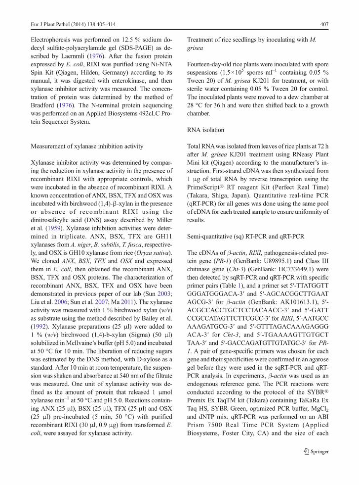
The ORF was cloned into expression vector pET-30a(+) arranged in frame with an N-terminal extensionincluding a His-tag to give the expression constructpET-30(a)+RIXI. The expressed protein was a fusionprotein with 71 additional amino acids at the N-terminusfrom the vector. The protein expression in E. coli in-duced by IPTG was secreted in the supernatant after 6 hinduction, the concentration of soluble protein extractswas about 230 mg/l. Using His-tagged Ni–NTA SpinKit, the concentration of purified protein was about30 mg/L. SDS-polyacrylamide electrophoresis analysisshowed that the purified proteins from E. coli containingpET30a+RIXI plasmid exhibited a clear band with amolecular weight of about 40 kDa (Fig. 1, Lane 2), intotal agreement with the predicted calculated mass in-cluding the extra 71 amino acids and a His-tag in the N-terminal from the vector. No band was observed in theextract from the control strain of E. coli containing pET-30a(+) vector (Fig. 1, Lane 1). After digested by entero-kinase, the N-terminal amino acid sequenceAMADIGSEFMVALGRRS for purified protein wasdetermined, thus in complete agreement with the pre-dicted peptide.
Table 1 Primers and PCR conditions for real time PCR
Gene GenBank accession No. Primers sequence (from 5′ to 3′) Ta (°C) Amplicon size (bp)
β-actin AK101613.1 TTATGGTTGGGATGGGACA 60 292AGCACGGCTTGAATAGCG
RIXI D55712.1 ACGCCACCTGCTCCTACAACC 60 203GATTCCGCCATAGTTCTTCGCC
Cht-3 HC733649 AATGCCAAAGATGCG 60 293GTTTAGACAAAGAGGGACA
PR-1 U89895.1 TGAAAAGTTGTGCTTAA 60 307GACCAGATGTTGTATGC
Ta annealing temperature,RIXI xylanase inhibitor gene,Cht-3 class IIIchitinase gene, PR-1 pathogenesis related (PR) protein 1 gene, β-actininternal control
408 Eur J Plant Pathol (2014) 138:405–414
Comparison of RIXI with other xylanase inhibitors
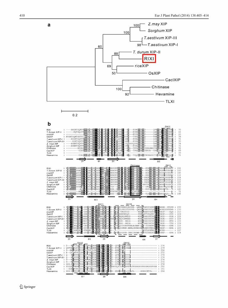
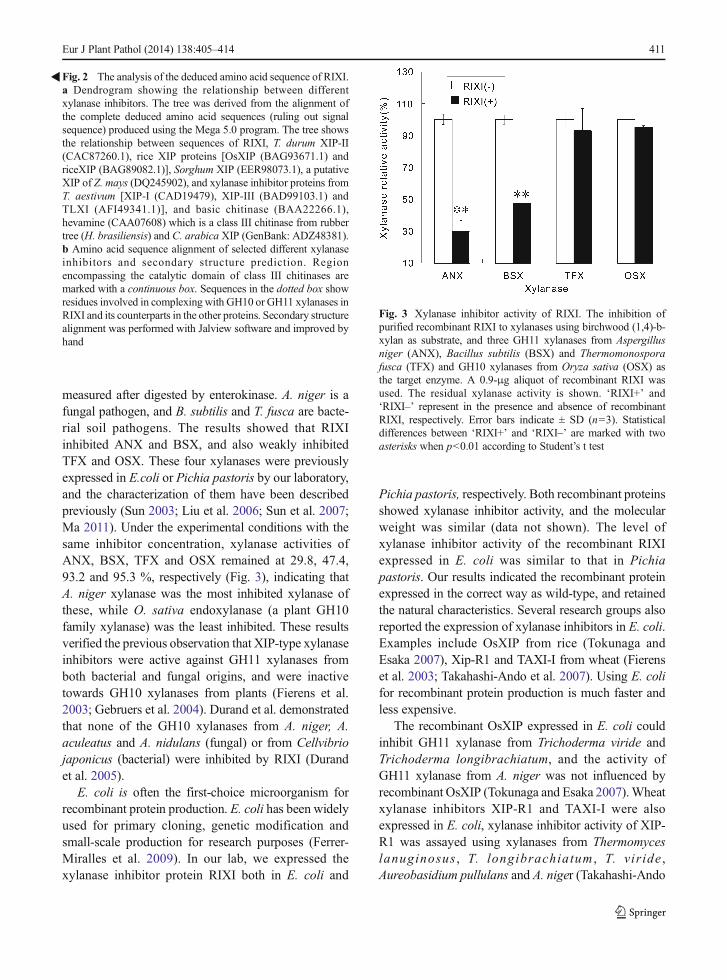
RIXI belongs to the GH18 family as well as class Шchitinases, despite its lack of enzymatic activity (Durandet al. 2005). The phylogenetic tree for plant members ofGH18 showed that RIXI belongs to the XIP-typexylanase inhibitor subfamily (Tokunaga and Esaka2007) . The a l ignment of RIXI (GenBank:BAA23810.1) with other xylanase inhibitors was per-formed by Mega 5.0 and Clustalx (Fig. 2a, b), and thesignal sequences of all sequences (RIXI and otherxylanase inhibitors) for the alignment was ruled out.According to the alignment, these proteins all show
the (β/α)8 barrel fold, and regions encompassing theformer chitinase catalytic moiety and the predicted sitesof interaction with xylanases GH10 and GH11 activesites are highlighted in Fig. 2b. On the protein level,RIXI shared more sequence identity with XIP-П (65 %,GenBank: CAC87260.1) from Triticum durum thanwith OsXIP (48 %, GenBank: BAG93671.1) andriceXIP (57 %, GenBank: BAG89082.1) from rice. Incontrast, lower identities were shared with the otherinhibitor sequences; i.e. 51, 50 and 3 % with XIP-I(GenBank: CAD19479) , XIP-Ш (GenBank:BAD99103.1), TLXI (GenBank: AFI49341.1) fromwheat, respectively, 51 % with a putative Zea maysXIP (GenBank: DQ245902) and Sorghum XIP(GenBank: EER98073.1), 41 % with basic chitinase(GenBank: BAA22266.1), 36 % with hevamine(GenBank: CAA07608) which is a class Ш chitinasefrom rubber tree (Hevea brasiliensis), and 30 % withCoffea arabica XIP (GenBank: ADZ48381). Figure 2ashows the relationship between these sequences.
The enzymes of the GH family 18 showing chitinaseactivity have a conserved aspartic acid and a catalyticglutamic acid residue in the active site, corresponding toAsp125 and Glu127 in hevamine. However, XIP-typexylanase inhibitors are devoid of chitinase activity. Inthe structures of RIXI, T. Durum XIP-П, OsXIP andriceXIP, Asp125 and Glu127 are replaced by Phe and Aspat positions 124–126, 122–124, 115–117 and 130–132,respectively (Fig. 2b), which mostly account for the lackof chitinase activity reported for these proteins. The factthat RIXI is closer to the T. durum xylanase inhibitorsequence than to other rice and wheat xylanase inhibitorproteins suggests that the different members of the XIPgene family underwent sequence conservation and/ordivergence during evolution. The profile of organ-specific expression of RIXI differs from other rice XIPgenes, OsXIP and riceXIP. RIXI was constitutivelyexpressed in shoot and not induced by defence-relatedphytohormones, while OsXIP and riceXIP mRNAswere not detected in basal conditions and were inducedin root by wounding and methyl jasmonate (Tokunagaand Esaka 2007). Thus we speculate that the defencemechanism against pathogens of RIXI may differ fromother rice XIPs.
Xylanase inhibition specificity of RIXI
RIXI’s inhibition varied among xylanases. Xylanaseinhibition activity of the recombinant RIXI was
Fig. 1 SDS-PAGE of the purified recombinant RIXI expressed inEscherichia coli BL21. M - marker of proteins, Lane 1 - cellextracts from control strain ofE. coli containing pET-30a(+) vectorand Lane 2 - cell extracts from E. coli containing pET-30a+RIXIvector induced by IPTG after 6 h. The sizes of the markers areindicated at the left of each gel
Eur J Plant Pathol (2014) 138:405–414 409
410 Eur J Plant Pathol (2014) 138:405–414
measured after digested by enterokinase. A. niger is afungal pathogen, and B. subtilis and T. fusca are bacte-rial soil pathogens. The results showed that RIXIinhibited ANX and BSX, and also weakly inhibitedTFX and OSX. These four xylanases were previouslyexpressed in E.coli or Pichia pastoris by our laboratory,and the characterization of them have been describedpreviously (Sun 2003; Liu et al. 2006; Sun et al. 2007;Ma 2011). Under the experimental conditions with thesame inhibitor concentration, xylanase activities ofANX, BSX, TFX and OSX remained at 29.8, 47.4,93.2 and 95.3 %, respectively (Fig. 3), indicating thatA. niger xylanase was the most inhibited xylanase ofthese, while O. sativa endoxylanase (a plant GH10family xylanase) was the least inhibited. These resultsverified the previous observation that XIP-type xylanaseinhibitors were active against GH11 xylanases fromboth bacterial and fungal origins, and were inactivetowards GH10 xylanases from plants (Fierens et al.2003; Gebruers et al. 2004). Durand et al. demonstratedthat none of the GH10 xylanases from A. niger, A.aculeatus and A. nidulans (fungal) or from Cellvibriojaponicus (bacterial) were inhibited by RIXI (Durandet al. 2005).
E. coli is often the first-choice microorganism forrecombinant protein production. E. coli has been widelyused for primary cloning, genetic modification andsmall-scale production for research purposes (Ferrer-Miralles et al. 2009). In our lab, we expressed thexylanase inhibitor protein RIXI both in E. coli and
Pichia pastoris, respectively. Both recombinant proteinsshowed xylanase inhibitor activity, and the molecularweight was similar (data not shown). The level ofxylanase inhibitor activity of the recombinant RIXIexpressed in E. coli was similar to that in Pichiapastoris. Our results indicated the recombinant proteinexpressed in the correct way as wild-type, and retainedthe natural characteristics. Several research groups alsoreported the expression of xylanase inhibitors in E. coli.Examples include OsXIP from rice (Tokunaga andEsaka 2007), Xip-R1 and TAXI-I from wheat (Fierenset al. 2003; Takahashi-Ando et al. 2007). Using E. colifor recombinant protein production is much faster andless expensive.
The recombinant OsXIP expressed in E. coli couldinhibit GH11 xylanase from Trichoderma viride andTrichoderma longibrachiatum, and the activity ofGH11 xylanase from A. niger was not influenced byrecombinant OsXIP (Tokunaga and Esaka 2007).Wheatxylanase inhibitors XIP-R1 and TAXI-I were alsoexpressed in E. coli, xylanase inhibitor activity of XIP-R1 was assayed using xylanases from Thermomyceslanuginosus , T. longibrachiatum , T. viride ,Aureobasidium pullulans and A. niger (Takahashi-Ando
Fig. 3 Xylanase inhibitor activity of RIXI. The inhibition ofpurified recombinant RIXI to xylanases using birchwood (1,4)-b-xylan as substrate, and three GH11 xylanases from Aspergillusniger (ANX), Bacillus subtilis (BSX) and Thermomonosporafusca (TFX) and GH10 xylanases from Oryza sativa (OSX) asthe target enzyme. A 0.9-μg aliquot of recombinant RIXI wasused. The residual xylanase activity is shown. ‘RIXI+’ and‘RIXI–’ represent in the presence and absence of recombinantRIXI, respectively. Error bars indicate ± SD (n=3). Statisticaldifferences between ‘RIXI+’ and ‘RIXI–’ are marked with twoasterisks when p<0.01 according to Student’s t test
Fig. 2 The analysis of the deduced amino acid sequence of RIXI.a Dendrogram showing the relationship between differentxylanase inhibitors. The tree was derived from the alignment ofthe complete deduced amino acid sequences (ruling out signalsequence) produced using the Mega 5.0 program. The tree showsthe relationship between sequences of RIXI, T. durum XIP-II(CAC87260.1), rice XIP proteins [OsXIP (BAG93671.1) andriceXIP (BAG89082.1)], Sorghum XIP (EER98073.1), a putativeXIP of Z. mays (DQ245902), and xylanase inhibitor proteins fromT. aestivum [XIP-I (CAD19479), XIP-III (BAD99103.1) andTLXI (AFI49341.1)], and basic chitinase (BAA22266.1),hevamine (CAA07608) which is a class III chitinase from rubbertree (H. brasiliensis) and C. arabica XIP (GenBank: ADZ48381).b Amino acid sequence alignment of selected different xylanaseinhibitors and secondary structure prediction. Regionencompassing the catalytic domain of class III chitinases aremarked with a continuous box. Sequences in the dotted box showresidues involved in complexing with GH10 or GH11 xylanases inRIXI and its counterparts in the other proteins. Secondary structurealignment was performed with Jalview software and improved byhand
�
Eur J Plant Pathol (2014) 138:405–414 411
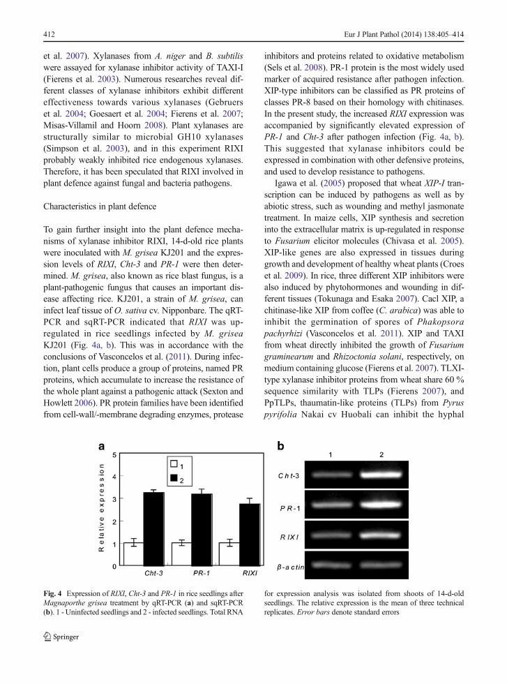
et al. 2007). Xylanases from A. niger and B. subtiliswere assayed for xylanase inhibitor activity of TAXI-I(Fierens et al. 2003). Numerous researches reveal dif-ferent classes of xylanase inhibitors exhibit differenteffectiveness towards various xylanases (Gebruerset al. 2004; Goesaert et al. 2004; Fierens et al. 2007;Misas-Villamil and Hoorn 2008). Plant xylanases arestructurally similar to microbial GH10 xylanases(Simpson et al. 2003), and in this experiment RIXIprobably weakly inhibited rice endogenous xylanases.Therefore, it has been speculated that RIXI involved inplant defence against fungal and bacteria pathogens.
Characteristics in plant defence
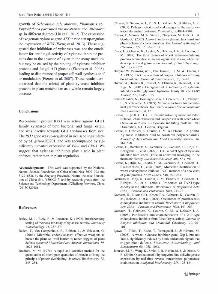
To gain further insight into the plant defence mecha-nisms of xylanase inhibitor RIXI, 14-d-old rice plantswere inoculated with M. grisea KJ201 and the expres-sion levels of RIXI, Cht-3 and PR-1 were then deter-mined. M. grisea, also known as rice blast fungus, is aplant-pathogenic fungus that causes an important dis-ease affecting rice. KJ201, a strain of M. grisea, caninfect leaf tissue of O. sativa cv. Nipponbare. The qRT-PCR and sqRT-PCR indicated that RIXI was up-regulated in rice seedlings infected by M. griseaKJ201 (Fig. 4a, b). This was in accordance with theconclusions of Vasconcelos et al. (2011). During infec-tion, plant cells produce a group of proteins, named PRproteins, which accumulate to increase the resistance ofthe whole plant against a pathogenic attack (Sexton andHowlett 2006). PR protein families have been identifiedfrom cell-wall/-membrane degrading enzymes, protease
inhibitors and proteins related to oxidative metabolism(Sels et al. 2008). PR-1 protein is the most widely usedmarker of acquired resistance after pathogen infection.XIP-type inhibitors can be classified as PR proteins ofclasses PR-8 based on their homology with chitinases.In the present study, the increased RIXI expression wasaccompanied by significantly elevated expression ofPR-1 and Cht-3 after pathogen infection (Fig. 4a, b).This suggested that xylanase inhibitors could beexpressed in combination with other defensive proteins,and used to develop resistance to pathogens.
Igawa et al. (2005) proposed that wheat XIP-I tran-scription can be induced by pathogens as well as byabiotic stress, such as wounding and methyl jasmonatetreatment. In maize cells, XIP synthesis and secretioninto the extracellular matrix is up-regulated in responseto Fusarium elicitor molecules (Chivasa et al. 2005).XIP-like genes are also expressed in tissues duringgrowth and development of healthy wheat plants (Croeset al. 2009). In rice, three different XIP inhibitors werealso induced by phytohormones and wounding in dif-ferent tissues (Tokunaga and Esaka 2007). Cacl XIP, achitinase-like XIP from coffee (C. arabica) was able toinhibit the germination of spores of Phakopsorapachyrhizi (Vasconcelos et al. 2011). XIP and TAXIfrom wheat directly inhibited the growth of Fusariumgraminearum and Rhizoctonia solani, respectively, onmedium containing glucose (Fierens et al. 2007). TLXI-type xylanase inhibitor proteins from wheat share 60 %sequence similarity with TLPs (Fierens 2007), andPpTLPs, thaumatin-like proteins (TLPs) from Pyruspyrifolia Nakai cv Huobali can inhibit the hyphal
Fig. 4 Expression of RIXI, Cht-3 and PR-1 in rice seedlings afterMagnaporthe grisea treatment by qRT-PCR (a) and sqRT-PCR(b). 1 - Uninfected seedlings and 2 - infected seedlings. Total RNA
for expression analysis was isolated from shoots of 14-d-oldseedlings. The relative expression is the mean of three technicalreplicates. Error bars denote standard errors
412 Eur J Plant Pathol (2014) 138:405–414
growth of Sclerotinia sclerotiorum, Phomopsis sp.,Phytophthora parasitica var nicotianae and Alternariasp. in different degrees (Liu et al. 2012). The expressionof exogenous xylanase gene ATX in rice can up-regulatethe expression of RIXI (Weng et al. 2013). These sug-gested that inhibition of xylanases was not the crucialfactor for antifungal activity of xylanase inhibitor pro-teins due to the absence of xylan in the assay medium,but may be caused by the binding of xylanase inhibitorproteins and fungal β-D-glucans (Fierens et al. 2008),leading to disturbance of proper cell wall synthesis and/or modulation (Fierens et al. 2007). These results dem-onstrated that the role(s) of plant xylanase inhibitorproteins in plant metabolism as a whole remain largelyobscure.
Conclusions
Recombinant protein RIXI was active against GH11family xylanases of both bacterial and fungal originand was inactive towards GH10 xylanases from rice.The RIXI gene was up-regulated in rice seedlings infect-ed by M. grisea KJ201, and was accompanied by sig-nificantly elevated expression of PR-1 and Cht-3. Wesuggest that xylanase inhibitors play a role in plantdefence, rather than in plant regulation.
Acknowledgments This work was supported by the NationalNatural Science Foundation of China (Grant Nos. 30971702 and31271632), by the Zhejiang Provincial Natural Science Founda-tion of China (No. Y3090247) and by research grants from theScience and Technology Department of Zhejiang Province, China(2013C32018).
References
Bailey, M. J., Biely, P., & Poutanen, K. (1992). Interlaboratorytesting of methods for assay of xylanase activity. Journal ofBiotechnology, 23, 257–270.
Belien, T., Van Campenhout, S., Robben, J., & Volckaert, G.(2006). Microbial endoxylanases: effective weapons tobreach the plant cell-wall barrier or, rather, triggers of plantdefense systems?Molecular Plant-Microbe Interactions, 19,1072–1081.
Bradford, M. M. (1976). A rapid and sensitive method for thequantitation of microgram quantities of protein utilising theprinciple of protein dye binding. Analytical Biochemistry, 72,248–254.
Chivasa, S., Simon, W. J., Yu, X. L., Yalpani, N., & Slabas, A. R.(2005). Pathogen elicitor-induced changes in the maize ex-tracellular matrix proteome. Proteomics, 5, 4894–4904.
Collins, T., Meuwis, M. A., Stals, I., Claeyssens, M., Feller, G., &Gerday, C. (2002). A novel family 8 xylanase, functional andphysicochemical characterization. The Journal of BiologicalChemistry, 277, 35133–35139.
Croes, E., Gebruers, K., Luyten, N., Delcour, J. A., & Courtin, C.M. (2009). The three classes of wheat xylanase-inhibitingproteins accumulate in an analogous way during wheat eardevelopment and germination. Journal of Plant Physiology,166, 1253–1262.
Debyser, W., Peumans, W. J., Van Damme, E. J. M., & Delcour, J.A. (1999). TAXI, a new class of enzyme inhibitors affectingbread volume. Journal of Cereal Science, 30, 39–43.
Durand, A., Hughes, R., Roussel, A., Flatman, R., Henrissat, B., &Juge, N. (2005). Emergence of a subfamily of xylanaseinhibitors within glycoside hydrolase family 18. The FEBSJournal, 272, 1745–1755.
Ferrer-Miralles, N., Domingo-Espin, J., Corchero, J. L., Vazquez,E., & Villaverde, A. (2009). Microbial factories for recombi-nant pharmaceuticals.Microbial Factories For RecombinantPharmaceuticals, 8, 17.
Fierens, E. (2007). TLXI, a thaumatin-like xylanase inhibitor:isolation, characterisation and comparison with other wheat(Triticum aestivum L.) xylanase inhibiting proteins. PhDDissertation, K.U. Leuven, Belgium.
Fierens, E., Gebruers, K., Courtin, C. M., & Delcour, J. A. (2008).Xylanase inhibitors bind to nonstarch polysaccharides.Journal of Agricultural and Food Chemistry Journal, 56,564–570.
Fierens, E., Rombouts, S., Gebruers, K., Goesaert, H., Brijs, K.,Beaugrand, J., et al. (2007). TLXI, a novel type of xylanaseinhibitor from wheat (Triticum aestivum) belonging to thethaumatin family. Biochemical Journal, 403, 583–591.
Fierens, K., Brijs, K., Courtin, C. M., Gebruers, K., Goesaert, H.,Raedschelders, G., et al. (2003). Molecular identification ofwheat endoxylanase inhibitor TAXI, member of a new classof plant proteins. FEBS Letters, 540, 259–263.
Gebruers, K., Brijs, K., Courtin, C. M., Fierens, K., Goesaert, H.,Rabijns, A., et al. (2004). Properties of TAXI-typeendoxylanase inhibitors. Biochimica et Biophysica Acta(BBA) - Proteins and Proteomics, 1696, 213-221.
Goesaert, H., Elliott, G.O., Kroon, P.A., Gebruers, K., Courtin, C.M., Robben, J., et al. (2004). Occurrence of proteinaceousendoxylanase inhibitor in cereals. Biochimica et BiophysicaActa (BBA) - Proteins and Proteomics, 1696, 193–202.
Goesaert, H., Gebruers, K., Courtin, C. M., & Delcour, J. A.(2005). Purification and characterization of a XIP-typeendoxylanase inhibitor from Rice (Oryza sativa). Journal ofEnzyme Inhibition and Medicinal Chemistry, 20, 95–101.
Igawa, T., Tokai, T., Kudo, T., Yamaguchi, I., & Kimura, M.(2005). A wheat xylanase inhibitor gene, Xip-I, but notTaxi-I, significantly induced by biotic and abiotic signals thattrigger plant defense. Bioscience, Biotechnology, andBiochemistry, 69, 1058–1063.
Johnson, M. R.,Wang, K., Smith, J. B., Heslin, M. J., & Diasio, R.B. (2000). Quantitation of dihydropyrimidine dehydrogenaseexpression by real-time reverse transcription polymerasechain reaction. Analytical Biochemistry, 278, 175–184.
Eur J Plant Pathol (2014) 138:405–414 413
Laemmli, U. K. (1976). Cleavage of structural proteins during theassembly of the head of bacteriophage T4.Nature, 227, 680–685.
Liu, D. Q., He, X., Li, W. X., Chen, C. Y., & Ge, F. (2012).Molecular cloning of a thaumatin-like protein gene fromPyrus pyrifolia and overexpression of this gene in tobaccoincreased resistance to pathogenic fungi. Plant Cell, Tissueand Organ Culture, 111, 29–39.
Liu, M. Q., Weng, X. Y., & Sun, J. Y. (2006). Expression ofrecombinant Aspergillus niger xylanase A in Pichia pastorisand its action on xylan. Protein Expression and Purification,48, 292–299.
Livak, K. J., & Schmittgen, T. D. (2001). Analysis of relative geneexpression data using Real-Time Quantitative PCR and the 2-ΔΔCt method. Methods, 25, 402–408.
Ma, M.Z. (2011) Cloning and expression of endoxylanase geneosx from Oryza sativa and research on inhibition of theinhibitor RIXI. M.S. Dissertation, Zhejiang University,China (English abstract).
Mclauchlan, W. R., Garcia-Conesa, M. T., Williamson, G., Roza,M., Ravestein, P., & Maat, J. (1999). A novel class of proteinfrom wheat which inhibits xylanases. Biochemical Journal,338, 441–446.
Miller, G. L., Blum, R., Glennom, W. E., & Burton, A. L. (1959).Measurement of methods for assay of xylanase activity.Analytical Biochemistry, 2, 127–132.
Misas-Villamil, J. C., & van der Hoorn, R. A. L. (2008). Enzyme-inhibitor interactions at the plant-pathogen interface. CurrentOpinion in Plant Biology, 11, 380–388.
Sels, J., Mathys, J., De Coninck, B. M., Cammue, B. P., & DeBolle, M. F. (2008). Plant pathogenesis-related (PR) proteins:a focus on PR peptides. Plant Physiology and Biochemistry,46, 941–950.
Sexton, A. C., & Howlett, B. J. (2006). Parallels in fungal patho-genesis on plant and animal hosts. Eukaryotic Cell, 5, 1941–1949.
Simpson, D. J., Fincher, G. B., Huang, A. H. C., & Cameron-Mills, V. (2003). Structure and function of cereal and related
higher plant (1→4)-β-xylan endohydrolases. Journal ofCereal Science, 37, 111–127.
Sun, J.Y. (2003) Construction and expression of hybrid geneencoding thermostable xylanase and property of hybrid en-zyme. PhDDissertation, Zhejiang University, China (Englishabstract).
Sun, J. Y., Liu, M. Q., Weng, X. Y., Qian, L. C., & Gu, S. H.(2007). Expression of recombinant Thermomonospora fuscaxylanase A in Pichia pastoris and xylooligosaccharides re-leased from xylans by it. Food Chemistry, 104, 1055–1064.
Takahashi-Ando, N., Inaba, M., Ohsato, S., Igawa, T., Usami, R.,& Kimura, M. (2007). Identification of multiple highly sim-ilar XIP-type xylanase inhibitor genes in hexaploid wheat.Biochemical and Biophysical Research Communications,360, 880–884.
Tokunaga, T., & Esaka, M. (2007). Induction of a novel XIP-typexylanase inhibitor by external ascorbic acid treatment anddifferential expression of XIP-family genes in rice. Plant andCell Physiology, 48, 700–714.
Tokunaga, T., Miyata, Y., Fujikawa, Y., & Esaka, M. (2008).RNAi-mediated knockdown of the XIP-type endoxylanaseinhibitor gene, OsXIP, has no effect on grain developmentand germination in rice.Plant and Cell Physiology, 49, 1122–1127.
Vasconcelos, E. A., Santana, C. G., Godoy, C. V., Seixas, C. D.,Silva, M. S., Moreira, L. R., et al. (2011). A new chitinase-like xylanase inhibitor protein (XIP) from coffee (Coffeaarabica) affects Soybean Asian rust (Phakopsora pachyrhizi)spore germination. BMC Biotechnology, 11, 14.
Weng, X. Y., Huang, Y. Y., Hou, C. X., & Jiang, D. A. (2013).Effects of an exogenous xylanase gene expression on thegrowth of transgenic rice and the expression level of endog-enous xylanase inhibitor gene RIXI. Journal of the Science ofFood and Agriculture, 93, 173–179.
Weng, X. Y., & Sun, J. Y. (2005). Construction, expression, andcharacterization of a thermostable xylanase. CurrentMicrobiology, 51, 188–192.
414 Eur J Plant Pathol (2014) 138:405–414