Embed Size (px)
Citation preview
MOLECULAR AND CELLULAR BIOLOGY, Sept. 2009, p. 4982–4993 Vol. 29, No. 180270-7306/09/$08.00�0 doi:10.1128/MCB.00285-09Copyright © 2009, American Society for Microbiology. All Rights Reserved.
Functional Connection between Deimination and Deacetylationof Histones�
Helene Denis,1 Rachel Deplus,1 Pascale Putmans,1 Michiyuki Yamada,2Raphaël Metivier,3 and Francois Fuks1*
Free University of Brussels, Faculty of Medicine, Laboratory of Cancer Epigenetics, 808 route de Lennik, 1070 Brussels, Belgium1;Graduate School of Integrated Science, Yokohama City University, 22-2 Seto, Kanazawa-ku, Yokohama 236-0027, Japan2;
and Equipe SPARTE, UMR CNRS 6026, Universite de Rennes I, Campus de Beaulieu, 35042 Rennes Cedex, France3
Received 4 March 2009/Returned for modification 28 March 2009/Accepted 29 June 2009
Histone methylation plays key roles in regulating chromatin structure and function. The recent identifica-tion of enzymes that antagonize or remove histone methylation offers new opportunities to appreciate histonemethylation plasticity in the regulation of epigenetic pathways. Peptidylarginine deiminase 4 (PADI4; alsoknown as PAD4) was the first enzyme shown to antagonize histone methylation. PADI4 functions as a histonedeiminase converting a methylarginine residue to citrulline at specific sites on the tails of histones H3 and H4.This activity is linked to repression of the estrogen-regulated pS2 promoter. Very little is known as to howPADI4 silences gene expression. We show here that PADI4 associates with the histone deacetylase 1 (HDAC1).Kinetic chromatin immunoprecipitation assays revealed that PADI4 and HDAC1, and the correspondingactivities, associate cyclically and coordinately with the pS2 promoter during repression phases. Knockdown ofHDAC1 led to decreased H3 citrullination, concomitantly with increased histone arginine methylation. In cellswith a reduced HDAC1 and a slightly decreased PADI4 level, these effects were more pronounced. Our datathus suggest that PADI4 and HDAC1 collaborate to generate a repressive chromatin environment on the pS2promoter. These findings further substantiate the “transcriptional clock” concept, highlighting the dynamicconnection between deimination and deacetylation of histones.
Until recently, it was unclear whether enzymes capable ofantagonizing histone methylation existed. However, recentstudies have revealed a growing number of lysine demethylasesthat can reverse histone lysine methylation, such as LSD1/KDM1 and the JmjC-domain-containing proteins (16, 21, 31,37, 43, 45, 47).
In addition to the methylation of lysine, histones can also bemethylated on arginine (6, 34). Recent work has identified theJMJD6 protein as an H3R2 and H4R3 demethylase (5). Analternative pathway for the reversal of arginine methylationhas been identified in mammals. In this pathway, a methylgroup is removed from a methylarginine residue by conversionof this residue to citrulline. The reaction is termed deiminationbecause the methyl group is removed along with the iminegroup of arginine (7, 42). The enzyme that catalyzes this reac-tion is peptidylarginine deiminase 4 (PADI4; also known asPAD4) (7, 42). PADI4 has a relatively broad substrate speci-ficity, since the enzyme can deiminate multiple arginine siteson histones H3 (R2, R8, R17, and R26) and H4 (R3) (42).PADI4 is a Ca2�-dependent enzyme (1). Functionally, theinduction of PADI4-mediated deimination has been best stud-ied as part of the estrogen signaling pathway, particularly in thecontext of the estrogen-regulated pS2 promoter.
Chromatin immunoprecipitation (ChIP)-based kinetic anal-yses performed on human breast cancer cell lines have shownthat in the presence of estrogen (E2), pS2 expression is con-
trolled by the estrogen receptor alpha (ER�), which cycles onits promoter. Not only does the receptor cycle, it also follows asequence of cofactor recruitment (20, 30). For example, duringtranscriptional activation by the estrogen receptor, argininemethylation of histone H3 appears transient and cyclic (20).After the initial hormone-induced transcriptional activationphase, PADI4 is recruited to the promoter region of pS2,where its presence correlates with loss of arginine methylation,acquisition of citrullinated histones, and disengagement ofRNA polymerase II (Pol II) from the gene (7, 42). Thus,methylation of arginine residues followed by deimination byPADI4 seems to participate in the normal cyclic on-and-offregulation of pS2 transcription. Although these studies havebegun to shed light on how PADI4-mediated histone deimina-tion silences gene expression, much remains to be discoveredin order to understand the mechanistic basis of this process.
In the present study we have sought to better understandhow PADI4 represses transcription. Our starting point was aneffort to identify proteins that associate with PADI4. We re-port that PADI4 interacts with histone deacetylase 1 (HDAC1)both in vitro and in vivo. We provide evidence that PADI4 andHDAC1, and their corresponding enzymatic activities, associ-ate cyclically with the pS2 promoter and are present simulta-neously on this promoter in the presence of estradiol. Finally,RNA interference (RNAi) experiments suggest a coordinatedaction of the PADI4 and HDAC1 enzymes on the pS2 pro-moter.
MATERIALS AND METHODS
Expression plasmids. We cloned full-length PADI4 into pcDNA3.1-HA andPADI4 fragments into the vector pGex (Pharmacia) using appropriate sets ofprimers. We also cloned various domains of HDAC1 into the pGex vector by
* Corresponding author. Mailing address: Free University of Brus-sels, Faculty of Medicine, Laboratory of Cancer Epigenetics, 808 routede Lennik, 1070 Brussels, Belgium. Phone: 32-2-555-62-45. Fax: 32-2-555-62-57. E-mail: [email protected].
� Published ahead of print on 6 July 2009.
4982
Dow
nloa
ded
from
http
s://j
ourn
als.
asm
.org
/jour
nal/m
cb o
n 17
Nov
embe
r 20
21 b
y 18
3.89
.93.
105.

PCR using appropriate primers. The following plasmids were used: pGex PADI4full-length (23), pcDNA3 Flag RbBP5 (Addgene), pcDNA3 HDAC1-Flag (3),and pING14A-HDAC1 (10).
GST fusion, in vitro translation, and pulldown assays. We expressed gluta-thione S-transferase (GST) and GST fusion proteins in Escherichia coli Top10 orBL21 using the pGex (Pharmacia) vector system. Bacteria were disrupted byusing an ultrasonicator (Bioruptor; Diagenode), and proteins from crude bacte-rial lysates were purified using the glutathione-Sepharose 4B (Pharmacia) usedaccording to the manufacturer’s instructions. We used the TNT system (Pro-mega) to carry out in vitro transcription and translation. The PADI4, HDAC1,and control RbBP5 genes were in vitro transcribed and/or translated frompcDNA3.1-HA PADI4, pcDNA3 HDAC1-Flag, and pcDNA3 Flag RbBP5, re-spectively. The GST pulldown experiments were performed as described previ-ously (40).
Cell culture and transient transfections. 293T cells were maintained in Dul-becco modified Eagle medium (DMEM; Sigma) supplemented with 5% fetal calfserum (FCS; BioWest) and antibiotics (Roche) at 37°C under 5% CO2. MDA-MB231 cells stably expressing ER (MDA::ER�) (19) were grown in DMEMcomplemented with 5% FCS. 17�-estradiol (E2) was purchased from Sigma.Transfections were performed with polyethylene imine (Euromedex) as de-scribed previously (9).
Immunoprecipitations and Western blot analyses. 293T cells were transientlytransfected in culture dishes (10-cm diameter) with a total of 6 �g of plasmids asdescribed previously (9). Standard procedures were used for coimmunoprecipi-tations and Western blotting (9). Antihemagglutinin (anti-HA; ab18181; Ab-cam), anti-Flag (M2; Sigma), and anti-HDAC1 (pAb-053-050; Diagenode) an-tibodies were used for immunoprecipitations from preparations of transfected293T cells. For immunoprecipitations of endogenous proteins from preparationsof MDA::ER� cells, antibodies were incubated at 4°C with MDA::ER� wholenuclear extracts in IPH buffer (4). Antibodies to HDAC1 were purchased fromDiagenode (pAb-053-050). Immunoglobulin G (IgG; ab3745; Abcam), IgG (sc-2027; Santa Cruz), HA (ab9110; Abcam), and RNA Pol II (sc-899; Santa Cruz)were used as control antibodies. PADI4 antibody has been described previously(24).
Histone deimination assay. The assay was done essentially as described pre-viously (7). 293T cells were transiently transfected with mammalian expressionplasmids encoding HA-PADI4 or HDAC1 or with both of these plasmids.Whole-cell extracts were prepared in IPH lysis buffer (4) and incubated over-night at 4°C with the relevant antibodies. Antibody complexes were collected onprotein A/G-Sepharose beads, washed, and then tested for histone deiminaseactivity. The reaction mixture containing 100 mM Tris-HCl (pH 7.6), 5 mMdithiothreitol (DTT), 10 mM CaCl2, and 10 �g of histones (Roche) was incu-bated at 37°C for 1 h. Reaction products were then resolved by sodium dodecylsulfate (SDS)-polyacrylamide gel electrophoresis and Western blotted. Anti-histone H3 (citrulline 17�2�8) antibody (ab5103; Abcam) was used to reveal thepresence of deiminated histones. Levels of PADI4 and HDAC1 in the immu-noprecipitates were checked by Western blot analysis with anti-HA (ab18184;Abcam) and anti-HDAC1 (pAb-053-050; Diagenode) antibodies, respectively.
ChIPs and Re-ChIPs. ChIP assays were performed essentially as describedpreviously (20). A total of 5 � 107 MDA::ER� cells were synchronized by 3 daysof culture in DMEM–0.5% dextran-charcoal-treated FCS (BioWest) and thentreated with 2.5 �M �-amanitin (Sigma) for 2 h, followed by exposure to 10�8 ME2 (Sigma) or ethanol. After cross-linking for 10 min with 1.5% formaldehyde(Sigma) at room temperature, the cells were collected in 5 ml of collection buffer
(100 mM Tris-HCl [pH 9.4] and 100 mM DTT) and incubated first on ice for 10min and then at 30°C for 10 min. The cells were then lysed sequentially, withpipetting and a 5-min centrifugation (at 2,000 � g and 4°C) between steps. Thelysis steps were as follows: (i) 5 ml of phosphate-buffered saline; (ii) 5 ml of bufferA (10 mM EDTA, 0.5 mM EGTA, 10 mM HEPES [pH 6.5], and 0.25% TritonX-100); (iii) 5 ml of buffer B (1 mM EDTA, 0.5 mM EGTA, 10 mM HEPES [pH6.5], and 200 mM NaCl); (iv) lysis for 15 min at room temperature in 2.5 ml oflysis buffer (10 mM EDTA, 50 mM Tris-HCl [pH 8.0], 1% SDS, 0.5% EmpigenBB [Sigma]). Then, 500-�l aliquots were sonicated three times by 15 pulses at50% duty and power 3 (Sonifier cell disruptor; Branson). After centrifugation,50 �l of the combined supernatants was used as inputs, and the remainder wasdiluted 2.5-fold in immunoprecipitation buffer (2 mM EDTA, 100 mM NaCl, 20mM Tris-HCl [pH 8.1], and 0.5%Triton X-100). This fraction was first preclearedby incubation for 3 h at 4°C with 15 �g of sheared salmon sperm DNA, and 1 mlof 50% protein A-Sepharose beads (Amersham Pharmacia Biosciences) slurry. Aportion (1/20) of the precleared chromatin was then subjected to immunopre-cipitation overnight at 4°C under rocking with appropriate antibodies (purchasedfrom Santa Cruz, Upstate Biotechnology, or Abcam). Complexes were recoveredafter 2 h of incubation at 4°C with 2 �g of sheared salmon sperm DNA and 50 �lof protein A-Sepharose. Precipitates were then serially washed with 300 �l ofwashing buffers WB I (2 mM EDTA, 20 mM Tris-HCl [pH 8.1], 0.1% SDS, 1%Triton X-100, 150 mM NaCl), WB II (2 mM EDTA, 20 mM Tris-HCl [pH 8.1],and specific combinations of detergent and salt, as indicated in Table 1), and WBIII (1 mM EDTA, 10 mM Tris-HCl [pH 8.1], 1% NP-40, 1% deoxycholate, 0.25M LiCl) and then twice with 1 mM EDTA–10 mM Tris-HCl (pH 8.1). Precipi-tated complexes were removed from the beads by three sequential 10-min incu-bations with 50 �l of 1% SDS–0.1 M NaHCO3.
In Re-ChIP assays, DNA-protein complexes were extracted twice from thebeads by adding 25 �l of 10 mM DTT, followed by incubation for 20 min at 37°Cwith vortexing every 5 min. Supernatants were then diluted 10 times in immu-noprecipitation buffer, and the immunoprecipitation procedure was performedagain. Cross-linking was reversed by an overnight incubation at 65°C. DNA waspurified on Qiaquick columns (Qiagen). In subsequent semiquantitative PCRanalysis (Taq; Qiagen), performed on a total volume of 25 �l including 15 mMMgCl2 (final concentration), 2 �l of input material or 3 �l of ChIP samples wasused, with the following primers (Proligo): left, 5�-GTTGTCAGGCCAAGCCTTTT-3�; and right, 5�-GAGCGTTAGATAACATTTGCC-3� (54°C; 15 mMMgCl2 [final concentration]).
In ChIP assays, quantitative PCRs were performed on 1 �l of input or ChIPsample DNA, with a Bio-Rad MyiQ apparatus and Bio-Rad iQ SYBR greensupermix, in a total volume of 20 �l containing 10 mM MgCl2, with the followingprimers (Proligo): left pS2 promoter, 5�-TTCCGGCCATCTCTCACTAT-3�;right pS2 promoter, 5�-CGGGGATCCTCTGAGACA-3�; forward unrelatedcontrol region (cf. Fig. 4B), 5�-AAGCTGCGTGTTGTCAGATG-3�; and reverseunrelated control region, 5�-AAGGAAGGGCCCTCTATTCA-3�.
The relative levels of the fragments of interest in the immunoprecipitatedDNA were determined from the threshold cycle (CT) for each PCR. To ensurethe reliability of our ChIP data, two control samples specific for the ChIPexperiment have been included, as we have previsously reported (20): the inputsample (indicative of the presence and amount of chromatin used in the ChIPreaction) and the control antibody (HA antibody) sample (indicative of theamount of background signal generated by the chromatin preparations and ChIPprocedure). The calculations of the relative enrichment values were as describedbelow. (i) We normalized the quantitative PCR signals obtained from the im-
TABLE 1. Antibodies used and ChIP conditions
Protein Provider (catalog no.) or reference Amt (�g) Washing conditionsa
ER� Santa Cruz (sc-543) 1.0 WB I, WB II (Det. I, 500 mM NaCl ��1), WB IIIPol II Santa Cruz (sc-899) 1.0 WB I, WB II (Det. II, 500 mM NaCl ��2)HA Santa Cruz (sc-805) 1.0 WB I, WB II (Det. I, 500 mM NaCl ��1), WB IIIH3K14ac Upstate Biotech (06-911) 0.5 WB I, WB II (Det. I, 500 mM NaCl ��1), WB IIIH3R17me2 Upstate Biotech (07-214) 0.5 WB I, WB II (Det. II, 500 mM NaCl ��2)H3R(17�2�8)cit Abcam (ab5103) 0.5 WB I, WB II (Det. I, 500 mM NaCl ��1), WB IIIH4R3me2 Upstate Biotech (07-213) 0.5 WB I, WB II (Det. I, 250 mM NaCl ��2)H4R3cit Upstate Biotech (07-596) 0.5 WB I, WB II (Det. I, 250 mM NaCl ��2)PADI4 Nakashima et al. (24) 0.2 WB I, WB II (Det. I, 250 mM NaCl ��3)HDAC1 Abcam (ab7028) 1.5 WB I, WB II (Det. II, 500 mM NaCl ��2)
a Washings used three washing buffers (WB), with WB II containing different salt concentrations and detergent (Det.) mixes as indicated. Det. I 0.1% SDS, 1%Triton X-100. Det. II 1% Triton X-100. The numbers in brackets indicate the number of washes done with WB II.
VOL. 29, 2009 DEIMINATION AND DEACETYLATION OF HISTONES 4983
Dow
nloa
ded
from
http
s://j
ourn
als.
asm
.org
/jour
nal/m
cb o
n 17
Nov
embe
r 20
21 b
y 18
3.89
.93.
105.

munoprecipitated ChIP sample to the input sample, i.e., CT input – CT ChIP. ThePCR efficiency, corresponding to the different sets of primers used in our quan-titative PCR, was then raised to the power of this CT difference, i.e., (primer PCRefficiency)(CT input � CT ChIP). (ii) The enrichment (n-fold) of the immunoprecipi-tated sequence of interest was obtained by normalizing the values to the ChIPbackground (relative to IP HA antibody). (iii) To ensure that the observedbinding of the tested proteins or histone marks reflect specific binding to thepS2/TFF1 promoter, we also amplified an unrelated control region (cf. Fig. 4B)in a quantitative PCR. The relative enrichment values were calculated by divid-ing the enrichment (n-fold) derived from the sequence of interest (pS2/TFF1locus) by the signal derived from this control locus (unrelated control region).
RNAi and retroviral infection. RNAi PADI4 and RNAi HDAC1 were gener-ated as previously described (2, 40). Briefly, the target sequence used to silenceHDAC1 or PADI4 was inserted as a short hairpin into the pRetroSuper (pRS)retroviral vector according to the manufacturer’s (OligoEngine) recommenda-tions to form RNAi PADI4 or RNAi HDAC1, respectively. Retrovirus produc-tion by 293T GP cells and infection of target MDA::ER� were performed asdescribed. Infected cells were selected with 1 �g of puromycin (Sigma)/ml. Theprimer sequences are available from the authors upon request.
RNA purification and RT-PCR analyses. Extraction of total RNA was carriedout with TriPure reagent (Roche) according to the manufacturer’s instructions.DNase treatment was performed with a DNA-free DNase kit (Ambion) accord-ing to the manufacturer’s protocol. cDNA was reverse transcribed from 1 �g ofRNA using random hexamers (Amersham/Pharmacia Biotech) and SuperscriptII reverse transcriptase (Life Technologies, Inc.). The reverse transcription (RT)reaction mixture was diluted with diethyl pyrocarbonate water (Fluka Bio-chemika) before addition to the PCR. To quantitatively evaluate expressionlevels under different conditions, real-time PCRs were performed in a Light-Cycler 480 system (Roche). The primers sequences for PADI4 and HDAC1amplifications were as follows: left PADI4, 5�-GACAAAGTGAGGGTGTTTCA-3�; right PADI4, 5�-AGAAGTCCATGTTGTGCTTT-3�; left HDAC1, 5�-GGATCGGTTAGGTTGCTTCA-3�; and right HDAC1, 5�-AGCATCAGCATAGGCAGGTT-3�. SDHA was amplified as an internal control to measure theamounts of the cDNAs (39).
Formaldehyde-assisted isolation of regulatory elements (FAIRE). After cross-linking for 10 min with 1.5% formaldehyde (Sigma) at room temperature, cellswere collected in 5 ml of collection buffer (100 mM Tris-HCl [pH 9.4], 100 mMDTT) and incubated first for 10 min and then at 30°C for 10 min. The cells werethen washed in 1 ml of PBS, lysed for 15 min at room temperature in 300 �l oflysis buffer (10 mM EDTA, 50 mM Tris-HCl [pH 8.0], 1% SDS, 0.5% EmpigenBB [Sigma]), and sonicated for 14 min in a BioRuptor apparatus (Diagenode),with 30-s on/off cycles. After centrifugation for 10 min at 10,000 � g, 30 �l of thesupernatant was used as input, and the remainder was subjected to three con-secutive phenol-chloroform-isoamyl ethanol (25:24:1) extractions. Inputs andextracted samples were then incubated overnight at 65°C to reverse formalde-hyde cross-linking linking with 5 �g of proteinase K (Sigma). After a subsequentincubation of the samples with 2 �g of RNase (Sigma) for 1 h at 37°C, the DNAwas then purified on NucleoSpin columns (Macherey-Nagel) in NTB buffer andquantified with a Nanodrop apparatus (Thermo Scientific). Then, 20 ng of inputor purified DNA was used for quantitative PCRs. Quantitative PCR conditionsand primers were as described above. We typically calculated the relative en-richment values using the comparative CT method. First, a ratio was calculatedusing the signal from the FAIRE sample relative to the signal from the inputsample. Second, consistent with other reports using the FAIRE technique (11,12), all ratios are then normalized to the unrelated control region (cf. Fig. 4B).
Statistical analysis. In ChIP and FAIRE experiments, statistical analysis of theresults was performed by using one-way analysis of variance, followed by Dunnett(bilateral) post hoc comparisons and by using the Kruskal-Wallis test, followedby Mann-Whitney comparisons when the conditions of the parametric tests werenot satisfied. The statistical software used was SPSS Statistics 17.0.
RESULTS
PADI4 interacts with HDAC1 in vitro and in vivo. To iden-tify proteins that associate with PADI4, we used an in vitroGST pull-down assay. We found that PADI4 associates withHDAC1. As shown in Fig. 1A, full-length PADI4 (residues 1 to663) fused to GST was able to bind in vitro-translated (IVT)radiolabeled full-length HDAC1 (lane 3), whereas GST didnot. As a further control for the specificity of the interaction,we show that in vitro translation of the transcriptional regula-
tor RbBP5 (IVT RbBP5) was unable to bind to GST PADI4(Fig. 1A). Mapping experiments revealed that the binding ofPADI4 to HDAC1 is mediated primarily by its C-terminal part,spanning residues 460 to 663 (Fig. 1B). As shown in Fig. 1B(lower part), the gel was Coomassie blue stained to check thatthe amounts of GST fusion proteins used were equal.
We next sought to determine which part of HDAC1 medi-ates the observed association with PADI4. To this end, variousGST fragments spanning the HDAC1 protein were incubatedwith IVT full-length PADI4. Figure 1C shows that the associ-ation with IVT PADI4 is mediated by the N-terminal portionof HDAC1, encompassing residues 1 to 90. The Coomassieblue-stained gel shows the input of GST fusion proteins used(Fig. 1C, lower part).
To further substantiate the interaction between PADI4 andHDAC1, we used coimmunoprecipitation. We cotransfected293T mammalian cells with a vector expressing HA-taggedfull-length PADI4, a vector expressing Flag-tagged full-lengthHDAC1, or with both vectors. We first analyzed the cell lysatesby immunoprecipitation with anti-HA antibody (for PADI4),followed by Western blotting with anti-Flag antibody (forHDAC1). Figure 2A shows that PADI4 interacts with HDAC1(lane 3). No precipitate was detected after transfection of cellswith either the HA-PADI4-encoding plasmid or the HDAC1-Flag-encoding plasmid alone (lanes 1 and 2, respectively). Theinput levels were checked by Western blotting with anti-Flag oranti-HA antibodies (input controls). The converse experiment,i.e., immunoprecipitation of HDAC1-Flag followed by West-ern blotting with HA-PADI4 detection, also revealed specificassociation of the proteins (Fig. 2B).
The interaction between HDAC1 and PADI4 was also dem-onstrated in untransfected cells. In this experiment, we usedMDA-MB231 cells stably expressing ER� (MDA::ER�), aspreviously described (19). We stimulated the cells with estro-gen, since this is known to increase their PADI4 level (7). Wefirst immunoprecipitated endogenous HDAC1 from estrogen-stimulated MDA::ER� cells and detected PADI4 in the im-munoprecipitates by Western blotting with anti-PADI4 anti-body. Figure 2C shows that endogenous PADI4 associates withnative HDAC1 (lane 1). This association is specific, as dem-onstrated by the following controls: no precipitation of PADI4was observed with an antibody against another transcriptionalregulator (RNA Pol II, lane 2), the unrelated anti-HA anti-body (lane 3), IgG (lanes 4 and 5), or the beads only (lane 6).Taken together, our data show that the histone deiminasePADI4 interacts with HDAC1 both in vitro and in vivo.
HDAC1 associates with PADI4-mediated histone deiminaseactivity. Given the ability of PADI4 to deiminate histoneswhen presented with core histones (7, 42) and, in the light ofthe above data showing an interaction between PADI4 andHDAC1 (Fig. 1 and 2), we decided to test whether HDAC1associates with PADI4-mediated histone deiminase activity(removal of an imino group from peptidyl arginine to producepeptidyl citrulline). The first step was to measure this activity in293T cells overexpressing HA-PADI4. We used these cellsrather than MDA::ER� cells because they can be transfectedat high efficiency. Whole-cell extracts were then precipitatedwith anti-HA antibody, and the immune complexes were addedto purified core histones. Histone deiminase activity was thenmonitored in the presence or absence of calcium (Fig. 3A),
4984 DENIS ET AL. MOL. CELL. BIOL.
Dow
nloa
ded
from
http
s://j
ourn
als.
asm
.org
/jour
nal/m
cb o
n 17
Nov
embe
r 20
21 b
y 18
3.89
.93.
105.

since binding of calcium ions is essential to PADI4 activation(41). The presence of deiminated histones H3 was revealed byWestern blotting with an antibody recognizing citrullinatedH3. This anti-CitH3 antibody specifically recognizes the tail ofhistone H3 when arginines R2, R8, and R17 are replaced bycitrulline (citrulline 17�2�8) (7). In our experiment, as shownin Fig. 3B, this antibody detected histone H3 when overex-pressed PADI4 (lane 2) was added to the histones. The ob-served histone deiminase activity was further shown to requirecalcium (Fig. 3B, lane 1). The levels of PADI4 in the citrulli-nation reaction are shown (Fig. 3B, bottom panel). Thus, inkeeping with earlier reports (7, 42), PADI4 is able to specifi-cally deiminate histone H3 when presented with core histonesas substrates.
The next step was to test the association of PAD14 deimi-
nase activity with HDAC1. We first transfected 293T cells withHDAC1, alone or together with HA-PADI4. Whole-cell ex-tracts were then precipitated with anti-HDAC1 antibody, andthe immune complexes were tested for histone deiminase ac-tivity in the presence or absence of calcium (Fig. 3C). Deimi-nated histone H3 was revealed by Western blotting with anti-CitH3 antibody. Figure 3C shows that immunoprecipitation ofHDAC1 purified histone deiminase activity only when the cellswere cotransfected with PADI4 (lane 4). No HDAC1-associ-ated histone deiminase activity was observed after transfectionwith HDAC1 alone (lane 2). As a control, we checked levels ofHDAC1 in the reaction by Western blotting with anti-HDAC1antibody (Fig. 3D, lower part). Equal amounts of histone H3were used in all reactions (Fig. 3D, middle part), and weperformed Western blotting with anti-HA to detect HA-
FIG. 1. The histone deiminase PADI4 interacts with the histone deacetylase HDAC1 in vitro. (A) The full-length PADI4 peptidylargininedeiminase protein was fused to GST and tested in GST pull-down experiments using IVT full-length HDAC1 (lanes 1 to 3) or IVT RbBP5 (asa negative control; lanes 4 to 6). Lanes 1 and 4, inputs (5%) of radiolabeled IVT-HDAC1 and IVT-RbBP5, respectively. A Coomassie blue-stainedgel shows the input of GST fusion proteins used. (B) HDAC1 binds specific regions of PADI4. A schematic representation of the PADI4, withits known domains highlighted, is shown in the upper part of the panel. The depicted PADI4 protein fragments were fused to GST and tested inGST pull-down experiments using IVT full-length HDAC1. Lane 1: radiolabeled IVT-HDAC1 Input (5%). The results are summarized on theright (from “��” [strong interaction] to “–” [no interaction]). The lower part shows a Coomassie blue-stained gel showing that equivalent GSTfusion proteins were used. (C) Representation of the HDAC1 regions fused to GST (upper panel). These proteins were tested for interaction withIVT-PADI4 in GST pull-down assays. The Coomassie blue-stained gel shows the input of GST fusion proteins used.
VOL. 29, 2009 DEIMINATION AND DEACETYLATION OF HISTONES 4985
Dow
nloa
ded
from
http
s://j
ourn
als.
asm
.org
/jour
nal/m
cb o
n 17
Nov
embe
r 20
21 b
y 18
3.89
.93.
105.

PADI4 (data not shown). Together, these results show thatHDAC1 associates in vivo with PADI4 histone deiminase ac-tivity.
Cycling of HDAC1 and PADI4 on the estrogen-responsivepromoter pS2. Next, we investigated the functional conse-quences of the PADI4-HDAC1 association. More precisely,we tested whether PADI4 and HDAC1 can be recruited con-comitantly to the same target gene promoter. It is known thatin the presence of estrogen, pS2 gene expression is controlledby ER�, which cycles on its promoter. Not only does thereceptor cycle, it also follows a sequence of cofactor recruit-ment ultimately resulting in binding of RNA Pol II (26). More-over, recent work has linked recruitment of PADI4 to the pS2promoter with downregulation of the gene (7, 42). These ob-servations prompted us to probe whether PADI4, togetherwith HDCA1, might associate with the pS2 promoter. To thisend, we performed ChIP kinetics at 5-min resolution withchromatin prepared from 3-day-synchronized MDA::ER�cells treated with 10�8 M estradiol (Fig. 4A). Synchronization,done with �-amanitin, was needed to obtain a synchronized
cell population whose pS2 promoters were devoid of trans-acting factors and whose local histones were not acetylated(26). In keeping with our previous observations (26), we foundthe endogenous pS2 promoter to become cyclically permissiveto the binding of E2-liganded ER� at a frequency of 40 to 45min, after an initial 20-min cycle (Fig. 4D). Although Pol II isnot recruited during the first cycle, afterward it lags 10 minbehind ER� association, defining transcriptionally productivecycles (Fig. 4D). We also observed that histone H3 becomesmodified by acetylation (ac H3) and histone H4 becomes mod-ified by dimethylation on arginine 3 (H4R3me2) during theinitial nonproductive cycle, so as to induce transcriptional com-petence within the pS2 promoter (Fig. 4E). Furthermore,methylation of arginine 17 of H3 (di-meH3R17) shows kineticssimilar to our previous observations (20). After this first tran-scriptionally silent cycle, the level of pS2 promoter bound toacetylated H3, along with dimethylation on H3R17, increasesdramatically at each cycle after ER� binding (Fig. 4E). Ofnote, we found in our previous work that the presence on thepS2 promoter of not only H4R3me2 but also of certain factors,
FIG. 2. PADI4 binds to HDAC1 in vivo. (A) 293T cells were transiently transfected with the expression vector(s) for HA-tagged full-lengthPADI4 and/or Flag-tagged full-length HDAC1. Cell extracts were precipitated with anti-HA antibody, and HDAC1-Flag was detected in theimmunoprecipitates by Western blot analysis with anti-Flag antibody. The respective amounts of the different proteins present in the inputs werechecked by Western blotting with the appropriate antibodies (Input controls). (B) Anti-Flag antibody was used to immunoprecipitate HDAC1-Flag, and anti-HA was used to probe immunoblots for HA-PADI4. The levels of the different proteins in the inputs were checked by Westernblotting with anti-HA or anti-Flag antibody (Input controls). (C) PADI4 coimmunoprecipitates with HDAC1 from untransfected cells. MDA::ER�cells treated with E2 for 4 h were lysed in IPH buffer. Whole-cell extracts were then immunoprecipitated with anti-HDAC1 or with the followingcontrol antibodies: anti-RNA Pol II, anti-HA, IgG, or the beads only. Precipitates were then probed with anti-PADI4 antibody.
4986 DENIS ET AL. MOL. CELL. BIOL.
Dow
nloa
ded
from
http
s://j
ourn
als.
asm
.org
/jour
nal/m
cb o
n 17
Nov
embe
r 20
21 b
y 18
3.89
.93.
105.

such as TATA box binding protein, persists over two cycles. Inaddition, rearrangement of nucleosome phasing changes atcompletion of every double cycle (20). We believe that theseevents reflect a sequential difference in the clearance phase ofalternating transcriptionally productive cycles, in which com-plete resetting of chromatin organization correlates, at least inpart, with removal of TATA box binding protein.
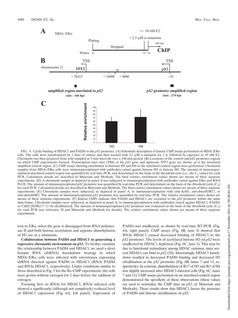
It is worth mentioning that the observed kinetics of recruit-ment to the pS2 promoter does not appear to reflect nonspe-cific binding of the tested proteins or histone marks to all DNAsequences, as indicated by an important ChIP control thatextends our initial findings (19, 20): we used an unrelated DNAsequence, located several kb upstream from the pS2 gene (Fig.4B). Kinetic ChIPs for ER�, RNA Pol II, AcH3, H3R17me2,and H4R3me2 showed only background binding to this controlregion (these values were used to normalize our ChIP data onpS2; see Materials and Methods).
We next examined PADI4 and HDAC1 binding to the pS2promoter. ChIP assays revealed that both PADI4 and HDAC1associate cyclically with this promoter when it becomes refrac-tory to ER� (Fig. 4F). The presence of PADI4 enzymaticactivity was determined with antibodies to citrullinated H3(citH3). These ChIP assays demonstrated that histone H3 be-comes modified by citrullination when PADI4 and HDAC1 areassociated with the pS2 promoter (Fig. 4F).
These bindings to the pS2 promoter are specific, since theywere not detected on an unrelated control region (values usedto normalize the ChIP data on pS2; see Materials and Meth-ods). Taken together, these results demonstrate that PADI4and HDAC1, and their corresponding enzymatic activities, as-sociate specifically with the estrogen-responsive pS2 promoter(during the transcriptionally unproductive phase) and cycleon it.
PADI4 and HDAC1 bind together to the pS2 promoter. Toinvestigate whether PADI4 and HDAC1 bind independentlyor coordinately to the pS2 promoter in vivo, we performedsequential ChIP (Re-ChIP). In these experiments, MDA::ER�cells were grown in the absence of estrogen for 2 days and thentreated with estrogen. A first round of immunoprecipitationwas carried out with anti-PADI4 antibody (Fig. 5, lane 2).Then, immunoprecipitated cross-linked DNA-protein com-plexes were isolated and reimmunoprecipitated with antibod-ies to HDAC1. Figure 5, lane 2, shows that the pS2 promoterregion was successfully amplified from both the fraction reim-munoprecipitated with anti-HDAC1 and the fraction reimmu-noprecipitated with anti-citH3, whereas only weak signal wasdetected when antibodies against ER, acH3, or dimethylatedH3R17 were used in the second round of immunoprecipita-tion. Control reactions performed with nonspecific anti-HAantibodies gave, as expected, no signal (Fig. 5, lane 1). Similarresults were obtained when anti-HDAC1 was used in the firstround of immunoprecipitation (Fig. 5, lane 3): a signal wasobserved with the fractions reimmunoprecipitated with eitheranti-PADI4 or anti-CitH3. Weak signal was detected withanti-ER or anti-acH3 antibodies used in the second round ofimmunoprecipitation (Fig. 5, lane 3).
Together, these data strongly suggest that the histone de-iminase PADI4 and the histone deacetylase HDAC1 are si-multaneously and cyclically present on the pS2 promoter, withmaximum occupation as the pS2 promoter is becoming refrac-
FIG. 3. HDAC1 associates with PADI4-mediated histone deimi-nase activity. (A) Outline of the experimental procedure used to mon-itor histone deiminase activity by PADI4. 293T cells were transientlytransfected with HA-PADI4. Whole-cell extracts were then immuno-precipitated with anti-HA antibody (IP PADI4), and the complexeswere incubated with histones in the presence or absence of calcium(Ca2�). This ion is required for the PADI4-catalyzed reaction involv-ing removal of an imine group from a peptidyl arginine of histone toproduce peptidyl citrulline. (B) Using the approach described in panelA, the histone deiminase activity was assayed by Western blotting withan antibody recognizing citrullinated residues (citrulline 17�2�8) inthe tail of histone H3 (7). This anticitrulline antibody detects histoneH3 only when overexpressed PADI4 is added to the histones. Levels ofPADI4 in the deiminase reaction were checked by Western blottingwith anti-HA antibody. (C) Outline of the experiment revealing asso-ciation of HDAC1 with PADI4-mediated histone deiminase activity.293T cells were transiently transfected with HDAC1 alone or withHA-PADI4. Whole-cell extracts were then immunoprecipitated withanti-HDAC1 antibody, and the complexes were incubated with his-tones in the presence or absence of calcium (Ca2�). (D) Histonedeiminase activity was assayed by Western blotting with anti-histoneH3 (citrulline 17�2�8). Immunoprecipitation of HDAC1 purified his-tone deiminase activity only when the cells were cotransfected withPADI4. The levels of HDAC1 in the reaction were checked by West-ern blotting with anti-HDAC1. The Coomassie blue-stained gel showsthat equal amounts of histones were used in all reactions.
VOL. 29, 2009 DEIMINATION AND DEACETYLATION OF HISTONES 4987
Dow
nloa
ded
from
http
s://j
ourn
als.
asm
.org
/jour
nal/m
cb o
n 17
Nov
embe
r 20
21 b
y 18
3.89
.93.
105.
tory to ER�, when the gene is disengaged from RNA polymer-ase II and both histone acetylation and arginine dimethylationof H3 are at a minimum.
Collaboration between PADI4 and HDAC1 in generating arepressive chromatin environment on pS2. To further examinethe relationship between PADI4 and HDAC1, we used a shorthairpin RNA (shRNA) knockdown strategy in whichMDA::ER� cells were infected with retroviruses expressingshRNA directed against PADI4 or HDAC1 (RNAi PADI4and RNAi HDAC1, respectively). Under conditions similar tothose described in Fig. 5 for the Re-ChIP experiments, the cellswere grown without estrogen for 2 days before the addition ofestrogen.
Focusing first on RNAi for HDAC1, RNAi infected cellsshowed a significantly (although not completely) reduced levelof HDAC1 expression (Fig. 6A, left panel). Expression of
PADI4 was unaffected, as shown by real-time RT-PCR (Fig.6A, right panel). ChIP assays (Fig. 6B, lane 3) showed thatRNAi HDAC1 caused decreased binding of HDAC1 to thepS2 promoter. The levels of acetylated histone H3 on pS2 wereunaffected by HDAC1 depletion (Fig. 6C, lane 3). This may bedue to functional redundancy among HDAC enzymes, since sev-eral HDACs can bind to pS2 (20). Interestingly, HDAC1 knock-down resulted in decreased PADI4 binding and decreased H3citrullination at the pS2 promoter (Fig. 6B, lanes 7 and 11, re-spectively). In contrast, dimethylation of R17 of H3 and R3 of H4was slightly increased after HDAC1-infected cells (Fig. 6C, lanes7 and 11). ChIP assays performed on an unrelated control regiondemonstrated the specificity of these observations (these valuesare used to normalize the ChIP data on pS2; cf. Materials andMethods). These results show that HDAC1 favors the presenceof PADI4 and histone citrullination on pS2.
FIG. 4. Cyclic binding of HDAC1 and PADI4 to the pS2 promoter. (A) Schematic description of kinetic ChIP assays performed on MDA::ER�cells. The cells were synchronized by 3 days of culture and then treated with 2.5 �M �-amanitin for 2 h, followed by exposure to 10 nM E2.Chromatin was then prepared from cells sampled at 5-min intervals over a 140-min period. (B) Locations of the control and pS2 promoter regionson which ChIP experiments focused. Transcription start sites (TSS) of the pS2 gene and upstream TFF2 gene are shown, as is the unrelatedamplified control region. (C) ChIP assays showing enrichment in histones H3 and H4 in the unrelated control region were performed. Chromatinsamples from MDA::ER� cells were immunoprecipitated with antibodies raised against histone H3 or histone H4. The amount of immunopre-cipitated unrelated control region was quantified by real-time PCR, and determined on the basis of the threshold cycle (i.e., the CT value) for eachPCR. Calculation details are described in Materials and Methods. The final relative enrichment values shown are means of three separateexperiments. (D) A chromatin sample as depicted in panel A was subjected to immunoprecipitation with antibodies raised against ER� and RNAPol II. The amount of immunoprecipitated pS2 promoter was quantified by real-time PCR and determined on the basis of the threshold cycle (CT)for each PCR. Calculation details are described in Materials and Methods. The final relative enrichment values shown are means of three separateexperiments. (E) Chromatin samples were subjected, as depicted in panel A, to immunoprecipitation with anti-AcH3, anti-dimeH3R17, oranti-dimeH4R3. The amount of immunoprecipitated pS2 promoter was quantified by real-time PCR. The relative enrichment values shown aremeans of three separate experiments. (F) Kinetic ChIPs indicate that PADI4 and HDAC1 are recruited to the pS2 promoter within the sametime-frame. Chromatin samples were subjected, as depicted in panel A, to immunoprecipitation with antibodies raised against HDAC1, PADI4,or CitH3 [H3R(17�2�8) citrullinated]. The amount of immunoprecipitated pS2 promoter was evaluated on the basis of the threshold cycle (CT)for each PCR (see reference 20 and Materials and Methods for details). The relative enrichment values shown are means of three separateexperiments.
4988 DENIS ET AL. MOL. CELL. BIOL.
Dow
nloa
ded
from
http
s://j
ourn
als.
asm
.org
/jour
nal/m
cb o
n 17
Nov
embe
r 20
21 b
y 18
3.89
.93.
105.

FIG. 4—Continued.
VOL. 29, 2009 DEIMINATION AND DEACETYLATION OF HISTONES 4989
Dow
nloa
ded
from
http
s://j
ourn
als.
asm
.org
/jour
nal/m
cb o
n 17
Nov
embe
r 20
21 b
y 18
3.89
.93.
105.

Regarding PADI4 knockdown in MDA::ER� cells, and de-spite tremendous efforts, we were able to reduce PADI4 ex-pression only slightly (Fig. 6A, right panel). The PADI4 RNAiconstruct we used is specific for PADI4, since we could use itto strongly and specifically decrease PADI4 levels in the U2OScell line (data not shown). The reason for this difference be-tween cell types is currently unclear. Consistent with the slighteffect of our construct on the PADI4 level in MDA::ER� cells,this knockdown approach barely affected the presence ofPADI4, HDAC1 or their corresponding enzymatic activities onthe pS2 promoter (Fig. 6B, lanes 2, 6, and 10 and 6C).
We subsequently investigated whether PADI4 and HDAC1might functionally collaborate on the pS2 promoter. We rea-soned that the limited reduction of PADI4 expression ob-served after infection of MDA::ER� cells with the shRNA forPADI4 (Fig. 6A, right panel) could provide a useful tool fortesting whether PADI4 collaborates with HDAC1. We gener-ated PADI4 and HDAC1 doubly infected MDA::ER� cellsdisplaying decreased PADI4 and HDAC1 levels comparable tothose observed in the corresponding singly infected cells (Fig.6A). Simultaneous expression of PADI4 and HDAC1 RNAi,compared to expression of HDAC1 RNAi alone, led to (i) afurther decrease in H3 citrullination (Fig. 6B, lane 12), (ii) asubstantial increase in acetylated H3 (Fig. 6C, lane 4), and (iii)an enhanced level of dimethylated R17 of histone H3 and R3of histone H4 (Fig. 6C, lanes 8 and 12). Control ChIP assaysperformed on an unrelated region showed no such effects(these values were used to normalize ChIP assays on pS2; cf.
Materials and Methods). Together, these observations suggestthat PADI4 and HDAC1 may favor H3 citrullination anddeacetylation of the pS2 promoter.
To complement the data described above, we used the tech-nique called FAIRE. This technique starts like a standardChIP: chromatin is cross-linked with formaldehyde, sheared bysonication, and phenol-chloroform extracted. After cross-linkreversion, the DNA is purified, and a real-time PCR is carriedout on the sequence of interest. DNA segments that activelyregulate transcription are typically characterized by eviction ofnucleosomes from chromatin, and this can be facilitated in partby increased acetylation of the nucleosome before activation oftranscription (27). Since DNA covalently linked to proteins(consisting mainly of histones) is sequestered in the organicphase, leaving only protein-free DNA fragments in the aque-ous phase, the FAIRE technique provides an estimate of nu-cleosome occupancy or of its relative loose association withDNA at a given region, correlating directly with its transcrip-tional status (Fig. 7A) (14, 22). We performed FAIRE on thepS2 promoter, which contains two phased nucleosomes (29,33), in PADI4 and HDAC1 single- and dual-interferenceMDA::ER� cells treated with E2 as before (Fig. 5). On thebasis of the results presented in Fig. 6, we hypothesized that adouble infection of cells with our shRNA would lead to en-hanced FAIRE enrichment. This hypothesis proved correct.After FAIRE, as shown in Fig. 7B, preparations from PADI4and HDAC1 dual-interference cells proved to be morestrongly enriched in pS2 promoter than preparations derivedfrom single-interference cells. This was not observed with anunrelated control region (these values were used to normalizethe FAIRE data on pS2; cf. Materials and Methods). Theseresults indicate that the association of nucleosomes at the pS2promoter is looser in PADI4 and HDAC1 dual-interferencecells compared to controls, supporting the view that there isfunctional cross talk between these two enzymes at the pS2promoter.
Taken together, these data suggest a functional interconnec-tion between the PADI4 and HDAC1 enzymes in the regula-tion of the pS2 gene.
DISCUSSION
The discovery of the histone deiminase PADI4, convertingmethylarginine to citrulline, has revealed that histone argininemethylation is not a static modification, as previously thought(7, 42). However, understanding how this newly discoveredenzyme functions in chromatin biology remains an importantchallenge. We here report a close connection between thePADI4 histone deiminase and HDAC1. We show that PADI4binds to HDAC1 and that HDAC1 associates with PADI4-mediated histone deiminase activity. The results of our kineticChIP assays show that PADI4 and HDAC1 appear transientlyand in a cyclic manner on the estrogen-responsive promoterpS2, in the presence of estradiol. Their presence correlateswith a loss of arginine methylation, acquisition of citrulline,histone deacetylation, and disengagement of RNA polymeraseII from the pS2 promoter.
We have previously proposed the concept of a “transcrip-tional clock” that synchronizes sequential waves of promoteraccessibility for given cofactors and thereby dictates an or-
FIG. 5. Simultaneous association of PADI4 and HDAC1 with thepS2 promoter in the presence of estradiol. Chromatin prepared fromMDA::ER� cells treated with 10�8 M E2 was subjected to the ChIPprocedure with anti-HA (used as a negative control), anti-PADI4, oranti-HDAC1 and then immunoprecipitated again (Re-ChIP) with theantibody shown on the left. The semiquantitative PCR results shownare representative of experiments performed at least three times.
4990 DENIS ET AL. MOL. CELL. BIOL.
Dow
nloa
ded
from
http
s://j
ourn
als.
asm
.org
/jour
nal/m
cb o
n 17
Nov
embe
r 20
21 b
y 18
3.89
.93.
105.

dered and dynamic sequence of events that initiate and sustaintranscription (20). Periodic limitation of transcription is causedby events that clear the pS2 promoter of transcription factors.The precise mechanisms underlying this promoter clearanceand the termination of each cycle are still unclear. Our current
work offers important insights into the molecular basis of theclearance phase of the pS2 promoter. We found that the jointprocesses of PADI4-mediated histone deimination andHDAC1-mediated histone deacetylation contribute, perhapspartially but importantly, to this phase. These concerted re-
FIG. 6. Collaboration between PADI4 and HDAC1 in generating a repressive pS2 chromatin environment. (A) Specific-RNAi-encodingvectors decrease HDAC1 expression substantially and PADI4 expression moderately. MDA::ER� cells were stably infected with the pRS/controlvector (RNAi), the pRS/PADI4 vector (RNAi PADI4), the pRS/HDAC1 vector (RNAi HDAC1), or both pRS/PADI4 and pRS/HDAC1 (RNAiPADI4 � RNAi HDAC1). After selection the cells were harvested, and quantitative RT-PCR was performed on their mRNA, with primerscorresponding to HDAC1 (left panel) or PADI4 (right panel). All transcript levels were normalized with respect to SDHA and then divided bythe normalized level recorded for control cells. Values are means of three independent experiments. (B) Chromatin was prepared fromMDA::ER� cells expressing a control RNAi, PADI4 RNAi, HDAC1 RNAi, or both PADI4 RNAi and HDAC1 RNAi (RNAi PADI4 � RNAiHDAC1) and treated with 10�8 M E2. Cross-linked chromatin samples were immunoprecipitated with antibodies to HDAC1 (lanes 1 to 4), PADI4(lanes 5 to 8), or CitH3 [H3R(17�2�8) citrullinated] (lanes 9 to 12). The amount of immunoprecipitated pS2 promoter was quantified by real-timePCR on the basis of the threshold cycle value (CT). Calculation details were done as previously described (20) (see also Materials and Methods).The data are presented as the means � the standard errors of four independent experiments, and asterisks indicate statistical significance asdetermined by Dunnett and Mann-Whitney comparisons (�, P � 0.05; ��, P � 0.01; ���, P � 0.001). (C) Chromatin samples were subjected, asdescribed in panel B, to immunoprecipitation with antibodies raised against AcH3 (lanes 1 to 4), dimeH3R17 (lanes 5 to 8), or dimeH4R3 (lanes9 to 12). The immunoprecipitated pS2 promoter was quantified by real-time PCR on the basis of the CT for each PCR (see reference 20 andMaterials and Methods for calculation details). The relative enrichment values shown are means of four independent experiments. Asterisksindicate the statistical significance as determined by Dunnett and Mann-Whitney comparisons (�, P � 0.05; ��, P � 0.01; ���, P � 0.001).
VOL. 29, 2009 DEIMINATION AND DEACETYLATION OF HISTONES 4991
Dow
nloa
ded
from
http
s://j
ourn
als.
asm
.org
/jour
nal/m
cb o
n 17
Nov
embe
r 20
21 b
y 18
3.89
.93.
105.

pressive histone modifications appear to participate in markingthe end of each cycle, allowing the pS2 promoter to be resetand making it possible for subsequent cycles to proceed. His-tone deimination and deacetylation may thus be particularlyimportant in enabling the cell to adjust continuously the tran-scription rate of the pS2 gene according to the level of estro-gen. We do not know, at this stage, which factors recruitPADI4 and HDAC1 when the pS2 promoter is becoming re-fractory to ER�. The Sp proteins might be involved, sincethese transcription factors are reported to interact withHDACs and since, in the absence of the ER�, their presenceon the pS2 promoter coincides with binding of HDACs to thepromoter (13, 35).
Our findings may illustrate nicely the concept of cross talk
between histone modifications leading to modulation of geneexpression. For example, deacetylation of lysine 9 of H3 isreported to be required to allow methylation of this residue(25). This is due, at least in some instances, to the concertedand coordinated action of the SU(VAR)3-9 methyltransferaseand HDAC1 (8). Recent studies have shown an interplay be-tween histone deacetylation and the LSD1/KDM1 H3K4 his-tone demethylase: lysine 9 deacetylation by HDAC1 rendershistone H3 more susceptible to LSD1/KDM1-mediated H3K4demethylation, and LSD1/KDM1 cofactors are required foroptimal HDAC1 activity (18, 32). Here, we show that HDAC1plays a role in PADI4-mediated histone citrullination at thepS2 promoter. Furthermore, our data suggest a functional linkbetween the PADI4 and HDAC1 enzymes in the regulation ofthe pS2 promoter. The challenge for the future will be tounravel the precise sequence of events of the coordinatedaction of PADI4 and HDAC1 at the pS2 promoter. Althoughour data suggest that this coordinated action may be related tothe physical association we have identified between PADI4 andHDAC1, further work will be needed to formally prove a directfunctional link between these two enzymes. Furthermore, thebiological or regulatory roles of the PADI4-HDAC1 associa-tion remain unclear and defining these roles will deserve futurestudies.
The deiminating activity of PADI4 has been implicated inthe pathophysiology of rheumatoid arthritis (RA) (36), oneof the most common human systemic autoimmune diseases. Inaddition, assays of antibodies against citrullinated peptides canbe used as valuable diagnostic tools (28, 38). Histone deacety-lase inhibitors are emerging as effective and valuable tools forboth chemotherapy and chemoprevention of cancer (46). Inaddition to the efficacy of such inhibitors in cancer therapy,recent work points to the HDAC inhibitor trichostatin A as apotential therapeutic agent in the treatment of RA (15). Itseems that trichostatin A sensitizes RA-related synovial fibro-blasts to TRAIL-induced apoptosis (15). We reveal here co-ordinated histone deimination and deacetylation, through thecombined action of the PADI4 and HDAC1 enzymes.Whether inhibition of histone deimination is an importantcomponent of the anti-RA effect is not known, but our presentfindings raise this attractive possibility. Future studies aimed atdeveloping PADI4-inhibiting drugs should make it possible totest this possibility and may contribute to progress in the med-ical care of RA.
ACKNOWLEDGMENTS
H.D. and R.D. were funded by the FNRS. F.F. is a ChercheurQualifie du FNRS of the Belgian Fonds National de la RechercheScientifique. Grants to R.M. were from the CNRS, University ofRennes I and from the Association pour la Recherche contre le Can-cer. This study was also funded by grants to F.F. from the Fondationcontre le Cancer and the FNRS, grants from the Action de RechercheConcertee de la Communaute Francaise de Belgique and from theInteruniversity Attraction Poles (IAP P6/28), by EU grant CANCERDIPFP7-200620, and by the European Molecular Biology OrganizationYoung Investigator Programme.
We thank A. Cook and T. Kouzarides for HDAC1 constructs.
REFERENCES
1. Arita, K., H. Hashimoto, T. Shimizu, K. Nakashima, M. Yamada, and M.Sato. 2004. Structural basis for Ca2�-induced activation of human PAD4.Nat. Struct. Mol. Biol. 11:777–783.
FIG. 7. Effects of PADI4 and HDAC1 on pS2 nucleosome occu-pancy. (A) Schematic description of the FAIRE technique. InfectedMDA::ER� cells were placed for 2 days in an E2-free medium andthen exposed to 10 nM E2 for 6 h (upper part). Chromatin was thenprepared and phenol-chloroform extracted without previous reversecross-linking (bottom part). (B) MDA::ER� chromatin samples wereprepared from cells expressing control RNAi, PADI4 RNAi, HDAC1RNAi, or PADI4 RNAi � HDAC1 RNAi (RNAi PADI4 � RNAiHDAC1). FAIRE enrichment in pS2 promoter was quantified by real-time PCR on the basis of the threshold cycle value (CT). Calculationdetails are described in Materials and Methods. The data are pre-sented as means � the standard errors of four independent experi-ments. Asterisks indicate the statistical significance as determined byDunnett post hoc comparisons (��, P � 0.01; ���, P � 0.001).
4992 DENIS ET AL. MOL. CELL. BIOL.
Dow
nloa
ded
from
http
s://j
ourn
als.
asm
.org
/jour
nal/m
cb o
n 17
Nov
embe
r 20
21 b
y 18
3.89
.93.
105.

2. Bernard, D., J. Gil, P. Dumont, S. Rizzo, D. Monte, B. Quatannens, D.Hudson, T. Visakorpi, F. Fuks, and Y. de Launoit. 2006. The methyl-CpG-binding protein MECP2 is required for prostate cancer cell growth. Onco-gene 25:1358–1366.
3. Brehm, A., E. A. Miska, D. J. McCance, J. L. Reid, A. J. Bannister, and T.Kouzarides. 1998. Retinoblastoma protein recruits histone deacetylase torepress transcription. Nature 391:597–601.
4. Brenner, C., R. Deplus, C. Didelot, A. Loriot, E. Vire, C. De Smet, A.Gutierrez, D. Danovi, D. Bernard, T. Boon, P. G. Pelicci, B. Amati, T.Kouzarides, Y. de Launoit, L. Di Croce, and F. Fuks. 2005. Myc repressestranscription through recruitment of DNA methyltransferase corepressor.EMBO J. 24:336–346.
5. Chang, B., Y. Chen, Y. Zhao, and R. K. Bruick. 2007. JMJD6 is a histonearginine demethylase. Science 318:444–447.
6. Chen, D., H. Ma, H. Hong, S. S. Koh, S. M. Huang, B. T. Schurter, D. W.Aswad, and M. R. Stallcup. 1999. Regulation of transcription by a proteinmethyltransferase. Science 284:2174–2177.
7. Cuthbert, G. L., S. Daujat, A. W. Snowden, H. Erdjument-Bromage, T.Hagiwara, M. Yamada, R. Schneider, P. D. Gregory, P. Tempst, A. J. Ban-nister, and T. Kouzarides. 2004. Histone deimination antagonizes argininemethylation. Cell 118:545–553.
8. Czermin, B., G. Schotta, B. B. Hulsmann, A. Brehm, P. B. Becker, G. Reuter,and A. Imhof. 2001. Physical and functional association of SU(VAR)3-9 andHDAC1 in Drosophila. EMBO Rep. 2:915–919.
9. Deplus, R., C. Brenner, W. A. Burgers, P. Putmans, T. Kouzarides, Y. deLaunoit, and F. Fuks. 2002. Dnmt3L is a transcriptional repressor thatrecruits histone deacetylase. Nucleic Acids Res. 30:3831–3838.
10. Fuks, F., W. A. Burgers, A. Brehm, L. Hughes-Davies, and T. Kouzarides.2000. DNA methyltransferase Dnmt1 associates with histone deacetylaseactivity. Nat. Genet. 24:88–91.
11. Giresi, P. G., J. Kim, R. M. McDaniell, V. R. Iyer, and J. D. Lieb. 2007.FAIRE (formaldehyde-assisted isolation of regulatory elements) isolatesactive regulatory elements from human chromatin. Genome Res. 17:877–885.
12. Giresi, P. G., and J. D. Lieb. 2009. Isolation of active regulatory elementsfrom eukaryotic chromatin using FAIRE (formaldehyde assisted isolation ofregulatory elements). Methods 48:233–239.
13. He, S., J. M. Sun, L. Li, and J. R. Davie. 2005. Differential intranuclearorganization of transcription factors Sp1 and Sp3. Mol. Biol. Cell 16:4073–4083.
14. Hogan, G. J., C. K. Lee, and J. D. Lieb. 2006. Cell cycle-specified fluctuationof nucleosome occupancy at gene promoters. PLoS Genet. 2:e158.
15. Jungel, A., V. Baresova, C. Ospelt, B. R. Simmen, B. A. Michel, R. E. Gay,S. Gay, C. A. Seemayer, and M. Neidhart. 2006. Trichostatin A sensitizesrheumatoid arthritis synovial fibroblasts for TRAIL-induced apoptosis. Ann.Rheum. Dis. 65:910–912.
16. Klose, R. J., K. Yamane, Y. Bae, D. Zhang, H. Erdjument-Bromage, P.Tempst, J. Wong, and Y. Zhang. 2006. The transcriptional repressorJHDM3A demethylates trimethyl histone H3 lysine 9 and lysine 36. Nature442:312–316.
17. Reference deleted.18. Lee, M. G., C. Wynder, D. A. Bochar, M. A. Hakimi, N. Cooch, and R.
Shiekhattar. 2006. Functional interplay between histone demethylase anddeacetylase enzymes. Mol. Cell. Biol. 26:6395–6402.
19. Metivier, R., G. Penot, R. P. Carmouche, M. R. Hubner, G. Reid, S. Denger,D. Manu, H. Brand, M. Kos, V. Benes, and F. Gannon. 2004. Transcriptionalcomplexes engaged by apo-estrogen receptor-alpha isoforms have divergentoutcomes. EMBO J. 23:3653–3666.
20. Metivier, R., G. Penot, M. R. Hubner, G. Reid, H. Brand, M. Kos, and F.Gannon. 2003. Estrogen receptor-alpha directs ordered, cyclical, and com-binatorial recruitment of cofactors on a natural target promoter. Cell 115:751–763.
21. Metzger, E., M. Wissmann, N. Yin, J. M. Muller, R. Schneider, A. H. Peters,T. Gunther, R. Buettner, and R. Schule. 2005. LSD1 demethylates repressivehistone marks to promote androgen-receptor-dependent transcription. Na-ture 437:436–439.
22. Nagy, P. L., M. L. Cleary, P. O. Brown, and J. D. Lieb. 2003. Genomewidedemarcation of RNA polymerase II transcription units revealed by physicalfractionation of chromatin. Proc. Natl. Acad. Sci. USA 100:6364–6369.
23. Nakashima, K., T. Hagiwara, A. Ishigami, S. Nagata, H. Asaga, M.Kuramoto, T. Senshu, and M. Yamada. 1999. Molecular characterization ofpeptidylarginine deiminase in HL-60 cells induced by retinoic acid and1�,25-dihydroxyvitamin D3. J. Biol. Chem. 274:27786–27792.
24. Nakashima, K., T. Hagiwara, and M. Yamada. 2002. Nuclear localization ofpeptidylarginine deiminase V and histone deimination in granulocytes.J. Biol. Chem. 277:49562–49568.
25. Rea, S., F. Eisenhaber, D. O’Carroll, B. D. Strahl, Z. W. Sun, M. Schmid, S.Opravil, K. Mechtler, C. P. Ponting, C. D. Allis, and T. Jenuwein. 2000.Regulation of chromatin structure by site-specific histone H3 methyltrans-ferases. Nature 406:593–599.
26. Reid, G., M. R. Hubner, R. Metivier, H. Brand, S. Denger, D. Manu, J.Beaudouin, J. Ellenberg, and F. Gannon. 2003. Cyclic, proteasome-mediatedturnover of unliganded and liganded ER� on responsive promoters is anintegral feature of estrogen signaling. Mol. Cell 11:695–707.
27. Reinke, H., and W. Horz. 2003. Histones are first hyperacetylated and thenlose contact with the activated PHO5 promoter. Mol. Cell 11:1599–1607.
28. Schellekens, G. A., H. Visser, B. A. de Jong, F. H. van den Hoogen, J. M.Hazes, F. C. Breedveld, and W. J. van Venrooij. 2000. The diagnostic prop-erties of rheumatoid arthritis antibodies recognizing a cyclic citrullinatedpeptide. Arthritis Rheum. 43:155–163.
29. Sewack, G. F., and U. Hansen. 1997. Nucleosome positioning and transcrip-tion-associated chromatin alterations on the human estrogen-responsive pS2promoter. J. Biol. Chem. 272:31118–31129.
30. Shang, Y., X. Hu, J. DiRenzo, M. A. Lazar, and M. Brown. 2000. Cofactordynamics and sufficiency in estrogen receptor-regulated transcription. Cell103:843–852.
31. Shi, Y., F. Lan, C. Matson, P. Mulligan, J. R. Whetstine, P. A. Cole, R. A.Casero, and Y. Shi. 2004. Histone demethylation mediated by the nuclearamine oxidase homolog LSD1. Cell 119:941–953.
32. Shi, Y. J., C. Matson, F. Lan, S. Iwase, T. Baba, and Y. Shi. 2005. Regulationof LSD1 histone demethylase activity by its associated factors. Mol. Cell19:857–864.
33. Sparmann, A., and M. van Lohuizen. 2006. Polycomb silencers control cellfate, development and cancer. Nat. Rev. Cancer 6:846–856.
34. Strahl, B. D., S. D. Briggs, C. J. Brame, J. A. Caldwell, S. S. Koh, H. Ma,R. G. Cook, J. Shabanowitz, D. F. Hunt, M. R. Stallcup, and C. D. Allis.2001. Methylation of histone H4 at arginine 3 occurs in vivo and is mediatedby the nuclear receptor coactivator PRMT1. Curr. Biol. 11:996–1000.
35. Sun, J. M., V. A. Spencer, L. Li, H. Yu Chen, J. Yu, and J. R. Davie. 2005.Estrogen regulation of trefoil factor 1 expression by estrogen receptor alphaand Sp proteins. Exp. Cell Res. 302:96–107.
36. Suzuki, A., R. Yamada, X. Chang, S. Tokuhiro, T. Sawada, M. Suzuki, M.Nagasaki, M. Nakayama-Hamada, R. Kawaida, M. Ono, M. Ohtsuki, H.Furukawa, S. Yoshino, M. Yukioka, S. Tohma, T. Matsubara, S. Wakitani,R. Teshima, Y. Nishioka, A. Sekine, A. Iida, A. Takahashi, T. Tsunoda, Y.Nakamura, and K. Yamamoto. 2003. Functional haplotypes of PADI4, en-coding citrullinating enzyme peptidylarginine deiminase 4, are associatedwith rheumatoid arthritis. Nat. Genet. 34:395–402.
37. Tsukada, Y., J. Fang, H. Erdjument-Bromage, M. E. Warren, C. H. Borch-ers, P. Tempst, and Y. Zhang. 2006. Histone demethylation by a family ofJmjC domain-containing proteins. Nature 439:811–816.
38. van Boekel, M. A., E. R. Vossenaar, F. H. van den Hoogen, and W. J. vanVenrooij. 2002. Autoantibody systems in rheumatoid arthritis: specificity,sensitivity and diagnostic value. Arthritis Res. 4:87–93.
39. Vandesompele, J., K. De Preter, F. Pattyn, B. Poppe, N. Van Roy, A. DePaepe, and F. Speleman. 2002. Accurate normalization of real-time quanti-tative RT-PCR data by geometric averaging of multiple internal controlgenes. Genome Biol. 3:RESEARCH0034.
40. Vire, E., C. Brenner, R. Deplus, L. Blanchon, M. Fraga, C. Didelot, L. Morey,A. Van Eynde, D. Bernard, J. M. Vanderwinden, M. Bollen, M. Esteller, L.Di Croce, Y. de Launoit, and F. Fuks. 2006. The Polycomb group proteinEZH2 directly controls DNA methylation. Nature 439:871–874.
41. Vossenaar, E. R., A. J. Zendman, W. J. van Venrooij, and G. J. Pruijn. 2003.PAD, a growing family of citrullinating enzymes: genes, features and involve-ment in disease. Bioessays 25:1106–1118.
42. Wang, Y., J. Wysocka, J. Sayegh, Y. H. Lee, J. R. Perlin, L. Leonelli, L. S.Sonbuchner, C. H. McDonald, R. G. Cook, Y. Dou, R. G. Roeder, S. Clarke,M. R. Stallcup, C. D. Allis, and S. A. Coonrod. 2004. Human PAD4 regulateshistone arginine methylation levels via demethylimination. Science 306:279–283.
43. Whetstine, J. R., A. Nottke, F. Lan, M. Huarte, S. Smolikov, Z. Chen, E.Spooner, E. Li, G. Zhang, M. Colaiacovo, and Y. Shi. 2006. Reversal ofhistone lysine trimethylation by the JMJD2 family of histone demethylases.Cell 125:467–481.
44. Reference deleted.45. Yamane, K., C. Toumazou, Y. Tsukada, H. Erdjument-Bromage, P. Tempst, J.
Wong, and Y. Zhang. 2006. JHDM2A, a JmjC-containing H3K9 demethylase,facilitates transcription activation by androgen receptor. Cell 125:483–495.
46. Yoo, C. B., and P. A. Jones. 2006. Epigenetic therapy of cancer: past, presentand future. Nat. Rev. Drug Discov. 5:37–50.
47. Zhang, Y. 2004. Molecular biology: no exception to reversibility. Nature431:637–639.
VOL. 29, 2009 DEIMINATION AND DEACETYLATION OF HISTONES 4993
Dow
nloa
ded
from
http
s://j
ourn
als.
asm
.org
/jour
nal/m
cb o
n 17
Nov
embe
r 20
21 b
y 18
3.89
.93.
105.