Embed Size (px)
Citation preview
Proc. Natl Acad. Sci. USAVol. 78, No. 7, pp. 4557-4561, July 1981Immunology
Functional association of idiotypic and I-J determinants on theantigen receptor of suppressor T cells
(T-cell receptor/hybridoma T cells/suppressor factor)
KENJI OKUDA, MUTSUHIKO MINAMI, SHYR-TE JU, AND MARTIN E. DORFDepartment of Pathology, Harvard Medical School, Boston, Massachusetts 02115
Communicated by Baruj Bennacerraf, April 15, 1981
ABSTRACT The serological characteristics of the antigen re-ceptor on 4-hydroxy-3-nitrophenylacetyl (NP) specific suppressorT cell hybridomas were analyzed. Three T-cell hybrids could belysed with anti-idiotype and complement. The reactivity patternobserved from a panel ofanti-idiotypic reagents indicated that NPbdeterminants were detected on all three hybrid lines. NP conju-gates with bovine serum albumin or caproic acid specifically in-hibited the complement-mediated lysis of these cells by both anti-NPb idiotype and anti-I-J antisera. These hapten conjugates failedto block lysis by anti-Thy 1 or anti-H-2K antisera on the same tar-get cell populations. The data indicate that both I-J and Igh vari-able region gene products are intimately involved in the recog-nition of antigen by suppressor T cells. Finally, the suppressor cellhybrids produce soluble factors that mediate antigen-specificsuppression. The characteristics of the cells and their factors in-dicate that the hybrids correspond to the Ts' or first-order sup-pressor cells in the suppressor cell pathway.
Immune responses to specific antigens are controlled by theactivity of suppressor T lymphocytes. The sequence of cellularinteractions resulting in suppression ofthe cellular and humoralimmune responses to antigen has been well characterized in the4-hydroxy-3-nitrophenylacetyl (NP) system. At least three dis-tinct cell populations have been implicated in this suppressorcell pathway (1-5), thereby making the NP system one of themost thoroughly characterized of the suppressor cell models.Furthermore, the conclusions derived from the NP system areconsistent with most other observations concerning the cellularinteractions involved in immune suppression (6-10). However,in spite of considerable data relating to the cell surface phe-notype of the T cells involved in immune suppression, little isactually known about the interrelationships of such membranemarkers and the nature of the antigen receptors on these cells.To investigate the nature of the T cell antigen receptor and
to characterize the molecules responsible for the communica-tion between cells, in the suppressor pathway, large numbersof functional suppressor T cells must be obtained. To achievethis goal we have utilized the techniques of somatic cell hy-bridization (11). These techniques have been used previouslyto prepare monoclonal cell lines that maintain the biologicalactivity of the original suppressor cell population, including theability to secrete biologically active suppressor factors (12-14).We now describe a series of suppressor cell hybrids that pro-
duce soluble products capable of specifically suppressing NPimmune responses. The relationship of these hybrids to theoverall suppressor cell pathway is analyzed. These hybridomacell lines also provide a source of monoclonal cells that are usedto probe the T cell antigen receptor. The data demonstrate that
both I-J and idiotypic determinants are intimately associatedwith the antigen receptor on suppressor T lymphocytes.
MATERIALS AND METHODSMice. C57BL/6J(B6), C3H/HeJ, and B10.BR mice were
purchased from The Jackson Laboratory (Bar Harbor, ME). Allother inbred strains were bred in the animal facilities ofHarvardMedical School. Mice were used at 2-12 months of age.
Antigens. The preparation ofhapten-conjugated proteins hasbeen described (15). The molar conjugation ratio of haptenicgroups used in this work was: dinitrophenyl (DNP)/bovineserum albumin, 12:1; NP/albumin, 12:1; and 4-hydroxy-5-iodo-3-nitrophenyl (NIP)/albumin, 7:1. The caproic acid (Cap) de-rivatives of DNP and NP were purchased from Biosearch (SanRaphael, CA). DNP-Cap and NP-Cap were dissolved in phos-phate-buffered saline at 10 mM and the pH was adjusted to 7.3with 1 M NaOH. NP-(CH2)6-NP (0.1 mM) was dissolved in 0.25M phosphate buffer (pH 7.8) containing 20% dimethyl sulfoxide.
Alloantisera. B10.A(3R) anti-B10.A(5R) (I-Jk), B10.A(5R)anti-B1O.A(3R) (anti-I-J ), (B10 x LP.RIII)F1 anti-B10.A(4R)(anti-Kk and I-Ak), and (B1O.A(4R) x B10.GD)F1 anti-B10 (antiKb + I-Ab) were produced by immunization with spleen andlymph node cells as described (4, 16). Anti-brain-associated T-cell antigen (anti-BAT) antibody was made by the techniquedescribed by Golub (17). Monoclonal anti-Thy 1.1 and Thy 1.2antibodies were purchased from New England Nuclear.
Anti-Idiotypic Antisera. C57BL/6, A/J, SJL, and C3H micewere immunized intraperitoneally with 100 ,g ofNP-BGG andpertussis vaccine (2). Primary anti-NP antibodies were specif-ically purified by addition ofpooled sera to Sepharose 4B beads(Pharmacia) conjugated with NP-albumin and elution with 0.2M glycine HCl buffer (pH 2.35) into tubes containing 2 MTris HCl at pH 8.0. Guinea pigs were immunized with 100 'gof anti-NP antibodies emulsified with complete Freund's ad-juvant, followed by repeated intramuscular and subcutaneousinjections with the same immunogen. Sera were rendered id-iotype specific by adsorption with Sepharose 4B beads coupledwith a gamma globulin fraction of MOPC 104E ascites and nor-mal serum from the appropriate mouse strains. The binding of"2I-labeled B6 anti-NP antibodies to its anti-idiotypic anti-serum could be strongly inhibited by 1 u1 of B6 anti-NP anti-serum but not by 30 A1 of B6 normal mouse serum or MOPC104E ascites. The idiotypic specificities detected were closelyassociated with NP binding sites because 0.06 ,umol of NP-Capor NIP-Cap but not 6 pgmol of p-aminoarsanilate completelyinhibited the binding. Furthermore, strain distribution studiesindicated that the anti-idiotypic antiserum defines NPb-idi-otypic specificities similar to those previously reported (18).
Abbreviations: NP, 4-hydroxy-3-nitrophenylacetyl; Cap, caproic acid;DNP, dinitrophenyl; NIP, 4-hydroxy-5-iodo-3-nitrophenyl; NBrP, 4-hydroxy-5-bromo-3-nitrophenyl.
The publication costs ofthis article were defrayed in part by page chargepayment. This article must therefore be hereby marked "advertise-ment" in accordance with 18 U. S. C. §1734 solely to indicate this fact.
4557

Proc. Natl. Acad. Sci. USA 78 (1981)
Anti-idiotypic antisera to B6 anti-4-hydroxyl-5-bromo-3-nitro-phenyl (NBrP) and B6 anti-NIP, characterized in a similar fash-ion, also detected NPb idiotypic specificities.
Anti-NP-i idiotypic antiserum was made in guinea pigsagainst NP-1 hybridoma anti-NP antibody (Au, A) secreted bya hybridoma cell line generated by fusion of SJL spleen cellsimmunized to NP-Ficoll with the SP2/0 cell line. The anti-idiotypic antiserum was adsorbed as described above. The ad-sorbed antiserum reacted strongly with NP-1 hybridoma anti-NP antibody but not with MOPC 104E or MOPC 315 myelomaprotein or SJL normal mouse serum. The specific idiotype bind-ing, termed "NP-1 idiotype," was inhibited completely in thepresence of 0.06 Amol of NP-Cap or NIP-Cap but not 6 Iumolof p-aminoarsanilate. NP-1 idiotypic specificities were sharedby B6, SJL, and A/J anti-NP antisera and were not detectablein seven other mouse strains not bearing Ighb or Ighe alleles.Additional serological analyses indicated that anti-idiotypicantisera against B6 anti-NP, B6 anti-NBrP, B6 anti-NIP, andSJL anti-NP crossreactively bound NP-1 hybridoma anti-NPantibody. However, the binding strength ofthese anti-idiotypicantisera were 'Ao to 1A that of anti-idiotypic antiserum to NP-1 hybridoma antibody.
Anti-idiotypic antisera againstCGAT and hybridoma F17. 148.3of B6 origin have been described'(i9).
Preparation of Antigen Specific Suppressor T Cells. Themethods for the preparation and enrichment ofNP-specific sup-pressor T cells have been described (1, 3).
Hybridization. Suppressor T-enriched C57BL/6J or CKBsplenic lymphocytes were hybridized with BW5147 T lym-
phoma cells. The hybridizing method was as described by Tan-iguchi et al. (12).To provide an additional specificity control, in some experi-
ments another T cell hybrid, prepared by fusion ofBW5147 andT cells from DBA/1 GAT nonresponder mice pretreated-withGAT to induce suppressorT cells, was studied. This hybrid line,F65,121 (termed "DI-121" in this report) was the gift ofRonaldN. Germain (Harvard Medical School).
Screening for I-J+ and Id' Hybrids. Soon after the hybridcells began to grow, both I-J+ and Id' hybrids were selectedby cytotoxic tests (20). Briefly, 2 ,1 of cell suspension (1 x 10⁶cells per ml) was treated with 2 ,ul of diluted antisera in L-15medium (Microbiological Bioproducts, Walkersville, MD) con-
taining 0.5% fetal calf serum. The reaction was carried out atroom temperature in Terasaki microplates (no. 3034, Falcon).After 20 min, the cells were washed with one drop ofserum-freeL-15 medium. Then 2 ,ul ofa 1:5 dilution ofselected rabbit com-plement in serum-free L-15 medium was allowed to react withthese washed cells for 40 min at 37°C. Then these cells were
stained with 0.2% nigrosine and the percentage of dead cellswas determined.
To inhibit the cytotoxicity test, 1 1,u of DNP-albumin, al-bumin, NP-albumin, or NIP-albumin solution (2 mg/ml) inphosphate-buffered saline or 0.01 M DNP-Cap, 0.01 M NP-Cap, or 0.1 mM NP-(CH2)6-NP were mixed with 1 ul of cellsuspension (2 x 10⁶ cells per ml) prior to the addition ofantiserum.
Adsorption and Elution of Suppressor Factor. NP-albumin,specifically purified antibodies, and gamma globulin of variousantisera were conjugated to CNBr-activated Sepharose 4Bbeads according to the manufacturer's protocol. Two to 4 mgof protein was coupled to 1 ml of beads and the product was
packed into a 3-ml syringe. Adsorptions were carried out atroom temperature by repeatedly passing 5 ml of'culture su-
pernatants through the columns. Unbound material was saved.After each column was washed with 10 ml ofphosphate-bufferedsaline, bound materials were eluted with 4.2 ml of 0.2 M gly-
cine'HCl (pH3.2) into tubes containing 0.5 ml offetal calfserumand 0.3 ml of2 M Tris'HCl (pH 8.0). The pH ofthe eluted frac-tions was adjusted to 7.4-7.6 when necessary. Fractions were
stored at -200C until used.Assay for Suppressive Activity of Culture Supernatant or
Fractionated Samples on Cutaneous Sensitivity Responses.The assay has been described in detail (21). Footpad swellingwas determined as the difference, in units of 10-3 cm, betweenleft and right footpad thicknesses.
Data Analysis. Statistical analysis of the experimental datawith respect to controls utilized the two-tailed Student's t test.Data are expressed as the mean (±SEM) incremental footpadswelling.
RESULTSSerological Analysis of T-Cell Hybridoma Cell Lines. Two
series offusions using either B6 (H-2b, Ighb) orCKB (H_2k, Ighb)antigen-enriched suppressor cells and the BW5147 AKR thy-moma were analyzed. The cell fusions were performed withpolyethylene glycol, and the hybrids were selected by growthin hypoxanthine/aminopterin/thymidine medium. Of 1050wells plated, 30% developed hybrids. All hybrids were screenedwith anti-I-J alloantisera and subsequently with guinea pig anti-NPb idiotype antisera; 16% of the hybrids were specificallylysed with allele-specific anti-I-J alloantisera and 29% of the I-J-bearing hybrids were specifically lysed with anti-NPb idiotypeantisera. For the present study one B6- and two CKB-derivedhybrids that were susceptible to lysis with both anti-I-J and anti-NP" antisera were selected. In addition one CKB hybrid thathad I-J determinants but lacked idiotypic determinants was se-
lected as a control.The specificity of lysis with these reagents is shown in Table
1. The anti-I-Jb antiserum only lysed hybrids ofB6 (H-2b) origin;the anti-I-Jk reagent only lysed cells of CKB origin. Severalbatches of anti-I-J antisera were used and all gave concordantresults.
The results from adsorption experiments indicated that thelytic activity of the anti-I-Jk antisera could be specifically ab-sorbed with spleen cells from H-2k, but not H-2b-bearing mice.
The specificity ofthe anti-idiotypic serum was demonstrated byits ability to lyse the B6-29, CKB-39, and CKB-17 hybrids butnot T-cell hybrids produced by fusion with GAT-induced sup-pressor T lymphocytes. As a reciprocal control, guinea pig anti-CGAT idiotype failed to react with the NP hybrids but lysedthe DBA/i-derived GAT suppressor cell hybrid. To verify theT-cell nature of these hybrids, each cell line was tested with
Table 1. Serological analysis of T-cell hybridomas% specific cytotoxicity*
Antisera B6-29 CKB-17 CKB-39 CKB-70 D1-121 BW5147
Anti-IjJb 35 0 0 0 0 5Anti-14J 0 70 81 90 0 5Anti-NPb 30 26 60 0 0 0Anti-CGAT 0 5 12 0 45 0Anti-Thy 1;2 100 94 0 70 95 11Anti-Thy 1.1 89 84 90 88 95 86
The results are from a single experiment, but each cell line has beentested on at least three separate occasions with comparable results.Each reagent was diluted 1:10 for use in cytotoxicity assays.* The data were calculated as 100 x (% experimental lysis - % controllysis) - (% total lysis - % control lysis). Control lysis was 5-20%.In a large series of experiments with control sera, we observed thatspecific lysis <15% is within control limits and specific lysis >20%is considered significant.
4558 Immunology: Okuda et al.
Proc. NatL. Acad. Sci. USA 78 (1981) 4559
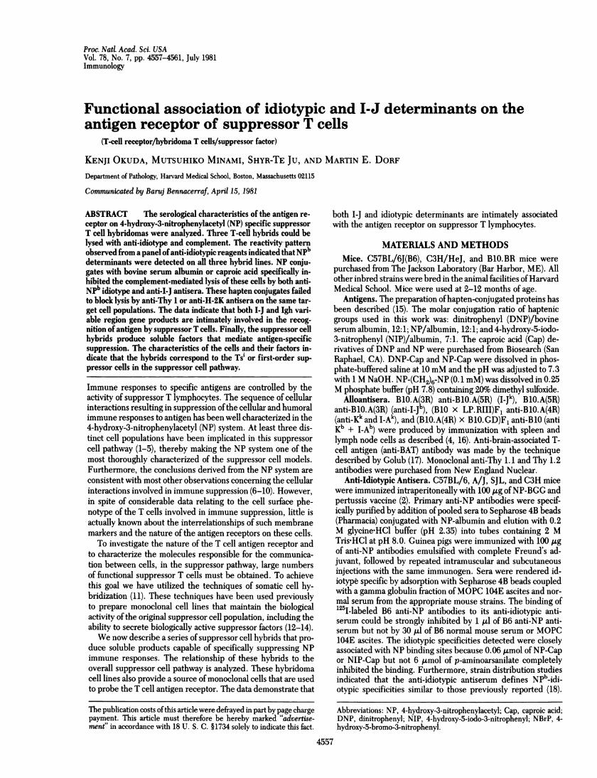
Table 2. Immunochemical characterization of B6-29 hybridomasuppressor factor
Source Immuno-of NP-O-Su adsorbent Column fraction
factor priming column Unbound EluateBW5147 + 26.8 + 4.6 (5)B6-29 + - 11.8 ± 2.3 (5)*B6-29 + AntiNPb 22.8 ± 2.4 (5) 12.0 ± 1.6 (6)*B6-29 + Anti-NIPb 25.2 ± 5.0 (5) 12.8 ± 2.1 (4)*B6-29 + Anti-Ig 10.5 ± 2.0 (4)* 25.0 ± 9.6 (4)B6-29 + Anti-IJb 27.4 ± 3.9 (5) 13.8 ± 0.7 (5)*B6-29 + Anti-I~Jk 10.8 ± 1.8 (5)* 30.0 ± 6.4 (5)B6-29 + NP-albumin 24.3 ± 3.5 (4) 14.5 ± 1.5 (4)*BW5147 - 8.8 ± 1.8 (5)
C57BL/6 mice were primed with NP-O-Su followed by daily intra-venous injections ofthe column fractions until the day prior to antigenchallenge. The results are expressed as the mean (±SEM) incrementof footpad swelling. The number of mice per group is indicated in pa-rentheses. The data are from one experiment; a second experimentyielded comparable results.* Significant suppression, P < 0.05.
monoclonal anti-Thy 1 reagents. With the exception of theCKB-39 cell line, all were lysed with the anti-Thy 1.2 reagentwhich fails to lyse the parental BW5147 line, indicating thatmost ofthese hybrids contained the Thy 1.2 allele derived fromthe suppressor T lymphocyte donor.The percentage of cells lysed with the anti-I-J and -NP" re-
agents varied considerably in different experiments, from 20%to 90%. Similar results were noted with cloned cell lines. Pre-liminary experiments suggest that this variation is correlatedwith the stage of the cell cycle (22). The percentage of cells sus-ceptible to lysis with anti-I-J and anti-idiotypic antisera ap-peared to decrease after prolonged maintenance in cell culture.
Functional Analysis of T-Cell Hybrids. Supernatants fromthe four hybridoma cells were assayed for their ability to inhibitNP-specific cutaneous sensitivity responses. The B6-29, CKB-39, CKB-17, and CKB-70 hybrid supernatants inhibited theNP-O-Su response 90%, 69%, 59%, and 12%, respectively, ifgiven daily from the time of antigen priming until the day priorto antigen challenge. The inhibition by the first three hybridswas antigen specific in that DNFB contact sensitivity responseswere not inhibited by the same supernatants (the inhibition was9%, 11%, and 11%, respectively). Controls for these experi-ments included testing supernatants from the BW5147 tumorline, which failed to inhibit cutaneous sensitivity responses.
To characterize the biologically active factors present in thesesupernatants, we selected supernatants from the B6-29 hybrid
for analysis. Culture supernatants from these cells were passedover immunoadsorbent columns and the adsorbed fractionswere eluted with glycine HCI buffer (pH 3.2). The suppressiveactivity was adsorbed by anti-idiotype, anti-I-jb, and NP-al-bumin columns (Table 2). Furthermore, suppressive activitycould be recovered specifically by elution from these columns.The suppressor factor lacked conventional immunoglobulin de-terminants because polyvalent guinea pig anti-immunoglobulinantisera failed to absorb the activity. Furthermore, a controlanti-I-Jk immunoabsorbent column failed to remove the activityof the H-2"-derived suppressor factor.
Idiotype Analysis of Hybridoma T-Cell Lines. In order tocharacterize which idiotypic determinants were present on thehybridoma cell lines, the hybrids were screened against a seriesof guinea pig anti-idiotypic reagents prepared against purifiedantibodies to NP or to NP derivatives such as NIP or NBrP(Table 3). The anti-idiotypic reagents prepared against B6 anti-NP, anti-NIP, or anti-NBrP were specifically lytic for the B6-29, CKB-39, and CKB-17 hybrids. In contrast, none ofthe otheranti-idiotypic reagents prepared against purified NP antibodiesderived from other strains of mice could lyse these cell lines.Furthermore, anti-idiotypic antibody prepared against the NPbidiotype bearing SJL B cell hybridoma were specifically lyticfor the three functional suppressor T cell hybrids (see below).As a control, guinea pig anti-idiotypic antibodies specific for theGAT-specific B-cell hybridoma F17. 148.3 were found to benonlytic.
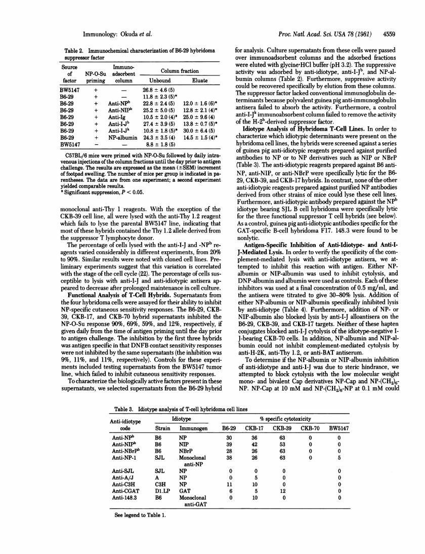
Antigen-Specific Inhibition of Anti-Idiotype- and Anti-I-J-Mediated Lysis. In order to verify the specificity of the com-plement-mediated lysis with anti-idiotype antisera, we at-tempted to inhibit this reaction with antigen. Either NP-albumin or NIP-albumin was used to inhibit cytolysis, andDNP-albumin and albumin were used as controls. Each oftheseinhibitors was used at a final concentration of 0.5 mg/ml, andthe antisera were titrated to give 30-80% lysis. Addition ofeither NP-albumin or NIP-albumin specifically inhibited lysisby anti-idiotype (Table 4). Furthermore, addition of NP- or
NIP-albumin also blocked lysis by anti-I-J alloantisera on theB6-29, CKB-39, and CKB-17 targets. Neither of these haptenconjugates blocked anti-I-J cytolysis of the idiotype-negative I-J-bearing CKB-70 cells. In addition, NP-albumin and NIP-al-bumin could not inhibit complement-mediated cytolysis byanti-H-2K, anti-Thy 1.2, or anti-BAT antiserum.
To determine if the NP-albumin or NIP-albumin inhibitionof anti-idiotype and anti-I-J was due to steric hindrance, we
attempted to block cytolysis with the low molecular weightmono- and bivalent Cap derivatives NP-Cap and NP-(CH2)6-NP. NP-Cap at 10 mM and NP-(CH2)6-NP at 0.1 mM could
Table 3. Idiotype analysis of T-cell hybridoma cell lines
Anti-idiotype Idiotype % specific cytotoxicitycode Strain Immunogen B6-29 CKB-17 CKB-39 CKB-70 BW5147
Anti-NPb B6 NP 30 36 63 0 0Anti-NIPb B6 NIP 39 42 53 0 0Anti-NBrPb B6 NBrP 28 26 63 0 0Anti-NP-1 SJL Monoclonal 38 26 63 0 5
anti-NPAnti-SJL SJL NP 0 0 0 0Anti-A/J A NP 0 5 0 0Anti-C3H C3H NP 11 10 0 0Anti-CGAT D1.LP GAT 6 5 12 0Anti-148.3 B6 Monoclonal 0 10 0 0
anti-GAT
See legend to Table 1.
Immunology: Okuda et al.
Proc. NatL Acad. Sci. USA 78 (1981)
Table 4. Antigen-specific inhibition of anti-idiotype and anti-I-Jcomplement-mediated lysis
% specific cytotoxicityCell DNP- NP- NIP-line Antiserum Albumin albumin albumin albumin
B6-29 Anti-NPb 29 29 5 0Anti-I-Jb 35 29 0 5Anti-Kb 70 65 79 70Anti-Thy 1.2 94 100 100 94
CKB-17 Anti-NP" 38 36 0 5Anti-NIPb 39 36 0 5Anti-I-Jk 63 68 12 5Anti-Kk 63 79 78 75Anti-Thy 1.2 75 79 68 63Anti-BAT 63 79 79 63
CKB-39 Anti-NP" 47 57 0 6Anti-NIP" 58 57 0 0Anti-I-Jk 89 93 5 5Anti-Kk 79 79 89 95Anti-Thy 1.2 0 0 0 0Anti-BAT 79 71 79 75
CKB-70 Anti-NP" 0 0 0 0Anti-NIPb 0 0 0 0Anti-IJk 47 53 47 37Anti-Kk 63 59 48 56Anti-Thy 1.2 63 59 59 75
Antisera were titrated in order to obtain 30-80% specific lysis. Allinhibitors were used at a final concentration of 0.5 mg/ml. See legendto Table 1.
block the lysis mediated by either antiTNPb or anti-NIPb anti-idiotypic antiserum (Table 5). Furthermore, these compoundsspecifically blocked anti-I-J lysis in the B6-29, CKB-17, andCKB-39 cell lines but failed to inhibit anti-I-J lysis of the non-functional CKB-70 hybrid or the CKB-59 cell line. Preliminaryanalyses suggest that the CKB-59 hybrid represents an effector-phase second-order suppressor T cell and presumably bears ananti-idiotypic receptor. The specificity of the inhibition wasdemonstrated by the inability of NP-Cap or NP-(CH2)6-NP toblock anti-H-2K or anti-Thy 1 lysis and the inability ofDNP-Capto block anti-idiotype- or anti-I-J-mediated lysis.
Finally, the data suggest that NP-(CH2)6-NP at 0.1 mM is abetter inhibitor of anti-idiotype and anti-I-J lysis than is a 100-fold excess ofmonovalent NP-Cap. Although this trend has beenobserved in nearly all experiments, more data are required toevaluate adequately the role of valency in suppressor T-cell-antigen interactions.
DISCUSSIONThe suppressor cell pathway resulting in the modulation of cel-lular and humoral NP responses has been well characterized.The first population of suppressor cells (Ts1 or Ts') bear I-J andidiotypic determinants and function during the induction phaseof the immune response (1, 3). The Ts, population induces asecond complementary population of suppressor cells (Ts2 orTse) which bear I-J determinants and anti-idiotypic receptors(3). The latter population functions during the effector phaseof the immune response in previously primed animals (2, 3).However, this second population does not contain the final ef-fector suppressor T cells. The available data suggest that the Ts2cells activate a third population of I-J- and idiotype-bearing sup-pressor T cells (Ts3) in the immune recipient (4, 9). The B6-29,
Table 5. Hapten inhibition of anti-I-J and anti-NPb lysis% specific cytotoxicity
NP-Cell No (CH2)rNP NP-Cap DNP-Capline Antiserum inhibitor (0.1 mM) (10 mM) (10 mM)
B6-29 Anti-NP" 37 3 6 30Anti-NIP" 48 6 3 33Anti-I-Jb 37 13 12 33Anti-Kb 74 77 82 76Anti-Thy 1.1 76 63 63 58
CKB-17 Anti-NPW 25 0 0 25Anti-NIP" 28 2 13 25Anti-14k 52 5 25 63Anti-Kk 63 55 81 63Anti-Thy 1.1 57 72 75 69
CKB-39 Anti-NP" 41 0 10 35Anti-NIP" 36 4 15 31Anti-I-Jk 55 9 19 56Anti-Kk 60 61 69 69Anti-Thy 1.1 63 63 70 72
CKB-70 Anti-I~Jk 47. 39 33 44Anti-Kk 50 67 - -
Anti-Thy 1.1 50 33 - -
CKB-59 Anti-NPb 0 0 0 0Anti-NIP" 3 3 6 0Anti-I-Jk 47 42 65 65Anti-Kk 58 63 53 53Anti-Thy 1.1 58 63 53 65
See legend to Tables 1 and 4.
CKB-17, and CKB-39 T-cell hybrids described here appear tocorrespond with the Ts, population. In support of this, we notethat (i) the cells were induced and selected by the same pro-cedures that have been shown previously to generate NP-spe-cific Ts, (1), (ii) these hybrid cells have the same surface phe-notype as the Ts, population (4), and (iii) preliminary dataindicate that the supernatants from these hybrid cells can gen-erate a Ts2 population (data not shown).The relationship of the B cell idiotypic determinants with
those on hybridoma suppressor T cell lines was compared byusing a panel of anti-idiotypic antisera. The anti-NPb, anti-NIP, and anti-NBrPb antisera have similar specificity pat-terns-i.e., each ofthese guinea pig antisera bind purified anti-NP antibodies from Ighb-bearing C57BL/6 mice and the pre-dominant idiotypic determinants detected by these reagentshave a strain distribution similar to that described for the al-lotype-linked NPb idiotype (18). Another reagent was also ca-pable of specifically lysing the Ts hybrids. Anti-idiotype againsta SJL monoclonal anti-NP hybridoma antibody which possessedsome NPb idiotypic determinants and demonstrated weak bind-ing of NPb ligand could also lyse the three functional T cell hy-brids. Because this anti-idiotypic reagent was prepared againstpurified monoclonal hybridoma antibodies, it excludes the pos-sibility that this reagent contained contaminating antibodiesagainst T cell-derived products that may have copurified withthe anti-NP antibodies obtained from conventional immuneserum.
Anti-idiotype reagents prepared against purified anti-NP oranti-NIP antibodies from C3H or A/J mice (which lack the Ighballotype) failed to lyse the NP-specific T cell hybrids. Finally,anti-idiotype prepared against purified SJL anti-NP antibodies,
4560 Immunology: Okuda et al.
Proc. Natl. Acad. Sci. USA 78 (1981) 4561
which carry the Ighb allotype but lack the NPb idiotype due toa defect in the production ofA light chain (18), also failed to lysethese hybrids. Anti-idiotype prepared against these SJL anti-bodies can bind the NP-1 hybridoma protein which possessessome of the NPb idiotypic determinants, but this reagent failedto lyse the T cell hybridoma lines. These findings suggest thatonly selected idiotypic determinants on NP-1 hybridoma anti-NP antibody are expressed on individual hybrid T cell lines.The availability of a homogeneous population ofantigen-spe-
cific suppressor T cells permitted serological analysis of the Tcell's antigen receptor. The ability of NP-albumin or NIP-al-bumin to inhibit lysis by anti-idiotype specifically verifies thespecificity of this lysis. The finding that NP-albumin or NIP-albumin could also block lysis by anti-I-J alloantisera on hybridsthat possessed both the NPb and I-J antigenic determinants in-dicated that the I-J determinant is closely associated with theantigen receptor of the T cell. Furthermore, monovalent NP-Cap and bivalent NP-(CH2)6-NP also were able to block anti-idiotype- and anti-I-J-mediated lysis. Because these compoundsare smaller than 500 daltons, the results indicate that (i) thereis a functional association of H-2 and Igh gene products on theTs cell surface, (ii) the predominant idiotypic determinants de-tected on Ts are associated with the combing site, not frame-work variable region, determinants, and (iii) I-J determinantsare intimately associated with the combining site. It is importantto note that these hapten conjugates failed to inhibit the lysisof anti-I-J antisera on the I-J-bearing but NPb-idiotype-negativeCKB-70 hybridoma. This hybridoma lacks NP-specific sup-pressor function and presumably lacks an anti-NP receptor.Furthermore, these NP conjugates could not block anti-H-2K-or anti-Thy 1-mediated lysis.
Finally, among the small number of hybrids tested, there isan apparent correlation between the expression of cell surfaceI-J and NPb determinants and the ability of factors from thesehybrids to mediate NP-specific suppression. Only one of thehybridoma factors was characterized. The antigen-binding B6-29 factor had both I-J and NPb determinants which correlateswith the phenotype of the antigen receptor on the cell fromwhich it was derived. The properties of this factor indicate thatit corresponds to a series ofsuppressor factors described in othersystems (10).
This work was supported by Grants AI-16677 from the National In-stitutes of Health and PCM 80-04573 from the National ScienceFoundation.
1. Weinberger, J. Z., Germain, R. N., Ju, S.-T., Greene, M. I.,Benacerraf, B. & Dorf, M. E. (1979)J. Exp. Med. 150, 761-776.
2. Weinberger, J. Z., Benacerraf, B. & Dorf, M. E. (1980)J. Exp.Med. 151, 1413-1423.
3. Weinberger, J. Z., Germain, R. N., Benacerraf, B. & Dorf, M.E. (1980) J. Exp. Med. 152, 161-169.
4. Sunday, M. E., Benacerraf, B. & Dorf, M. E. (1981)J. Exp. Med.153, 811-822.
5. Suzuki, G., Kumagai, Y., Shiratori, Y., Karasuyama, H., Kita-hara, T., Abe, R., Hayakawa, K., Okumura, K. & Tada, T. (1980)Proc. Jpn. Soc. Immunot 10, 121-122.
6. Tada, T. & Okumura, K. (1980) Adv. ImmunoL 28, 1.7. Eardley, D. D., Shen, F. W., Cantor, H. & Gershon, R. K.
(1979) J. Exp. Med. 150, 44.8. Dohi, Y. & Nisonoff, A. (1979) J. Exp. Med. 150, 909.9. Sy, M.-S., Miller, S. D., Moorhead, J. W. & Claman, H. N.
(1979) J. Exp. Med. 149, 1197-1207.10. Germain, R. N. & Benacerraf, B. (1981) Scand. J. Immunot 13,
1-10.11. Kohler, G. & Milstein, C. (1975) Nature (London), 256, 495-497.12. Taniguchi, M., Saito, T. & Tada, T. (1979) Nature (London) 278,
555-558.13. Kapp, J. A., Araneo, B. A. & Clevinger, B. L. (1980)J. Exp. Med.
152, 235-240.14. Taniguchi, M., Takei, I. & Tada, T. (1980) Nature (London) 283,
227.15. Weinberger, J. Z., Greene, M. I., Benacerraf, B. & Dorf, M. E.
(1979)1. Exp. Med. 149, 1336-1348.16. Perry, L. L., Dorf, M. E., Benacerraf, B. & Greene, M. I. (1979)
Proc. Nati Acad. Sci. USA 76, 920-924.17. Golub, E. S. (1971) CelL ImmunoL 2, 353.18. Makela, 0. & Karjalainen, K. (1977) ImmunoL Rev. 34, 119-138.19. Ju, S.-T., Pierres, M., Waltenbaugh, C., Germain, R. N., Ben-
acerraf, B. & Dorf, M. E. (1979) Proc. Nat. Acad. Sci. USA 76,2942-2946.
20. Dorf, M. E., Eguro, S. Y., Cabrera, G., Yunis, E. J., Swanson,J. & Amos, D. B. (1972) Vox Sang. 22, 447-456.
21. Sunday, M. E., Weinberger, J. Z., Benacerraf, B. & Dorf, M. E.(1980)J. ImmunoL 125, 1601-1605.
22. Kanno, M., Takei, I., Suzuki, N., Tomioka, H. & Taniguchi, M.(1980) Proc. Jpn. Soc. ImmunoL 10, 41-42.
Immunology: Okuda et al.