Embed Size (px)
Citation preview
Ophthalmic Procedures Assessment*
Functional Indications for Upper and Lower Eyelid Blepharoplasty
American Academy of Ophthalmology
*The purpose of the Committee on Ophthalmic Procedures Assessment is to evaluate on a scientific basis new and existing ophthalmic tests, devices, and procedures for their safety, efficacy, clinical effectiveness, and appropriate uses. Evaluations include examination of available literature, epidemiological analyses when appropriate, and compilation of opinions from recognized experts and other interested parties. After appropriate review by all contributors, including legal counsel, assessments are submitted to the Academy's Board of Trustees for consideration as official Academy policy.
"Blepharoplasty" is defined by Stedman's Medical Dictionary as "any operation for the restoration of a defect in the eyelid." 1 Under this broad description, blepharoplasty includes procedures to repair ptosis, eyelid retraction, entropion, ectropion, trichiasis, or defects following excision of tumors. In common usage, however, the term "blepharoplasty" refers to an operation in which redundant tissues (skin, muscle, or fat) are excised from an eyelid.
A blepharoplasty may be performed for either functional or cosmetic purposes. The goal of functional or reconstructive surgery is to restore normalcy to a structure that has been altered by trauma, infection, inflammation, degeneration, neoplasia, or developmental errors. Cosmetic surgery attempts to improve the appearance of tissue or structures that are histologically and functionally normal. Although many blepharoplasty operations in the United States are performed to enhance cosmesis, there are numerous functional indications for the procedure.
The most common functional indication for blepharoplasty is a superior visual field defect secondary to redundant upper eyelid tissue (dermatochalasis) that overhangs the eyelid margin. This condition is one cause of "pseudoptosis" as described by Beard2; the upper eyelids may be abnormally low because of the mechanical weight
Prepared by the Committee on Ophthalmic Procedures Assessment and revised and approved by the Academy's Board of Trustees, September 24, 1994.
of the excess upper eyelid tissues, but also there may be concomitant levator aponeurosis dehiscence or disinsertion.3 "Myogenic" (including aponeurotic) or neurologic factors usually are found in true blepharoptosis.2 Because dermatochalasis and true ptosis often co-exist, blepharoplasty at the time of ptosis repair often is indicated. 2·4-9
Failure to excise redundant tissues at the time of ptosis repair may result in accentuating the overhanging of skin and muscle at the eyelid margin.4- 6 Excision of redundant skin and muscle often is performed concurrently with ptosis repair utilizing a frontalis sling technique, 5 internal approach for tarsectomy or Muellerectomy, 6 external tarsectomy/ or levator aponeurosis advancement.8•9 In the latter instances, where the coexisting problems of dermatochalasis and ptosis are corrected using an aponeurotic approach, the surgeon may excise redundant skin and/or orbicularis muscle at the time of initial skin incision8 or toward the end of the operation.9 Additionally, brow ptosis may co-exist with dermatochalasis, necessitating concomitant browplasty and blepharoplasty in some instances.10
Visual field loss from malpositioned upper eyelids was confirmed by Cahill and associates11 and quantitated by Meyer, Linberg, and co~workers. 12 Impairment of the superior visual field ranged from 20% for mild ptosis to 64% in advanced cases where the eyelid crosses the middle of the pupil. Excessive upper eyelid skin also may produce asthenopic symptoms, persistent blepharoconjunctivitis ("functional dermatochalasia")13 and dry eye symptoms. 14
693
Ophthalmology Volume 102, Number 4, April1995
Blepharoplasty may be indicated to treat the sequelae of several inflammatory disorders of the orbit or eyelids. For example, a common feature of Graves' ophthalmopathy is edema and fullness of the eyelids with anterior prolapse of orbital fat and lacrimal gland tissue. Blepharoplasty may be included in the surgical rehabilitation of these patients, and often can be performed concomitantly with repair of eyelid retraction. 15 Blepharochalasis (not to be confused with dermatochalasis) is an unusual disease affecting primarily young persons in which recurrent episodes of idiopathic eyelid edema result in stretching and redundancy of the eyelid tissues; blepharoplasty, usually in combination with advancement of the levator aponeurosis, may be indicated. 16•17 Floppy eyelid syndrome, another unusual disorder that may be related to blepharochalasis18 and which causes a chronic papillary conjunctivitis, 19•20 is treated effectively with surgery that may include blepharoplasty.21 •22
Trauma to the eyelids and orbit may result in the need for a functional blepharoplasty. Blowout fractures of the orbit commonly lead to loss or atrophy of orbital fat, resulting in enophthalmos and a deep superior palpebral sulcus. Putterman23 has emphasized that it is possible to camouflage the orbital asymmetry by performing an upper eyelid blepharoplasty on the contralateral side in lieu of more extensive surgery on the traumatized orbit. An additional traumatic setting in which blepharoplasty may be useful is when skin grafts are required to replace or cover avulsed or burned eyelid tissue; subsequent trimming of the healed grafts (in effect, a blepharoplasty) may be required.
There are at least two situations in which a functional lower eyelid blepharoplasty is indicated. One is middleaged or elderly patients with massive lower eyelid edema that may be secondary to systemic corticosteroid therapy, myxedema, Graves' disease, nephrotic syndrome, or a number of other metabolic or inflammatory disorders. The excessive eyelid bulk, even after satisfactory treatment
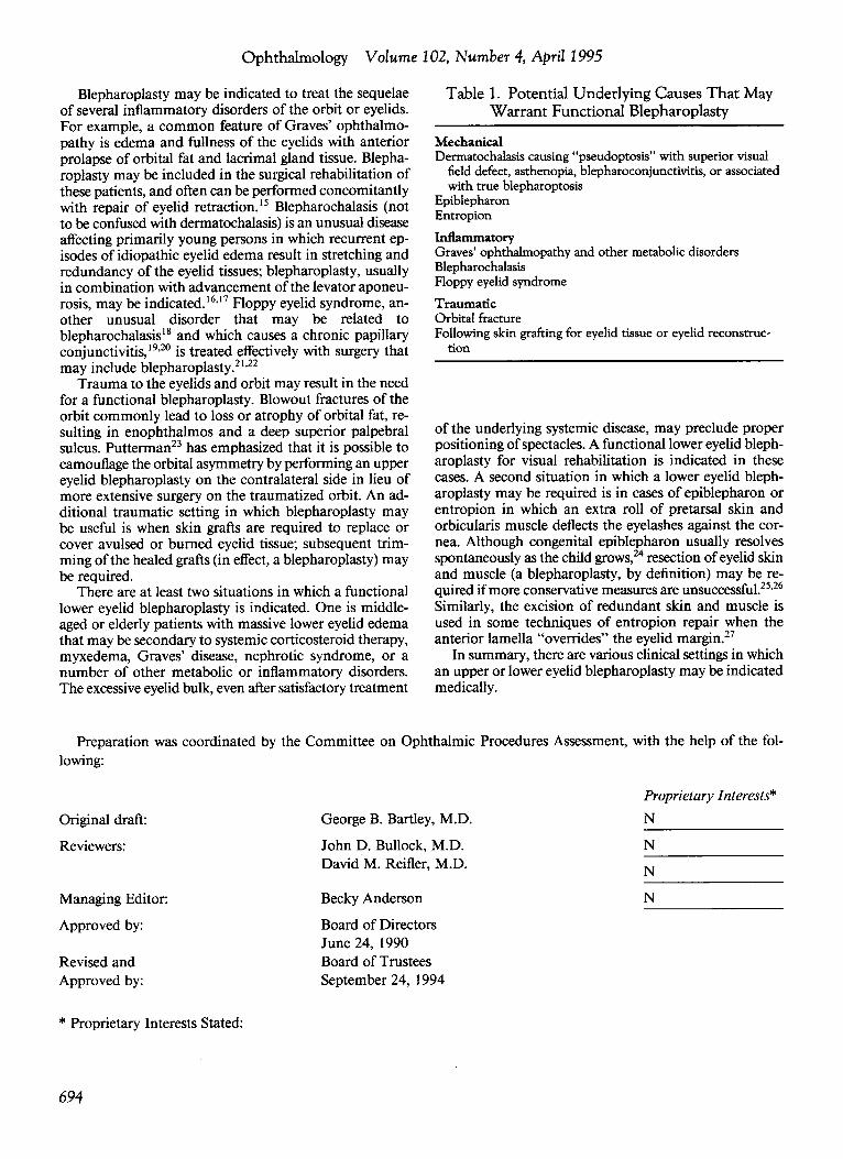
Table 1. Potential Underlying Causes That May Warrant Functional Blepharoplasty
Mechanical Dermatochalasis causing "pseudoptosis" with superior visual
field defect, asthenopia, blepharoconjunctivitis, or associated with true blepharoptosis
Epiblepharon Entropion
Inflammatory Graves' ophthalmopathy and other metabolic disorders Blepharochalasis Floppy eyelid syndrome
Traumatic Orbital fracture Following skin grafting for eyelid tissue or eyelid reconstruc
tion
of the underlying systemic disease, may preclude proper positioning of spectacles. A functional lower eyelid blepharoplasty for visual rehabilitation is indicated in these cases. A second situation in which a lower eyelid blepharoplasty may be required is in cases of epiblepharon or entropion in which an extra roll of pretarsal skin and orbicularis muscle deflects the eyelashes against the cornea. Although congenital epiblepharon usually resolves spontaneously as the child grows, 24 resection of eyelid skin and muscle (a blepharoplasty, by definition) may be required if more conservative measures are unsuccessful.25•26
Similarly, the excision of redundant skin and muscle is used in some techniques of entropion repair when the anterior lamella "overrides" the eyelid margin.27
In summary, there are various clinical settings in which an upper or lower eyelid blepharoplasty may be indicated medically.
Preparation was coordinated by the Committee on Ophthalmic Procedures Assessment, with the help of the following:
Original draft:
Reviewers:
Managing Editor:
Approved by:
Revised and Approved by:
* Proprietary Interests Stated:
694
George B. Bartley, M.D.
John D. Bullock, M.D. David M. Reifler, M.D.
Becky Anderson
Board of Directors June 24, 1990 Board of Trustees September 24, 1994
Proprietary Interests*
N
N
N
N
AAO · Blepharoplasty
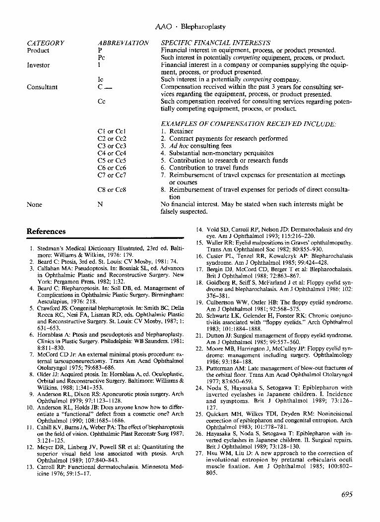
CATEGORY Product
ABBREVIATION p
SPECIFIC FINANCIAL INTERESTS
Pc Investor I
Financial interest in equipment, process, or product presented. Such interest in potentially competing equipment, process, or product. Financial interest in a company or companies supplying the equipment, process, or product presented.
Ic c_
Such interest in a potentially competing company. Consultant Compensation received within the past 3 years for consulting ser
vices regarding the equipment, process, or product presented. Cc Such compensation received for consulting services regarding poten
tially competing equipment, process, or product.
EXAMPLES OF COMPENSATION RECEIVED INCLUDE: 1. Retainer Cl or Ccl
C2 or Cc2 C3 or Cc3 C4 or Cc4 C5 or Cc5 C6 or Cc6 C7 or Cc7
2. Contract payments for research performed 3. Ad hoc consulting fees 4. Substantial non-monetary perquisites 5. Contribution to research or research funds 6. Contribution to travel funds 7. Reimbursement oftravel expenses for presentation at meetings
or courses C8 or Cc8 8. Reimbursement of travel expenses for periods of direct consulta
tion None N No financial interest. May be stated when such interests might be
falsely suspected.
References
1. Stedman's Medical Dictionary Illustrated, 23rd ed. Baltimore: Williams & Wilkins, 1976: 179.
2. Beard C: Ptosis, 3rd ed. St. Louis: CV Mosby, 1981: 74. 3. Callahan MA: Pseudoptosis. In: Bosniak SL, ed. Advances
in Ophthalmic Plastic and Reconstructive Surgery. New York: Pergamon Press, 1982; 1 :32.
4. Beard C: Blepharoptosis. In: Soli DB, ed. Management of Complications in Ophthalmic Plastic Surgery. Birmingham: Aesculapius, 1976: 218.
5. Crawford JS: Congenital blepharoptosis. In: Smith BC, Della Rocca RC, Nesi FA, Lisman RD, eds. Ophthalmic Plastic and Reconstructive Surgery. St. Louis: CV Mosby, 1987; 1: 631-653.
6. Hornblass A: Ptosis and pseudoptosis and blepharoplasty. Clinics in Plastic Surgery. Philadelphia: WB Saunders, 1981: 8:811-830.
7. McCord CD Jr: An external minimal ptosis procedure: external tarsoaponeurectomy. Trans Am Acad Ophthalmol Otolaryngol 1975; 79:683-686.
8. Older JJ: Acquired ptosis. In: Hornblass A, ed. Oculoplastic, Orbital and Reconstructive Surgery. Baltimore: Williams & Wilkins, 1988; 1:341-353.
9. Anderson RL, Dixon RS: Aponeurotic ptosis surgery. Arch Ophthalmol 1979; 97:1123-1128.
10. Anderson RL, Holds JB: Does anyone know how to differentiate a "functional" defect from a cosmetic one? Arch Ophthalmol 1990; 108:1685-1686.
11. Cahill KV, Burns JA. Weber PA: The effect ofblepharoptosis on the field of vision. Ophthalmic Plast Reconstr Surg 1987; 3:121-125.
12. Meyer DR, Linberg JV, Powell SR et al: Quantitating the superior visual field loss associated with ptosis. Arch Ophthalmol 1989; 107:840-843.
13. Carroll RP: Functional dermatochalasia. Minnesota Medicine 1976; 59:15-17.
14. Void SD, Carroll RP, Nelson JD: Dermatochalasis and dry eye. Am J Ophthalmol 1993; 115:216-220.
15. Waller RR: Eyelid malpositions in Graves' ophthalmopathy. Trans Am Ophthalmol Soc 1982; 80:855-930.
16. Custer PL, Tenzel RR, Kowalczyk AP: Blepharochalasis syndrome. Am J Ophthalmol 1985; 99:424-428.
17. Bergin DJ, McCord CD, Berger T et al: Blepharochalasis. Brit J Ophtha1mol 1988; 72:863-867.
18. Goldberg R, Seiff S, McFarland J et al: Aoppy eyelid syndrome and blepharochalasis. Am J Ophthalmol 1986; 102: 376-381.
19. Culbertson WW, Ostler HB: The floppy eyelid syndrome. Am J Ophthalmol 1981; 92:568-575.
20. Schwartz LK, Ge1ender H, Forster RK: Chronic conjunctivitis associated with "floppy eyelids." Arch Ophthalmol 1983; 101:1884-1888.
21. Dutton JJ: Surgical management of floppy eyelid syndrome. Am J Ophthalmo1 1985; 99:557-560.
22. Moore MB, Harrington J, McCulley JP: Aoppy eyelid syndrome: management including surgery. Ophthalmology 1986; 93:184-188.
23. Putterman AM: Late management of blow-out fractures of the orbital floor. Trans Am Acad Ophtha1mol Otolaryngol 1977; 83:650-659.
24. Noda S, Hayasaka S, Setogawa T: Epiblepharon with inverted eyelashes in Japanese children. I. Incidence and symptoms. Brit J Ophthalmol 1989; 73:126-127.
25. Quickert MH, Wilkes TDI, Dryden RM: Nonincisional correction of epiblepharon and congenital entropion. Arch Ophthalmol 1983; 101:778-781.
26. Hayasaka S, Noda S, Setogawa T: Epiblepharon with inverted eyelashes in Japanese children. II. Surgical repairs. Brit J Ophthalmol 1989; 73:128-130.
27. Hsu WM, Liu D: A new approach to the correction of involutional entropion by pretarsal orbicularis oculi muscle fixation. Am J Ophthalmol 1985; 100:802-805.
695