Embed Size (px)
Citation preview

The PDF of the article you requested follows this cover page.
This is an enhanced PDF from The Journal of Bone and Joint Surgery
2005;87:9-21. doi:10.2106/JBJS.E.00628 J Bone Joint Surg Am.Anil Bhave, Michael Mont, Scott Tennis, Michele Nickey, Roland Starr and Gracia Etienne
Joint ArthroplastyFunctional Problems and Treatment Solutions After Total Hip and Knee
This information is current as of March 17, 2008
Reprints and Permissions
Permissions] link. and click on the [Reprints andjbjs.orgarticle, or locate the article citation on
to use material from thisorder reprints or request permissionClick here to
Publisher Information
www.jbjs.org20 Pickering Street, Needham, MA 02492-3157The Journal of Bone and Joint Surgery
COPYRIGHT © 2005 BY THE JOURNAL OF BONE AND JOINT SURGERY, INCORPORATED
9
Functional Problems and Treatment Solutions After Total Hip and
Knee Joint ArthroplastyBY ANIL BHAVE, PT, MICHAEL MONT, MD, SCOTT TENNIS, PT,
MICHELE NICKEY, PT, ROLAND STARR, MS, AND GRACIA ETIENNE, MD
Introductionlthough most patients who undergo total hip or kneejoint arthroplasty have an excellent clinical result withroutine postoperative interventions, substantial dys-
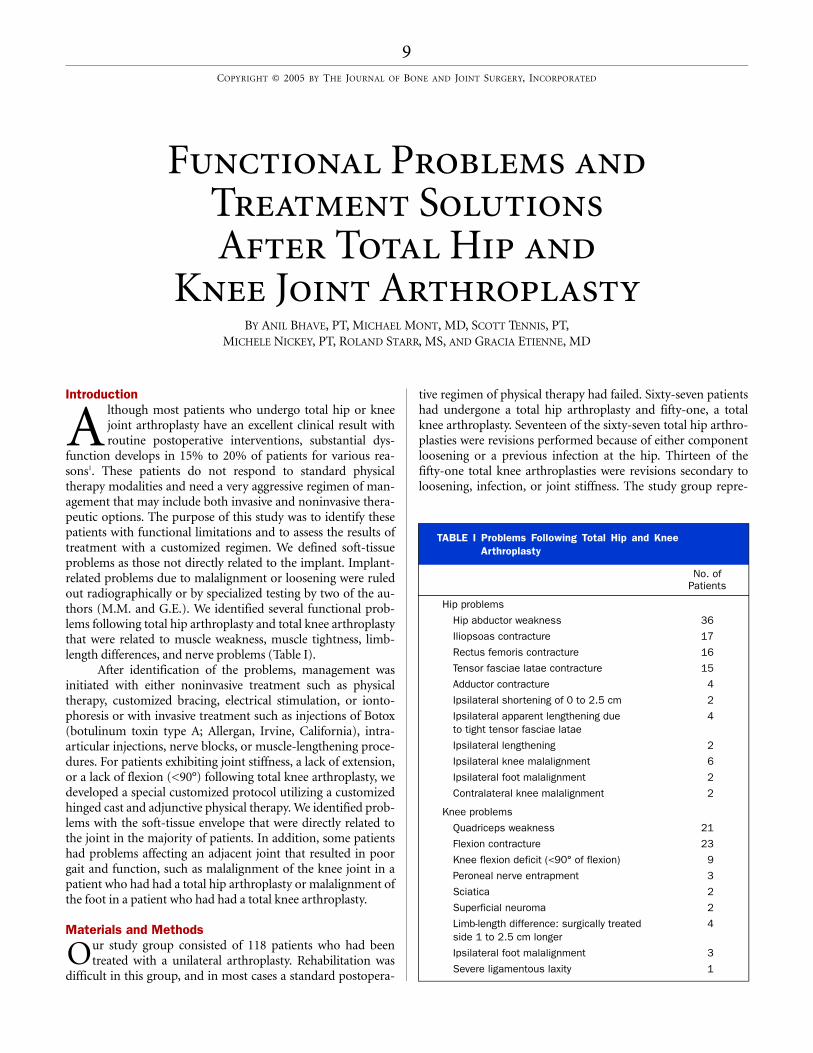
function develops in 15% to 20% of patients for various rea-sons1. These patients do not respond to standard physicaltherapy modalities and need a very aggressive regimen of man-agement that may include both invasive and noninvasive thera-peutic options. The purpose of this study was to identify thesepatients with functional limitations and to assess the results oftreatment with a customized regimen. We defined soft-tissueproblems as those not directly related to the implant. Implant-related problems due to malalignment or loosening were ruledout radiographically or by specialized testing by two of the au-thors (M.M. and G.E.). We identified several functional prob-lems following total hip arthroplasty and total knee arthroplastythat were related to muscle weakness, muscle tightness, limb-length differences, and nerve problems (Table I).
After identification of the problems, management wasinitiated with either noninvasive treatment such as physicaltherapy, customized bracing, electrical stimulation, or ionto-phoresis or with invasive treatment such as injections of Botox(botulinum toxin type A; Allergan, Irvine, California), intra-articular injections, nerve blocks, or muscle-lengthening proce-dures. For patients exhibiting joint stiffness, a lack of extension,or a lack of flexion (<90°) following total knee arthroplasty, wedeveloped a special customized protocol utilizing a customizedhinged cast and adjunctive physical therapy. We identified prob-lems with the soft-tissue envelope that were directly related tothe joint in the majority of patients. In addition, some patientshad problems affecting an adjacent joint that resulted in poorgait and function, such as malalignment of the knee joint in apatient who had had a total hip arthroplasty or malalignment ofthe foot in a patient who had had a total knee arthroplasty.
Materials and Methodsur study group consisted of 118 patients who had beentreated with a unilateral arthroplasty. Rehabilitation was
difficult in this group, and in most cases a standard postopera-
tive regimen of physical therapy had failed. Sixty-seven patientshad undergone a total hip arthroplasty and fifty-one, a totalknee arthroplasty. Seventeen of the sixty-seven total hip arthro-plasties were revisions performed because of either componentloosening or a previous infection at the hip. Thirteen of thefifty-one total knee arthroplasties were revisions secondary toloosening, infection, or joint stiffness. The study group repre-
A
O
TABLE I Problems Following Total Hip and Knee Arthroplasty
No. of Patients
Hip problems
Hip abductor weakness 36
Iliopsoas contracture 17
Rectus femoris contracture 16
Tensor fasciae latae contracture 15
Adductor contracture 4
Ipsilateral shortening of 0 to 2.5 cm 2
Ipsilateral apparent lengthening due to tight tensor fasciae latae
4
Ipsilateral lengthening 2
Ipsilateral knee malalignment 6
Ipsilateral foot malalignment 2
Contralateral knee malalignment 2
Knee problems
Quadriceps weakness 21
Flexion contracture 23
Knee flexion deficit (<90° of flexion) 9
Peroneal nerve entrapment 3
Sciatica 2
Superficial neuroma 2
Limb-length difference: surgically treated side 1 to 2.5 cm longer
4
Ipsilateral foot malalignment 3
Severe ligamentous laxity 1
10
TH E JO U R NA L OF BONE & JOINT SURGER Y · JBJS .ORG
VO LU M E 87-A · SUPPLEMENT 2 · 2005FU N C T I O N A L PROBL EM S AND TREA T M ENT SOLUTIONS AFTER TO T A L HIP A N D KN E E JOI NT AR TH ROPLA ST Y
sents about 5% of the patient population at our center. Therewere fifty men and sixty-eight women, and the age range wasforty-seven to seventy-two years (mean age, 65.3 years).
Patients were screened carefully for radiographic evi-dence of loosening. Additional assessment included a detailedphysical examination, isokinetic strength testing, videotapedgait analysis, assessment of balance, and, for the few patientswith malalignment, a three-dimensional gait study. Patientswith neuropathic pain underwent sensory nerve testing andelectromyography, and superficial nerve blocks were used toidentify specific etiologies in some patients. On the basis ofthese detailed examinations, we identified certain functionalproblems associated with total hip arthroplasty and total kneearthroplasty. Functional limitations due to range-of-motiondeficits in the patients who had had a total hip arthroplasty weremost commonly identified around two to 2.5 months after thesurgery, usually during the second or third follow-up visit.Problems related to joint stiffness in the patients who had had atotal knee arthroplasty were typically detected at six to nineweeks postoperatively and also coincided with the second orthird follow-up visit. All of the other functional limitations weredetected late in the follow-up period. Problems related to mala-lignment were usually discovered much later than the otherfunctional deficits (approximately one year after the surgery).
We questioned all patients regarding their ability to par-ticipate in light recreational sports and whether they had beenactive prior to the surgery. We also posed specific questionsregarding their activities of daily living, including stair-climbing, showering, dressing and undressing, and walkingvarious distances, as well as other relevant activities, includ-ing sexual relations.
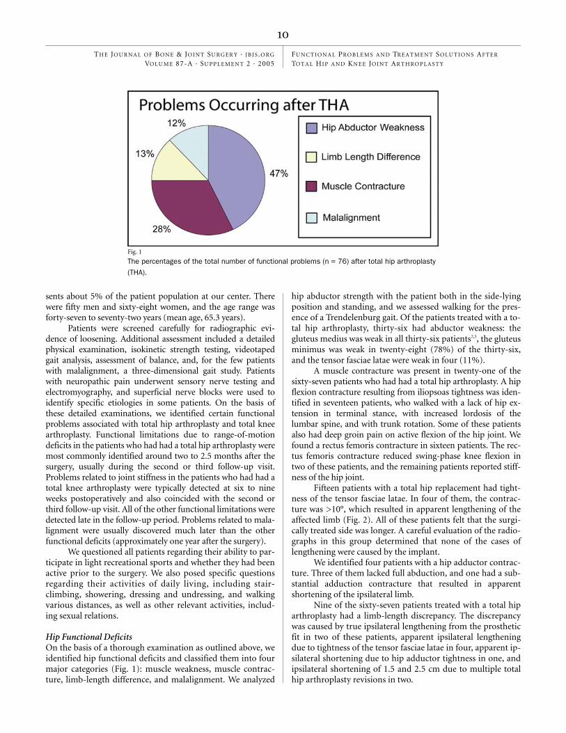
Hip Functional DeficitsOn the basis of a thorough examination as outlined above, weidentified hip functional deficits and classified them into fourmajor categories (Fig. 1): muscle weakness, muscle contrac-ture, limb-length difference, and malalignment. We analyzed
hip abductor strength with the patient both in the side-lyingposition and standing, and we assessed walking for the pres-ence of a Trendelenburg gait. Of the patients treated with a to-tal hip arthroplasty, thirty-six had abductor weakness: thegluteus medius was weak in all thirty-six patients2,3, the gluteusminimus was weak in twenty-eight (78%) of the thirty-six,and the tensor fasciae latae were weak in four (11%).
A muscle contracture was present in twenty-one of thesixty-seven patients who had had a total hip arthroplasty. A hipflexion contracture resulting from iliopsoas tightness was iden-tified in seventeen patients, who walked with a lack of hip ex-tension in terminal stance, with increased lordosis of thelumbar spine, and with trunk rotation. Some of these patientsalso had deep groin pain on active flexion of the hip joint. Wefound a rectus femoris contracture in sixteen patients. The rec-tus femoris contracture reduced swing-phase knee flexion intwo of these patients, and the remaining patients reported stiff-ness of the hip joint.
Fifteen patients with a total hip replacement had tight-ness of the tensor fasciae latae. In four of them, the contrac-ture was >10°, which resulted in apparent lengthening of theaffected limb (Fig. 2). All of these patients felt that the surgi-cally treated side was longer. A careful evaluation of the radio-graphs in this group determined that none of the cases oflengthening were caused by the implant.
We identified four patients with a hip adductor contrac-ture. Three of them lacked full abduction, and one had a sub-stantial adduction contracture that resulted in apparentshortening of the ipsilateral limb.
Nine of the sixty-seven patients treated with a total hiparthroplasty had a limb-length discrepancy. The discrepancywas caused by true ipsilateral lengthening from the prostheticfit in two of these patients, apparent ipsilateral lengtheningdue to tightness of the tensor fasciae latae in four, apparent ip-silateral shortening due to hip adductor tightness in one, andipsilateral shortening of 1.5 and 2.5 cm due to multiple totalhip arthroplasty revisions in two.
Fig. 1
The percentages of the total number of functional problems (n = 76) after total hip arthroplasty
(THA).

11
TH E JO U R NA L OF BONE & JOINT SURGER Y · JBJS .ORG
VO LU M E 87-A · SUPPLEMENT 2 · 2005FU N C T I O N A L PROBL EM S AND TREA T M ENT SOLUTIONS AFTER TO T A L HIP A N D KN E E JOI NT AR TH ROPLA ST Y
Ten of the sixty-seven patients had malalignment, not ofthe prosthesis but of a more distal joint. Five patients hadmalalignment of the ipsilateral knee joint due to a genu valgusdeformity, and one had ipsilateral genu varum with a flexioncontracture that resulted in gait and functional abnormalities.Four patients in this group did not have symptoms in the kneeuntil after they underwent the total hip arthroplasty, eventhough the knee deformity had been long-standing. Thus, thebiomechanical abnormality may have been unmasked by the
total hip arthroplasty. Two patients had malalignment of thecontralateral knee; both had a substantial genu varum defor-mity that resulted in increased knee pain with walking. Twopatients had an ipsilateral planovalgus abnormality of thefoot. These two patients had constant foot pain and a gait dis-turbance related to the collapsed longitudinal arch.
Knee Functional DeficitsThe knee functional deficits were broadly divided into six
Fig. 2
Anteroposterior standing radiograph showing an apparent limb-length difference caused by an
abduction contracture. Note that the line drawn from the ischial tuberosity passes close to the
lesser trochanter on both sides, indicating that there is no true difference caused by the implant.
Fig. 3
The percentages of the total number of functional problems (n = 68) after total knee arthro-
plasty (TKA).

12
TH E JO U R NA L OF BONE & JOINT SURGER Y · JBJS .ORG
VO LU M E 87-A · SUPPLEMENT 2 · 2005FU N C T I O N A L PROBL EM S AND TREA T M ENT SOLUTIONS AFTER TO T A L HIP A N D KN E E JOI NT AR TH ROPLA ST Y
categories: weakness, peroneal nerve symptoms, flexion con-tracture, limb-length difference, knee flexion deficit, and mal-alignment (Fig. 3). Twenty-one of the fifty-one patients whohad had a total knee arthroplasty had a clinically relevantquadriceps strength deficit as demonstrated by an isokinetictest with a Biodex dynamometer (Biodex Medical Systems,Shirley, New York). Quadriceps weakness was defined as anactive extension lag exceeding 15° in the early postoperativephase or <50% of the strength of the contralateral limb asshown by isokinetic testing in the later stages of recovery.
There was a high rate of knee flexion contracture (definedas a lack of extension of ≥10°), which was identified in twenty-three of the fifty-one patients who had had a total knee arthro-plasty (Fig. 4). The common causes of the knee flexion contrac-tures were adaptive muscle-shortening, exuberant scar-tissueadhesions, quadriceps inhibition and hamstring overactivity,unrecognized tightness of the gastrocnemius, limb-length dif-ference, and peroneal nerve entrapment. A knee flexion deficit(defined as knee flexion of <90°) was present in nine of the fifty-one patients. The common causes of the knee flexion deficitswere joint effusion, abnormal pain response, quadriceps scar-ring, tightness of the patellar tendon, tightness of the rectusfemoris, and tightness of the iliotibial band.
We identified several nerve problems related to peronealnerve entrapment as well as neuromas of the sciatic and su-perficial nerves in this group. Three patients had peronealnerve entrapment that produced burning pain down to thedorsum of the foot, paresthesias of the foot, increased footpain, mild weakness of the extensor hallucis longus, and apositive Tinel sign (in one case). Two patients had a history ofsciatica that resulted in increased peroneal nerve symptomspostoperatively. In addition, two patients had a superficialneuroma involving the saphenous nerve, with symptoms thatresolved with nerve blocks.
Four of the fifty-one patients who had had a total knee ar-throplasty demonstrated a limb-length difference, with the sideof the arthroplasty longer than the contralateral side. These dis-crepancies resulted in a flexed knee posture and a resultant flex-
ion contracture. We determined that these flexion contractureshad developed as compensation for the limb-length difference.Two of the four patients who had a limb-length difference afterthe unilateral total knee arthroplasty had preoperative bilateralgenu varum deformity. The discrepancy occurred in those twopatients when the surgical side gained length as a result of thecorrection of the varus deformity.
There was malalignment on the side of the total kneearthroplasty in four patients, three of whom had substantialplanovalgus deformity of the foot. Two of the three had a dys-functional posterior tibial tendon with hindfoot valgus and apronated hindfoot, and one had a tarsal coalition and a plano-valgus foot deformity. The fourth patient had substantial liga-mentous laxity of the knee in both flexion and extension.This patient was dissatisfied with the result and went on tohave a revision total knee arthroplasty to improve the stabil-ity of the joint.
Correlation of Functional Impairment with Symptoms and Physical FindingsWe attempted to correlate the physical impairments with thesymptoms in the patients in our study. In the populationtreated with a total hip arthroplasty, we identified four majorimpairments: hip flexion contracture, hip abduction contrac-ture, hip abductor weakness, and a true or functional limb-length difference (Table II). The major symptoms in thepatients who exhibited a contracture included anterior hip painor groin pain, abnormal gait, and low back pain. These im-pairments were found in most of the patients. Many patients,especially younger ones, reported that their sexual relationshad been altered. The main symptoms in the patients with hipabductor weakness were increased energy consumption dur-ing gait, as suggested by the patient tiring too easily, and anabnormal appearance. The weakness also resulted in an inabil-ity to participate in light sports or recreational activities suchas golf, tennis, dancing, or gardening. The main problems as-sociated with a limb-length difference were low back pain andabnormal gait.
Fig. 4
A 25° knee flexion contracture seen five weeks following a primary total knee arthroplasty in a
sixty-nine-year-old woman. The contracture was due to joint swelling, capsular contracture, and
previously unrecognized tightness of the gastrocnemius.

13
TH E JO U R NA L OF BONE & JOINT SURGER Y · JBJS .ORG
VO LU M E 87-A · SUPPLEMENT 2 · 2005FU N C T I O N A L PROBL EM S AND TREA T M ENT SOLUTIONS AFTER TO T A L HIP A N D KN E E JOI NT AR TH ROPLA ST Y
In the population with a total knee arthroplasty, weidentified five impairments: knee flexion contracture, kneeflexion deficit, quadriceps weakness, peroneal nerve entrap-ment syndrome or nerve symptoms, and malalignment (Ta-ble III). Knee flexion contractures frequently resulted inanterior knee pain or retropatellar pain with quadriceps fa-tigue pain, back pain, limping gait, difficulty walking longdistances, or the inability to participate in light sports. A flex-ion deficit did not result in a clinically relevant gait abnor-mality but did result in difficulty sitting in a chair or risingfrom a seated position, difficulty ascending or descendingstairs, inability to use a bicycle for recreation, and difficultywith sexual relations. Patients with quadriceps weakness hadquadriceps fatigue pain, buckling, or a giving-way sensation,especially those who demonstrated a quadriceps lag of >15°when they were tested in a seated or straight-leg-raise posi-tion. These patients lacked quadriceps control at the initialstance phase of the gait cycle and experienced some instabil-ity with a giving-way sensation. Quadriceps weakness also re-sulted in difficulty with walking on uneven ground and onramps, difficulty climbing stairs and walking long distances,and the inability to participate in sports.
Peroneal nerve symptoms caused difficulty with sleep-ing at night, radiating pain to the dorsum of the foot, and dif-ficulty walking long distances, especially at heel-strike duringthe gait cycle, with tripping or stumbling occurring in severecases. Malalignment usually resulted in knee pain and laxitywith buckling at the knee. Patients also needed to use braces,which some found cumbersome.
Treatment of Problems Following Total Hip ArthroplastyMuscle Contractures
n our study, we identified hip flexion contractures due totightness of the iliopsoas, rectus femoris, sartorius, adduc-
tors, and sometimes the tensor fasciae latae. Our initial strat-egy for treatment of hip flexion contractures involved manualtherapy. This included an aggressive customized stretchingprotocol with at least seven to ten stretches of each affectedmuscle during each physical therapy session, at a frequency offour or five times a week for the first two to three weeks fol-lowed by a frequency of three times a week (Fig. 5). All pa-tients were also provided with a home exercise regimen, whichthey performed either by themselves or with assistance fromfamily members. In addition to aggressive physical therapy tostretch the hip flexor contracture, we injected 2 mL of triamci-nolone with 3 mL of 1% lidocaine into the iliopsoas tendonwith image-intensifer-guided assistance (Fig. 6). Tendinitiswas diagnosed when resisted hip flexion was found to be pain-ful in a seated position. Stretching was initiated forty-eighthours after the injection.
Of our four patients with adductor tightness, one re-sponded well to standard manual therapy, one underwent ad-ductor lengthening surgery, one received a Botox injectioninto the adductor muscle, and one had resolution of the ad-ductor spasm after a revision total hip arthroplasty. Of the fif-teen tensor fasciae latae contractures, four resulted in anapparent limb-length difference, with the tight side beinglonger by 1 to 2.5 cm. The patients with this complication re-
I
TABLE II Symptoms and Physical Findings Related to Problems Following Total Hip Arthroplasty
Impairment Symptoms Physical Findings
Hip flexion contracture Deep anterior hip pain, abnormal gait, diffi-culty with sexual relations, low back pain
Iliopsoas contracture, rectus femoris contracture, sartorius and tensor fasciae latae contracture, decreased step length and increased pelvic rota-tion during gait
Hip abduction contracture Lateral hip pain, limb-length difference, ab-normal gait, back pain, unable to participate in light sports or recreational activity*
Tensor fasciae latae tightness, apparent limb-length difference, lateral trunk-lean during gait, trochanteric bursitis
Hip abductor weakness Lateral hip pain, abnormal gait, need to use cane, back pain, hip-muscle fatigue pain, diffi-culty walking long distances, unable to partici-pate in light sports or recreational activity*
Weakness of gluteus medius and minimus, posi-tive Trendelenburg sign and gait, trochanteric bur-sitis, poor balance, lateral trunk-lean during gait
True limb-length difference (shortening on the surgically treated side)
Back pain, abnormal gait Trunk lean on surgically treated side during gait, contralateral knee flexion and ipsilateral equinus posture, increased patellofemoral stress and pain on contralateral side, tightness of contralateral quadratus lumborum
Apparent limb-length difference Back pain, abnormal gait, unable to partici-pate in light sports or recreational activity*
Pseudo-Trendelenburg gait, ipsilateral tensor fasciae latae contracture, contralateral adduc-tion contracture, contralateral hip or knee flexion contracture
*Golf, doubles tennis, light dancing, gardening, etc.

14
TH E JO U R NA L OF BONE & JOINT SURGER Y · JBJS .ORG
VO LU M E 87-A · SUPPLEMENT 2 · 2005FU N C T I O N A L PROBL EM S AND TREA T M ENT SOLUTIONS AFTER TO T A L HIP A N D KN E E JOI NT AR TH ROPLA ST Y
sponded well to a specific tensor fasciae latae stretching regi-men as shown in Figure 7. Stabilizing the hip with the patientin either the prone or side-lying position stretched the tensorfasciae latae, after which the therapist performed extensionand adduction mobilization of the hip joint to increase ten-sion in the tensor fasciae latae. In addition to stretching, man-ual massage and soft-tissue mobilization techniques involvingdeep massage had good results in patients with tightness ofthe tensor fasciae latae. All of the patients with muscle con-tractures were asked to perform home-stretching regimensdaily. We avoided giving a shoe lift initially to patients inwhom the limb-length difference was due to tightness of thetensor fasciae latae; instead, we instructed them to walk in or-der to stretch the tensor fasciae latae. In our opinion, a shoelift would have prevented effective stretching.
Abductor WeaknessHip abductor weakness was a major problem in a large pro-portion (54%) of our patients. Our treatment strategies de-pended on the degree of weakness. If the muscle weakness wasless than grade 2 (of 5), we always began with aquatic therapy4.Aquatic therapy improves muscle performance by utilizingbuoyancy to reduce gravitational forces, and the warm tem-perature of the water increases blood flow to the affected mus-cles. Once muscle strength was increased to at least grade 3, webegan a land-based strengthening regimen. We used electrical
stimulation to augment the strengthening program for thegluteus minimus and medius muscles (Fig. 8). Instead of com-pleting a standard hip-abduction exercise program with use ofresistance, we focused mainly on muscle pattern techniquesthat strengthened hip abduction through the use of correctpositioning to enhance muscle performance. For example, thegluteus medius is best strengthened in the side-lying positionwith the hip in about 10° of extension and external rotationwhile the abduction motion is completed. This maneuver spe-cifically addresses the role of the gluteus medius in abduction.The gluteus minimus was strengthened in pure abductionwith the patient in the side-lying position, whereas the tensorfasciae latae was strengthened with the patient in the supineposition with the hip held in external rotation and abductioncompleted in a 45° arc. As the muscle performance increased,we applied weights, first at the knee and then at the ankle, toincrease the lever arm of the resistance. The maximum weightused with this specific regimen never exceeded 10 lb (4.5 kg).In addition to hip abductor strengthening with the patient inthe side-lying position, we utilized functional retraining withthe Balance Master System (NeuroCom International, Clacka-mas, Oregon).
Once the patient graduated from treatment with thesetechniques, balance enhancement with use of functional exer-cises, such as standing on one leg in front of a mirror to obtainbiofeedback and use of the BAPS (Biomechanical Ankle Plat-
TABLE III Symptoms and Physical Findings Related to Problems Following Total Knee Arthroplasty
Impairment Symptoms Physical Findings
Knee flexion contracture Anterior knee pain (retropatellar or patellar ten-don), quadriceps fatigue pain, back pain, limp, difficulty walking long distances, unable to parti-cipate in light sports or recreational activity*
Gastrocnemius contracture, hamstrings contrac-ture, posterior capsule and cruciate ligament con-tracture, hip flexion contracture compensated for by knee flexion contracture, flatfoot gait with quad-riceps avoidance, inflammation of patellar tendon, peroneal nerve entrapment or sciatica, true or ap-parent limb-length difference
Knee flexion deficit (flexion <90°)
Difficulty sitting in and rising from a chair, descend-ing and ascending stairs, bicycling, and with sex-ual relations. Unable to participate in light sports or recreational activity*
Rectus femoris contracture, quadriceps or patellar tendon adhesions, retropatellar scarring or adhe-sions, joint effusion, reflex sympathetic dystrophy, abnormal pain response
Quadriceps weakness Quadriceps fatigue pain, buckling or giving-way especially on uneven ground, difficulty climbing steps, difficulty walking long distances, unable to participate in light sports or recreational activity*
Active extension lag, quadriceps atrophy, anterior trunk-lean in stance phase, lack of knee flexion or quadriceps-avoidance gait in early stance phase
Peroneal nerve entrapment syndrome
Numbness, weakness, radiating pain in dorsum of foot; difficulty walking, with tripping or stumbling
Positive Tinel sign; weakness of extensor hallucis longus, extensor digitorum longus, tibialis anterior; hypesthesia or anesthesia at dorsum of foot; knee extension increases radiating pain; fixed flexion posture of knee to avoid peroneal nerve symptoms
Malalignment Knee pain, laxity with buckling, need to use brace or cane
Planovalgus foot, tibialis posterior weakness or rupture, ligamentous laxity
*Golf, doubles tennis, light dancing, gardening, etc.

15
TH E JO U R NA L OF BONE & JOINT SURGER Y · JBJS .ORG
VO LU M E 87-A · SUPPLEMENT 2 · 2005FU N C T I O N A L PROBL EM S AND TREA T M ENT SOLUTIONS AFTER TO T A L HIP A N D KN E E JOI NT AR TH ROPLA ST Y
form System) board (Spectrum Therapy Products, Jasper,Michigan) were encouraged to increase the total limb functionand pelvic stability in single-limb stance. This was the pro-gression of hip abductor strengthening for all patients whohad major hip abductor weakness. After most revision totalhip arthroplasties, and some primary arthroplasties, the aver-age duration of the exercise program with and without a su-pervised physical therapy regimen was three to six monthsuntil the patient returned to full function. In two patients inthis series, the hip abductor weakness was extensive and didnot respond to the treatment regimen described above. Whenwe found no improvement in these two patients, we referredthem for electromyography and nerve conduction studies.Both demonstrated L4-L5 radiculopathy, and they were re-ferred to spine physicians for specific interventions. One pa-tient underwent surgical decompression of the spine withfusion, and had improvement, and the other patient refusedadditional treatment.
We always attempted to address apparent limb-lengthdifferences due to tightness of the hip abductors with aggres-sive stretching and manual physical therapy techniques to im-prove the length of the tensor fasciae latae. One patient had noimprovement with this treatment, and one patient did notcomply with our program and chose to have a surgical length-ening. Patients who had a true limb-length difference, withthe surgically treated side longer than the uninvolved side, hadtrochanteric pain and pain around the total hip arthroplastyincision. These symptoms appeared to be due to a pelvic tilt
Fig. 6
Image-intensifier-guided injection into the iliopsoas tendon just above
the lesser trochanter. Intra-articular and extra-articular hip injections
were used to provide pain relief and to decrease inflammation so that
patients could tolerate stretching of the hip contracture and improve
the range of motion.
Fig. 5
Iliopsoas stretching with the patient stabilizing the lumbar spine and pelvis by maintaining a “back-flat”
posture. This exercise is best done at the end of a table.

16
TH E JO U R NA L OF BONE & JOINT SURGER Y · JBJS .ORG
VO LU M E 87-A · SUPPLEMENT 2 · 2005FU N C T I O N A L PROBL EM S AND TREA T M ENT SOLUTIONS AFTER TO T A L HIP A N D KN E E JOI NT AR TH ROPLA ST Y
compensation that placed the long limb in adduction and theshort limb in abduction. This problem resolved only after anappropriate shoe lift was worn on the contralateral side, and itdid not resolve in all patients. The patients with a longer limbon the contralateral side than on the surgically treated sideand some stiffness of the hip joint were always given a shoe liftthat was 0.5 to 1 cm shorter than the true limb-length differ-ence in order to aid in foot clearance.
Ten patients had malalignment that was not directlyrelated to the hip joint but rather was in the knee or foot andresulted in pain or an abnormal gait. Two patients with apronated foot on the surgically treated side were treated witha medial wedge and corrective footwear that improved thegait and reduced the pain in the foot. Two patients had sub-stantial genu varum on the contralateral side, which was cor-rected with a high tibial osteotomy in one and with a total
Fig. 8
Fig. 7
Stretching of the tensor fasciae latae with the contralateral hip positioned in 90° of flexion. The table stabi-
lizes the pelvis, and the affected limb is stretched into extension and adduction.
Neuromuscular electrical stimulation to strengthen the gluteus medius. The electrodes are placed posterior
and posterosuperior to the greater trochanter, and the patient is instructed to abduct, extend, and externally
rotate the hip while the stimulator is active.

17
TH E JO U R NA L OF BONE & JOINT SURGER Y · JBJS .ORG
VO LU M E 87-A · SUPPLEMENT 2 · 2005FU N C T I O N A L PROBL EM S AND TREA T M ENT SOLUTIONS AFTER TO T A L HIP A N D KN E E JOI NT AR TH ROPLA ST Y
knee arthroplasty in the other. Five patients had substantialipsilateral genu valgum. Three of them also had substantialabductor weakness and, as a result of positioning of thetrunk over the foot in order to reduce the abductor lurch, thelateral knee pain was considerably increased. As the abductor
lurch was reduced with improvements in strength, the lateralknee pain was reduced as well. The other three patients hadno resolution of symptoms; substantial pain in the lateralcompartment continued. Two patients had an osteotomy ofthe distal part of the femur to correct the mechanical axis,
Fig. 10
Closed-chain exercise for improving concentric and eccentric quadriceps strength. This exercise
promotes full knee extension to train proper gait, helps to gain knee flexion, and provides propri-
oceptive input.
Fig. 9
The customized knee device was used to increase the range of knee extension and to eliminate a knee flexion
contracture. The patient lies supine with the knee extended. The brace is applied with tension produced by a
Theraband in a figure-of-eight fashion to create a knee extension moment and a sustained end-range stretch of
the posterior knee structures. Patients used the device three times a day for thirty minutes each time, before
physical therapy. In some recalcitrant cases, the device was used for an extended period of time, for up to
eight hours in total.

18
TH E JO U R NA L OF BONE & JOINT SURGER Y · JBJS .ORG
VO LU M E 87-A · SUPPLEMENT 2 · 2005FU N C T I O N A L PROBL EM S AND TREA T M ENT SOLUTIONS AFTER TO T A L HIP A N D KN E E JOI NT AR TH ROPLA ST Y
and one patient used an off-loader brace and was satisfiedwith the outcome.
Treatment of Problems Following Total Knee Arthroplasty
e identified five types of impairment that resulted insubstantial dysfunction following total knee arthro-
plasty. A knee flexion contracture was found in twenty-threeof the fifty-one patients. The causes of the knee flexion con-tracture were preoperative loss of motion with muscle-short-ening, previous surgery with exuberant scar formation, kneeeffusion resulting in pain and quadriceps inhibition causinghamstring overactivity, unrecognized gastrocnemius tight-ness, a limb-length difference with the side of the total kneearthroplasty being longer and resulting in a flexed-knee pos-ture, periarticular remodeling, joint subluxation, and pero-neal nerve entrapment.
Our preferred method for treatment was aggressive andcustomized. All patients with a knee flexion contracture wereassigned to be treated with our aggressive regimen, which wascarried out five times a week for the first few weeks. In addi-tion, they were all fitted with a customized knee device that wehad developed5. This device allows knee-extension positioningwith use of a Theraband attached in a figure-of-eight fashionto produce an extension moment at the knee (Fig. 9). We usedpolyester-based casting tape (Dynacast PII; BSN Medical,Charlotte, North Carolina) to fabricate the splint. This mate-rial is lightweight and conforms well. At the same time, it isrigid enough to transmit optimal force. Patients wore the cus-tomized knee device at maximally tolerated tension for thirty
W
Fig. 11
The custom knee device that is used to improve knee flexion. The patient sits while a posterior
force from the elastic tubing maintains maximal knee flexion for thirty minutes, two to three times
per day. Note the use of a cylindrical tube to achieve a flexion force perpendicular to the tibia.
Fig. 12
The joint mobilization technique to improve knee flexion. Poste-
rior glide of the tibia on the femur mobilizes the knee with the
patient prone. This mobilization directly addresses tight struc-
tures and increases the range of knee flexion.

19
TH E JO U R NA L OF BONE & JOINT SURGER Y · JBJS .ORG
VO LU M E 87-A · SUPPLEMENT 2 · 2005FU N C T I O N A L PROBL EM S AND TREA T M ENT SOLUTIONS AFTER TO T A L HIP A N D KN E E JOI NT AR TH ROPLA ST Y
to forty-five minutes three times a day. The heel was proppedup on a pillow to utilize gravity and promote knee extensionwith the patient in a long-sitting or supine position.
In addition, we used an aggressive adjunctive therapyconsisting of application of moist heat and soft-tissue mobiliza-tion at the posterior aspect of the knee (at the distal hamstringinsertion and the proximal gastrocnemius insertion) with thepatient prone and the knee in maximal extension. Anterior-posterior joint mobilization of the femur while the patient wasin the supine position, and the proximal part of the tibia wassupported by a bolster, was used to promote end-range knee ex-tension. Gastrocnemius stretching with the patient supine, theheel propped, and the knee in maximum extension was alsoused. Neuromuscular electrical stimulation involved applyingelectrodes to the quadriceps over the vastus medialis obliquusfibers, and over the proximal part of the vastus lateralis. It wasused for twenty to thirty minutes with a five-second on-timeand a fifteen-second off-time and a waveform at 70 to 90 pulsesper second with 400-microsecond pulse duration. The intensityof the stimulation was the maximum that could be tolerated6.We instructed patients to complete a quadriceps set when theelectrical stimulation was active. Closed-chain weight-bearingexercise with use of a leg-press to promote end-range knee ex-tension was also utilized (Fig. 10).
The majority of the patients responded well to this regi-men, but we used Botox injections into the hamstring and gas-trocnemius muscles to reduce muscle spasm in two patients.This resulted in the resolution of symptoms, and aggressive
therapy could continue. Three other patients were not doingwell because of persistent effusions of the joint, and they re-sponded to the vigorous physical therapy and bracing only afteraspiration of the joint followed by intra-articular injections of 1mL of triamcinolone with 4 mL of 1% lidocaine. One patientunderwent arthroscopic lysis of adhesions followed by a contin-uous epidural block and continuous passive motion with ag-gressive manual therapy to increase knee extension. This patientwent on to have a persistent knee flexion contraction of 5° butwas functioning well. Another patient in this group did not re-spond to any of these interventions and chose to have a revisiontotal knee arthroplasty and scar-tissue release.
As stated, our treatment regimen for flexion contracturewas successful in the majority of cases. It was aggressive to ad-dress joint positioning with careful assessment of joint sublux-ation (if any) and use of an anterior drawer maneuver tomobilize the knee into extension. A customized knee devicewas used for prolonged knee-extension positioning at homeand sometimes at night. Aggressive mobilization of the jointand electrical stimulation of the quadriceps muscle were usedto increase the passive range of motion achieved with activeextension. Gait training was employed to improve active heelstrike at initial contact once the flexion contracture had re-solved, and any limb-length difference was managed with anappropriate shoe lift (used for two patients).
Knee flexion deficits (<90° of flexion) resulted in func-tional deficits in the ability to ascend and descend stairs, risefrom and sit in a chair, sit for a prolonged period of time, and
Fig. 13
Rectus femoris stretch with inferior patellar mobilization. This technique promotes true elonga-
tion of the quadriceps without increasing joint contact forces of the patellofemoral joint. It also
reduces pain during flexion mobilization.

20
TH E JO U R NA L OF BONE & JOINT SURGER Y · JBJS .ORG
VO LU M E 87-A · SUPPLEMENT 2 · 2005FU N C T I O N A L PROBL EM S AND TREA T M ENT SOLUTIONS AFTER TO T A L HIP A N D KN E E JOI NT AR TH ROPLA ST Y
sexual activities. These problems were mainly related to tight-ness of the rectus femoris, patellar tracking problems, andtightness or inflammation of the patellar tendon. Four pa-tients in this group required manipulation under anesthesiafollowed by aggressive physical therapy to improve knee flex-ion. We also devised a customized knee device to increase kneeflexion (Fig. 11), as we had to increase extension. The patientswere instructed to use the device for thirty to forty-five min-utes three times a day to improve knee flexion. In addition,careful knee joint mobilization with posterior glides of thetibia (Fig. 12), inferior patellar mobilization (Fig. 13), andmobilization of the quadriceps tendon and the patellar tendonwere all techniques that were applied to increase knee flexion.Treatment with physical therapy and customized bracingalone was successful in five of the nine patients. The other fourpatients required manipulation under anesthesia as outlinedabove, and one of the four also required arthroscopic lysis ofadhesions. With this combination of invasive and noninvasivemodalities, all of the nine patients had an increase in kneeflexion and most had an increase of at least 30°.
We defined quadriceps weakness as either an active exten-sion lag of >15° or quadriceps strength, in the available range ofmotion tested isokinetically, that was <50% of that on the con-tralateral side. For patients with quadriceps weakness, we usedthe muscle-stimulation protocol to augment the muscle con-traction during strengthening. This regimen is more effectivethan are voluntary contractions alone, presumably because ofthe neural overflow produced by the electrical stimulation. Useof this regimen improved quadriceps strength in most patients.
Three patients had peroneal nerve entrapment resultingin a lack of knee extension. These patients had no frank motorinvolvement except for mild weakness of the extensor hallucislongus in one patient. Knee extension made the foot pain
worse, and knee flexion made it better. Sensory testing withuse of a pressure-specified sensory device and additional test-ing with electromyography in both the extended-knee and theflexed-knee position were used to test the sensitivity of theperoneal nerve at the fibular head. Frank electromyographicchanges with knee extension or substantial clinical symptomswere present in these three patients. Two underwent surgicalrelease (Fig. 14), and one responded well to medications. Twopatients with sciatica had less severe symptoms and could betreated with medications; the symptoms resolved within threemonths. A saphenous neuroma was found in two patients.One underwent excision of the neuroma followed by release ofscar tissue and had an excellent outcome. The second patientrefused further treatment for this problem.
Three patients had planovalgus malalignment of the foot,which resulted in maltracking at the knee. The changes in thepressure distribution on the foot were managed with appropri-ate shoe modifications, and all three patients had substantialimprovement of gait and decreased pain after that intervention.One patient had ligamentous laxity of the knee, which requireda revision total knee arthroplasty with an adjustment in theheight of the spacer. This patient eventually had 0° to 110° ofmotion with a substantially improved gait pattern.
Overview verall, our aggressive, customized, invasive and noninva-sive treatment protocols that addressed limitations fol-
lowing total hip or knee arthroplasty reduced symptoms andrestored function in 92% (108) of the 118 patients in the se-ries. The 8% for whom this treatment failed required a majorsurgical intervention.
Although such an aggressive regimen is not required fol-lowing most total hip and knee arthroplasties, some patientsdo require, to a lesser degree, a structured program. A carefulphysical examination of selected patients can help the clini-cian to identify specific physical findings and impairments,which can then be treated with the noninvasive or invasivetherapeutic modalities described above. �
Corresponding author:Anil Bhave, PTRehabilitation Services, Rubin Institute of Advanced Orthopedics, Sinai Hospital, 2401 West Belvedere Avenue, Baltimore, MD 21215. E-mail ad-dress: [email protected] or [email protected]
The authors did not receive grants or outside funding in support of their research or preparation of this manuscript. They did not receive pay-ments or other benefits or a commitment or agreement to provide such benefits from a commercial entity. No commercial entity paid or di-rected, or agreed to pay or direct, any benefits to any research fund, foun-dation, educational institution, or other charitable or nonprofit organization with which the authors are affiliated or associated.
doi:10.2106/JBJS.E.00628
O
Fig. 14
Peroneal nerve release to decrease peroneal nerve symptoms so that
the patient can gain functional knee extension.

21
TH E JO U R NA L OF BONE & JOINT SURGER Y · JBJS .ORG
VO LU M E 87-A · SUPPLEMENT 2 · 2005FU N C T I O N A L PROBL EM S AND TREA T M ENT SOLUTIONS AFTER TO T A L HIP A N D KN E E JOI NT AR TH ROPLA ST Y
References
1. Ranawat CS, Ranawat AS, Mehta A. Total knee arthroplasty rehabilitation protocol: what makes the difference? J Arthroplasty. 2003;18(3 Suppl 1):27-30.
2. Sicard-Rosenbaum L, Light KE, Behrman AL. Gait, lower extremity strength, and self-assessed mobility after hip arthroplasty. J Gerontol A Biol Sci Med Sci. 2002;57:M47-51.
3. Bertocci GE, Munin MC, Frost KL, Burdett R, Wassinger CA, Fitzgerald SG. Isokinetic performance after total hip replacement. Am J Phys Med Rehabil. 2004;83:1-9.
4. Ruoti R, Morris D, Cole A, editors. Aquatic rehabilitation. Philadelphia: Lippincott Williams and Wilkins; 1997.
5. Bhave A, Mont M, Brown C, Tennis S, Etienne G. New cost-effective customized knee device for knee flexion contractures after total knee arthroplasty. Presented as a podium presentation at the Annual Meeting of the American Academy of Or-thopaedic Surgeons; 2005 Feb 23-27; Washington, DC.
6. Stevens JE, Mizner RL, Snyder-Mackler L. Quadriceps strength and volitional activation before and after total knee arthroplasty for osteoarthritis. J Orthop Res. 2003;21:775-9.